
Anesthetic complications in pregnancy
28
Anesthetic Complications in Pregnancy Amie Hoefnagel, MD*, Albert Yu, DO, Anna Kaminski, DO THROMBOCYTOPENIA/EPIDURAL HEMATOMA Introduction An epidural hematoma is symptomatic bleeding within the spine where accumulating blood outside the dura can cause rare but potentially catastrophic compression of neural tissue by direct injury or ischemia. Reported rates range from 1:2600 to 1:220,000. Even though pregnancy is a prothrombotic, hypercoagulable state, a decreased number or function of platelets (ie, with pre-eclampsia or hemolysis, Department of Anesthesiology, University of Rochester, 601 Elmwood Avenue, Rochester, NY 14642, USA * Corresponding author. E-mail address: [email protected] KEYWORDS Thrombocytopenia Epidural hematoma Aspiration Neuraxial blockade Failed intubation Postdural puncture headache Neuropathy Transient neurologic symptoms KEY POINTS An epidural hematoma is symptomatic bleeding within the spine whereby accumulating blood outside the dura can cause rare but potentially catastrophic compression of neural tissue by direct injury or ischemia. There is no recommended absolute minimum platelet count that would contraindicate neuraxial procedures. Neuraxial blockade consists of either epidural or spinal blockade, the goal of which is to provide analgesia for delivery of the infant. Common complications of neuraxial blockade include hypotension, high spinal, local anesthetic systemic toxicity, persistent neuropa- thy, transient neurologic symptoms, and postdural puncture headache. Anticoagulant use is not an absolute contraindication to neuraxial placement; however, one must know the medication dosage, frequency, and last administration. National guidelines exist to aid in providing safe care in the setting of ever-changing anticoagula- tion medications. During pregnancy, many anatomic and physiologic changes occur that can lead to difficult airway management and increased the risk of aspiration. In the event of maternal cardiac arrest, the patient should be positioned with left uterine tilt and prepared for delivery of the fetus via cesarean section if there is not a return of maternal circulation after 4 minutes. Crit Care Clin 32 (2016) 1–28 http://dx.doi.org/10.1016/j.ccc.2015.08.009 criticalcare.theclinics.com 0749-0704/16/$ – see front matter Ó 2016 Elsevier Inc. All rights reserved.
-
Upload
rodolfo-granados -
Category
Automotive
-
view
753 -
download
1
Transcript of Anesthetic complications in pregnancy
Anesthetic Complicationsin Pregnancy
Amie Hoefnagel, MD*, Albert Yu, DO, Anna Kaminski, DO
KEYWORDS
� Thrombocytopenia � Epidural hematoma � Aspiration � Neuraxial blockade� Failed intubation � Postdural puncture headache � Neuropathy� Transient neurologic symptoms
KEY POINTS
� An epidural hematoma is symptomatic bleeding within the spine whereby accumulatingblood outside the dura can cause rare but potentially catastrophic compression of neuraltissue by direct injury or ischemia. There is no recommended absolute minimum plateletcount that would contraindicate neuraxial procedures.
� Neuraxial blockade consists of either epidural or spinal blockade, the goal of which is toprovide analgesia for delivery of the infant. Common complications of neuraxial blockadeinclude hypotension, high spinal, local anesthetic systemic toxicity, persistent neuropa-thy, transient neurologic symptoms, and postdural puncture headache.
� Anticoagulant use is not an absolute contraindication to neuraxial placement; however,one must know the medication dosage, frequency, and last administration. Nationalguidelines exist to aid in providing safe care in the setting of ever-changing anticoagula-tion medications.
� During pregnancy, many anatomic and physiologic changes occur that can lead to difficultairway management and increased the risk of aspiration.
� In the event of maternal cardiac arrest, the patient should be positioned with left uterine tiltand prepared for delivery of the fetus via cesarean section if there is not a return ofmaternal circulation after 4 minutes.
THROMBOCYTOPENIA/EPIDURAL HEMATOMAIntroduction
An epidural hematoma is symptomatic bleeding within the spine where accumulatingblood outside the dura can cause rare but potentially catastrophic compressionof neural tissue by direct injury or ischemia. Reported rates range from 1:2600 to1:220,000. Even though pregnancy is a prothrombotic, hypercoagulable state, adecreased number or function of platelets (ie, with pre-eclampsia or hemolysis,
Department of Anesthesiology, University of Rochester, 601 Elmwood Avenue, Rochester, NY14642, USA* Corresponding author.E-mail address: [email protected]
Crit Care Clin 32 (2016) 1–28http://dx.doi.org/10.1016/j.ccc.2015.08.009 criticalcare.theclinics.com0749-0704/16/$ – see front matter � 2016 Elsevier Inc. All rights reserved.
Hoefnagel et al2
elevated liver enzymes, low platelet count [HELLP] syndrome) or anticoagulant usecan predispose the parturient to development of an epidural hematoma.1,2
Risks
In ameta-analysis of spinal epidural hematomas from 1926 to 1996, themost commoncause was idiopathic; the second was anticoagulant medications; fifth was spinal/epidural procedures with anticoagulant use, and the tenth on the list was spinal/epidural procedure without anticoagulant.1
Other risk factors include
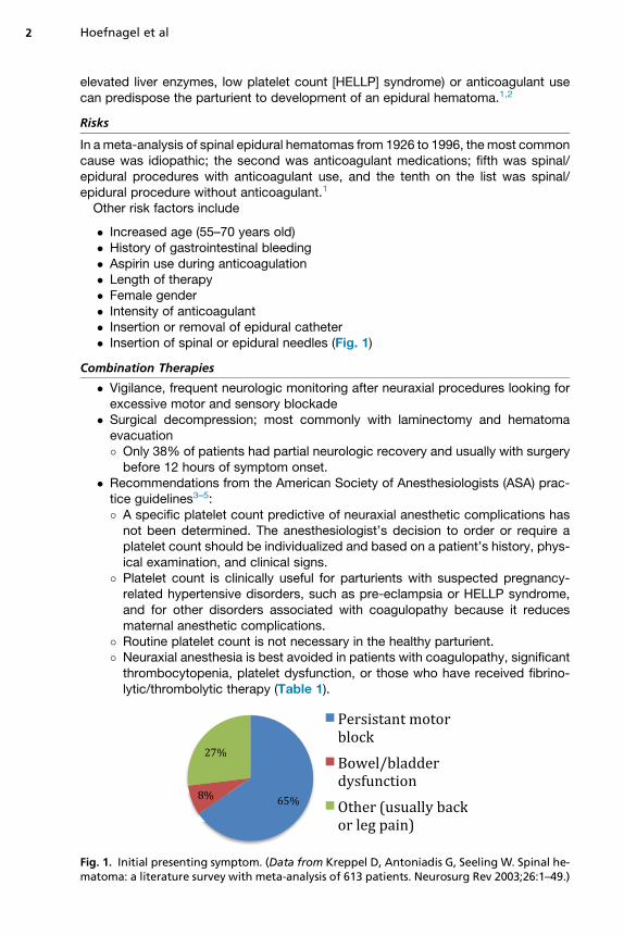
� Increased age (55–70 years old)� History of gastrointestinal bleeding� Aspirin use during anticoagulation� Length of therapy� Female gender� Intensity of anticoagulant� Insertion or removal of epidural catheter� Insertion of spinal or epidural needles (Fig. 1)
Combination Therapies
� Vigilance, frequent neurologic monitoring after neuraxial procedures looking forexcessive motor and sensory blockade
� Surgical decompression; most commonly with laminectomy and hematomaevacuation
Figma
� Only 38% of patients had partial neurologic recovery and usually with surgerybefore 12 hours of symptom onset.
� Recommendations from the American Society of Anesthesiologists (ASA) prac-tice guidelines3–5:� A specific platelet count predictive of neuraxial anesthetic complications hasnot been determined. The anesthesiologist’s decision to order or require aplatelet count should be individualized and based on a patient’s history, phys-ical examination, and clinical signs.
� Platelet count is clinically useful for parturients with suspected pregnancy-related hypertensive disorders, such as pre-eclampsia or HELLP syndrome,and for other disorders associated with coagulopathy because it reducesmaternal anesthetic complications.
� Routine platelet count is not necessary in the healthy parturient.� Neuraxial anesthesia is best avoided in patients with coagulopathy, significantthrombocytopenia, platelet dysfunction, or those who have received fibrino-lytic/thrombolytic therapy (Table 1).
. 1. Initial presenting symptom. (Data from Kreppel D, Antoniadis G, Seeling W. Spinal he-toma: a literature survey with meta-analysis of 613 patients. Neurosurg Rev 2003;26:1–49.)
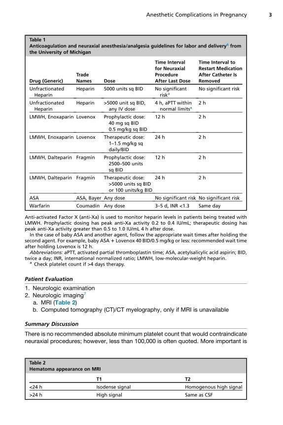
Table 1Anticoagulation and neuraxial anesthesia/analgesia guidelines for labor and delivery6 fromthe University of Michigan
Drug (Generic)TradeNames Dose
Time Intervalfor NeuraxialProcedureAfter Last Dose
Time Interval toRestart MedicationAfter Catheter IsRemoved
UnfractionatedHeparin
Heparin 5000 units sq BID No significantriska
No significant risk
UnfractionatedHeparin
Heparin >5000 unit sq BID,any IV dose
4 h, aPTT withinnormal limitsa
2 h
LMWH, Enoxaparin Lovenox Prophylactic dose:40 mg sq BID0.5 mg/kg sq BID
12 h 2 h
LMWH, Enoxaparin Lovenox Therapeutic dose:1–1.5 mg/kg sqdaily/BID
24 h 2 h
LMWH, Dalteparin Fragmin Prophylactic dose:2500–500 unitssq BID
12 h 2 h
LMWH, Dalteparin Fragmin Therapeutic dose:>5000 units sq BIDor 100 units/kg BID
24 h 2 h
ASA ASA, Bayer Any dose No significant risk No significant risk
Warfarin Coumadin Any dose 3–5 d, INR <1.3 Same day
Anti-activated Factor X (anti-Xa) is used to monitor heparin levels in patients being treated withLMWH. Prophylactic dosing has peak anti-Xa activity 0.2 to 0.4 IU/mL; therapeutic dosing haspeak anti-Xa activity greater than 0.5 to 1.0 IU/mL 4 h after dose.
In the case of baby ASA and another agent, follow the appropriate wait times after holding thesecond agent. For example, baby ASA1 Lovenox 40 BID/0.5 mg/kg or less: recommended wait timeafter holding Lovenox is 12 h.
Abbreviations: aPTT, activated partial thromboplastin time; ASA, acetylsalicylic acid aspirin; BID,twice a day; INR, international normalized ratio; LMWH, low-molecular-weight heparin.
a Check platelet count if >4 days therapy.
Anesthetic Complications in Pregnancy 3
Patient Evaluation
1. Neurologic examination2. Neurologic imaging7
a. MRI (Table 2)b. Computed tomography (CT)/CT myelography, only if MRI is unavailable
Summary Discussion
There is no recommended absolute minimum platelet count that would contraindicateneuraxial procedures; however, less than 100,000 is often quoted. More important is
Table 2Hematoma appearance on MRI
T1 T2
<24 h Isodense signal Homogenous high signal
>24 h High signal Same as CSF
Hoefnagel et al4
the relative decline from baseline, rate of decline, and overall clinical picture of that pa-tient, that is, if there are disease processes that would predispose the patient to a spi-nal epidural hematoma. As always, clinical decision-making and risk/benefit analysisremain key for optimum patient care.
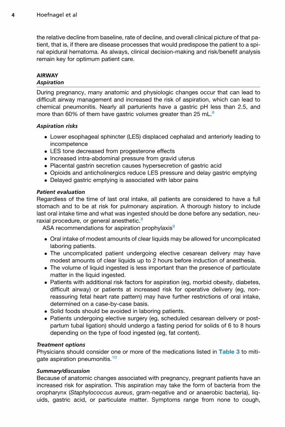
AIRWAYAspiration
During pregnancy, many anatomic and physiologic changes occur that can lead todifficult airway management and increased the risk of aspiration, which can lead tochemical pneumonitis. Nearly all parturients have a gastric pH less than 2.5, andmore than 60% of them have gastric volumes greater than 25 mL.8
Aspiration risks
� Lower esophageal sphincter (LES) displaced cephalad and anteriorly leading toincompetence
� LES tone decreased from progesterone effects� Increased intra-abdominal pressure from gravid uterus� Placental gastrin secretion causes hypersecretion of gastric acid� Opioids and anticholinergics reduce LES pressure and delay gastric emptying� Delayed gastric emptying is associated with labor pains
Patient evaluationRegardless of the time of last oral intake, all patients are considered to have a fullstomach and to be at risk for pulmonary aspiration. A thorough history to includelast oral intake time and what was ingested should be done before any sedation, neu-raxial procedure, or general anesthetic.8
ASA recommendations for aspiration prophylaxis9
� Oral intake of modest amounts of clear liquids may be allowed for uncomplicatedlaboring patients.
� The uncomplicated patient undergoing elective cesarean delivery may havemodest amounts of clear liquids up to 2 hours before induction of anesthesia.
� The volume of liquid ingested is less important than the presence of particulatematter in the liquid ingested.
� Patients with additional risk factors for aspiration (eg, morbid obesity, diabetes,difficult airway) or patients at increased risk for operative delivery (eg, non-reassuring fetal heart rate pattern) may have further restrictions of oral intake,determined on a case-by-case basis.
� Solid foods should be avoided in laboring patients.� Patients undergoing elective surgery (eg, scheduled cesarean delivery or post-partum tubal ligation) should undergo a fasting period for solids of 6 to 8 hoursdepending on the type of food ingested (eg, fat content).
Treatment optionsPhysicians should consider one or more of the medications listed in Table 3 to miti-gate aspiration pneumonitis.10
Summary/discussionBecause of anatomic changes associated with pregnancy, pregnant patients have anincreased risk for aspiration. This aspiration may take the form of bacteria from theoropharynx (Staphylococcus aureus, gram-negative and or anaerobic bacteria), liq-uids, gastric acid, or particulate matter. Symptoms range from none to cough,

Table 3Useful medication for mitigation of aspiration pneumonitis
Drug Class Example with Dose and Route Benefit
Clear nonparticulateantacid
Sodium citrate 15–30 mL orallyevery 3 h
Maintains gastric pH >2.5
H-2 antagonist Ranitidine 100–150 mg orallyor 50 mg IV
Reduces gastric acid volumeand pHa
Prokinetic Metoclopramide 10 mg orallyor IV
Decreases gastric volume, increases LEStone, decreases peripartum nauseaand vomiting
a No effect on gastric contents already present.
Anesthetic Complications in Pregnancy 5
bronchospasm, cyanosis, tachypnea, pulmonary edema, and rarely, hypotension andhypoxemia for liquid aspiration. Treatment is primarily prevention; if that fails, thepatient should be repositioned into the Trendelenburg position and oropharyngealsuction tried next. Intubation follows if the patient becomes apneic, hypoxic, or is un-able to protect her airway. Once the endotracheal tube (ETT) is in place, a soft suctioncatheter should be passed, and the ETT should be suctioned before positive pressureventilation is commenced to prevent pushing aspirated material further into the air-spaces. If the patient has become hypoxic, there may not be time for this maneuverbecause oxygenation should be the primary goal. Finally, bronchoscopy, pulmonarylavage, and antibiotics are usually not necessary unless there has been aspiration ofparticulate matter10 (Fig. 2).
Difficult Airway
IntroductionA study of the Centers for Disease Control and Prevention material mortality datashowed that anesthesia-related mortality from 1979 to 2002 most often was associ-ated with cesarean section (86%), intubation failure (23%), and respiratory failure(20%)11 In these cases of difficult airways, the potential is not only for mortality andmorbidity to themother but also to her unborn child. Changes with pregnancy increasethe risk of these patients to be a difficult airway. The best treatment for a difficultairway is a good assessment and preparation with backup plans available.
Fig. 2. Management of aspiration.

Hoefnagel et al6
Risks of difficult airway
� Rapid desaturation of oxygen during apnea or hypoventilation due to
� Increased oxygen consumption� Decreased functional residual capacity by 20% to 30%, cephalad displace-ment of diaphragm� Functional residual capacity (FRC) even closer to closing volume in patientswith pre-existing lung disease, obesity, scoliosis, smoking, supine, or Trende-lenburg position
� Relative anemia of pregnancy
Patient evaluation (special consideration of)
� Increased edema of upper airway from an increase in extracellular fluid anddecreased oncotic pressure
� Worse with steep Trendelenburg or pre-eclampsia� Friable mucosa, especially nasal mucosa due to capillary engorgement� Enlarged breasts, which may impede laryngoscope handle positioning� Increased Mallampati scores (Fig. 3)� Full dentition
Planning for airway managementProper positioning
1. Left uterine displacement to offload aortocaval compression2. Sniffing position in nonobese3. Ramped position (Fig. 4) with external auditory meatus and sternal notch aligned in
a horizontal plane for obese patients
Have emergency airway equipment available:
1. Multiple laryngoscope blades/types (Macintosh 3, 4; Miller 2, 3)2. Short laryngoscope handle (Fig. 5)3. Several sizes of ETTs6–8
4. Oral and nasal airways5. Stylet and bougie6. Supraglottic device, laryngeal mask airway7. Fiberoptic bronchoscope8. Video laryngoscopy equipment9. Availability of transtracheal jet ventilation or surgical airway (Figs. 6 and 7)
NEURAXIAL BLOCKADEIntroduction
Neuraxial blockade consists of either epidural or spinal blockade, the goal of which isto provide analgesia for delivery of the infant. For vaginal delivery, loss of pain sensa-tion lower than T8 is the goal. Uterine contraction and cervical dilation result in visceralpain, which is transmitted at the T10 to L1 levels. As labor progresses and the fetalhead descends, pressure on the pelvic floor, vagina, and perineum generate somaticpain transmitted by the pudendal nerve (S2–4). For cesarean delivery, a T4 level mustbe obtained (Table 4).
Hypotension
� Very common 30% to 40% of the time (Fig. 8)

Fig. 3. Mallampati score. (Courtesy of Fotosearch. � Fotosearch.com; with permission.)
Anesthetic Complications in Pregnancy 7
� Defined as systolic blood pressure less than 100 mmHg or a 20% decrease frombaseline12
� A large percentage of distress after epidural can be due to maternal hypotension� Sympathetic efferents exit the spinal cord from T1 to L212,13
� T1–T4 sympathectomy causes warm vasodilated hands, large reduction in sys-temic vascular resistance, and Horner syndrome and may lead to bradycardiadue to blockade of cardiac accelerator fibers
� T5–L2 sympathectomy causes pooling of blood in the splanchnic vessels,reducing venous return and cardiac output14
� Inferior vena cava obstruction by gravid uterus further exacerbates a decrease invenous return13
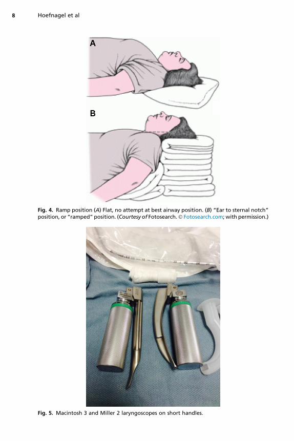
Fig. 4. Ramp position (A) Flat, no attempt at best airway position. (B) “Ear to sternal notch”position, or “ramped” position. (Courtesy of Fotosearch.� Fotosearch.com;with permission.)
Fig. 5. Macintosh 3 and Miller 2 laryngoscopes on short handles.
Hoefnagel et al8

Fig. 6. Steps after trial of failed intubation during general anesthesia in obstetric patient.a Laryngeal mask airway; b Transtracheal jet ventilation. (From Jadon A. Complications ofregional and general anaesthesia in obstetric practice. Indian J Anaesth 2010;54(5):415–20.)
Anesthetic Complications in Pregnancy 9
Patient evaluation overviewMonitor maternal blood pressure frequently after starting neuraxial blockade.
Summary/discussionMaternal hypotension is a very common result of neuraxial anesthesia that should beanticipated and treated early. Before placement of a spinal or epidural block, consid-eration should be allowed for intravenous hydration to ensure the patient hasadequate preload, which minimizes the effects of sympathectomy and vasodilation.There is little downside to prophylactically treating small decreases in maternal
blood pressure with vasoactive medications, expecting that left untreated, blood pres-sure will continue to decrease. The placenta does not have the ability to autoregulate;therefore, when maternal blood pressure is decreased, blood flow to the fetus isdecreased, leading to fetal distress. Ensure that the mother is positioned such thatthe gravid uterus is not compressing the great vessels, optimize the fluid status inthe mother, and treat with vasoactive medications as needed to restore maternalblood pressure normal levels (Tables 5 and 6).

Fig. 7. ASA difficult airway algorithm. a Invasive airway access includes surgical or percuta-neous airway, jet ventilation, and retrograde intubation. b Confirm ventilation, trachealintubation, or SGA placement with exhaled CO2.
c Other options include (but are not limitedto): surgery utilizing face mask or supraglottic airway (SGA) anesthesia (eg, LMA, ILMA,laryngeal tube), local anesthesia infiltration or regional nerve blockade. Pursuit of theseoptions usually implies that mask ventilation will not be problematic. Therefore, these op-tions may be of limited value if this step in the algorithm has been reached via the Emer-gency Pathway. d Alternative difficult intubation approaches include (but are not limitedto): video-assisted laryngoscopy, alternative laryngoscope blades, SGA (eg, LMA or ILMA)as an intubation conduit (with or without fiberoptic guidance), fiberoptic intubation, intu-bating stylet or tube changer, light wand, and blind oral or nasal intubation. e Emergencynon-invasive airway ventilation consists of a SGA. f Consider re-preparation of the patientfor awake intubation or canceling surgery. (From Apfelbaum JL, Hagberg CA, Caplan RA,et al. Practice guidelines for management of the difficult airway: an updated report bythe American Society of Anesthesiologists Task Force on Management of the DifficultAirway. Anesthesiology 2013;118(2):251–70; with permission.)
Hoefnagel et al10

Table 4Common local anesthetics for spinal anesthesia
Drug Dose (mg) for T10 Dose (mg) for T4
Duration (min)
Onset (min)Plain DEpi
Lidocaine 5% 50–75 75–100 60–70 75–100 3–5
Bupivacaine 0.75% 8–12 14–20 90–110 100–150 5–8
Data from NYSORA. Spinal anesthesia. Available at: http://www.nysora.com/techniques/neuraxial-and-perineuraxial-techniques/landmark-based/3423-spinal-anesthesia.html.
Anesthetic Complications in Pregnancy 11
High Spinal
� A respiratory and cardiac emergency� Rapidly increasing sensory levels, dyspnea, bradycardia, apnea, and cardiac ar-rest after spinal or epidural bolus dose
� Most common with dosing an epidural that has unintentionally becomeintrathecal
� Occurs in w1% of spinal anesthetics15
� Occurs when anesthetic level increases to higher than a T1 dermatome level(Fig. 9)
Patient evaluation overview
Fig. 8. Spinal hypotension cycle.
Hypotension15 Nausea
Bradycardia Anxiety
Respiratory compromise Cranial nerve involvement
Apnea Arm/hand dyaesthesia or pain
Reduced oxygen saturation Loss of consciousness
Difficulty speaking/coughing Cardiac arrest (asystole)

Table 5Pharmacologic treatment options
Intravenous Fluid (IVF) 500 mL to 1 L rapid bolus. Preload with 1 L IVF before blockade
Phenylephrine 100 mg IV, repeat as needed (may lead to reflex bradycardia)
Ephedrine 5–10 mg IV, repeat as needed (may lead to tachyphylaxis)
Data from Pellegrini JE. Prevention and treatment of spinal induced hypotension. Available at: http://www.aana.com/meetings/meeting-materials/annualcongress/Documents/Pellegrini_Handouts.pdf.
Hoefnagel et al12
Combination therapies
1. Notify attending anesthesiologista. Consider calling a STAT
2. Inform obstetrician or surgeon3. Support ventilation and intubate if necessary4. Support blood pressure with intravenous (IV) fluid bolus and vasopressors (Table 7)5. Treat bradycardia with epinephrine (preferred to atropine)6. Consider:
a. Emergency cesarean section if signs of fetal distressb. Need for more venous accessc. Sedation until able to extubate (midazolam)d. Treat cardiac arrest with advanced cardiac life support (ACLS) protocols
Causes
� Excessive dose of local anesthetic� Failure to reduce normally accepted dose in patients susceptible to excessivespread (pregnant, obese, or short patients)
� Unusual excessive spread� Spinal anesthetic after recent epidural bolus dose16
Table 6Nonpharmacologic treatment options
Oxygen If there is fetal bradycardia
Reposition Left uterine displacement, hands and knees, etc
Adapted from Pellegrini JE. Prevention and treatment of spinal induced hypotension. Available at:http://www.aana.com/meetings/meeting-materials/annualcongress/Documents/Pellegrini_Handouts.pdf; with permission.

Fig. 9. Dermatome Map. (Courtesy of Fotosearch. � Fotosearch.com; with permission.)
Anesthetic Complications in Pregnancy 13
Summary/discussionHigh or total spinal blockade occurs when spinal blockade increase to higher than theT1 dermatomal level and can lead to cardiovascular collapse and respiratory arrest.Symptoms and signs of high spinal usually occur within minutes of placement; how-ever, delay of up to 30 minutes has been reported.15 Nausea and anxiety, both fromcerebral hypoperfusion, are usually the first reported symptoms.Prevention of high spinal anesthesia, while not always possible, is based on
frequent monitoring of sensory levels, patient positioning to influence spread of medi-cation via baricity, and recognition for timely intervention in the event of circulatorycollapse.
Local Anesthetic Systemic Toxicity
Table 8
� Local anesthetics act on sodium channels, and therefore, local anesthetic sys-temic toxicity (LAST) is seen in organs of the body that depend on these sodium
Table 7Pharmacologic treatment options
Epinephrine 5–10 mg IV bolus dosing
Vasopressin 1–2 units IV bolus dosing
Atropine 0.5–1 mg IV bolus

Table 8Local anesthetic maximum doses (mg/kg)
Plain DEpi
Lidocaine 5 7
Mepivacaine 5 7 (400 mg max)
Bupivacaine/Ropivacaine 3 3
Data from Open Anesthesia. Local anesthetics: systemic toxicity. Available at https://www.openanesthesia.org/local_anesthetics_systemic_toxicity/.
Hoefnagel et al14
channels for proper function, such as the central nervous system (CNS) and theheart.
� The CNS is more sensitive to the effects of local anesthetics than the cardiac sys-tem and will generally manifest symptoms of toxicity first.
Signs/symptomsTable 9
Combination therapies
1. Notify attending anesthesiologist17
a. Consider calling a STAT2. Notify surgical team (if in operating room)3. Change FiO2 to 100% (avoid hypoxia) and hyperventilate (avoid acidosis). Intubate
if not already done.4. Treat seizures with benzodiazepines.5. Call for lipid emulsion and cardiac bypass (if cardiotoxicity).6. Administer lipid emulsion 20% (DO NOT USE propofol; it is only 10% and is likely to
lead to severe hypotension).7. Start a second IV.8. Consider ACLS modifications:
a. Administer low-dose epinephrine, 1 mg/kg IVb. Avoid vasopressin (can cause pulmonary edema)
9. Consider:a. Monitor patient postoperatively in intensive care unit.b. Perform cardiopulmonary bypass if refractory cardiac arrest (STAT page perfu-
sion for setup) (Fig. 10).
Table 9Signs and Symptoms of Local Anesthesitc Toxicity (LAST)
Central Nervous System Cardiac
Excitation Depression Nonspecific —
Agitation Drowsiness Diplopia Hypotension
Tremors Coma Dizziness Bradycardia
Seizure Slurred speech — Asystole
Metallic taste — — Atrial/ventricular ectopy
Circumoral numbness — — —
Tinnitus — — —
Data from Neal JM, Bernards CM, Butterworth JM, et al. ASRA practice advisory of local anestheticsystemic toxicity. Reg Anesth Pain Med 2010;35(2):152–61; and Checklist for treatment of localanesthetic systemic toxicity. ASRA.
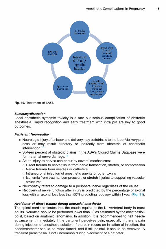
Fig. 10. Treatment of LAST.
Anesthetic Complications in Pregnancy 15
Summary/discussionLocal anesthetic systemic toxicity is a rare but serious complication of obstetricanesthesia. Rapid recognition and early treatment with intralipid are key to goodoutcomes.
Persistent Neuropathy
� Neurologic injury after labor and deliverymay be intrinsic to the labor/delivery pro-cess or may result directory or indirectly from obstetric of anestheticintervention.18
� Sixteen percent of obstetric claims in the ASA’s Closed Claims Database werefor maternal nerve damage.19
� Acute injury to nerves can occur by several mechanisms:
� Direct trauma to nerve tissue from nerve transection, stretch, or compression� Nerve trauma from needles or catheters� Intraneuronal injection of anesthetic agents or other toxins� Ischemia from trauma, compression, or stretch injuries to supporting vascularstructures� Neuropathy refers to damage to a peripheral nerve regardless of the cause.� Recovery of nerve function after injury is predicted by the percentage of axonalloss with an axonal loss less than 50% predicting recovery within 1 year (Fig. 11).
Avoidance of direct trauma during neuraxial anesthesiaThe spinal cord terminates into the cauda equina at the L1 vertebral body in mostadults. Neuraxial should be performed lower than L3 as estimated by the anesthesiol-ogist, based on anatomic landmarks. In addition, it is recommended to halt needleadvancement immediately if the parturient perceives pain, especially if there is painduring injection of anesthetic solution. If the pain recurs on initiation of injection, theneedle/catheter should be repositioned, and if still painful, it should be removed. Atransient parasthesia is not uncommon during placement of a catheter.

Fig. 11. Innervation of the lower extremity. (From Haymaker W, Woodhall B. Peripheralnerve injuries, 2d ed. Philadelphia: Saunders; 1953.)
Hoefnagel et al16
Evaluation overviewInitial evaluation of the patient should include the presence of lower extremity pain,numbness, or weakness (Table 10). This information should be used in the risk/benefitdiscussion when discussing neuraxial anesthesia with the parturient (Fig. 12).Evaluation of postpartum complaints of lower extremity numbness, weakness, and
pain should occur promptly, and rare life- or limb-threatening causes should be ruledout. Symptoms present immediately after delivery, and those that have improved orstayed the same, are less likely to be more life-threatening than those that occur sud-denly and after a symptom-free period. A complete history and physical examinationshould include determination of the onset time of symptoms and details of the laborand delivery process (eg, mode of delivery, use of forceps, maternal positioning).The presence of a fever and elevated white blood count suggest the presence of
infection, whereas sensory and motor deficits without pain suggest an intrinsic obstet-ric palsy (see Table 10). A detailed neurologic examination will help in differentiationbetween central, radicular, plexus, and peripheral nerve lesions may be considered.More complex symptoms, particularly motor deficits, or bilateral symptoms, should

Table 10Obstetric Palsies
Palsy Cause/Risks Symptoms
Lateral femoralcutaneous
Trauma, prolonged hip flexion,exaggerated lumbar lordosis
Loss of sensation to the lateral thigh
Femoral Pfannenstiel incision, self-retainingretractors, thigh flexion withexternal rotation and abduction
Weakness of hip flexion, loss ofsensation medial lower leg
Lumbosacralplexus
Compression by fetal head againstthe pelvis, forceps delivery
Foot drop (Table 11)
Peroneal External compression from pressureat the head of the fibula, usuallyfrom stirrups
Numbness of the lateral leg anddorsum of the foot, weakness offoot dorsiflexion, and eversion
Sciatic Stretch, injection trauma Weakness of foot inversion, absentankle jerk
Anesthetic Complications in Pregnancy 17
prompt consultation with a neurologist or neurosurgeon. Immediate MRI is the currentgold standard to rule out central lesions. Electromyography (EMG) may aid in deter-mining the site of injury as well as the degree of axonal loss; however, EMG only mea-sures large nerve fiber changes and may take as long as 3 weeks after injury to showchanges. An abnormal EMG in the first postpartum week suggests pre-existinginjury.18,19
Table 11Differential diagnosis of foot drop
L5 NerveRoot
LumbarPlexus
SciaticNerve
PeronealNerve
Motor Weakness ofparaspinousmuscles
Weakness ofgluteal musclesand analsphincter
— —
Ankleinversiona
Weak Weak Normal or weak Normal
Plantarflexion
Normal Normal Normal or weak Normal
Toeflexion
Weak Weak Normal or weak Normal
Sensoryloss
Poorlydemarcated,predominatelybig toe
Well demarcatedto L5dermatome
Lower 2/3 of lateralleg, and dorsumof foot
Lower 2/3 of lateralleg and dorsum offoot
Anklejerk
Normalb Normalb Normal or weak Normal
Pain Common,radicular
Common, may beradicular
Can be severe Rare
a Attempt at inversion should be made with the foot dorsiflexed passively to 90�.b Weak with S1 involvement.
From Katirji B. Entrapment and other focal neuropathies. Peroneal Neuropathy. Neurol Clin1999;17:567–91; with permission.
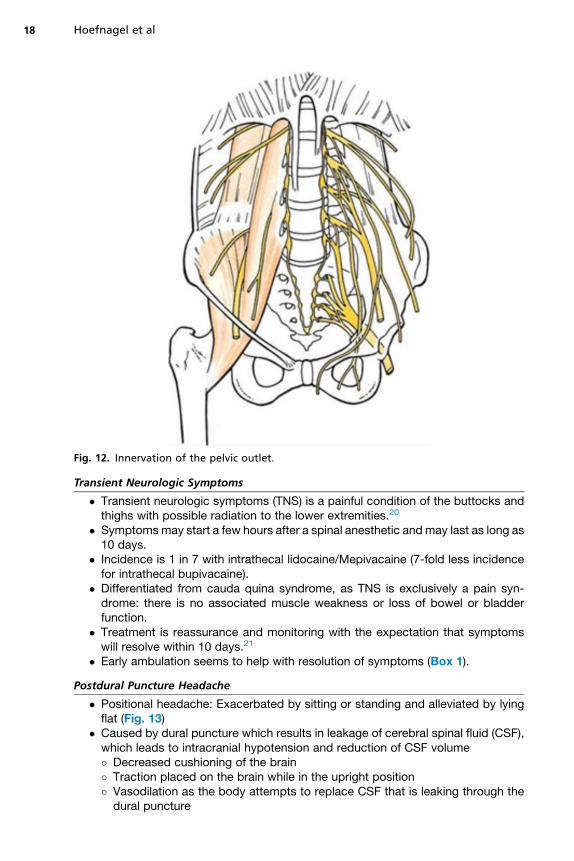
Fig. 12. Innervation of the pelvic outlet.
Hoefnagel et al18
Transient Neurologic Symptoms
� Transient neurologic symptoms (TNS) is a painful condition of the buttocks andthighs with possible radiation to the lower extremities.20
� Symptomsmay start a few hours after a spinal anesthetic andmay last as long as10 days.
� Incidence is 1 in 7 with intrathecal lidocaine/Mepivacaine (7-fold less incidencefor intrathecal bupivacaine).
� Differentiated from cauda quina syndrome, as TNS is exclusively a pain syn-drome: there is no associated muscle weakness or loss of bowel or bladderfunction.
� Treatment is reassurance and monitoring with the expectation that symptomswill resolve within 10 days.21
� Early ambulation seems to help with resolution of symptoms (Box 1).
Postdural Puncture Headache
� Positional headache: Exacerbated by sitting or standing and alleviated by lyingflat (Fig. 13)
� Caused by dural puncture which results in leakage of cerebral spinal fluid (CSF),which leads to intracranial hypotension and reduction of CSF volume
� Decreased cushioning of the brain� Traction placed on the brain while in the upright position� Vasodilation as the body attempts to replace CSF that is leaking through thedural puncture
Fig
Box 1
Risk factors for transient neurologic symptoms
5% Lidocaine
Lithotomy position
Microcatheter use
Outpatient status
Data from Sime AC. AANA Journal course: transient neurological symptoms and spinal anes-thesia. AANA J 2000;68:163–827.
Anesthetic Complications in Pregnancy 19
� Expected rate of dural puncture (wet tap) with lumbar epidural is w1.5%.22
� Independent of performer skill or expertise� Risk of developing postdural puncture headache (PDPH) as a result of a wettap is w52% (Table 12)
� PDPH from spinal anesthesia is related to the gauge and shape of the needleused, with smaller-gauge pencil-point needles having the lowest incidence ofPDPH (Fig. 14).� In the picture on the left in Fig. 14, Whitacre and Sprote are examples of pencil-point needles.
� The Quincke has a cutting tip and is associated with a higher incidence ofPDPH.
Differential diagnosis of postpartum headache
� Lactation headache23
� Migraine� Subdural hematoma� Brain mass/tumor� Arterial venous malformation� Cortical vein/dural sinus thrombosis� Tension headache� Pre-eclampsia� Pneumocephalus� Dehydration
. 13. PDPH signs and symptoms.

Table 12Risk factors for dural puncture and PDPH
Risk Factors for Dural Puncture Risk Factors for PDPH
Increased body mass indexIncreased age
Low body mass indexPregnancyPrior history of PDPHYounger agea
Needle gauge/type
a Elderly patients will have reduced elasticity of the dura mater, which increases the resistance toCSF leakage.
20 Hoefnagel et al
� Caffeine withdrawal� Meningitis/encephalitis
Treatment optionsTreatment options for PDPH are outlined in Fig. 15.
Epidural blood patchEpidural blood patching (EBP) involves the injection of autologous blood into theepidural space.
Fig. 14. Spinal needles. (From Peterman SB. Postmyelography headache rates with Whitacreversus Quincke 22-gauge spinal needles. Radiology 1996;200:771–8; with permission.)
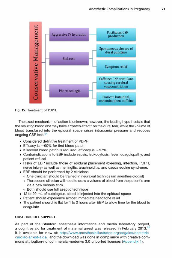
Fig. 15. Treatment of PDPH.
Anesthetic Complications in Pregnancy 21
The exact mechanism of action is unknown; however, the leading hypothesis is thatthe resulting blood clot may have a “patch effect” on the dural tear, while the volume ofblood transfused into the epidural space raises intracranial pressure and reducesongoing CSF leak.24
� Considered definitive treatment of PDPH� Efficacy is w90% for first blood patch� If second blood patch is required, efficacy is w97%� Contraindications to EBP include sepsis, leukocytosis, fever, coagulopathy, andpatient refusal
� Risks of EBP include those of epidural placement (bleeding, infection, PDPH,nerve injury) as well as meningitis, arachnoiditis, and cauda equine syndrome.
� EBP should be performed by 2 clinicians.
� One clinician should be trained in neuraxial technics (an anesthesiologist)� The second clinician will need to draw a volume of blood from the patient’s armvia a new venous stick� Both should use full aseptic technique� 12 to 20 mL of autologous blood is injected into the epidural space� Patient should experience almost immediate headache relief� The patient should lie flat for 1 to 2 hours after EBP to allow time for the blood tocoagulate
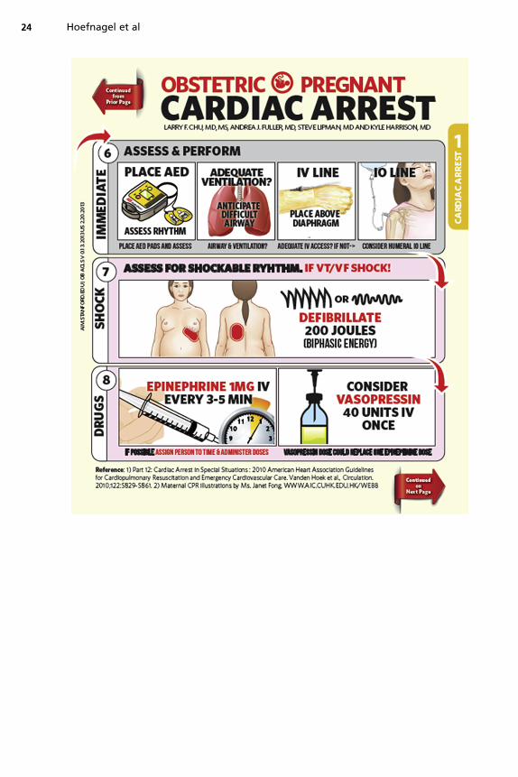
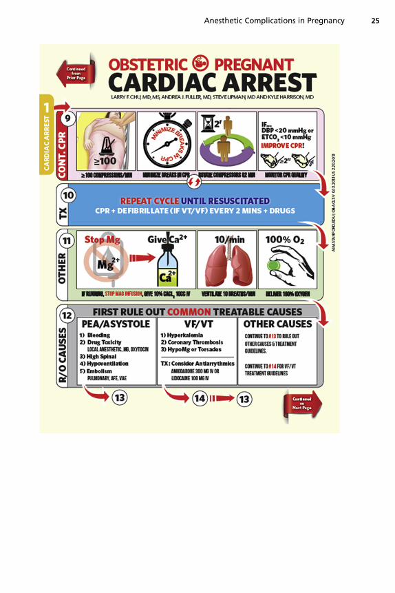
OBSTETRIC LIFE SUPPORT
As part of the Stanford anesthesia informatics and media laboratory project,a cognitive aid for treatment of maternal arrest was released in February 2013.25
It is available for view at: http://www.anesthesiaillustrated.org/cogaids/obstetric-cardiac-arrest-aids/, and the download was done in compliance with creative com-mons attribution-noncommercial-noderivs 3.0 unported licenses (Appendix 1).
Hoefnagel et al22
REFERENCES
1. Kreppel D, Antoniadis G, Seeling W. Spinal hematoma: a literature survey withmeta-analysis of 613 patients. Neurosurg Rev 2003;26:1–49.
2. Levine MN, Raskob G, Landefelt S, et al. Hemorrhagic complications of anti-coagulant treatment. Chest 2001;110:108S–21S.
3. Vandermeulen EP, Van Aken H, Vermylen J, et al. Anticoagulants and spinal-epidural anesthesia. Anesth Analg 1994;79:1165–77.
4. Available at: https://www.asra.com/advisory-guidelines/article/1/anticoagulation-3rd-edition.
5. Available at: http://www.asahq.org/w/media/Sites/ASAHQ/Files/Public/Resources/standards-guidelines/guidelines-for-neuraxial-anesthesia-in-obstetrics.pdf.
6. Available at: http://anes.med.umich.edu/vault/1007575-AnticoagulationNeuraxialGuidelines.pdf.
7. Lawton MT, Porter RW, Heiserman JE, et al. Surgical management of spinalepidural hematoma: relationship between surgical timing and neurologicaloutcome. J Neurosurg 1995;83(1):1–7.
8. Kodali B, Chandrasekhar S, Bulich LN. Airway changes during labor and delivery.Anesthesiology 2008;108:357–62.
9. American Society of Anesthesiologists Committee. Practice guidelines for preop-erative fasting and the use of pharmacologic agents to reduce the risk ofpulmonary aspiration: application to healthy patients undergoing elective proce-dures. An updated report by the American Society of Anesthesiologists Commit-tee on Standards and Practice Parameters. Anesthesiology 2011;114(3):495–511.
10. Marik PE. Aspiration pneumonitis and aspiration pneumonia. N Engl J Med 2001;344(9):665–71.
11. Hawkins JL, Chang J, Palmer SK, et al. Anesthesia-related maternal mortality inthe United States. 1979-2002. Obstet Gynecol 2011;117(1):69–74.
12. Kiohr S, Roth R, Hormann T, et al. Definitions of hypotension after spinal anaes-thesia for caesarean section: literature search and application to parturients.Acta Anaesthesiol Scand 2010;54(8):909–21.
13. Clark RB. Hypotension and caesarean section. Br J Anaesth 2008;101(6):882.
14. NYSORA. Spinal anesthesia. Available at: http://www.nysora.com/techniques/neuraxial-and-perineuraxial-techniques/landmark-based/3423-spinal-anesthesia.html.
15. Neuman B. Complete spinal block following spinal anaesthesia. Anaesthesiatutorial of the week. 24th May 2010.
16. Furst SR, Reisner LS. Risk of high spinal anesthesia following failed epiduralblock for cesarean delivery. J Clin Anesth 1995;7(1):71–4.
17. Checklist for treatment of local anesthetic systemic toxicity. ASRA.18. Cynthia Wong. Neurologic Deficit and Labor Analgesia.19. ASA Closed Claims Project.20. Open Anesthesia. Local Anesthetics: Transient Neurologic Symptoms.21. Sime AC. AANA journal course: transient neurological symptoms and spinal
anesthesia. AANA J 2000;68:163–8.22. Choi PT, Galinski SE, Takeuchi L, et al. PDPH is a common complication of neu-
raxial blockade in parturients: a meta-analysis of obstetrical studies. Can JAnaesth 2003;50(5):406–9.

Anesthetic Complications in Pregnancy 23
23. Jadon A. Complications of regional and general anaesthesia in obstetric practice.Indian J Anaesth 2010;54(5):415–20.
24. Jane Campbell FRCA. Effective management of the post-dural puncture head-ache. Anesthesia tutorial of the week. 31st May 2010.
25. Chu L, Harrison K. CogAIDs Project, Stanford AIM Lab (Anesthesia Informaticsand Media Lab. Anesthesiaillustrated.org).
APPENDIX 1: Obstetric life support cognitive aide. From Anesthesia Informatics andMedia Lab, Stanford, CA. Designed by Larry Chu, MD. Available at: http://aim.stanford.edu/project/anesthesiaillustrated/.
Hoefnagel et al24
Anesthetic Complications in Pregnancy 25

Hoefnagel et al26

Anesthetic Complications in Pregnancy 27

Hoefnagel et al28