
Anestesiologia UIS Hipoxemia perioperatoria
46
LOGO Anestesiologia UIS Hipoxemia perioperatoria Dr. Raúl Vásquez
-
Upload
summer-sanford -
Category
Documents
-
view
32 -
download
1
description
Anestesiologia UIS Hipoxemia perioperatoria. Dr. Raúl Vásquez. Historia. La anestesia general se asocia con hipoxemia. Fisiologia. Consumo O2 (VO2). Oxigenación. Entrega O2 (DO2). Metabolismo celular aerobico. Oxygenation and mechanisms of hypoxemia. Definicion. - PowerPoint PPT Presentation
Transcript of Anestesiologia UIS Hipoxemia perioperatoria
LOGO
Anestesiologia UIS
Hipoxemia perioperatoriaAnestesiologia UIS
Hipoxemia perioperatoriaDr. Raúl Vásquez
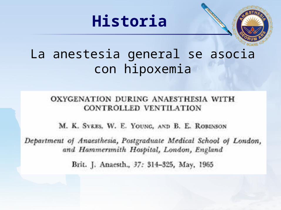
LOGOHistoria
La anestesia general se asocia con hipoxemia
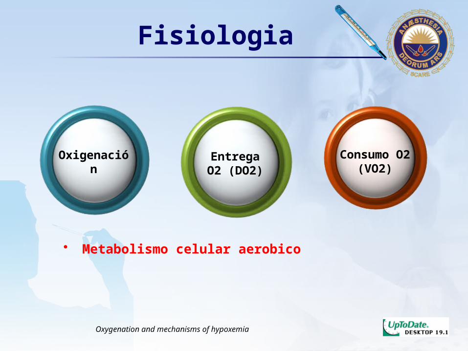
LOGOFisiologia
Oxigenación EntregaO2 (DO2)
Consumo O2 (VO2)
• Metabolismo celular aerobico
Oxygenation and mechanisms of hypoxemia

LOGO
• Disminucion de la entrega de oxigeno de la atmosfera a la sangre arterial (oxigenación
insuficiente)
Definicion
Hipoxemia
• Disminucion de la entrega de oxigeno de la sangre arterial a
los tejidosHipoxia
Monitoring Respiratory Function
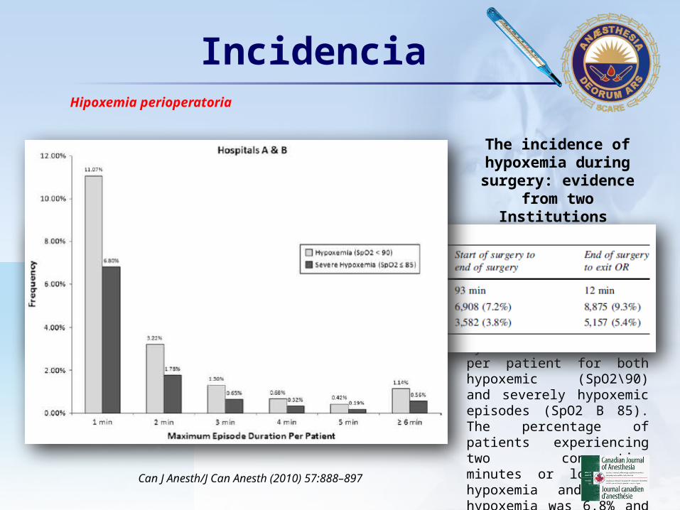
LOGOIncidencia
The incidence of hypoxemia during
surgery: evidence from two Institutions
Duration of Hypoxemic Episodes at Hospitals A & B. The incidence and maximum duration of intraoperative hypoxemic episodes. Episodes are grouped by maximum duration per patient for both hypoxemic (SpO2\90) and severely hypoxemic episodes (SpO2 B 85). The percentage of patients experiencing two consecutive minutes or longer of hypoxemia and severe hypoxemia was 6.8% and 3.5%, respectively
Can J Anesth/J Can Anesth (2010) 57:888–897
Hipoxemia perioperatoria

LOGOIncidencia
Can J Anesth/J Can Anesth (2010) 57:888–897
Hipoxemia perioperatoria
• 1 : 15 Hipoxemia por dos minutos• 1 : 64 Hipoxemia ≥ 5 minutos
• ˃3 millones pacientes hipoxemia ≥ 5 min
234 Millones Cx Año
LOGOEfectos Deletereos
• Altera cicatrización, integridad anatomosis y resistencia a infección
Arch Surg 1997; 132: 991-6. Arch Surg 1997; 132: 997-1005. N Engl J Med 2000; 342: 161-7
• Translocacion bacteriana GI – SepsisArch Surg 1996; 131: 57-62
• Disfunción cognitiva – delirioAm J Med 1981; 79:1247-54. Br J Anaesth 1994; 72: 286-90
• ↓↑GC, precipita arritmias, hipertensión arterial, Taquicardia, isquemia
Anesthesiology 1999; 91: 1246-52. Br Heart J 1993; 69: 3-5
Hipoxemia perioperatoria
LOGOMedidas Oxigenacion
• Examen físico– Cianosis franca 5 g/dl deoxihemoglobina
SaO2 67%
• GASA gold standard PaO2
• SaO2 “Quinto signo vital”– Proyecto global pulso oximetria OMS
• 100000 pulso oximetros
Hipoxemia perioperatoria
Pulse oximetry

LOGOOximetría de Pulso
Hipoxemia perioperatoria
Monitoring Respiratory Function
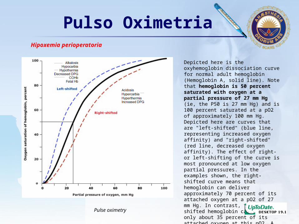
LOGOPulso Oximetria
Depicted here is the oxyhemoglobin dissociation curve for normal adult hemoglobin (Hemoglobin A, solid line). Note that hemoglobin is 50 percent saturated with oxygen at a partial pressure of 27 mm Hg (ie, the P50 is 27 mm Hg) and is 100 percent saturated at a pO2 of approximately 100 mm Hg. Depicted here are curves that are "left-shifted" (blue line, representing increased oxygen affinity) and "right-shifted" (red line, decreased oxygen affinity). The effect of right- or left-shifting of the curve is most pronounced at low oxygen partial pressures. In the examples shown, the right-shifted curve means that hemoglobin can deliver approximately 70 percent of its attached oxygen at a pO2 of 27 mm Hg. In contrast, the left-shifted hemoglobin can deliver only about 35 percent of its attached oxygen at this pO2. A high proportion of fetal hemoglobin, which has high oxygen affinity, shifts this curve to the left in newborns.
Hipoxemia perioperatoria
Pulse oximetry

LOGOOximetria de Pulso
• Aumenta deteccion hipoxemia 20 veces• Dx temprano
– Intubación endobronquial– Hipoventilacion
• Pacientes monitorizado– 50% menos eventos isquemia miocárdica
Hipoxemia perioperatoria
Global Pulse Oximetry Project
LOGOOximetría de Pulso
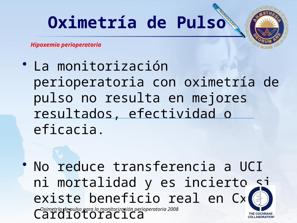
• La monitorización perioperatoria con oximetría de pulso no resulta en mejores resultados, efectividad o eficacia.
• No reduce transferencia a UCI ni mortalidad y es incierto si existe beneficio real en Cx Cardiotoracica
Hipoxemia perioperatoria
Oximetría de pulso para la monitorización perioperatoria 2008
LOGOOximetría de Pulso
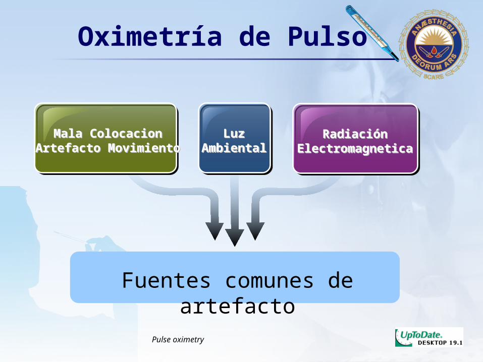
Mala ColocacionArtefacto Movimiento
Mala ColocacionArtefacto Movimiento
LuzAmbiental
LuzAmbiental
RadiaciónElectromagnetica
RadiaciónElectromagnetica
Fuentes comunes de artefacto
Pulse oximetry

LOGO
Hg
Anormales
Hipoperfusion
Hipotermia
Congestion
Venosa
Pigmentacion
Piel
Colorantes
Vitales
Esmalte
Anemia
Oximetría de Pulso
Errores relacionadosCon el Paciente
Pulse oximetry
LOGOMedidas Oxigenacion
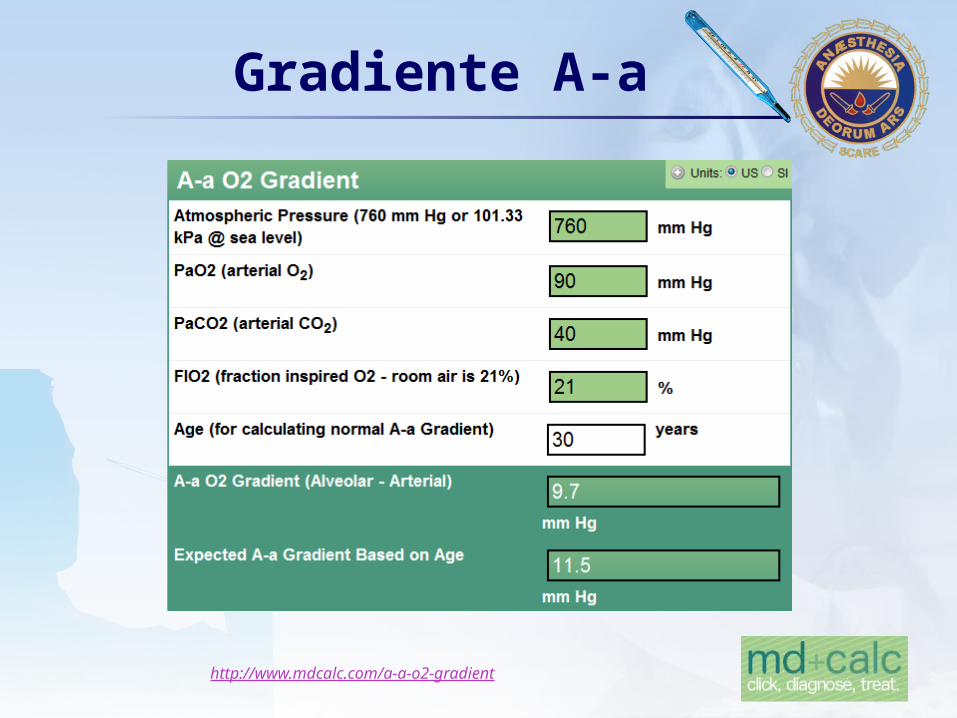
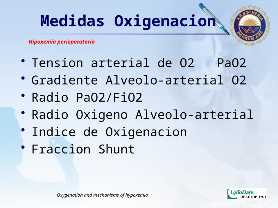
• Tension arterial de O2 PaO2• Gradiente Alveolo-arterial O2• Radio PaO2/FiO2• Radio Oxigeno Alveolo-arterial• Indice de Oxigenacion• Fraccion Shunt
Hipoxemia perioperatoria
Oxygenation and mechanisms of hypoxemia
LOGOGradiente A-a
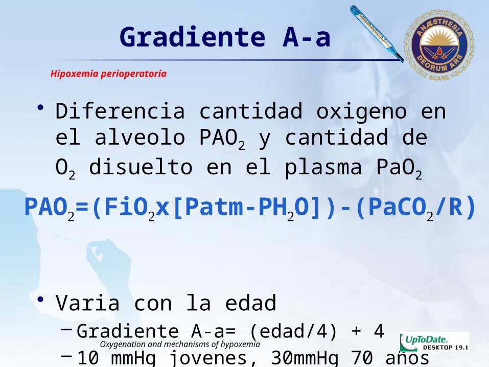
• Diferencia cantidad oxigeno en el alveolo PAO2 y cantidad de O2 disuelto en el plasma PaO2
• Varia con la edad– Gradiente A-a= (edad/4) + 4– 10 mmHg jovenes, 30mmHg 70 años
Hipoxemia perioperatoria
Oxygenation and mechanisms of hypoxemia
PAO2=(FiO2x[Patm-PH2O])-(PaCO2/R)
LOGOGradiente A-a
Hipoxemia perioperatoria
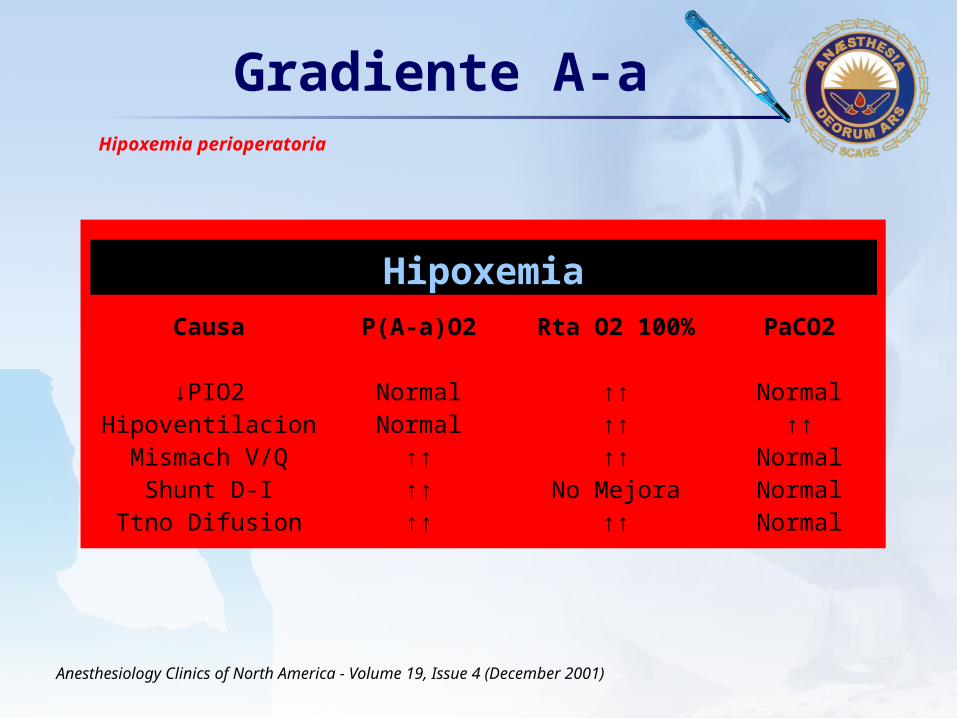
Hipoxemia
Causa
↓PIO2HipoventilacionMismach V/Q
Shunt D-ITtno Difusion
P(A-a)O2
NormalNormal
↑↑↑↑↑↑
Rta O2 100%
↑↑↑↑↑↑
No Mejora↑↑
PaCO2
Normal↑↑
NormalNormalNormal
Anesthesiology Clinics of North America - Volume 19, Issue 4 (December 2001)
LOGOGradiente A-a
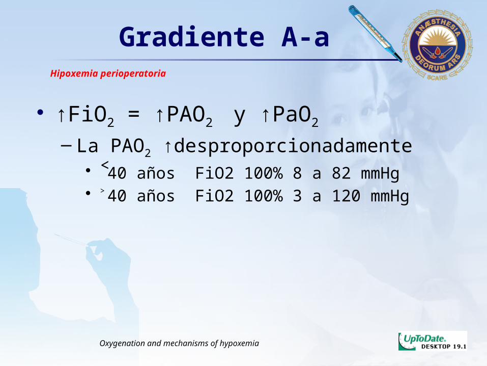
• ↑FiO2 = ↑PAO2 y ↑PaO2
– La PAO2 ↑desproporcionadamente• ˂40 años FiO2 100% 8 a 82 mmHg• ˃40 años FiO2 100% 3 a 120 mmHg
Hipoxemia perioperatoria
Oxygenation and mechanisms of hypoxemia

LOGOFraccion Shunt D-I
• Gold standar de oxigenacion eficiente en los pulmones
• Shunt 50%= Falla respiratoria severa• Shunt 5%= Normal
Monitoring Respiratory Function
Qs/Qt=(CcO2-CaO2)/(CcO2-CvO2)
CaO2=(1.34xHgbxHgbO2)+(0.003xPO2)
LOGOMedidas Oxigenacion
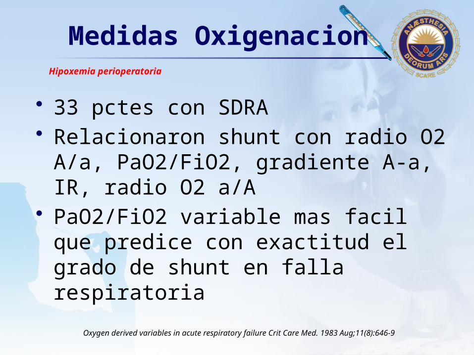
• 33 pctes con SDRA• Relacionaron shunt con radio O2 A/a,
PaO2/FiO2, gradiente A-a, IR, radio O2 a/A
• PaO2/FiO2 variable mas facil que predice con exactitud el grado de shunt en falla respiratoria
Hipoxemia perioperatoria
Oxygen derived variables in acute respiratory failure Crit Care Med. 1983 Aug;11(8):646-9
LOGORadio PaO2/FiO2
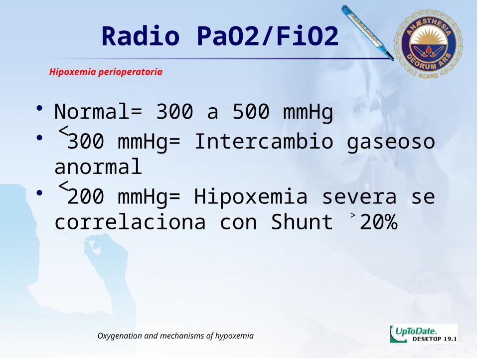
• Normal= 300 a 500 mmHg• ˂300 mmHg= Intercambio gaseoso
anormal• ˂200 mmHg= Hipoxemia severa se
correlaciona con Shunt ˃20%
Hipoxemia perioperatoria
Oxygenation and mechanisms of hypoxemia

LOGOCausas
↓Oxígeno inspirado
Hipoventilación
Alteración V/Q
Difusión limitada
.
Hipoxemica
Anemica
Circulatoria
Afinidad
Histotoxica
Hipoxemia Hipoxia
Monitoring Respiratory FunctionOxygenation and mechanisms of hypoxemia
Shunt Derecha-Izquierda
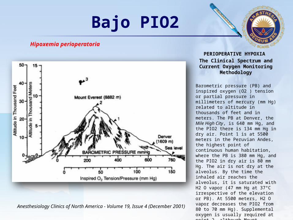
LOGOBajo PIO2
PERIOPERATIVE HYPOXIA
The Clinical Spectrum and Current Oxygen Monitoring Methodology
Barometric pressure (PB) and inspired oxygen (O2 ) tension or partial pressure in millimeters of mercury (mm Hg) related to altitude in thousands of feet and in meters. The PB at Denver, the Mile High City, is 640 mm Hg, and the PIO2 there is 134 mm Hg in dry air. Point 1 is at 5500 meters in the Peruvian Andes, the highest point of continuous human habitation, where the PB is 380 mm Hg, and the PIO2 in dry air is 80 mm Hg. The air is not dry at the alveolus. By the time the inhaled air reaches the alveolus, it is saturated with H2 O vapor (47 mm Hg at 37°C irrespective of the elevation or PB). At 5500 meters, H2 O vapor decreases the PIO2 from 80 to 70 mm Hg). Supplemental oxygen is usually required at point 2, although Mount Everest has been climbed without O2 . Point 3 represents the highest ascent with O2 but without superatmospheric pressure.
Anesthesiology Clinics of North America - Volume 19, Issue 4 (December 2001)
Hipoxemia perioperatoria

LOGOEcuación 1
• ↓PIO2 = ↓PAO2
PIO2 = FiO2 X (PB – PH20)
Anesthesiology Clinics of North America - Volume 19, Issue 4 (December 2001)
LOGO
Morgan, Edward: Anestesiologia clinica – Seccion I capitulo 3 y 4. 2007
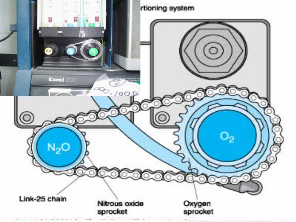
Flujometros
• Guarda Hipóxica– Mecánica, neumática o electrónica– Garantiza FiO2 25%
• Flujo Metabolico O2: Peso¾ x 10
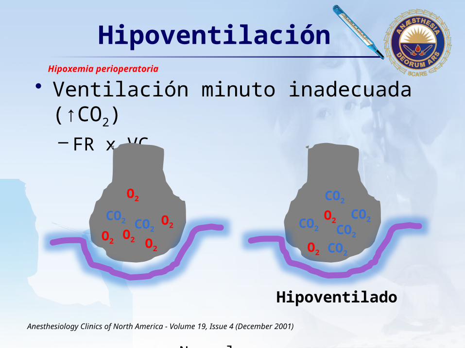
LOGOHipoventilación
• Ventilación minuto inadecuada (↑CO2)– FR x VC
Normal
Anesthesiology Clinics of North America - Volume 19, Issue 4 (December 2001)
Hipoxemia perioperatoria
CO2 CO2O2
O2
O2
O2
CO2 CO2
CO2
CO2
O2O2
O2
Hipoventilado
CO2
LOGO
PAO2=0.21x(760-47)-PaCO2/0.8
100=0.21x(760-47)-40/0.8 Ventilacion Normal PaCO2 40
50=0.21x(760-47)-80/0.8Hipoventilacion PaCO2 80
FiO2 21%
114=0.3x(760-47)-80/0.8Hipoventilacion PaCO2 80
FiO2 30%
• Corrige al ↑FiO2
• Gradiente A-a normal
Hipoventilación
Oxygenation and mechanisms of hypoxemiaAnesthesiology Clinics of North America - Volume 19, Issue 4 (December 2001)
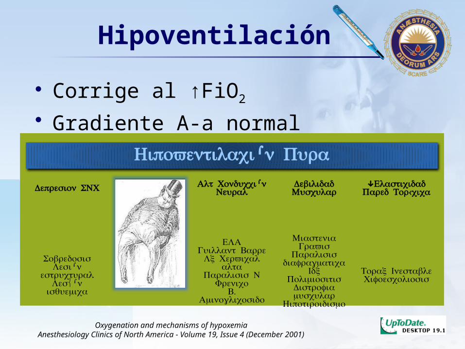
Hipoventilación Pura
Depresion SNC Obesidad Alt ConducciónNeural
DebilidadMuscular
↓ Elasticidad Pared Torácica
Sobredosis Lesión
estructural Lesíón
isquemica
Sx Pickwickian
ELA Guillant Barre
Lx Cervicalalta
Paralisis NFrenico
. BAminoglicosido
MiasteniaGravis
Paralisis diafragmatica
IdxPolimiositis
Distrofiamuscular
Hipotiroidismo
Torax InestableCifoescoliosis

LOGO
• El balance V/Q es complejo– Volumen ventilatorio– Presion alveolar– Compliance pulmon y caja toracica– Resistencia de la via aerea– Gravedad– Posicion del paciente– Flujo sanguineo pulmonar– Modo ventilatorio
Imbalance V/Q
Anesthesiology Clinics of North America - Volume 19, Issue 4 (December 2001)
V sin Q Espacio Muerto
Q sin V Shunt

LOGOImbalance V/Q
• Enfermedad pulmonar obstructiva• Enfermedad vascular pulmonar• Enfermedad intersticial
Anesthesiology Clinics of North America - Volume 19, Issue 4 (December 2001)

LOGOFalla Respiratoria
HipoxemicaAguda
HipoventilacionAlveolar
PerioperatoriaAguda
HipoperfusionShock
Tipo IV
Tipo I
Tipo II
Tipo III Atelectasias, dolor incisionalanalgesia inadecuada, alteracion tos,
uso de tabaco 6 semanas precx sobrehidratación
Critical Care. Just the Facts 2007

LOGOAtelectasias
In normal lungs (A), the alveolar inflation and vascular prfusion are associated with low stress and are not injurious. Two separate barriers form the alveolar– capillary barrier, the microvascular endothelium, and the alveolar epithelium. e
Pulmonary Atelectasis
A Pathogenic Perioperative Entity
Anesthesiology, V 102, No 4, Apr 2005
In contrast, with atelectasis (B), alveolar inflation and deflation may be heterogeneous, and the resulting airway stress causes epithelial injury. Because the blood vessels are compressed, perfusion may be traumatic because of flowinduced disruption of the microvascular endothelium. Both epithelial and endothelial injury may initiate or ropagate lung injury. This figure depicts the advanced stage of lung injury caused by atelectasis. The initial injury is simple collapse of alveoli. However, with time, this leads to an inflammatory reaction. As the derecruited lungs cause epithelial injury and loss of epithelial integrity, both type I and type II alveolar cells are damaged. Injury to type II cells disrupts normal epithelial fluid transport, impairing the removal of edema fluid from the alveolar space. In ddition to collapse, derecruited lungs also become fluid filled. Neutrophils adhere to the injured capillary endothelium and migrate through the interstitium into the alveolar airspace.In the airspace, alveolar macrophages secrete cytokines, interleukin (IL)-1, -6, -8, and -10, and tumor necrosis factor (TNF)-, which act locally to stimulate chemotaxis and activate neutrophils. IL-1 can also stimulate the production of extracellular matrix by fibroblasts. Neutrophils can release oxidants, proteases, leukotrienes, and other proinflammatory molecules, such as platelet-activating factor (PAF). MIF macrophage inhibitory factor.
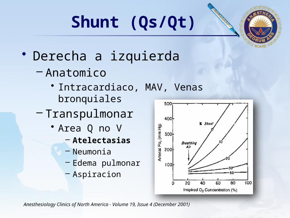
LOGOShunt (Qs/Qt)
• Derecha a izquierda– Anatomico
• Intracardiaco, MAV, Venas bronquiales
– Transpulmonar• Area Q no V
– Atelectasias– Neumonia– Edema pulmonar– Aspiracion
Anesthesiology Clinics of North America - Volume 19, Issue 4 (December 2001)

LOGOAtelectasias
Anesthesiology, V 102, No 4, Apr 2005
ReabsorcionGas
CompresionPulmonar
AlteracionSurfactante
Tres sets de mecanismos causan o contribuyena la formacion de atelectasias
Etiologia y Patogenesis
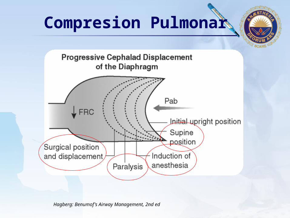
LOGOCompresion Pulmonar
Hagberg: Benumof's Airway Management, 2nd ed
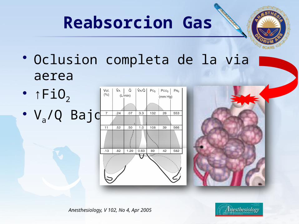
LOGOReabsorcion Gas
Anesthesiology, V 102, No 4, Apr 2005
• Oclusion completa de la via aerea• ↑FiO2
• Va/Q Bajo

LOGOAtelectasias
Anesthesiology, V 102, No 4, Apr 2005

LOGOAtelectasias
Hipoxemia
Alt Compliance Pulmonar
↑RVP
Lesion pulmonar
VC bajo, hiperoxia (microatelectasias)Reversada por hiperinflacion
Reduccion volumen pulmonar, macroatelectasias.Empeora oxigenacion sistemica
Vasoconstriccion hipoxica pulmonar ↓ Tension Oxigeno alveolar y venoso mixto
Atelectasias + VC ↑Prevenida con PEEP
Anesthesiology, V 102, No 4, Apr 2005
Complicaciones
LOGOAtelectasias
Anesthesiology, V 102, No 4, Apr 2005
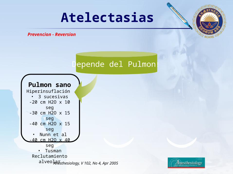
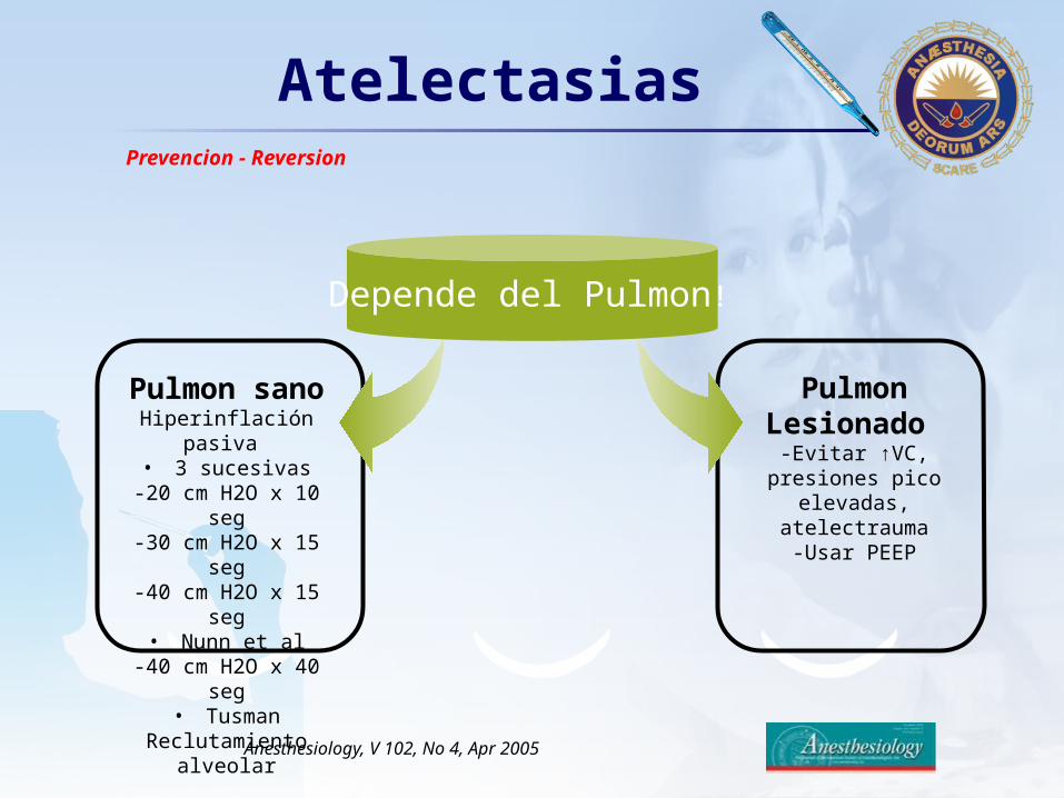
Pulmon sano Hiperinsuflación • 3 sucesivas
-20 cm H2O x 10 seg-30 cm H2O x 15 seg-40 cm H2O x 15 seg
• Nunn et al-40 cm H2O x 40 seg
• TusmanReclutamiento alveolar
Depende del Pulmon!
Prevencion - Reversion
LOGO
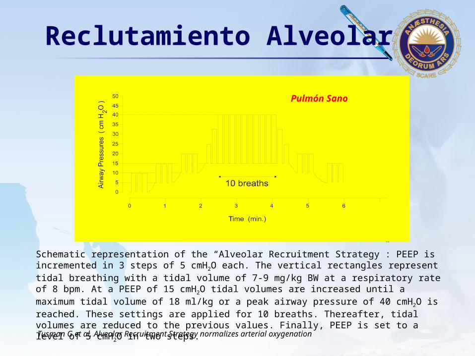
Schematic representation of the “Alveolar Recruitment Strategy”: PEEP is incremented in 3 steps of 5 cmH2O each. The vertical rectangles represent tidal breathing with a tidal volume of 7-9 mg/kg BW at a respiratory rate of 8 bpm. At a PEEP of 15 cmH2O tidal volumes are increased until a maximum tidal volume of 18 ml/kg or a peak airway pressure of 40 cmH2O is reached. These settings are applied for 10 breaths. Thereafter, tidal volumes are reduced to the previous values. Finally, PEEP is set to a level of 5 cmH2O in two steps.
Reclutamiento Alveolar
Tusman G et al. Alveolar Recruitment Strategy normalizes arterial oxygenation
Pulmón Sano
LOGOAtelectasias
Anesthesiology, V 102, No 4, Apr 2005
Pulmon sano Hiperinflación pasiva
• 3 sucesivas-20 cm H2O x 10 seg-30 cm H2O x 15 seg-40 cm H2O x 15 seg
• Nunn et al-40 cm H2O x 40 seg
• TusmanReclutamiento alveolar
Pulmon Lesionado
-Evitar ↑VC, presiones pico elevadas, atelectrauma-Usar PEEP
Depende del Pulmon!
Prevencion - Reversion
LOGO
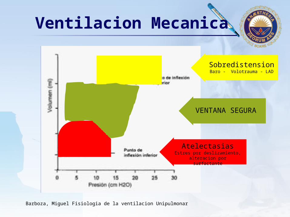
SobredistensionBaro - Volotrauma - LAD
VENTANA SEGURA
AtelectasiasEstres por deslizamiento, alteracion por surfactante
Ventilacion Mecanica
Barboza, Miguel Fisiologia de la ventilacion Unipulmonar

LOGO
LOGO

LOGO
Gracias!Gracias!Por la Paciencia
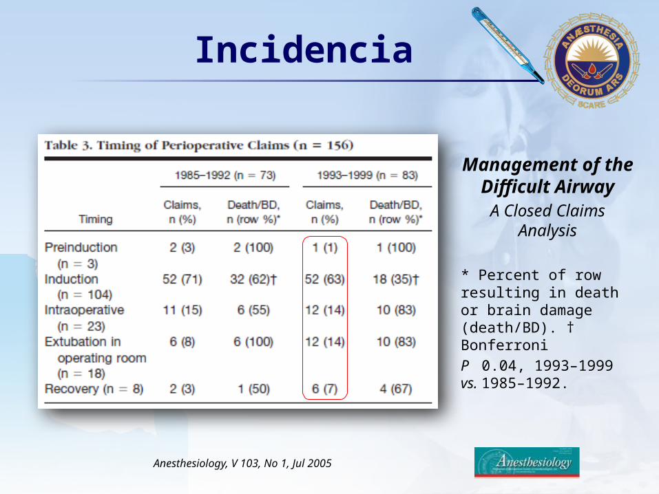
LOGOIncidencia
Management of the Difficult AirwayA Closed Claims
Analysis
* Percent of row resulting in death or brain damage (death/BD). † Bonferroni
P 0.04, 1993–1999 vs. 1985–1992.
Anesthesiology, V 103, No 1, Jul 2005