Anemia patama5march2015
28
Patama Gomutbutra Consultant : Lect. Lalita Norasetthada Anemia Ambulatory setting
-
Upload
patama-gomutbutra -
Category
Health & Medicine
-
view
88 -
download
0
Transcript of Anemia patama5march2015

Patama Gomutbutra Consultant : Lect. Lalita Norasetthada
Anemia
Ambulatory setting
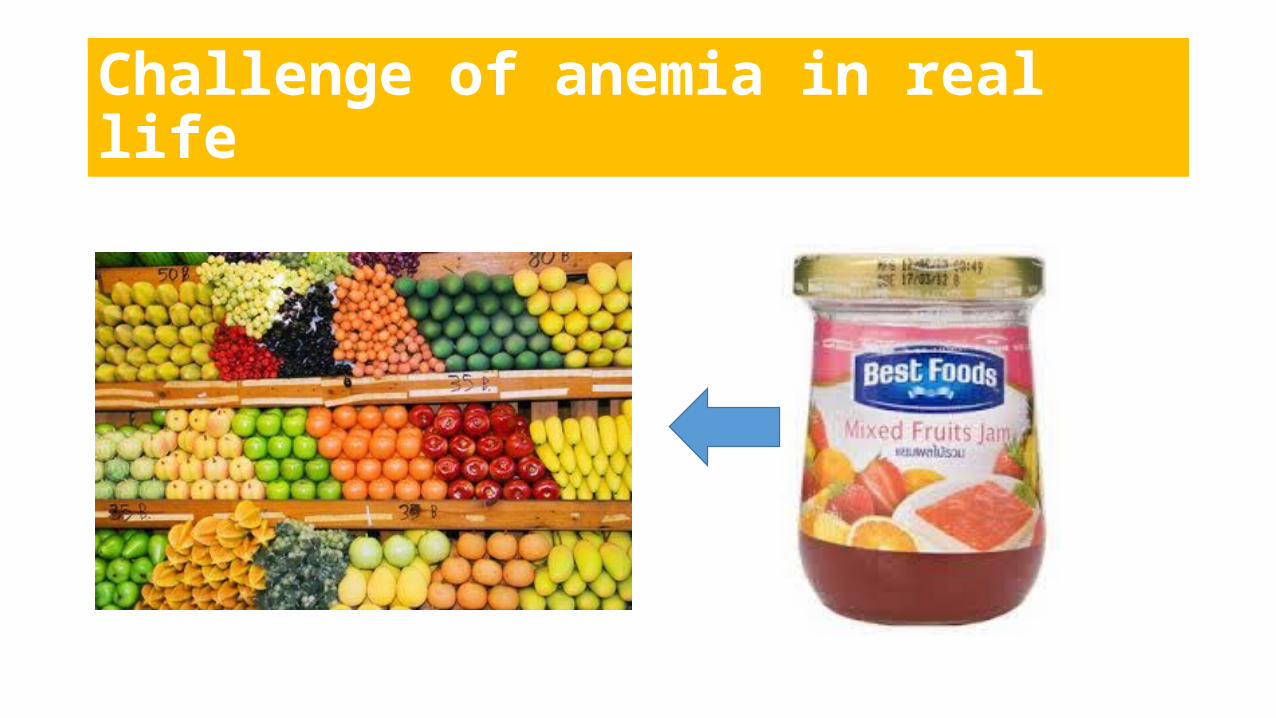
Challenge of anemia in real life
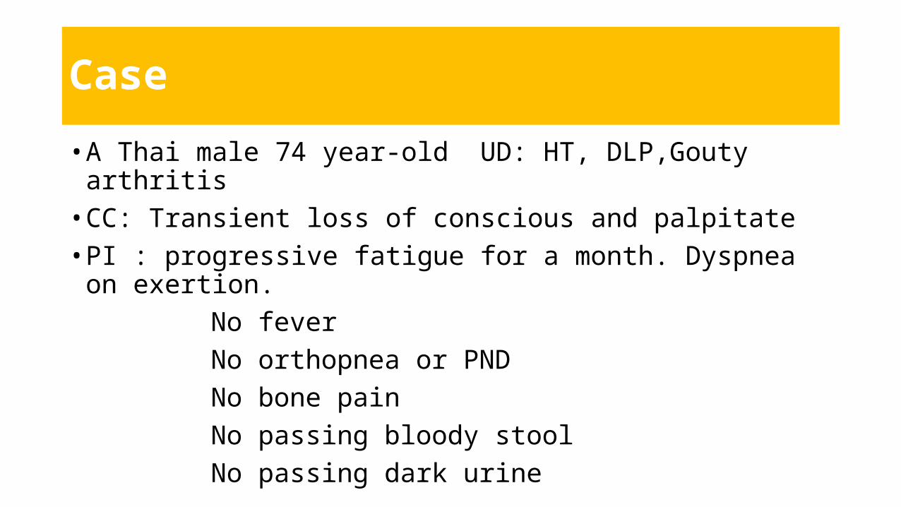
Case
• A Thai male 74 year-old UD: HT, DLP,Gouty arthritis• CC: Transient loss of conscious and palpitate• PI : progressive fatigue for a month. Dyspnea on exertion. No fever No orthopnea or PND No bone pain No passing bloody stool No passing dark urine
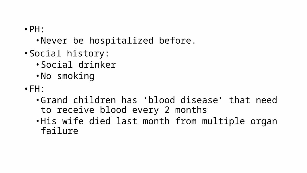
• PH: • Never be hospitalized before.
• Social history:• Social drinker• No smoking
• FH: • Grand children has ‘blood disease’ that need to receive blood
every 2 months• His wife died last month from multiple organ failure
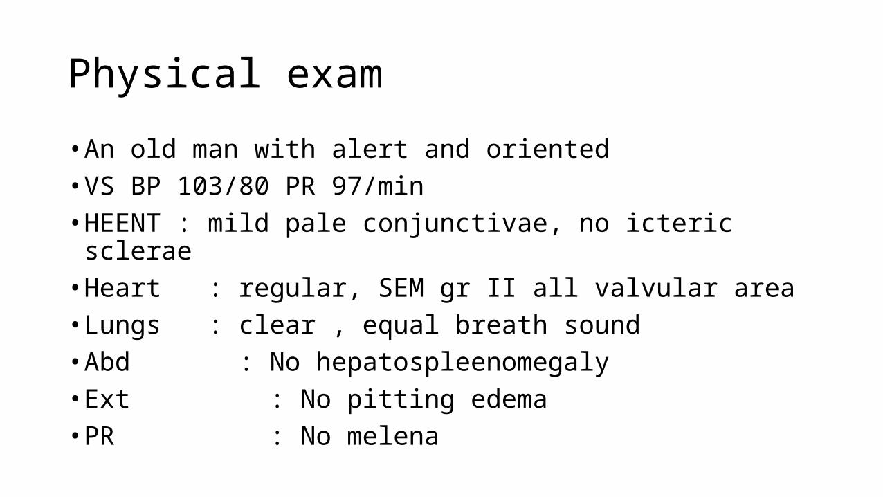
Physical exam
• An old man with alert and oriented• VS BP 103/80 PR 97/min• HEENT : mild pale conjunctivae, no icteric sclerae• Heart : regular, SEM gr II all valvular area• Lungs : clear , equal breath sound• Abd : No hepatospleenomegaly• Ext : No pitting edema• PR : No melena

• Corrected RC = 6.62 X (18.3 /45) = 2.68
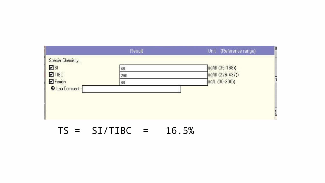
TS = SI/TIBC = 16.5%

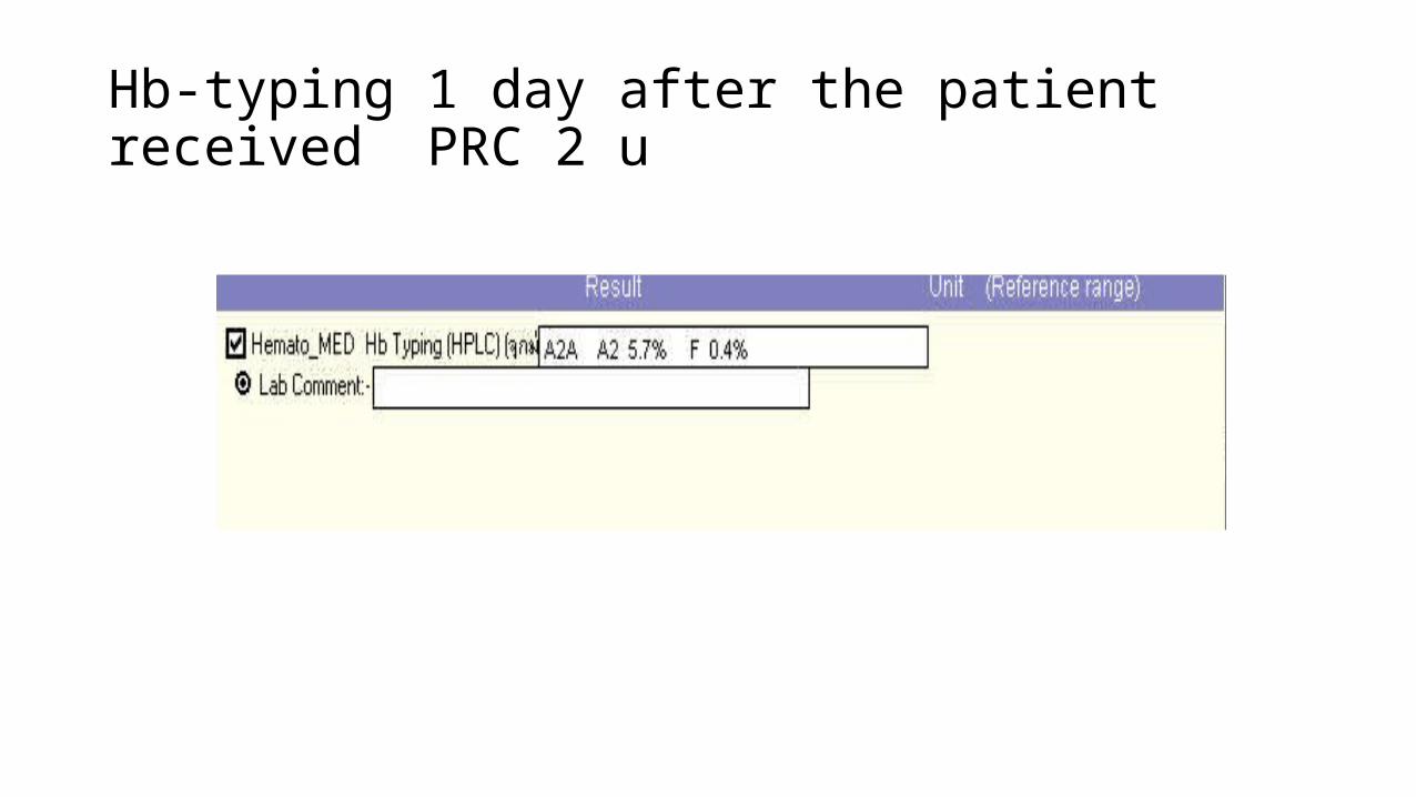
Hb-typing 1 day after the patient received PRC 2 u
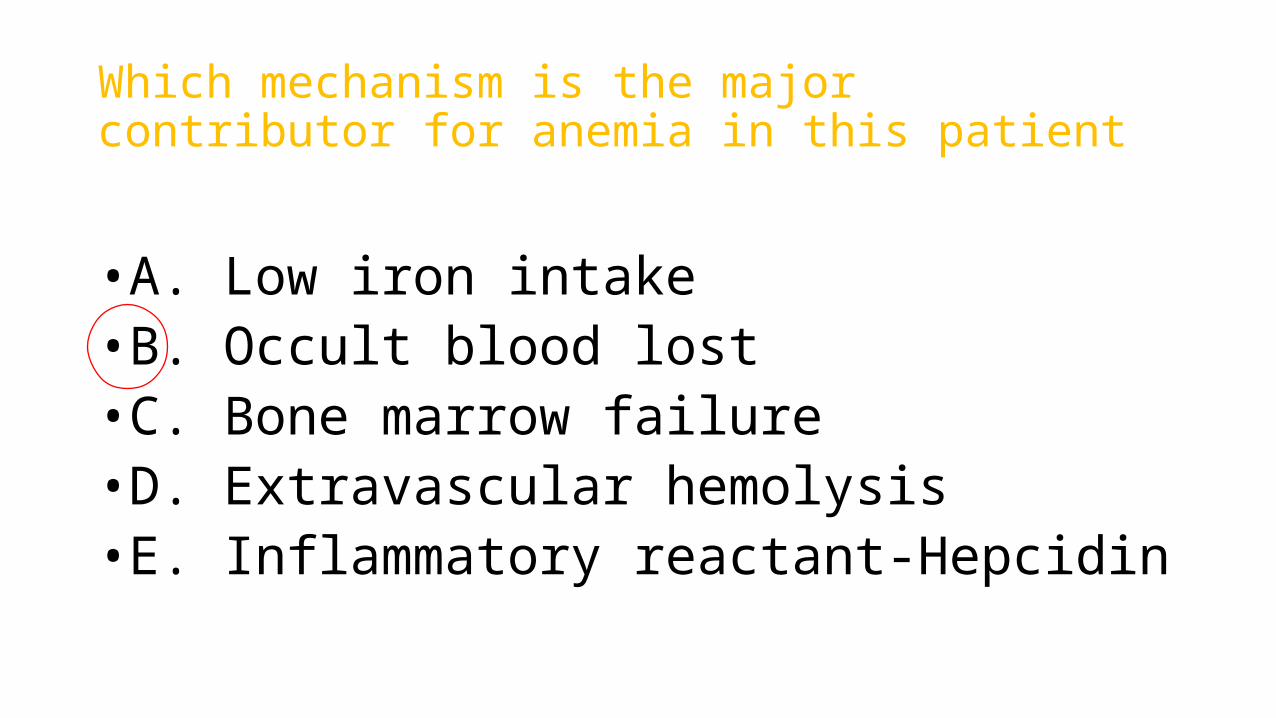
Which mechanism is the major contributor for anemia in this patient
•A. Low iron intake •B. Occult blood lost•C. Bone marrow failure•D. Extravascular hemolysis•E. Inflammatory reactant-Hepcidin

PBS• Hypochromic microcytic. Anisopoikilocyte 2+ ,No nRBC • Increased polychromasia Imp: Thallasemia trait with occult blood lost
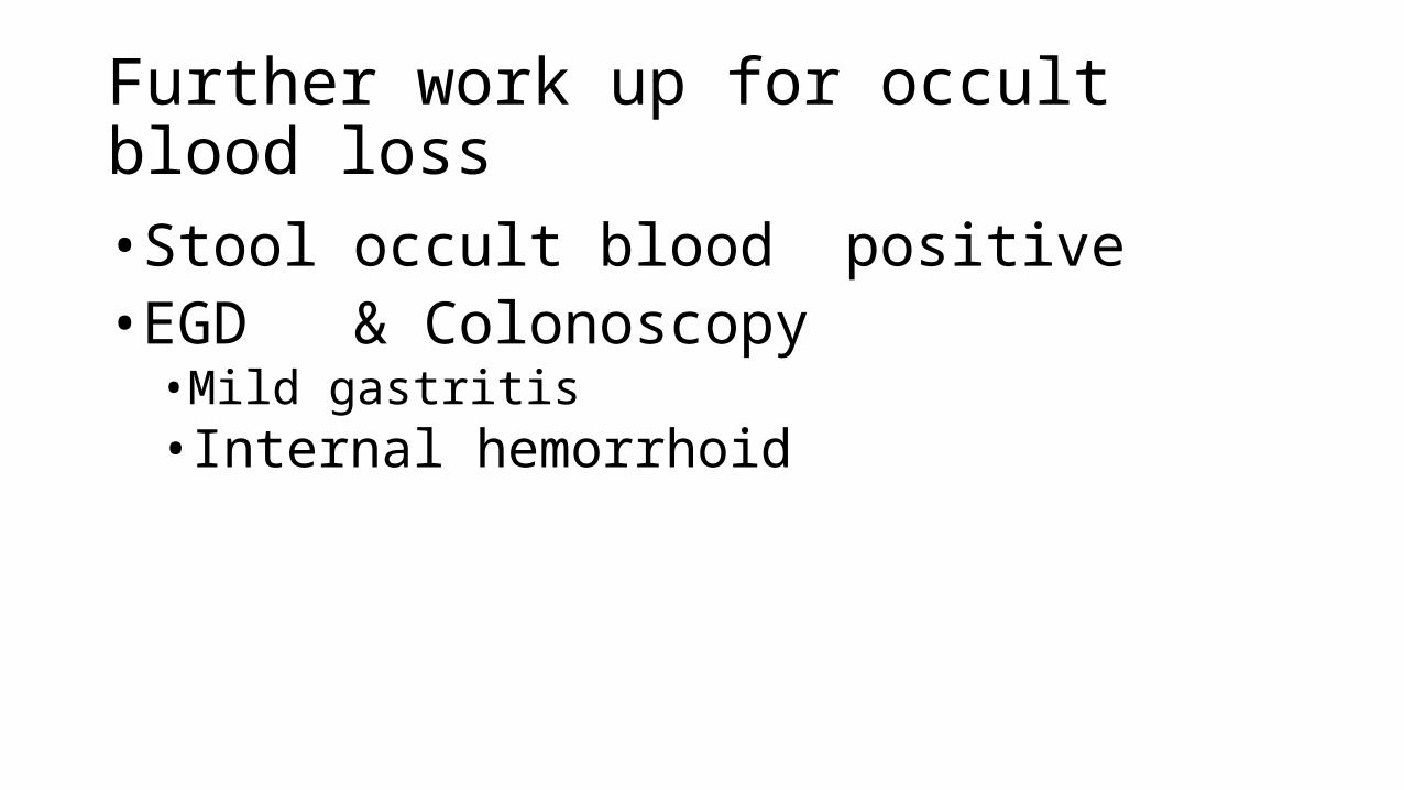
Further work up for occult blood loss
•Stool occult blood positive•EGD & Colonoscopy •Mild gastritis• Internal hemorrhoid

Hb < 12 for femaleHb < 13 for male
MCV, RC
Low MCV <80
High RC
Low RC
Normal MCV 80-100
High RC
Low RC
High MCV >100
High RC
Low RC
!
Adapted from Norasettada L et al. 2013
! !
Hemolysis ?- LDH - Bilirubin w Indirect bilirubin - Haptoglobin (likely intravas)
SI/TIBC high• Lead poisoning• Sideroblastic
Hb typing normal• PNH• IDA w Iron
suppl
Bi/pancytopenia• Aplastic • MDS• BM infiltrate
Coomb’s positive• AIHA (E)
Megaloblastic : hypersegmented neutrophil• B12 def• Folic def
SI/TIBC low <20• Iron def
anemia• ACD• Thal trait
Hb typing abn• Thalasemia
major
Occult bleeding ?
Pure anemia• Endocrine Thyroid, AI• BM defect
MM, PRCA
Non megaloblastic• Liver disease/Alcohol• Hypothyroid• BM defect
Coomb’s negative• MAHA (I )• G-6-PD (I )• Malaria (I)• Spherocytosis(E)
!
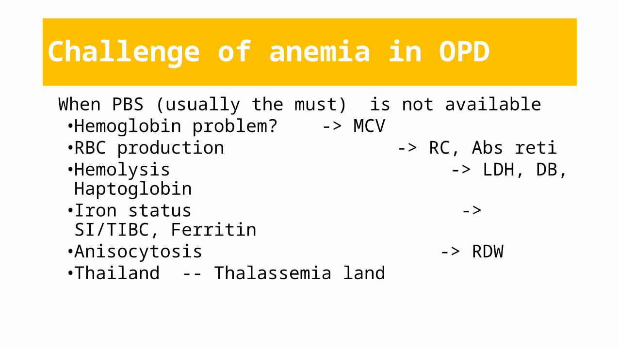
Challenge of anemia in OPD
When PBS (usually the must) is not available•Hemoglobin problem? -> MCV•RBC production -> RC, Abs reti•Hemolysis -> LDH, DB, Haptoglobin• Iron status -> SI/TIBC, Ferritin •Anisocytosis -> RDW• Thailand -- Thalassemia land

MCV < 80
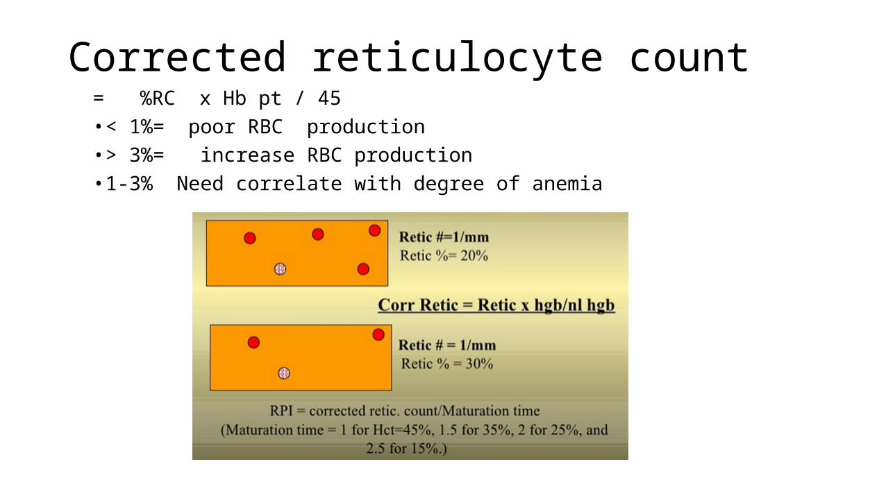
Corrected reticulocyte count= %RC x Hb pt / 45• < 1%= poor RBC production• > 3%= increase RBC production• 1-3% Need correlate with degree of anemia
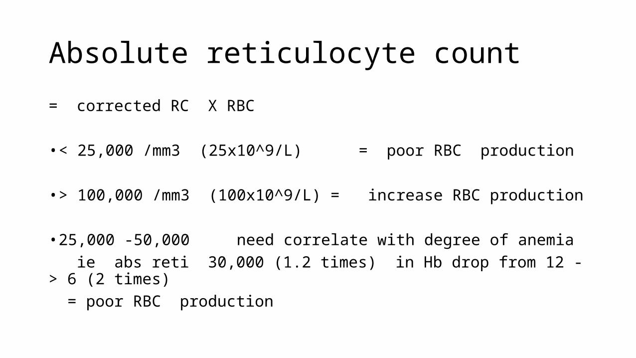
Absolute reticulocyte count
= corrected RC X RBC
• < 25,000 /mm3 (25x10^9/L) = poor RBC production
• > 100,000 /mm3 (100x10^9/L) = increase RBC production
• 25,000 -50,000 need correlate with degree of anemia ie abs reti 30,000 (1.2 times) in Hb drop from 12 -> 6 (2 times) = poor RBC production
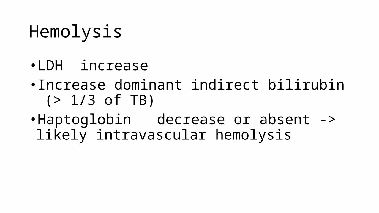
Hemolysis
• LDH increase• Increase dominant indirect bilirubin (> 1/3 of TB)•Haptoglobin decrease or absent -> likely
intravascular hemolysis

Free Hb bind to
Haptoglobin

Accumulate PTP intermediate = Porphyrias (Greek – Purple) Acute intermittent porphyria Porphyria cutanea tada
Lack of Protoporphyrin lead to excess iron
= Sideroblastic anemia (Greek – iron) B6 deficiency
Lead poisoning
Fe
PTP

• Feritin is the best indicator for Iron storage status
cut point 100 sens = 70 % spec = 95 %
• TIBC is Transferin ‘seeking for Fe’ Lower Fe -> Higher TIBC
(SI/TIBC)
350 ug/dl
100 ug/dl
50 ug/dl 300 ug/dl
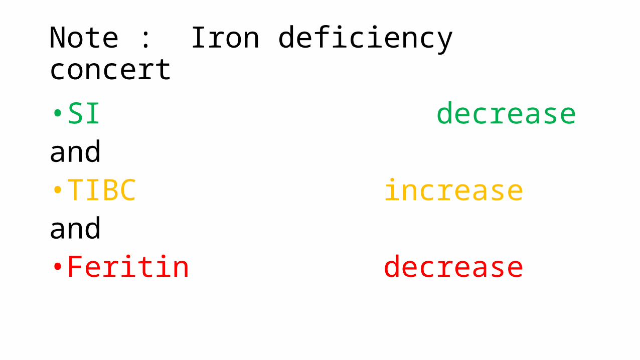
Note : Iron deficiency concert
•SI decreaseand•TIBC increase and•Feritin decrease

RDW = Level of varies in size (anisocytosis)
Normal range of RDW-CV < 16-18 %
Why divide by MCV ?= To fair with higher upper limit of RBC population

Thallasemia minor/ trait
• Hb normal or mildly reduced • MCV reduced• Hb electrophoresis • HbA2: <5 % (<3.5 = alpha , > 3.5 = beta)• Hb F: < 5 %• HbCS 1-2%
• Not cause of significant hemolytic anemia• PBS- milder anisopoikilocytosis. No nRBC

Exercise case A 25 year old waiter with recurrent pulmonary TB and long term Isoniazid treatment
• Hb = 3.8 • MCV = 72• RC corrected = 1.7• RDW = 22• SI = 246 (50-150) TIBC = 254 (250-450) Feritin = 446 (27-224)• TB/DB = 1.0/0.2
What is the most likely diagnosisA. Iron def anemiaB. ThallasemiaC. Anemia of chronic diseaseD. Sideroblastic anemiaE. Lead poisoning


![Marinov - Anemia and haemorrhagic diatheses 2016 [Eng].ppt - Anemia and... · ANEMIA Time Anemias due to impaired ... Pathway Common Pathway. 4/13/2016 21 ... Marinov - Anemia and](https://static.fdocuments.us/doc/165x107/5d15387088c993e8108c4415/marinov-anemia-and-haemorrhagic-diatheses-2016-eng-anemia-and-anemia.jpg)