and p66Shc Generates Reactive Oxygen Species that Trigger...
13
Cell, Vol. 122, 221–233, July 29, 2005, Copyright ©2005 by Elsevier Inc. DOI 10.1016/j.cell.2005.05.011 Electron Transfer between Cytochrome c and p66 Shc Generates Reactive Oxygen Species that Trigger Mitochondrial Apoptosis Marco Giorgio, 1,2,8, * Enrica Migliaccio, 1,2,8 degenerative diseases, and aging. Mitochondria play a central role in apoptosis. They contain a number of pro- Francesca Orsini, 1,2 Demis Paolucci, 3 Maurizio Moroni, 4 Cristina Contursi, 4 apoptotic factors, such as cytochrome c (cyt c), aif, smac/diablo, and endoG, which are released into the Giovanni Pelliccia, 2 Lucilla Luzi, 2 Saverio Minucci, 1,4 Massimo Marcaccio, 3 Paolo Pinton, 6 cytosol during apoptosis, where they activate a series of enzymatic activities that lead to the specific degra- Rosario Rizzuto, 6 Paolo Bernardi, 5 Francesco Paolucci, 3 and Pier Giuseppe Pelicci 1,2,7,8, * dation of proteins and DNA (Danial and Korsmeyer, 2004; Pelicci, 2004). 1 Experimental Oncology Department European Institute of Oncology The release of proapoptotic factors from mito- chondria is due to the disruption of the organelle integ- Milan 2 FIRC Institute of Molecular Oncology rity, which is achieved through multiple and concomi- tant events. In the case of cyt c, for example, its release Milan 3 G Ciamician Chemistry Department requires permeabilization of the outer membrane, re- modeling of the mitochondrial cristae, and dissociation University of Bologna Bologna of cyt c from cardiolipin, a constituent of the inner membrane (Green and Kroemer, 2004). These events 4 Congenia srl Milan are triggered by the opening of a high-conductance channel, the permeability transition pore (PTP) (Ber- 5 Biomedical Sciences Department University of Padova nardi et al., 2001). Opening of the PTP provokes an increase of inner membrane permeability to ions and Padova solutes (the so-called permeability transition, PT), fol- 6 Experimental and Diagnostic Medicine Department lowed by net water influx toward the mitochondrial ma- University of Ferrara trix, swelling of the organelle, and physical rupture of its Ferrara outer membrane, with the consequent release of proteins 7 Department of Medicine and Surgery of the intermembrane space, including cyt c (Bernardi University of Milan et al., 2001). Another mechanism leading to alterations Milan of the mitochondrial membrane integrity is the translo- Italy cation of proapoptotic proteins such as Bax and Bid from the cytosol to the outer mitochondrial membrane, where they form channels and/or regulate the function Summary of preexisting channels (Scorrano and Korsmeyer, 2003). Whether these events then trigger the PT or are Reactive oxygen species (ROS) are potent inducers of per se sufficient to mediate complete cyt c release re- oxidative damage and have been implicated in the regu- mains, however, controversial. lation of specific cellular functions, including apoptosis. Reactive oxygen species (ROS; superoxide, hydro- Mitochondrial ROS increase markedly after proapoptotic gen peroxide, and hydroxyl radicals) are potent intra- signals, though the biological significance and the un- cellular oxidants, which have been proposed as critical derlying molecular mechanisms remain undetermined. regulators of apoptosis (Danial and Korsmeyer, 2004). P66 Shc is a genetic determinant of life span in mammals, Indeed, ROS induce the opening of the PTP through which regulates ROS metabolism and apoptosis. We re- oxidation-dependent mechanisms and are potent in- port here that p66 Shc is a redox enzyme that generates ducers of apoptosis, both in cultured cells and in vivo mitochondrial ROS (hydrogen peroxide) as signaling (Petronilli et al., 1994; Danial and Korsmeyer, 2004). Be- molecules for apoptosis. For this function, p66 Shc uti- yond their documented proapoptotic activity when ad- lizes reducing equivalents of the mitochondrial electron ministered exogenously, endogenously produced ROS transfer chain through the oxidation of cytochrome c. are implicated in the execution of the apoptotic pro- Redox-defective mutants of p66 Shc are unable to in- gram itself, regardless of the type of triggering signal. duce mitochondrial ROS generation and swelling in In fact, a marked increase of intracellular ROS is con- vitro or to mediate mitochondrial apoptosis in vivo. sistently associated with apoptosis, and treatments These data demonstrate the existence of alternative with antioxidants protect cells from apoptosis (Mayer redox reactions of the mitochondrial electron transfer and Noble, 1994). chain, which evolved to generate proapoptotic ROS The molecular mechanisms underlying increased in response to specific stress signals. ROS generation during apoptosis are unclear. It has been proposed that ROS accumulation is secondary to Introduction the progress of the apoptotic process and is caused by the interruption of the mitochondrial electron transfer Apoptosis is essential for the maintenance of tissue ho- chain (ETC), a consequence of the PT and of cyt c re- meostasis, and its deregulation is implicated in cancer, lease (Cai and Jones, 1998). Alternatively, ROS might function to trigger mitochondrial apoptosis, serving as *Correspondence: [email protected]; piergiuseppe. signaling molecules to initiate the PT. Notably, intracel- [email protected] 8 These authors contributed equally to this work. lular ROS arise prior to cyt c release during the acti-
Transcript of and p66Shc Generates Reactive Oxygen Species that Trigger...

Cell, Vol. 122, 221–233, July 29, 2005, Copyright ©2005 by Elsevier Inc. DOI 10.1016/j.cell.2005.05.011
Electron Transfer between Cytochrome cand p66Shc Generates Reactive OxygenSpecies that Trigger Mitochondrial Apoptosis
Marco Giorgio,1,2,8,* Enrica Migliaccio,1,2,8
Francesca Orsini,1,2 Demis Paolucci,3
Maurizio Moroni,4 Cristina Contursi,4
Giovanni Pelliccia,2 Lucilla Luzi,2 Saverio Minucci,1,4
Massimo Marcaccio,3 Paolo Pinton,6
Rosario Rizzuto,6 Paolo Bernardi,5
Francesco Paolucci,3 and Pier Giuseppe Pelicci1,2,7,8,*1Experimental Oncology DepartmentEuropean Institute of OncologyMilan2FIRC Institute of Molecular OncologyMilan3G Ciamician Chemistry DepartmentUniversity of BolognaBologna4Congenia srlMilan5Biomedical Sciences DepartmentUniversity of PadovaPadova6Experimental and Diagnostic Medicine DepartmentUniversity of FerraraFerrara7Department of Medicine and SurgeryUniversity of MilanMilanItaly
Summary
Reactive oxygen species (ROS) are potent inducers ofoxidative damage and have been implicated in the regu-lation of specific cellular functions, including apoptosis.Mitochondrial ROS increase markedly after proapoptoticsignals, though the biological significance and the un-derlying molecular mechanisms remain undetermined.P66Shc is a genetic determinant of life span in mammals,which regulates ROS metabolism and apoptosis. We re-port here that p66Shc is a redox enzyme that generatesmitochondrial ROS (hydrogen peroxide) as signalingmolecules for apoptosis. For this function, p66Shc uti-lizes reducing equivalents of the mitochondrial electrontransfer chain through the oxidation of cytochrome c.Redox-defective mutants of p66Shc are unable to in-duce mitochondrial ROS generation and swelling invitro or to mediate mitochondrial apoptosis in vivo.These data demonstrate the existence of alternativeredox reactions of the mitochondrial electron transferchain, which evolved to generate proapoptotic ROSin response to specific stress signals.
Introduction
Apoptosis is essential for the maintenance of tissue ho-meostasis, and its deregulation is implicated in cancer,
*Correspondence: [email protected]; [email protected]
8 These authors contributed equally to this work.degenerative diseases, and aging. Mitochondria play acentral role in apoptosis. They contain a number of pro-apoptotic factors, such as cytochrome c (cyt c), aif,smac/diablo, and endoG, which are released into thecytosol during apoptosis, where they activate a seriesof enzymatic activities that lead to the specific degra-dation of proteins and DNA (Danial and Korsmeyer,2004; Pelicci, 2004).
The release of proapoptotic factors from mito-chondria is due to the disruption of the organelle integ-rity, which is achieved through multiple and concomi-tant events. In the case of cyt c, for example, its releaserequires permeabilization of the outer membrane, re-modeling of the mitochondrial cristae, and dissociationof cyt c from cardiolipin, a constituent of the innermembrane (Green and Kroemer, 2004). These eventsare triggered by the opening of a high-conductancechannel, the permeability transition pore (PTP) (Ber-nardi et al., 2001). Opening of the PTP provokes anincrease of inner membrane permeability to ions andsolutes (the so-called permeability transition, PT), fol-lowed by net water influx toward the mitochondrial ma-trix, swelling of the organelle, and physical rupture of itsouter membrane, with the consequent release of proteinsof the intermembrane space, including cyt c (Bernardiet al., 2001). Another mechanism leading to alterationsof the mitochondrial membrane integrity is the translo-cation of proapoptotic proteins such as Bax and Bidfrom the cytosol to the outer mitochondrial membrane,where they form channels and/or regulate the functionof preexisting channels (Scorrano and Korsmeyer,2003). Whether these events then trigger the PT or areper se sufficient to mediate complete cyt c release re-mains, however, controversial.
Reactive oxygen species (ROS; superoxide, hydro-gen peroxide, and hydroxyl radicals) are potent intra-cellular oxidants, which have been proposed as criticalregulators of apoptosis (Danial and Korsmeyer, 2004).Indeed, ROS induce the opening of the PTP throughoxidation-dependent mechanisms and are potent in-ducers of apoptosis, both in cultured cells and in vivo(Petronilli et al., 1994; Danial and Korsmeyer, 2004). Be-yond their documented proapoptotic activity when ad-ministered exogenously, endogenously produced ROSare implicated in the execution of the apoptotic pro-gram itself, regardless of the type of triggering signal.In fact, a marked increase of intracellular ROS is con-sistently associated with apoptosis, and treatmentswith antioxidants protect cells from apoptosis (Mayerand Noble, 1994).
The molecular mechanisms underlying increasedROS generation during apoptosis are unclear. It hasbeen proposed that ROS accumulation is secondary tothe progress of the apoptotic process and is caused bythe interruption of the mitochondrial electron transferchain (ETC), a consequence of the PT and of cyt c re-lease (Cai and Jones, 1998). Alternatively, ROS mightfunction to trigger mitochondrial apoptosis, serving assignaling molecules to initiate the PT. Notably, intracel-lular ROS arise prior to cyt c release during the acti-

Cell222
vation of several apoptotic pathways, such as those hinvolving p53 or TNFα or those triggered by hypergly- pcemia, ischemia/reperfusion, or viral infection (Trinei etal., 2002; Sakon et al., 2003; Brownlee, 2001; Becker, R2004).
ROS are generated accidentally, as by products of Paerobic metabolism, and mitochondria are the major osource of intracellular ROS (Wallace, 1999). During res- Tpiration, electrons are extracted from substrates and care then transferred to molecular oxygen through a schain of enzymatic complexes (I–IV). In the final step of cthe ETC, cyt c oxidase (COX; complex IV) ensures the Mreduction of molecular oxygen to water, without forma- ltion of oxygen radicals. However, partial reduction of (oxygen with generation of ROS can occur if molecular toxygen interacts with the ETC upstream of complex IV. 2Experimental data indicate that ROS are indeed contin- nuously produced during mitochondrial respiration and Cthat up to 2% of the total oxygen consumption is con- (verted to ROS (Chance et al., 1979). How ROS are regu- clated during apoptosis is unclear. c
P66Shc deletion in mice (p66Shc−/−) decreases the in- mcidence of aging-associated diseases, such as athero-sclerosis, and prolongs life span (Migliaccio et al., 1999; wNapoli et al., 2003; Francia et al., 2004). The underlying imolecular mechanisms are, however, unknown. P66Shc
mis a splice variant of p52Shc/p46Shc, two cytoplasmic �adaptor proteins involved in the propagation of intracel- blular signals from activated tyrosine kinases to Ras d(Pelicci et al., 1992). P66Shc has the same modular astructure of p52Shc/p46Shc (SH2-CH1-PTB) and con- [tains a unique N-terminal region (CH2); however, it is anot involved in Ras regulation but rather functions in dthe intracellular pathway(s) that regulates ROS metabo- Tlism and apoptosis (Migliaccio et al., 1997; Migliaccio set al., 1999; Trinei et al., 2002). t
Intracellular ROS levels are decreased in p66Shc−/−m
cells, as revealed by the reduced oxidation of ROS-sen-c
sitive probes and the reduced accumulation of endoge-R
nous markers of oxidative stress (8-oxo-guanosine)i(Trinei et al., 2002; Francia et al., 2004). Likewise,wp66Shc−/− mice have diminished levels of both systemicA(isoprostane) and intracellular (nytrotyrosines, 8-oxo-pguanosine) oxidative stress (Napoli et al., 2003; Franciaaet al., 2004; Trinei et al., 2002).nP66Shc−/− cells are resistant to apoptosis induced bypa variety of different signals, including hydrogen perox-mide, ultraviolet radiation, staurosporine, growth factorPdeprivation, calcium ionophore, and CD3-CD4 cross-
linking (Migliaccio et al., 1999; Orsini et al., 2004; PaciniPet al., 2004). Similarly, the p66Shc−/− mice are resistantIto apoptosis induced by paraquat, hypercholestero-Plemia, and ischemia (Migliaccio et al., 1999; Napoli etcal., 2003; Zaccagnini et al., 2004).tExpression of p66Shc is indispensable for the mi-ttochondrial depolarization and the release of cyt c(observed after treatment of cells with various proapo-iptotic signals, suggesting that it regulates the mito-echondrial pathway of apoptosis. Since inhibition of PTPtopening blocks the proapoptotic function of p66Shc, thelPTP itself might be the target of p66Shc activity (Trineiaet al., 2002; Orsini et al., 2004). We report here that
p66Shc is a redox enzyme that generates mitochondrial t
ydrogen peroxide for the induction of PT and apo-tosis.
esults
66Shc Induces Permeability Transitionf Isolated Mitochondriao investigate whether the effect of p66Shc on the mito-hondrial PTP is direct, we analyzed its ability to inducewelling of purified mitochondria, measured as de-rease of light absorbance of the organelle suspension.itochondria were purified from wild-type (wt) mouse
iver and pretreated with a low concentration of Ca2+
7 �M), a condition that does not induce PT but sensi-izes PTP opening to other signals (Bernardi et al.,001). Addition of recombinant p66Shc (5–20 �M) didot induce swelling, as compared to a large load ofa2+ (0.2 mM), a well-characterized inducer of the PT
Figure 1A). Since p66Shc is not imported by mito-hondria in vitro (Ventura et al. 2004), this finding indi-ates that p66Shc is unable to alter mitochondrial per-eability when acting from the outside of the organelle.We then investigated the effect of p66Shc on the PTPhen the protein is allowed to enter the mitochondrial
ntermembrane space. To this end, we treated purifieditochondria with a low concentration of digitonin (25M), which selectively permeabilizes the outer mem-rane without affecting PT, as shown by the fact thatigitonin-treated mitochondria retain cyt c, respire, andre sensitive to Ca2+-induced swelling (Diwan et al.1988] and data not shown). Under these conditions,ddition of recombinant p66Shc induced marked andose-dependent swelling of mitochondria (Figure 1B).his effect was abolished in the absence of respiratoryubstrates, indicating that it depends on an intactransmembrane potential (data not shown). Further-ore, it was Ca2+ dependent (data not shown) and
omparable to that obtained by exogenously addedOS (H2O2; Figure 1C). Notably, p66Shc was unable to
nduce swelling when digitonin-treated mitochondriaere pretreated with PT inhibitors such as cyclosporine(CsA), a drug that binds the PTP component cyclo-
hilin D, or N-ethyl-maleimide (NEM), a sulfhydryl re-gent that blocks PTP opening by thiol oxidants (Petro-illi et al., 1994) (Figure 1D). These results suggest that66Shc is biologically active when it localizes within theitochondrial intermembrane space, where it inducesT through oxidation-dependent mechanisms.
66Shc Localizes within the Mitochondrialntermembrane Space66Shc is enriched in the submitochondrial fractionontaining inner membranes (Orsini et al., 2004). To fur-her investigate its localization, mitochondria prepara-ions from MEFs (Figure 1E) were treated with digitoninwhich allows extraction of soluble proteins from thentermembrane space) or digitonin and high salt (whichxtracts membrane bound proteins). After centrifuga-ion, the resulting pellets and supernatants were ana-yzed by Western blotting (WB) by using anti-p66Shc,nti-cyt c, anti-mtHsp70, or anti-porin antibodies. Digi-onin or digitonin/high-salt treatments did not modify

P66Shc Is a Redox Enzyme that Generates H2O2223
Figure 1. P66Shc Localizes within the Mito-chondrial Intermembrane Space and In-duces Swelling of Isolated Mitochondria
Absorbance changes of purified liver mito-chondria, either intact (A) or treated with 25�M digitonin (B–D), pretreated with 7 �MCaCl2 and supplemented with the following:(A) 20 �M collagenase as inactive control(control [ctrl]; black trace), 20 �M p66Shc
(red), or 200 �M CaCl2 (brown); (B) 20 �Mcollagenase in 0.25 M sucrose (ctrl, black) ordifferent concentrations of p66Shc (5, 10, and20 �M, as indicated; red); (C) different con-centrations of H2O2 (blue) or control buffer(gray); (D) 20 �M p66Shc (added to all kine-tics) and 1 �M CsA (violet), 20 �M NEM(green) or control buffer (red). Each experi-ment was performed six times using differentmitochondrial preparations and two distinctpreparations of the p66Shc. (E) (Left panels)WB analysis of MEF subcellular fractionsusing anti-p66Shc, anti-mtHp70, anti-cal-nexin, and anti-cytoplasmic Hsp70 antibod-ies. T, total extract; Mt, mitochondria; ER,endoplasmic reticulum; Ct, cytosol. (Rightpanels) ER and Mt fractions were treatedwith Proteinase K (PK) and analyzed by WBwith anti-calnexin or anti-p66Shc antibodies.(F and G) Mitochondrial fractions from MEFswere treated with the indicated concentra-tions of NaCl and/or digitonin. After centrifu-gation, pellets (P) and supernatants (S) wereanalyzed by WB.
the localization of porin (a marker of the outer mem-brane) and of mtHsp70 (matrix), indicating that theseexperimental conditions did not provoke gross struc-tural alterations of purified mitochondria (Figures 1Fand 1G). As expected, digitonin/high salt but not digi-tonin alone induced cyt c release in the supernatant(Griparic et al., 2004). P66Shc, instead, was partially andequally released by either digitonin or digitonin/highsalt, suggesting that it exists within the inner mitochon-drial space in the form of both soluble and nonsolubleprotein (mitochondrial p66Shc is about 50 ng/mg of totalproteins; see Figure S1 in the Supplemental Data avail-able with this article online). Similar results were ob-tained by morphometric analysis of immunoelectronmicroscopy sections of wt MEFs, which revealed asso-ciation of mitochondrial gold particles with the innermembrane (55.7%), intermembrane space (35.4%), andmatrix (8.9%) (data not shown).
P66Shc Stimulates Generation of ROSby Energized MitochondriaP66Shc expression is indispensable for the upregulationof intracellular ROS during p53- or ischemia-inducedapoptosis (Trinei et al., 2002; Pacini et al., 2004; Zac-cagnini et al., 2004). To investigate whether p66Shc-reg-ulated ROS derive from mitochondria, we analyzed in-tact liver mitochondria from wt or p66Shc−/− mice upontreatment with carbon tetrachloride (CCl4; 2 mg/kg), achemical that induces ROS- and p66Shc-dependentapoptosis. We had previously shown that in vivo treat-ment with CCl4 induces apoptosis of hepatocytes in wtbut not p66Shc−/− mice and that apoptosis in wt animalsis prevented by treatment with the antioxidant N-acetylcysteine. Likewise, CCl4 induces p66Shc- and ROS-dependent apoptosis of MEFs (Figure S2). Liver mito-chondria were collected 20 hr after treatment, and ROSwere measured by spectrofluorimetry, using a mole-

Cell224
cular probe that becomes fluorescent upon oxidation pRby ROS (2#,7#-dichlorofluorescin diacetate; H2DCFDA)
(LeBel et al., 1992). The rate of mitochondrial H2DFCDA poxidation increased significantly after CCl4 treatment inthe wt mice, while it remained unchanged in the e
fp66Shc−/− mice (Figures 2A–2C). Notably, H2DCFDA oxi-dation was consistently detected only in the presence s
wof respiratory substrates, such as succinate (Figures2A–2C) or glutamate/malate (data not shown). Together, S
kthese findings indicate that p66Shc expression is indis-
Figure 2. P66Shc Stimulates Mitochondrial Generation of ROS
(A and B) Fluorescence changes (ex. 498, em. 527; I, intensity of fluorescence) and corresponding bar graph (rate of fluorescence intensity/min) (C) of mitochondria from wt (black lines in [A] and [B] and empty bars in [C]) or p66Shc−/− (red lines and red bars) mouse livers, treatedwith placebo ([A], CTRL) or CCl4 ([B], +CCl4), after sequential additions of H2DCFDA and ant A (black and red lines) or control ethanol (grayline, wt; green line, p66Shc−/−) with (plain) or without (dashed) succinate. (A) and (B) report one set of experiments representative of four; (C)reports the average of the four experiments ± SEM (#, wt ctrl versus wt +CCl4, p < 0,01; #, p66Shc−/− + CCl4 versus wt + CCl4, p < 0.01).Respiration was controlled for each mitochondrial preparation. (D) Fluorescence changes (ex. 498, em. 527) of purified liver mitoplasts aftersequential additions of H2DCFDA, succinate and ant A (blue), p66Shc (red), or 20 �M collagenase (ctrl, black). (D) reports one set of experi-ments. The average of six experiments is reported in the graph of the right panel ± SEM, where results are expressed as fold increase influorescence rate induced by p66Shc, ant A, or control buffer (*, p66Shc versus ctrl, p < 0.01). (E) Fluorescence changes (ex. 498, em. 527) ofpurified liver mitoplasts after sequential additions of H2DCFDA and succinate (green), p66Shc (red), or ant A (blue). (F) Respiration analysis ofmitoplasts after additions of p66Shc (red), ant A (blue), or control buffer (ctrl, black). ADP (100 �m) was added to all samples to maximizerespiration. (G and H) Fluorescence changes in intact wt (black) or p66Shc−/− (red) MEFs incubated with H2DCFDA (G) or HE (ex. 410, em.490) (H) and treated with ant A (plain) or ethanol as control (dashed).
ensable for the increased generation of mitochondrialOS after challenge with proapoptotic stimuli and that66Shc requires a functional ETC for this activity.To analyze the mechanisms underlying the regulatory
ffect of p66Shc on mitochondrial ROS generation, weirst investigated ROS scavenging. The total peroxidecavenging activities in mitochondrial preparations fromt and p66Shc−/− mouse livers were comparable (Figure3). Furthermore, we found no differences in any of thenown ROS scavenging systems (catalase, SODs, and

P66Shc Is a Redox Enzyme that Generates H2O2225
GSH-peroxidase) in samples from wt or p66Shc−/− fibro-blasts and tissues (Figure S3), suggesting that the ef-fect of p66Shc on mitochondrial ROS is due to increasedROS production rather than diminished scavenging.
The effect of p66Shc on mitochondrial ROS produc-tion might be direct or, rather, be the consequence ofthe organelle swelling induced by p66Shc. To distinguishbetween these two possibilities, we investigated the ef-fect of recombinant p66Shc on ROS generation by puri-fied mitoplasts. Mitoplasts are unable to swell after PTPopening, yet they respire (due to a fraction of cyt c thatremains associated to the inner membrane; Figure 2F).Strikingly, addition of recombinant p66Shc to mitoplastsincreased H2DCFDA oxidation (Figure 2D). These dataindicate that the effect of p66Shc on mitochondrial ROSproduction is not a consequence of the organelle swell-ing and suggest that p66Shc directly stimulates mito-chondrial ROS generation.
P66Shc Produces Mitochondrial Hydrogen Peroxidewithout Inhibiting RespirationMitochondrial respiration generates hydrogen peroxide(H2O2) by dismutation of superoxide anion (O2
-·), whichis produced by reduction of oxygen by semiquinone(Balaban et al., 2005). A well-characterized mechanismleading to mitochondrial ROS generation is inhibition ofETC downstream to quinone. Antimycin A (ant A), forexample, blocks the ETC at the level of complex III andis a potent inducer of O2
-· and H2O2 production (Bala-ban et al., 2005). Therefore, we investigated the effectsof p66Shc on mitochondrial respiration and H2O2/O2
-·
metabolism.Like ant A, p66Shc did not induce ROS production in
nonrespiring mitochondria (data not shown) or mi-toplasts (Figures 2D and 2E). Unlike ant A, however,p66Shc did not decrease oxygen consumption of mi-toplasts (Figure 2F) or mitochondria (data not shown),indicating that it does not block respiration. To obtainin vivo confirmation, oxygen consumption was mea-sured in purified liver and heart mitochondria and inintact primary fibroblasts from wt and p66Shc−/− miceunder either basal or uncoupled conditions and usingdifferent substrates. In no case did we observe sig-nificant differences in respiration between wt andp66Shc−/− mitochondria (data not shown).
To characterize ROS production by p66Shc, we usedH2DCFDA or hydroetidine (HE), two fluorescent probesthat are oxidized by H2O2 or O2
-·, respectively (LeBelet al., 1992; Zhao et al., 2003). Their specificity in ourexperimental system was confirmed by the findingsthat basal and p66Shc-induced H2DCFDA oxidation bymitoplasts was inhibited by catalase (an H2O2 scaven-ger) but not by superoxide dismutase (SOD) (whichgenerates H2O2 from O2
-·), while HE oxidation was in-hibited by SOD but not by catalase (Figure S4). Analysisof basal and ant A-induced oxidation of H2DCFDA orHE in wt and p66Shc−/− MEFs (Figures 2G and 2H) andpurified liver mitochondria (data not shown) revealeddecreased levels of H2O2 but not O2
-· in the p66Shc−/−
samples. It appears, therefore, that p66Shc stimulatesH2O2 generation without affecting mitochondrial respi-ration or production of O2
-·. Since, however, it requiresa functional ETC, these data suggest that p66Shc favors
oxygen reduction using reducing equivalents from therespiratory chain. We estimate the rate of H2O2 produc-tion by p66Shc in the range of 10 nM/min/mg of mito-chondrial protein, both in vivo (Figures 2A and 2B)and in vitro (Figure 2D) (using standard curves of H2O2-induced H2DFCDA fluorescence in mitoplasts or mito-chondria suspensions; data not shown).
P66Shc Is a Redox Protein that MediatesElectron TransferWe therefore investigated whether p66Shc is capableitself to execute an electron transfer (ET) reaction. Tothis end, we measured the ability of recombinantp66Shc to mediate ET when adsorbed onto a probeelectrode by cyclic voltammetry (CV) (Armstrong, 2002).Protein adsorption was performed through the carbox-ylic group of 11-mercaptoundecanoic acid self-assem-bled as a monolayer (SAM) onto a gold electrode. In theabsence of adsorbed proteins, the CV curve reflectedthe unperturbed and symmetrical capacitive responseof the SAM electrode to alternate oxidation and reduc-tion scans, which is typical of long-chain alkylthiolsSAMs (Figure 3A, left panel). Accordingly, baselinesubtraction from the oxidation and reduction scansresulted to zero (Figure 3A upper panel; red line). Strik-ingly, when the SAM electrode was coated with recom-binant p66Shc, the two CV scans were modified by thesuperimposition of additional oxidation and reductionevents, in the regions of 100 and −170 mV, respectively.Baseline subtraction resulted in two sharp peaks (Fig-ure 3A). The intensity of the two peaks was alwaysproportional to the scan rate (data not shown). Fur-thermore, the calculated concentration of p66Shc thatcontributed to the faradaic ET reaction was about 6pmol/cm2, consistent with a homogenous and thinlayer of an electrode-coated protein of about 66 kDa.These data indicate that electrode-coated p66Shc un-dergoes an ET process.
P66Shc Oxidizes Cyt c In VitroThe average of the oxidation and reduction peaks is adirect measure of the redox potential of an ET protein,which, in the case of p66Shc, is about −35 mV. Amongthe redox systems that are present within the mito-chondria, the redox value of cyt c (17 mV) is one of theclosest to that of p66Shc (Figure 3A and Chen et al.[2002]). Therefore, we evaluated whether cyt c andp66Shc can exchange electrons. Absorption of cyt c (butnot of glucose oxidase, used as negative control; datanot shown) on the p66Shc-coated electrode provoked adramatic change of its CV response (Figure 3B). First,the reduction/oxidation peaks typical of p66Shc were nolonger detected, while a novel, single ET event was re-corded with a redox potential of 10 mV (Figure 3B), dis-tinct from that of either p66Shc or cyt c alone (Figure3A). Second, the calculated electrode capacitance ofp66Shc was reduced of about 30% in the presence ofcyt c, suggesting that cyt c formed an additional filmonto the p66Shc-coated SAM electrode. These findingsindicate that p66Shc and cyt c interacted physically andwere integrated in the same ET event. Finally, the re-duction and oxidation peaks of p66Shc became muchcloser in the presence of cyt c (from 270 to 35 mV),
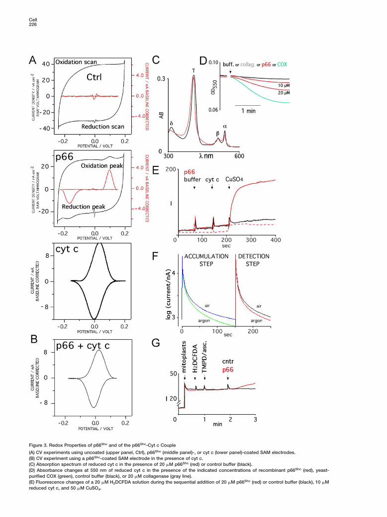
Cell226
Figure 3. Redox Properties of p66Shc and of the p66Shc-Cyt c Couple
(A) CV experiments using uncoated (upper panel, Ctrl), p66Shc (middle panel)-, or cyt c (lower panel)-coated SAM electrodes.(B) CV experiment using a p66Shc-coated SAM electrode in the presence of cyt c.(C) Absorption spectrum of reduced cyt c in the presence of 20 �M p66Shc (red) or control buffer (black).(D) Absorbance changes at 550 nm of reduced cyt c in the presence of the indicated concentrations of recombinant p66Shc (red), yeast-purified COX (green), control buffer (black), or 20 �M collagenase (gray line).(E) Fluorescence changes of a 20 �M H2DCFDA solution during the sequential addition of 20 �M p66Shc (red) or control buffer (black), 10 �Mreduced cyt c, and 50 �M CuSO4.
P66Shc Is a Redox Enzyme that Generates H2O2227
ally in the presence of air-saturated buffer (Figure 3F; dase, are essential for the interaction and ET reaction
(F) Current measurement at −100 (accumulation step) or +650 (detection step) mV of the glassy carbon electrode coated with a gel containingp66Shc and cyt c. Recordings were performed in aerated (air, blue and black traces) or argon-flushed (argon, green and red traces) phosphatebuffer, as indicated.(G) Fluorescence changes of purified liver mitoplasts after sequential additions of H2DCFDA, 0.5 mM ascorbate, 0.2 mM TMPD, and p66Shc
(red) or control buffer (black).
indicating that the ET kinetics of p66Shc is acceleratedby cyt c. Therefore, it appears that p66Shc executes aredox reaction with cyt c under the CV experimentalconditions.
We then investigated the effect of p66Shc on the re-dox status of cyt c by spectrophotometry. Addition ofp66Shc modified the absorbance spectrum of reducedcyt c, provoking a slight shift of the γ peak and a reduc-tion of the amplitude of the α peak (Figure 3C). Thesemodifications are consistent with a strong interactionbetween p66Shc and the prosthetic group of cyt c, andthe partial oxidation of cyt c. Analysis of the kinetics ofabsorbance at 550 nm (α peak) revealed that the effectof p66Shc on cyt c oxidation status is dose dependentand comparable to that of purified yeast COX (Figure3D). Together, these findings indicate that p66Shc is aredox protein that oxidizes cyt c in vitro.
Electron Transfer between p66Shc and Cyt cGenerates Hydrogen Peroxide In VitroWe then investigated whether p66Shc oxidizesH2DCFDA when reacting with cyt c. Addition of p66Shc
to reduced cyt c did not provoke H2DFCDA oxidation(data not shown). A significant fluorescence boost fromoxidized H2DCFDA was, instead, recorded in the pres-ence of copper (but not zinc or iron) ions (Figure 3E;red trace), indicating that, in vitro, the copper atom cat-alyzes generation of free radicals from the redox centerof the p66Shc-cyt c complex. Notably, under anoxicconditions, H2DCFDA oxidation was significantly de-creased, suggesting a role for oxygen intermediates inthis process (Figure 3E; dashed trace). Since H2DCFDAoxidation is specifically sensitive to H2O2, these datasuggest that this is the molecular species that formsduring the reaction.
To obtain a direct proof that H2O2 is generated duringthe electrochemical reaction of p66Shc with cyt c, wemeasured current generation at 650 mV (which corres-ponds to the oxidation potential of H2O2) in an electrode-confined p66Shc-cyt c system (Callegari et al., 2004).P66Shc and cyt c were entrapped within a conductingpolymeric film and immobilized onto a glassy carbonelectrode. The coated electrode was then transferredinto an air- or argon-saturated phosphate buffer and a−100 mV potential applied for 150 s (this potential in-duces the continuous reduction of the p66Shc-cyt ccomplex, has no direct effect on molecular oxygen, andallows accumulation of oxygen radicals, if formed, inthe gel; accumulation step, Figure 3F). Analysis of cur-rent generation at −100 mV revealed early oxygenreduction current generated by the p66Shc/cyt celectrode, only in the presence of air-saturated buffer(Figure 3F; left panel). After switching of the electrodepotential to 650 mV (to measure accumulated H2O2; de-tection step, Figure 3F), current was recorded specific-
right panel). Furthermore, when the initial potential wasset at +100 mV (which cannot reduce the p66Shc/cyt credox center), neither catalytic oxygen reduction norH2O2 oxidation was detected (data not shown). To-gether, these results provide direct demonstration thatET between p66shc and cyt c leads to oxygen reductionand formation of H2O2.
P66Shc Generates Hydrogen Peroxidefrom Reduced Cyt c In VivoWe then investigated whether, in vivo, the ETC site ofp66Shc-induced ROS production is compatible with thedescribed in vitro activity of p66Shc on reduced cyt c.To this end, mitochondria were treated with ascorbate/N,N#-tetramethyl-p-phenyldiamine (TMPD) in the ab-sence of energetic substrates. Under these experimen-tal conditions, mitochondrial respiration is supportedby reduction of cyt c (by TMPD), while complexes I–IIIremain inactive. As expected, since the ETC sites ofROS production are excluded in this system, ascor-bate/TMPD-treated mitochondria did not generatedetectable ROS (Figure 3G; black trace). Addition of re-combinant p66Shc, instead, induced H2DFCDA oxida-tion, indicating that p66Shc is able to generate ROS byacting downstream to reduced cyt c. Consistently,p66Shc did not increase ROS generation induced byinhibitors of complexes I (rotenone) or III (ant A) (Fig-ure S4).
Mapping of the Redox Center of p66Shc
To map the region of p66Shc interacting with cyt c, wecompared, by ELISA, the cyt c binding properties ofp66Shc with those of p52Shc or p46Shc, which are notinvolved in the regulation of ROS and apoptosis (Mig-liaccio et al., 1999). Increasing amounts of coated cytc (0.25–4 �g) were incubated with increasing amountsof Shc proteins (0.08–4 �g) and bound proteins re-vealed with anti-Shc antibodies. The p66Shc-cyt c asso-ciation was dose dependent at each concentration ofthe two proteins (Figure 4B). A residual binding activity(about 10%) was detected for p52Shc, while p46Shc didnot bind at all, suggesting that the N-terminal region ofp66Shc (CH2-PTB) is involved in cyt c binding.
To narrow down the cyt c binding region of p66Shc,we expressed various portions of the CH2-PTB regionas GST fusion proteins (Figure 4C) and measured theirability to recover cyt c from cellular lysates throughpull-down experiments. Results showed that a 52amino acid region N terminal to the PTB domain is criti-cal for the binding to cellular cyt c (denominated CB,for cyt c binding; Figures 4A and 4C). This region ishighly conserved among the known p66Shc vertebrateorthologs (Figure 4D) and contains glutamic (E125,E132, E133) and tryptophan (W134 and W148) residues,which, in the context of COX IV and yeast cyt c peroxi-

Cell228
Figure 4. Mapping of the p66Shc Cyt c-Interacting Region
(A) Modular organization of Shc proteins (p66, p52, p46) and p66Shc mutants (qq and f ). The positions of the qq and f mutations are indicatedwithin the CB region.(B) ELISA binding assay of recombinant p66Shc, p52Shc, p46Shc, p66Shcqq, and p66Shcf proteins added at different concentrations to dishwells preadsorbed with different concentrations of cyt c, as indicated.(C) In vitro pull-down experiment (right panel) using MEF lysates and the schematized (left panel) portions of p66Shc expressed in bacteria asGST fusion proteins. Recovered proteins were analyzed by Western blotting using anti-cyt c antibodies.(D) Amino acid sequence of the p66Shc N-terminal region. Amino acid sequence alignment of the CH2-PTB N-terminal regions (aa 1–155) ofthe available p66Shc vertebrate horthologs. Identities are highlighted in gray. The most conserved region (CB) is boxed in red. The relevantglutamic (E132 and E133) and triptophan (W134) residues are indicated in the sequence (by asterisks and circle, respectively) and in thep66Shc modular organization scheme below.

P66Shc Is a Redox Enzyme that Generates H2O2229
with cyt c (Zhen et al., 1999). We engineered similarmutations within p66Shc (p66ShcE132Q-E133Q muta-tion, p66Shcqq; p66ShcW134F mutation, p66Shcf ) andevaluated their effects on the ability of the protein tobind cyt c, to transfer electrons, and to stimulate mito-chondrial ROS generation. Notably, both qq and f muta-tions abrogated the ability of recombinant p66Shc tobind cyt c, as revealed by ELISA and pull-down experi-ments (Figures 4B and 4C).
The CV curves of the p66Shcqq and p66Shcf mutantsshowed symmetric peaks with redox potentials of −151mV and −140 mV, respectively (Figures 5A and 5B).These values are markedly lower than that of wt p66Shc
(−35 mV), suggesting that the E132-E133 and W134 res-idues influence the p66Shc redox properties. In the pres-ence of cyt c, the CV responses of both mutant proteinsresulted in two asymmetric peaks (Figures 5A and 5B),which could be easily deconvoluted into two compo-nents associated to the ET processes of cyt c (17 mV;Figures 5A and 5B, green traces) and the p66Shcqq(−150 mV; Figure 5A, orange trace) or p66Shcf (−141 mV;Figure 5B, pink trace) mutants. The detection of sepa-rated ET processes is consistent with a decreased in-teraction between the two p66Shc mutants and cyt c,as also revealed by ELISA and pull-down experiments(Figures 4B–4D). Therefore, both properties of execut-ing ET and interacting with cyt c are impaired in the qqor f mutants of p66Shc, suggesting that the E132-E133and W134 residues of p66Shc concur in the formationof its redox center and cyt c binding surface.
We then compared the ability of p66Shc, p66Shcqq,and p66Shcf to regulate production of ROS in vitro andin vivo. Addition of recombinant p66Shcqq and p66Shcfto digitonin-treated mitochondria did not increaseH2DCFDA oxidation (Figure 5C). Likewise, expressionof p66Shcqq and and p66Shcf into p66Shc−/− MEFs didnot increase the cellular fluorescence of H DCFDA-
Figure 5. Impaired Redox Properties ofp66Shc Mutants Defective for Cyt c Binding
(A) Voltammograms of cyt c (green), p66Shcqq(orange), and p66Shcqq + cyt c (black).(B) Voltammograms of cyt c (green), p66Shcf(pink), and p66Shcf + cyt c (black).(C) Fluorescence changes of purified livermitoplasts during the sequential additions ofH2DCFDA; succinate; and buffer (ctrl, black),p66Shc (red), p66Shcqq (orange), or p66Shcf(violet) proteins.(D) Ratio of the H2DCFDA mean fluorescence,obtained by FACS analysis of p66Shc−/−
MEFs after infection with control, p66Shc,p66Shcqq, or p66Shcf retroviruses; results arethe average ± SEM of three experiments.
2
stained cells (Figure 5D). Therefore, mutations that im-pair the p66Shc redox center also inactivate the abilityof p66Shc to stimulate ROS generation in vivo.
Mutation of the Redox Center of p66Shc Impairsits Ability to Mediate Mitochondrial ApoptosisWe then investigated whether the function of p66Shc toregulate mitochondrial apoptosis depends on its elec-trochemical properties, using the p66Shcqq mutant. Toensure that the qq mutation had not interfered withother functions of p66Shc, we first evaluated the abilityof the two neighboring domains (PTB and CH2) to bindactivated receptors and undergo phosphorylation afteroxidative stress, respectively, and the property ofp66Shcqq to localize within mitochondria. In the contextof the qq mutation, the p66Shc PTB domain maintainedits ability to bind activated EGF receptors (Figure 6A).Likewise, p66Shcqq was efficiently phosphorylated onserine following expression into p66Shc−/− MEFs andtreatment with H2O2 (Figure 6B). Like the wt protein,p66Shcqq localizes within mitochondria (Figure 6C).Mitochondrial p66Shc is PK resistant (Figure 6D); it isreleased by digitonin treatment (Figure 6E) and forms acomplex with mtHsp70, from which it dissociates afterproapoptotic stimuli (Figure 6F).
We then measured the ability of the p66Shcqq mutantto regulate mitochondrial PT in vitro and apoptosis in vivo.P66Shcqq was unable to induce swelling of digitonizedmitochondria (Figure 7A), nor did it restore the apo-ptotic response of p66Shc−/− MEFs upon treatment withH2O2 or staurosporine, as measured by cyt c release(Figure 7B), caspase activation (Figure 7C), and cell via-bility (Figure 7D). Notably, CsA inhibited the ability ofreexpressed p66Shc to restore mitochondrial apoptosisfollowing H2O2 (Figure 7E) or staurosporine (data notshown), supporting the theory that a functional PT isneeded for the proapoptotic activity of p66Shc. It ap-

Cell230
Figure 6. Receptor Binding, Phosphorylation,Localization, and Hsp70 Association of thep66Shc Redox Mutants
(A) In vitro pull-down experiment usinglysates of MEFs and treated or not withEGF, and GST, GST-PTBp66Shc, or GST-PTBp66Shcqq. Recovered proteins were ana-lyzed by WB using anti-EGFR antibodies.(B) P66Shc−/− MEFs were reconstituted withp66Shc or p66Shcqq by retroviral-mediatedgene transfer and treated or not with H2O2.Anti-Shc immunoprecipitates were analyzedby WB using antibodies against phosphory-lated p66Shc (upper panel) or total pool ofp66Shc (lower panel).(C) WB analysis of subcellular fractions fromp66Shc−/− MEFs reexpressing p66Shc orp66Shcqq, using the indicated antibodies.(D) Mitochondria (Mt) and ER-enriched frac-tions (ER) were treated with PK and analyzedby WB with indicated antibodies.(E) MEFs mitochondrial fractions (as in [C])were treated with the indicated concentra-tions of digitonin. After centrifugation, pel-lets (P) and supernatants (S) were analyzedby WB.(F) MEFs mitochondrial fractions (as in [C])were immunopreciptated with anti-p66Shc
antibodies. Specific immunoprecipitates wereanalyzed by WB using anti-mtHsp70 (upperpanel) or anti-p66Shc (lower panel) antibodies.
pears, therefore, that mutation of the p66Shc redox cen- cnter abrogates the functions of p66Shc to induce mito-
chondrial PT in vitro and to mediate cellular apoptosis swafter stress.tsDiscussionmmThe present findings provide a mechanistic explanationmfor the proapoptotic function of p66Shc and support a(model whereby p66Shc generates H2O2 within mito-pchondria, which, in turn, induces opening of the PTPmand cellular apoptosis (Figure 8). ROS production byap66Shc appears to be a specialized function wherebymelectrons are subtracted from the mitochondrial ETC topcatalyze the partial reduction of molecular oxygen. ThisFis the first demonstration that the mitochondrial respi-pratory chain can generate ROS not only accidentallytbut also through a specific enzymatic system. In thistcontext, p66Shc can be regarded as an atypical signalctransducer that converts proapoptotic into redox signals.eIn vitro and in vivo experiments demonstrated thatep66Shc oxidizes cyt c and generates H2O2. Therefore, aifraction of the mitochondrial electron flow, which is(mostly used by COX to reduce oxygen to water, is devi-aated from cyt c by p66Shc for the production of ROSm(Figure 8). P66Shc expression, however, does not influ-wence the mitochondrial transmembrane potential undermsteady-state conditions, while it is indispensable for itsscollapse following proapoptotic signals (Orsini et al.,s2004). These findings are compatible with the existence
of two functional states of p66Shc: inactive, under basal r
onditions, and active, after proapoptotic signals. Aumber of modifications of p66Shc have been de-cribed, which occur within minutes after treatmentith various proapoptotic signals and might lead to ac-
ivation of p66Shc: the cytosolic pool of p66Shc becomeserine phosphorylated (Migliaccio et al., 1999), theitochondrial pool of p66Shc is released from a higholecular weight complex (Orsini et al., 2004), and bothitochondrial and cytosolic p66Shc pools are increased
Trinei et al., 2002; Pacini et al., 2004). In the case ofhosphorylation, genetic evidence indicates that thisodification is required for the ability of p66Shc to medi-
te apoptosis (Migliaccio et al., 1999). The underlyingechanism, however, is unknown. The mitochondrialool of p66Shc is not serine phosporylated (E.M. and.O., unpublished data), and significant translocation of66Shc from cytosol to mitochondria does not occur af-
er proapoptotic signals (Orsini et al., 2004), suggestinghat serine phosphorylation might serve other, nonmito-hondrial activities of p66Shc that are also needed toxert its proapoptotic function. Mitochondrial p66Shc
xists within a high-molecular-weight complex, whichncludes members of the TIM-TOM import complexTIM44, TIM20, TIM23, and mtHsp70; Orsini et al. [2004]nd E.M. and F.O., unpublished data). The p66Shc-tHsp70 complex is destabilized following treatmentith proapoptotic signals, leading to the release ofonomeric p66Shc. Since recombinant p66Shc pos-
esses constitutive redox activity, it is tempting topeculate that association with the TIM-TOM complexesults with the inactivation of p66Shc and that pro-

P66Shc Is a Redox Enzyme that Generates H2O2231
Figure 7. The p66Shcqq Mutant Does Not In-duce PT, Nor Does It Activate Apoptosis
(A) Absorbance changes of digitonized livermitochondria pretreated with Ca2+ andadded of p66Shc (red trace) or p66Shcqq (or-ange) proteins.(B) WT and p66Shc−/− MEFs infected with con-trol, p66Shc, or p66Shcqq retroviruses weretreated with 2 �M staurosporine and, after 6or 12 hr, cytosolic fractions analyzed by WB(left panels). Cytosolic cyt c was analyzed byELISA in the same cells after 6 hR treatmentwith 2 �M staurosporine or 800 �M H2O2
(right bar graph).(C and D) The same cells as in (B) weretreated with 2 �M staurosporine or 800 �MH2O2 and analyzed for caspase-3 activation(by flow cytometry using antibodies againstcleaved caspase-3; [C]) or trypan blue (D).(E) The same cells as in (B) were treated withH2O2 and 5 �m CsA and analyzed by trypanblue. Results represent the mean of triplicatecultures ± SEM.
providing the molecular basis for a function of p66ferent proapoptotic mechanisms may lead to increased
Figure 8. Model of p66Shc Redox Activityduring Mitochondrial Apoptosis
Proapoptotic signals induce release of p66Shc
from a putative inhibitory complex. Activep66Shc then oxidizes reduced cyt c (red) andcatalyzes the reduction of O2 to H2O2. PTPopening by H2O2 then leads to swelling andapoptosis. NADH-cyt b5 reductase is indi-cated as additional putative source of re-duced cyt c.
apoptotic signals activate p66Shc by releasing it fromthis inhibitory complex.
A further mechanism of regulation of the proapo-ptotic function of mitochondrial p66Shc might be linkedto its intrinsic redox properties. Since the calculatedKeq of the [p66Shc] + [cyt c] 4 [p66Shc]− + [cyt c]+ redoxreaction is 0.1, cyt c oxidation by p66Shc is unfavoredwhen reduced cyt c is present at a concentration com-parable to p66Shc. The reaction might, instead, occur ifan excess of reduced cyt c is present, a condition thatis achieved when COX activity is decreased (e.g., dur-ing hypoxia) or by physiological inhibition by nitric ox-ide (NO) (Sarti et al., 2003). Notably, reduced cyt c mightalso accumulate during tBid/Bax-induced apoptosis,since both molecules induce remodeling of the innermembrane and increase of free intermembrane cyt c(Scorrano et al., 2002), which is then fully reduced bythe outer membrane rotenone-insensitive NADH-cyt b5
reductase (Bernardi and Azzone, 1981). Therefore, dif-
concentrations of reduced cyt c, thus firing the ET reac-tion of p66Shc (Figure 8).
The levels of intracellular ROS and of oxidation-dam-aged DNA are significantly reduced in p66Shc−/− primarycultures (fibroblasts, endothelial cells, lymphocytes;Nemoto and Finkel [2002]; Trinei et al. [2002]; Zaccag-nini et al. [2004]; Pacini et al. [2004]). Likewise, markersof oxidative stress (8-oxo-guanosine, isoprostane, nit-rotyrosine) are decreased in tissues from p66Shc−/− mice(Trinei et al., 2002; Napoli et al., 2003; Francia et al.,2004), suggesting that p66Shc might regulate ROS pro-duction also in the absence of acute (proapoptotic)stress signals and that a fraction of intracellular ROS(and of oxidative stress) depends, under basal condi-tions, on the expression of p66Shc. Since p66Shc is acti-vated by virtually every type of stress, the basal pro-duction of ROS by p66Shc may reflect its moderateactivation by chronic stress. The redox balance hasprofound effects on metabolism and transcription, thus
Shc

Cell232
Ain the cellular adaptation to chronic stress. Regulationof redox equilibrium and apoptosis by p66Shc might
Wequally contribute to its effect on life span.cf
Experimental ProcedurescfCells, Vectors, and AntibodieshMEFs were treated with 2 �M staurosporine or 800 �M H2O2. Cyto-
solic cyt c was measured after 6 hr by ELISA and after 6/12 hr byWB of cytosolic fractions; anti-caspase-3 positive cells after 6 hr, Rby FACS; and cell survival after 20 hr, by trypan blue. Intracellular RROS were measured incubating cells for 30 min with 10 �M AH2DCFDA (Molecular Probes) in complete culture medium; cells Pwere then recovered and suspended in PBS for FACS analysis. Mi-toplast (0.5 mg/ml) or 0.4 mg/ml mitochondrial suspension and 30
R�M DH2CFDA were sequentially added to a spectrofluorimetric cu-vette. Fluorescence (excitation wavelength [ex.] 498; emission
Awavelength [em.] 525) was registered using a Perkin Elmer Ls55mspectrofluorimeter at 25°C. Anti-p66Shc antibodies have been de-Cscribed (Orsini et al., 2004). Antibodies against caspase-3,
mtHsp70, cytHsp70, calnexin, porin, phosphorylated p66Shc Ser36 Bresidue, phosphotyrosine, and cyt c were obtained from commer- Bcial sources. The p66Shcqq and f mutants were generated using the cQuikChange Site-Directed Mutagenesis Kit (Stratagene) and 4cloned into the PINCO vector (Migliaccio et al., 1999).
BsCellular Subfractionation, Mitochondria,Band Mitoplast PreparationBCell lysates were subjected to differential centrifugations to sepa-crate nuclei (700 rgf), mitochondria (8000 rgf), and the endoplasmic
reticulum (15,000 rgf). Mice were intraperitoneally injected with 2 1mg CCl4/g of body weight CCl4 20 hr before liver collection. Each Bmitochondrial preparation was assayed for respiration by using a dClark type oxygraph and swelling, as described (Petronilli et al.,
C1994), with minor modifications (OD620; 0.4 mg/ml mitochondrialdsuspension with 5 mM succinate as substrate). Mitoplasts were1obtained by hypotonic shock (1:10 dilution in deionized water for 5Cmin on ice).GFProtein Film VoltammetryiWe used a three electrode cell composed of a saturated calomelMelectrode (reference), platinum spiral wire (counter electrode), and
gold disk (working electrode). The SAM was formed by soaking the Cgold electrode for 5 hr in 100 �M 11-mercaptoundecanoic acid. oPurified horse cyt c (Sigma), p66Shc, p66Shcqq, or p66Shcf solutions
Cwere deposited on the electrode surface and left to evaporatepslowly. Measurements were performed in phosphate buffer andlLiClO4 at constant ionic strength (100 mM). Before each experi-1ment, the electrolyte solution was purged with argon transistorD(lower than 1 ppm O2), and the voltammetric curves were recordedpmaintaining an argon blanket over the solution. CV experiments
were carried out at 5–50 mV/s with an Autolab Model PGSTAT 30 at D25°C. Protein midpoint potentials (E1/2) were calculated as average (between the potential of oxidation (Epa) and reduction (Epc) peaks. n
lRecombinant Proteins, ELISA, and In Vitro Binding Assay
FAll recombinant proteins were produced in E. coli as GST fusionvproteins. GST was removed by prescission treatment after affinityVcolumn purification. For in vitro binding assays, total protein ly-tsates (1 mg) were incubated for 2 hr at 4°C with 10 �g of the appro-2priate GST fusion protein. ELISA plates were coated with 4, 1, orG0.25 �g purified horse cyt c (Sigma) by using a 4 �g/ml coatingcsolution in carbonate buffer (0.1 ml/well). After washing with PBS,
different concentrations of recombinant Shc proteins were added Gto each well and incubated for 2 hr at 37°C. Binding was revealed dusing an anti SH2-SHC antibody (Transduction Laboratories) and Mhorseradish peroxidase-conjugated anti-rabbit antibodies. Bound lantibodies were detected using the TMB detection reagent (SIGMA).
LtgSupplemental Data2Supplemental Data include four figures and can be found with thisMarticle online at http://www.cell.com/cgi/content/full/122/2/221/
DC1. t
cknowledgments
e are grateful to S. Cattadori and D. Triarico for excellent techni-al support, C. Tacchetti and C. Puri for EM analysis, and V. Rakeror some of the mitochondrial fractionation experiments. F.O. is re-ipient of a FIRC fellowship. This work was supported by grantsrom A.I.R.C. and M.I.U.R. M.G.; S.M., P.B., and P.G.P. are share-olders of Genextra Spa.
eceived: November 10, 2004evised: March 22, 2005ccepted: May 6, 2005ublished: July 28, 2005
eferences
rmstrong, F.A. (2002). Insights from protein film voltammetry intoechanisms of complex biological electron-transfer reactions. J.hem. Soc., Dalton Trans. 5, 661.
alaban, R.S., Nemoto, S., and Finkel, T. (2005). Cell 120, 483–495.
ecker, L.B. (2004). New concepts in reactive oxygen species andardiovascular reperfusion physiology. Cardiovasc. Res. 61, 461–70.
ernardi, P., and Azzone, G.F. (1981). Cytochrome c as an electronhuttle between the outer and inner mitochondrial membranes. J.iol. Chem. 256, 7187–7192.
ernardi, P., Petronilli, V., Di Lisa, F., and Forte, M. (2001). A mito-hondrial perspective on cell death. Trends Biochem. Sci. 26,12–117.
rownlee, M. (2001). Biochemistry and molecular cell biology ofiabetic complications. Nature 414, 813–820.
ai, J., and Jones, D.P. (1998). Superoxide in apoptosis. Mitochon-rial generation triggered by cytochrome c loss. J. Biol. Chem. 273,1401–11404.
allegari, A., Cosnier, S., Marcaccio, M., Paolucci, D., Paolucci, M.,eorgakilas, V., Tagmatarchis, N., Vázquez, E., and Prato, M. (2004).unctionalised single wall carbon nanotubes/polypyrrole compos-
tes for the preparation of amperometric glucose biosensors. J.at. Chem. 14, 807–810.
hance, B., Sies, H., and Boveris, A. (1979). Hydroperoxide metab-lism in mammalian organs. Physiol. Rev. 59, 527–605.
hen, X., Ferrigno, R., Yang, J., and Whitesides, G.M. (2002). Redoxroperties of cytochrome c adsorbed on self-assembled mono-
ayers: a probe for protein conformation and orientation. Langmuir8, 7009–7015.
anial, N.N., and Korsmeyer, S.J. (2004). Cell death: critical controloints. Cell 116, 205–219.
iwan, J.J., Yune, H.H., Bawa, R., Haley, T., and Mannella, C.A.1988). Enhanced uptake of spermidine and methylglyoxal-bis(gua-ylhydrazone) by rat liver mitochondria following outer membrane
ysis. Biochem. Pharmacol. 37, 957–961.
rancia, P., delli Gatti, C., Bachschmid, M., Martin-Padura, I., Sa-oia, C., Migliaccio, E., Pelicci, P.G., Schiavoni, M., Luscher, T.F.,olpe, M., and Cosentino, F. (2004). Deletion of p66shc gene pro-ects against age-related endothelial dysfunction. Circulation 110,889–2895.
reen, D.R., and Kroemer, G. (2004). The pathophysiology of mito-hondrial cell death. Science 305, 626–629.
riparic, L., van der Wel, N.N., Orozco, I.J., Peters, P.J., and vaner Bliek, A.M. (2004). Loss of the intermembrane space proteingm1/OPA1 induces swelling and localized constrictions along the
engths of mitochondria. J. Biol. Chem. 279, 18792–18798.
eBel, C.P., Ischiropoulos, H., and Bondy, S.C. (1992). Evaluation ofhe probe 2#,7#-dichlorofluorescin as an indicator of reactive oxy-en species formation and oxidative stress. Chem. Res. Toxicol. 5,27–231.
ayer, M., and Noble, M. (1994). N-acetyl-L-cysteine is a pluripo-ent protector against cell death and enhancer of trophic factor-

P66Shc Is a Redox Enzyme that Generates H2O2233
mediated cell survival in vitro. Proc. Natl. Acad. Sci. USA 91,7496–7500.
Migliaccio, E., Mele, S., Salcini, A.E., Pelicci, G., Lai, K.M., Superti-Furga, G., Pawson, T., Di Fiore, P.P., Lanfrancone, L., and Pelicci,P.G. (1997). Opposite effects of the p52shc/p46shc and p66shcsplicing isoforms on the EGF receptor-MAP kinase fos signallingpathway. EMBO J. 16, 706–716.
Migliaccio, E., Giorgio, M., Mele, S., Pelicci, G., Reboldi, P., Pan-dolfi, P.P., Lanfrancone, L., and Pelicci, P.G. (1999). The p66shcadaptor protein controls oxidative stress response and life span inmammals. Nature 402, 309–313.
Napoli, C., Martin-Padura, I., de Nigris, F., Giorgio, M., Mansueto,G., Somma, P., Condorelli, M., Sica, G., De Rosa, G., and Pelicci, P.(2003). Deletion of the p66Shc longevity gene reduces systemicand tissue oxidative stress, vascular cell apoptosis, and early ath-erogenesis in mice fed a high-fat diet. Proc. Natl. Acad. Sci. USA18, 2112–2116.
Nemoto, S., and Finkel, T. (2002). Redox regulation of forkhead pro-teins through a p66shc dependent signaling pathway. Science 295,2450–2452.
Orsini, F., Migliaccio, E., Moroni, M., Contursi, C., Raker, V.A., Pic-cini, D., Martin-Padura, I., Pelliccia, G., Trinei, M., Bono, M., et al.(2004). The lifespan determinant p66Shc localizes to mitochondriawhere it associates with mtHsp70 and regulates trans-membranepotential. J. Biol. Chem. 11, 25689–25695.
Pacini, S., Pellegrini, M., Migliaccio, E., Patrussi, L., Ulivieri, C., Ven-tura, A., Carraro, F., Naldini, A., Lanfrancone, L., Pelicci, P., andBaldari, C.T. (2004). P66Shc promotes apoptosis and antagonizesmitogenic signaling in T cells. Mol. Cell. Biol. 24, 1747–1757.
Pelicci, P.G. (2004). Do tumor-suppressive mechanisms contributeto organism aging by inducing stem cell senescence? J. Clin. In-vest. 113, 4–7.
Pelicci, G., Lanfrancone, L., Grignani, F., McGlade, J., Cavallo, F.,Forni, G., Nicoletti, I., Grignani, F., Pawson, T., and Pelicci, P.G.(1992). A novel transforming protein (SHC) with an SH2 domain isimplicated in mitogenic signal transduction. Cell 70, 93–104.
Petronilli, V., Costantini, P., Scorrano, L., Colonna, R., Passamonti,S., and Bernardi, P. (1994). The voltage sensor of the mitochondrialpermeability transition pore is tuned by the oxidation-reductionstate of vicinal thiols. Increase of the gating potential by oxidantsand its reversal by reducing agents. J. Biol. Chem. 269, 16638–16642.
Sakon, S., Xue, X., Takekawa, M., Sasazuki, T., Okazaki, T., Kojima,Y., Piao, J.H., Yagita, H., Okumura, K., Doi, T., and Nakano, H.(2003). NF-kappaB inhibits TNF-induced accumulation of ROS thatmediate prolonged MAPK activation and necrotic cell death. EMBOJ. 22, 3898–3909.
Sarti, P., Arese, M., Bacchi, A., Barone, M.C., Forte, E., Mastroni-cola, D., Brunori, M., and Giuffre, A. (2003). Nitric oxide and mito-chondrial complex IV. IUBMB Life 55, 605–611.
Scorrano, L., Ashiya, M., Buttle, K., Weiler, S., Oakes, S.A., Man-nella, C.A., and Korsmeyer, S.J. (2002). A distinct pathway remod-els mitochondrial cristae and mobilizes cytochrome c during apo-ptosis. Dev. Cell 2, 55–67.
Scorrano, L., and Korsmeyer, S.J. (2003). Mechanisms of cyto-chrome c release by proapoptotic BCL-2 family members. Bio-chem. Biophys. Res. Commun. 304, 437–444.
Trinei, M., Giorgio, M., Cicalese, A., Barozzi, S., Ventura, A., Migliac-cio, E., Milia, E., Padura, I.M., Raker, V.A., Maccarana, M., et al.(2002). A p53-p66Shc signalling pathway controls intracellular re-dox status, levels of oxidation-damaged DNA and oxidative stress-induced apoptosis. Oncogene 21, 3872–3878.
Ventura, A., Maccarana, M., Raker, V.A., and Pelicci, P.G. (2004).A cryptic targeting signal induces isoform-specific localization ofp46Shc to mitochondria. J. Biol. Chem. 279, 2299–2306.
Wallace, D.C. (1999). Mitochondrial diseases in man and mouse.Science 283, 1482–1488.
Zaccagnini, G., Martelli, F., Fasanaro, P., Magenta, A., Gaetano, C.,Di Carlo, A., Biglioli, P., Giorgio, M., Martin-Padura, I., Pelicci, P.G.,
and Capogrossi, M.C. (2004). P66ShcA modulates tissue responseto hindlimb ischemia. Circulation 109, 2917–2923.
Zhao, H., Kalivendi, S., Zhang, H., Joseph, J., Nithipatikom, K.,Vasquez-Vivar, J., and Kalyanaraman, B. (2003). Superoxide reactswith hydroethidine but forms a fluorescent product that is distinctlydifferent from ethidium: potential implications in intracellular fluo-rescence detection of superoxide. Free Radic. Biol. Med. 34,1359–1368.
Zhen, Y., Hoganson, C.W., Babcock, G.T., and Ferguson-Miller, S.(1999). Definition of the interaction domain for cytochrome c oncytochrome c oxidase. I. Biochemical, spectral, and kinetic charac-terization of surface mutants in subunit ii of Rhodobacter sphaeroi-des cytochrome aa(3). J. Biol. Chem. 274, 38032–38041.