Anatomy of the Dwarf Mistletoe Shoot System - fs.fed.us · reported for leaves of numerous...
18
CHAPTER 10 Anatomy of the Dwarf Mistletoe Shoot System Carol A. Wilson and Clyde L. Calvin * In this chapter, we present an overview of the structure of the Arceuthobium shoot system. Anatomical examination reveals that dwarf mistletoes are indeed well adapted to a parasitic habit. An exten- sive endophytic system (see chapter 11) interacts physiologically with the host to obtain needed resources (water, minerals, and photosynthates); and the shoots provide regulatory and reproductive func- tions. Beyond specialization of their morphology (Le., their leaves are reduced to scales), the dwarf mistle- toes also show peculiarities of their structure that reflect their phylogenetic relationships with other mistletoes and illustrate a high degree of specialization for the parasitic habit. From Arceuthobium globosum, the largest described species with shoots 70 cm tall and 5 cm in diameter, toA. douglasii, a small species with shoots 3 cm tall and 0.3 cm in diameter, the anatomical features are consistent. We have studied diverse species of Arceuthobium, including large primitive species and highly reduced specialized species. For this account, however, we concentrated on the shoots of A. globosum and A. tsu- gense. We chose these species for several reasons: (1) material on developmental stages was available; (2) A. globosum presumably represents a primitive member within the genus, whereas A. tsugense repre- sents intermediate specialization; and (3) these species represent the two geographical areas of greatest speci- ation, central Mexico and northern California. Where appropriate, we compare features of Arceuthobium with the related genera Korthalsella, Phoradendron, and Viscum. These three genera share features with Arceuthobium, and Korthalsella had been proposed as a sister genus to Arceuthobium (Wiens and Barlow 1971, but see chapter 15). In this chapter, we focus on leaf, stem, and fruit structure, and, wherever possible, attempt to relate structure to function. General morphology presents the best starting point for discussing the shoot system of Arceuthobium. Morphology of Shoots Arceuthobium does not produce shoots immedi- ately after germination. The endophytic system first develops within the host branch. Oftentimes, the only evidence of infection is swelling of the tissues near the infection site (Scharpf 1967). After 1 to 3 years, the first shoots are produced (table 2.1). All shoots arise from the endophytic system and thus are root-borne shoots (Groff and Kaplan 1988). In emerging shoots, the leaves of adjacent nodes overlap and conceal the stem. As the internodes elongate, stem segments become visible; but the shoot apex remains tightly enclosed by newly developing leaf primordia (fig. 10.lA). Two oppositely arranged leaves, joined at their bases, occur at each node (fig. 10.lA-B). This decussate phyllotaxis characterizes the entire genus. The mature, paired leaves form a boat-shaped structure that encircles the main stem and its branches at the node (fig. 10.lB-C). The merged leaf bases typi- cally extend some distance into the internode below the attachment point (fig. 10.lD). This contributes to the much larger diameter of the internode below the point of attachment than above (fig. 10.lB). Generally, the mean diameter of the stem 1 to 2 mm below the node measures almost twice the diameter an equal dis- tance above. The merger of leaf structure into the main axis results in a stem morphology in which internodes widen in an acropetal direction, particularly in the upper third of the internode. The functions of the widened upper portions of internodes are discussed more fully in a later section on the epidermis. Several different branching patterns occur in Arceuthobium (fig. 2.1). The A. tsugense shoot (fig. 10.lB) displays decussate branching, a pattern com- mon for shoots with decussate phyllotaxis. The evolu- tionary and systematic importance of these patterns was established by Kuijt (1970) and further refined by Hawksworth and Wiens (1972) and by Mark and Hawksworth (1981). • of Biol?gy, University of California, Berkeley and Department of Biology, Portland State University, Portland, OR, respectively; contnbuted as EnVironmental SCiences and Resources Program Publication No. 242. Anatomy of the Dwarf Mistletoe Shoot System 95 This file was created by scanning the printed publication. Errors identified by the software have been corrected; however, some errors may remain.
Transcript of Anatomy of the Dwarf Mistletoe Shoot System - fs.fed.us · reported for leaves of numerous...
CHAPTER 10
Anatomy of the Dwarf Mistletoe Shoot System
Carol A. Wilson and Clyde L. Calvin *
In this chapter, we present an overview of the structure of the Arceuthobium shoot system. Anatomical examination reveals that dwarf mistletoes are indeed well adapted to a parasitic habit. An extensive endophytic system (see chapter 11) interacts physiologically with the host to obtain needed resources (water, minerals, and photosynthates); and the shoots provide regulatory and reproductive functions. Beyond specialization of their morphology (Le., their leaves are reduced to scales), the dwarf mistletoes also show peculiarities of their structure that reflect their phylogenetic relationships with other mistletoes and illustrate a high degree of specialization for the parasitic habit. From Arceuthobium globosum, the largest described species with shoots 70 cm tall and 5 cm in diameter, toA. douglasii, a small species with shoots 3 cm tall and 0.3 cm in diameter, the anatomical features are consistent.
We have studied diverse species of Arceuthobium, including large primitive species and highly reduced specialized species. For this account, however, we concentrated on the shoots of A. globosum and A. tsugense. We chose these species for several reasons: (1) material on developmental stages was available; (2) A. globosum presumably represents a primitive member within the genus, whereas A. tsugense represents intermediate specialization; and (3) these species represent the two geographical areas of greatest speciation, central Mexico and northern California. Where appropriate, we compare features of Arceuthobium with the related genera Korthalsella, Phoradendron, and Viscum. These three genera share features with Arceuthobium, and Korthalsella had been proposed as a sister genus to Arceuthobium (Wiens and Barlow 1971, but see chapter 15).
In this chapter, we focus on leaf, stem, and fruit structure, and, wherever possible, attempt to relate structure to function. General morphology presents the best starting point for discussing the shoot system of Arceuthobium.
Morphology of Shoots Arceuthobium does not produce shoots immedi
ately after germination. The endophytic system first develops within the host branch. Oftentimes, the only evidence of infection is swelling of the tissues near the infection site (Scharpf 1967). After 1 to 3 years, the first shoots are produced (table 2.1). All shoots arise from the endophytic system and thus are root-borne shoots (Groff and Kaplan 1988). In emerging shoots, the leaves of adjacent nodes overlap and conceal the stem. As the internodes elongate, stem segments become visible; but the shoot apex remains tightly enclosed by newly developing leaf primordia (fig. 10.lA). Two oppositely arranged leaves, joined at their bases, occur at each node (fig. 10.lA-B). This decussate phyllotaxis characterizes the entire genus.
The mature, paired leaves form a boat-shaped structure that encircles the main stem and its branches at the node (fig. 10.lB-C). The merged leaf bases typically extend some distance into the internode below the attachment point (fig. 10.lD). This contributes to the much larger diameter of the internode below the point of attachment than above (fig. 10.lB). Generally, the mean diameter of the stem 1 to 2 mm below the node measures almost twice the diameter an equal distance above. The merger of leaf structure into the main axis results in a stem morphology in which internodes widen in an acropetal direction, particularly in the upper third of the internode. The functions of the widened upper portions of internodes are discussed more fully in a later section on the epidermis.
Several different branching patterns occur in Arceuthobium (fig. 2.1). The A. tsugense shoot (fig. 10.lB) displays decussate branching, a pattern common for shoots with decussate phyllotaxis. The evolutionary and systematic importance of these patterns was established by Kuijt (1970) and further refined by Hawksworth and Wiens (1972) and by Mark and Hawksworth (1981).
• Depa~ment of In~egrative Biol?gy, University of California, Berkeley and Department of Biology, Portland State University, Portland, OR, respectively; contnbuted as EnVironmental SCiences and Resources Program Publication No. 242.
Anatomy of the Dwarf Mistletoe Shoot System 95
This file was created by scanning the printed publication.Errors identified by the software have been corrected;
however, some errors may remain.

Chapter 10
c D Figure 10.1 -Surface (A-B) and transectional views (C-D) of Arceuthobium tsugense shoots. A: shoot tip showing opposite, paired leaves and decussate phyllotaxis, x60. B: nodal region showing stem (st), mature leaves (I) and axillary branches (ab), x25. C: leaf 300 ~m above point of attachement to stem, unlabeled arrows at points where leaf margins join, x31. D: stem at point of leaf base fusion, x31.
96 --------------------------- Anatomy of the Dwarf Mistletoe Shoot System

Chapter 10
Shoot Apical Organization In median longitudinal section, a shoot tip of
Arceuthobium (fig. 10.2A) shows a highly stratified apex. Two stratified layers were present in all shoot apices that we examined. Although we did not analyze a large enough sample (Gifford 1954) to state authoritatively the number of tunica layers present, our observations suggest that the tunica is biseriate. A biseriate tunica has also been reported for Phoradendron (Cutter 1955). In the apices we examined, axillary buds were visible in the axils of the third leaf pair and were well developed in the axils of the fifth leaf pair. Studying several parasitic and "saprophytic" angiosperms, Cutter (1955) found no anomaly of shoot apical organization associated with these nutritional modes. Mauseth and others (1985) examined shoots of the mistletoe Tristerix aphyllus (Loranthaceae) and arrived at the same conclusion. Through our observations of Arceuthobium (fig. 10.2A-B), we concur.
Leaf Anatomy Although Arceuthobium is described as "leafless,"
shoots are in fact squamate; that is, they bear simple, scale-like leaves. Leaves are initiated at the periphery of the shoot apex by periclinal divisions in the subsurface layer (fig. 10.2B). When leaves are initiated, primordia are more or less circular in transverse section, and each primordium is independent. As development continues, however, meristematic activity is limited to the lower leaf zone, so that by the third leaf pair the leaf bases are visible as a Single unit due to congenital fusion. Leaf bases continue to expand and are tubular at maturity; but the upper leaf zone, which was prominent in early development (fig. 10.lA), becomes almost indistinguishable (fig. 10.lB).
Three stages of leaf development in Arceuthobium tsugense are shown in transverse sections in figure 10.2C-E. In figure 10.2C, a more or less continuous plate of procambial tissue occupies the center of the leaf, and one mature tracheary element is visible (at arrow). No other cells have matured from procambiurn at this level. At a slightly later developmental stage (fig. 10.2D), one mature tracheary element (at arrow), plus several adjacent cells in which secondary wall deposition is beginning, can be seen. In figure 10.2E, an older developmental stage, all pro cambial derivatives have matured. Several tracheary elements are present (at unlabeled arrows), but the majority of derivatives have matured as sclerified parenchyma cells, each with numerous, large, simple pits.
Anatomy a/the DwaifMistletoe Shoot System
Sclerified parenchyma cells abut directly against the tracheary elements (fig. 10.2E). No sieve elements were seen in any of the leaves we examined. In older leaves, sclerified parenchyma forms a mostly continuous plate of tissue that continues into the leaf base. This sclerified tissue is absent in the internode beneath the leaf base. Some of the cells contiguous with this sclerenchymatous layer contain rhomboidal (prismatic) crystals (fig. 10.2E). The sclerified parenchyma in A. globosum veins generally extends acropetallya greater distance than the tracheary elements; but in some cases, the two tissues appear to terminate at about the same level. Some tracheary elements at vein endings appeared to be tracheids. In more proximal positions, however, vessel members were present.
The ground tissue in leaves of Arceuthobium tsugense consists mainly of parenchyma, and only an occasional sclereid is present. As viewed basipetally in developing leaves (figs. 10.lC-D), chlorenchyma tissue begins as small groups of large cells in the midvein region and fused margins of the leaf. Moving basipetally, chlorenchyma becomes more abundant at the margins where the leaves are joined (at unlabeled arrows in fig. 10.lC). At a lower level where the leaf joins the stem, chlorenchyma occurs all the way around the sheathing leaf base (fig. 10.lD). This chlorenchyma layer is 3 to 5 cells thick around the entire stem (fig. 10.lD). No differentiation of the mesophyll into palisade and spongy tissue occurred in any of the species we examined, and intercellular space was minimal.
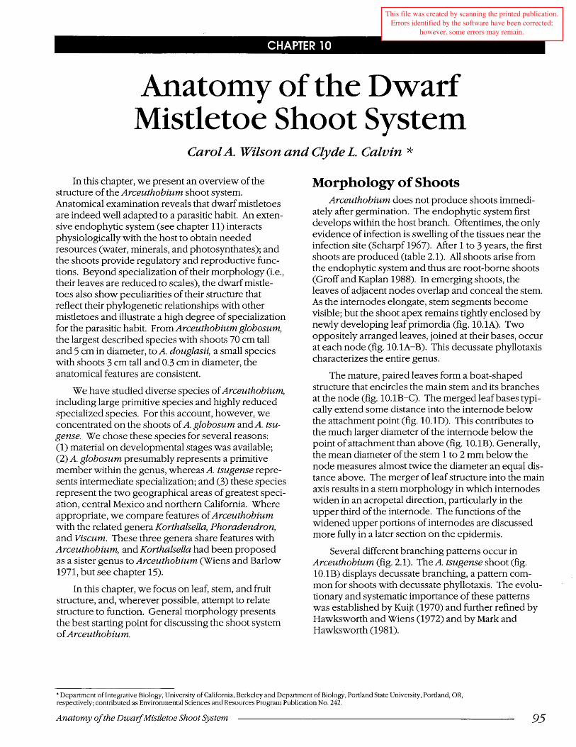
The leaf epidermis contains ordinary epidermal cells and stomatal complexes consisting of guard cells and subsidiary cells. No trichomes are present. Stomata are abundant in the abaxial epidermis but are sparse or absent in the adaxial epidermis. The abaxial stomata density on fully developed leaves reaches 38 per mm2 (table 10.1). This value compares to those reported for leaves of numerous nonparasitic angiosperms (Meyer and Anderson 1952). As reported for many other members of Santalales (Butterfass 1987), stomata have a transverse orientation with respect to the plant axis. As viewed in transverse (fig. 10.3A) and longitudinal (fig. 10.3B) sections, guard cells are recessed beneath over-arching subsidiary cells. The subsidiary cells project above the surface and form a small crypt (fig. 10.3C) at the bottom of which occurs the stomatal aperture (fig. 10.3B). A small substomatal chamber is present (fig. 10.3B); but generally, cells of the stomatal complex have wall contacts with cells in the subepidermal layer (figs. 10.3A-B).
Epidermal cells are covered by a thick cuticular layer (fig. 10.3A) that may be waxy (fig. 10.3C). The
97

Chapter 10
Figure 10.2 -Longitudinal sections of stem tips of Arceuthobium globosum (A-B) and transverse sections of leaves of A. tsugense (C-E). A: shoot tip showing meristem and leaf primordia, x40. B: apical meristem, peric1inal divisions in subepidermal layer at unlabeled arrows, xlOO. C-E: leaves at successive stages of maturation, showing xylem elements at unlabeled arrows, rhomboidal crystals (c), guard cells (g), subsidiary cell (s), and sc1erified parenchyma (sp), x250.
98 --------------------------- Anatomy a/the Dwarf Mistletoe Shoot System
Chapter 10
Figure 10,3 -Leaves in transverse (A), longitudinal (B), and suface view (C); stem in transverse view (D); and individual macerated cell (E), of Arceuthobium tsugense (A-D) and A. durangense (E). A: guard cell (g) x300. B: subsidiary cell (s) and guard cells, x300. C: SEM view showing emergent subsidiary cells, x36o; D: vascular cylinder, x120. E: primary phloem fiber, x48.
Anatomy of the Dwaif Mistletoe Shoot System 99

TABLE 10.1- Stomatal distribution in Arceuthobium americanum
plant part
Leaf (N=9) abaxial surface
Internode (N=2) upper third middle third lower third
Fruit (N=9) distal portion proximal portion
Stomata (no./mm2)
38.0 (3.0)
9.3 (0.2) 2.7 (0.0) 0.0
48.0 (35) 0.0
Source: unpublished data from L. Kirkpatrick. Values are mean and standard error in parentheses eN = sample size).
cuticular layer covering the abaxial epidermis is thicker than that on the adaxial surface. On the older (pair 7) Arceuthobium tsugense leaves that we examined, the abaxial cuticular layer had a mean thickness of 17 !-1m, whereas the adaxial cuticular layer had a mean thickness of 11 !-1m. Leaves occasionally develop a cuticular epithelium, a secondary protective layer that is well developed on stems (see discussion of epidermis).
Leaf reduction occurs in other genera ofViscaceae but not in all species. Arceuthobium and Korthalsella are entirely squamate, but only a few of the more than 150 Phoradendron species are squamate. Leaves of Arceuthobium undergo early development similar to other dicotyledons and possess certain features typical for autotrophic plants, such as a discontinuous pattern of xylem differentiation. At maturity, these leaves have a high density of stomata, a well-developed vascular system, and an extensive mesophyll. Leaf primordia and mature leaves have the dorsoventral symmetry typical of leaves. Development of the upper leaf zone stops at an early stage but continues in the basal zone. The presence of stomata and chlorenchyma suggests that these specialized leaves play important roles in gas exchange and photosynthesis. Also, young leaves of Arceuthobium protect the shoot apex during development, and mature leaves protect lateral branch primordia.
Although we know about some aspects of leaf reduction, interesting points remain unexplained. For example, while the squamate condition is common among Viscaceae, it is virtually absent in Loranthaceae.
Chapter 10
It remains as a future challenge to determine how the squamate habit serves the dwarf mistletoes.
Stem Anatomy The shoot system of a plant consists of the stems
and attached leaves. The division of the shoot into stem and leaf is somewhat arbitrary because the boundary between the two is often imprecise. This is particularly true in Arceuthobium. To illustrate, stomata are relatively more abundant on the distal third of the internode than elsewhere on the stem (table 10.1). The region of highest stomate density corresponds to the area where the leaf base extends down the stem. Thus, in the upper internode region the stem converges structurally and functionally with the leaf. Even in the uppermost portion of the internode, however, stomata are far less abundant (per unit area) than on abaxial leaf surfaces.
During primary growth, the stems of Arceuthobium have a structure similar to that of many terrestrial autotrophic dicotyledons. A transverse section through an internode of A. tsugr-mse near the completion of primary growth shows that the stem bears a single ring of vascular bundles delimited internally by the pith and externally by the cortex (fig. 10.3D). The collateral vascular bundles contain, from inside to outside, protoxylem, metaxylem, residual procambium, and phloem tissue. The phloem mayor may not contain sieve elements, but primary phloem fibers (fig. 10.3E), which in some species are only partially differentiated, and parenchyma cells are present (figs.10.4A-B). As shown below, secondary growth complicates this otherwise typical stem anatomy.
Primary Growth Primary growth forms the complete plant body;
secondary growth only adds to this body. The extent of secondary growth in Arceuthobium varies greatly from species to species. Extensive secondary growth produces large plants such as A. globosum. An understanding of stem structure in Arceuthobium, particularly the vasculature, provides valuable insights into the physiology, evolution, and ecology of the genus.
The primary xylem contains tracheary elements and parenchyma. In the material we examined, all tracheary elements (where type could be determined) were vessel members with simple perforations (fig. 10.4C). Even in protoxylem elements with helical thickening, clearly delimited perforation plates were often visible. The presence of vessel members only agrees with Kuijt's (1960b) observation that in the four Arceuthobium species studied he had "yet to find a
100 -------------------------- Anatomy o/the DwarjMistletoe Shoot System

Chapter 10
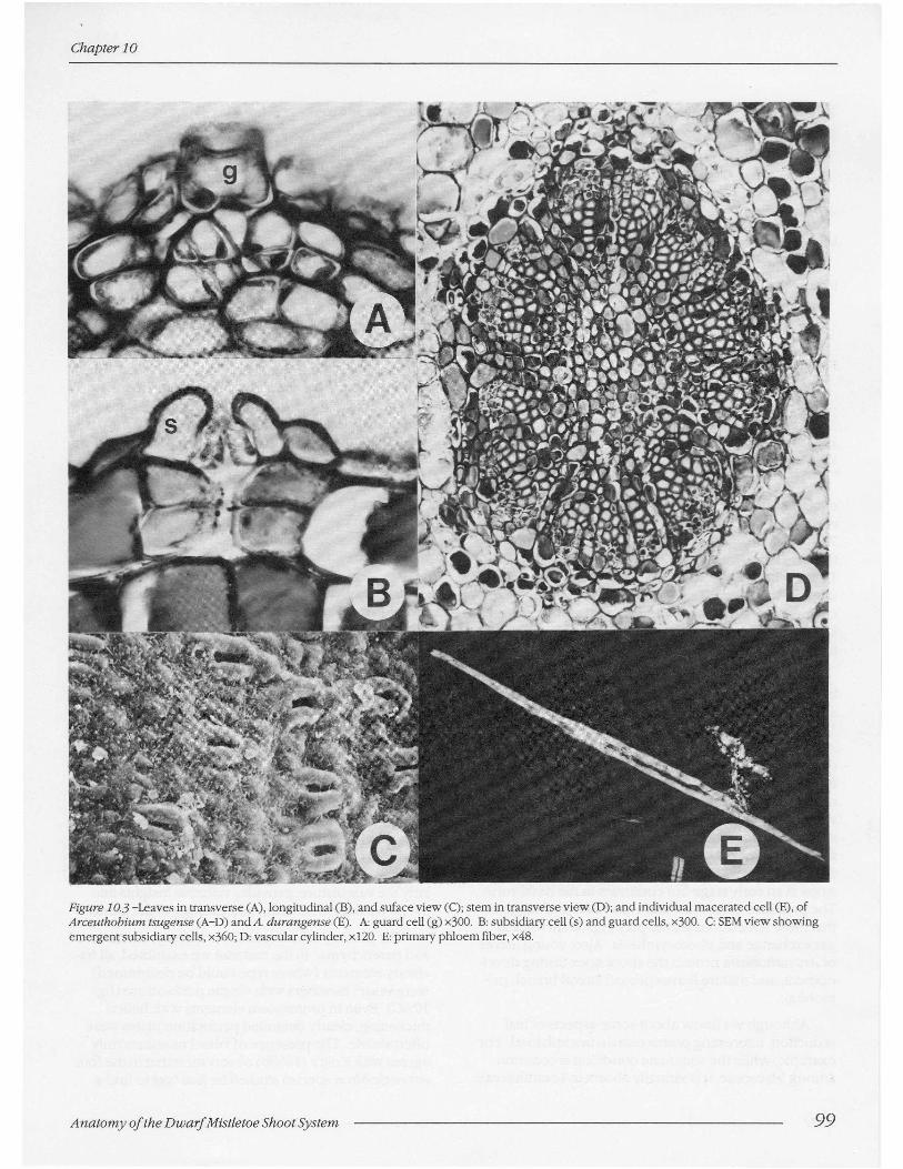
Figure 10.4 -Transverse (A) and longitudinal (B-F) sections of stems (A-C), leaf (D), and fruits (E-F) of Arceulhobfl4m ISl4gense (A-B), A. gfobosum (C-D),andA. cya1Hxarpl4m (E-F). A: vascular bundle showing xyiem(x), phloem parenchyma (ph), phloem fibers (pt), and cortex (co), ><100. B: vascular bundle, ><100. C: primary xylem (x) showing simple perforation (p) and breaks (b) in xylem continuity, ><480. D-F: graniferous tracheary elements, granules at unlabeled arrows (D, ><400; E-F, >(250).
Allalomy of the Dwarf Mistletoe Shoot System 101
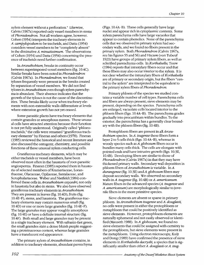
xylem element without a perforation." Likewise, Calvin (1967a) reported only vessel members in stems of Phoradendron. Not all workers agree, however. Cohen (1954) interprets the tracheary elements in A. campylopodum to be tracheids, and Datta (1954) considers vessel members to be "completely absent" in the diminutive A. minutissimum. The observations of Cohen (1954) and Datta (1954) concerning the presence of tracheids need further confirmation.
In Arceuthobium, breaks in continuity occur between superimposed vessel members (fig. 10.4C). Similar breaks have been noted in Phoradendron (Calvin 1967a). In Phoradendron, we found that tyloses frequently were present in the breaks created by separation of vessel members. We did not find tyloses in Arceuthobium even though xylem parenchyma is abundant. Their absence indicates that the growth of the tyloses is not the cause of the discontinuities. These breaks likely occur when tracheary elements with non-extensible walls differentiate at levels where extension growth has not yet ceased.
Some parasitic plants have tracheary elements that contain granules or amorphous masses. These unusual cells have attracted attention because their function is not yet fully understood. Originally called "phloeotracheids," the cells were renamed "graniferous tracheary elements" by Fineran and others (1978). Fineran (1985) reviewed the historical and recent literature and also discussed the ontogeny, chemistry, and possible functions of these unusual xylem conducting cells.
Graniferous tracheary elements, which may be either tracheids or vessel members, have been observed most often in the haustoria of root-parasitic angiosperms. Fineran (1985) reported them in haustoria of selected members of Krameriaceae, Loranthaceae, Olacaceae, Opilaceae, Santalaceae, and Scrophulariaceae. Weber and Nietfeld (1984) confirmed these cells in Arceuthobium oxycedri, not only in haustoria but also in stems. We also have observed graniferous tracheary elements in Arceuthobium. They are present in leaves (fig. 10.4D), fruits (fig. 10.4E-F), stems, and haustoria. The graniferous tracheary elements may contain numerous small (fig. 10.4D) or one or more large granules (fig. 10.4E-F). The large granules may appear roughly amorphous (fig. 10.4E) or have a definite internal structure (fig. 10.4F). Both small and large granules may be present in a single tracheary element. With safranin-fast green, the small granules stain a dense bluish purple suggesting a proteinaceous content, whereas large granules have a translucent red appearance.
The primary xylem of Arceuthobium contains, in addition to tracheary elements, abundant parenchyma
Chapter 10
(Figs. 10.4A-B). These cells generally have large nuclei and appear rich in cytoplasmic contents. Some xylem parenchyma cells have large vacuoles that appear to contain phenolics. None of the parenchyma cells that we observed in primary xylem had secondary walls, and we found no fibers present in the primary xylem. Both Phoradendron (Calvin 1967a, see his figures 55 and 56) and Viscum (von Tubeuf 1923) have groups of primary xylem fibers, as well as sclerified parenchyma cells. In Korthalsella, Touw (1984) reports that intraxylary fibers occur and that these fibers may also occur interior to the xylem. It is not clear whether the intraxylary fibers of Korthalsella are of primary or secondary origin, but the fibers "central to the xylem" are interpreted to be equivalent to the primary xylem fibers of Phoradendron.
Primary phloem of the species we studied contains a variable number of cell types. Parenchyma cells and fibers are always present; sieve elements may be present, depending on the species. Parenchyma cells are enlarged, vacuolate cells located interior to the phloem fibers (figs. 10.4A-B). The parenchyma grades gradually into pro cambium within bundles. To the exterior, the parenchyma has a generally clear boundarywith the phloem fibers (fig. 10.4B).
Protophloem fibers are present in all Arceuthobium species. In A. tsugense these fibers form a layer 2 to 5 cells thick (figs. 10.4A-B), whereas in woody species such as A. globosum fibers occur in bundles many cells thick. The cells are elongate with pointed ends and have intrusive growth (figs. 10.3E, 10.4B). Developing fibers are similar to those of Phoradendron (Calvin 1967a) in that they may have thickened primary walls. Secondary wall deposition in phloem fibers of Arceuthobium is variable. In A. durangense (fig. 10.3E) andA. globosum fibers may deposit secondary walls. We observed no secondary walls inA. tsugense (fig. 10.4B) or A. americanum. Mature fibers in the advanced species (A. tsugense and A. americanum) are morphologically similar to juvenile fibers in the more primitive species.
Sieve elements are absent or sparse in primary phloem. In Arceuthobium tsugense and A. douglasii, no cells were present in either the protophloem or metaphloem that could be positively identified as sieve elements. However, protophloem elements are naturally ephemeral and not easily observed or identified (Mauseth 1988). In A. globosum, we found no sieve elements that could be assigned with certainty to the protophloem, but sieve elements were present in the metaphloem. Using electron microscopy, Kuijt and Dong (1989) have confirmed the presence of sieve elements in Korthalsella dacrydii, a species that is significantly smaller than either A. douglasii or A. tsug-
102 ------------------------- Anatomy a/the Dwaif Mistletoe Shoot System

Chapter 10
ense. The reported absence of sieve elements in primary phloem of species such as A. tsugense and A. douglasii should be reevaluated utilizing electron microscopy.
Secondary Growth The presence and extent of secondary growth
varies greatly among the Arceuthobium species we studied. When the cambial state begins and how this state can be identified (Larson 1982) remain controversial. We limit our discussion of secondary vascular tissues to A. globosum, a species in which secondary growth is pronounced.
Secondary xylem of Arceuthobium globosum contains both parenchyma cells and tracheary elements (fig. 10.5A), but we observed no fibers. Parenchyma cells occur in both the axial and ray systems and may have thickened primary walls. Secondary walls were absent, however, even in woody stems exceeding 1 em diameter. Contents of parenchyma cells vary. Most axial parenchyma cells are unspecialized. Their protoplasts stain a rich green with safranin-fast green Qensen 1962) and contain prominent reddish nuclei. In our studies, a smaller percentage of cells stained a deep red, suggesting phenolic contents. Ray system cells were mainly of the latter type. We discovered no starch in parenchyma cells and no crystal-containing cells in the xylem.
Tracheary elements in Arceuthobium globosum occur as radial bands of cells, 1,2, or occasionally 3 cells wide (fig. lO.5A). All tracheary elements we observed were vessel members with oblique to transverse simple perforations (fig. 10.5B). Perforations are small in diameter relative to cell width (fig. 10.5A at unlabelled arrows). Secondary wall thickening forms a reticulate pattern in cells, and bordered pits of lateral walls range from almost circular to strongly elliptical (fig.10.5B). However, even the most "horizontally elongate" pit apertures do not exceed a wall face in width. Morphologically similar lateral-wall pitting has been reported for Misodendron (Cariquist 1985), the sole genus within the mistletoe family Misodendraceae. Myoschilos of the Santalaceae (Metcalfe and Chalk 1950) also displays this form oflateral-wall pitting. Cariquist (1988) noted a correlation between the presence of "horizontally elongate" pits and the abundance of axial parenchyma in wood. In A. globosum, virtually all tracheary elements contact parenchyma cells. Cariquist (1988) describes the horizontally elongate pits in Misodendron as scalariform-like, but he adds that such pits may not be relictual.
Secondary phloem of Arceuthobium globosum contains sieve elements and companion cells, plus axial and ray parenchyma (fig. 10.6). No fibers are present in secondary phloem, but groups of sclerified parenchyma cells and occasionally sclereids, probably
Figure 1 0.5 -Transverse (A) and longitudinal (B) sections of Arceuthobium globosum wood. A: Simple perforations at unlabeled arrows in upper right corner, x lOO. B: vessel members showing simple perforations (p) and lateral wall pitting (unlabeled arrows), x250.
Anatomy of the Dwarf Mistletoe Shoot System 103

of secondary origin, do occur (fig. 10.7). Groups of sclerified cells are surrounded by short cells containing rhomboidal crystals. Walls of these crystalcontaining cells also may sclerify. Rhomboidal crystals have been reported for some members of Loranthaceae, Misodendraceae, Santalaceae, and Viscaceae, as well as families not included within Santalales (Carlquist 1988). Within the Viscaceae, both Viscum (Fahn and others 1986) and Phoradendron contain rhomboidal crystals in their ray cells. Druses are also present in Viscum (Carlquist 1988) and Phoradendron, but we did not observe them in Arceuthobium.
Sieve elements of Arceuthobium, as described previously (Calvin and others 1984), have transverse to oblique, usually simple sieve plates (fig. 10.6B) and small but numerous diffuse lateral pores (fig. 10.6C-D). Sieve elements show a pattern of callose deposition and subsequent removal similar to that reported for other dicotyledons. As in Phoradendron (Calvin 1967b), companion cells are generally as large or larger in cross-section than accompanying sieve elements. Sieve elements do not appear to be particularly numerous in secondary phloem. Sieve tubes like those shown in figure 10.6 were difficult to find, even using sensitive fluorescence techniques to detect callose.
Chapter 10
Phloem parenchyma, in contrast, is abundant. Contents of axial parenchyma cells normally turn green with safranin-fast green stain, whereas protoplasts of ray cells often turn reddish, possibly due to phenolics.
Anomalous patterns of secondary growth, termed "cambial variants" by Carlquist (1988), occur in many plant families including the Loranthaceae and Misodendraceae. Successive cambia are formed in stems of the terrestrial mistletoe Nuytsia (Loranthaceae) and in Misodendron, subgenus Angelopogon (Carlquist 1988). However, the pattern of development is not identical. In Nuytsia, successive cambia form centrifugally; but they form centripetally in Misodendron. Reports of anomalous growth in Arceuthobium (Datta 1954, Metcalfe and Chalk 1950) and Korthalsella (Stevenson 1934) are problematic because they may refer to anomalous primary growth.
We observed anomalous secondary growth in Arceuthobium. For discussion here, we will exemplify the pattern by describing secondary growth for A. globosum. Cross sections of an older A. globosum stem (1 cm diameter) show discrete blocks of vascular tissue, and each block is separated by a broad band of
Figure 10.6 -Longitudinal sections through secondary phloem of Arceuthobiumglobosum. A: sieve tube (p) and adjacent primary xylem (px), x500. B: sieve tubes enlarged to show sieve plates and lateral sieve areas, xl,250. C: lateral sieve areas in surface view, xl,250. 0 : sieve tube showing oblique sieve plate and lateral sieve areas, x960.
104 -------------------------- Anatomy of the Dwaif Mistletoe Shoot System

Chapter 10
parenchyma (fig. 10.7 A). In such blocks (fig. 10.7B), vascular cambium occurs not only centrifugal to secondary xylem but also extends radially along each flank of the block as far as the pith (fig. 10.7C). The extending cambial arms curve inward at this depth, initially towards each other, but gradually forming virtu-
ally a 1800 turn (fig. 10.7D). At its inner limits, the cambial zone is interior to the protoxylem (fig. 10.7C-D). Due to the unusual configuration of the cambial zone within a vascular block, tracheary elements are differentiating in several directions. In secondary xylem (fig. 10.7D), for example, tracheary elements (at
Figure 10.7 -Transverse sections of stems of Arceuthobium g/obosum illustrating variant (anomalous) secondary growth. A: whole stem, x3. B: individual block of vascular tissue, note that vascular cambium nearly encircles block, x40. C: vascular tissue showing vascular cambium (c) along flank of block (see upper left corner) ~nd groups of protoxylem elements (px), xlOO. D: centrifugal direction of xylem differentiation at unlabe;ed arrows, note protoxylem at left of figure, xlOO.
Anatomy of the Dwarf Mistletoe Shoot System 105

unlabeled arrows) are differentiating centripetally while at the outer limit of the block xylem differentiates centrifugally. Along the block flanks differentiation is outward along a tangent. Because of the unusual configuration of the vascular cambium, some recently formed secondary xylem (fig. 10.7C-D) and phloem (fig. 10.6A) may lie very near the earliest protoxylem elements.
In all of the Arceuthobium species we studied, the blocks of vascular tissue with secondary growth arose from 2 or more vascular bundles. We found 4 to 5 or more discrete regions of protoxylem inside each vascular block of A. durangense. The number of blocks of vascular tissue in a stem with secondary growth varies between and within species. In general, for a given species, greater stem diameter allows for more blocks of vascular tissue. This situation suggests that as secondary growth progresses, a block of vascular tissue (fig. 10.7 A-B) may split into 2 or more blocks, possibly through expansion of rays present within the wood. We counted 10, and sometimes more, vascular blocks in older A. globosum stems (fig. 10.7 A) but found only 2 blocks in the oldest A. tsugense stems.
Epidermis The stem epidermis in Arceuthobium is composed
of ordinary epidermal cells and stomatal complexes. Trichomes are absent. Each stomatal complex consists of 2 guard cells and over-arching subsidiary cells. Salient features of the stomatal complexes have been described above and need not be repeated here.
Species of Arceuthobium form an epidermis with a thick cuticular layer. Even on relatively young stems (fig.,10.8A), the cuticular layer is prominent, with a mean thickness of 17 !Jm. As stems age, this extracellular layer becomes remarkably thick, develops cuticular pegs, and acquires a definite morphological heterogeneity (figs. 10.8B-D). The layer's thickness at the stage shown in figures 10.8B-D averages 24 !Jm. Development of a thick cuticular layer decreases the ability of the epidermis to expand as the stem's circumference continues to increase (Calvin 1970).
Chemical composition of the cuticular layer also varies. Under primary fluorescence (fig. 10.8C), outer portions of the cuticular layer fluoresce considerably more than inner portions, suggesting a higher cutin content. In contrast, when polarized light (fig. 10.8D) is used to detect crystalline cellulose, inner portions of the cuticular layer are highly birefringent, whereas outer portions have almost no birefringence. The weak birefringence seen in outer portions is possibly due to some birefringent waxes embedded in the
Chapter 10
cutin. These observations agree with those of Sitte and Rennier (1963) that the cuticular layer is chemically variable with a substantial region composed similarly to that of the cuticle proper and an inner region with a major cellulose component. Cuticular pegs also appear to be high in cellulose (fig. 10.8D).
Cuticular Epithelium Seed plants show striking differences in the extent
to which their epidermis is modified during development. Many herbaceous dicotyledons complete their life cycles in a short time, have little or no secondary growth, and retain their epidermis with, at the most, only slight modification. At the other extreme are plants that increase greatly in circumference. The woody dicotyledons and gymnosperms are well known for this secondary growth. In these plants, the epidermis is commonly replaced at an early age by a protective tissue of secondary origin, the periderm. Between these extremes are many plants in which the epidermis remains active for a time, keeping pace with the increasing circumference of the enlarging organ. Generally, however, the epidermis eventually gives way to a well-defined periderm. Such is not the case in members of the Viscaceae. In Viscum (Damm 1902) and Phoradendron (Calvin 1970), the epidermis is gradually replaced by a secondary protective layer that is strikingly different than the periderm. This complex layer is characterized by many unique features (Calvin 1970) and was termed by Damm (1902) the "cuticular epithelium."
Developmental and structural features of the epidermis were outlined above (figs. 10.3A-C and 10.8A-D), and we noted that development of a thick cuticular layer decreased the ability of the epidermis to expand with the stem (Calvin 1970). With increasing stem enlargement and resulting tangential stresses, cuticular pegs (p in fig. 10.8E) develop between epidermal cells. As development continues, these pegs become wider and eventually begin to isolate epidermal cells from cortex cells (see cells at arrows, fig. 10.8E). Eventually, cells of the epidermis lose direct contact with underlying cells, and at about this time the underlying cells themselves begin to form a cuticular layer (fig. 10.8F). Calvin (1970) has shown that in Phoradendron some of the cuticular material produced by the underlying cells is deposited within the confines of the cell, not outside the outer periclinal wall. Cells in the underlying layer soon become isolated by a similar process, and production of cuticular material shifts to a still deeper layer of cells (fig. 10.8G). Over time, 3, 4, or even more strata of isolated, necrotic cells may be included within cuticular epithelium.
106 -------------------------- Anatomy of the Dwarf Mistletoe Shoot System

Chapter 10
Figure lo.S-Epidermis (A-E) and cuticular epithelium (F-I) of Arceuthobium tsugense (A-D, F, H-I) andA. globosum (E-G) stems as seen in transverse (A-G) and longitudinal (H-I) sections. A: cuticular layer (el) of intermediate thickness, x250. B-D: thick cuticular layer as seen using brightfield, fluorescence, and polarization optics, note cuticular pegs (p) between adjacent cells, x400. E: individual epidermal cells becoming isolated (at arrows), x250. F: cuticular layer formed by subepidermal cells, xlOO. G: thick cuticular epithelium (ce), xlOO. H-I: crushed stomata showing subsidiary (s) and guard (g) cells, note small substomatal chambers, x250.
Anatomy of the Dwarf Mistletoe Shoot System 107

Ultimately, thickness of the cuticular epithelium may approach 1 00 ~m.
The dramatic developmental changes leading to a cuticular epithelium disrupt stomata. As the cuticular layer covering the epidermal cells thickens, subsidiary cells collapse (fig. 10.8H-I), and the already small substomatal chambers (fig. 10.3B) almost disappear (fig. 10.8H-I). The guard cells degenerate. In older stems, remnants of the stomatal complex are still evident near the surface of the cuticular epithelium. On stems of this age, we could find no structures that could be interpreted as the functional equivalent of lenticels. Calvin (1970) listed several developmental features accompanying the formation of a cuticular epithelium in Phoradendron. Except for the loss of trichomes (trichomes are present in Phoradendron), developmental events in Arceuthobium mirror those in Phoradendron and in Viscum (Damm 1902).
Fruit Structure Fruits of Arceuthobium (fig. 10.9) are divided into
2 distinct morphological zones. The upper one-third of the fruit is darker and attenuated distally (fig. 10.9A-B). Epidermis in this zone contain stomata (fig. 10.9C) that are as elsewhere on the shoot, oriented tranverse to the' plant axis. Stomatal density in nearly mature fruits (fig. 10.9C) approaches 48 per mm2, a value considerably greater than that for any other part of the shoot (table 10.1). The lower two-thirds of the fruit (proximal zone) is completely devoid of stomata (fig. 10.9B and table 10.1).
A near-median, longitudinal section of a developing fruit of Arceuthobium americanum (fig. 10.9D) shows prominent structural features: (1) perianth segments (sepals), (2) stigma, (3) pericarp, (4) endosperm with globular embryo, (5) crushed remnants of mamelon, and (6) the flower stalk or pedicel. In the proximal zone, cells of the mesocarp have greatly thickened walls. Viscin, which arises at a later stage of development, reportedly originates primarily in the distal zone from tissue interior to the vascular tissue and ultimately forms a dome-shaped covering at the top of the "seed" (Bhandari and Vohra 1983). Trachearyelements comprising the vascular tissue are mainly of the graniferous type. The elongate open areas within the pericarp are presumably artifacts.
The fruit shown in figure 10.9D contains a single developing embryo, a feature characteristic of the genus. According to Hawksworth (1961b), however, about 1% of Arceuthobium americanum produce "abnormal fruits with 2 seeds and seeds with 2 embryos and endosperms." All 39 fruits that we examined had only a single embryo. Polyembryony is a
Chapter 10
common feature in Viscum (Salle 1983). Other aspects of fruit biology are discussed in chapter 2 (life cycle), chapter 3 (sexual reproduction), and chapter 8 (biotic associates).
Discussion The shoot system of Arceuthobium has a number
of distinctive structural charateristics, including anomalous secondary growth, neotenic features in its vascular tissues, unusual orientation and distribution of stomata, a cuticular epithelium, and several xeromorphic features. An understanding of these structural characteristics provides important insights into plant function, reproductive biology, evolution, and ecology.
Arceuthobium clearly demonstrates anomalous secondary growth. Recent discussions of the topic include those of Carlquist (1988), Mauseth (1988), and Metcalfe (1979a). As far as is known, all Arceuthobium species display anomalous secondary. growth. The~ may also display an anomalous or vanant form of pnmary growth. Both Dobbins (1969,1971,1981) and van der Walt and others (1973) indicated that anomalous patterns of secondary growth were often directly correlated with the primary vascular tissue. Our observations suggest that this may apply to Arceuthobium. Metcalfe and Chalk (1950) describe stems of A. oxycedri as having "two opposite pairs of bundles." Datta (1954) observed a similar arrangement of vascular bundles in A. minutissimum. Stevenson (1934) noted that Korthalsella internodes are traversed by 2 main vascular bundles. In A. tsugense, we observed that some internodal regions have 2 separate sets of vascular bundles. We did not determine the relationship between this vascular arrangement and the variant secondary growth described below.
Carlquist (1988) reported that cambial variants (anomalous growth) fall into 3 main categories: (1) successive cambia, (2) a Single cambium that yields interxylary phloem, and (3) cambia that typically begin as single, normal cambia that produce phloem externally and xylem internally and have or develop a conformation other than cylindrical. With the caveat that some Arceuthobium species produce little or no phloem (at least sieve elements), Arceuthobium falls within Carl quist' s category three.
Carlquist (1988) commented that genera with cambial variants are specialized within families where most genera have normal cambial activity. This is certainly true within the Viscaceae. Besides Arceuthobium, anomalous secondary growth has also been reported but not described for Korthalsella (Stevenson 1934). Both genera are highly specialized members of
108 ------------------------- Anatomy of the Dwarf Mistletoe Shoot System

Chapter 10
Figure 10.9 -Fruits of Arceuthobium americanum as seen in surface view SEM (A-C) and sectional view using brightfield microscopy (D). A: whole fruit showing distal and proximal zones, x40. B: enlarged view showing juncture of zones, x70. C: distal section showing dense concentration of stomata, x80. D: longitudinal section of developing fruit showing crushed mamelon (c), embryo (e), endosperm (en), pericarp (p) pedicel (pe), remnants of stigma (s), and remnants of sepal (se), x40.
Anatomy of the Dwarf Mistletoe Shoot System 109

the Viscaceae, suggesting a close relationship. However, the nature of the cambial variant in Korthalsella has not been described, so the apparent anomalous secondary growth in both Arceuthobium and Korthalsella cannot be used to support a close affinity within the Viscaceae. Much more must be known about the nature and distribution of the cambial variants within the family before evolutionary relationships based on this anatomical feature can be formulated.
Presumably, anomalous growth somehow benefits Arceuthobium. But how? Cambial variants are particularly abundant in lianas and vines (Carlquist 1988). An anomalous growth pattern also characterizes certain storage organs, such as those of beet and sweet potato. One feature that lianas, vines, and storage organs share is abundant parenchyma tissue in the variant organ. Anomalous growth in Arceuthobium also augments the already abundant parenchyma, especially between the blocks of vascular tissue. The added parenchyma has the effect of dispersing the individual blocks of vascular tissue. Carlquist (1988) presented several hypothetical advantages conferred by abundant parenchyma in lianas and vines with anomalous growth. Some of these generalizations have recently been confirmed experimentally. Fisher and Ewers (1989) experimentally injured stems of severallianas, some of which had an anomalous arrangement of secondary vascular tissues. Their results showed that stems with variant growth could heal damaged vascular tissues more rapidly, as well as limit xylem dysfunction. Whether or not these presumed advantages apply to Arceuthobium is unknown.
Carlquist (1962) called attention to the significance of neoteny in plants. Within Arceuthobium the evolutionary trend towards a herbaceous habit has resulted in the retention of numerous juvenile characteristics, which are most pronounced in the more specialized members of the genus. Examples illustrated here include (1) primary phloem fibers, which at maturity lack secondary walls; (2) parenchyma cells and/or parenchyma-like cells as the only axial components of xylem other than vessel members; and (3) wide, sclariform-like pits on the side walls of pitted tracheary elements. Regarding taxa he studied, Carlquist (1962) commented, "juvenile features are held to be retained in secondary xylem because these plants are adjusted to, or are tending toward, a habit other than truly woody." This trend characterizes Arceuthobium.
Stomata in Arceuthobium show unique orientation and distribution. They are dense on distal portions of fruits and are moderately abundant on abaxial leaf surfaces. A highly effective transpiring surface is essential
Chapter 10
to mistletoes because of their need to maintain negative water potentials relative to their hosts (Fisher
" 1983). Ehleringer and others (1985) have shown that stomata are the main pathway of evaporative water loss in mistletoes and that through stomatal action they can maintain a tight control over rates of water loss. Fisher (1983) proposed that Arceuthobium fruits, due to their abundant stomata and absence of a thick cuticular layer, must play an important role in evaporative water loss from shoots. We share this view, based on anatomical and reproductive features. Arceuthobium fruits have abundant xylem tissue. Further, this elaborate vascular network ends close to the area of greatest stomate density. Fruits of Arceuthobium have a long maturation period, with individual crops staying on the plant as long as 19 months in A. gillii. In A. hawksworthii, fruits may mature within about 4 months of fertilization. It would be interesting to know if tropical species such as this species have a standing crop of fruits in some stage of development throughout the year.
Even as one crop of fruits is discharged, a new crop is maturing; so at least one crop of fruit is always present on the plant. We suggest that the long period of fruit maturation in dwarf mistletoes is tied evolutionarily to maintaining the effective transpiring surface provided by fruits. If true, fruits serve not only in the traditional roles of protection and dispersal but also in a role normally assumed by leaves that is, transpiration. We speculate that this transfer of leaf function to fruits represents an interesting evolutionary advance. Transpiring surfaces can be controlled with fruit production and drop, and the separation of vegetative and reproductive roles is blurred. Even if this proves true, stomata are not the only adaptation for regulating water loss.
The presence of a cuticular epithelium in Arceuthobium, and other members ofViscaceae, also has important physiological ramifications. Opinions about transpiratory water losses by mistletoes have changed over time. The concept of uncontrolled water losses has given way to the concept of carefully regulated water losses (Ehleringer and others 1985). Results of our studies suggest that, on areas of stem where a cuticular epithelium is present, lenticels (or their functional equivalent) are absent. If this is the case, then transpiration water losses should be limited largely to those areas where an epidermis is present, which would be younger shoot regions and fruits. The development of a cuticular epithelium would give dwarf mistletoes the ability to regulate gas exchange more judiciously than occurs in woody plants in general, because gas exchange via stomata only is under tighter regulation than is gas exchange through stomata plus lenticels.
110 -------------------------- Anatomy a/the Dwarf Mistletoe Shoot System

Chapter 10
Alosi (1979) listed several modifications of Arceuthobium shoots that may reduce transpiration. These include (1) low external surface to volume ratio, (2) sunken guard cells, (3) small substomatal chambers, (4) lack of intercellular space in mesophyll tissue, and (5) thick cuticular layer. To this list should be added transversely oriented stomata, which are more often seen in xeromorphic species (Butterfass 1987). Alosi concludes that these seemingly xerophytic adaptations appear inconsistent with known high transpiration rates in Arceuthobium.
Metcalfe (1979b) discusses the distinction between xeromorphs and xerophytes. Accordingly, xeromorphs are plants that, based on their morphology and histology, give the impression that they would be found only in dry conditions. Xerophytes, on the other hand, are actually restricted to dry conditions. Is Arceuthobium a xerophyte, or does it simply display
Anatomy of the Dwarf Mistletoe Shoot System
xeromorphic features? In a thought-provoking article, Niklas (1989) discusses the cellular mechanics of plants relative to ecology and evolution. In essence, in settings where water deprivation is unlikely, plants tend to utilize hydrostatic support mechanisms (turgidity of thin-walled cells) because they are "cheaper," whereas if water supply is unpredictable they tend to "invest" in thick-walled cells or water-insensitive tissues such as wood. Analysis of anatomy as related to evolution within Arceuthobium indicates that, with increasing specialization, species place greater emphasis on hydrostatic support mechanisms. Thus while Arceuthobium species display several xeromorphic features, they also display the mesomorphic feature of a loss of woodiness and the resulting reliance on hydrostatic support. We consider Arceuthobium to have xeromorphic features, but we do not consider it to be xerophytic.
111