Anatomy of Paranasal Sinuses
23
ANATOMY OF PARANASAL SINUSES
-
Upload
meghna-rai -
Category
Documents
-
view
48 -
download
0
Transcript of Anatomy of Paranasal Sinuses
ANATOMY OF PARANASAL SINUSES

Introduction
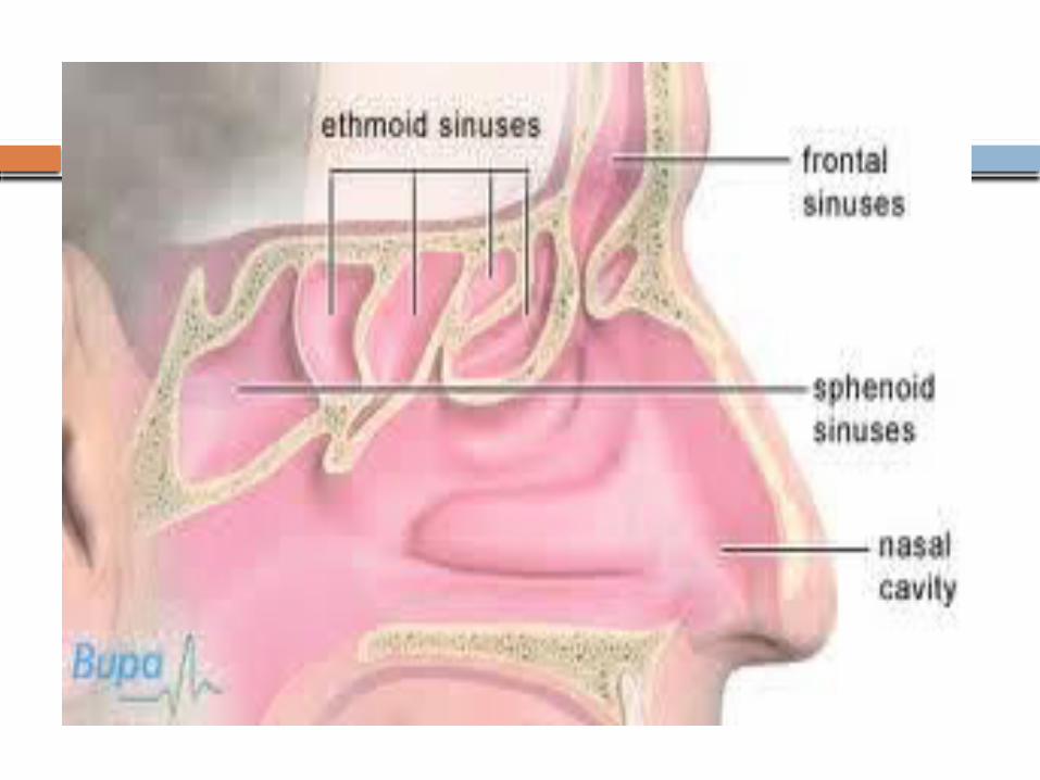
Air-containing spaces 4 on each side Clinically:
a. Anterior : Maxillary, frontal, anterior ethmoidal (middle meatus)
b. Posterior: Posterior ethmoidal (superior meatus) and sphenoidal (sphenoethmoidal recess)
Function: Makes skull lighter; Adds resonance


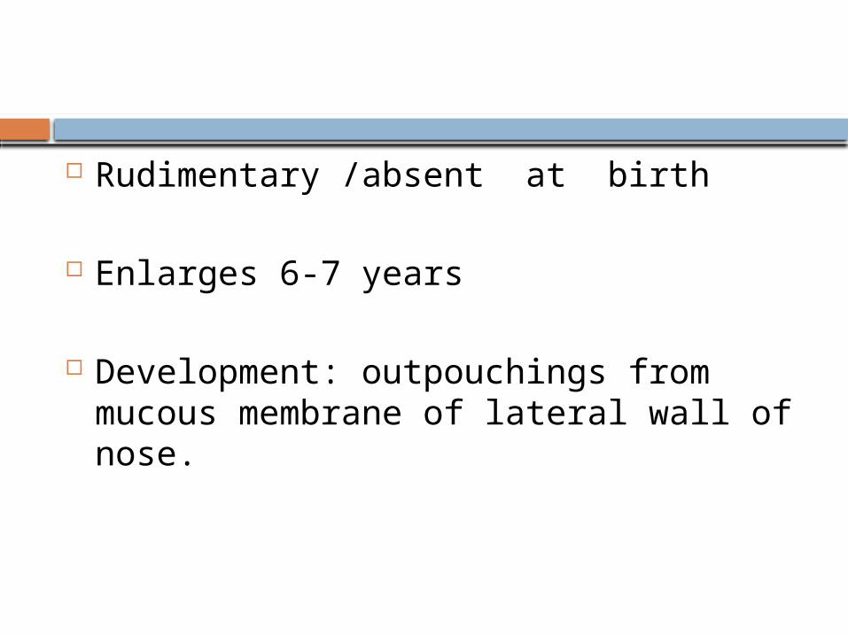
Rudimentary /absent at birth
Enlarges 6-7 years
Development: outpouchings from mucous membrane of lateral wall of nose.
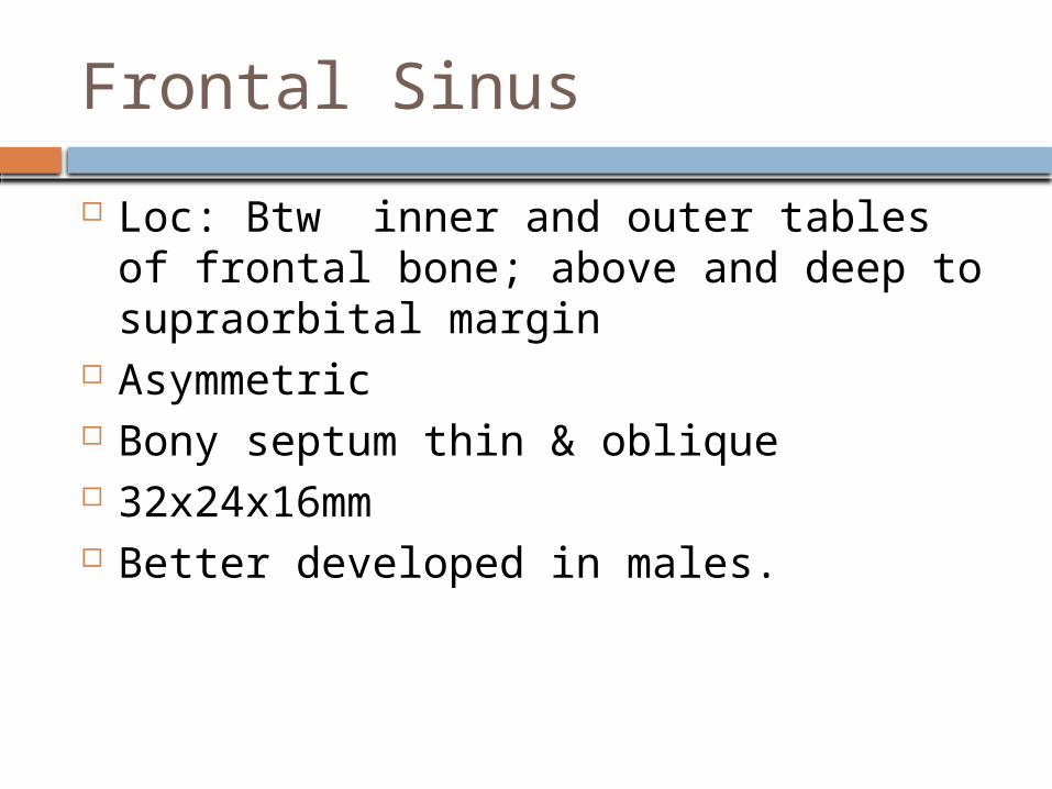
Frontal Sinus
Loc: Btw inner and outer tables of frontal bone; above and deep to supraorbital margin
Asymmetric Bony septum thin & oblique 32x24x16mm Better developed in males.
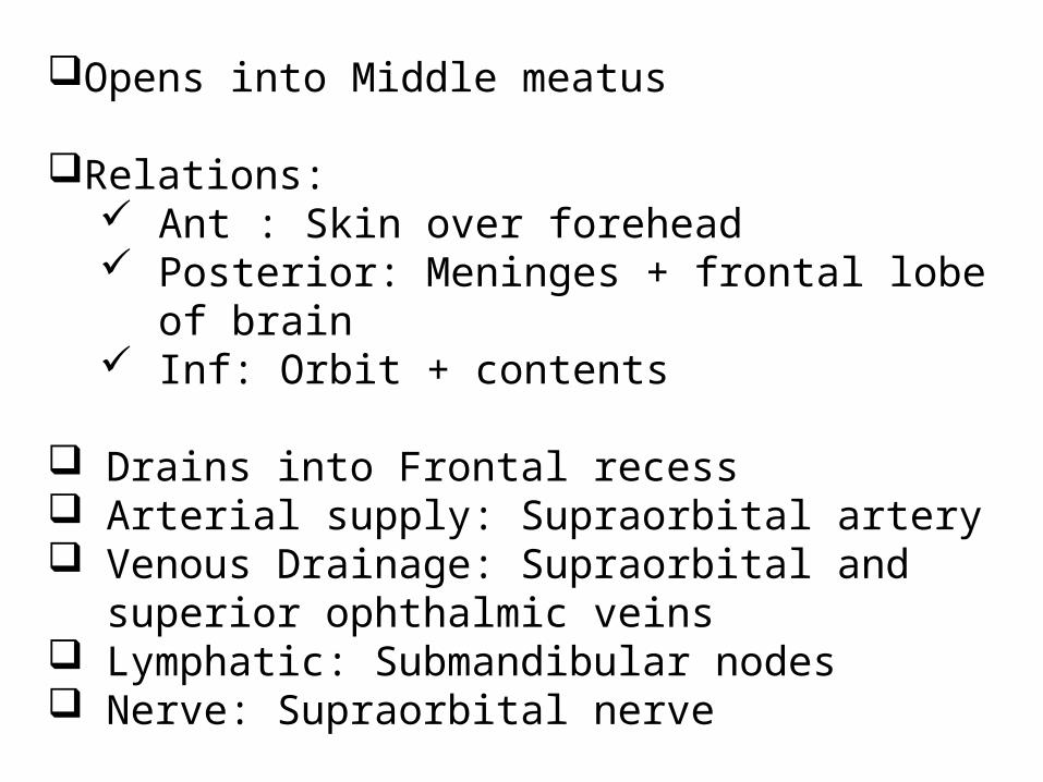
Opens into Middle meatus
Relations: Ant : Skin over forehead Posterior: Meninges + frontal lobe of
brain Inf: Orbit + contents
Drains into Frontal recess Arterial supply: Supraorbital artery Venous Drainage: Supraorbital and superior
ophthalmic veins Lymphatic: Submandibular nodes Nerve: Supraorbital nerve

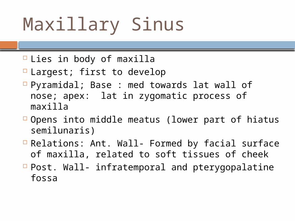
Maxillary Sinus
Lies in body of maxilla Largest; first to develop Pyramidal; Base : med towards lat wall of nose;
apex: lat in zygomatic process of maxilla Opens into middle meatus (lower part of hiatus
semilunaris) Relations: Ant. Wall- Formed by facial surface of
maxilla, related to soft tissues of cheek Post. Wall- infratemporal and pterygopalatine
fossa

Medial wall- middle and inferior meatuses. At places uncinate process, ant and post fontanelle and inferior turbinate
Floor-Alveolar and palatine processes of maxilla Roof- Floor of orbit 3.4x2.5x3.5cm Arterial: Facial, infraorbital, greater palatine
arteries Venous: facial vein, pterygoid plexus Lymphatic: Submandibular nodes Nerve: Infraorbital, ant, middle, post alveolar
nerves

Ethmoidal Sinus
Numerous (3-18) Lie within labyrinth of ethmoid bone Relations:
Above: orbital plate of frontal bone Behind: Sphenoidal conchae+ orbital
process of palatine Ant: lacrimal bone
Divided into anterior, middle and posterior groups.
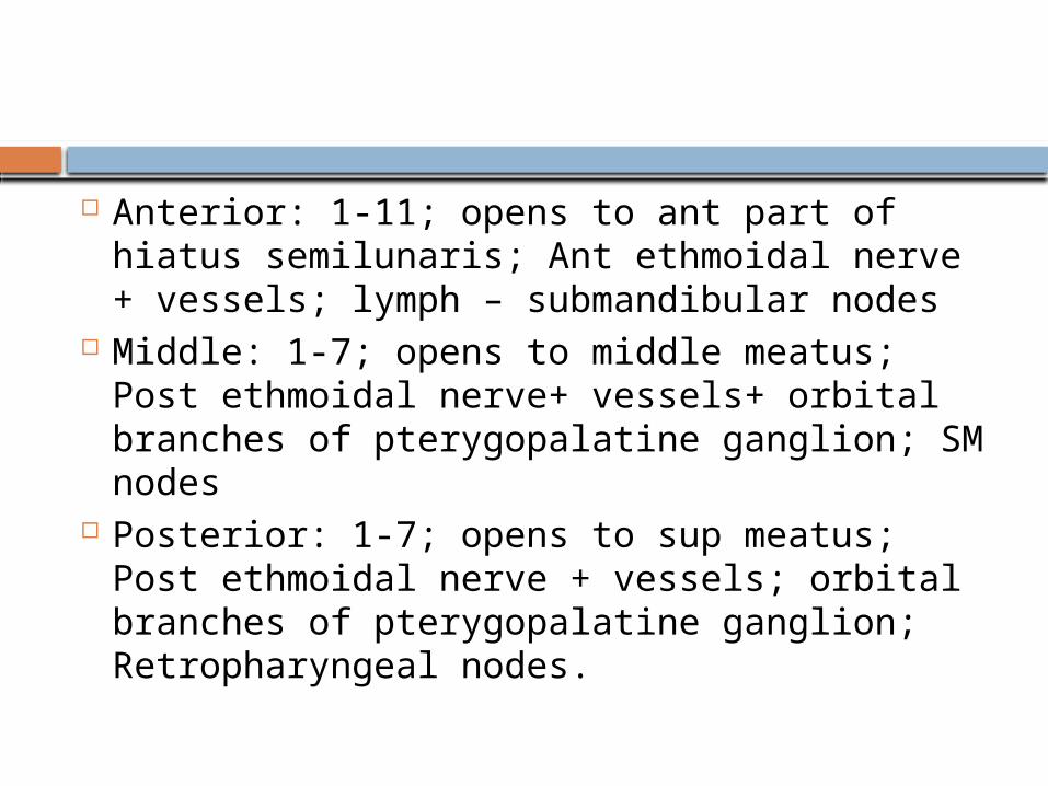
Anterior: 1-11; opens to ant part of hiatus semilunaris; Ant ethmoidal nerve + vessels; lymph – submandibular nodes
Middle: 1-7; opens to middle meatus; Post ethmoidal nerve+ vessels+ orbital branches of pterygopalatine ganglion; SM nodes
Posterior: 1-7; opens to sup meatus; Post ethmoidal nerve + vessels; orbital branches of pterygopalatine ganglion; Retropharyngeal nodes.

Important cells in anterior group- Agger nasi cells, ethmoidal bulla, supraorbital cells, frontoethmoid cells, Haller cells
Important cell in posterior group- Sphenoethmoid or Onodi cell

Sphenoidal Sinus
Within body of sphenoid bone Separated from each by thin bony
septum Asymmetric Opens to shpenoethmoidal recess Relations:
Sup: Optic chiasma+ hypophysis cerebri Lat: int carotid artery+ cavernous sinus

Arterial supply: Post ethmoidal + int carotid
Venous: Pterygoid venous plexus + cavernous sinus
Lymph: Retropharyngeal nodes Nerve: Post ethmoidal nerve+
pterygopalatine ganglion branches.

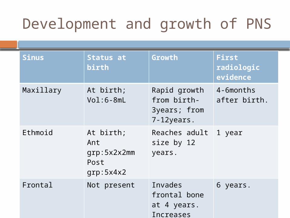
Development and growth of PNS
Sinus Status at birth
Growth First radiologic evidence
Maxillary At birth; Vol:6-8mL
Rapid growth from birth-3years; from 7-12years.
4-6months after birth.
Ethmoid At birth;Ant grp:5x2x2mmPost grp:5x4x2
Reaches adult size by 12 years.
1 year
Frontal Not present Invades frontal bone at 4 years. Increases until teens. Till 20y.
6 years.
Sphenoid Not present. Reach sella turcica 7years, dorsum sellae late teens, basisphenoid adult
4 years.

Clinical Aspects
Acute Sinusitis acute inflammation of sinus mucosa. Most common:
Maxillary>ethmoid>frontal>sphenoid Can be open/closed type- drainage of the
inflammatory products into nasal cavity. Aetiology:
Exciting causes: Nasal infections, swimming and diving(bacteria, chlorine), trauma, dental infection(Max Sinus; molar/pre-molar tooth extraction)
Predisposing causes: Local: obstruction to sinus ventilation and
drainage (DNS, nasal packing, hypertrophic turbinates, nasal polypi, structural abnormalities of ethmoidal air cells, neoplasm)
Stasis of secretions in nasal cavity: adenoids, choanal atresia, cystic fibrosis
Previous attacks of sinusitis
General Environment:pollution, smoke, dust Poor general health: exanthematous fever,
nutritional deficiency, systemic disorder
Chronic Sinusitis Sinus infection lasting for months/years
Complications of sinusitis Local:Mucocele, mucous retention cyst,
osteomyelitis Orbital: Preseptal inflm edema of lids,
subperiosteak abscess, orbital cellulitis, orbital abscess, superior orbital fissure syndrome
Intra-cranial: Meningitis, extradural abscess, subdural abscess, brain abscess, cavernous sinus thrombosis
Descending infections Focal infection.

Neoplasms of PNS
Benign: Osteomas, fibrous dysplasia, ossifying fibroma, ameloblastoma
Malignant:CommonMostly Maxillary>ethmoid>frontal>sphenoid.80% squamous cell type. Rest
adenocarcinoma, adenoid cystic carcinoma, melanoma, sarcoma.

Thank you