Anatomy of Haplo mastodon chimborazi (Mammalia...
59
663 GEODIVERSITAS • 2010 • 32 (4) © Publications Scientifiques du Muséum national d’Histoire naturelle, Paris. www.geodiversitas.com KEY WORDS Mammalia, Proboscidea, Gomphotheriidae, Haplomastodon, Stegomastodon, South America, Ecuador, Pleistocene, osteology, phylogeny. Ferretti M. P. 2010. — Anatomy of Haplomastodon chimborazi (Mammalia, Proboscidea) from the late Pleistocene of Ecuador and its bearing on the phylogeny and systematics of South American gomphotheres. Geodiversitas 32 (4): 663-721. ABSTRACT I present here a revision of the late Pleistocene Haplomastodon chimborazi (Proaño, 1922) material from Bolivar, Ecuador and a comparison with other New World trilophodont gomphotheres, and provide new morphological data in order to develop a novel phylogenetic hypothesis of South American (SA) proboscideans. Haplomastodon Hoffstetter, 1950 includes a single SA species whose valid name is H. chimborazi. Haplomastodon waringi (Holland, 1920) is considered to be an invalid taxon as it is based on undiagnosed material. Phylogenetic analysis supports the monophyly of SA gomphotheres (Cuvieroniinae) H. chimborazi, Cuvieronius hyodon (Fischer de Waldheim, 1814), and “Stegomastodon” platensis (Ameghino, 1888), based on five unambiguous characters. Conflicting evidence regarding the interrelationships of SA gomphotheres leads to three possible alternative hypotheses: two paired associations ((H. chimborazi,“S.” platensis) C. hyodon) and ((C. hyodon,“S.” platensis) H. chimborazi), and a trichotomy. ese imply that the ancestral separation of the three SA taxa might be either the result of two successive dichotomous branchings or of a single trichotomous branching event. e latter hypothesis would be consistent with the disjunct fossil distribution of the three SA gomphothere species. “Stegomastodon” plat- ensis is shown to be not closely related to North American (NA) Stegomastodon Pohlig, 1912, supporting its removal from the latter genus. e NA species Rhynchotherium cf. falconeri Osborn, 1923 is placed as the sister taxon of SA gomphotheres, on the basis of four unequivocal characters. NA Stegomastodon and the Asian Sinomastodon Tobien, Chen & Li, 1986 form successive out- groups to the previous clade together with whom they form a monophyletic group which includes all the brevirostrine species considered, along with the “depressed-beaked” gomphothere Rhynchotherium Falconer, 1868. e results of the present phylogenetic analysis indicate a rather high level of homoplasy in the evolution of New World gomphotheres. Marco P. FERRETTI Università di Firenze, Dipartimento di Scienze della Terra, via G. La Pira 4, I-50121 Firenze (Italy) mferretti@unifi.it Anatomy of Haplomastodon chimborazi (Mammalia, Proboscidea) from the late Pleistocene of Ecuador and its bearing on the phylogeny and systematics of South American gomphotheres
-
Upload
nguyenkhue -
Category
Documents
-
view
223 -
download
0
Transcript of Anatomy of Haplo mastodon chimborazi (Mammalia...

663GEODIVERSITAS • 2010 • 32 (4) © Publications Scientifi ques du Muséum national d’Histoire naturelle, Paris. www.geodiversitas.com
KEY WORDSMammalia,
Proboscidea, Gomphotheriidae,
Haplo mastodon, Stegomastodon,
South America,Ecuador,
Pleistocene,osteology,
phylogeny.
Ferretti M. P. 2010. — Anatomy of Haplo mastodon chimborazi (Mammalia, Proboscidea) from the late Pleistocene of Ecuador and its bearing on the phylogeny and systematics of South American gomphotheres. Geodiversitas 32 (4): 663-721.
ABSTRACTI present here a revision of the late Pleistocene Haplo mastodon chimborazi (Proaño, 1922) material from Bolivar, Ecuador and a comparison with other New World trilophodont gomphotheres, and provide new morphological data in order to develop a novel phylogenetic hypothesis of South American (SA) proboscideans. Haplo mastodon Hoff stetter, 1950 includes a single SA species whose valid name is H. chimborazi. Haplo mastodon waringi (Holland, 1920) is considered to be an invalid taxon as it is based on undiagnosed material. Phylogenetic analysis supports the monophyly of SA gomphotheres (Cuvieroniinae) H. chimborazi, Cuvieronius hyodon (Fischer de Waldheim, 1814), and “Stegomastodon” platensis (Ameghino, 1888), based on fi ve unambiguous characters. Confl icting evidence regarding the interrelationships of SA gomphotheres leads to three possible alternative hypotheses: two paired associations ((H. chimborazi,“S.” platensis) C. hyodon) and ((C. hyodon,“S.” platensis) H. chimborazi), and a trichotomy. Th ese imply that the ancestral separation of the three SA taxa might be either the result of two successive dichotomous branchings or of a single trichotomous branching event. Th e latter hypothesis would be consistent with the disjunct fossil distribution of the three SA gomphothere species. “Stegomastodon” plat-ensis is shown to be not closely related to North American (NA) Stegomastodon Pohlig, 1912, supporting its removal from the latter genus. Th e NA species Rhynchotherium cf. falconeri Osborn, 1923 is placed as the sister taxon of SA gomphotheres, on the basis of four unequivocal characters. NA Stegomastodon and the Asian Sinomastodon Tobien, Chen & Li, 1986 form successive out-groups to the previous clade together with whom they form a monophyletic group which includes all the brevirostrine species considered, along with the “depressed-beaked” gomphothere Rhynchotherium Falconer, 1868. Th e results of the present phylogenetic analysis indicate a rather high level of homoplasy in the evolution of New World gomphotheres.
Marco P. FERRETTIUniversità di Firenze, Dipartimento di Scienze della Terra,
via G. La Pira 4, I-50121 Firenze (Italy)mferretti@unifi .it
Anatomy of Haplo mastodon chimborazi (Mammalia, Proboscidea) from the late Pleistocene of Ecuador and its bearing on the phylogeny and systematics of South American gomphotheres

664 GEODIVERSITAS • 2010 • 32 (4)
Ferretti M. P.
INTRODUCTION
South American (SA) gomphotheres have received extensive attention from vertebrate paleontologists for over 200 years, thanks in particular to the very rich fossil record from Ecuador, Bolivia, Chile, Brazil, and Argentina (see Hoff stetter 1952; Prado et al. 2005; and Ferretti 2008a for an overview). Despite this, many problems associated with the phylogeny, classifi cation, and paleobiogeographic patterns of this proboscidean group still exist. Within SA gomphothere systematics, the taxonomic status of Haplo mastodon chimborazi
(Proaño, 1922) is a particularly contentious issue (Hoff stetter 1955; Simpson & Paula Couto 1957; Ficcarelli et al. 1995; Prado et al. 2005; Shoshani & Tassy 2005; Lucas 2008, 2009). In fact, both the valid-ity of the species H. chimborazi, considered by some authors as a junior synonym of “Mastodon” waringi Holland, 1920 and that of the genus Haplo mastodon Hoff stetter, 1950 as a distinct taxon from Stegomastodon Pohlig, 1912 have been questioned. Th e relationships between H. chimborazi and the other Pleistocene SA gomphotheres, Cuvieronius hyodon (Fischer de Wald-heim, 1814) and “S.” platensis (Ameghino, 1888),
MOTS CLÉSMammalia,
Proboscidea,Gomphotheriidae,
Haplo mastodon,Stegomastodon,
Amérique du Sud,Équateur,
Pléistocène,ostéologie,
phylogénie.
RÉSUMÉAnatomie de Haplo mastodon chimborazi (Mammalia, Proboscidea) du Pléistocène supérieur de l’Équateur et ses implications sur la phylogénie et sur la systématique des gomphothères de l’Amérique du Sud.La révision du matériel du Pléistocène tardif de Bolivar, Équateur, associé à Haplo mastodon chimborazi (Proaño, 1922), et sa comparaison avec les autres gomphothères trilophodontes du Nouveau Monde, fournissent des données morphologiques inédites permettant de développer une nouvelle hypothèse phylogénétique pour les proboscidiens d’Amérique du Sud (SA). Haplo mastodon Hoff stetter, 1950 comprend une seule espèce SA dont le nom valide est H. chim-borazi. Le taxon H. waringi (Holland, 1920), fondé sur l’étude d’un matériel non diagnostique, est considéré invalide. L’analyse cladistique soutient la monophylie des gomphothères SA (Cuvieroniinae) H. chimborazi, Cuvieronius hyodon (Fischer de Waldheim, 1814), et “Stegomastodon” platensis (Ameghino, 1888), défi nis par cinq caractères non ambigus. Des résultats contradictoires quant aux relations internes des gomphothères SA conduisent à trois hypothèses alternatives : deux paires exclusives ((H. chimborazi,“S.” platensis) C. hyodon) et ((C. hyodon,“S.” pla-tensis) H. chimborazi)), ou une trifurcation. Elles impliquent que la séparation ancestrale des trois taxons SA serait le résultat, soit de deux branchements dicho-tomiques successifs, soit d’un unique évènement de trifurcation. La dernière hypothèse serait cohérente avec la distribution fossile séparée des trois espèces de gomphothères SA. “Stegomastodon” platensis n’apparait pas directement lié aux Stegomastodon Pohlig, 1912 nord-américains (NA), ce qui soutient son exclusion de ce genre. L’espèce NA Rhynchotherium cf. falconeri Osborn, 1923 se place en groupe frère des gomphothères SA, sur la base de quatre caractères non équi-voques. Les Stegomastodon NA et Sinomastodon Tobien, Chen & Yuqing, 1986 asiatiques forment les groupes externes successifs du clade précédent avec lequel ils forment un groupe monophylétique qui inclut toutes les espèces brévirostres considérées, associées au gomphothère à symphyse mandibulaire inclinées vers le bas Rhynchotherium Falconer, 1868. Les résultats de l’analyse phylogénétique présente indiquent un niveau relativement élevé d’homoplasie au sein de l’évo-lution des gomphothères du Nouveau Monde.

665
Anatomy and phylogeny of Haplomastodon (Mammalia, Proboscidea)
GEODIVERSITAS • 2010 • 32 (4)
are still not completely resolved, though there is a general consensus in considering the three taxa as forming a monophyletic group (Hoff stetter 1952; Tobien 1973; Tassy 1985; Webb 1992; Shoshani 1996; Lambert & Shoshani 1998; Ferretti 2008a; Prado & Alberdi 2008). Th e phylogenetic relation-ships of SA gomphotheres with other gomphothere taxa (both from North America and Eurasia) is also problematic. In fact, it is questionable as to whether “S”. platensis should be included in Stegomastodon which is otherwise an exclusively North American taxon (Madden 1984; Webb 1992).
Th e species name “Masthodon” chimborazi was based on a complete skeleton from the Late Pleistocene of Quebrada Chalán, near Punin, Chimborazo Prov-ince, Ecuador (Proaño 1922; Spillmann 1928) and was later selected by Hoff stetter (1950) as the type species of the new genus Haplo mastodon. A second skeleton was discovered in 1928 at Quebrada Cal-lihuaico, near Alangasi, Quito Province (Spillmann 1928; 1931). Th ese two remarkable specimens were almost completely lost in a fi re that destroyed part of the fossil collections of the University of Quito in 1929. Subsequently, Haplo mastodon remains have been found from various other Ecuadorian sites, including both high plains and coastal localities (Hoff stetter 1952).
Th e principal works on the dental, cranial and postcranial anatomy of Ecuadorian Haplo mastodon are those of Spillmann’s (1928, 1931) and Hoff stet-ter’s (1950, 1952). Little attempt was made by these authors at detailed comparison of skeletal features (especially cranial) with other gomphotheres, so that the phylogenetic information of the skeletal anatomy of Haplo mastodon has remained largely unexplored. In 1991, fi eldwork in Northern Ecuador by the Dipartimento di Scienze della Terra and Museo di Storia Naturale, Sezione di Geologia e Paleontologia of the University of Firenze, in collaboration with the Museo Ecuatoriano de Ciencias Naturales of Quito, and the Escuela Politecnica National of Quito, re-sulted in the discovery of new fossil vertebrate locali-ties near Bolivar, Carchi province (Fig. 1; Ficcarelli et al. 1992, 1993). Two collecting sites in the Bolivar area (known as Quebrada Pistud and Q. Cuesaca, respectively) provided abundant material, mostly representing mylodonts and gomphotheres. In par-
ticular, an almost complete skeleton of H. chimborazi was retrieved from the Cangahua Formation at the Q. Pistud site.
Th e Bolivar sample represents the most complete material of this species and is also, at present, the only sample suitable for a comprehensive diagnosis of this taxon which includes cranial, dental and postcranial elements, complemented by precise stratigraphic information. Ficcarelli et al. (1993, 1995) provided a general description of the skull, mandible, and atlas of the Bolivar skeleton.
Th e aim of this paper is to review the taxonomy of Haplo mastodon and to provide a new comprehensive and detailed description of the dental and skeletal anatomy of Haplo mastodon chimborazi. A compara-tive analysis of dental and osteological characters of American trilophodont gomphotheres is presented in order to develop a new phylogenetic hypothesis of New World gomphotheres, focusing on the South American taxa. Th e classifi cation and taxonomy of South American proboscideans are thus discussed based on the results of the cladistic analysis.
79°W
COLOMBIA
PERU
ECUADOR
AN
DES
Bolivar
Quito
S. Elena
Punin
0°
PacificOcean
100 km
S Am.
FIG. 1. — Map of Ecuador, showing the location of the Bolivar area and of other Ecuadorian sites with Haplo mastodon chim-borazi (Proaño, 1922).

666 GEODIVERSITAS • 2010 • 32 (4)
Ferretti M. P.
MATERIAL AND METHODS
Th e present description and revision of Haplo mastodon chimborazi are based on the Bolivar sample (mate-rial kept at MECN). Observations made from other Ecuadorian H. chimborazi specimens, including the S. Elena sample (material kept at EPN) described by Hoff stetter (1952), were also integrated in the description, as they have details not preserved in the Bolivar material. Comparisons with the two specimens from Punin (holotype of H. chimborazi) and Alangasi, were based on the published descrip-tions and fi gures made by Proaño (1922), Spillmann (1928, 1931), and Osborn (1936). Th e individual from Punin had the M3 in use. Th e specimen from Alangasi was a young adult with both M1 and M2 in use and the M3 completely formed but yet unerupted. In the comparative analysis, dental and skeletal material of the following elephantoid taxa were studied (repository of directly studied mate-rial and published sources are indicated within brackets): Mammut americanum (Kerr, 1792) from North America (Late Pleistocene; AMNH, NHM); Gomphotherium sylvaticum Tassy, 1985 from Artenay, France (Early Miocene; MNHN; Tassy 1977); Gom-photherium angustidens (Cuvier, 1817) from Sansan, France (Middle Miocene; MNHN; Tassy 1985) and the Dinotheriensande, Germany (Middle Miocene; SMNS); Gomphotherium productum (Cope, 1875) from Clarendon, Texas (Late Miocene; AMNH; Os-born 1936); Eubelodon morilli Barbour, 1914 from Brown County, Nebraska (Late Miocene; AMNH; Osborn 1936); Rhynchotherium edensis Frick, 1921 from Mt Eden, California (Early Pliocene; AMNH; Osborn 1936); Rhynchotherium cf. falconeri Osborn, 1923 from Arizona (Late Pliocene; LVMNH; Miller 1990; Ferretti 2008a); Cuvieronius hyodon from Tarija, Bolivia (Late Pleistocene; MLP, MACN, MNHN, NMR; Boule & Th evenin 1920); “Stegomastodon” platensis from various localities of the Provinces of Buenos Aires and Entre Rios, Argentina (Late Pleistocene; MLP, MACN, NHM; Cabrera 1929); Stegomastodon texanus Osborn, 1924 (= S. mirifi cus Leidy, 1858, according to Savage 1955) from Llano Estacado, Texas (Late Pliocene; AMNH; Osborn 1936); Anancus perimensis (Falconer & Cautley, 1847) from Perim Island, India (Late Miocene;
NHM); Anancus arvernensis (Croizet & Jobert, 1828) from Asti, Italy (Early Pliocene; MGT, MGB), and Valdarno Inferiore, Italy (Middle Pliocene; IGF); Elephas maximus Linnaeus, 1758 (Recent; ACM, MCZR, MNHN, MSNFZ, NHM); Loxodonta africana (Blumenbach, 1792) from various African localities (Recent; MNHN, MSNFZ, NHM). Data on the dental and skeletal anatomy of Megabelodon lulli Barbour, 1914, Gnathabelodon thorpei Barbour & Sternberg, 1935, Sinomastodon intermedius (Teilhard de Chardin & Trassaert, 1935), and Sinomastodon hanjiangensis Tang & Zong, 1987, were obtained from the following publications: Barbour (1914), Barbour & Sternberg (1935), Osborn (1936), To-bien (1973), Tobien et al. (1986) and Zong et al. (1989).
Th e Peruvian gomphothere Amahuacatherium peruvium Romero-Pittman, 1996 was not included in the analysis as the interpretation of its anatomy and age is still highly controversial (Campbell et al. 2000; Alberdi et al. 2004; Shoshani & Tassy 2005; Ferretti 2008a; Campbell et al. 2009).
Th e nomenclature of dental characters follows Tassy (1985, 1996a).
To test hypotheses about the phylogenetic relation-ships of New World gomphotheres, a parsimony analysis was performed based on the matrix shown in Appendix 7. Th e matrix consists 11 taxa and 24 characters (Appendix 6).
Th e 11 taxa analyzed in the cladistic analysis include representatives of all the trilophodont non-amebelodontine gomphothere genera recognized in North America by Lambert & Shoshani (1998) and the three South American taxa recognized by Prado et al. (2005) and Ferretti (2008a). Th e Old World brevirostrine gomphothere Sinomastodon Tobien, Chen & Li, 1986 was also included in the analysis as several authors consider this genus closely related to South American proboscideans (Tobien et al. 1986; Tassy 1990; Shoshani 1996; Prado & Alberdi 2008). Fifteen characters are taken or modifi ed from previ-ous studies (Tassy 1990; Shoshani 1996). Eleven are new characters. Polarity was defi ned by outgroup comparison using G. angustidens as outgroup. Th e analysis was performed using PAUP 4.0b10 (Swof-ford 2003). All 24 characters were weighted equally and treated as ordered (except for the multistate

667
Anatomy and phylogeny of Haplomastodon (Mammalia, Proboscidea)
GEODIVERSITAS • 2010 • 32 (4)
character number 9). A set of exhaustive searches was conducted, implementing both the ACCTRAN (accelerated transfomation) and DELTRAN (delayed transformation) optimization criteria.
ABBREVIATIONS
InstitutionsACM Museo di Anatomia Comparata, Bologna;AMNH American Museum of Natural History, New
York;EPN Escuela Politecnica Nacional, Quito;IGF Museo di Storia Naturale – Sezione di Geologia
e Paleontologia, Firenze;MCZR Museo Civico di Zoologia, Roma; MECN Museo Ecuatoriano de Ciencias Naturales,
Quito;MGB Museo di Paleontologia “G. Capellini”, Bo-
logna;MGT Museo di Geologia, Torino;MICN Museo de Historia Natural del Instituto de Cien-
cias Naturales, Universidad Central, Quito;MLP Museo de La Plata, La Plata; MNHN Muséum national d’Histoire naturelle, Paris;MSNFZ Muso di Storia Naturale (sezione di Zoologia),
Firenze;MUT Museo Nacional Paleontologico-Arquelogico,
Tarija;NHM Natural History Museum, London;NMR Swedish Museum of Natural History, Stock-
holm;SMNS Staatlichen Museum Naturkunde, Stuttgart;UCE Universidad Central de Ecuador, Quito;UCMP University of California, Museum of Paleon-
tology, Berkeley.
AnatomyC cervical vertebra;DP upper deciduous premolar;dp lower deciduous premolar;I upper incisor (tusk);L lumbar vertebra;M upper molar;m lower molar;Mc metacarpal;Mt metatarsal;T thoracic vertebra.
GEOLOGY AND TAPHONOMY OF THE BOLIVAR GOMPHOTHERE SITE
Th e Cangahua Formation is a Late Pleistocene loess-like pyroclastic unit, deposited under periglacial
conditions, present in the interandean depression of northern Ecuador (Sauer 1965; Ficcarelli et al. 1992; Coltorti et al. 1998). It yielded abundant vertebrate remains, including articulated skeletons of large mammals (Hoff stetter 1952). At Quebrada Pistud (Bolivar), two main vertebrate-rich levels and other minor ones were recognized in the Cangahua sequence. Th e lower and upper main fossiliferous levels, consisting of torrential stream sediments, were dated to 18 720±90 and 12 350±70 y respectively, on the base of radiometric analysis of bone carbonate (Coltorti et al. 1998). Both ages are consistent with the biochronological interpretation of the fauna from this site, which indicates a Lujanian age (Ficcarelli et al. 1992; 1993; 1997).
Th e H. chimborazi skeleton was recovered from the lowermost main fossiliferous bed, in an area of about 5 m2. It consists of the cranium, mandible and 68 postcranial elements (Table 1).
Diff erent catalogue numbers were given to the various skeletal elements of this individual as they were collected during successive excavations. Nev-ertheless, in the following description the skeleton will be referred to as individual “MECN 82”.
Some of the bones of MECN 82 were in ana-tomical connection (e.g., right humerus and ulna), position of other parts, when not in anatomical connection, was however anatomically coherent. Th e skull was lying on its dorsal part on the fi rst cervical vertebrae, the mandible was in front of the skull. Bone disposition indicates that the animal was decomposed in situ and that the skeleton was only slightly displaced by a current responsible of the skull upturning and mandible disarticulation. Bones do not show any sign of butchering nor car-nivore tooth marks.
Th e exploited area at Q. Pistud, some tens of square meters, allowed recovery of 160 proboscidean elements, representing at least seven individuals, including the MECN 82 skeleton. Vertebrae and ribs are the most abundant elements. Smaller ele-ments (sesamoids, phalanges) are somewhat under-represented, indicating a certain size bias. On the other hand, large but fragile parts, as skulls, scapulae and pelvis are extremely rare. Finally, elements of the anterior limbs are slightly more abundant than that of the posterior ones.

668 GEODIVERSITAS • 2010 • 32 (4)
Ferretti M. P.
TAXONOMY OF ECUADORIAN GOMPHOTHERES
Th e complex taxonomy of Ecuadorian gomphotheres has been revised by Hoff stetter (1950, 1952, 1955), Simpson & Paula Couto (1957) and Ficcarelli et al. (1993, 1995; see Ferretti 2008a and Lucas 2008 for an overview). In this section, we shall discuss the taxonomy and nomenclature of those specimens from Ecuador allocated by Hoff stetter (1952) to Haplo mastodon.
HAPLO MASTODON CHIMBORAZI
In 1894, J. F. Proaño reported the discovery of a partial proboscidean skeleton at Quebrada de Chalán, a small gorge near Punin (Chimborazo Province, Central Ecuador), providing a summary description and pictures of the skull and mandible (Arauz 1950; Costales 1950; Hoff stetter 1952). Th e author referred to the specimen as “Mastodonte del Chimborazo”, considering it a new, exclusively South American taxon. Proaño (1894) mentioned as distinctive characters of the Punin gomphothere, distinguishing it from the other species known from the Andean highplains, i.e. Mastodon an-dium Cuvier, 1824 (= Cuvieronius hyodon Fischer de Waldheim, 1814), the higher cranium and the more massive upper tusks with converging tips and no enamel band.
Elements of the Punin skeleton, namely the skull, the atlas, a humerus and other postcranials, were then fi gured in an anonymous paper published in the French magazine Cosmos in 1903 (Hoff stetter 1952).
Th e species name “Masthodon” chimborazi was fi rst published by Proaño in 1922.
Th e fact that the name appeared in the caption of a picture of the Punin skull in an unpaged leafl et inserted in a religious tract (see Arauz 1950 and Hoff stetter 1952), has led some authors to question the adequacy of Proano’s publication to the purpose of zoological nomenclature. However, the work by Proaño meets all the criteria set by the ICZN code (ICZN 1999) to be regarded as published and therefore the species name “M.” chimborazi should be considered as valid, within the meaning of the Code.
Spillmann (1931), in his review of the Pleistocene mammals from Ecuador, provided a new description of the Punin skull, based on data and photographs taken before the specimen was destroyed in 1929. He referred this specimen to Bunolophodon ayora Spillmann, 1928, a specifi c name he had previ-ously created for the Punin skeleton (Spillmann, 1928), completely disregarding Proaño’s species. In the same paper, Spillmann named a nearly com-plete skeleton from Q. Callihuaico near Alangasi (Quito Province) as B. postremus Spillmann, 1931. Hoff stetter (1950, 1952, 1955) revised all the gomphothere material from Ecuador then known. Th e author concluded that the Punin and Alan-gasi gomphotheres were conspecifi c and that the specifi c name “M.” chimborazi had priority over Spillmann’s B. ayora and B. postremus. Hoff stet-ter (1950) created the new name Haplo mastodon as a subgenus of Stegomastodon to accommodate “M.”chimborazi (Hoff stetter 1950). Later, based on the presence/absence of transverse foramina on the atlas and axis, Hoff stetter (1952) established a full generic separation of Haplo mastodon (atlas without foramina) from Stegomastodon (atlas and axis with foramina) and based the separation of the former into the two subgenera Haplo mastodon (axis lacks the foramina) and Aleamastodon Hoff stetter, 1952 (axis with foramina). Th e latter subgenus was es-tablished on material from the S. Elena peninsula, on the southern coast of Ecuador, and attributed to the new species H. (Aleamastodon) guayasensis Hoff stetter, 1952. Simpson & Paula Couto (1957), based on a larger Haplo mastodon sample from Aguas do Araxà (Brazil) demonstrated that the absence/presence of transversal foramina in the fi rst and sec-ond cranial vertebrae is a variable character in this genus. Th ey considered therefore H. guayasensis as a junior synonym of Haplo mastodon chimborazi. On the other hand, Simpson & Paula Couto (1957), following Hoff stetter’s (1950) original diagnosis, accepted Haplo mastodon as a valid genus distinct from Stegomastodon. Based on the comparison of the Ecuadorian and Brazilian material referred to Haplo mastodon, Simpson & Paula Couto (1957) concluded that this represents one and the same spe-cies and that the nominal taxon “Mastodon” waringi Holland, 1920, a species based on a Brazilian type

669
Anatomy and phylogeny of Haplomastodon (Mammalia, Proboscidea)
GEODIVERSITAS • 2010 • 32 (4)
specimen (see below), has priority over “Mastho-don” chimborazi Proaño, 1922. Simpson & Paula Couto (1957) treated then Haplo mastodon waringi (Holland, 1920) as the type species Haplo mastodon, contrary to the rule of the Code (Lucas 2008, 2009). Hoff stetter (1955, 1986), however, strongly ques-tioned the validity of Holland’s species, considering it as a nomen dubium. Ficcarelli et al. (1993), based on new material from Bolivar, demonstrated that Ecuadorian Haplo mastodon display a variability in the presence/absence of transverse foramina similar to that known in the Aguas do Araxà population. Following Hoff stetter (1955), they accepted the validity of the species name H. chimborazi over “M.” waringi. However, considering that the type material, in particular the skull, of the former species was lost, Ficcarelli et al. (1995) proposed as neo-
type of H. chimborazi the complete skeleton from Q. Pistud, Bolivar. More recently, Lucas (2009) presented a case to the International Commission on Zoological Nomenclature to conserve the usage of the name Mastodon waringi by designating as neotype the Bolivar specimen. As noted by Lucas (2009), H. chimborazi would still retain its status as the type species of Haplo mastodon under the rules of the Code. Th e usage of the name H. waringi is indeed far more widespread in the literature, after the systematic revison of Simpson & Paula Couto (1957). To note, however, is that, contrary to com-mon opinion, the type skeleton of H. chimborazi was not completely lost in the 1929 fi re of the Quito University. Hoff stetter (1952: 195) mentioned in fact an atlas and two humeri (left and right), then housed in the collection of the UCE, as probably
TABLE 1. — Haplo mastodon chimborazi (Proaño, 1922) from Bolivar, Ecuador. List of skeletal elements of individual MECN 82.
Element Catalogue number Element Catalogue number
skull MECN 82 right humerus MECN 95mandible MECN 133 left humerus MECN 100atlas MECN 83 right ulna MECN 134axix MECN 84 left ulna MECN 418C3 MECN 85 right radius MECN 210C5 MECN 102 left radius MECN 421C6 MECN 94 right unciform MECN 216C7 MECN 403 right magnum MECN 217 T2 MECN 404 right pyramidal MECN 219T3 or T4 MECN 405 left scaphoid MECN 428T4 or T5 MECN 406 left trapezoid MECN 429T6 or T7 MECN 407 left lunar MECN 427 T7 or T8 MECN 408 left pisiform MECN 426T9 or T10 MECN 135a right Mc3 MECN 218T11 or T12 MECN 135b right Mc4 MECN 220T12 or T13 MECN 135c right Mc5 MECN 221T13 or T14 MECN 214 pelvis MECN 415T16-lastT MECN 177 right femur MECN 420T16-lastT MECN 211 left femur MECN 98L1 MECN 101 right patella MECN 90L3 MECN 113 right tibia MECN 99L4 MECN 106 left tibia MECN 424sacrum MECN 107 right fi bula MECN 423caudal MECN 436 left fi bula MECN 422caudal MECN 435 right astragalus MECN 425caudal MECN 87 right Mt1 MECN 4341st left rib MECN 409 right Mt4 MECN 4301st right rib MECN 410 left Mt3 MECN 4329 ribs MECN 91, 92, 93, 207, 208,
411, 412, 457, 470left Mt4 MECN 431
right scapula MECN 82 phalanx MECN 89left scapula MECN 82 left phalanx MECN 433

670 GEODIVERSITAS • 2010 • 32 (4)
Ferretti M. P.
representing what was left of the type specimen of H. chimborazi described by Proaño (1922). Th e left and right humeri are currently kept at the MICN (catalog numbers MICN-UCE 1982 and 1981, respectively), along with other post-cranial material from Punin (T. Gordón pers. comm.). Th e atlas, however, is not among the MICN gompho there material. Th ough this specimen might probably be in other collections of the UCE, it has not yet been located by the author.
Th e two humeri housed at the MICN still bear an old label reporting the site of provenance (Chalán, Punin). Indeed, a comparison with the published fi gures of the Punin skeleton leaves no doubt that the two humeri pertain to the type specimen of H. chimborazi. For these reasons, the proposal of Ficcarelli’s et al. (1995) and Lucas (2009) to designate the Bolivar skeleton as the neotype of the species is not valid according to the Code (Ferretti 2009).
Alberdi & Prado (1995), accepted the synony-mies and species-level nomenclature proposed by Simpson & Paula Couto (1957), but included waringi within Stegomastodon, considering Haplo-mastodon a junior synonym of the former.
Based on new character information and on the results of the cladistic analysis presented here, Haplo mastodon is conservatively considered a valid genus, characterized by two autapomorphic features, with respect to both “Stegomastodon” platensis and the North American (NA) repre-sentatives of the genus.
STATUS OF “MASTODON” WARINGI
Th e type series of the species is from Pedra Vermelha, Bahia (Brazil). Holland (1920) did not fi gure the material, providing only a general description of the remains that does not distinguish “M.” waringi from other similar taxa. Th e surviving material consists of the tip of a tusk, a tusk dentine fragment, three molar fragments, and the distal end of a tibia, rep-resenting at least two individuals (Simpson & Paula Couto 1957; Ficcarelli et al. 1995; Ficcarelli pers. comm.; Lucas 2008, 2009). Th e remains are too poorly preserved to provide any useful systematic information. Th us, “M.” waringi should be eff ec-tively considered as a nomen dubium.
NOMENCLATURE
Th e taxonomic evidence outlined above leads to the following conclusions concerning the nomenclature of Ecuadorian gomphotheres:– the species “M.” chimborazi is valid, as the original description and available published fi gures of the type skeleton from Punin, clearly distinguish it from other similar brevirostrine gomphotheres. Th e left and right humeri, and possibly the atlas (not found yet among the UCE collections) represent what is left of the type skeleton described by Proaño (1922);– Haplo mastodon chimborazi is characterized by a set of autapomorphic characters that distinguish it from both SA and NA Stegomastodon species (see below). It is thus proposed that the generic name Haplo mastodon is retained, with Haplo mastodon chimborazi as the only species;– Bunolophodon ayora (sometimes spelled ayorae in later papers) is a junior objective synonym of H. chimborazi;– Bunolophodon postremus and H. guayasensis are junior subjective synonyms of H. chimborazi;– “Mastodon” waringi is a nomen dubium because it is based on undiagnostic material.
It should be kept in mind, however, that such a treatment has no formal standing until ratifi ed by the ICZN.
SYSTEMATICS
Order PROBOSCIDEA Illiger, 1811Superfamily ELEPHANTOIDEA Gray, 1821Family GOMPHOTHERIIDAE Hay, 1922
Genus Haplo mastodon Hoff stetter, 1950
TYPE SPECIES. — Masthodon chimborazi Proaño, 1922 by monotypy.
DIAGNOSIS. — As for the only species H. chimborazi, see Revised diagnosis below.
Haplo mastodon chimborazi (Proaño, 1922)
Mastodonte del Chimborazo Proaño, 1894: unnum-bered page.

671
Anatomy and phylogeny of Haplomastodon (Mammalia, Proboscidea)
GEODIVERSITAS • 2010 • 32 (4)
Mastodon waringi Holland, 1920: 229, nomen dubium.
Masthodon chimborazi Proaño, 1922: unnumbered page.
Tetrabelodon ayora Spillmann, 1928: unnumbered page preceding p. 70.
Bunolophodon ayorae (Spillmann, 1931): 67.
Bunolophodon postremus Spillmann, 1931: 73.
Cuvieronius ayora – Osborn 1936: 567.
Cuvieronius postremus – Osborn 1936: 595.
Haplo mastodon (Haplo mastodon) chimborazi – Hoff stet-ter 1952: 192.
Haplo mastodon (Aleamastodon) guayasensis Hoff stetter, 1952: 208.
Haplo mastodon waringi – Simpson & Paula Couto 1957: 171.
Haplo mastodon waringi – Ficcarelli et al. 1993: 233. — Casamiguela et al. 1996: 316.
Stegomastodon waringi – Alberdi & Prado 1995: 283. — Alberdi et al. 2004: 433. — Prado & Alberdi 2005: 4. — Prado & Alberdi 2008: 905.
HOLOTYPE. — A nearly complete adult skeleton of which only the right (MICN-UCE 1981) and left (MICN-UCE 1982) humeri are now preserved.
OTHER MATERIAL EXAMINED. — See Tables 1-19.
ORIGINAL DIAGNOSIS. — Gomphothere with high-domed cranium; upper tusks massive, with converging tips, lack-ing an enamel band (Proaño 1922). Hoff stetter (1952) listed the following diagnostic characters of Haplo mastodon chimborazi: 1) cranium high, elephant-like; 2) mandible with short symphysis (brevirostrine); 3) atlas and axis lack transverse foramina; 4) lower tusks absent; 5) up-per tusks are relatively short, upturned with no torsion; 6) enamel band present in some juvenile tusks, never at adult age; 7) molars show slight tendency to anancoidy (displacement of buccal and lingual half-lophs); 8) post-trite central conules absent to poorly developed; 9) third molar tetra- to pentalophodont. Characters 1, 2, 4, and 7 are shared with “Stegomastodon” platensis, whilst characters 3, 8 and 9 have a wider distribu-tion within SA gomphotheres, with character 3 showing intraspecifi c variability. Characters 5 and 6 are derived relative to the condition in Cuvieronius hyodon. Th e phylogenetic signifi cance of these two latter characters, in particular concerning the relationships between Haplo mastodon and SA “Stegomastodon” will be discussed below.
REVISED DIAGNOSIS. — Medium to large brevirostrine gomphotheriid of trilophodont grade that diff ers from other South American gomphotheres by the presence of relatively short, massive and upwardly curved upper tusk, which always lacks an enamel band in the adult growth stage, and in possessing only slightly divergent tusk alveoli in the premaxillaries. In addition, H. chimborazi has a unique combination of the following characters: 1) high, elephantine skull; 2) infl ated frontals and parietals forming sagittaly a wide fronto-parietal plane; 3) anterior border of bony orbit laying forward of the mesialmost cheek tooth in use; 4) nasal aperture wide and shallow, separated by a thin bony lamina from a deep subnasal fossa; 5) supraor-bital foramen of maxillary absent; 6) alveolar portion of premaxillaries relatively long and robust; 7) presence of a shallow fossa for muscular insertion (lateral coronoid fossa) at the base of the ascending ramus of the mandible; 8) position of the mandibular foramen on the medial side of the ascending ramus markedly higher than the occlusal plane; 9) transversal foramina of the fi rst and second cervical vertebrae with tendency to obliterate; 10) dorsal arch of atlas very thick, with strong dorsal concavity; 11) large and robust ventral tubercle of transverse process of atlas; 12) lower tusks absent; 13) DP3/dp3 trilophodont; 14) P3-P4/p3-p4 absent; 15) M3 with fully developed tretraloph; m3 with 4 to 5 lophids; 16) posttrite central conules (forming secondary trefoils under wear) absent to weakly developed.
Character 1, is shared with “S.” platensis. Characters 2-10 and 12-15 are shared with both “S.” platensis and C. hyodon, whilst character states 11 and 16 are as in C. hyodon.
TYPE LOCALITY. — Quebrada de Chalán near Punin, Chimborazo province, Central Ecuador (Proaño 1922; Hoff stetter 1952).
TYPE HORIZON. — No stratigraphic data were provided by Proaño (1894, 1922). Recent geological investigations in the Punin area, however, indicate that the sedimentary sequence cropping out at Quebrada Chalán is part of the Late Pleistocene Cangahua Formation and contains a typical Lujanian fauna (Ficcarelli et al. 1997; Coltorti et al. 1998). A radiometric dating of bone carbonate from mammalian fossil remains newly collected from Q. Chalán gave an age of 20 980±530 years BP (Col-torti et al. 1998).
OCCURRENCE. — Late Pleistocene to earliest Holocene (Ficcarelli et al. 2003; Coltorti et al. 1998). Ecuador: it is distributed both in the Andean provinces (Carchi, Pichincha, Chimborazo) and on the coast (Santa Elena Peninsula, Guayas province). Remains referable to this species have been collected from many other sites in South America, including Brazil (Simpson & Paula Couto 1957), Colombia, Venezuela (Hoff stetter 1986), and Peru (Alberdi et al. 2004).

672 GEODIVERSITAS • 2010 • 32 (4)
Ferretti M. P.
ANATOMICAL DESCRIPTION AND COMPARISONS
Cranium (Figs 2-5; Table 2)Th e description of the cranium of H. chimborazi is mainly based on the study of the Bolivar specimen (MECN 82). Additional data were obtained from Spillmann’s (1928, 1931) description and fi gures of the Punin (Fig. 2) and Alangasi skulls. Th ough these three specimens are at slightly diff erent onto genetic stage (see the Material and methods section), they represent adult individuals as in all of them the M3 is completely formed. Th e skull of the Bolivar skeleton suff ered a dorso-ventral crushing of the neurocranium and a distortion that altered in part the orientation of the distal end of the premaxillar-ies, that of the right tusk, and of the molars along the tooth row (see below). Th e Punin and Alangasi skulls were, on the other hand, in good state of preservation. Unfortunately, some aspects of these skulls were not fi gured before they were lost, so we have a partial knowledge of their morphology. Of the Punin skull only the left lateral aspect was fi g-ured by both Proaño (1894, 1922) and Spillmann (1928, 1931). Of the Alangasi skull the anterior, antero-lateral and ventral aspects were fi gured by Spillmann (1931).
Antero-dorsal view (Figs 3A, B; 4A). Th e parietal bones are transversally expanded and dorsally form two small bulges separated by a sagittal depression. Th e forehead (fronto-parietal plane) is wide, sagittally convex, and transversely fl at. Th e temporal lines are very faint and smooth postero-ventrally. Th e postor-bital process of the frontal bone is relatively small.
Th e nasals are bounded posteriorly by a sulcus for the attachment of the m. maxillo labialis (levator of the trunk), marking the limit between the nasals and the frontals. Th e sulcus is shallow medially and deeper laterally. It continues laterally into the nasal process of the premaxillary bone, along the limit between the premaxillary and frontal. Rostrally, the nasals consti-tute the dorsal limit of the external nasal aperture. Th e nasal processes are very small. Th e external nasal aperture is wide and low (Fig. 4A). Th e dorsal margin, formed by the nasals and the nasal processes of the premaxillaries, is thick, forming a step-like border. Laterally, the nasal aperture is delimited by the nasal processes of the premaxillaries. Th e thickness of the lateral border of the nasal aperture rapidly decreases ventrally. Th e ventral border, made up by the body of the premaxillary, is not very distinct. Th e lateral and dorsal inner walls of the nasal aperture do not show any opening communicating with the paranasal sinuses, or with the lacrimal conduct. Medially, just below the ventral margin of the nasal aperture is a fossa that deepens into the body of the premaxillary (Fig. 4A). Th is structure, here called the sub-nasal fossa, is contiguous anteriorly with the incisive fossa, from which it is clearly distinct. Th e alveolar processes of the premaxillaries, bearing the alveoli for the tusks, are long and very robust. In particular, the distal anterior margin is very thick. Seen in anterior view, the tusk alveoli only slightly diverge distally. Th e maximum distal width of the premaxillaries do not exceed the diameter between the orbital processes of the frontals. Th e incisura dentalis only slightly separates distally the two premaxillaries.
TABLE 2. — Haplo mastodon chimborazi (Proaño, 1922) from Bolivar, Ecuador. Measurements (in mm) of the skull (see Appendix 1).
Measures Specimen MECN 82
1. Total length: akrokranion-prosthion 9302. Akrokranion-rhinion (base of nasal aperture) 2703. Tusk alveolus length 5904. Length of zygomatic arch 4505. Greatest breadth of neurocranium c. 7806. Diameter between most lateral points of orbital processes of frontals c. 5507. Breadth of tusk alveoli between infraorbital foramina 4228. Greatest breadth of tusk alveoli 5159. Maximum dorso-ventral diameter of tusk alveoli 213
10. Greatest length of occipital condyle 11011. Transversal diameter of occipital condyle 75

673
Anatomy and phylogeny of Haplomastodon (Mammalia, Proboscidea)
GEODIVERSITAS • 2010 • 32 (4)
Lateral view (Figs 2; 5A, B). In lateral view, the outline of the cranial vault is regularly convex (Punin skull; Fig. 2). Th e skull vertex is on the perpendicular passing just behind the tuber max-illa. Th e orbits are large, with their anterior margin laying at the front of the mesial-most molar. Th e proximo-distal axis of the tusk alveoli is parallel to the plane of the forehead, and forms a wide open angle with the occlusal plane. Th e dorsal side of the alveolar processes of the premaxillaries is lon-gitudinally concave.
Th e maxillary bones have a single large orbital perforation, ventral to the orbit, corresponding to the infraorbital foramen. Th e foramen is oval-shaped and relatively large, with a maximum diameter comparable to that of extant Loxodonta africana. Th e infraorbital process of the maxillaries, forming the lateral wall of the infraorbital foramen, is thick and antero-posteriorly expanded. Th e zygomatic arch is deep and robust. Th e anterior half of the zygomatic is deep, with a straight dorsal margin. On the ventral margin, is a slightly concave surface, possibly representing the origin of the m. masseter. A marked step separates dorsally the anterior half of the zygomatic from the shallower posterior portion (pars temporalis), whose dorsal side is occupied by the elongated articular surface for the zygomatic process of the temporal. Th e zygomatic ends pos-teriorly in a small tuberosity.
Posterior view (Fig. 3C). Th e dorso-ventral crush-ing of the MECN 82 cranium caused the posterior tilting of the occipital squama. Dorsally and later-ally, the occipital squama is bounded by the nuchal crest. Th e latter is a thick and markedly wrinkled crest, evidence of a powerful dorsal nuchal muscu-lature (m. splenius capits and m. semispinalis capitis). Viewed from behind and slightly from above, the cranium shows two lateral swellings separated by a median depressed area at the bottom of which is the nuchal fossa.
Ventral view (Fig. 3D). Because of the post-mortem crushing, the occipital and basicranial regions of the Bolivar cranium are quite damaged preventing a detailed anatomical description. Anterior to the condyles is the stoutly built basilar process (basioc-
cipital). Along its lateral margins are two contigu-ous depressed areas, likely representing the area for insertion of the m. rectus capitis ventralis and m. longus capitis. Th e auditory bullae are not preserved, except for the anterior portion of the right one, represented by a thin bony lamina (muscular proc-ess). Anterior and lateral to the muscular process is an aperture here interpreted as the foramen lacerum orale (= foramen lacerum medium + foramen ovale; Eales 1928). Anterior and lateral to the basioccipital are, on both sides, the pterygoid processes of the sphenoids, extending to the palatine region.
Th e angle between the plane of the basicranium and the occlusal plane in H. chimborazi is greater than in G. angustidens and G. productum, approaching the condition in L. africana. Th e articular-mastoid region is characterized by the stout zygomatic proc-ess of the temporal and by its auricular part. Th e area posterior to the articular fossa is not preserved in the Bolivar skull, so it is not possible to control the occurrence of a retro articular fossa (present in elephants and stegodonts, and absent in primitive gomphotheres and in mammutids; Tassy 1985).
Th e palate is relatively long and narrow. Sagittally there is a prominent crista palatina. Lateral to the palatine crista, on both sides, are two sulci deepening posteriorly. No palatine foramina are discernable in both the MECN 82 and Alangasi skull. Both the palatine crista and the palatine sulci gradually weaken and eventually disappear anteriorly. Th e palatines reach posteriorly and laterally the pterygoid proc-esses at the contact with the sphenoids, where they
FIG. 2. — Diagrammatic representation of the skull of Haplo-mastodon chimborazi (Proaño, 1922) from Q. Chalán, Punin, Ecuador (holotype, now destroyed), left lateral view (based on Spillmann 1931). Scale bar: 10 cm.
674 GEODIVERSITAS • 2010 • 32 (4)
Ferretti M. P.
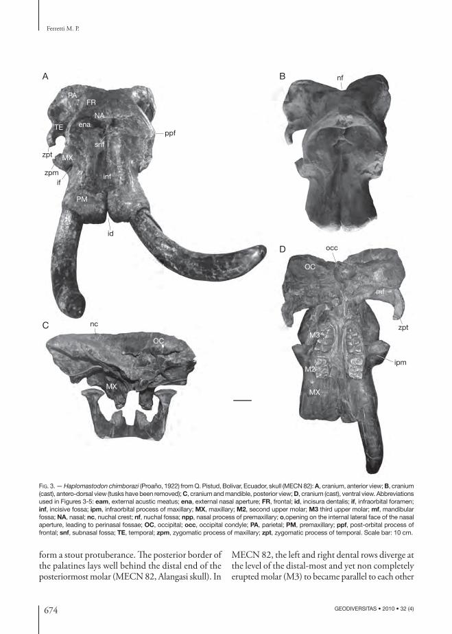
form a stout protuberance. Th e posterior border of the palatines lays well behind the distal end of the posteriormost molar (MECN 82, Alangasi skull). In
MECN 82, the left and right dental rows diverge at the level of the distal-most and yet non completely erupted molar (M3) to became parallel to each other
A
C
D
nc
snf
ppf
zpt
zpt
zpmif
id
PAFR
MX
NA
TE
MX
mf
M2
M3
inf
PM
OC
OC
B nf
ipm
occ
MX
ena
FIG. 3. — Haplo mastodon chimborazi (Proaño, 1922) from Q. Pistud, Bolivar, Ecuador, skull (MECN 82): A, cranium, anterior view; B, cranium (cast), antero-dorsal view (tusks have been removed); C, cranium and mandible, posterior view; D, cranium (cast), ventral view. Abbreviations used in Figures 3-5: eam, external acustic meatus; ena, external nasal aperture; FR, frontal; id, incisura dentalis; if, infraorbital foramen; inf, incisive fossa; ipm, infraorbital process of maxillary; MX, maxillary; M2, second upper molar; M3 third upper molar; mf, mandibular fossa; NA, nasal; nc, nuchal crest; nf, nuchal fossa; npp, nasal process of premaxillary; o,opening on the internal lateral face of the nasal aperture, leading to perinasal fossae; OC, occipital; occ, occipital condyle; PA, parietal; PM, premaxillary; ppf, post-orbital process of frontal; snf, subnasal fossa; TE, temporal; zpm, zygomatic process of maxillary; zpt, zygomatic process of temporal. Scale bar: 10 cm.

675
Anatomy and phylogeny of Haplomastodon (Mammalia, Proboscidea)
GEODIVERSITAS • 2010 • 32 (4)
at the level of the molars in use (M2). Medial to the mesial-most molar (M2) originates, on both side, the interalveolar crest. Th e two interalveolar crests diverge anteriorly so that their anterior ends are well separated from one another.
Discussion. Compared to Gomphotherium angustidens and G. productum, H. chimborazi is more derived in possessing larger and more robust premaxillar-ies, whose dorsal face is slightly upwardly concave, a wider and more rounded forehead, and a more pronounced pneumatization of the bones forming the dorsal, lateral, and posterior walls of the brain-case, which produces a moderate lateral swelling of the parieto-occipital bulges. Th ese characters are also present in Rhynchotherium cf. falconeri (LVNHM 871; Ferretti 2008: fi g. 3), as well as in the other American brevirostrine gomphotheres considered (i.e. Stegomastodon texanus, C. hyodon, and “S.” platensis). Sinomastodon hanjiangensis, the only species of this Old World brevirostrine gomphothere genus whose cranial morphology is suffi ciently well-known (Zong et al. 1989), possesses a similar derived morphology of the neurocranium, while retaining a primitive premaxillary morphology. Haplo mastodon chimborazi and the other brevirostrine gomphotheres consid-ered are derived with respect to G. angustidens and G. productum in having the anterior margin of the orbit laying just at the front of the mesial end of the tooth row. Th e more forward position of the orbit in the brevirostrine forms with respect to the condi-tion in G. angustidens and other longirostrine gom-photheres (e.g., Eubelodon morilli, R. cf. falconeri), is correlated to a modifi cation of the skull toward a more “elephant-like” morphology, characterized by a relatively higher, vertically tilted, and more for-aft compressed cranium than that of primitive gomphotheres. Such derived skull shape is likely linked to the development of a large proboscis, and evolved in parallel in other elephantoid lineages, such as Elephantidae Gray, 1821 and Stegodonti-dae Hopwood, 1935. Another derived character of the skull of H. chimborazi with respect to primitive gomphotheriids, is the absence of a supraorbital foramen. Th is last character is shared, among Ameri-can gomphotheres, with R. cf. falconeri, C. hyodon (Tarija sample), and “S.” platensis (MLP 8-1, MLP
8-3). It is noteworthy that Stegomastodon texanus (AMNH 10622) retains a well-defi ned supraorbital foramen (Osborn 1936). On the basis of the fi g-ures provided by Zong et al. (1989), Sinomastodon hanjiangensis seems also to possess a supraorbital foramen. Haplo mastodon chimborazi shares with “S.” platensis similarly large and robust premaxil-laries, but in the latter (skulls MLP 8-1 and NHM M-19951) these are relatively shorter and distally wider than in Haplo mastodon. In these characters, “S.” platensis appears morphologically intermediate between C. hyodon, that possesses extremely fl aring
FIG. 4. — Anterior view of skull, showing anatomical details of the anterior nasal aperture: A, Haplo mastodon chimborazi (Proaño, 1922) from Q. Pistud, Bolivar, Ecuador (MECN 82); B, Elephas maximus Linnaeus, 1758 (Recent; MCZR). Abbreviations: see Figure 3. Scale bar: 10 cm.
A
B
ena
PA
PM
o
FR
FR
NA
NA
ena
snf
PA
PM
MX
npp

676 GEODIVERSITAS • 2010 • 32 (4)
Ferretti M. P.
premaxillaries, distally divided by a deep and wide incisura, and H. chimborazi. Th e confi guration of the external nasal aperture of H. chimborazi, with the occurrence of a deep sub-nasal fossa, diff ers from the condition seen in Mammut americanum, G. angustidens, G. productum, Anancus Aymard, 1855 and elephantines, whereas it is shared by R. cf. falconeri, C. hyodon, and “S.” platensis (MLP 8-1, MLP 8-3, NHM M-19951). Th e wide exter-nal nasal aperture, the evidence for a strong trunk musculature, and the large infraorbital foramen indi-cate that H. chimborazi possessed a well-developed, elephant-like proboscis.
Mandible (Figs 3C; 5A; 6; Table 3)Th e mandibular corpus is relatively long and its labial side is moderately infl ated. Th e corpus be-comes deeper anteriorly at the level of the posterior
mental foramen. Th e symphyseal portion is short (“brevirostrine”), massive, with no tusk or vestigial tusk alveoli (Fig. 6A-C). In MECN 82, on the labial side, there are three mental foramina: the most posterior one is the largest and is positioned at the level of mesial root of the m2 (Fig. 6D). Th e ascending ramus is slightly posteriorly inclined. Its anterior and posterior borders are parallel to one another. Th e posterior margin of the ramus is straight. It continues dorsally into the condylar process. Th is is dorso-caudally directed and bears a large and transversely elongated condyle. In an-terior view, the condyle is very slightly medially inclined (Fig. 6B). Th e coronoid process is sig-nifi cantly lower than the condyle. On the lateral side of the ramus, there is a deep masseteric fossa, dorsally positioned, just below the sigmoid inci-sure (Fig. 6D). Th e posteroventral margin of the
TABLE 3. — Haplo mastodon chimborazi (Proaño, 1922) from Bolivar, Ecuador. Measurements (in mm) of the mandible (see Appendix 1).
Specimen/site
MeasuresMECN 82Q. Pistud
MECN 437Q. Pistud
MECN 147Q. Quesaca
1. Length: most aboral margin of condyle-infradentale 765 – –2. Length: gonion caudale-infradentale c. 740 – –
3. L ength: infradentale-most oral point of the anterior marginof the ascending ramus
523 – –
4. Length: infradentale-anterior origin of ascending ramus 450 – –5. Length: infradentale-oral border of m2 210 175 –6. Length: gonion caudale-oral border of m2 590 – – 7. Length: oral border of m2-anterior origin
of ascending ramus270 270 –
8. L ength: gonion caudale-most oral point of the anterior margin of the ascending ramus
239 – –
9. Horizontal antero-posterior diameter of symphysis projection in sagittal plane
169 – –
10. Oral height of ascending ramus: gonion ventrale-corion c. 316 – – 11. Aboral height of ascending ramus: gonion ventrale
-heighest point of condyle 473 – –
12. Height of the mandible body at midpoint of the cheektooth row
154 155 135
13. Greatest thickness of the mandible body at midpoint of cheektooth row
110 125 139
14. Maximum breadth between interalveolar crests 105 – –15. Breadth between the most lingual points
of the trigoni retro-molari 235 – –
16. Breadth between anterior margin of ascending rami 430 – –17. Breadth between most lateral points of the condyles 570 – –18. Mandibular breadth: gonion laterale-gonion laterale 478 – –19. Transverse diameter of condyle 141 – –20. Antero-posterior diameter of condyle 54 – –

677
Anatomy and phylogeny of Haplomastodon (Mammalia, Proboscidea)
GEODIVERSITAS • 2010 • 32 (4)
fossa makes a marked step with the lateral surface of the ascending ramus. Anteriorly, the fossa be-comes gradually shallower and the anterior margin is poorly defi ned.
At the very base of the anterior margin of the ramus, posterior to the linea obliqua, is a small
and not well-delimited depressed surface (Fig. 6D). Th is structure is here named the lateral coronoid fossa (LCF). Th e LCF could represent the ven-tral-most point of insertion of the m. temporalis on the lateral side of the coronoid process (see Laub 1996).
FIG. 5. — Haplo mastodon chimborazi (Proaño, 1922) from Q. Pistud, Bolivar, Ecuador, skull (MECN 82); A, cranium and mandible in right lateral view; B, cranium in left lateral view; C-E, right molar in medial, lateral, and anterior views. Abbreviations: see Figure 3. Scale bars: 10 cm.
B
A
C
PA FR
PM
MX
ppf
eam
ifTE
D E

678 GEODIVERSITAS • 2010 • 32 (4)
Ferretti M. P.
Th e mandibular foramen is small and dorsally positioned on the medial side of the ramus, about 5 cm from the posterior border of the ramus. Th e ventral border of the foramen is V-shaped (Fig. 6E). Th e anterior border bears a small tuberosity, here interpreted as homologous to the lingula (linguoid process) described in elephants (Beden 1979) and mammutids (Laub 1996). Behind the mandibular foramen, at the posterior margin of the ascending ramus, below the condyle, there is a small depres-sion probably for the m. pterygoideus externus. Still, on the medial side of both rami, near to the trigonus retromolares, is a well-developed coronoid foramen (Fig. 6E).
Discussion. Th e mandible of H. chimborazi closely resembles those of C. hyodon and “S.” platensis. Among the C. hyodon sample examined there are specimens (e.g., MUT J1, MUT J2) with relatively long and downward defl ected symphyses. Th is primi-tive morphotype is unknown in H. chimborazi and “S.” platensis. Several juvenile mandibles of C. hyodon from Tarija, Bolivia (MUT) present small alveoli for the deciduous lower tusks (Hoff stetter 1952; Ferretti 2008b). In one specimen (MUT-J69), a left deciduous tusk was indeed found in situ (Fer-retti pers. obs). Both the tusk and the alveolus have an oval cross-section. No traces of lower incisors were found in the mandibles of any of the other brevirostrine gomphotheres considered. Stegomas-todon texanus (AMNH 10622) diff ers from the SA gomphotheres here examined in possessing a lower and more backwardly oriented ascending ramus and the posterior opening of the mandibular canal (mandibular foramen) placed more ventrally on the medial side of the ascending ramus. Absence of a lateral coronoid fossa as described in H. chimborazi, in M. americanum, G. angustidens, G. productum, R. cf. falconeri, S. texanus, Anancus arvernensis, and elephants and its presence in “S.” platensis and C. hyodon suggests this is a derived feature of SA gomphotheres. Th e mandibles of Sinomastodon in-termedius and S. hanjiangensis are morphologically very similar to those of South American gompho-theres, but apparently lack a coronoid fossa (Teilhard de Chardin & Trassaert 1937; Tobien et al. 1986; Zong et al. 1989).
DentitionUpper incisor (tusk; Figs 3A; 5A, B). Adults of H. chimborazi posses massive and relatively short upper tusks, oval in cross-section. Th e longitudinal axis of the tusk is distinctly upwardly curved and with no trace of torsion. Th e curvature becomes more evident as the tusk increases its length dur-ing growth, so that adult or, more in general, larger tusks are more curved than smaller juvenile tusks. In MECN 82, the left tusk has a maximum diameter of 115 mm and the length of the extralveolar part on the outer side is of 880 mm.
All known adult tusks of H. chimborazi lack enamel. Contrary to what was reported by Fic-carelli et al. (1995), the juvenile tusk MECN 258 from Bolivar also has no trace of enamel. However, a juvenile fragmentary skull with the DP4 in use from the Late Pleistocene of Quebrada Los Mila-gros near Llano Chico (EPN V-1980; Hoff stetter 1952), bears a tusk with a distinct lateral enamel band. Th e enamel band is very thin and straight. Th e tusk is rectilinear, with a fl attened sub-circular cross section and no torsion, that would exclude it from C. hyodon.
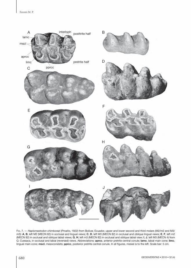
Cheek teeth (Figs 7; 8; Table 4). Cheek tooth categories represented at the Bolivar sites are DP4-M3 and m1-m3. No permanent premolars (P3-P4-p3-p4) are present in the Bolivar sample nor among the La Carolina and Punin samples stud-ied by Hoff stetter (1952). All intermediate cheek teeth (DP4-M2 and m1-m2) are trilophodont. M3s posses four lophs and a small distal talon (Fig. 7C, D, I, J). Th e fourth loph is sensibly nar-rower and with a more simple structure than the preceding ones. Th e lower m3s have four to fi ve lophids and a distal talonid. Th ey possess well-developed pretrite central conules. Th e emerging wear fi gure is a typical trefoil pattern. In all molar categories, posttrite central conules could be either absent (morphotype a) or moderately developed (morphotype b). In the latter morphotype a poorly defi ned secondary trefoil pattern emerges in ad-vanced stages of wear. Cement is absent or fi lling the very base of the interloph(id)s. Enamel is even and never wrinkled as in “S.” platensis. Two com-plete m3s from Bolivar (MECN 133, MECN 438;

679
Anatomy and phylogeny of Haplomastodon (Mammalia, Proboscidea)
GEODIVERSITAS • 2010 • 32 (4)
Fig. 8F, H) preserve their roots. Th e root of the second lophid is divided into two branches, with the mesial one joining the main anterior root and
the posterior one coalescing with the posterior root system, formed by the fusion of the roots of the third to fi fth lophids.
FIG. 6. — Haplo mastodon chimborazi (Proaño, 1922) from Q. Pistud, Bolivar, Ecuador, mandible (MECN 82): A, B, D, mandible in oc-clusal, anterior (cast), and left lateral view; C, detail of the ventral aspect of the symphysis (anterior to the bottom); E, medial view of the left ascending ramus. Abbreviations: cf, coronoid foramen; lcf, lateral coronoid fossa; ling, linguoid process; m2, second lower molar; m3, third lower molar; maf, masseteric fossa; mf, mandibular foramen; pmf, posterior mental foramen. Scale bar: 10 cm.
m2
pmf
lcf
maf
m3
A
C
D
E
B
ling
mfcf
680 GEODIVERSITAS • 2010 • 32 (4)
Ferretti M. P.
A
G
I
B
C
E
D
F
H
J
interloph
lamc
mscl
aprcc
limc
pprcc
pretrite half
posttrite half
FIG. 7. — Haplo mastodon chimborazi (Proaño, 1922) from Bolivar, Ecuador, upper and lower second and third molars (M2/m2 and M3/m3): A, B, left M2 (MECN 82) in occlusal and lingual views; C, D, left M3 (MECN 82) in occlusal and oblique lingual views; E, F, left m2 (MECN 82) in occlusal and oblique labial views; G, H, left m3 (MECN 82) in occlusal and oblique labial view; I, J, left M3 (MECN 4) from Q. Cuesaca, in occlusal and labial (reversed) views. Abbreviations: aprcc, anterior pretrite central conule; lamc, labial main cone; limc, lingual main cone; mscl, mesoconelets; pprcc, posterior pretrite central conule. In all fi gures, mesial is to the left. Scale bar: 5 cm.

681
Anatomy and phylogeny of Haplomastodon (Mammalia, Proboscidea)
GEODIVERSITAS • 2010 • 32 (4)
mar
A C
DB
FE
I
J
G
H
FIG. 8. — Haplo mastodon chimborazi (Proaño, 1922) from Bolivar, Ecuador, mandibles and lower molars: A, B, left mandibular body with m3 (MECN 437; Q. Pistud) in lateral and occlusal views; C, D, incomplete right half mandible with m3 (MECN 147; Q. Cuesaca), in lateral (reversed) and occlusal view; E, F, right m3 (MECN 189; Q. Pistud), in occlusal and lingual views; G, H, right m3 (MECN 438; Q. Pistud), in occlusal and lingual views; I, J, left m3 (MECN 272; Q. Cuesaca), in occlusal and lingual (reversed) views. Abbreviation: mar, main anterior root. In all fi gures, mesial is to the left. Scale bar: 5 cm.

682 GEODIVERSITAS • 2010 • 32 (4)
Ferretti M. P.
Th ough no DP3s are present in the Bolivar dental sample, information on the anatomy of this tooth is given by Hoff stetter (1952) who describes, without fi guring them, two isolated H. chimborazi DP3s from the Late Pleistocene of Alangasi and Calderon, in the surrounding of Quito. Th e specimen from Alangasi (EPN V1244) is formed by three lophs and a small distal talon. Its size (length 56 mm; width 43 mm) is comparable to that the DP3 of C. hyodon from Tarija, where complete upper molar series are known (Boule & Th evenin 1920). Specimen EPN V1244 is, on the other hand, signifi cantly smaller than the DP4s of C. hyodon and also than that from Q. Pistud, described above. In EPN V1244 only pretrite trefoils are present. Between the fi rst and second lophs, at the labial end of the interlpohid, is a small cone (“bouton”). Th e enamel sectioned on the occlusal surface is rather wrinkled.
Th e second DP3 from Calderon (EPN V1231) is very similar to that from Alangasi both in size (57.3 mm in length; 47.5 mm in width) and mor-phology.
Discussion. Th e tusks of H. chimborazi diff er from those of primitive gomphotheres (e.g., G. angusti-dens) in being more robust, not downwardly curved and with no enamel band in adults. With respect to “S.” platensis and C. hyodon, the tusks of H. chim-borazi are relatively shorter. Th e type specimen of “S.” platensis (MLP-8-1) possesses long and nearly straight tusks, a morphotype absent in Ecuadorian H. chimborazi. No adult tusks from Ecuador show traces of an enamel cover. Also, in the Aguas do Araxá sample described by Simpson & Paula Couto (1957), all tusks are without enamel. On the other hand, this occurs in some “S.” platensis isolated tusks from Argentina (MLP; material previously referred to a distinct taxon, Notiomastodon orna-tus Cabrera, 1929) belonging to both juvenile and adult individuals (Ferretti pers. obs.). Th erefore, even though the occurrence of an enamel band is a variable character in both H. chimborazi and “S.” platensis, the latter has a tendency to retain an enamel band in later stages of growth.
Th e morphology of H. chimborazi molars is gen-erally conservative (as far as South American gom-photheres is concerned), very similar to the condition
in C. hyodon, but with morphotypes that approach the more derived complex structure of “S.” platen-sis. Haplo mastodon chimborazi is more progressive than species of Gomphotherium Burmeister, 1837 and Rhynchotherium Falconer, 1868 in the greater development of the accessory conules and in pos-sessing well-developed tetra loph and pentalophid on M3 and m3 respectively. Th e most primitive NA bona-fi de species of Stegomastodon, S. primitivus Osborn, 1936 from the Late Hemphillian to Early Blancan (Latest Miocene-Early Pliocene) of North America, is already more derived than H. chimbo-razi in possessing a more developed pentaloph on the M3 (Osborn 1936). Ecuadorian H. chimborazi possesses DP3 with three lophs and a distal talon. Th is character is also present in C. hyodon and “S.” platensis. As evidenced by Tassy (1990) and Shoshani (1996), the full development of a third loph in the DP3/dp3 could represent a distinct feature of SA gomphotheres, and a convergence with tetralophodont gomphotheres (Tetralophodon Falconer, 1857, Anancus, Paratetralophodon Tassy, 1983; Tassy 1985, 1990). Primitive trilophodon gomphotheres, like G. angustidens and G. produc-tum, posses DP3/dp3s formed by two loph(id)s and a distal talon. Savage (1955) described a dp3 (UCMP 44749) of Stegomastodon mirifi cus from Cita Canyon, Texas, as consisting of three lophids, though the third one is much lower and structur-ally less complex than the anterior ones. A similar incipient development of the third loph(id) of the third deciduous premolars is observed in the juve-nile material of Rhynchotherium edensis from Mt Eden, California, described by Frick (1926) and Osborn (1936).
Available evidence from Ecuador and Brazil (Simp-son & Paula Couto 1957) indicates that in Haplo-mastodon deciduous premolars are not replaced by permanent premolars. Th is derived feature is also known in C. hyodon (Boule & Th evenin 1920), in “S.” platensis (Cabrera 1929; Ferretti pers. obs.) and in NA Stegomastodon (Shoshani 1996). In contrast, P3-P4 are present in more primitive gomphother-iids from NA and Europe, like G. productum and G. angustidens (Frick 1926; Tassy 1985, 1990). Evi-dence from a juvenile skull and mandible (AMNH 18218, 18216a, 18216b) of Rhynchotherium edensis
683
Anatomy and phylogeny of Haplomastodon (Mammalia, Proboscidea)
GEODIVERSITAS • 2010 • 32 (4)
from Mt. Eden, with the DP2-DP3/dp2-dp3 in use and the M1/m1 already formed, indicates that this taxon also lacks permanent premolars (Frick 1926; Osborn 1936). Loss of permanent premolars and the acquisition of a “horizontal tooth succes-sion” is a derived trait among proboscideans, that evolved independently in several elephantoid lin-eages (e.g., mammutids, Anancus, and elephants; Tassy 1990). Shoshani (1996) regarded this trait as a possible synapomorphy of SA gomphotheres and NA Stegomastodon.
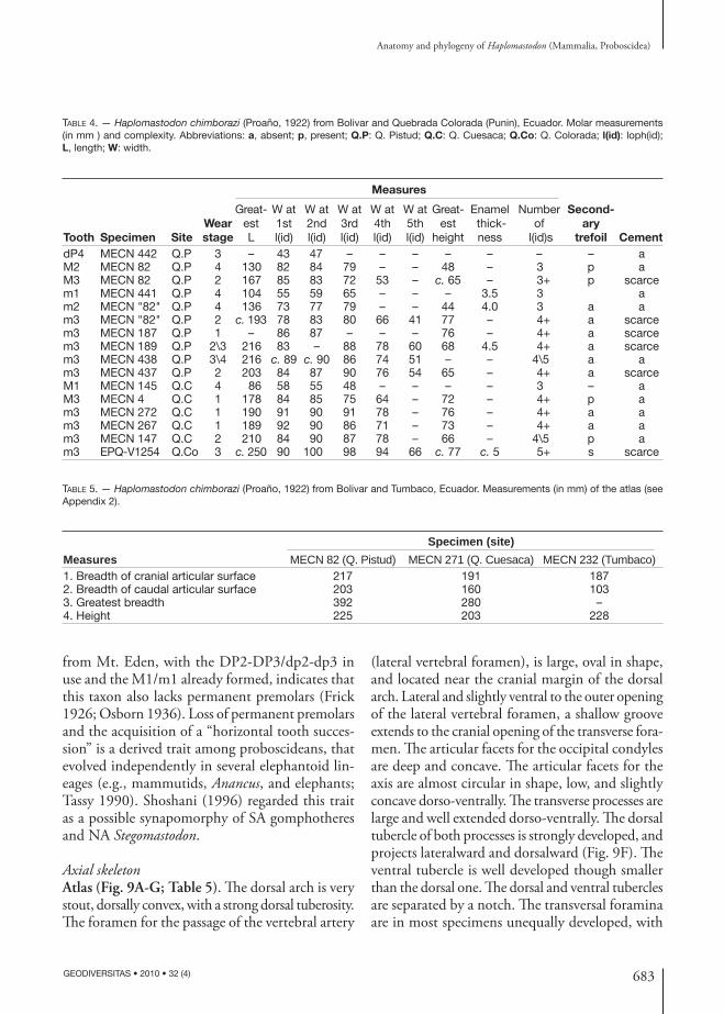
Axial skeletonAtlas (Fig. 9A-G; Table 5). Th e dorsal arch is very stout, dorsally convex, with a strong dorsal tuberosity. Th e foramen for the passage of the vertebral artery
(lateral vertebral foramen), is large, oval in shape, and located near the cranial margin of the dorsal arch. Lateral and slightly ventral to the outer opening of the lateral vertebral foramen, a shallow groove extends to the cranial opening of the transverse fora-men. Th e articular facets for the occipital condyles are deep and concave. Th e articular facets for the axis are almost circular in shape, low, and slightly concave dorso-ventrally. Th e transverse processes are large and well extended dorso-ventrally. Th e dorsal tubercle of both processes is strongly developed, and projects lateralward and dorsalward (Fig. 9F). Th e ventral tubercle is well developed though smaller than the dorsal one. Th e dorsal and ventral tubercles are separated by a notch. Th e transversal foramina are in most specimens unequally developed, with
TABLE 4. — Haplo mastodon chimborazi (Proaño, 1922) from Bolivar and Quebrada Colorada (Punin), Ecuador. Molar measurements (in mm ) and complexity. Abbreviations: a, absent; p, present; Q.P: Q. Pistud; Q.C: Q. Cuesaca; Q.Co: Q. Colorada; l(id): loph(id); L, length; W: width.
Measures
Tooth Specimen SiteWear stage
Great-est L
W at 1st l(id)
W at 2nd l(id)
W at 3rd l(id)
W at 4th l(id)
W at 5th l(id)
Great-est
height
Enamel thick-ness
Number of
l(id)s
Second-ary
trefoil CementdP4 MECN 442 Q.P 3 – 43 47 – – – – – – – aM2 MECN 82 Q.P 4 130 82 84 79 – – 48 – 3 p aM3 MECN 82 Q.P 2 167 85 83 72 53 – c. 65 – 3+ p scarcem1 MECN 441 Q.P 4 104 55 59 65 – – – 3.5 3 am2 MECN "82" Q.P 4 136 73 77 79 – – 44 4.0 3 a am3 MECN "82" Q.P 2 c. 193 78 83 80 66 41 77 – 4+ a scarcem3 MECN 187 Q.P 1 – 86 87 – – – 76 – 4+ a scarcem3 MECN 189 Q.P 2\3 216 83 – 88 78 60 68 4.5 4+ a scarcem3 MECN 438 Q.P 3\4 216 c. 89 c. 90 86 74 51 – – 4\5 a am3 MECN 437 Q.P 2 203 84 87 90 76 54 65 – 4+ a scarceM1 MECN 145 Q.C 4 86 58 55 48 – – – – 3 – aM3 MECN 4 Q.C 1 178 84 85 75 64 – 72 – 4+ p am3 MECN 272 Q.C 1 190 91 90 91 78 – 76 – 4+ a am3 MECN 267 Q.C 1 189 92 90 86 71 – 73 – 4+ a am3 MECN 147 Q.C 2 210 84 90 87 78 – 66 – 4\5 p am3 EPQ-V1254 Q.Co 3 c. 250 90 100 98 94 66 c. 77 c. 5 5+ s scarce
TABLE 5. — Haplo mastodon chimborazi (Proaño, 1922) from Bolivar and Tumbaco, Ecuador. Measurements (in mm) of the atlas (see Appendix 2).
Specimen (site)Measures MECN 82 (Q. Pistud) MECN 271 (Q. Cuesaca) MECN 232 (Tumbaco)1. Breadth of cranial articular surface 217 191 1872. Breadth of caudal articular surface 203 160 1033. Greatest breadth 392 280 –4. Height 225 203 228

684 GEODIVERSITAS • 2010 • 32 (4)
Ferretti M. P.
one foramen markedly larger than the opposite one (e.g., MECN 82; Fig. 9F). Noticeably, in some specimens, as fi rst noted by Hoff stetter (1952), one or both foramina are completely obliterated (e.g., EPN V2010; Fig. 9C, D). A similar variability in size and presence/absence of transverse foramina of the atlas has been observed in the C. hyodon and “S.” platensis samples analyzed for this study. On the other hand, none of the other proboscideans examined show this feature.
Cervical vertebrae 2 to 7 (Figs 9H-K; 10A-H; Tables 6; 7). Five other cervical vertebrae of the Bolivar skeleton are preserved, identifi ed as C2, C3, C5, C6, and C7 respectively. Th e axis possesses a very high dorsal arch (Fig. 9H) Th e spinous process is transversally enlarged and terminates with two dorsal tuberosities, separated by a central depres-sion. On the caudal aspect of the process runs a narrow crest, and, lateral to this, on both sides, two intensely wrinkled grooves are present. Th e pedicles are long and robust. Th e vertebral body has a ventral crest that terminates caudally in a small tuberosity. Lateral to this crest, the ventral face of the body is cranio-caudally concave and transversally convex. Cranially, the two wide articular surfaces for the atlas are situated lateral to the stout odontoid proc-ess. Th e transverse processes are short and weak and are pierced by an oval-shaped transverse foramen. Th e process ends laterally with a tuberosity. Medial and ventral to the transverse process there is a small bony spine. A second, nearly complete axis from Punin (EPN V 3744) shows no traces of transverse foramina. On the other hand, two axes from La
Carolina (EPN 1265, EPN 1287) described by Hoff stetter (1952) possess well-developed trans-verse foramina.
Th e body of C3 has a sub-circular shape in cranial view (Fig. 10A, B). On the ventral side runs a thin median crest. Th e laminae are thick. Th e spinous process is broken just above its base. It seems, how-ever, to have been slender and not caudally bent. Th e vertebral foramen is triangular-shaped with a rounded vertex. Th e caudal vertebral incision con-sists, on both sides, in a groove running from the vertebral foramen to the transverse foramen. Th e transverse process is slender, and its dorsal root is ventrally sloping. Th e dorsal tubercle of the transverse process is caudally directed. Th e ventral tubercle forms a thin process, cranially directed.
Th e morphology of C5 is very similar to that of C3 (Fig. 10C, D). Th e transversal foramina are however larger than those of C3, while the ventral tubercle of the transverse process is smaller.
Th e body of C6 of MECN 82 is crushed dorsally (Fig. 10E, F). Th e vertebral foramen is wide. Th e spinous process is slender. Th e transverse processes are more robust relative to those of the previous vertebrae. Ventrally and caudally there is a robust process whose extremity forms a large tubercle (tuberculum caroticus) cranially directed.
In C7 the spinous process is incomplete; however, it was evidently larger than in the previous cervical vertebrae. Th e ventral portion of the body and the transverse processes are crushed and partly broken off . Th e facet for the articulation with the fi rst left rib is partly preserved on the caudal face of the body. Th e transverse processes are dorso-ventrally expanded, laterally fl attened and without trans-versal foramina.
Th oracic vertebrae (Fig. 10I-T; Table 7). Eleven vertebrae of the thoracic segment of the backbone of MECN 82 are preserved. Th e approximate posi-tion of each vertebra along the vertebral column was assessed by comparison with associated vertebrae of E. maximus and C. hyodon from Tarija. Th e cranial-most preserved thoracic vertebra is identifi ed as a T2 or T3 (thereafter T2/T3) based on the size of the spinous process and the fact that it seems not to articulate with C7. Th e body is heart-shaped and
TABLE 6. — Haplo mastodon chimborazi (Proaño, 1922) from Bolivar, Ecuador. Measurements (in mm) of the axis (see Appendix 2).
MeasuresSpecimenMECN 82
1. Greatest breadth 2512. Breadth of cranial articular surface 2083. Breadth across the postzygapophyses 1544. Breadth of caudal articular surface 1545. Height of caudal articular surface 1346. Greatest height 2967. Height of the dorsal process 1068. Greatest length 125

685
Anatomy and phylogeny of Haplomastodon (Mammalia, Proboscidea)
GEODIVERSITAS • 2010 • 32 (4)
its lateral sides are concave (Fig. 10I, J). On both sides, ventral to the base of the transverse process, there are two demi-facets, which articulate, with the heads of the second and third ribs. Th e incom-
plete spinous process is large, caudally bent, and originally terminated into an apical tubercle. Th e cranial margin of the spinous process is sharp. Th e caudal one, on the contrary, is characterized by a
tfdt
A
B
C
D
E
F
H
I
J
K
G
vt
ovf
FIG. 9. — Haplo mastodon chimborazi (Proaño, 1922), from Ecuador (various localities), fi rst (atlas) and second (axis) cervical vertebrae: A, B, atlas (MECN 271; Q.Cuesaca, Bolivar) in cranial and caudal views; C, D, atlas (EPN V2010; La Carolina, S. Elena Peninsula), in cranial and caudal views; E-G, atlas (MECN 82; Q. Pistud, Bolivar), in cranial, caudal, and left lateral views; H-K, axis (MECN 82), in cranial, caudal, ventral and left lateral views. Abbreviations: dt, dorsal tubercle of transverse process; ovf, outer opening of lateral vertebral foramen; tf, transversal foramen; vt, ventral tubercle of transverse process. Scale bar: 5 cm.

686 GEODIVERSITAS • 2010 • 32 (4)
Ferretti M. P.
the preceding vertebrae. Th e cranial articular facet for the head of the fourth/fi fth rib is concave and dorso-ventrally elongated. Th e caudal one is wide, concave, semicircular-shaped and positioned lateral to the corpus.
T6/T7 has the same general characteristics as T4/T5. It is missing its spinous process (Fig. 10O, P). Th e transverse processes are more dorsally posi-tioned than in the preceding vertebrae. Th e caudal facet for the sixth/seventh rib is smaller than that of T4/T5.
Also in T7/T8 the spinous process is broken (Fig. 10Q, R). Th is vertebra is very similar in shape to the previous one. Th e transverse processes are in-complete, laterally and slightly cranially directed.
Th e shape of T9/T10 is similar to that of the preceding one (Fig. 10S, T). Only the proximal half of the spinous process is preserved. Th e groove occurring on the caudal side of the process is very deep, and the spine has a V-shaped cross section. Th e lateral processes are broken.
T11/T12 has a heart-shaped, and dorso-ventrally elongated body. Th e costal facets are close to each other and positioned dorsally on the body. Th eir dorsal margin is situated at about half of the ver-
feeble median groove. Th e vertebral foramen is triangle-shaped. Th e very thick transverse processes end in a robust lateral tubercle, on the cranial side of which is present a small, dorsally oriented spine. Th e cranial articular processes (prezygapophyses) are well separated from each other.
In T3/T4 the body is longer than that of the preceding vertebra (Fig. 10K, L). Th e vertebral fo-ramen is narrow, due to the position of the cranial articular processes. Th e spinous process is similar to that of T2/T3. Th e transverse processes are stout and laterally directed. Th e articular facet for the tubercle of the fourth rib is not evident. Also the cranial articular facet for the head of the rib is not well defi ned; it seems however to be positioned on the cranio-ventral margin of the transverse process. Th e caudal one is positioned laterally with respect to the body, near the dorsal margin.
Th e corpus of T4/T5 is heart-shaped (Fig. 10M, N). Th e spinous process is strongly defl ected caudally, probably as a consequence of taphonomic distor-tion. Th e cranial margin of the process is fl attened, while on the caudal one runs a very deep groove. Th e transverse processes are more slender and the lateral tubercle is less pronounced with respect to
TABLE 7. — Haplo mastodon chimborazi (Proaño, 1922) from Bolivar, Ecuador. Measurements (in mm) of cervical (C3-C7), thoracic, and lumbar vertebrae (see Appendix 2).
Specimen MECN 82
Measures1. Greatest
breadth2. Breadth of cranial
articular surface3. Breadth of caudal
articular surface4. Greatest
height5. Height of neural spine
6. Greatest length of corpus
C3 250 140 149 – – 61C5 256 150 150 – – –C6 c. 275 133 156 > 304 > 76 c. 42C7 117 165 c. 179 > 100 – c. 62T2 380 149 c. 186 > 450 > 270 c. 63T3 or T4 339 161 209 > 455 330 c. 70T4 or T5 – 147 205 > 270 > 200 68T6 or T7 306 136 208 – – c. 61T7 or T8 – 136 195 – – c. 68T9 or T10 – 125 c. 176 > 260 > 207 c. 75T11 or T12 – 128 186 > 290 > 280 70T12 or T13 290 126 – > 270 > 280 66T13 or T14 292 117 160 310 207 73T16–lastT – 119 c. 143 – – 77T16–lastT – 132 c. 135 265 172 71L1 – 137 132 – – 72L3 – 139 155 – – 82L4 310 162 155 – > 109 89
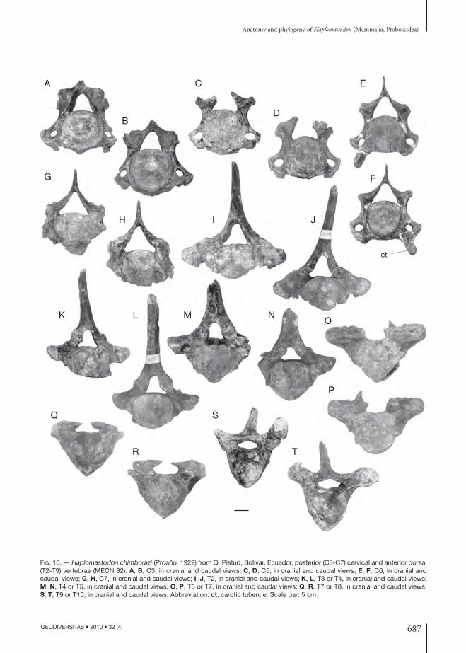
687
Anatomy and phylogeny of Haplomastodon (Mammalia, Proboscidea)
GEODIVERSITAS • 2010 • 32 (4)
K L M
Q S
T
J
E
FG
H
O
R
C
DB
I
N
A
ct
P
FIG. 10. — Haplo mastodon chimborazi (Proaño, 1922) from Q. Pistud, Bolivar, Ecuador, posterior (C3-C7) cervical and anterior dorsal (T2-T9) vertebrae (MECN 82): A, B, C3, in cranial and caudal views; C, D, C5, in cranial and caudal views; E, F, C6, in cranial and caudal views; G, H, C7, in cranial and caudal views; I, J, T2, in cranial and caudal views; K, L, T3 or T4, in cranial and caudal views; M, N, T4 or T5, in cranial and caudal views; O, P, T6 or T7, in cranial and caudal views; Q, R, T7 or T8, in cranial and caudal views; S, T, T9 or T10, in cranial and caudal views. Abbreviation: ct, carotic tubercle. Scale bar: 5 cm.

688 GEODIVERSITAS • 2010 • 32 (4)
Ferretti M. P.
tebral foramen. Th e vertebral foramen has an oval outline. Th e spinous process is rather long and possesses a very sharp cranial margin. Th e lateral processes are broken.
T12/T13 is very similar in shape to the preceding vertebra. Th e spinous process is slender, long, and caudally inclined. A narrow notch with a rounded fl oor is found in a median position on the cranial margin of the dorsal arch. Th e transverse processes are laterally and dorsally directed. Th e costal ar-ticular facets are close to one another. Th e cranial facet is particularly deep and concave.
In T14/T15, the cranial and caudal costal ar-ticular facets are small, and well separated from one another. Th e spinous process is short and not so much inclined caudally. Th e cranial notch on the dorsal arch is deep. Th e transverse processes are dorso-laterally directed. Th ey have a dorso-ventrally fl attened base and each ends in a clubbed extremity. A stout crest, possibly homologous to the accessory tubercle of the lumbar vertebrae, is present on caudal margin of the transverse processes.
Two more posterior thoracic vertebrae are present, whose position is between T15 and the last tho-racic vertebra. Th e corpus of the more anterior one is rather long. Th e spinous process is broken at its very base. A deep cranial groove runs on the dorsal arch. Th e right transverse process is broken, whilst the left one is only slightly damaged on its ventral side. Th e left process is short, dorso-ventrally fl attened and horizontal in direction. Dorso-cranially there is an elongated tubercle, which is homologous to the mammillary process of the lumbar vertebrae.
Th e second posterior thoracic is morphologi-cally similar to the preceding one. It is damaged on the right side. It has, on both sides, only one cranial facet for the head of a rib. It bears a short spinous process, caudally inclined. Th e mammil-lary processes are large and positioned laterally to the cranial articular processes. Th e right transverse process is short, fl attened and dorsalward and lat-eralward directed. It articulates with a rudimen-tary rib. Between the latter and the ventral side of the transverse process persists a small fi ssure. Th e caudal articular facet for the rib is absent.
Lumbar vertebrae (Fig. 11A-C, F; Table 7). Th ree lumbar vertebrae are preserved in MECN 82. Th e body of L1 is wide and thick (Fig. 11A). Th e pedicles diverge. Th e spinous process is broken. Th e cranial articular processes are very close to one another, and are separated only by a narrow fi ssure. Th e left transverse process is not preserved. Th e right one is short, thick and latero-caudally directed. Th e mam-millary processes end in two small tubercles. Lateral to each mammillary process arises a bony lamina that reaches the cranio-dorsal margin of transverse processes, bounding a small foramen dorsally.
Th e body of L3 is subcylindrical in shape and dorso-ventrally fl attened (Fig. 11B). Th e dorsal arch is thin. Th e distal part of spinous process is not preserved. It has a wide base. Th e vertebral fo-ramen is wide. Th e transverse processes are broken at their bases. Th e mammillary processes end into two small tubercles cranially directed.
L4 has a markedly dorso-ventrally fl attened body (Fig. 11C). Th e spinous process has a wide base. It is laterally fl attened, short and caudally inclined. Th e cranial articular processes and the mammillary processes, on both sides, are fused together into a unique strong process, cranio-dorsally oriented. Th e transverse processes (costal processes) are thick and are laterally expanded, making up, caudally, a wide articular surface for the ileum (Fig. 11F).
Sacrum (Fig. 11D-F; Table 8). Th e sacrum of MECN 82 consists of the fi rst, second and third sacral vertebra (in adult elephants the sacrum is formed usually by fi ve sacral vertebrae that are fused at diff erent times during the individual life history). Th e width of the vertebrae diminishes in a caudal direction. Th e ventral face of each verte-bra is concave (Fig. 11E). Th e neural arch is low. All three vertebrae bear a spinous process strongly inclined caudally (in the third one it is broken) and two cranial articular processes, dorso-cranially directed (Fig. 11F). Th e vertebral canal is fl attened and it narrows caudally. Th e fi rst sacral vertebra has, lateral to the articular facets, a large mammil-lary process. Th e transverse processes are wide and fl attened, ventrally directed and blended together to form the wrinkled lateral crest. Th e sacral wing is poorly developed. Laterally, the vertebrae are

689
Anatomy and phylogeny of Haplomastodon (Mammalia, Proboscidea)
GEODIVERSITAS • 2010 • 32 (4)
A
E FG
L3
L4
sacrum
H
I
KA-F
J
L
M
N
O
P
Q
R
S
T
U
V
W
B C D
G-L
M-W
FIG. 11. — Haplo mastodon chimborazi (Proaño, 1922) from Q. Pistud, Bolivar, Ecuador, lumbar vertebrae, sacrum and caudal vertebrae (MECN 82): A, L1, cranial view; B, L3, cranial view; C, L4, cranial view; D, E, sacrum, in cranial and ventral view; F, L3-L4 and sacrum in anatomical connection, dorsal view; G-L, caudal vertebrae in dorsal and cranial views; M, N, fi rst left and right costae; O-R, anterior costae; S-U, intermediate costae; V, W, posterior costae. Scale bars: A-L, 5 cm; M-W, 10 cm.

690 GEODIVERSITAS • 2010 • 32 (4)
Ferretti M. P.
separated from each other by a wide intervertebral space, where the supra- and sub-sacral foramina, respectively, are visible.
Caudal vertebrae (Fig. 11G-L). Th ree caudal ver-tebrae are attributable to individual MECN 82. All three vertebrae have an elongated body. Th e two most anterior caudals have cranial and caudal convex articular surfaces. Transverse processes are fl attened, horizontal, and their length diminishes from the anterior-most to the posterior-most one. Th e dorsal arches have a base cranio-distally ex-panded. Cranial and caudal articular processes are button-like. Th e spinous process is extremely reduced and caudally oriented.
Ribs (Fig. 11M-W). Among the collected mate-rial, four ribs surely pertain to individual MECN 82. Most probably also specimens MECN 112, MECN 207, MECN 208, MECN 409, MECN 410, MECN 411, MECN 412, MECN 457 and MECN 470 belong to the same individual. Th e fi rst rib is complete. It is short and slightly curved (Fig. 11M, N). Th e tuberculum is large and sepa-rated by a notch from the head. Th e latter has two facets, respectively for the seventh cervical and the fi rst thoracic vertebrae. Th e tubercula of the con-secutive ribs become gradually smaller and even-tually disappear, while the bodies become longer and more curved.
Discussion. Th e observed asymmetry and variability in the degree of closure of the transverse foramina on the atlas and axis of H. chimborazi, C. hyodon and “S.” platensis could be consistent with a regres-
sive process aff ecting skeletal structures no longer functional. A similar pattern can be observed in C7 of most mammals. In humans, for instance, the transverse foramina of the seventh cervical vertebra rarely give passage to the vertebral artery and vein, and they are generally smaller on one side or even absent (Bell et al. 1950). Th e vertebral artery in humans usually passes in front of the transverse process of C7. A similar condition for the atlas and axis of H. chimborazi (and the other SA gom-photheres) is suggested here. According to this hy-pothesis, in H. chimborazi the vertebral artery ran cranially through the transverse foramina of C6-C3 and then just below the transverse processes of the axis and the atlas (Fig. 9). Th en it turned upward passing laterally to the articular surface for the oc-cipital condyle and entered the vertebral foramen passing through the lateral vertebral foramen. Th e latter is present in all the atlases of H. chimborazi examined, even in those with completely closed transversal foramina. It is diffi cult to assign any adap-tive signifi cance to the lack of transverse foramina on the fi rst cervicals. Rather, it might represent an anatomic variation, quite frequent among SA gomphotheres, while extremely rare (if present at all) in other proboscideans (actually no other case has been described so far). In this case, it could represent the results of the so-called founder eff ect, supporting the hypothesis that all South American taxa derived from a single dispersal event.
Th e preserved vertebrae of MECN 82 indicate that the trunk region of H. chimborazi contained a minimum of 16-17 vertebrae (extant elephants typically possess from 19 to 21 thoracic vertebrae). All thoracic vertebrae of H. chimborazi possess backwardly bent spinous processes. Th e inclina-tion increases in the anterior thoracics (T2 to T5), remains nearly constant from T5 to about T12 and then decreases in the remaining posterior vertebrae. Compared to G. sylvaticum and G. productum, the spinous processes of the anterior thoracics of H. chimborazi are more backwardly bent, suggest-ing the head was kept in a more elevated posture in the latter taxon. In H. chimborazi, the length of the spinous processes increases from (T1) T2 to T5 and rapidly decreases passing from the fi fth thoracic to consecutive vertebrae.
TABLE 8. — Haplo mastodon chimborazi (Proaño, 1922) from Bolivar, Ecuador. Measurements (in mm) of the sacrum (see Appendix 2).
MeasuresSpecimenMECN 82
1. Greatest length c. 2802. Greatest breadth 2903. Breadth of second sacral vertebra 2804. Breadth of thirdh sacral vertebra 2535. Breadth of cranial articular surface 1506. Height of cranial articular surface 92

691
Anatomy and phylogeny of Haplomastodon (Mammalia, Proboscidea)
GEODIVERSITAS • 2010 • 32 (4)
A B
C
D EF G
ec
dt
mt
lt
FIG. 12. — Haplo mastodon chimborazi (Proaño, 1922) from Q. Pistud, Bolivar, Ecuador, anterior limb bones (MECN 82): A-C, right scapula, in lateral, medial, and articular views; D-G, right humerus, in anterior, lateral, posterior, and medial views. Abbreviations: dt, deltoid tuberosity; ec, epicondilar crest; lt, lateral tuberosity; mt, medial tuberosity. Scale bar: 10 cm.

692 GEODIVERSITAS • 2010 • 32 (4)
Ferretti M. P.
On the basis of the preserved thoracic vertebrae of MECN 82, it can therefore be deduced that the dorsal outline originally produced by their spinous
processes (Fig. 20) should have been similar to that observed in complete specimens of M. americanum (see Olsen 1979) and in “S.” platensis (Ferretti 2008a).
Appendicular skeletonScapula (Fig. 12A-C; Table 9). In both scapulae of the Bolivar skeleton, the ossifi ed dorsal cartilage is not fused, and only that of the right scapula was retrieved (Fig. 12A). Th e supraspinous fossa is sub-rectangular and fairly concave. Th e infraspinous fossa, more extended caudally than the former, is sub-triangular. Its cranial portion is concave and the caudal portion is convex. Th e scapular spine is prominent and terminates ventrally into an anterior acromian process (processus hamatus) and a posterior metacromian process (processus suprahamatus), separated by a shallow triangular surface. Th e acromian process is more developed than the metacromian one. Th e medial (costal) face cranially displays a shallow subscapular fossa and is rather convex in the central portion (Fig. 12B). In the dorsal half of the costal surface there is a wide, extremely rough triangular area for the origin the m. serratus ventralis. Caudally, the surface is fl at and is delimited by the caudal angle of the scapula. Th e glenoid cavity is sub-rectangular, with the main axis directed cranio-caudally. Th e cavity is mark-edly concave cranio-caudally, and is less concave transversally. Th e supraglenoid tubercle is situated cranially to the glenoid cavity. Th e coracoid process is poorly developed and forms a short crest along the medial aspect of the glenoid cavity. Th e sup-raglenoid tubercle is not ventrally bent to a great extent (Fig. 12A).
Humerus (Fig. 12D-G; Table 10). In the MECN 82 skeleton the left humerus is fractured at the epicondylar crest, while the right one is complete and undeformed. Th e epiphyses are fused to the shaft. Th e bone appears rather massive. Th e epi-condylar crest, for the extensor of the forearm, is well developed. Th e deltoid tuberosity is moder-ately developed.
Th e tuberosity for m. teres major and m. latis-simus dorsi is weakly pronounced and proximally positioned. Th e proximal epiphysis is massive and
TABLE 9. — Haplo mastodon chimborazi (Proaño, 1922) from Bolivar and La Carolina, Ecuador. Measurements (in mm) of the scapula (see Appendix 3).
Specimen/siteQ. Pistud La Carolina
Measures MECN 82, r MECN 82, l EPN V-20101. Height 785 – –2. Length of
dorso-posterior margin
700 696 –
3. Length of thespine (to the tip of the acromion)
– 530 –
4. Smallest length of collum scap-ulae
233 228 –
5. Greatest length of the glenoid process
245 250 –
6. Length ofglenoid cavity
193 191 185
7. Breatdth ofglenoid cavity
107 110 120
TABLE 10. — Haplo mastodon chimborazi (Proaño, 1922) from Bolivar and La Carolina, Ecuador. Measurements (in mm) of the humerus.
Measures
Specimen/site
Q. Pistud La Carolina
MECN95
MECN 100
EPNV-165
EPNV-166
1. Greatest length from caput
791 790 741 810
2. Greates length of lateral part
866 870 778 846
3. Greatest breadth of caput
218 206 – –
4. Smallest breadth of diaphysis
125 120 – –
5. Smallest depth of diaphysis
138 142 – –
6. Length of epitroclear crest
330 325 – –
7. Breadth of distalarticular surface
230 226 c. 214 c. 214
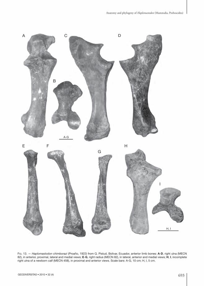
693
Anatomy and phylogeny of Haplomastodon (Mammalia, Proboscidea)
GEODIVERSITAS • 2010 • 32 (4)
A
E F H
I
H, I
G
B
C D
A-G
FIG. 13. — Haplo mastodon chimborazi (Proaño, 1922) from Q. Pistud, Bolivar, Ecuador, anterior limb bones: A-D, right ulna (MECN 82), in anterior, proximal, lateral and medial views; E-G, right radius (MECN 82), in lateral, anterior and medial views; H, I, incomplete right ulna of a newborn calf (MECN 458), in proximal and anterior views. Scale bars: A-G, 10 cm; H, I, 5 cm.
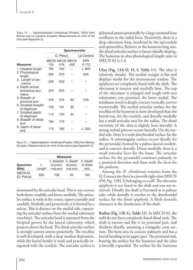
694 GEODIVERSITAS • 2010 • 32 (4)
Ferretti M. P.
dominated by the articular head. Th is is very convex both dorso-caudally and latero-medially. Th e articu-lar surface is wide at the center, tapers cranially and caudally. Medially and posteriorly, it is limited by a sulcus. Th is is distinct on the medial side, separat-ing the articular surface from the medial tuberosity (trochine). Th e articular head is separated from the bicipital groove by the lateral tuberosity, which projects above the head. Th e distal articular surface is strongly convex antero-posteriorly. Th e trochlea is well developed, with a deep median depression, while the lateral border is weak and practically in-tegrated with the condyle. Th e articular surface is
delimited antero-proximally by a large coronoid fossa confl uent to the radial fossa. Posteriorly, there is a deep olecranon fossa, bordered by the epicondyle and epitrochlea. Relative to the humerus long axis, the distal articular surface is latero-distally sloping. Th e humerus to ulna physiological length ratio in MECN 82 is 1.4.
Ulna (Fig. 13A-D, H, I; Table 11). Th e ulna is relatively slender. Th e medial margin is fl at and displays marks for the interosseous tendon. Th e epiphyses are completely fused with the shaft. Th e olecranon is massive and medially bent. Th e top of the olecranon is enlarged and rough with two tuberosites, one proximal, the latter medial. Th e semilunar notch is deeply concave vertically, convex transversally. Th e medial articular surface for the trochlea of the humerus is more developed than the lateral one, for the condyle, and dorsally-medially has a small articular area for the radius. Th e distal extremity of the ulna is slightly bent laterally. A strong styloid process occurs laterally. On the me-dial side, there is a wide diarthrodial surface for the radius. A subtriangular surface occurs distally for the pyramidal, formed by a palmo-lateral condyle, and is concave dorsally. Dorso-medially there is a small articular facet for the lunar. Th e articular surface for the pyramidal continues palmarly in a proximal direction and fuses with the facet for the pisiform.
Among the H. chimborazi remains from the Q. Cuesaca site there is a juvenile right ulna (MECN 458; Fig. 13H, I) belonging to a calf. Th e olecranic epiphysis is not fused to the shaft and was not re-trieved. Distally the shaft is fractured at is palmar side, while dorsally it reaches to the diarthroidal surface for the distal epiphysis. A likely juvenile character is the slenderness of the shaft.
Radius (Fig. 13E-G; Table 12). In MECN 82, the radii do not have completely fused distal ends. Th e shaft is narrow and fl at is its proximal part, and thickens distally, assuming a triangular cross sec-tion. Th e bone axis in concave palmarly and has a lateral bending in its upper part. Th e proximal end, bearing the surface for the humerus and the ulna is laterally expanded. Th e surface for the humerus
TABLE 11. — Haplo mastodon chimborazi (Proaño, 1922) from Bolivar and La Carolina, Ecuador. Measurements (in mm) of the ulna (see Appendix 3).
Specimen/site
Q. Pistud La Carolina
MeasuresMECN
134MECN
418MECN
458EPN
V-1721. Greatest length 765 750 – c. 8002. Physiological
length585 570 – 635
3. Length of ole-cranon
230 230 – –
4. Depth across processus aco-naeus
224 222 – –
5. Breadth of proximal end
220 224 80 240
6. Smallest breadth of diaphysis
100 101 30 –
7. Smallest depth of diaphysis
114 120 34 –
8. Breadth of distal end
184 173 – –
9. Depth of distal end
174 190 – c. 250
TABLE 12. — Haplo mastodon chimborazi (Proaño, 1922) from Bolivar, Ecuador. Measurements (in mm) of the radius (see Appendix 3).
Measures
Specimen (Site)
1.Length
2. Breadth of proxi-mal end
3. Depth of proxi-mal end
4. Depth of distal
endMECN 82(Q. Pistud)
605 108 64 150

695
Anatomy and phylogeny of Haplomastodon (Mammalia, Proboscidea)
GEODIVERSITAS • 2010 • 32 (4)
is trapezoidal and concave antero-posterirorly. It is divided in three parts: the lateral-most part con-tributes with the ulna to the articulation with the condyle of the humerus. Th e middle part is convex and forms the coronoid process. Th e medial-most part is part of the articulation for the humeral tro-chlea. Palmarly and distally is the articular facet for the ulna and distally to it, an extremely rough triangular area for the insertion of tendons. Th e distal end, more voluminous than the proximal one, displays a triangular cross section. Th e articular facets for the lunar and the scaphoid are found on distal face of the radius. Both facets continue for a short distance on the palmar side. Th e articular facet for the lunar is laterally positioned and has a saddle form: palmarly convex and dorsally concave. Th e surface for the scaphoid is medial to the convex portion of the articulation for the lunar, and is also concave. In dorsal view the two surfaces form an angle of about 160°.
Carpus (Figs 14; 15; Table 13). Only three carpals from the right side and four from the left side of the Bolivar skeleton are preserved. Articular surfaces of each element of the carpus are schematically depicted in Figure 14.
In lateral view the scaphoid (radial carpal bone; Figs 14B, C; 15A-C) is triangular. Th e medial as-pect is concave and rough. A large tuberosity, that projects above the proximal margin, is found on the dorsal margin of the scaphoid. A groove occurs distal to this tuberosity, and reaches to the distal margin of the bone. With its upper extremity it articulates with the radius (proximally) and with the lunar (medially). Th e lower extremity presents the facets for the lunar (dorsally), the magnum (palmarly), the trapezoid, and the trapezium (dis-tally and palmarly).
Th e lunar (intermedium carpal bone; Figs 14D-F; 15D-H) is proximo-distally fl attened, and sub-triangular in proximal view. Th e proximal face bears the articulation for the radius and the ulna. Th e former is saddle-like. A strong margin divides, laterally, the surface for the radius from that for the ulna. Th e latter is very small, laterally facing, palmo-dorsally concave and convex proximo-distally. Th e dorsal aspect of the bone is sub-rectangular and very
rough. Th e palmar angle is formed by a voluminous square tuberosity. Th e lateral and medial faces are irregular and dorso-palmarly elongated. Both bear on the proximal and distal margins the articular surfaces for the neighbouring carpals.
Th e lateral process of the pyramidal (ulnar carpal bone; Figs 14G; 15I-K) is not palmarly curved as in elephants. Th e pisiform (accessory carpal bone; Fig. 14H) is massive, with the distal tubercle slightly bent medially. It possesses two articular surfaces: proximally is a smaller one for the tibia and below, on the anterior face, a larger one for the pyramidal. Th e trapezoid (second carpal bone; Figs 14I-L; 15L-O) has a triangle-like shape in proximal view. Th e proximal face is occupied by the articulation for the scaphoid. Th is is wide dorsally and narrow palmarly. Th e medial margin is straight, while the lateral one forms a saddle-like articulation for the magnum. In specimen MECN 82 the sinuosity of the lateral margin of the trapezoid is indeed not particularly pronounced. Th ere is no evidence of a distinct facet for the lunar on the proximal face of the trapezoid,
TABLE 13. — Haplo mastodon chimborazi (Proaño, 1922) from Bolivar, Ecuador. Measurements (in mm) of carpal bones (see Appendix 3).
Measures
Specimen/site 1. Breadth 2. Depth 3. Heightleft lunar, MECN 45
(Q. Cuesaca)144 137 78
left magnum, MECN 23(Q. Cuesaca)
79 104 82
left unciform, MECN 46(Q. Cuesaca)
88 102 78
right scaphoid, MECN 82(Q. Pistud)
67 130 109
right lunar, MECN 82(Q. Pistud)
133 136 79
right pyramidal, MECN 82(Q. Pistud)
157 124 55
right trapezoid, MECN 82(Q. Pistud)
60 114 58
right trapezoid, MECN 184(Q. Pistud)
70 114 67
right magnum, MECN 82(Q. Pistud)
88 127 87
right unciform, MECN 82(Q. Pistud)
132 130 92
left unciform, MECN 183(Q. Pistud)
107 132 90

696 GEODIVERSITAS • 2010 • 32 (4)
Ferretti M. P.
magnum
unciform
metacarpal (Mc) 5
phalanges
pyramidal lunar
pisiformA
B C D E F
G H
MO
N
Q R S T U
P
I KJ
L
scaphoid
trapezoideum
trapezium
Mc2Mc1
Mc3Mc4
ra
pipy
py
ra
sc
sc
sc
sclu
td
lu
lu
lu?
ul
ul
ulMc2
Mc2
Mc2
Mc4
Mc5
Mc3
Mc3
ulsc
tr
tr
td
td
mama ma
ma
ma
ma
ma
un
un
ma
ma
td? py
I
II
III
un
FIG. 14. — Schematic representation of Haplo mastodon chimborazi (Proaño, 1922) carpals and third metapodial, showing extention of articular surfaces for contiguous bones (based on the Bolivar sample). All bones are from the right manus. A, reconstruction of the right manus, dorsal aspect (based on “S.” platensis, MNHN-PAM191) showing the position of various autopodial bones; B, C, scaphoid in lateral and distal views; D-F, lunar, in dorsal, lateral, and medial views; G, pyramidal, proximal view; H, pisiform, dorsal view; I-L, trapezoid, in distal, proximal, medial and lateral views. It is not possible to asses if the trapezoid articulates with the lunar (area enclosed by dotted line); M-P, magnum, in proximal, distal, medial, and lateral views; Q-S, unciform, in proximal, distal, and medial views; T, U, proximal end of right Mc3, in dorsal and proximal views. Abbreviations: lu, lunar; ma, magnum; Mc1, fi rst meta-carpal; Mc2, second metacarpal; Mc3, third metacarpal; Mc4, fourth metacarpal; Mc5, fi fth metacarpal; pi, pisiform; py, pyramidal; ra, radius; sc, scaphoid; td, trapezoid; tr, trapezium; ul, ulna; un, unciform.

697
Anatomy and phylogeny of Haplomastodon (Mammalia, Proboscidea)
GEODIVERSITAS • 2010 • 32 (4)
A B C
E
H
L M
P Q
TU V W
R S
N O
F
I
G
KJ
D
FIG. 15. — Haplo mastodon chimborazi (Proaño, 1922) from Q. Pistud, Bolivar, Ecuador, carpal bones (MECN 82): A-C, left scaphoid, in dorsal, medial and lateral views; D-H, left lunar, in dorsal, medial, lateral, proximal and distal views; I-K, right pyramidal, in dorsal, proximal and distal views; L-O, left trapezoid, in dorsal, medial, lateral and proximal views; P-S, right magnum, in dorsal, lateral, medial and proximal views; T-W, right unciform, in dorsal, lateral, medial and proximal views. Scale bar: 5 cm.

698 GEODIVERSITAS • 2010 • 32 (4)
Ferretti M. P.
even though the latter might have articulated with the lunar along its dorso-lateral edge. Th e distal face has the articular face for Mc2. Th e dorsal face is planar and rough. Th e palmar margin is made up by a rounded tuberosity. Th e articulation for the trapezium is found on the medial face of the py-ramidal. On the lateral face there are three facets for the magnum. Th e dorsal one is the larger, running from the proximal to the distal margin. A third, smaller facet for the magnum occurs in a palmar position, near the distal margin of the bone.
Th e magnum (third carpal bone; Figs 14M-P; 15P-S) is a quadrangular bone. Th e dorsal face has a squared outline and is very rough. Th e lateral face is trapezoidal and bears a large facet for the unciform. A second oval facet for the unciform is present dor-sally, in proximity of the distal margin of the bone. Palmarly and distally there is a large hooked tubercle, distally directed. Th e medial side of the magnum has centrally a deep fossa for the interosseous ligaments. A quadratic articular facet for the trapezoid is present along the dorsal margin of the medial side. Th is facet has a prolongation running palmarly and proximally bordening the central fossa. Th e prolongation reaches the proximo-palmar facet for the trapezoid. A small fi issure separates the proximal facet from the distal facet for the trapezoid. Th e latter is linked to the dorsal facet through a dorsally sloping crest. Th e proximal face is quadrangular. Palmarly, on the medial margin, a process bears the articular surface
for the scaphoid. For this reason, the medial margin has a sinuous outline, complementary to that of the lateral margin of the trapezoid. Th e proximal face is occupied by the articular surface for the lunar and, on the palmar angle, by that for the scaphoid. Th e articular surface is markedly convex palmarly and concave dorsally. Th e palmar face has a triangular outline. At the distal vertex of the triangle is a large tubercle, distally bent. Th e distal face is concave palmo-dorsally, and bears the articular facets for Mc2 and Mc3. Th e articular facet for the Mc2 does not reach the dorsal margin.
Th e unciform (fourth carpal bone; Figs 14Q-S; 15T-W) is a massive bone, possessing a trapezoidal outline. Th e proximal face slopes laterally and articu-lates with the pyramidal. Th e unciform articulates distally with the third, fourth and fi fth metacarpals. Palmarly, the surface for the Mc5 is contiguous with that of the pyramidal. Two large surfaces for the magnum occur on the medial face.
Metacarpus (Fig. 16; Table 14). Th ree metacarpals are preserved. Mc5 is very short (Fig. 16A, E, H). Th e proximal articular surface is divided into two facets: a medial one for the unciform and a lateral one articulating with the lateral process of the py-ramidal. Th e lateral face is proximally protruding, forming a tubercle from which a crest departs, dividing the facet for the unciform from that for the pyramidal.
TABLE 14. — Haplo mastodon chimborazi (Proaño, 1922) from Bolivar. Measurements (in mm) of metapodials (see Appendix 3). Ab-breviations: Mc, metacarpal; Mt, metatarsal.
Measures
Metapodial Specimen 1. Length
2. Small-est depth of diaphysis
3. Smallest breadth of diaphysis
4. Depth of proximal
end
5. Breadth of proximal
end6. Depth of distal end
7. Breadth of distal
endMc2 MECN 48 134 55 56 102 57 69 78Mc2 MECN 462 125 50 54 – 51 63 65Mc2 MECN 60 121 48 47 c. 79 c. 54 62 67Mc3 MECN 218 148 55 74 110 86 88 90Mc3 MECN 461 134 43 68 – 73 65 74Mc3 MECN 17 149 45 63 – 78 67 80Mc4 MECN 220 121 54 71 101 82 77 87Mc5 MECN 82 130 64 66 94 82 83 83Mt3 MECN 432 105 40 51 70 40 52 62Mt4 MECN 431 95 47 56 70 70 64 69Mt4 MECN 430 94 45 58 – 71 63 69

699
Anatomy and phylogeny of Haplomastodon (Mammalia, Proboscidea)
GEODIVERSITAS • 2010 • 32 (4)
FIG. 16. — Haplo mastodon chimborazi (Proaño, 1922) from Q. Pistud, Bolivar, Ecuador, metacarpals and partial manus: A, E, H, right Mc5 (MECN 82), in dorsal, medial, and proximal view; B, F, I, right Mc4 (MECN 82), in dorsal, medial and proximal views; C, G, J, right Mc3 (MECN 82), in dorsal, medial and proximal views; D, right Mc2 (MECN 462), dorsal view; K, partial manus (cast) with magnum (ma), unciform (un), third (Mc3), fourth (Mc4), and Fifth (Mc5) metacarpals in anatomical connection, dorsal view. Scale bar: 5 cm.
A
E
H
Kun
Mc5
Mc4 Mc3
ma
I J
F G
B C D

700 GEODIVERSITAS • 2010 • 32 (4)
Ferretti M. P.
Mc4 is shorter than Mc3 (Fig. 16B, F, I). Th e proximal articular surface is convex and articulates with the unciform. Medially, the surface for the Mc3 does not reach the dorsal margin. Th e palmar tubercle is more slender than that of the Mc3.
Th e body of Mc3 is sub-cylindrical, sturdy, and fl attened dorso-palmarly (Fig. 16C, G, J). Its dorsal face is convex, the lateral and medial faces are fl at, and the palmar face is slightly concave proximo-distally. Th e proximal extremity is palmo-dorsally elongated. Proximally, it bears an extended articular surface di-vided by a sagittal crest. Th e medial articular facet is for the magnum. Th is has a trapezoidal outline and narrows palmarly. It is dorso-palmarly convex and medially sloping. Th e lateral facet articulates with the unciform. It is dorso-palmarly convex, transversely concave except in its palmar edge, where it is strongly inclined laterally. It is less extended palmarly than the facet for the magnum. Th e two facets are at an angle to one another. Th e crest dividing the two facets is dorsally elevated, forming a prominence, which slopes and then rises again palmarly. In proximal view the crest appears slightly wavy. Articulations for Mc4 and Mc2 are located, respectively, on the lateral and medial sides. A large tubercle is present palmarly, hanging over the palmar face. Th e distal extremity is larger than the proximal one. Th e large articulation for the proximal phalanx is concave transversely and convex dorso-palmarly. A slightly convex facet for the sesamoids bones is found palmarly.
A second right metacarpal (MECN 462; Fig. 16D), not belonging to the MECN 82 skeleton, is present among the Bolivar sample. Th e facet for the trap-ezoid is dorso-palmarly extended, while that for the trapezium occupies only the palmar half of the proximal surface.
Pelvis (Fig. 17; Table 15). Both halves of the pelvis of the MECN 82 skeleton are preserved. Th e acetabu-lum is directed caudally and ventrally. It is bounded by a sub-circular rim, and interrupted ventrally by a deep acetabular notch. Th e cranial portion of the lunate surface is less developed than the caudal one. Dorsally to the acetabulum, the ischial spine forms a sturdy and short tuberosity. On the cranial surface, at the point of junction with the pubis, the prominent ileo-pubic eminence reaches dorsally and laterally the short and weak arcuate line. Lateralward to the iliac eminence, a wide surface for the attachment of the m. rectus femuris is present.
Th e iliac wing arises from the median part of the hipbone through a short, cranially fl attened and transversely broad, neck. Th e iliac wing is greatly extended transversally, and is cranially inclined. Th e gluteal (external) surface is plane, and it bends cau-dally at the level of the sacral tuberosity. Th e gluteal lines are well marked. Th e pelvic (internal) surface has a sub-triangular outline, is cranially oriented and it is bounded by the arcuate line medially and by the ventral margin of the ilium laterally. Th e
TABLE 15. — Haplo mastodon chimborazi (Proaño, 1922) from Q. Pistud, Bolivar, Ecuador. Measurements (in mm) of the pelvis (see Ap-pendix 4).
Measures MECN 821. Maximum length of pelvis 11102. Maximum length of ilium: from tuber coxae to tuber sacrale 8403. Length of the symphysis 4204. D iagonal height of pelvic aperture: from the pubic symphysis to the lowest point
of the tuber sacrale490
5. D iagonal height of pelvic aperture: from the pubic symphysis to the most caudal point of the tuber sacrale
480
6. Maximum width of pelvic aperture, between the iliac crests 4607. Maximum breadth across the acetabula 7608. Width between eminentiae iliopubicae 4209. Smallest height of caudal margin of cranial branch of pubis 80
10. Smallest height of cranial margin of cranial branch of pubis 6311. Smallest height of medial branch of ileum 5412. Smallest breadth of the shaft of ileum 207
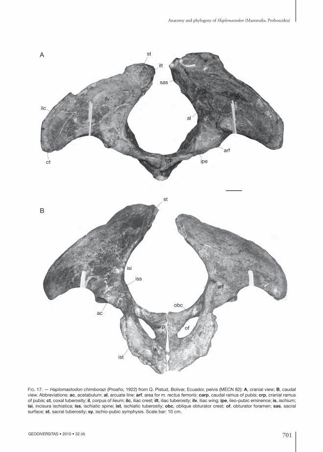
701
Anatomy and phylogeny of Haplomastodon (Mammalia, Proboscidea)
GEODIVERSITAS • 2010 • 32 (4)
ipe
st
iss
isi
ac
obc
arf
carp
ist
issy
of
crp
arf
al
sas
stA
B
ilt
ilv
ilc
il
ct
FIG. 17. — Haplo mastodon chimborazi (Proaño, 1922) from Q. Pistud, Bolivar, Ecuador, pelvis (MECN 82): A, cranial view; B, caudal view. Abbreviations: ac, acetabulum; al, arcuate line; arf, area for m. rectus femoris; carp, caudal ramus of pubis; crp, cranial ramus of pubis; ct, coxal tuberosity; il, corpus of ileum; ilc, iliac crest; ilt, iliac tuberosity; ilv, iliac wing; ipe, ileo-pubic eminence; is, ischium; isi, incisura ischiatica; iss, ischiatic spine; ist, ischiatic tuberosity; obc, oblique obturator crest; of, obturator foramen; sas, sacral surface; st, sacral tuberosity; sy, ischio-pubic symphysis. Scale bar: 10 cm.

702 GEODIVERSITAS • 2010 • 32 (4)
Ferretti M. P.
internal surface is slightly concave. Along the arcu-ate line no tuberculum for the m. psoas minoris has been observed.
Th e arcuate line reaches dorsally the sacral tu-berosity, and separates the pelvic surface from the sacral one. Th e latter is medially oriented, at an angle with the pelvic surface. Th e sacral surface is rough. Ventrally, an amphiarthrosial surface articulates with the sacrum, just below the iliac tuberosity. A crani-ally oriented amphiarthrosis for the fourth lumbar vertebra is situated cranially to the sacral surface. Th e crest of the ilium is thick and rough, ventrally and laterally directed. Th e crest ends ventro-laterally into an expanded coxal tuberosity. Th e ischiopubic part of the hipbone has a squared outline, with one of the vertex represented by the acetabulum. At the center, the great obturate foramen displays an oval outline, with the greater axis sagittally oriented. Th e body of the pubis enters into the formation of the acetabulum, of which it forms the ventral and cranial parts. Th e cranial branch of the pubis is robust and has an oval cross-section. Its dorsal surface is transversely concave, while the anterior one (pectineal surface) is planar. Th e caudal surface is crossed by the oblique obturator crest, which forms part of the medial margin of the obturator sulcus. Th e medio-cranial angle of the ischiopubic part is thick. Ventrally, it supports a robust ventral pubic spine. Th e caudal branch of the pubis is thin, depressed and fully fused to the ischium. Its medial border presents the symphysial surface.
Th e medial branch of the ischium is fl attened and very short. It is continuous with the caudal branch of the pubis. Th e lateral branch of the ischium is characterized by torsion and joins the body of the ischium. Th e latter forms the caudal portion of the acetabulum. Th e cranial portion of the medial margin of the lateral branch of the ischium partici-pates in the formation of the pubic symphysis. Th e caudal margin of the ischiatic table forms a thick and rough vertical margin, ventrally and medially directed. Th e dorsal extremity forms the tuberosity of the ischium. With its fellow the caudal margin constitutes the ischiatic arch. Th is has a V shape and its vertex is open ventrally.
Femur (Fig. 18A-E; Table 16). Th e femora of MECN 82 have both epiphyses fused to the shaft. Th e shaft is long, subcylindrical and fl attened cranio-caudally. Narrow in the central portion, it widens approaching the extremities. Proximally, at about one third of the length of the medial margin, there is an elongated prominence slightly caudally placed, probably a homologue of the lesser trochanter. Th e head of the femur is perfectly hemispheric and delimited by a thick crest from the neck (Fig. 18E, B). Caudally there is, along the crest, a small round depressed surface for the femural ligament. Th e trochanter is massive, less elevated than the head and expanded cranio-caudally. Distally, the trochanter forms a robust margin, which integrates into the caudal margin of the lateral face of the shaft.
TABLE 16. — Haplo mastodon chimborazi (Proaño, 1922) from Bolivar and La Carolina, Ecuador. Measurements (in mm) of the femur (see Appendix 5). *, measured at the same location as measure 5.
Specimen/site
Measures
Q. Pistud La CarolinaMECN
98MECN
420EPN
V-168EPN
V-170EPN
V-2009EPN
V-2010EPN
V-38461. Greatest length from caput 965 960 990 – – 1005 9952. Lateral length 900 890 941 – 942 945 9153. Greatest breadth
of proximal endc. 370 360 – – – – –
4. Greatest depth of caput 155 150 157 – – – 1585. Smallest breadth of diaphysis 132 131 158 146 157 145 1546. Depth of diaphysis* 88 82 98 92 93 88 947. Greatest breadth of distal end 221 230 210 202 210 – 2238. Depth of distal end 226 223 212 210 236 – 232

703
Anatomy and phylogeny of Haplomastodon (Mammalia, Proboscidea)
GEODIVERSITAS • 2010 • 32 (4)
A B D
C
E
F
G
N
M
L
J
H
A-E, H-N
F, G
I
K
FIG. 18. — Haplo mastodon chimborazi (Proaño, 1922), from Q. Pistud, Bolivar, Ecuador, hindlimb bones (MECN 82): A-E, left femur, in anterior, proximal, posterior, medial and distal views; F, G, right patella, in anterior and posterior views; H-L, left tibia, in proximal (anterior to the bottom), anterior view, posterior, distal (anterior to the bottom) and lateral views; M, N, right tibia and fi bula in anatomi-cal connection, in posterior and distal views. Scale bars: A-E, H-N, 10 cm; F, G, 5 cm.

704 GEODIVERSITAS • 2010 • 32 (4)
Ferretti M. P.
Th e distal extremity presents the condyles and, anteriorly, the patellar surface (Fig. 18E). Th e me-dial condyle is larger and more elongated than the lateral one. It is also slightly bent laterally, while the lateral one is rotated clockwise. From this it fol-lows that the lateral condyle reaches a more distal plane than the medial one. Th e medial epicondyle is more developed than the lateral one. Two deep fossae, possibly for the insertion of the lateral knee ligament and m. popliteus, respectively, are found on the lateral side of the lateral condyle. Th e patellar surface is relatively large and characterized by wide median concavity.
Patella (Fig. 18F, G). Th e kneecap has an elliptic outline. Th e cranial face is convex and rough. Th e caudal face is fl at and bears the articulation for the trochlea. Th e proximal margin has a small tubercle. Th e distal margin is fl at.
Tibia (Fig. 18H-M; Table 17). Th e tibia has a prismatic-shaped shaft. Th e proximal portion is more expanded than the distal one. Th is is very evident in lateral view. Th e articular surface for the lateral condyle of the femur is sub-oval and concave. Caudally is the small facet for the fi bula. Th e articulation for the medial condyle is circular and more expanded cranio-caudally. Th e distal extremity is trapezoidal in outline. On the distal face there is a square surface for the astragalus and latero-caudally a smaller facet of the fi bula. A strong medial malleolus borders medially the articulation for the astragalus, on the medial side.
Fibula (Fig. 18M; Table 18). In the MECN 82 skeleton, the left fi bula lacks the proximal epiphysis, while the right one is complete. Th e proximal ex-tremity of the fi bula widens to form the head of the bone. On the cranio-medial faces a rounded surface for the tibia occurs. Caudally and laterally there are two tuberosities for ligaments. Th e lateral face of the distal extremity is convex and very rugose. Th e medial face bears a complex set of articular surfaces. At the base of a large fossa there is the articulation for the tibia, dorsally oriented. Ventrally and laterally to this there is a triangular articulation surface for the astragalus. On the ventral face there is a concave surface, which articulates with the calcaneum.
TABLE 17. — Haplo mastodon chimborazi (Proaño, 1922) from Bolivar and La Carolina, Ecuador. Measurements (in mm) of the tibia (see Appendix 5).
Specimen/site
Q. Pistud La Carolina
MeasuresMECN
83MECN
424EPN
V-167EPN
V-169EPN
V-171EPN
V-173EPN
V-2010 EPN
V-38471. Greatest length 605 580 649 606 638 695 > 613 6812. Lateral length 475 480 – – – – – –3. Medial length 570 575 620 580 – – – –4. Breadth at mid diaphysis 95 96 116 102 111 109 104 1145. Depth at mid diaphysis 89 91 – – – – – –6. Breadth of proximal articular
surface220 215 246 246 252 269 247 272
7. Depth of proximal articular surface
160 170 181 176 193 206 165 216
8. Breadth of distal articular surface
193 198 180 197 186 196 200 206
9. Depth of distal articular surface
129 132 148 132 139 161 142 172
TABLE 18. — Haplo mastodon chimborazi (Proaño, 1922) from Bolivar, Ecuador. Measurements (in mm) of the fi bula (see Appendix 5).
Measures
Specimen/site 1. Length2. Greatest
breadth3. Great-est depth
MECN 422 (Q. Pistud) 560 84 118

705
Anatomy and phylogeny of Haplomastodon (Mammalia, Proboscidea)
GEODIVERSITAS • 2010 • 32 (4)
A B
D
F
J
N
O P Q
K ML
I
H
sus
ecs
tis
fis
nas
G
E
C
FIG. 19. — Haplo mastodon chimborazi (Proaño, 1922) from Q. Pistud, Bolivar, Ecuador, astragalus, metatarsals and phalanx (MECN 82): A-E, right astragalus, in dorso-distal, proximal, medial, dorsal and distal views; F, G, N, right Mt1, in dorsal, medial and proximal views; G, K, O, left Mt3, in dorsal, medial and proximal views; H, L, P, left Mt4,in dorsal, medial and proximal views; I, M, Q, left proxi-mal phalanx, in dorsal, medial and proximal views. Abbreviations: ecs, ectal surface; fi s, articular surface for the fi bula; nas, articular surface for the navicular; sus, sustentacular surface; tis, articular surface of the tibia. Scale bar: 5 cm.
706 GEODIVERSITAS • 2010 • 32 (4)
Ferretti M. P.
Tarsus (Fig. 19A-E; Table 19). Of the tarsus of the MECN 82 skeleton, only the astragalus is represented. No other tarsals are present in the Bolivar sample. Th e right astragalus is complete and not deformed. Th e surface for the tibia is rectangular, and medio-laterally more elongated in comparison with that of elephants. On the lateral margin is the surface for the fi bula. Th e medial tubercle is poorly developed. Th e surface for the navicular (central tarsal bone) occupies the two thirds of the antero-distal (dorso-distal) side. Th e two surfaces for the calcaneum are well separated from each other by a deep sulcus for the attachment of ligaments on the postero-distal (palmo-distal) side (Fig. 19E). Th e ligamentous surface widens anteriorly. Th e medial surface (sustentacular) for the calcaneum is bean-shaped and dorso-palmarly directed. Th e lat-eral surface (ectal) is oval, with its major axis directed laterally and anteriorly (dorsally).
Metatarsus (Fig. 19F-Q; Table 14). Five metatarsals of the Bolivar skeleton are preserved. Th ese are markedly shorter than the metacarpals. Th e proximal extremity of Mt1 (Fig. 19 F, J, N) has an oval outline. Th e distal one is much more transversely expanded.
Th e proximal articular surface of Mt3 (Fig. G, K, O) is triangular and transversely concave. Th e articulation for the sesamoids forms two distinct condyles.
Th e proximal extremity of Mt4 is medially rotated (Fig. 19H, L, P). Th e proximal articular surface is triangular and almost entirely devoted to the articu-lation with the cuboid.
Discussion. Th e postcranial material of H. chimborazi from Bolivar is characterized by a large amount of size variability, without exhibiting any clear (sexual) dimorphic pattern. Th e mean size of the gompho-
there from Bolivar, as can be deduced from the skel-etal elements (Tables 2-19), is greater than that of C. hyodon, while it is similar to that of “S.” platensis. Th e postcranium of H. chimborazi shows a typical elephantoid pattern. Haplomastodon chimborazi displays, however, a more robust skeleton than both Gomphotherium (e.g., G. sylvaticum; Tassy 1977) and more derived elephantoids such as A. arvernensis and elephantids.
Brevirostrine and longirostrine elephantoids are characterized by a diff erent proportion between the bones of the anterior limb (Ferretti 1998). In many longirostrine forms (e.g., Gomphotherium), the hu-merus to ulna physiological length ratio is about 1, whilst in brevirostrines it is usually higher than 1.2 (e.g., A. arvernensis = 1.3-1.4; in elephantines is >1.25). In H. chimborazi (MECN 82) the humerus to ulna ratio is 1.4, displaying thus a typical “brevirostrine” limb proportion.
Th e structure of the basipodial of H. chimborazi is typically elephantoid. Haplo mastodon chimborazi possesses relatively short basipodials, similarly to other gomphotheres (Fig. 16K). Some elements posses in-terlocking surfaces. Th e whole structure appears very robust and fi rm, a possible adaptation to traversing uneven grounds. Th e available material prevents us from knowing whether in H. chimborazi the lunar articulated distally with the trapezoid, producing the so-called aserial carpus (Weithofer 1889). However, the condition in C. hyodon and “S.” platensis, as re-ported by Boule & Th evenin (1920: 60), is aserial. In A. arvernensis, a brevirostrine ghomphotere from the Old World, the superposition of the lunar on the trapezoid is markedly more developed than in SA gomphotheres. Such diff erences could have important functional implications related to limb posture and feeding habits (Ferretti & Croitor 2001).
TABLE 19. — Haplo mastodon chimborazi (Proaño, 1922) from Bolivar, Ecuador. Measurements (in mm) of the astragalus (see Ap-pendix 5).
Measures
Specimen/site 1. Depth 2. Breadth 3. Height4. Greatest breadth
of ectal surface5. Greatest breadth
of sustentacular surfaceMECN 425 (Q. Pistud) 140 163 99 95 85MECN 143 (Q. Cuesaca) c. 152 c. 173 c. 97 103 –MECN 47 (Q. Cuesaca) 143 153 96 100 101
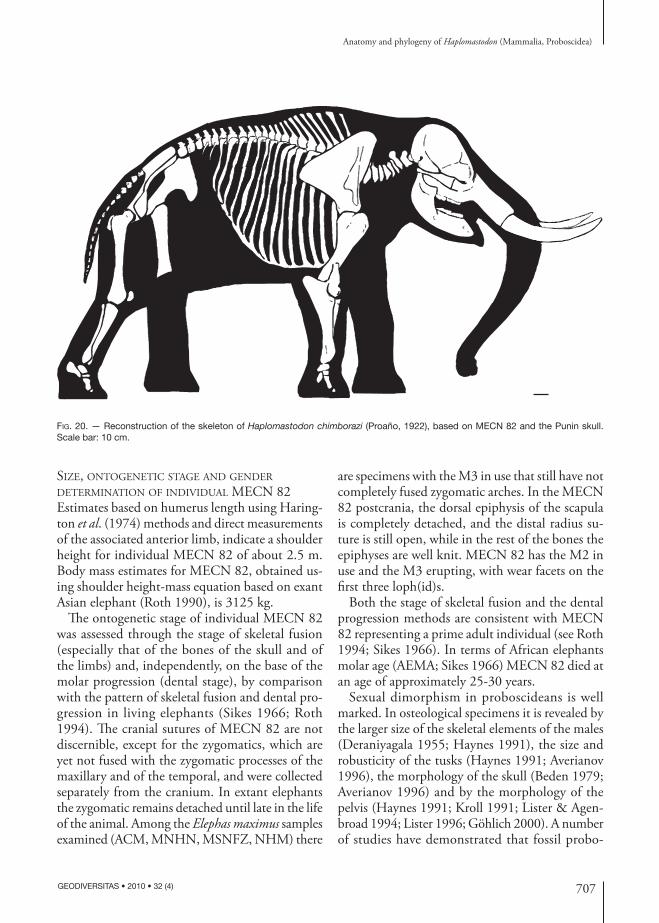
707
Anatomy and phylogeny of Haplomastodon (Mammalia, Proboscidea)
GEODIVERSITAS • 2010 • 32 (4)
SIZE, ONTOGENETIC STAGE AND GENDER DETERMINATION OF INDIVIDUAL MECN 82Estimates based on humerus length using Haring-ton et al. (1974) methods and direct measurements of the associated anterior limb, indicate a shoulder height for individual MECN 82 of about 2.5 m. Body mass estimates for MECN 82, obtained us-ing shoulder height-mass equation based on exant Asian elephant (Roth 1990), is 3125 kg.
Th e ontogenetic stage of individual MECN 82 was assessed through the stage of skeletal fusion (especially that of the bones of the skull and of the limbs) and, independently, on the base of the molar progression (dental stage), by comparison with the pattern of skeletal fusion and dental pro-gression in living elephants (Sikes 1966; Roth 1994). Th e cranial sutures of MECN 82 are not discernible, except for the zygomatics, which are yet not fused with the zygomatic processes of the maxillary and of the temporal, and were collected separately from the cranium. In extant elephants the zygomatic remains detached until late in the life of the animal. Among the Elephas maximus samples examined (ACM, MNHN, MSNFZ, NHM) there
are specimens with the M3 in use that still have not completely fused zygomatic arches. In the MECN 82 postcrania, the dorsal epiphysis of the scapula is completely detached, and the distal radius su-ture is still open, while in the rest of the bones the epiphyses are well knit. MECN 82 has the M2 in use and the M3 erupting, with wear facets on the fi rst three loph(id)s.
Both the stage of skeletal fusion and the dental progression methods are consistent with MECN 82 representing a prime adult individual (see Roth 1994; Sikes 1966). In terms of African elephants molar age (AEMA; Sikes 1966) MECN 82 died at an age of approximately 25-30 years.
Sexual dimorphism in proboscideans is well marked. In osteological specimens it is revealed by the larger size of the skeletal elements of the males (Deraniyagala 1955; Haynes 1991), the size and robusticity of the tusks (Haynes 1991; Averianov 1996), the morphology of the skull (Beden 1979; Averianov 1996) and by the morphology of the pelvis (Haynes 1991; Kroll 1991; Lister & Agen-broad 1994; Lister 1996; Göhlich 2000). A number of studies have demonstrated that fossil probo-
FIG. 20. — Reconstruction of the skeleton of Haplo mastodon chimborazi (Proaño, 1922), based on MECN 82 and the Punin skull. Scale bar: 10 cm.

708 GEODIVERSITAS • 2010 • 32 (4)
Ferretti M. P.
scideans display an amount of sexual dimorphism comparable to that of modern elephants (see Tassy 1996b and Haynes 1991 and citations therein). Th e strength and size of the skeletal elements, and of the tusks, indicate that MECN 82 is a male. Th e proportions of the pelvis, in particular the relative width of the pelvic aperture and the thickness of the acetabular region, are comparable with those reported for male elephants (Table 20; Haynes 1991; Kroll 1991; Lister & Agenbroad 1994; Lister 1996; Göhlich 2000).
PHYLOGENETIC ANALYSIS
RESULTS
Th e phylogenetic analysis was performed on a matrix of 11 taxa (Appendix 7) and 25 characters (Appendix 6).Th ree most parsimonius trees (MPT) of 49 steps each were obtained. Th ey have a consistency index (CI) of 0.65, a retention index (RI) of 0.79, and a homoplasy index (HI) of 0.35. Th e rescaled con-sistency index (RC) is of 0.51.
Th e three trees (Fig. 21) express the confl ict-ing evidence in the phylogenetic resolution of H. chimborazi,“S.” platensis, and C. hyodon, i.e. the South American gomphotheres. Th e three possible hypotheses consist of two paired associa-tions ((H. chimborazi,“S.” platensis) C. hyodon) and ((C. hyodon,“S.” platensis) H. chimborazi)) respec-tively, and a trichotomy, (C. hyodon,“S.” platensis, H. chimborazi). Th e strict consensus tree is identical to the MPT with the SA gomphotheres’ trichoto-mous topology (Fig. 21A). Each of the three SA
taxa is diagnosed by a set of apomorphies. Two homoplastic apomorphies characterize H. chimbo-razi, both concerning modifi cations of the upper tusk: presence of markedly upturned upper tusk (13 (2)), and absence of the enamel band of upper tusk at the adult growth stage (14 (2)). One of the MPT (Fig. 21C, node 8’) supports a H. chimborazi, “S.” platensis clade, based on three equivocal homo-plastic characters (ACCTRAN option): slightly diverging tusk alveoli (4 (1)), enamel band of upper tusk usually absent (14 (1)), and enamel band, when present, without helical torsion (15 (0)), the latter representing a reversal. Th e alternative hypothesis (Fig. 21B, node 8), that “S.” platensis is more closely related to C. hyodon, is weakly supported by one equivocal character reversal (DELTRAN option): enamel band of upper tusk sometimes present at the adult growth stage (14 (1)). Th e monophyly of South American gomphotheres (Fig. 21A, node 7) is, on the other hand, well supported by fi ve unam-biguous characters, of which four (12 (1): presence of a coronoid fossa; 19 (2): fully trilophodont DP3/dp3; 23 (1): transverse foramen of atlas sometimes absent; 24 (1); atlas with a large dorsal tuberosity) are unique synamoporphies (RI = 1.0).
According to the result of the analysis, Rhyncho-therium cf. falconeri is the sister taxon of South American gomphotheres (Fig. 21A, node 6), a hy-pothesis supported by four unequivocal characters of which two (5 (1): loss of supraorbital foramen and 6 (1): presence of a subnasal fossa) represent non-homoplastic synapomorphies. Noteworthy, Stegomastodon texanus is not closely related nei-ther to “S.” platensis nor to H. chimborazi, but is placed as the sister taxon of the Rhynchotherium + South American gomphotheres clade (Fig. 21A, node 5), based on one non-homoplastic synapo-morphy (3 (1): robust premaxillaries), and one homoplastic character (21 (1): third loph in DP3 outlined). Th e Asian Sinomastodon hanjiangensis (Fig. 21A, node 4) is united to the New world brevirostrine taxa by fi ve unequivocal characters, three of which are unique synapomorphies (2 (1): wide forehead; 8 (1): symphysis downturned; 18 (1): loss of P2-P4). Gnathabelodon thorpei (node 3), Eubelodon morilli (node 2), and Megabelodon lulli (node 1) are successive outgroups to the bre-
TABLE 20. — Comparison of measurements ratios of pelvis of Haplomastodon chimborazi (MECN 82) and of recent elephants, Loxodonta africana (Blumenbach, 1792) and Elephas maximus Linnaeus, 1758 (data from Kroll 1991). Labels of measures as in Table 15.
Pooled sample of male
Pooled sample of female
ratio N min-Max N min-Max MECN 828/3 3 0.94-1.01 3 1.25-1.47 01:108/12 3 1.97-2.28 4 2.27-3.34 02:224/12 3 2.05-2.56 3 2.90-3.50 02:37

709
Anatomy and phylogeny of Haplomastodon (Mammalia, Proboscidea)
GEODIVERSITAS • 2010 • 32 (4)
virostrine clade. Gomphotherium productum and G. angustidens are basal to all the rest of the New world gomphothere taxa.
A bootstrap analysis (1000 replicates) showed that all the clades present in the strict consensus topology have relatively high bootstrap proportion
FIG. 21. — The three most parsimonius trees (MPT) of New World gomphotheres from the current study (length = 49 steps; CI = 0.65; RI = 0.79). The topology of the MPT depicted in A is identical to that of the strict consensus tree. List of synapomorphies (in bold, non-homoplastic character; *, homoplastic character; in italics, unbiguous character; -, reversal): A, node 1: 16 (1)*; node 2: 9 (1), 14 (2)*; node 3: 13 (1)*; node 4: 1 (1)*, 2 (1), 7 (1)*, 8 (1), 10 (1)*, 18 (1), 19 (1), 20 (1)*; node 5: 3 (1), 21 (1)*, 22 (1); node 6: 4 (1)*, 5 (1), 6 (1), -14 (1)*; node 7: 11 (1)*; 12 (1), 19 (2), 23 (1), 24 (1). B, node 8: -14 (1)*; C, node 8’: -4 (1)*; 14 (1)*; -15 (0)*.
A
1
2
3
Gomphotherium angustidens (Cuvier, 1817)
Gomphotherium productum (Cope, 1875)
Megabelodon lulli Barbour, 1914
Eubelodon morilli Barbour, 1914
Gnathabelodon thorpei Barbour�& Sternberg, 1935
Sinomastodon hanjiangensis Tang & Zong, 1987
Stegomastodon texanus Osborn, 1924
Rhynchotherium cf. falconeri Osborn, 1923
Haplomastodon chimborazi (Proaño, 1922)
Cuvieronius hyodon (Fischer de Waldheim, 1814)
“Stegomastodon” platensis (Ameghino, 1888)
1
2
3
4
5
6
7
Gomphotherium angustidens
Gomphotherium productum
Megabelodon lulli
Eubelodon morilli
Gnathabelodon thorpei
Sinomastodon hanjiangensis
Stegomastodon texanus
Rhynchotherium cf. falconeri
Haplomastodon chimborazi
Cuvieronius hyodon
“Stegomastodon” platensis
4
5
67’
8’
B
C
Gomphotherium angustidens
Gomphotherium productum
Megabelodon lulli
Eubelodon morilli
Gnathabelodon thorpei
Sinomastodon hanjiangensis
Stegomastodon texanus
Rhynchotherium cf. falconeri
Haplomastodon chimborazi
Cuvieronius hyodon
“Stegomastodon” platensis
1
2
34
5
6
78
710 GEODIVERSITAS • 2010 • 32 (4)
Ferretti M. P.
values (BP > 60), except for the clades defi ned by nodes 3 and 4, which have BP values of 47 and 35, respectively.
DISCUSSION
Results of the present phylogenetic analysis provide further morphological evidence for a monophyletic South American clade, corresponding to the gompho-theriid subfamily Cuvieroniinae Cabrera, 1929. A major diff erence from previous studies, however, is that NA Stegomastodon is shown not to group within the Cuvieroniinae. Th e analysis demonstrates that “S.” platensis is in fact not closely related to NA Stegomastodon, as was already suggested by Osborn (1936), Savage (1955), Madden (1984), and Webb (1992), and therefore its removal from Stegomas-todon is supported. Th e morphological dataset used in the analysis does not provide unequivocal support for any single, dichotomously branching species tree linking H. cimborazi, “S.” platensis and C. hyodon. Th e hypothesis that H. chimborazi and “S.” platensis are more closely related to each other than either is to C. hyodon, as suggested by several authors (Hoff stetter 1952; Savage 1955; Tassy 1985; Prado & Alberdi 2008) is, in fact, just one of the three most parsimonius solutions found. Among these, the hypothesis that the ancestral separation of the three SA gomphothere species is actually the result of a single trichotomous branching event should also be taken into consideration. Th is last hypothesis would be consistent with the mostly disjunct fossil distribution of the three taxa (Prado et al. 2005; Ferretti 2008a). For these reasons, the generic name Haplo mastodon is conservatively retained as a distinct taxon from South American “Stegomastodon”.
Th e analysis supports the hypothesis, advocated by Savage (1955), Miller (1990), and Shoshani (1996), that Rhynchotherium is the sister taxon of SA gomphotheres.
Th is result diff ers from that provided by Prado & Alberdi (2008), which indicates a sister group rela-tionship between SA gomphotheres and Sinomasto-don, a hypothesis also presented by Shoshani (1996: 152) as one of the most parsimonious solutions of his own analysis of the Proboscidea. In the Prado & Alberdi’s (2008) study, the grouping of Sinomas-
todon with SA gomphotheres is supported by two characters: loss of lower tusk and occurrence of a brevirostrine mandible. However, the fi rst character actually represents a more inclusive feature among NA gomphothere, as also Eubelodon Barbour, 1914 and Gnathabelodon Barbour & Sternberg, 1935 possess tuskless mandibles (Barbour & Sternberg 1935; Osborn 1936; Lambert & Shoshani 1998). As a matter of fact, the codings of this character for Eubelodon (0, i.e. lower tusk present) and Gnathabe-lodon (1, i.e. lower tusk reduced) in the data matrix provided by Prado & Alberdi (2008: table 2) is either a mistake or is based on new observations, which are not discussed or mentioned by the authors. On the other hand, the new characters used in the present analysis and the improved morphological data for Rhynchotherium cf. falconeri (LVNHM 871) and Sinomastodon (based on Zong et al. 1989), pro-vide substantial support for a Rhynchotherium-SA gomphotheres clade, based on four unequivocal characters. Finally, the analysis does not support the proposed synonymy of Megabelodon and Gompho-therium advocated by Tobien (1973) and Shoshani (1996), although they are only removed from each other by a single step.
Eleven of the 23 synapomorphies that defi ne the seven nodes depicted by the strict consensus tree topology (Fig. 21A) are homoplastic. Among these homplasies, parallelisms are far more frequent than reversals.
CONCLUSIONS
Th e knowledge gained from the abundant and well preserved Haplomastodon chimborazi material from Bolivar, Ecuador provided new and improved morphological data to test phylogenetic hypotheses of New World gomphotheres, focusing on the relationships and systematics of South American taxa. New anatomical research and additional speci-mens are, however, needed to remove much of the missing data and to provide a taxonomically more comprehensive dataset. Th e systematic revision of Haplo mastodon indicates that this genus contains only a single South American species, whose valid name is H. chimborazi. Haplo mastodon waringi is

711
Anatomy and phylogeny of Haplomastodon (Mammalia, Proboscidea)
GEODIVERSITAS • 2010 • 32 (4)
considered a nomen dubium. Body size estimates based on an adult male skeleton from Bolivar shows that H. chimborazi was comparable is size to the extant Asian elephant, with a shoulder height of about 2.5 m and a body mass of three metric tons. Th e results of the cladistic analysis provided fur-ther morphological support for a monophyletic SA gomphothere clade (the Cuvieroniinae), though the interrelationships within the group could not be defi nitively resolved. Th e analysis demonstrates that “Stegomastodon” platensis represents an endemic SA taxon distinct from NA Stegomastodon, the latter be-ing in fact not closely related to the Cuvieroniinae. According to the results of the phylogenetic analysis and based on the known stratigraphic distribution of the taxa considered (see Lambert & Shoshani 1998 and Tobien et al. 1986), the Cuvieroniinae originated in North America during the Pliocene from a derived Rhynchotherium-like ancestor and then dispersed to South America, where the group diff erentiated during the Pleistocene, likely triggered by geographic factors. Th e earliest representatives of NA Stegomastodon (referred to S. primitivus) are reported from Late Hemphillian sites in the United States (Lambert & Shoshani 1998), indicating that the NA Stegomastodon and the Rhynchotherium-Cu-vieroniinae lineages diverged by the latest Miocene. Th e split between the Asian Sinomastodon and the New World brevirostrine gomphothere lineages can be also traced back to the Late Miocene, correspond-ing to the age of the oldest fi nds of S. intermedius from China (Tobien et al. 1986).
Th e analysis revealed that, rather than a linear progression from longirostrine to brevirostrine “elephant-like” morphologies, New World trilo-phodont gomphothere evolution is characterized by extensive homoplasy, especially in the form of parallelisms.
AcknowledgementsI dedicate this work to Jeheskel “Hezy” Shoshani (1943-2008), a true teacher and a good friend, whose fundamental contribution to the sudy of proboscidean anatomy and evolution, and dedication to the study and preservation of living elephants, have been, and still are, truly inspiring.
For access to collection, discussions and useful insights, I am grateful to: M. S. Bargo, A. Cione, E. P. Tonni, S. Vizcaino (MLP), M. Moreno Espinosa, G. Roman Jimenez (MECN), F. Rios Paredes (MUT), A. Currant (NHM), J. Meng (AMNH), P. Tassy (MNHN), and L. Werdelin (NMR). I thanks T. Gordón A. and N. E. Báez T., (UCE), for help in locating the type material of H. chimborazi in the UCE collections. Particular thanks go to G. Fic-carelli (IGF) for providing the original impetus to my research on SA proboscideans and for his help and encouragements throughout this study. I am also indebted to M.T. Alberdi (Madrid), V. Borselli, F. Cozzini, F. Landucci, L. Rook, M. Rossi, G. Tito (IGF), M. Coltorti, P. L. Pieruccini (University of Siena), U. B. Göhlich (Naturhistorisches Museum Wien), R. Debruyne (MNHN) for help, discus-sion and constructive criticism. Victoria Herridge (NHM) helped with the English text. I thank Cy-rille Delmer (NHM) and Regis Debruyne for the French translation of the abstract. H. Shoshani and L. Rook critically read and commented on an early draft of the paper greatly improving it. I gratefully thank R. W. Graham, A. Ohler, W. J. Sanders, and P. Tassy for helpful review of an early version of the manuscript.
Work supported by grants from the University of Firenze (Fondi di Ateneo), the Italian C.N.R., and M.I.U.R (Cofi n 2000, 2002).
REFERENCES
ALBERDI M. T. & PRADO J. L. 1995. — Los mastodontes de América del Sur, in ALBERDI M. T., LEONE G. & TONNI E. P. (eds), Evolucion biologica y climatica de la Region Pampeana durante los ultimos 5 miliones des años. Un ensayo de corrélacion con el Mediterraneo occidental. Monografi as de Museo Nacional de Ciencias Naturales y Consejo Superior de Investigaciones Científi cas, Madrid 12: 277-292.
ALBERDI M. T., PRADO J. L. & SALAS R. 2004. — Th e Pleis-tocene gomphotheres (Gomphotheriidae, Proboscidea) from Peru. Neues Jahrbuch für Geologie und Paläontologie Abhandlungen 231: 423-452.
ANONYMOUS 1903. — Le Mastodonte du Chimborazo. Cosmos Paris, n.s., 48/936: 6-9.
ARÁUZ J. 1950. — Nueva historia de los Mastodontes ecu-atorianos. Boletin de Informaciones Científi cas Nacional 3 (26-27): 419-425.

712 GEODIVERSITAS • 2010 • 32 (4)
Ferretti M. P.
AVERIANOV A. O. 1996. — Sexual dimorphism in mammoth skull, teeth, and long bones, in SHOSHANI J. & TASSY P. (eds), Th e Proboscidea. Evolution and Palaeoecology of Elephants and their Relatives. Oxford University Press, Oxford: 260-267.
BARBOUR E. H. 1914. — Mammalian fossils from Devil’s Gulch. Nebraska Geological Survey 4: 177-190.
BARBOUR E.H. & STERNBERG G. 1935 — Gnathabe-lodon thorpei, gen. et sp. nov. A new mud-grubbing mastodont. Bulletin of the Nebraska State Museum 1: 395-404.
BEDEN M. 1979. — Les éléphants (Elephas et Loxodonta) d’Afrique orientale : systématique, phylogénie, intérêt biochronologique. PhD Th esis, Université de Poitiers, 567 p. (unpublished).
BELL R. H., SWIGART L. L. & ANSON B. J. 1950. — Th e relation of the vertebral artery to the cervical verte-brae. Based on a study of 200 specimens. Quarterly Bullettin of the Northwestern University Medical School 24: 184-185.
BOULE M. & THEVENIN A. 1920. — Mammifères fossiles de Tarija. Imprimerie nationale, Mission scientifi que G. de Créqui-Monfort et F. Sénechal de la Grange, Paris, 256 p.
CABRERA A. 1929. — Una revision de los mastodontes ar-gentinos. Revista del Museo de La Plata 32: 61-144.
CAMPBELL K. E. JR, FRAILY C. D. & ROMERO-PITTMAN L. 2000. — Th e late Miocene Gomphothere Amahua-catherium peruvium (Proboscidea: Gomphotheriidae) from Amazonian Peru: implications for the Great American Faunal Interchange. Boletin de Estudios Regionales IGEMMET, Serie D, 23: 1-152.
CAMPBELL K. E. JR, FRAILEY C. D. & ROMERO-PITTMAN L. (2009). — In defense of Amahuacatherium (Pro-boscidea: Gomphotheriidae). Neues Jahrbuch für Geologie und Paläontologie Abhandlungen 252 (1): 113-128.
CASAMIGUELA R. M., SHOSHANI J. & DILLEHAY T. D. 1996. — South American proboscideans: general intro-duction and refl ections on Pleistocene extinctions, in SHOSHANI J. & TASSY P. (eds), Th e Proboscidea. Evolu-tion and Palaeoecology of Elephants and their Relatives. Oxford University Press, Oxford: 316-320.
COLTORTI M., FICCARELLI G., JAHREN H., ROOK L. & TORRE D. 1998. — Th e last occurrence of Pleistocene megafauna in the Ecuadorian Andes. Journal of South American Earth Sciences 11 (6): 581-586.
COSTALES SAMANIEGO A. 1950. — Masthodon Chimborazi Proaño. Boletin de Informaciones Científi cas Nacional 3 (35): 372-375.
DERANIYAGALA P. E. P. 1955. — Some Extinct Elephants, their Relatives and the Two Living Species. Ceylon Na-tional Museum Publication, Colombo, 161 p.
EALES N.B. 1928. — Th e anatomy of a foetal African elephant (Loxodonta africana). Transactions of the Royal Society of Edinburgh 56: 206-246.
FERRETTI M. P. 1998. — Gli Elefanti del Plio-Pleistocene dell’Italia. Doctoral thesis, Modena, Bologna, Firenze, and Roma associated Universities, 272 p. (unpub-lished).
FERRETTI M. P. 2008a. — A review of South American gomphotheres. New Mexico Natural History and Sci-ence Museum Bulletin 44: 381-391.
FERRETTI M. P. 2008b. — Enamel structure of Cuviero-nius hyodon (Proboscidea, Gomphotheriidae) with a discussion on enamel evolution in elephantoids. Journal of Mammalian Evolution 15 (1): 37-58.
FERRETTI M. P. 2009. — Comment on the proposed conservation of usage of Mastodon waringi Holland, 1920 (currently Haplo mastodon waringi; Mammalia, Proboscidea) by designation of a neotype. Bulletin of Zoological Nomenclature 66 (2): 358-359.
FERRETTI M. P. & CROITOR R. 2001. — Functional mor-phology and ecology of Villafranchian Proboscideans from Central Italy, in CAVARETTA G., GIOIA P., MUSSI M. & PALOMBO M. R. (eds), Th e World of Elephants - Proceedings of the 1st International Congress, Rome, 16-20 October, Consiglio Nazionale delle Ricerche, Roma: 103-108.
FICCARELLI G., AZZAROLI A., BORSELLI V., COLTORTI M., DRAMIS F., FEJFAR O., HIRTZ A. & TORRE D. 1992. — Stratigraphy and Paleontology of Upper Pleistocene deposits in the Interandean Depression, northern Ecuador. Journal of South American Earth Sciences 6 (3): 145-150.
FICCARELLI G., BORSELLI V., MORENO ESPINOSA M. & TORRE D. 1993. — New Haplo mastodon fi nds from the Late Pleistocene of Nor thern Ecuador. Geobios 26 (2): 231-240.
FICCARELLI G., BORSELLI V., MORENO ESPINOSA M. & TORRE D. 1995. — Taxonomic remarks on the South American mastodonts referred to Haplo mastodon and Cuvieronius. Geobios 28 (6): 745-756.
FICCARELLI G., AZZAROLI A., BERTINI A., COLTORTI M., MAZZA P., MEZZABOTTA C., MORENO ESPINOSA M., ROOK L. & TORRE D. 1997. — Hypothesis on the cause of extinction of the south American Masto-donts. Journal of South American Earth Sciences 10 (1): 29-38.
FICCARELLI G., COLTORTI M., MORENO ESPINOSA M., PIERUCCINI P., ROOK L. & TORRE D. 2003. — A model for the Holocene extinction of the mammal mega-fauna in Ecuador, South America. Journal of South American Earth Sciences 15: 835-845.
FRICK C. 1926. — Tooth sequence in certain trilophodont tetrabelodont mastodonts. Bulletin of the American Museum of Natural History 56: 122-176.
GÖHLICH U. B. 2000. — On a pelvis of the straight-tusked elephant Elephas antiquus (Proboscidea, Mammalia) from Binsfeld near Speyer (Rhineland-Palatinate, Ger-many). Paläontologisches Zeitschriftes 74: 205-214.
HARINGTON C. R., TIPPER H. W. & MOTT R. J. 1974. —

713
Anatomy and phylogeny of Haplomastodon (Mammalia, Proboscidea)
GEODIVERSITAS • 2010 • 32 (4)
Mammoth from Babine Lake, British Columbia. Canadian Journal of Earth Science 11: 285-303.
HAYNES G. 1991. — Mammoth Mastodonts & Elephants, Biology Behavior and the Fossil Record. Cambridge University Press, Cambridge, 413 p.
HOFFSTETTER R. 1950. — Observaciones sobre los mastodontes de Sud America y especialmente del Ecuador. Haplomastodon, subgen. nov. de Stegomast-odon. Publicaciones de la Escuela Politecnica Nacional de Quito, 1950, 1: 1-49.
HOFFSTETTER R. 1952. — Les Mammifères pléistocènes de la République de l’Équateur. Mémoires de la Societé géologique de France 66: 1-391.
HOFFSTETTER R. 1955. — Remarques sur la classifi ca-tion et la phylogénie des mastodontes sud-américains. Bulletin du Muséum national d’Histoire naturelle, Paris, 2e sér., 27 (6): 484-491.
HOFFSTETTER R. 1986. — High Andean mammalian faunas during the Plio-Pleistocene, in VUILLEUMIER F. & MONASTEIRO M. (eds), High Altitude Tropical Biogeography. Oxford University Press, New York: 218-245.
HOLLAND W. J. 1920. — Fossil mammal collected at Pedra Vernelha, Bahia, Brazil, by G. A. Waring. An-nals of the Carnegie Museum 13: 224-232.
ICZN 1999. — International Code of Zoological Nomen-clature. International Trust for Zoological Nomen-clature, London, 436 p.
KROLL W. 1991. — Der Waldelefant von Crumstadt. Ein Beitrag zur Osteologie des Waldelefanten, E. (Pa-laeoloxodon) antiquus Falconer & Cautley (1847). Inaugural-Dissertation zur Erlangung der tiermedizi-nischen Doktorwürde der Tierärztlichen Fakultät der Ludwig-Maximilians-Universität München, 31 p.
LAMBERT W. D. & SHOSHANI H. 1998. — Chapter 43. Proboscidea in JANIS C. M., SCOTT K. M. & JACOBS L. L. (eds), Evolution of Tertiary Mammals of North America. Volume 1. Cambridge University Press, Cambridge: 606-621.
LAUB R. S. 1996. — Th e masticatory apparatus of the American mastodon (Mammut americanum), in STEWART K. M. & SEYMOUR K. L. (eds), Palaeoecology and Palaeoenvironments of Late Cenozoic Mammals – Tributes to the Career of C.S. (Rufus) Churcher. University of Toronto Press, Toronto: 375-405.
LISTER A. 1996. — Sexual dimorphism in the mammoth pelvis: an aid to gender determination, in SHOSHANI J. & TASSY P. (eds), Th e Proboscidea. Evolution and Palaeoecology of Elephants and their Relatives. Oxford University Press, Oxford: 203-213.
LISTER A. & AGENBROAD L. D. 1994. — Gender deter-mination of the Hot Spring mammoths, in AGEN-BROAD L. D. & MEAD J. I. (eds), Th e Hot Spring Mammoth Site. Mammoth Site Inc., Hot Spring, South Dakota: 208-214.
LUCAS S. G. 2009. — Taxonomic nomenclature of Cu-
vieronius and Haplo mastodon, proboscideans from the Plio-Pleistocene of the NewWorld. New Mexico Museum of Natural History and Science Bulletin 44: 409-415.
LUCAS S. G. 2009. — Mastodon waringi Holland, 1920 (currently Haplo mastodon waringi; Mammalia, Pro-boscidea): proposed conservation of usage by designa-tion of a neotype. Bulletin of Zoological Nomenclature 66 (2): 164-167.
MADDEN C. T. 1984. — Th e Proboscidea of South America. Geological Society of America, abstracts with programs 12: 474.
MILLER W. 1990. — A Rhynchotherium skull and mandible from southeastern Arizona. Brigham Young University Geology Studies 36: 57-67.
OLSEN S. J. 1979. — Osteology for the Archaeologist. N.3: Th e american mastodon and the woolly mam-moth. Papers of the Peabody Museum of Archaeology and Ethnology, Harvard University 56 (3): 1-45.
OSBORN H. F. 1936. — Proboscidea. Volume 1. Americam Museum of Natural History, New-York, 802 p.
PRADO J. L. & ALBERDI M. T. 2008. — A cladistic ana-lysis among trilophodont gomphotheres (Mammalia, Proboscidea) with special attention to the South Ame-rican genera. Paleontology 51 (4): 903-915.
PRADO J. L., ALBERDI M. T., AZANZA B., SANCHEZ B. & FRASSINETTI D. 2005. — Th e Pleistocene Gomphothe-riidae (Proboscidea) from South America. Quaternary International 126-128C: 21-30.
PROAÑO J. F. 1894. — Mastodonte del Chimborazo. Memorias del Liceo Chimborazo, 1894, Riobamba, Ecuador: 31.
PROAÑO J. F. 1922. — La Virgen del Dios Chimborazo, Tradiciones Puruhaes. Imprenta “El Observador”, Riobamba, Ecuador, 23 p.
ROTH V. L. 1990. — Insular dwarf elephants: a case study in body mass estimation and ecological infer-ence, in DAMUTH J. & MACFADDEN B. J. (eds), Body size in Mammalian Palaeobiology: Estimation and Biological Implications. Cambridge University Press, Cambridge: 151-179.
ROTH V. L. 1994. — How elephants grow: Heterochrony and the calibration of developmental stages in some living and fossil species. Journal of Vertebrate Paleon-tology 4: 126-145.
SAUER W. 1965. — Geologia del Ecuador. Editorial del Min-isterio de Educacion del Ecuador, Quito, 383 p.
SAVAGE D. 1955. — A survey of various late Cenozoic faunas of the Panhandle of Texas. Part 2: Proboscidea. University of California Publication in Geological Sci-ences 31: 51-74.
SHOSHANI J. 1996. — Para- or monophyly of the gom-photheres and their position within Proboscidea, in SHOSHANI J. & TASSY P. (eds), Th e Proboscidea. Evolu-tion and Palaeoecology of Elephants and their Relatives. Oxford University Press, Oxford: 149-177.

714 GEODIVERSITAS • 2010 • 32 (4)
Ferretti M. P.
SHOSHANI J. & TASSY P. 2005. — Advances in pro-boscideans taxonomy and classifi cation, anatomy and physiology, and ecology and behavior. Quaternary International 126-128: 5-20.
SIKES S. K. 1966. — Th e african elephant, Loxodonta africana: a fi eld method for the extimation of age. Journal of Zoology 150: 279-295.
SIMPSON G. G. & DE PAULA COUTO C. 1957. — Th e mastodons of Brazil. Bulletin of the American Museum of Natural History 112: 131-189.
SPILLMANN F. 1928. — Tetrabelodon nov. spec. ayora Spillm. El Ecuador Comercial 6 (57): 1-70.
SPILLMANN F. 1931. — Die Säugetiere Ecuadors im Wandel der Zeit. Quito Universitad Central, 122 p.
SWOFFORD D. L. 2003. — PAUP * Phylogenetic Analysis Using Parsimony (* and Other Methods). Version 4. Sinauer Associates: Sunderland, Massachusetts.
TASSY P. 1977. — Le plus ancien squelette de gompho-thère (Proboscidea Mammalia) dans la formation burdigalienne des sables de l’Orléanais, France. Mé-moires du Muséum national d’Histoire naturelle, Série C, Sciences de la Terre 37: 3-51.
TASSY P. 1985. — La place des mastodontes miocènes de l’ancien Monde dans la phylogénie des Proboscidea (Mammalia): hypothèses et conjectures. Th èse de Doc-torat, Université Pierre et Marie Curie, Paris, 862 p. (unpublished).
TASSY P. 1990. — Phylogénie et classifi cation des Pro-boscidea (Mammalia) : historique et actualité. An-nales de Paléontologie (Vertebrates-Invertebrates) 76 (3): 159-224.
TASSY P. 1996a. — Dental homologies and nomencla-ture in the Proboscidea, in SHOSHANI J. & TASSY P.
(eds), Th e Proboscidea. Evolution and Palaeoecology of Elephants and their Relatives. Oxford University Press, Oxford: 21-25.
TASSY P. 1996b. — Growth and sexual dimorphism among Miocene elephantoids: the example of Gom-photherium angustidens, in SHOSHANI J. & TASSY P. (eds), Th e Proboscidea. Evolution and Palaeoecology of Elephants and their Relatives. Oxford University Press, Oxford: 92-100.
TEILHARD DE CHARDIN P. & TRASSAERT M. 1937. — Th e Proboscideans of south-eastern Shansi. Palaeontologia Sinica, Series C, 13: 1-58.
TOBIEN H. 1973. — On the evolution of mastodonts (Proboscidea, Mammalia). Prt 1: Th e bunodont trilo-phodont Groups. Notizblatt des Hessischen Landesamtes für Bodenforschung zu Wiesbaden 101: 202-208.
TOBIEN H., CHEN G. & LI Y. 1986. — Mastodonts (Proboscidea, Mammalia) from the late Neogene and early Pleistocene of the People’s Republic of China. Mainzer Geowissenschaftliches Mitteilungen 15: 119-181.
WEBB S. D. 1992. — A brief history of New World Pro-boscidea with emphasis on their adaptations and interac-tions with man, in FOX J. W., SMITH C. B. & WILKINS K. T. (eds), Proboscidean and Paleoindian Interactions. Baylor University Press, Waco, Texas: 16-34.
WEITHOFER K. A. 1889. — Einige Bemerkungen über den Carpus der Proboscidier. Gegenbaurs Morpholo-gisches Jahrbuch 14: 507-517.
ZONG G., TANG Y., LEI Y. & LI S. 1989. — Th e Hanjiang mastodont (Sinomastodon hanjiangensis nov. sp.). Academic Press, Beijing (in Chinese with English abstract), 84 p.
Submitted on 14 September 2007;accepted on 27 May 2010.

715
Anatomy and phylogeny of Haplomastodon (Mammalia, Proboscidea)
GEODIVERSITAS • 2010 • 32 (4)
APPENDIX 1
Measurements of Haplo mastodon chimborazi (Proaño, 1922): A, cranium (see Table 2) ; B, mandible (see Table 3). Material catalog numbers: see Table 1.
1
2
5
68
8
20
9
4
73
10
1
3
A
B
2
811
107
45
12
6
15
19
16
9
14
13

716 GEODIVERSITAS • 2010 • 32 (4)
Ferretti M. P.
APPENDIX 2
Measurements of vertebrae of Haplo mastodon chimborazi (Proaño, 1922): A, atlas (see Table 5); B, axis (see Table 6); C, vertebra C3-L4 (see Table 7); D, sacrum (see Table 8). Material catalog numbers: see Table 1.
1
2 6
2
5
1
3
4
3
6
7
281
4
5
5
1
6
2
BA
C
D
3
4

717
Anatomy and phylogeny of Haplomastodon (Mammalia, Proboscidea)
GEODIVERSITAS • 2010 • 32 (4)
APPENDIX 3
Measurements of forelimb bones of Haplo mastodon chimborazi (Proaño, 1922): A, scapula (see Table 9); B, humerus (see Table 10); C, ulna (see Table 11); D, radius (see Table 12); E, carpal (see Table 13); F, metapodial (see Table 14). Material catalog numbers: see Table 1.
6
53
9
5
2 3
4
6
2
45
7
3
1
2
8
6 A
B
C
E
D
F
2
1
7
3
4
1 24
3
6
7
2
1
1
1
4
7
3

718 GEODIVERSITAS • 2010 • 32 (4)
Ferretti M. P.
APPENDIX 4
Measurements of pelvis of Haplo mastodon chimborazi (Proaño, 1922) (see Table 15). Material catalog numbers: see Table 1.
7
8
1
2
3
4
12
10
5
6
9

719
Anatomy and phylogeny of Haplomastodon (Mammalia, Proboscidea)
GEODIVERSITAS • 2010 • 32 (4)
APPENDIX 5
Measurements of hindlimb bones of Haplo mastodon chimborazi (Proaño, 1922): A, femur (see Table 16); B, tibia (see Table 17); C, fi bula (see Table 18); D, astragalus (see Table 19). Material catalog numbers: see Table 1.
2 1
1
8
13
24 1 3
7
3
6 7
8
3
2
4
9
5
5
5 4
A
CD
B

720 GEODIVERSITAS • 2010 • 32 (4)
Ferretti M. P.
APPENDIX 6
Skeletal and dental characters used in the cladistic analysis and their primitive (0) and derived (1, 2) states.
Cranium
1. Position of the anterior border of the bony orbit (in lateral
view). 0) laying above or posterior to the mesial-most cheek tooth,
1) forward of the mesial-most tooth.
Modifi ed from character 83 of Shoshani (1996).
2. Forehead (frontoparietal plane between temporal lines).
0) narrow, 1) wide.
Modifi ed from character 37 of Tassy (1990).
3. Premaxillaries (in frontal view). 0) slender, distal margin much
narrower than interorbital width, 1) robust, distal margin equal or
wider than interorbital width.
Modifi ed from character 35 of Tassy (1990).
4. Tusk alveoli (in frontal view). 0) nearly parallel, 1) distally
slightly diverging, 2) distally greatly diverging.
Modifi ed from character 9 of Shoshani (1996).
5. Supraorbital foramen. 0) present, 1) absent.
Adapted from character 33 of Tassy (1990).
6. Subnasal fossa. 0) absent, 1) present.
Mandible
7. Symphysis. 0) long, 1) short.
Taken from character 52 of Tassy (1990).
8. Symphysis (in lateral view). 0) parallel to ventral margin of
mandibular body, 1) downturned.
Modifi ed from character 51 of Tassy (1990).
9. Symphysis (dorsal view). 0) slightly widens distally, 1) tapers
distally, 2) greatly widens distally.
10. Ascending ramus. 0) dorso-posteriorly directed, 1) dorsally
directed.
11. Position of the mandibular foramen. 0) at or slightly higher
than the level of the occlusal plane, 1) much higher than the oc-
clusal plane.
12. Coronoid fossa. 0) absent, 1) present.
Teeth
13. Upper tusk, curvature in adult growth stage. 0) downturned,
1) straight or slightly upturned, 2) upturned, much.
Modifi ed from character 76 of Tassy (1990) and character 7 of
Shoshani (1996).
14. Upper tusk, enamel band in adult growth stage. 0) present,
1) sometimes present, 2) absent.
Modifi ed from character 72 of Tassy (1990) and character 10 of
Shoshani (1996).
15. Upper tusk, helicoidal torsion of the enamel band. 0) absent
or moderate, 1) strong.
Modifi ed from character 8 of Shoshani (1996).
16. Lower permanent tusk (i2). 0) present, 1) absent.
Taken from character 70 of Tassy (1990).
17. Lower tusk: cross section. 0) piriform, 1) subcircular.
Taken from character 74 of Tassy (1990).
18. Defi nitive premolars (P2-P4). 0) P2 absent 1) P2-P4 absent.
Adapted from character 86 of Tassy (1990).
19. Th ird deciduous premolars (DP3/dp3). 0) bilophodont,
1) third loph/id outlined, 2) trilophodonty achieved.
20. Number of complete lophids in lower third molar. 0) four,
1) fi ve, 2) six or more.
Modifi ed from character 105 of Tassy (1990).
21. Posttrite central conules in molars (M1-M3/m1/m3).
0) absent, 1) outlined, 2) present, large.
Modifi ed from character 113 of Tassy (1990).
22. Crown cement in cheek teeth. 0) absent, 1) little present,
2) abundant.
Modifi ed from character 116 of Tassy (1990).
Post-cranial
23. Atlas, transversal foramen. 0) present, 1) sometimes absent.
Based on Hoff stetter (1952) and Tassy (1986).
24. Atlas, large dorsal tuberosity of dorsal arch.
0) absent, 1) present.

721
Anatomy and phylogeny of Haplomastodon (Mammalia, Proboscidea)
GEODIVERSITAS • 2010 • 32 (4)
APPENDIX 7
Character-taxon matrix used in the cladistic analysis. Missing data (either due to non-preservation or because non-applicable) are coded as “?”.
Taxon/character 1 2 3 4 5 6 7 8 9 10 11 12 13 14 15 16 17 18 19 20 21 22 23 24Gomphotherium angustidens 0 0 0 0 0 0 0 0 0 0 0 0 0 0 0 0 0 0 0 0 0 0 0 0Gomphotherium productum 0 0 0 0 0 0 0 0 0 0 0 0 0 0 0 0 1 0 0 0 0 0 0 0Megabelodon lulli 0 0 0 0 0 0 0 0 0 0 0 0 0 0 ? 1 ? 0 0 0 0 0 0 0Eubelodon morilli 0 0 0 0 0 0 0 0 2 0 0 0 0 2 ? 1 ? ? ? 0 1 1 0 0Gnathabelodon thorpei 0 0 0 0 0 0 0 0 1 0 0 0 1 2 ? 1 ? 0 0 0 0 0 0 0Sinomastodon hanjiangensis 1 1 0 0 0 0 1 1 2 1 1 0 1 2 ? 1 ? 1 ? 1 0 0 0 0Stegomastodon texanus 1 1 1 1 0 0 1 1 2 0 0 0 2 2 ? 1 ? 1 1 2 2 2 0 0Rynchotherium cf. falconeri 0 1 1 2 1 1 0 1 2 1 0 0 1 0 1 0 1 1 1 0 1 0 0 0“Stegomastodon” platensis 1 1 1 1 1 1 1 1 2 1 1 1 1 1 0 1 ? 1 2 1 2 1 1 1Cuvieronius hyodon 1 1 1 2 1 1 1 1 2 1 1 1 1 0 1 1 1 1 2 1 1 1 1 1Haplo mastodon chimborazi 1 1 1 1 1 1 1 1 2 1 1 1 2 2 0 1 ? 1 2 1 1 1 1 1