Anatomy of anal canal
36
ANATOMY OF ANAL CANAL Presented by : AYESHA JALIL Roll# 43
-
Upload
ayesha-jalil -
Category
Science
-
view
131 -
download
2
Transcript of Anatomy of anal canal

ANATOMY OF ANAL
CANALPresented by : AYESHA JALIL
Roll# 43
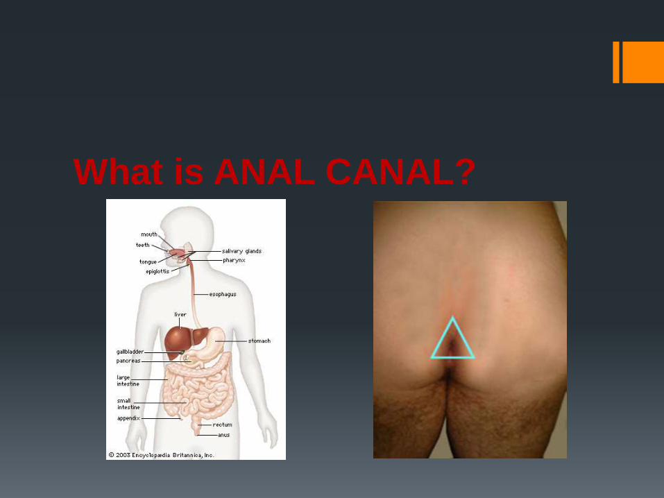
What is ANAL CANAL?

SURGICAL ANATOMY The anal canal is about
1.5”(4 cm) long,
commences at the level
where the rectum passes
through the pelvic
diaphragm, downward and
backward from rectal
ampulla and ends at the
anal verge

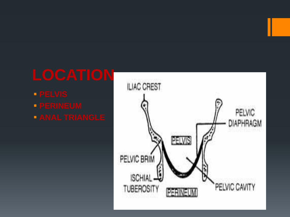
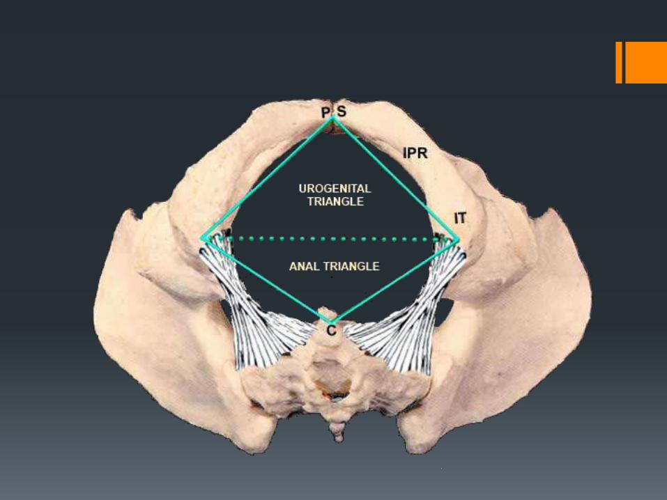
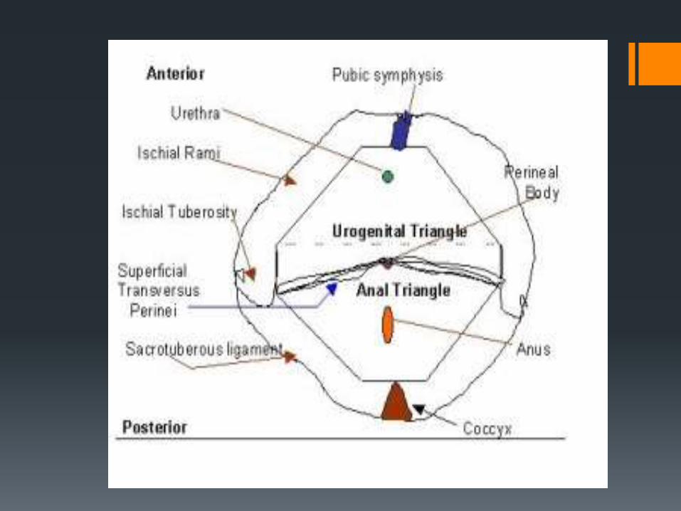
LOCATION PELVIS
PERINEUM
ANAL TRIANGLE


RELATIONS
POSTERIORLY
Anococcygeal body
LATERALLY
Ischiorectal fossas

ANTERIORLY IN MALE
Perineal body
Urogenital diaphragm
Membranous part of urethra
Bulb of penis
ANTERIORLY IN FEMALE
Perineal body
Urogenital diaphragm
Lower part of vagina

STRUCTURE EMBRYOLOGY

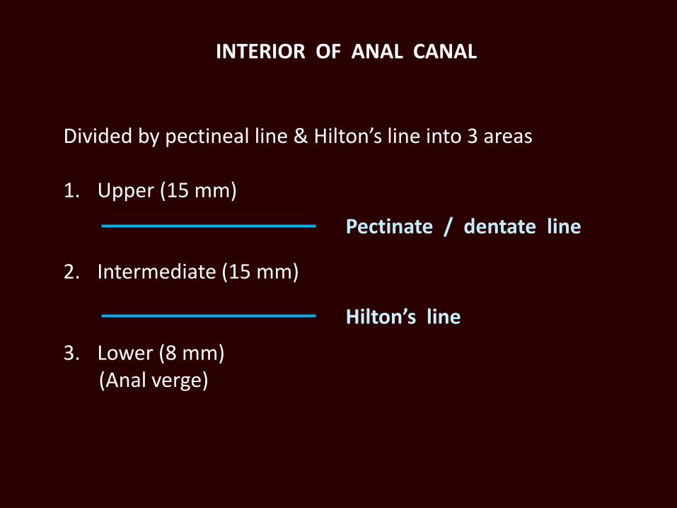
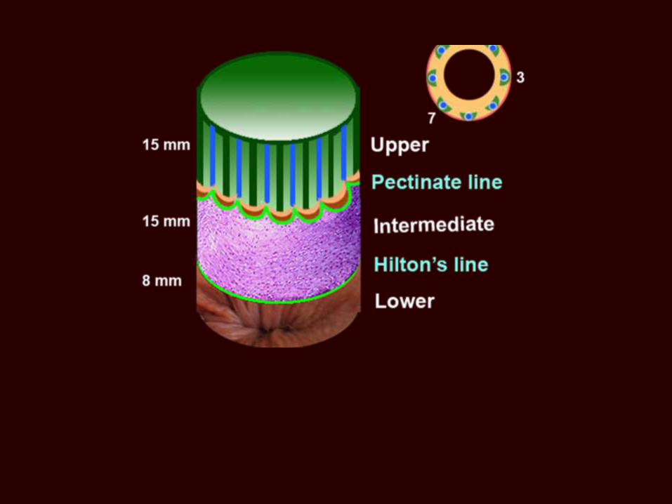
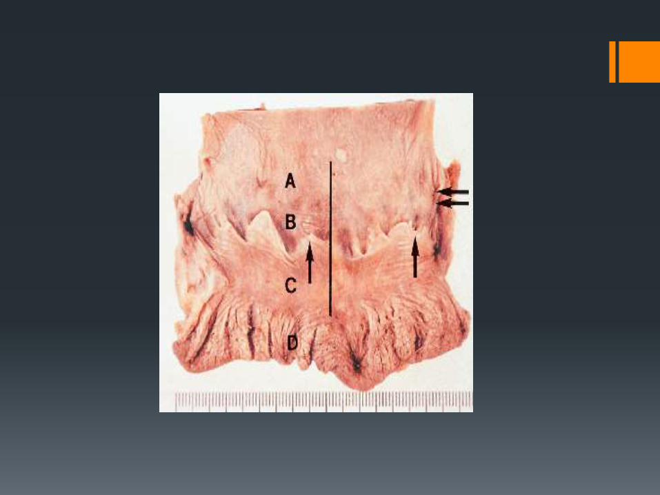
INTERIOR OF ANAL CANAL
Divided by pectineal line & Hilton’s line into 3 areas
1. Upper (15 mm)
2. Intermediate (15 mm)
3. Lower (8 mm)(Anal verge)
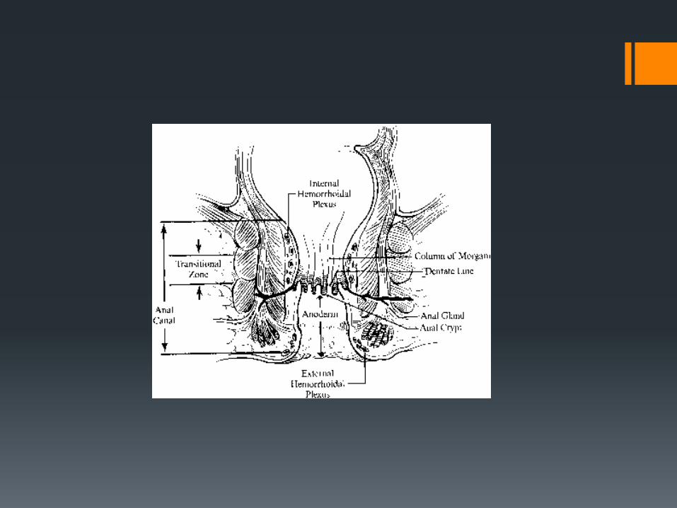
Pectinate / dentate line
Hilton’s line
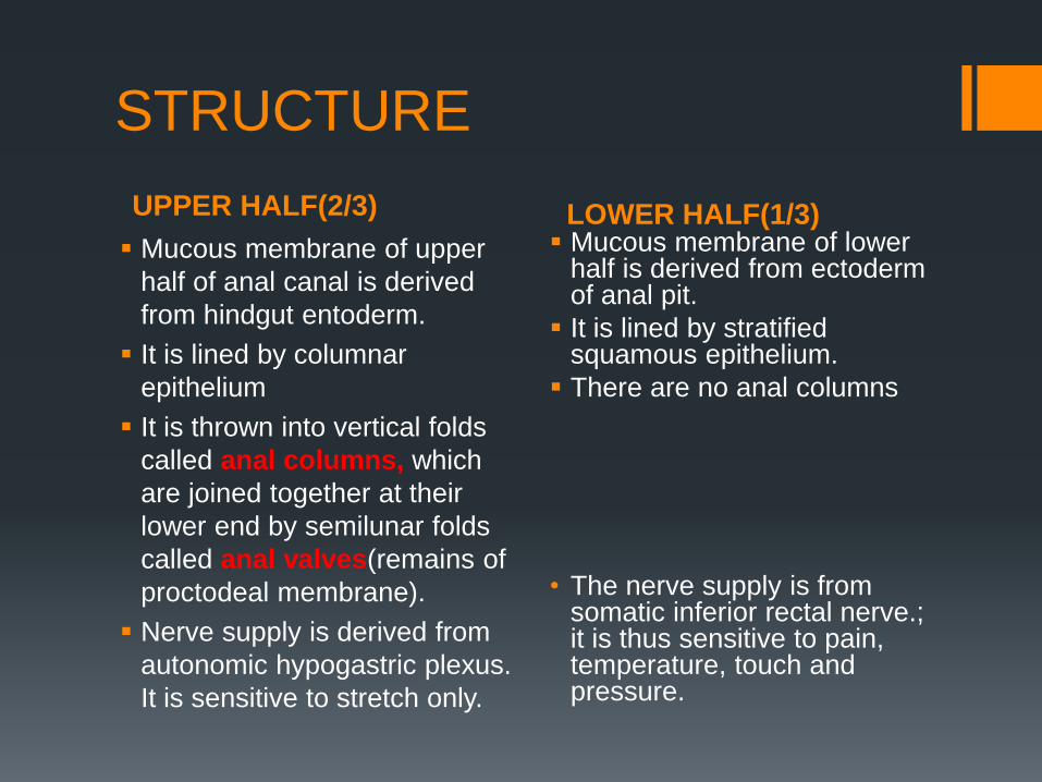
UPPER HALF(2/3) LOWER HALF(1/3)
STRUCTURE
Mucous membrane of upper
half of anal canal is derived
from hindgut entoderm.
It is lined by columnar
epithelium
It is thrown into vertical folds
called anal columns, which
are joined together at their
lower end by semilunar folds
called anal valves(remains of
proctodeal membrane).
Nerve supply is derived from
autonomic hypogastric plexus.
It is sensitive to stretch only.
Mucous membrane of lower half is derived from ectoderm of anal pit.
It is lined by stratified squamous epithelium.
There are no anal columns
• The nerve supply is from somatic inferior rectal nerve.; it is thus sensitive to pain, temperature, touch and pressure.

BLOOD SUPPLY
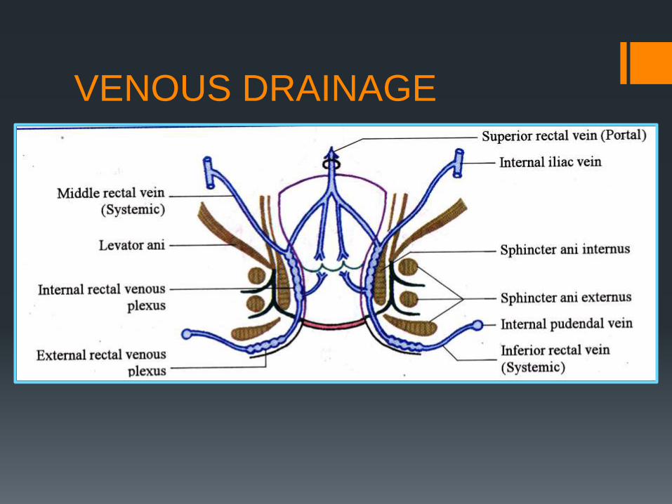
VENOUS DRAINAGE

LYMPHATIC DRAINAGE

The arterial supply is that of hindgut- superior rectal artery, branch of inferior mesenteric artery.
The venous drainage is by the superior rectal vein, a tributary of inferior mesenteric vein, and the portal vein.
The lymphatic drainage is to the pararectal nodes and then eventually to inferior mesenteric nodes.
The arterial supply is inferior rectal artery, a branch of internal pudendal artery.
The venous drainage is by inferior rectal vein, a tributary of the internal pudendal vein, which drains into the internal iliac vein.
The lymphatic drainage is downward to the medial group of superficial inguinal nodes.
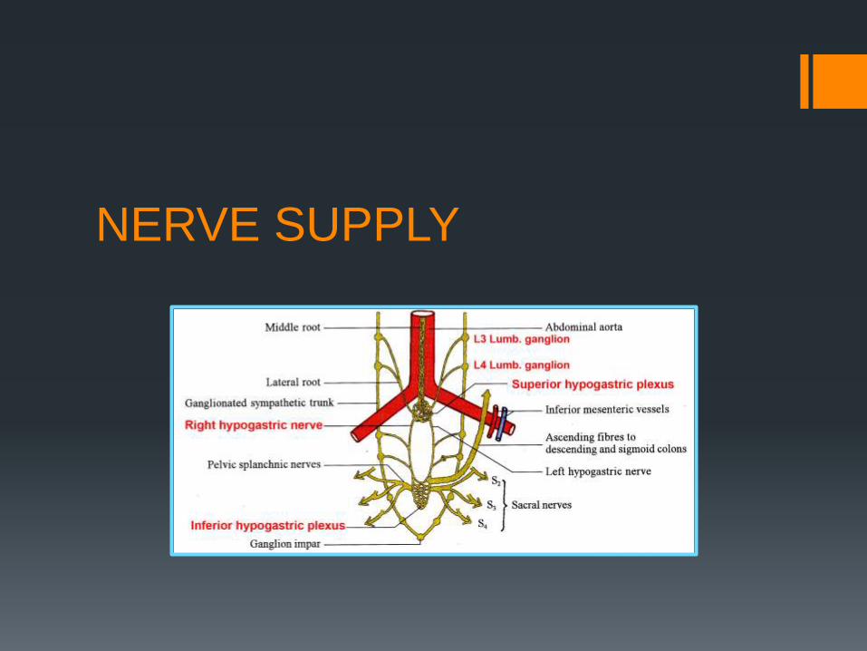
NERVE SUPPLY

IMPORTANT!!! When considering metastasis of cancer cells, tumors in
superior part are painless, and those in inferior part painful.

MUSCULARIS EXTERNA As in the upper parts of GIT, it is divided into an outer
longitudinal and inner circular layer of smooth muscle.

ANAL FOLDS AND COLUMNS The mucosa and submucosa above the dentate line is
uneven and thrown into folds, the so-called anal cushions.
there are usually three, the left lateral, right posterior and
right anterior positions, and they continue proximally as the
primary rectal foldings. Secondary foldings (the rectal
columns of Morgagni) lie both over and between the primary
folds.

MUCOSA SUSPENSORY
LIGAMENT the increased density of fibres that insert into the mucosa of
the anal crypts at the level of the dentate line, termed the
‘mucosal suspensory Ligament.

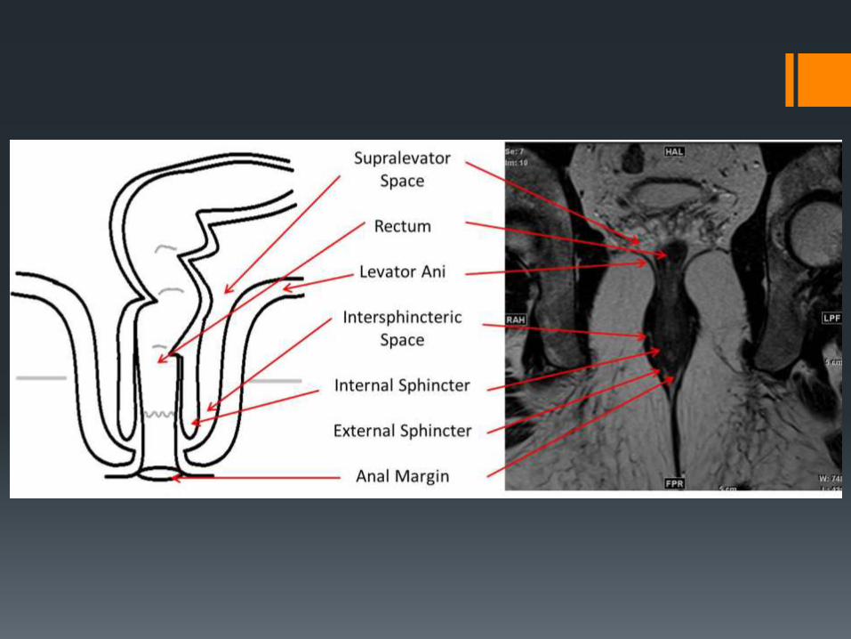
INVOLUNTARY INTERNAL VOLUNTARY EXTERNAL
ANAL SPHINCTERS
2-5 mm of thickening of the smooth muscle of the circular coat at the upper end of the anal canal.
Supplied by autonomic plexus and remains in tonic state of contraction.
It has three parts
Subcutaneous
Superficial
Deep
Supplied by pudendal
nerve.


ANORECTAL RING At the junction of rectum
and anal canal, the internal
sphincter, the deep part of
external sphincter,
conjoined longitudinal
muscle and puborectalis
muscles form anorectal
ring.

INTERSPHINCTERIC PLANE Between the external
sphincter muscle laterally and the longitudinal muscle medially exists a potential space, the intersphinctericplane.
IMPORTANCE
Contains anal glands
Route for spread of pus
The plane can be opened up surgically to provide access for operations on the sphincter muscles.
HILTON’S LINEALSO CALLED WHITE LINE/ANOCUTANEOUS INE.It is a color contrast bet’ bluish pink area above and black skin below.
The line is represented by inter-sphincteric groove at the lower end of the internal sphincter .Indicates lower end of internal sphincter.
Ischiorectal abscess when communicates with anal canal
usually opens at or below Hilton’s line.
ANAL GLANDS Anal glands may be found in the submucosa and
intersphincteric space, and normally number between 0 and
10 in an individual.
Their function is unknown, although they secrete mucin.
which perhaps lubricates the anal canal to ease defaecation.
The importance of intersphincteric anal glands is that they
are widely considered to be the potential source of anal
sepsis, either acute, presenting as perianal, ischiorectal or
even pelvic sepsis, or chronic, presenting as a
cryptoglandular (non-specific) anal fistula.