ANATOMY - Knee Jointccikinesiology.weebly.com/.../anatomy_-_knee_joint.pdf · 2019-09-13 ·...
8
ANATOMY Knee Joint Cobourg Collegiate Institute 1 2015 • Femur • Patella • Tibia • Fibula • Medial Collateral Ligament • Lateral Collateral Ligament • Posterior Cruciate Ligament • Anterior Cruciate Ligament • Medial Meniscus • Lateral Meniscus • Articular Cartilage • Quadriceps • Hamstrings • Quadriceps Tendon • Patellar Ligament KNEE JOINT PAGE #39
Transcript of ANATOMY - Knee Jointccikinesiology.weebly.com/.../anatomy_-_knee_joint.pdf · 2019-09-13 ·...

ANATOMY Knee Joint
Cobourg Collegiate Institute 1
2015
• Femur• Patella• Tibia• Fibula•Medial Collateral Ligament• Lateral Collateral Ligament• Posterior Cruciate Ligament•Anterior Cruciate Ligament
•Medial Meniscus• Lateral Meniscus•Articular Cartilage•Quadriceps•Hamstrings•Quadriceps Tendon• Patellar Ligament
KNEE JOINT
PAGE #39
ANATOMY Knee Joint
Cobourg Collegiate Institute 2
2015
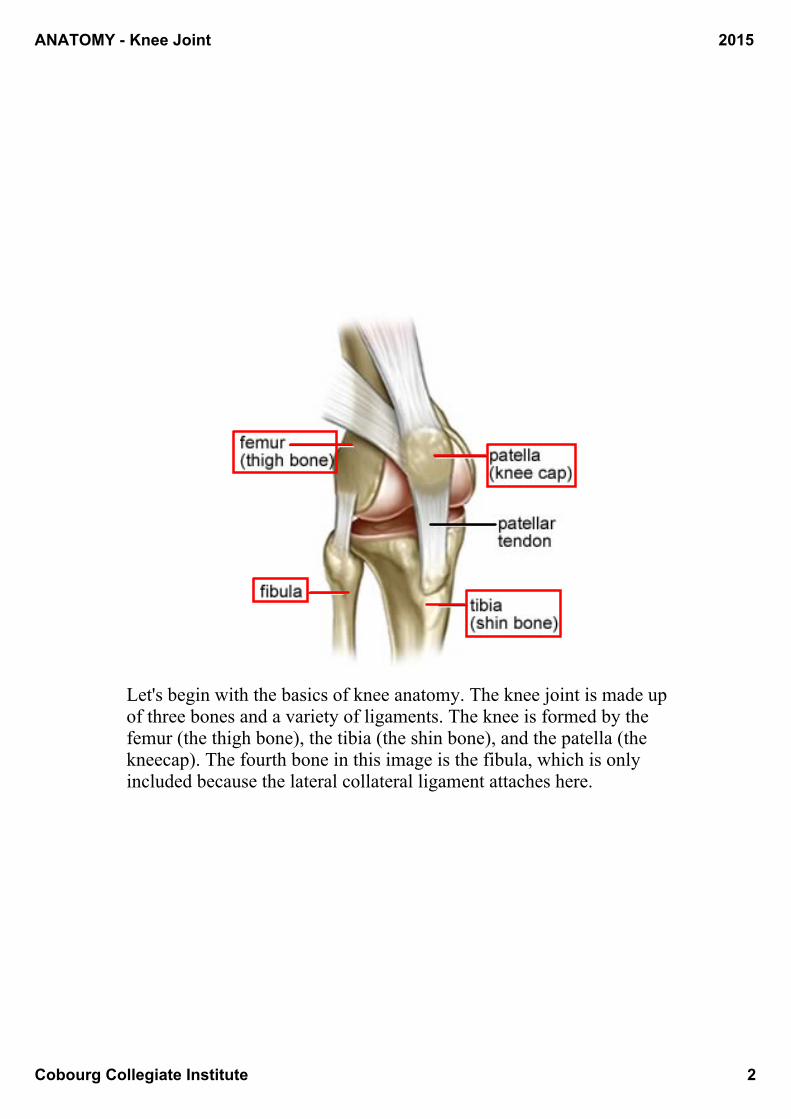
Let's begin with the basics of knee anatomy. The knee joint is made up of three bones and a variety of ligaments. The knee is formed by the femur (the thigh bone), the tibia (the shin bone), and the patella (the kneecap). The fourth bone in this image is the fibula, which is only included because the lateral collateral ligament attaches here.
ANATOMY Knee Joint
Cobourg Collegiate Institute 3
2015
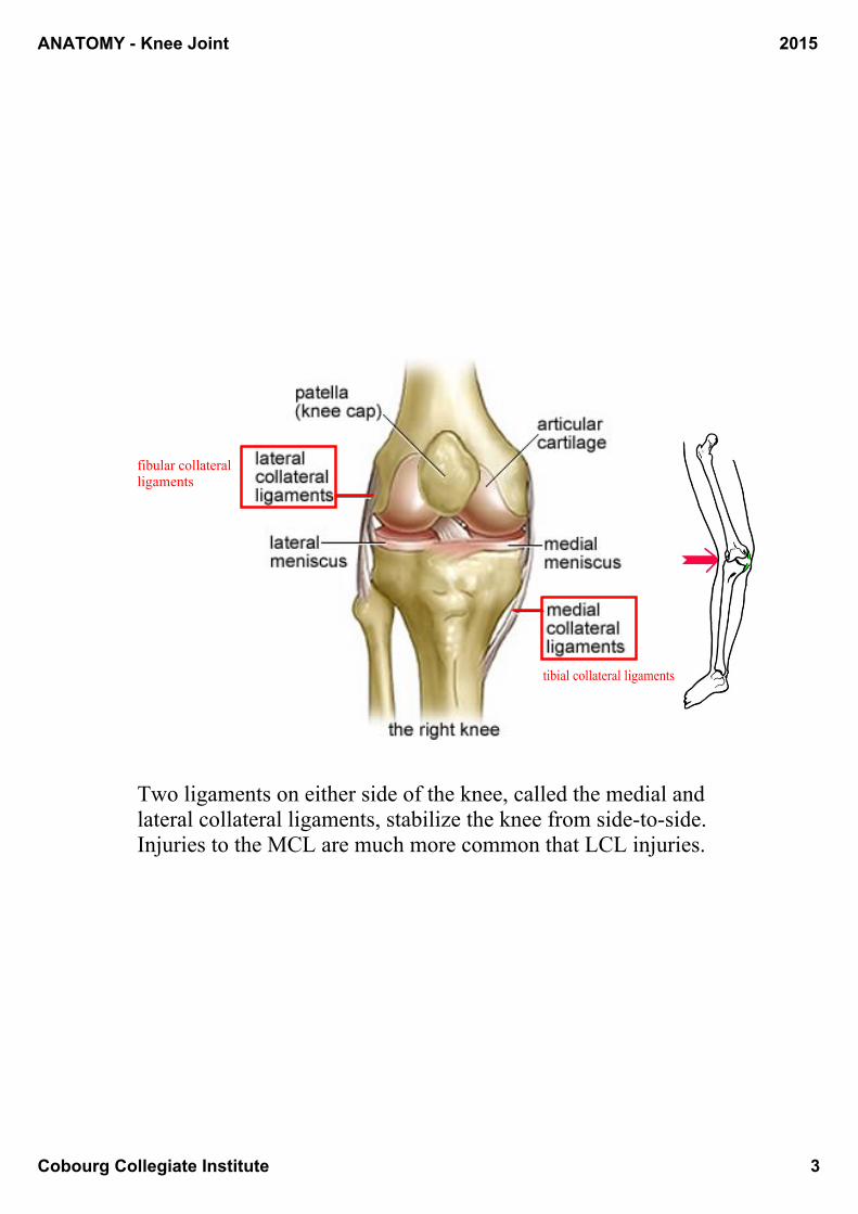
Two ligaments on either side of the knee, called the medial and lateral collateral ligaments, stabilize the knee from sidetoside. Injuries to the MCL are much more common that LCL injuries.
fibular collateral ligaments
tibial collateral ligaments
ANATOMY Knee Joint
Cobourg Collegiate Institute 4
2015
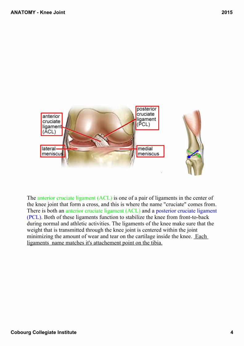
The anterior cruciate ligament (ACL) is one of a pair of ligaments in the center of the knee joint that form a cross, and this is where the name "cruciate" comes from. There is both an anterior cruciate ligament (ACL) and a posterior cruciate ligament (PCL). Both of these ligaments function to stabilize the knee from fronttoback during normal and athletic activities. The ligaments of the knee make sure that the weight that is transmitted through the knee joint is centered within the joint minimizing the amount of wear and tear on the cartilage inside the knee. Each ligaments name matches it's attachement point on the tibia.
ANATOMY Knee Joint
Cobourg Collegiate Institute 5
2015
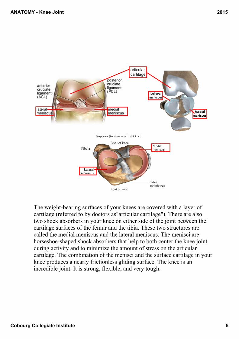
The weightbearing surfaces of your knees are covered with a layer of cartilage (referred to by doctors as"articular cartilage"). There are also two shock absorbers in your knee on either side of the joint between the cartilage surfaces of the femur and the tibia. These two structures are called the medial meniscus and the lateral meniscus. The menisci are horseshoeshaped shock absorbers that help to both center the knee joint during activity and to minimize the amount of stress on the articular cartilage. The combination of the menisci and the surface cartilage in your knee produces a nearly frictionless gliding surface. The knee is an incredible joint. It is strong, flexible, and very tough.
articular cartilage
ANATOMY Knee Joint
Cobourg Collegiate Institute 6
2015
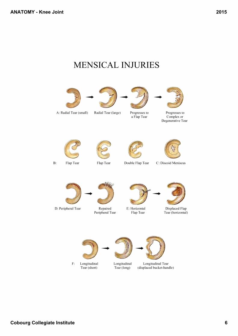
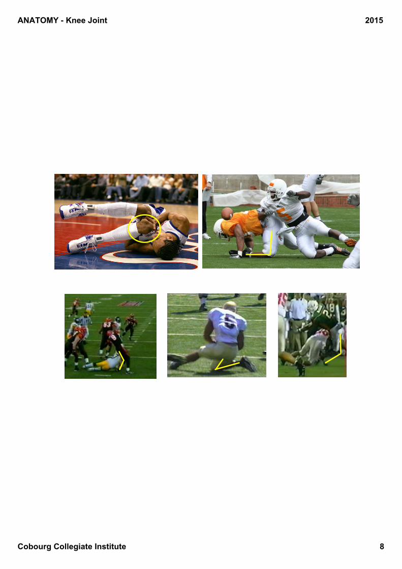
MENSICAL INJURIES
ANATOMY Knee Joint
Cobourg Collegiate Institute 7
2015
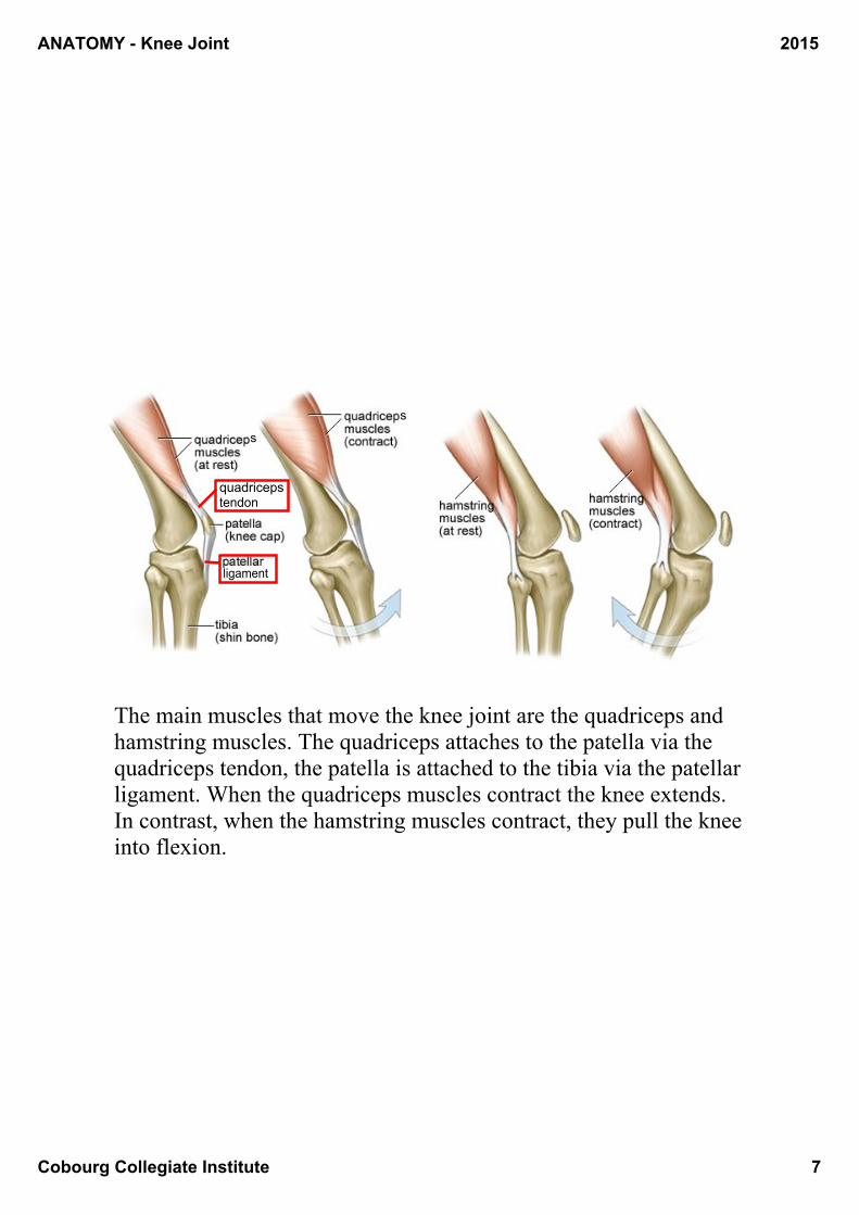
The main muscles that move the knee joint are the quadriceps and hamstring muscles. The quadriceps attaches to the patella via the quadriceps tendon, the patella is attached to the tibia via the patellar ligament. When the quadriceps muscles contract the knee extends. In contrast, when the hamstring muscles contract, they pull the knee into flexion.
quadriceps tendon
s
s
ligament
ANATOMY Knee Joint
Cobourg Collegiate Institute 8
2015