Anatomy and radiological evaluation of the wrist€¦ · Anatomy and radiological evaluation of the...
45
Anatomy and radiological evaluation of the wrist Andreas Panagopoulos, M.D., Ph. D. Assistant Professor in Orthopaedics, University Hospital of Patras
Transcript of Anatomy and radiological evaluation of the wrist€¦ · Anatomy and radiological evaluation of the...

Anatomy and radiological evaluation of the wrist
Andreas Panagopoulos, M.D., Ph. D. Assistant Professor in Orthopaedics,
University Hospital of Patras

- distal portions of radius and ulna,
- the proximal and distal rows
- bases of the metacarpals
The proximal carpal row is termed an
intercalated segment because forces
acting on its proximal and distal
articulations determine its position
Osseous anatomy

1. Radiocarpal
2. Midcarpal
3. Pisiform-triquetral
4. Common carpometacarpal
5. First carpometacarpal
6. Intermetacarpal
7. Inferior (distal) radioulnar
Articular Compartmental Anatomy

Ligamentous Anatomy - scapholunate
- lunotriquetral - capitotrapezoid - capitohamate - capitotriquetral - capitoscaphoid - triquetrohamate - scaphotrapeziumtrapezoid - trpeziotrapezoid
Intrinsic ligaments

Ligamentous Anatomy
Extrinsic ligaments
Mayefield
palmar dorsal dorsal palmar
Berger
dorsal palmar
Taleisnik

Poirier
V-ligament { Radiocapitate
Radioscapholunate
Radiolunotriquetral
Ulnotriquetral
Palmar ligaments

dorsal radio(scapho)triquetral Dorsal (inter)(radio)carpal
Dorsal ligaments

Functional “V” concept
Proximal volar Distal volar Dorsal volar

ulnocarpal complex and the radiolunotriquetral (RLT) ligament, both of which are extrinsic
Proximal volar V group
radiolunotriquetral (RLT) ligament ulnocarpal complex

Distal volar V group
Radioscaphocapitate (RSC), Scaphocapitate (SC), Triquetrocapitate (TC)
Triquetrocapitate (TC) Scaphocapitate (SC)

Dorsal V group
dorsal intercarpal (DIC) dorsal radio(scapho)triquetral (DRT)
dorsal radio(scapho)triquetral (DRT) dorsal intercarpal (DIC)
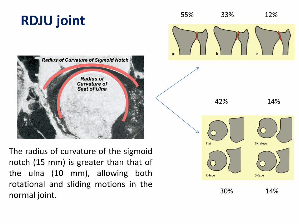
The radius of curvature of the sigmoid notch (15 mm) is greater than that of the ulna (10 mm), allowing both rotational and sliding motions in the normal joint.
55% 33% 12%
42% 14% 30% 14%
RDJU joint

Extrinsic stabilizers
(1) dynamic tensioning of the ECU (2) the semirigid sixth dorsal compartment subseath (3) the dynamic support provided by the superficial and deep heads of the pronator quadratus, and (4) the interosseous ligament of the midforearm

Intrinsic stabilizers
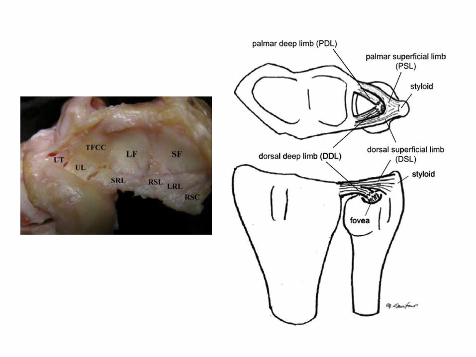
The articular disc is responsible for transferring load from the medial carpus to the pole of the distal ulna.
The TFC complex: superficial (green) and deep (blue) radioulna fibers, the two disc-carpal ligaments (disc-lunate and disc-triquetral), and the central articular disc (white).
fovea styloid

DRJU Biomechanics
84%

Common radiographic views
Posteroanterior
Oblique
Lateral

PA View
supination

Carpal arcs
Variations 1. short triquetrum = lunotriquetral step-off 2. proximally prominent hamate (H) with type II lunate = bilobate second and third carpal arc

Ulnar Variance
PUV = ulnar impaction syndrome
NUV = Kienbocks disease
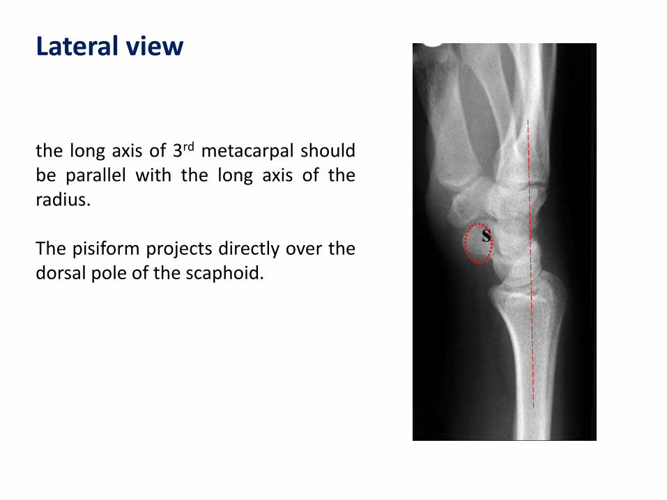
Lateral view
the long axis of 3rd metacarpal should be parallel with the long axis of the radius. The pisiform projects directly over the dorsal pole of the scaphoid.

Lateral view
The axes of the radius, lunate, and capitate should superimpose (0 to 30° is the normal capitate-lunate angle) The scapholunate angle ranges between 30 and 60°
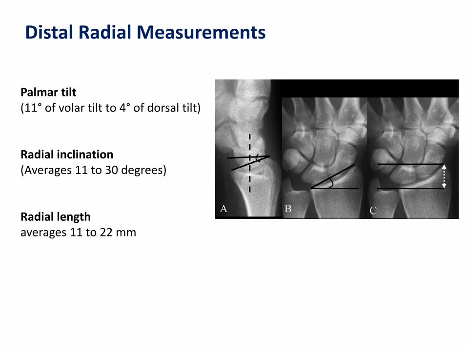
Distal Radial Measurements
Palmar tilt (11° of volar tilt to 4° of dorsal tilt) Radial inclination (Averages 11 to 30 degrees) Radial length averages 11 to 22 mm

Oblique views
Semisupinated oblique: the pisiform bone is separated from the remaining carpal bones. Semipronated oblique: allows examination of the radial aspect of the wrist, particularly the scaphoid and radial styloid

Deviation views
In radial deviation, the scaphoid rotates appears foreshortened. The distal scaphoid appears as a circular density The SCL distance remains normal (less than 2 mm). In ulnar deviation, the scaphoid is seen in full length. The scaphoid rotates and appears elongated The SLD interval may increase slightly

Carpal tunnel view
Hyperextension of the hand and the central ray is directed along the volar aspect at an angle of 25 to 30°
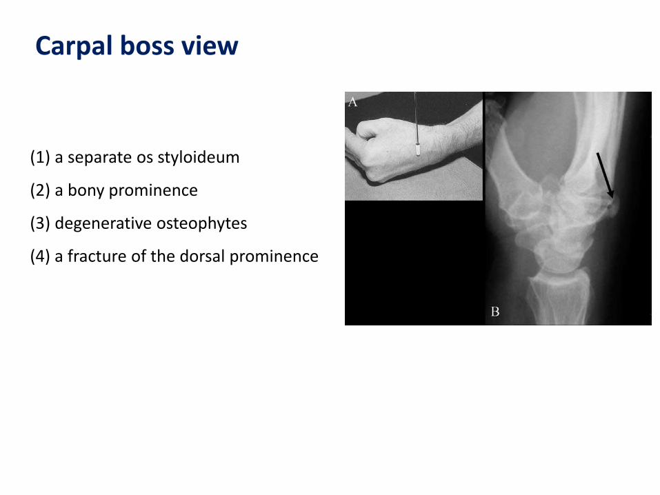
Carpal boss view
(1) a separate os styloideum
(2) a bony prominence
(3) degenerative osteophytes
(4) a fracture of the dorsal prominence
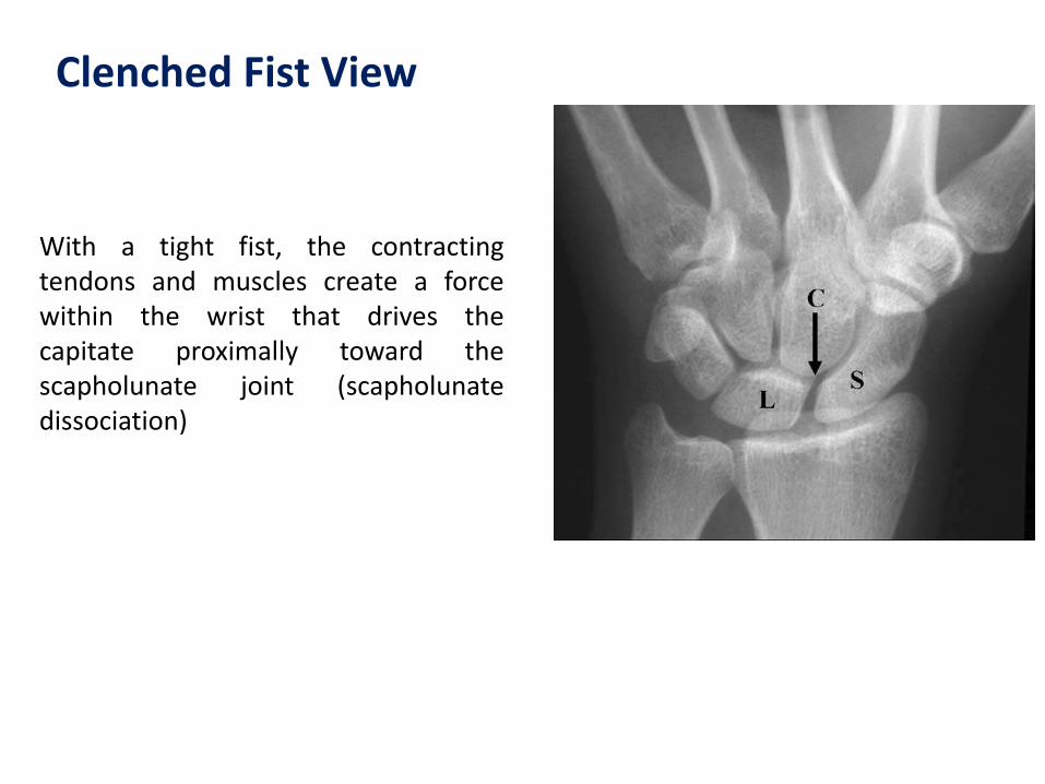
Clenched Fist View
With a tight fist, the contracting tendons and muscles create a force within the wrist that drives the capitate proximally toward the scapholunate joint (scapholunate dissociation)
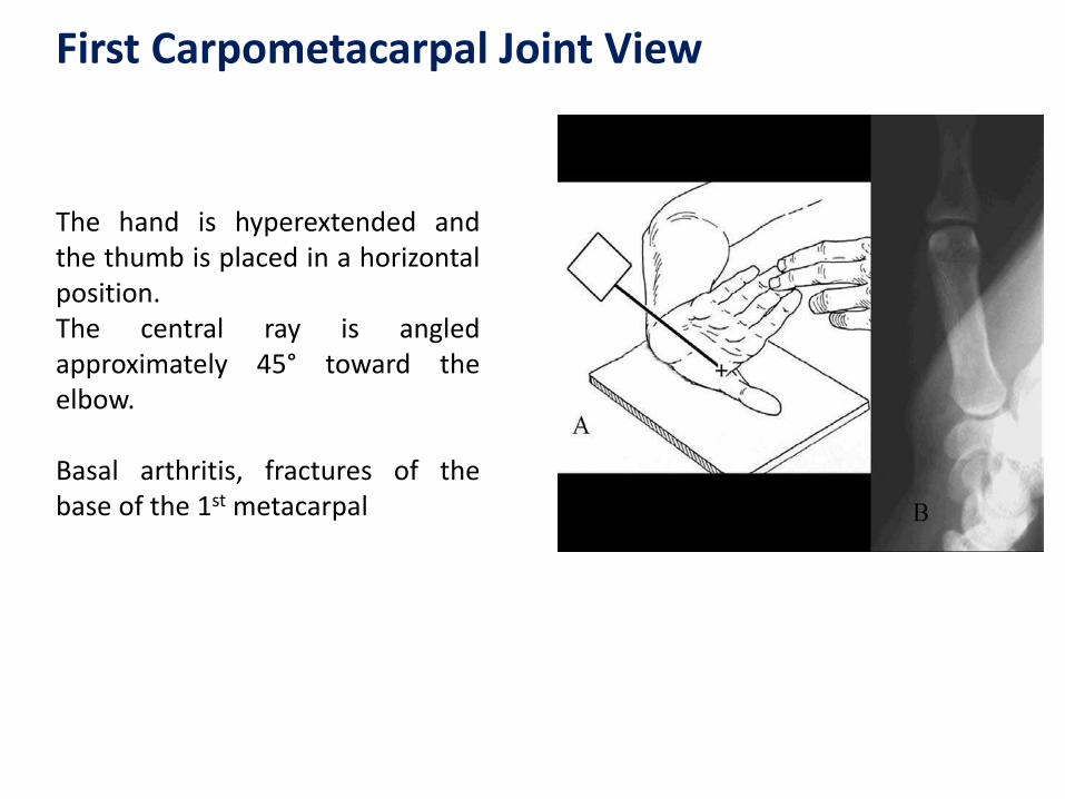
First Carpometacarpal Joint View
The hand is hyperextended and the thumb is placed in a horizontal position. The central ray is angled approximately 45° toward the elbow. Basal arthritis, fractures of the base of the 1st metacarpal

New method to evaluate the wrist ligaments
and radiocarpal cartilage. The hand and wrist
can be placed freely in any desired position
overall accuracy of 82–86 %
specificity 81–91 %.
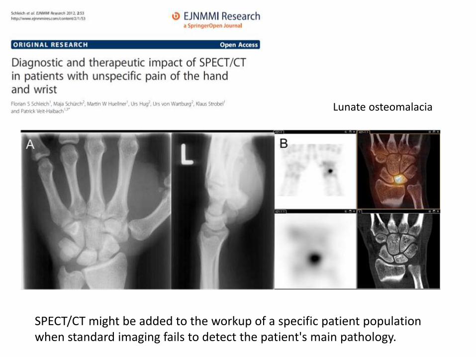
SPECT/CT might be added to the workup of a specific patient population when standard imaging fails to detect the patient's main pathology.
Lunate osteomalacia

Dynamic CT technique for assessment of wrist joint instabilities
4D CT (3D and time) technique to detect joint instability in a cadaveric model In the future using special segmentation techniques it will be possible to evaluate real time wrist kinematics

Obtaining the best possible image quality enables optimal interpretation and relies on several factors including field strength (a minimum of 1.0 T is recommended), and Positioning (“superman”)

MRI of the wrist
• Occult fracture
• Ganglion Cyst
• Tumor
• Ligament tear
• Avascular necrosis
• Arthritis
• Tendon Pathology
• Nerve Impingement
• Infection

MRI of the wrist
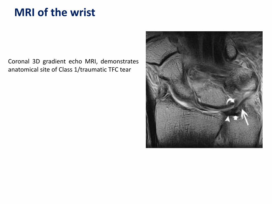
Coronal 3D gradient echo MRI, demonstrates anatomical site of Class 1/traumatic TFC tear
MRI of the wrist

… both CT and MRI might be incorporated in
the initial investigation of patients with wrist
trauma and a strong index of clinical suspicion.
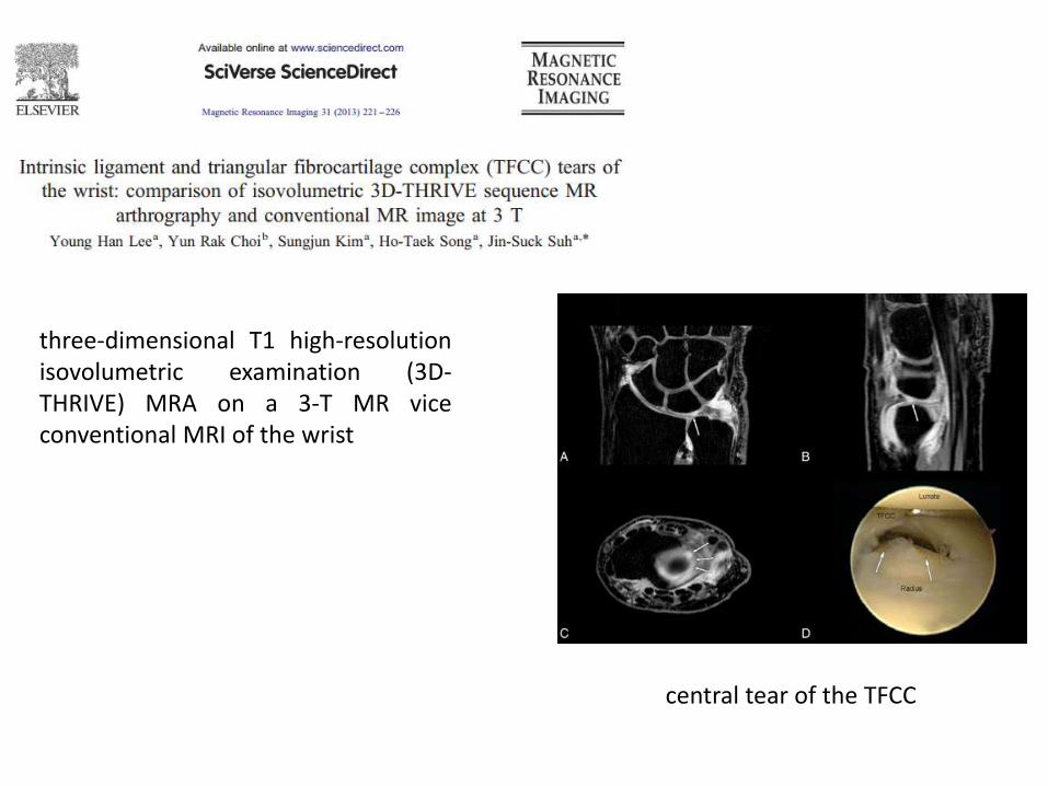
three-dimensional T1 high-resolution isovolumetric examination (3D-THRIVE) MRA on a 3-T MR vice conventional MRI of the wrist
central tear of the TFCC

Andreas Panagopoulos
Jonathan Compson
Richard Allom
The beneficial role of arthroscopy
in the investigation of wrist disorders:
A retrospective evaluation of 116 cases
Upper Limb Surgery &
Reconstruction Unit
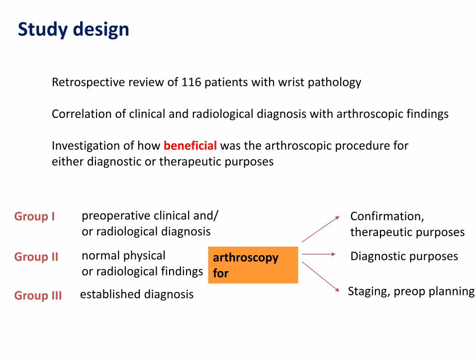
Study design
Retrospective review of 116 patients with wrist pathology Correlation of clinical and radiological diagnosis with arthroscopic findings Investigation of how beneficial was the arthroscopic procedure for either diagnostic or therapeutic purposes
Group I
Group II
Group III
preoperative clinical and/ or radiological diagnosis
normal physical or radiological findings
established diagnosis
arthroscopy for
Confirmation, therapeutic purposes
Diagnostic purposes
Staging, preop planning

Material
116 consecutive wrist arthroscopies
Seven year period (2002-2009)
49 male, 76 female
Mean age at operation 38 years (range 17-64 years)
57 patients (45.6%) had a documented previous injury
320 conventional diagnostic tests and 456 imagine studies!
Group I
Group II
Group III
preoperative clinical and/ or radiological diagnosis
Pain, but normal physical or radiological findings
established diagnosis
94 patients (75.2%)
12 patients
19 patients

Results
Group I: Arthroscopy was beneficial in 51/94 patients (54%) from in whom the pre-operative diagnosis was changed or augmented sufficiently to alter subsequent management. Group II: A beneficial arthroscopy establishing a definitive diagnosis was conducted for 9/12 patients (75%) Group III: Arthroscopy was of benefit to 14/19 patients (74%) for whom the subsequent definite management plan was modified. For all groups, arthroscopy was deemed of benefit when a therapeutic intervention was successfully conducted, independently of the ultimate outcome. There were 66/125 (53%) such patients.

Speculations…
9/12 (25%) of the patients in Group II (no diagnosis) had a normal arthroscopic appearance (9.4 investigations per patient!!!) - work compensation, malingering, simulation? - undiagnosed chronic wrist pain? 31/51 (61%) arthroscopies in Group I where the pre-operative diagnosis was changed, revealed significant unsuspected intra-articular pathology - unrelated to the clinical findings or misdiagnosed?

Conclusion
These data demonstrate the importance of wrist arthroscopy both as a diagnostic and therapeutic tool in the management of wrist disorders A thorough clinical examination is still the best way to reach the diagnosis Correlation of the unexpected arthroscopic findings with the symptoms of the patient to avoid over-treatment Useful tool in preoperative planning when a diagnosis is already exist

Thank you

![Radiological anatomy of_temporal_bone[1]](https://static.fdocuments.us/doc/165x107/5a6d2f6c7f8b9a10428b4ed5/radiological-anatomy-oftemporalbone1.jpg)


![Radiological anatomy of_abdomen[1]](https://static.fdocuments.us/doc/165x107/5a6d2f9f7f8b9ab3418b5eaf/radiological-anatomy-ofabdomen1.jpg)