Anatomy and Histochemistry of Flight Muscles in a Wing...
17
Anatomy and Histochemistry of Flight Muscles in a Wing-Propelled Diving Bird, the Atlantic Puffin, Fratercula arctica Christopher E. Kovacs 1 and Ron A. Meyers 2 * 1 Department of Ecology and Evolutionary Biology, Brown University, Providence, Rhode Island 2 Department of Zoology, Weber State University, Ogden, Utah ABSTRACT Twenty-three species within the avian fam- ily Alcidae are capable of wing-propelled flight in the air and underwater. Alcids have been viewed as Northern Hemisphere parallels to penguins, and have often been studied to see if their underwater flight comes at a cost, compromising their aerial flying ability. We examined the anatomy and histochemistry of select wing muscles (Mm. pectoralis, supracoracoideus, latissimus dorsi caudalis, coracobrachialis caudalis, triceps scapularis, and scapulo- humeralis caudalis) from Atlantic puffins (Fratercula arc- tica) to assess if the muscle fiber types reveal the existence of a compromise associated with “dual-medium” flight. Pectoralis was found to be proportional in size with that of nondiving species, although the supracoracoideus was proportionally larger in puffins. Muscle fiber types were largely aerobic in both muscles, with two distinct fast- twitch types demonstrable: a smaller, aerobic, moderately glycolytic population (FOg), and a larger, moderately aer- obic, glycolytic population (FoG). The presence of these two fiber types in the primary flight muscles of puffins suggests that aerial and underwater flight necessitate a largely aerobic fiber complement. We suggest that alcids do not represent an adaptive compromise, but a stable adaptation for wing-propelled locomotion both in the air and underwater. J. Morphol. 244:109 –125, 2000. © 2000 Wiley-Liss, Inc. Of the 9,700-plus species of birds, 32 are capable of flight under water in addition to “typical” (aerial) flight. These birds fall into three unrelated groups: alcids, diving petrels, and dippers. Alcids (which include puffins, murres, and auklets) inhabit the Northern Hemisphere and have often been com- pared to penguins, their Southern Hemisphere counterparts. Clearly, these two groups are “coun- terparts” in the loosest sense, as alcids are capable of both aerial and aquatic flight, whereas penguins are restricted to “flight” in the aquatic medium only. Storer (1960) proposed that the capacity for flight in air and water in alcids and diving petrels must represent a compromise stage between birds well suited for flight in air and those suited only to aquatic propulsion such as penguins. He suggested that compromise adaptations are reflected in the maximum and minimum body sizes attainable in wing-propelled divers. Larger birds require propor- tionately larger wings that are less effective under- water; he concluded that this restricts the maximum body size possible for alcids. The concept of alcids as compromise species between nondiving birds and penguins has led many researchers to probe the differences between alcids, penguins, and nondiving birds in an effort to identify adaptations specific to dual-medium fliers. Stettenheim (1959) compared the skeletal and muscular anatomy of a common murre with two nondiving species in the genera Larus and Limosa, and provided many anatomical examples which he believed important for wing- propelled diving. Raikow et al. (1988) examined fore- limb joint mobility in alcids and noted that in con- trast to penguins, alcids possess intrinsic wing muscles and do not show wing rigidity. Raikow and colleagues suggest that the presence of intrinsic wing muscles in alcids constrains the evolution of a flipper-like wing because subtle wing movements are required for aerial flight. These two important studies, along with more recent flight and physiolog- ical research, provide a foundation for our discussion of the adaptations in alcids. There have been a few accounts of the wing and shoulder anatomy of alcids and their involvement in underwater locomotion (Stettenheim, 1959; Storer, 1960; Spring, 1971), but to date, there has been no comprehensive study of the histochemical character- istics of muscles in alcids. For avian species limited to aerial flight, the pectoralis and supracoracoideus muscles are specialized to accommodate a variety of flight styles, from high frequency flapping to pro- Contract grant sponsors: Department of Ecology and Evolutionary Biology at Brown University, the Company of Biologists Ltd., the National Science Foundation; Contract grant number: IBN 9220097. *Correspondence to: Ron A. Meyers, Department of Zoology, Weber State University, Ogden, UT 84408-2505. E-mail: [email protected] JOURNAL OF MORPHOLOGY 244:109 –125 (2000) © 2000 WILEY-LISS, INC.
Transcript of Anatomy and Histochemistry of Flight Muscles in a Wing...

Anatomy and Histochemistry of Flight Muscles in aWing-Propelled Diving Bird, the Atlantic Puffin,Fratercula arcticaChristopher E. Kovacs1 and Ron A. Meyers2*
1Department of Ecology and Evolutionary Biology, Brown University, Providence, Rhode Island2Department of Zoology, Weber State University, Ogden, Utah
ABSTRACT Twenty-three species within the avian fam-ily Alcidae are capable of wing-propelled flight in the airand underwater. Alcids have been viewed as NorthernHemisphere parallels to penguins, and have often beenstudied to see if their underwater flight comes at a cost,compromising their aerial flying ability. We examined theanatomy and histochemistry of select wing muscles (Mm.pectoralis, supracoracoideus, latissimus dorsi caudalis,coracobrachialis caudalis, triceps scapularis, and scapulo-humeralis caudalis) from Atlantic puffins (Fratercula arc-tica) to assess if the muscle fiber types reveal the existenceof a compromise associated with “dual-medium” flight.Pectoralis was found to be proportional in size with that of
nondiving species, although the supracoracoideus wasproportionally larger in puffins. Muscle fiber types werelargely aerobic in both muscles, with two distinct fast-twitch types demonstrable: a smaller, aerobic, moderatelyglycolytic population (FOg), and a larger, moderately aer-obic, glycolytic population (FoG). The presence of thesetwo fiber types in the primary flight muscles of puffinssuggests that aerial and underwater flight necessitate alargely aerobic fiber complement. We suggest that alcidsdo not represent an adaptive compromise, but a stableadaptation for wing-propelled locomotion both in the airand underwater. J. Morphol. 244:109–125, 2000.© 2000 Wiley-Liss, Inc.
Of the 9,700-plus species of birds, 32 are capableof flight under water in addition to “typical” (aerial)flight. These birds fall into three unrelated groups:alcids, diving petrels, and dippers. Alcids (whichinclude puffins, murres, and auklets) inhabit theNorthern Hemisphere and have often been com-pared to penguins, their Southern Hemispherecounterparts. Clearly, these two groups are “coun-terparts” in the loosest sense, as alcids are capableof both aerial and aquatic flight, whereas penguinsare restricted to “flight” in the aquatic medium only.
Storer (1960) proposed that the capacity for flightin air and water in alcids and diving petrels mustrepresent a compromise stage between birds wellsuited for flight in air and those suited only toaquatic propulsion such as penguins. He suggestedthat compromise adaptations are reflected in themaximum and minimum body sizes attainable inwing-propelled divers. Larger birds require propor-tionately larger wings that are less effective under-water; he concluded that this restricts the maximumbody size possible for alcids. The concept of alcids ascompromise species between nondiving birds andpenguins has led many researchers to probe thedifferences between alcids, penguins, and nondivingbirds in an effort to identify adaptations specific todual-medium fliers. Stettenheim (1959) comparedthe skeletal and muscular anatomy of a commonmurre with two nondiving species in the generaLarus and Limosa, and provided many anatomical
examples which he believed important for wing-propelled diving. Raikow et al. (1988) examined fore-limb joint mobility in alcids and noted that in con-trast to penguins, alcids possess intrinsic wingmuscles and do not show wing rigidity. Raikow andcolleagues suggest that the presence of intrinsicwing muscles in alcids constrains the evolution of aflipper-like wing because subtle wing movementsare required for aerial flight. These two importantstudies, along with more recent flight and physiolog-ical research, provide a foundation for our discussionof the adaptations in alcids.
There have been a few accounts of the wing andshoulder anatomy of alcids and their involvement inunderwater locomotion (Stettenheim, 1959; Storer,1960; Spring, 1971), but to date, there has been nocomprehensive study of the histochemical character-istics of muscles in alcids. For avian species limitedto aerial flight, the pectoralis and supracoracoideusmuscles are specialized to accommodate a variety offlight styles, from high frequency flapping to pro-
Contract grant sponsors: Department of Ecology and EvolutionaryBiology at Brown University, the Company of Biologists Ltd., theNational Science Foundation; Contract grant number: IBN 9220097.
*Correspondence to: Ron A. Meyers, Department of Zoology, WeberState University, Ogden, UT 84408-2505.E-mail: [email protected]
JOURNAL OF MORPHOLOGY 244:109–125 (2000)
© 2000 WILEY-LISS, INC.

longed gliding and soaring (e.g., Rosser and George,1986a,b; Rosser et al., 1994). In penguins, thesemuscles are highly aerobic and contribute entirely tothrust generation and not body support (Bannasch,1994). We predict that the flight muscles of puffinswill contain attributes found in both aerial fliers andpenguins and that their histochemical compositionwill reflect this.
In this report, we have two major objectives. First,we describe the anatomical features of the wing andshoulder apparatus in the Atlantic puffin (Frater-cula arctica) we believe to be important for wingpropulsion underwater. The anatomical arrange-ment of the pectoral girdle musculature in puffinswill be compared with that of other alcids and divingand nondiving birds. Second, we will document thehistochemical profiles of the primary wing muscles.The correlation between muscle fiber histochemistryand muscle function has been described in mammals(Burke et al., 1971; Armstrong et al., 1982; Herman-son and Hurley, 1990; Hermanson et al., 1993) andbirds (Welsford et al., 1991; Meyers, 1992a; Sokoloffet al., 1998), and we will use this foundation fordetermining any specializations associated with theAtlantic puffin’s ability for aerial and underwaterflight. We hope to determine if alcids represent anintermediate in the evolution of wing-propelleddivers such as penguins, or if they are an adaptivecompromise for dual-medium flying. Additionally,we will analyze 1) interindividual differences be-tween individual puffins, and 2) the variation inmean muscle fiber diameter between multiple birdsin six different muscles.
MATERIALS AND METHODS
Twenty Atlantic puffins (Fratercula arctica) werepurchased from local hunters in Heimaey, Iceland.Muscles from eight birds were collected for histo-chemical analysis and the remaining birds were dis-sected both fresh and preserved for anatomical stud-ies. All muscles for histochemistry were excisedwithin 10 h of death. Samples were collected and allmaterial exported with permission of the IcelandicMinistry of the Environment. No birds were killedfor the sole purpose of this project. Muscle sampleswere collected during July 1995 and 1996 at theUniversity of Iceland’s Fisheries Research Unit inHeimaey, Iceland.
Anatomical Study
Dissections were made of five specimens, fixed informalin and stored in 2% phenoxyethanol. Aniodine solution (Bock and Shear, 1972) was used tohelp contrast muscle from connective tissue. Ana-tomical nomenclature is from Nomina AnatomicaAvium (Baumel et al., 1993). Names of processesand fossae to which muscles attach are given de-
scriptively and are then referred to by their NAAname.
Figures were prepared by tracing the dissectionswith a holbein camera lucida. The outline was thenenlarged to cover a surface of approximately 20 3 30cm. This drawing was compared to the original dis-section specimen, then corrected and details added.Final drawings were traced in ink and labeled.
Muscle Histochemistry
The following muscles were removed and weighed:pectoralis pars thoracicus, supracoracoideus, latissi-mus dorsi caudalis, triceps scapularis, coracobra-chialis caudalis, and scapulohumeralis caudalis.Samples (,1 cm3) from each muscle were mountedon wooden tongue depressors using Tissue-Tek em-bedding medium (Sakura Finetek, Torrance, CA)and rapidly frozen in isopentane cooled to about-150°C in liquid nitrogen. Samples were stored inliquid nitrogen until being transferred to dry-ice fortransport back to Brown University, where theywere then stored at -70°C. Transverse serial sections(12 mm) were cut in a cryostat (Leica, Jung Frigocut2800N) at -20°C. Sections were transferred to glasscover slips and air-dried for 30 to 120 min. Foridentification of fiber types, serial sections werestained for the presence of myofibrillar adenosinetriphosphatase (mATPase) following either alkaline(pH 10.4) or acidic (pH 4.3–4.6 in 0.1 increments)preincubation (Padykula and Herman, 1955; Guthand Samaha, 1969, 1970; Green et al., 1982). Sam-ples were incubated in an ATP buffer solution (pH9.4) for 30 min at 37°C and rinsed in calcium chlo-ride (1%), cobalt chloride (2%), and sodium barbital(0.01 M). Immersion in ammonium sulfide (1%)formed a precipitate identifying the stable mATPase.Sections were dehydrated in successive grades ofethanol and mounted on microscope slides usingxylene based Permount (Fisher Scientific, FairLawn, NJ).
Two additional sections from each block werestained for a glycolytic enzyme (a-glycerophosphatedehydrogenase, a-GPD) and an oxidative enzyme(nicotinamide adenine dinucleotide diaphorase,NADH-D) following the protocols of Novikoff et al.(1961) and Meijer (1968), respectively. Sections weredehydrated and mounted onto microscope slides asabove.
Slides were viewed under a Nikon microscope anddigital (8 bit gray scale) images of each section wereacquired at 200X magnification using a CCD cam-era, NIH Image software (W. Rasband, NationalInstitutes of Health) and a Macintosh Centris 650computer. The area of each fiber was measured us-ing the alkaline preincubation section as the fiberboundaries following this reaction were most clearlydefined.
110 C.E. KOVACS AND R.A. MEYERS

Nomenclature
George and Naik (1959) identified muscle fibertypes in mammals as either red, white, or interme-diate. These fiber type descriptors were expanded toinclude physiological parameters; red became fast-twitch red, white became fast-twitch white, and in-termediate fibers became slow-twitch intermediate.Peter et al. (1972) argued that fibers of the samecolor are composed of different quantities of meta-bolic enzymes and renamed fibers based on threecriteria: 1) contraction time relative to other fibersin the muscle, 2) glycolytic capacity, and 3) oxidativecapacity. Thus, fast-twitch white fibers were termedfast-twitch glycolytic fibers (FG); fast-twitch red fi-bers became fast-twitch oxidative-glycolytic (FOG)and slow-twitch intermediate fibers were more ap-propriately named slow-twitch-oxidative fibers (SO).In addition to these three categories of twitch fibers,tonic fibers have been identified in several species ofbirds (Rosser and George, 1986a,b; Rosser et al.,1987; Meyers, 1992a). Slow tonic fibers have differ-ent physiological and morphological characteristicsthat distinguish them from twitch fibers (Barnard etal., 1982). Tonic fibers are multiply innervated anddo not propagate an action potential (Moore et al.,1983; Johnston, 1985); they also have substantiallyslower contraction times than twitch fibers. Mam-malian fast-twitch-glycolytic fibers can have twitchtimes of 17.8-129 msec, and fast-twitch-oxidative-glycolytic fibers, 58-193 msec (Henneman and Ol-son, 1965). In contrast, avian slow tonic fibers takeseveral seconds to fully contract and relax (Barnardet al., 1982).
Rosser and George (1986b) showed that manyavian flight muscles contain only fast-twitch glyco-lytic, fast-twitch oxidative-glycolytic, and tonic fi-bers. However, Barnard et al. (1982) described anavian equivalent to the mammalian slow-oxidativefiber in chickens. These fibers have characteristicsthat include moderate mATPase staining at low pHand moderate NADH-D staining. Morphologically,these avian “SO” fibers are smaller in diameter thanFG or FOG fibers and have a high mitochondrialconcentration. Slow-oxidative fibers have been fur-ther documented in the flight muscles of other avianspecies (Meyers, 1997; Meyers and Mathias, 1997).
Throughout this study, we will use the conventionof Peter et al. (1972) to describe the muscle fibertypes present in six flight muscles of the Atlanticpuffin that are thought to be important for under-water and aerial propulsion. Prior work of manyresearchers provides a strong foundation for com-paring the muscle fiber types of an alcid to nondiv-ing, foot-propelled diving birds, and penguins(Hikida and Bock, 1974; Rosser and George, 1984,1986a,b; Rosser et al., 1987, 1994; Hikida, 1987;Meyers, 1992a, 1997; Meyers and Mathias, 1997;Ponganis et al., 1997).
Fiber Type Comparisons
Distinguishing between FG and FOG fibers wasnot as obvious using mATPase activity, glycolytic, oroxidative enzyme activity as reported for other spe-cies (Rosser and George, 1986a; Sokoloff et al.,1998). Several authors (Peter et al., 1972; Gollnickand Hodgson, 1986) cite a continuous staining inten-sity of metabolic enzymes as a compelling reason fornot using oxidative enzymes exclusively for classify-ing fibers. According to Gollnick and Hodgson(1986), the subdivisions using oxidative enzymes“may be more artificial than real.” A preferredmethod is to separate fibers using the glycolyticstaining intensity of a-GPD which correlates wellwith the glycolytic potential of fibers (Meijer, 1968).For these reasons, after the area of an individualfiber was measured using the mATPase stain (alka-line preincubation), the corresponding serial sectionstained for a-GPD was observed for final fiber typ-ing. The vast majority of fibers were identified asfast-twitch based on the stability of mATPase follow-ing alkaline and acidic preincubation, and then eval-uated on the basis of intensity of oxidative and gly-colytic activity (Peter et al., 1972). When the criteriafor slow-twitch-oxidative or slow-tonic fibers weresatisfied, such fibers were separated from the fast-twitch fibers and analyzed separately.
For this aspect of the study, all six muscles wereexamined in two birds. Between 476 and 593 fiberswere classified and measured in each muscle. Usingthe statistical program JMP (SAS Institute, Cary,NC) an analysis of variance (ANOVA) was per-formed comparing the cross-sectional areas of fastfibers for each muscle among the birds.
Fiber Size Comparisons
To compare fiber sizes, the areas of 100 to 2,000muscle fibers were measured in each of the six mus-cles. Eight birds were used for the comparison of thepectoralis and supracoracoideus muscles. Threebirds were used for comparison of the remainingfour muscles. While the entire muscle cross-sectioncould be sampled in the smallest muscles, the su-pracoracoideus and pectoralis muscles had to be di-vided into three and four individual blocks, respec-tively, and those blocks sampled. Thus, in these twolargest muscles not every fiber could be sampled.The regions sampled in the pectoralis and suprac-oracoideus muscles are shown in Figure 1.
The statistical program JMP (SAS Institute) wasused to compare the different regions of the pecto-ralis and supracoracoideus among the differentbirds. ANOVA was performed which included twofactors, the area of the fibers (response) and theregions sampled (predictor).
111PUFFIN FLIGHT MUSCLE HISTOCHEMISTRY

RESULTS
Various morphological features measured in theAtlantic puffin are listed in Table 1, along withsimilar measurements for other alcids and nondiv-ing birds. The average body mass of our Atlanticpuffins was 433 g, a value comparable with thosereported by Pennycuick (1987) and Hansen (1995).Wing span (0.568 m), wing area (0.0376 m2) andwing-loading (114.9 Nm-2) are also consistent withvalues reported by these authors.
Anatomy — Introduction
Muscles were selected based on their presumptivefunction in flight. Mm. pectoralis and supracoracoi-
deus are the primary downstroke and upstroke mus-cles, respectively. Additional muscles, Mm. latissi-mus dorsi pars caudalis, and scapulohumeraliscaudalis, coracobrachialis caudalis, and tricepsscapularis, were also selected due to their anatomi-cal orientation. Muscles are described relative to theavian anatomical position (see Raikow, 1985;Baumel et al., 1993), in which the wing is in anextended position. Thus, the wing can be dividedinto dorsal and ventral aspects, with the “leadingedge” cranial and the “trailing edge” caudal.
The anatomical description of each muscle in-cludes a brief orientation of the muscle, its relation-ship to surrounding structures, and a description ofthe origin and insertion. A statement of possiblefunction, based on muscle attachment, is also in-cluded (see Raikow, 1985). Comparisons, when ap-propriate, are made with muscles described in Stet-tenheim (1959) for murres (Uria), Hudson et al.(1969) for alcids, McKitrick (1991) for diving petrels(Pelecanoides), and Schreiweis (1982) for penguins.
Three different muscle fiber types were identifiedin the puffin: fast twitch, slow twitch, and slow tonic.All fast fibers reacted positively for both oxidativeand glycolytic enzymes, but differed in intensity.Based on the level of glycolytic staining (see above),two distinct populations could be identified. Wecould categorize fibers as either fast twitch, highlyoxidative, and moderately glycolytic, or fast twitch,moderately oxidative, and highly glycolytic. We willuse the abbreviations FOg and FoG, respectively, torefer to these two fiber populations. No fast twitchglycolytic fibers (FG) as described in the pigeon orchicken pectoralis were observed in Fratercula.
MusculatureM. latissimus dorsi pars caudalis (LD). The
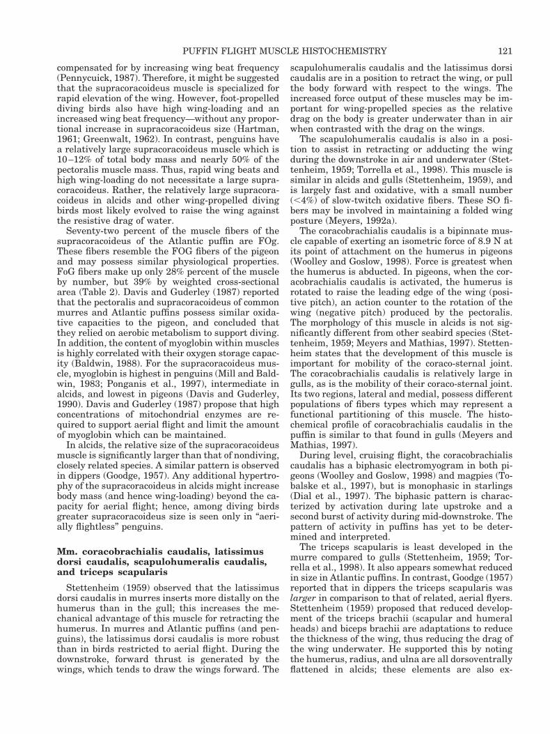
latissimus dorsi pars caudalis forms a largetriangular-shaped sheet with the pars cranialis, andforms the most superficial muscle layer of the back.It is triangular and extends from the vertebral col-umn to the humerus. LD arises from the caudal halfof the notarium and also from the fascia overlyingthe M. iliotrochantaricus caudalis. It tapers greatlyas it passes over the scapula toward the humerus.LD inserts onto the humeral shaft (margo caudalis)just caudal to the deltoideus major, and dorsal to thetriceps humeralis (Fig. 2). At the insertion, it liesventral to the fascicles of pars cranialis, but thefasciae of both muscles are fused. LD is in a positionto retract the humerus. Both Stettenheim (1959)and Hudson et al. (1969) ascribed its relatively largesize in alcids to a functional role in underwaterflight.
LD is comprised of 52% FoG fibers and 48% FOg.There are no slow fibers in the puffin LD, a findingconsistent with most avian species (Bock andHikida, 1968).
Fig. 1. Fratercula arctica. Lateral view of the right pectoralregion of the Atlantic puffin. A: Superficial view showing thora-cobrachialis (TB) and sternobrachialis (SB) regions of M. pecto-ralis. B: Deep view showing Mm. supracoracoideus, coracobra-chialis caudalis, and scapulohumeralis caudalis. Numbersindicate muscle regions sampled (see text). CBC, coracobrachialiscaudalis; c, coracoid; f, furcula; h, humerus; PT, insertion ofpectoralis onto humerus; s, scapula; SB, sternobrachialis regionof pectoralis; SC, supracoracoideus; scc, sterno-coraco-clavicularmembrane; SH, scapulohumeralis caudalis; st, sternum; TB, tho-racobrachialis region of pectoralis.
112 C.E. KOVACS AND R.A. MEYERS

M. pectoralis pars thoracicus (PT). The pecto-ralis muscle, the largest in birds, is elongate in al-cids. It is a broad, complex muscle that covers theentire ventral surface of the chest region. PT arisesfrom the distal edge of the sternal keel, from thelateral and caudal surfaces of the sternal body, fromthe lateral surface of the furcula, and from adjacentmembranes. PT is divided into distinct sternobra-chialis (SB) and thoracobrachialis (TB) portions byan internal sheet of connective tissue (Fig. 1). Theseportions have discrete fascicle arrangements. ThoseSB fascicles originating from the furcula are alignedtransversely (due to the cranial “bowing” of thisbone) and can function in wing protraction. The TBis arranged longitudinally, arising from the caudalsurface of the sternum and adjacent membranes,and is in a position to retract the wing. PT mainlyinserts onto the ventral surface of the pectoral crestof the humerus, but also has a connective tissueconnection to the bicipital crest of the humerus (seealso George and Berger, 1966; Meyers, 1992b).
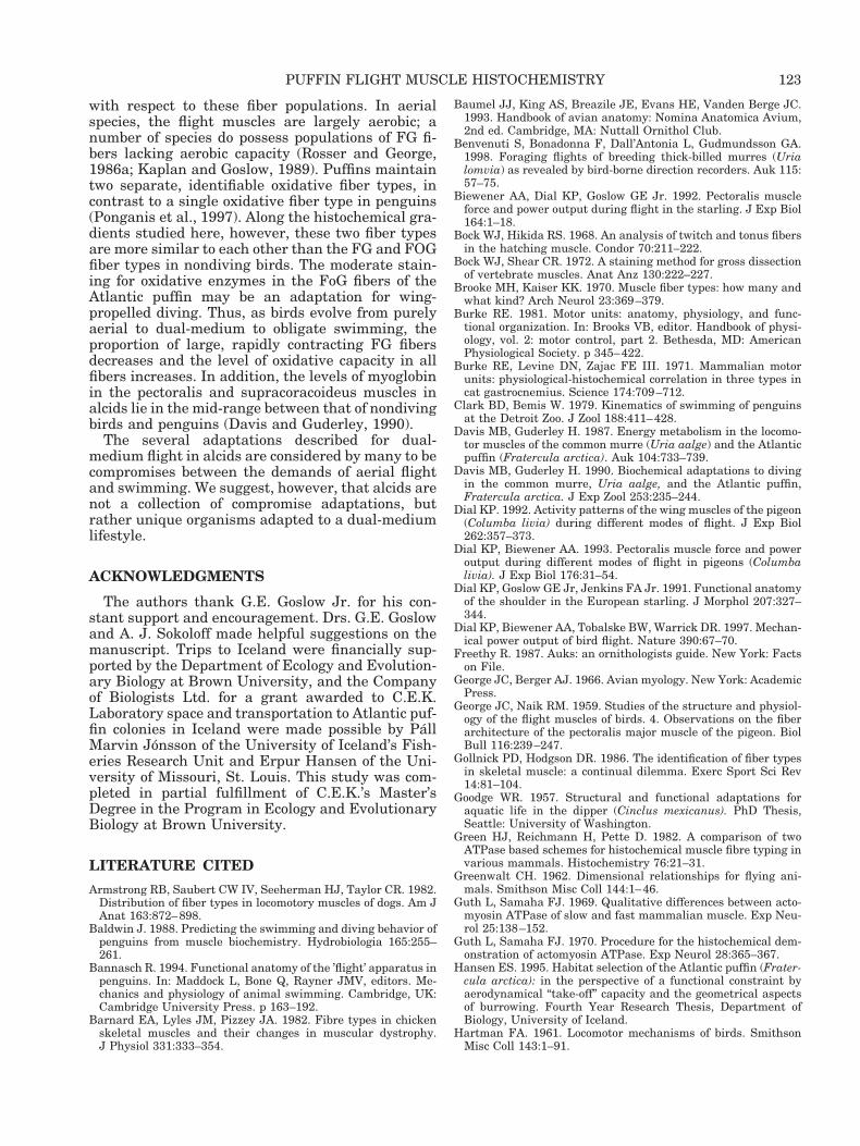
The pectoralis muscle is approximately 15% of thetotal body mass and 70–75% of the flight musclemass (Fig. 3). Eighty-seven percent of the muscle
fibers in the pectoralis of the Atlantic puffin are FOgfibers (Fig. 4) and have a mean area of approxi-mately 751 6 294 mm2 (30–33 mm diameter) (Table2). The remaining 13% of the fibers are FoG and aresignificantly larger than the FOg fibers, possessing amean cross-sectional area and diameter of 1,195 6393 mm2 and 40–41 mm, respectively. Fibers in theTB portion (region 4 in Fig. 1) were significantlylarger (895 6 292 mm2; P , 0.05) than those of theregions 1, 2, and 3 in the SB portion (768 6 253 mm2,790 6 275 mm2 and 764 6 239 mm2, respectively).
M. supracoracoideus (SC). The supracoracoi-deus muscle is a fusiform-shaped muscle, lying deepto the pectoralis on the ventral surface of the body.SC arises from the sternal body and keel (Fig. 1) andfrom the sterno-coraco-clavicular membrane. Thethick tendon emerges from the triosseal foramenonto the dorsal surface of the wing, deep to M. del-toideus minor (Fig. 2). The insertion of SC onto thetuberculum dorsalis of the humerus is very broad,and consists of two parts. The main tendon attachesalong a depression on the caudal surface of the hu-merus. A smaller tendon attaches cranially to it, butlies more proximal, adjacent (and proximal) to the
TABLE 1. Morphometrics of alcids and other birds
SpeciesMass(kg)
Wingspan(m)
Wingarea(m2)
Wingload
(Nm-2) Source
AlcidaeLeast auklet (Aethia pusilla) 0.104 0.325 0.0144 72.2 Spear and Ainley (1997)Dovekie (Alle alle) 0.153 0.320 0.0146 104.8 Pennycuick (1987)
0.096 0.0146 65.8 Poole (1938)Cassin’s auklet (Ptychoramphus aleuticus) 0.190 0.441 0.0262 72.5 Spear and Ainley (1997)Marbled murrelet (Brachyramphus
marmoratus)0.226 0.443 0.0240 94.2 Spear and Ainley (1997)
Parakeet auklet (Cyclorrhynchus psittacula) 0.282 0.502 0.0334 84.4 Spear and Ainley (1997)Atlantic puffin (Fratercula arctica) 0.433 0.568 0.0370 117.0 This study
0.398 0.549 0.0369 107.9 Pennycuick (1987)Female 0.424 0.571 0.0348 122.0 Hansen (1995)Male 0.474 0.593 0.0367 129.3 Hansen (1995)
Pigeon guillemot (Cepphus columba) 0.470 0.578 0.0422 111.4 Spear and Ainley (1997)Rhinoceros auklet (Cerorhinca monocerata) 0.560 0.615 0.0440 127.3 Spear and Ainley (1997)Razorbill (Alca torda) 0.620 0.661 0.0462 134.2 Pennycuick (1987)
0.74 Piatt and Nettleship (1985)Horned puffin (Fratercula corniculata) 0.663 0.607 0.0434 152.8 Spear and Ainley (1997)Tufted puffin (Fratercula cirrhata) 0.799 0.662 0.0512 156.1 Spear and Ainley (1997)Common murre (Uria aalge) 0.950 0.707 0.0544 174.6 Pennycuick (1987)
0.93 Piatt and Nettleship (1985)Thick-billed murre (Uria lomvia) 1.033 0.727 0.0560 184.5 Spear and Ainley (1997)Great auk (Pinguinus impennis) 5 0.0150 2200 Livezey (1988)
LaridaeLaughing gull (Larus atricilla) 0.299 0.590 0.1014 29.5 Hartman (1961)Herring gull (Larus argentatus) 0.940 1.310 0.1810 51.9 Pennycuick (1987)
SpheniscidaeLittle penguin (Eudyptula minor) 1.200 Montague (1985)Emperor penguin (Aptenodytes forsteri) 22 Kooyman et al. (1971)
GaviidaeBlack-throated loon (Gavia arctica) 2.425 0.1358 178.6 Poole (1938)
PelecanoididaeCommon diving petrel (Pelecanoides urinatrix) 0.137 0.393 0.0220 62.3 Pennycuick (1982)
0.150 0.425 0.0221 67.9 Spear and Ainley (1997)S. Georgia diving petrel (Pelecanoides georgicus) 0.114 0.381 0.0200 57.0 Spear and Ainley (1997)
ColumbidaePigeon (Columba livia) 0.314 0.0576 54.5 Poole (1938)
113PUFFIN FLIGHT MUSCLE HISTOCHEMISTRY
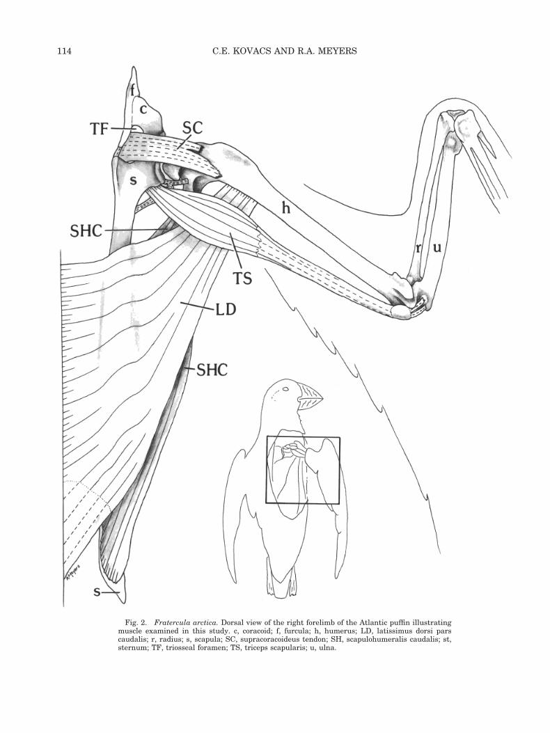
Fig. 2. Fratercula arctica. Dorsal view of the right forelimb of the Atlantic puffin illustratingmuscle examined in this study. c, coracoid; f, furcula; h, humerus; LD, latissimus dorsi parscaudalis; r, radius; s, scapula; SC, supracoracoideus tendon; SH, scapulohumeralis caudalis; st,sternum; TF, triosseal foramen; TS, triceps scapularis; u, ulna.
114 C.E. KOVACS AND R.A. MEYERS

insertion of the deltoid minor on the dorsal deltopec-toral crest (Fig. 2). In alcids the insertion is neitheron the proximal nor the leading edge of the humerusas in many aerial species (George and Berger, 1966),but is situated more caudally, and somewhat dis-tally. McKitrick (1991) described a deep layer of SCin Pelecanoides; however, this layer (also present inFratercula) is more accurately defined as a deephead of the M. deltoideus minor due to its innerva-tion (see also George and Berger, 1966; Baumel etal., 1993).
The supracoracoideus muscle comprises 4–5% ofthe total body mass, 20–22% of the flight musclemass (Fig. 3), and is approximately 30% of the pec-toralis muscle mass. Seventy-two percent of themuscle fibers of the SC are highly aerobic FOg andare significantly smaller in area (762 6 259 mm2)than the 28% FoG fibers (1,230 6 392 mm2) (Fig. 5;Table 2). There was no significant difference in fiberarea between any of the three regions sampled.
M. scapulohumeralis caudalis (SHC). Thescapulohumeralis caudalis lies on the dorsal surfaceof the body, beneath the latissimus dorsi complex(Fig. 2). It is a thick, triangular muscle, extendingfrom the scapula to the proximal humerus. SHCarises from the dorsal surface of the scapula, caudalto M. subscapularis caput laterale. The muscle fas-cicles taper to a short, stout tendon and insert ontothe distal process of the tuberculum ventrale of theproximal humerus (Fig. 1), adjacent to the origin ofthe triceps humeralis. SHC is in a position to retractand adduct the humerus (Stettenheim, 1959).
The scapulohumeralis caudalis is made up of amajority (61%) of FoG fibers which are significantlylarger (P , 0.05) in cross-sectional area than theFOg fibers (Table 2). Approximately 4% of the fiberssampled were stable at pH 4.3 for mATPase (Fig. 4).These fibers satisfy the criteria for mammalianslow-twitch-oxidative fibers (Barnard et al., 1982),and are reported as such.
M. triceps scapularis (scapulotriceps) (TS).The triceps scapularis lies on the caudal surface ofthe brachium, and extends from the scapula to theulna. TS arises via a complex of fleshy and tendinoustissue from the caudal border of the glenoid, adja-cent scapular shaft, and the dorsal scapulohumeralligament (Fig. 2). At about the level of the insertionof the latissimus dorsi pars cranialis, a humeralanchor connects TS to the humeral shaft. TS be-comes tendinous about halfway down the length ofthe humerus. The tendon of TS lies cranial to that ofM. triceps humeralis, to which it fuses at the distalhumerus. Two sesamoid bones are present in theconjoined tendons near the elbow joint (Fig. 1). Inthe resting folded wing position, these sesamoidbones lie between the humerus and the ulna. Whenthe forearm is extended to approximately 80°, thesesamoid bones slide cranially and lie within thefossae at the distal end of the humerus (sulcusscapulotricipitalis and sulcus humerotricipitalis).
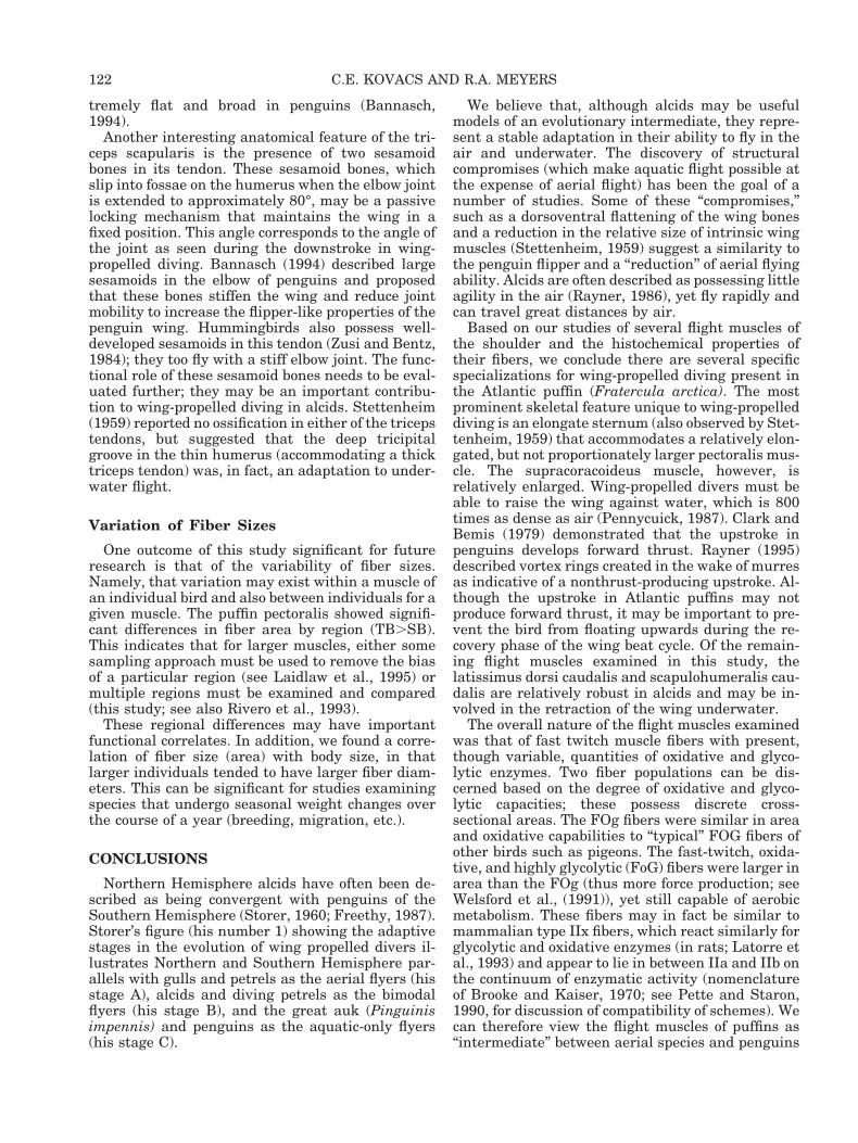
Fig. 3. Comparison of pectoralis and supracoracoideus musclemasses across a variety of wing-propelled and foot-propelled div-ing birds. A: Pectoralis muscle mass as a percent of body mass. Nosignificant difference between wing-propelled and foot-propelleddivers. B: Supracoracoideus muscle mass as a percent of bodymass. A significant difference (P , 0.001) was found betweenwing-propelled and foot-propelled divers. C: Ratio of supracora-coideus mass to pectoralis mass. For wing-propelled divers, themass of the supracoracoideus is approximately 30% of the pecto-ralis mass, a value much greater than that for foot-propelleddivers. Key: a, Atlantic puffin, Fratercula arctica (this study); b,razorbill, Alca torda; c, common murre, Uria aalge; d, Atlanticpuffin; e, dovekie, Alle alle; f, red-breasted merganser, Mergusserrator; g, common merganser, M. merganser; h, smew, Mergel-lus albellus; i, great crested grebe, Podiceps cristatus; j, red-necked grebe, P. grisegena; k, little grebe, Tachybaptus rufolava-tus; l, red-throated loon, Gavia stellata; m, common loon, G.immer; n, all other birds. All values are single data points fromGreenwalt (1962) except “a” which is a mean (6SD) from eightindividuals.
115PUFFIN FLIGHT MUSCLE HISTOCHEMISTRY

Fig. 4. Fratercula arctica. Serial sections of histochemistry of M. pectoralis (A–D) and M.scapulohumeralis caudalis (E). A: Alkaline preincubation (pH 10.4). B: Acidic preincubation (pH4.6). C: NADH. D: GPD. E: Acidic preincubation (pH 4.4) showing avian slow-twitch fibers, S. Notethat the FOg fibers react moderately for NADH, and weakly for GPD, whereas the FoG fibers reactweakly for NADH and strongly for GPD. Scale 5 25 mm.

These sesamoid bones are thought to contribute tothe stiffening of the wing for underwater propulsionin penguins (Bannasch, 1994). They may function tolock or otherwise assist in the maintenance of theflexed wing of puffins during the downstroke in un-derwater flight. Neither Stettenheim (1959) nor Mc-Kitrick (1991) reported any ossification in either ofthe triceps tendons in murres or diving petrels, re-spectively. From the united tendons, a thin tendonpasses cranially to insert onto the impressio scapu-lotriceps of the ulna (Fig. 2). TS plays an importantrole in aerial flight (Dial et al., 1991; Dial, 1992).
Triceps scapularis is composed mostly (79%) ofFOg fibers. Of the 480 fibers sampled, four (1.4%)were classified as slow-tonic fibers (ST). These STfibers were smaller in area (Table 2) than either ofthe FoG or FOg populations and stained moderatelyfor both a-GPD and NADH-D. Slow-tonic fibersstained lightly for mATPase at all pH levels, whilesurrounding fast-twitch fibers were alkali-stableand acid-labile. It is doubtful that these slow fiberscould contribute much force generation. No tonicfibers, but a few slow twitch fibers were found in thetriceps scapularis of one individual of the gull, Laruscalifornicus (Meyers and Mathias, 1997). No slowfibers of any kind were reported in this muscle of thegull L. cachinnans (Torella et al., 1998)
M. coracobrachialis caudalis (CBC). The cor-acobrachialis caudalis arises from the lateral mar-gin of the sternum along the costo-sternal joint, andfrom the lateral surface of the linea intermuscularisventralis of the coracoid and adjacent membranes. Itascends dorso-laterally and inserts on the tubercu-lum ventrale of the ventral surface of the humerusalong with the subcoracoideus muscle (Fig. 1) . Thisbipinnate muscle is divided by its internal tendoninto lateral and medial regions, which have distinctmuscle fiber types and different fiber morphologies.
The lateral region of the coracobrachialis caudalismuscle is composed primarily of FOg fibers withvery few FoG fibers. Slow fibers are entirely absentin this region. The medial region is predominantlymade up of FoG fibers with SO fibers comprisingonly 9% (Fig. 5). The SO fibers are significantly
smaller (P , 0.05) in area than FoG and FOg fibers(Table 2). The proportion and distribution of SOfibers in the Atlantic puffin is similar to that ob-served in the California gull (Meyers and Mathias,1997).
Comparisons of Fiber Types
A typical series of micrographs from the musclesexamined is represented by the pectoralis sections inFigure 4. Individual fibers are labeled as FoG or FOg.The FoG fibers are not only larger than the FOg fibersbut they tend to have more stable mATPase followingalkaline preincubation and are also most stable atlow pH. Peter et al. (1972) noted similar findings forFG fibers at low pH. The full range of fiber typepercentages can be found in Table 2. ANOVA wasconducted to compare the cross-sectional areas ofthe FoG and FOg fibers. The mean area of FoG fibersis significantly larger than that of FOg fibers in thepectoralis, supracoracoideus, and scapulohumeraliscaudalis muscles, and tends to be larger, but notsignificantly, in the coracobrachialis caudalis, latis-simus dorsi caudalis, and the triceps scapularismuscles. An effect of the variation between individ-ual birds exists in three muscles, the pectoralis,supracoracoideus, and scapulohumeralis caudalis.
FoG muscle fibers represented between 13% and52% of the fibers sampled in the six muscles (Table 2).The pectoralis had the lowest percentage of FoG fiberswhile latissimus dorsi had the highest. Slow-tonic fi-bers comprised less than 1.4% of the fibers in thetriceps scapularis, and slow-twitch oxidative musclefibers comprised 7% and 4% of the fiber population ofthe coracobrachialis caudalis and scapulohumeraliscaudalis muscles, respectively (Table 2).
Comparisons of Fiber Sizes
One goal of this study was to assess the inter- andintramuscular variation in mean muscle fiber areawithin the pectoralis and supracoracoideus muscles.In the pectoralis, fibers in regions 1, 2, and 3 (Fig. 1)have similar mean fiber areas, 768.5, 789.4, and
TABLE 2. Muscle fiber type comparisons
Muscle
Mean fiber area (mm2)(standard deviation)
% of total fiberssampled
Weighted cross-sectional area (%)
FoG FOg Slow FoG FOg Slow FoG FOg Slow
Latissimus dorsi caudalis 799 (385) 799 (336) — 52% 48% — 52% 48% —Pectoralis 1,195a (396) 751 (294) — 13% 87% — 19% 81% —Supracoracoideus 1,230a (392) 762 (260) — 28% 72% — 39% 61% —Scapulohumeralis caudalis 1,152a (270) 851 (221) 777c (177) 35% 61% 4% 42% 54% 3%Triceps scapularis 1,100 (199) 988 (253) 688b,d (173) 21% 78% 1% 23% 76% 1%Coracobrachialis caudalis 1,257 (511) 931 (361) 480b,c (83) 25% 68% 7% 32% 65% 3%
aSignificantly larger than FOg fibers (p , 0.05).bSignificantly smaller than FoG and FOg fibers (p , 0.05).cSlow-twitch.dSlow-tonic.
117PUFFIN FLIGHT MUSCLE HISTOCHEMISTRY
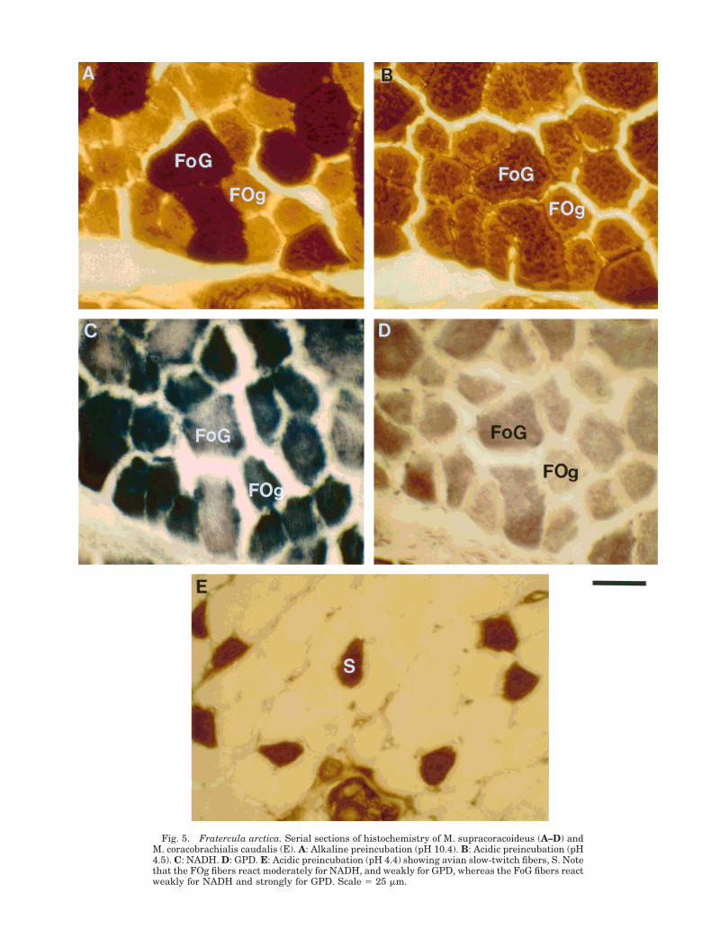
Fig. 5. Fratercula arctica. Serial sections of histochemistry of M. supracoracoideus (A–D) andM. coracobrachialis caudalis (E). A: Alkaline preincubation (pH 10.4). B: Acidic preincubation (pH4.5). C: NADH. D: GPD. E: Acidic preincubation (pH 4.4) showing avian slow-twitch fibers, S. Notethat the FOg fibers react moderately for NADH, and weakly for GPD, whereas the FoG fibers reactweakly for NADH and strongly for GPD. Scale 5 25 mm.
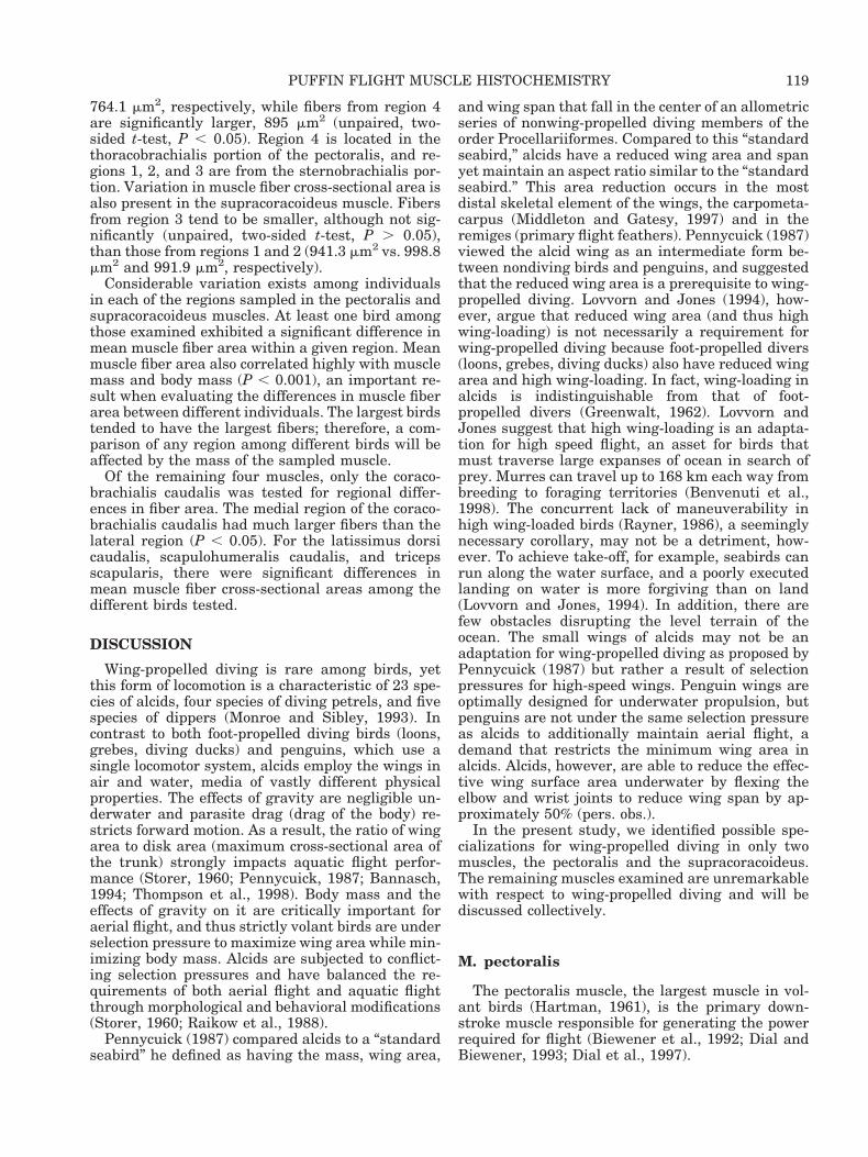
764.1 mm2, respectively, while fibers from region 4are significantly larger, 895 mm2 (unpaired, two-sided t-test, P , 0.05). Region 4 is located in thethoracobrachialis portion of the pectoralis, and re-gions 1, 2, and 3 are from the sternobrachialis por-tion. Variation in muscle fiber cross-sectional area isalso present in the supracoracoideus muscle. Fibersfrom region 3 tend to be smaller, although not sig-nificantly (unpaired, two-sided t-test, P . 0.05),than those from regions 1 and 2 (941.3 mm2 vs. 998.8mm2 and 991.9 mm2, respectively).
Considerable variation exists among individualsin each of the regions sampled in the pectoralis andsupracoracoideus muscles. At least one bird amongthose examined exhibited a significant difference inmean muscle fiber area within a given region. Meanmuscle fiber area also correlated highly with musclemass and body mass (P , 0.001), an important re-sult when evaluating the differences in muscle fiberarea between different individuals. The largest birdstended to have the largest fibers; therefore, a com-parison of any region among different birds will beaffected by the mass of the sampled muscle.
Of the remaining four muscles, only the coraco-brachialis caudalis was tested for regional differ-ences in fiber area. The medial region of the coraco-brachialis caudalis had much larger fibers than thelateral region (P , 0.05). For the latissimus dorsicaudalis, scapulohumeralis caudalis, and tricepsscapularis, there were significant differences inmean muscle fiber cross-sectional areas among thedifferent birds tested.
DISCUSSION
Wing-propelled diving is rare among birds, yetthis form of locomotion is a characteristic of 23 spe-cies of alcids, four species of diving petrels, and fivespecies of dippers (Monroe and Sibley, 1993). Incontrast to both foot-propelled diving birds (loons,grebes, diving ducks) and penguins, which use asingle locomotor system, alcids employ the wings inair and water, media of vastly different physicalproperties. The effects of gravity are negligible un-derwater and parasite drag (drag of the body) re-stricts forward motion. As a result, the ratio of wingarea to disk area (maximum cross-sectional area ofthe trunk) strongly impacts aquatic flight perfor-mance (Storer, 1960; Pennycuick, 1987; Bannasch,1994; Thompson et al., 1998). Body mass and theeffects of gravity on it are critically important foraerial flight, and thus strictly volant birds are underselection pressure to maximize wing area while min-imizing body mass. Alcids are subjected to conflict-ing selection pressures and have balanced the re-quirements of both aerial flight and aquatic flightthrough morphological and behavioral modifications(Storer, 1960; Raikow et al., 1988).
Pennycuick (1987) compared alcids to a “standardseabird” he defined as having the mass, wing area,
and wing span that fall in the center of an allometricseries of nonwing-propelled diving members of theorder Procellariiformes. Compared to this “standardseabird,” alcids have a reduced wing area and spanyet maintain an aspect ratio similar to the “standardseabird.” This area reduction occurs in the mostdistal skeletal element of the wings, the carpometa-carpus (Middleton and Gatesy, 1997) and in theremiges (primary flight feathers). Pennycuick (1987)viewed the alcid wing as an intermediate form be-tween nondiving birds and penguins, and suggestedthat the reduced wing area is a prerequisite to wing-propelled diving. Lovvorn and Jones (1994), how-ever, argue that reduced wing area (and thus highwing-loading) is not necessarily a requirement forwing-propelled diving because foot-propelled divers(loons, grebes, diving ducks) also have reduced wingarea and high wing-loading. In fact, wing-loading inalcids is indistinguishable from that of foot-propelled divers (Greenwalt, 1962). Lovvorn andJones suggest that high wing-loading is an adapta-tion for high speed flight, an asset for birds thatmust traverse large expanses of ocean in search ofprey. Murres can travel up to 168 km each way frombreeding to foraging territories (Benvenuti et al.,1998). The concurrent lack of maneuverability inhigh wing-loaded birds (Rayner, 1986), a seeminglynecessary corollary, may not be a detriment, how-ever. To achieve take-off, for example, seabirds canrun along the water surface, and a poorly executedlanding on water is more forgiving than on land(Lovvorn and Jones, 1994). In addition, there arefew obstacles disrupting the level terrain of theocean. The small wings of alcids may not be anadaptation for wing-propelled diving as proposed byPennycuick (1987) but rather a result of selectionpressures for high-speed wings. Penguin wings areoptimally designed for underwater propulsion, butpenguins are not under the same selection pressureas alcids to additionally maintain aerial flight, ademand that restricts the minimum wing area inalcids. Alcids, however, are able to reduce the effec-tive wing surface area underwater by flexing theelbow and wrist joints to reduce wing span by ap-proximately 50% (pers. obs.).
In the present study, we identified possible spe-cializations for wing-propelled diving in only twomuscles, the pectoralis and the supracoracoideus.The remaining muscles examined are unremarkablewith respect to wing-propelled diving and will bediscussed collectively.
M. pectoralis
The pectoralis muscle, the largest muscle in vol-ant birds (Hartman, 1961), is the primary down-stroke muscle responsible for generating the powerrequired for flight (Biewener et al., 1992; Dial andBiewener, 1993; Dial et al., 1997).
119PUFFIN FLIGHT MUSCLE HISTOCHEMISTRY

In alcids, PT is relatively elongated, producing anincrease in the fascicle length while decreasing theoverall thickness of the muscle (Stettenheim, 1959;Spring, 1971). This muscle was described as havingtwo layers in Pelecanoides (McKitrick, 1991), butthis is probably not related to underwater flightsince diving petrels are related to gannets, petrels,and albatrosses, all of which possess a deep andsuperficial layer (Kuroda, 1961; Pennycuick, 1982).Despite the relative increase in length of the pecto-ralis in alcids, the relative mass has not changed. Inthe Atlantic puffin, the pectoralis is 15.4% of thetotal body mass. Greenwalt (1962) predicted the pec-toralis muscle of volant birds should be 15.5% oftotal body mass. Therefore, despite the structuralrearrangement of the fascicles, the pectoralis musclein the Atlantic puffin does not exhibit the samerelative hypertrophy seen in the supracoracoideusmuscle.
The fiber type composition of the pectoralis in theAtlantic puffin is somewhat different from that ofnondiving birds. The only previous study of pectora-lis histochemistry from an alcid (Cassin’s Auklets,Ptychoramphus aleuticus) observed mostly FOG fi-bers (Meyers et al., 1992). In the present study, weidentified two populations of fast, oxidative, and gly-colytic muscle fibers that could be separated on thebasis of enzyme staining intensity and fiber diame-ter. Eighty-seven percent of the muscle fibers in thepectoralis of the Atlantic puffin are FOg fibers andhave a mean diameter of 30–33 mm. The remainingfibers (13%) are FoG and are significantly larger indiameter (40–41 mm) than the FOg fibers. Thissmall proportion of FoG fibers may play a significantrole in power production. Laidlaw et al. (1995) cal-culated the weighted cross-sectional area, or thepercentage of the total cross-sectional area, occupiedby a single fiber type of several hindlimb muscles inthe turtle. They determined that a small proportionof relatively large fibers can contribute significantlyto the total cross-sectional area of the muscle andtherefore to force output. Thus, while our FoG fibersmake up 13% of fibers by percentage, they make up19% by weighted cross-sectional area (Table 2).Sokoloff et al. (1998) showed similar results from asmall percentage of FG fibers in the pigeon. Theyconcluded that a pectoralis comprised of 10% FGfibers could contribute 36% of the muscle’s totalpower. This is because FG fibers in pigeons have a5.5-fold greater area than the FOG fibers (Kaplanand Goslow, 1989).
In the Atlantic puffin, FOg fibers have similarareas to those reported for FOG fibers by Kaplanand Goslow (1989) in pigeons, and by Talesara andGoldspink (1978) for pigeons, gulls, and sparrows.However, puffin FoG fibers are considerably smallerthan FG fibers in these other birds (mean diametersof 40 mm in the puffin vs. 78 mm in the pigeon;Kaplan and Goslow, 1989). Oxidative fibers are typ-ically smaller in diameter than those of lower oxida-
tive capacity and have higher diffusion rates of ox-ygen (Burke, 1981; Hermanson and Hurley, 1990).The relatively small size and proportion of FoG fi-bers in the Atlantic puffin may not provide the samefunctional qualities (i.e., force production) of the FGfibers observed in pigeons; however, these fibersmay be important as they can produce more forcethan the smaller FOg fibers, and sustain this aero-bically due to their greater oxidative capacity. Thus,the highly aerobic pectoralis of the puffin is capableof producing rapid, sustained wing beats in air andcan store large quantities of oxygen for increasingthe aerobic dive capacity during wing-propelled div-ing (Davis and Guderley, 1987).
Based on the anatomy of the pectoralis muscleand corresponding histochemistry, we conclude thatthere are few modifications of this muscle specific towing-propelled diving. The relative muscle mass issimilar to nondiving birds (and penguins), and thepresence of oxidative enzymes in all of the musclefibers may be an adaptation to limit fatigue of thesemotor units in both air and water. Davis and Gud-erley (1990) found similar oxidative capacities be-tween murres and puffins and pigeons (though moremyoglobin in alcids) and suggested that evolving adiving physiology does not require much modifica-tion from a prior aerobic system. Diving, in general,seems to require a highly aerobic locomotor system,as evidenced by penguins (Ponganis et al., 1997), aswell as seals (Kanatous et al., 1999).
M. supracoracoideus
The avian supracoracoideus has a bipinnate archi-tecture, robust tendon and broad attachment to thedelto-pectoral crest that reflect the high stressesimparted by this muscle during the upstroke. Stet-tenheim (1959) proposed that elongation of the su-pracoracoideus in alcids does not translate intogreater shortening distance, but rather greater forceproduction. He also suggested that SC can elevatethe wing with the elbow and humerus adducted withmore power in the murre than in gulls or godwitsdue to the longer in-lever of its relatively distalinsertion. Poore et al. (1997) reported the supracora-coideus in pigeons and European starlings is capableof producing 39.4 6 6.2 N and 6.5 6 1.2 N, respec-tively. In these species and most other nondivingbirds, the supracoracoideus is only 1.6% of the totalbody mass and 10% of the pectoralis mass (Green-walt, 1962; Poore et al., 1997). In contrast, the su-pracoracoideus muscle of the Atlantic puffin is 4–5%of total body mass and 28–30% of the pectoralismass, figures in agreement with Greenwalt (1962)(Fig. 3).
Poore et al. (1997) illustrated that the primaryrole of the supracoracoideus is to produce a highvelocity, forceful rotation about the long axis of thehumerus which repositions the wing for the subse-quent downstroke. High wing-loading in birds is
120 C.E. KOVACS AND R.A. MEYERS
compensated for by increasing wing beat frequency(Pennycuick, 1987). Therefore, it might be suggestedthat the supracoracoideus muscle is specialized forrapid elevation of the wing. However, foot-propelleddiving birds also have high wing-loading and anincreased wing beat frequency—without any propor-tional increase in supracoracoideus size (Hartman,1961; Greenwalt, 1962). In contrast, penguins havea relatively large supracoracoideus muscle which is10–12% of total body mass and nearly 50% of thepectoralis muscle mass. Thus, rapid wing beats andhigh wing-loading do not necessitate a large supra-coracoideus. Rather, the relatively large supracora-coideus in alcids and other wing-propelled divingbirds most likely evolved to raise the wing againstthe resistive drag of water.
Seventy-two percent of the muscle fibers of thesupracoracoideus of the Atlantic puffin are FOg.These fibers resemble the FOG fibers of the pigeonand may possess similar physiological properties.FoG fibers make up only 28% percent of the muscleby number, but 39% by weighted cross-sectionalarea (Table 2). Davis and Guderley (1987) reportedthat the pectoralis and supracoracoideus of commonmurres and Atlantic puffins possess similar oxida-tive capacities to the pigeon, and concluded thatthey relied on aerobic metabolism to support diving.In addition, the content of myoglobin within musclesis highly correlated with their oxygen storage capac-ity (Baldwin, 1988). For the supracoracoideus mus-cle, myoglobin is highest in penguins (Mill and Bald-win, 1983; Ponganis et al., 1997), intermediate inalcids, and lowest in pigeons (Davis and Guderley,1990). Davis and Guderley (1987) propose that highconcentrations of mitochondrial enzymes are re-quired to support aerial flight and limit the amountof myoglobin which can be maintained.
In alcids, the relative size of the supracoracoideusmuscle is significantly larger than that of nondiving,closely related species. A similar pattern is observedin dippers (Goodge, 1957). Any additional hypertro-phy of the supracoracoideus in alcids might increasebody mass (and hence wing-loading) beyond the ca-pacity for aerial flight; hence, among diving birdsgreater supracoracoideus size is seen only in “aeri-ally flightless” penguins.
Mm. coracobrachialis caudalis, latissimusdorsi caudalis, scapulohumeralis caudalis,and triceps scapularis
Stettenheim (1959) observed that the latissimusdorsi caudalis in murres inserts more distally on thehumerus than in the gull; this increases the me-chanical advantage of this muscle for retracting thehumerus. In murres and Atlantic puffins (and pen-guins), the latissimus dorsi caudalis is more robustthan in birds restricted to aerial flight. During thedownstroke, forward thrust is generated by thewings, which tends to draw the wings forward. The
scapulohumeralis caudalis and the latissimus dorsicaudalis are in a position to retract the wing, or pullthe body forward with respect to the wings. Theincreased force output of these muscles may be im-portant for wing-propelled species as the relativedrag on the body is greater underwater than in airwhen contrasted with the drag on the wings.
The scapulohumeralis caudalis is also in a posi-tion to assist in retracting or adducting the wingduring the downstroke in air and underwater (Stet-tenheim, 1959; Torrella et al., 1998). This muscle issimilar in alcids and gulls (Stettenheim, 1959), andis largely fast and oxidative, with a small number(,4%) of slow-twitch oxidative fibers. These SO fi-bers may be involved in maintaining a folded wingposture (Meyers, 1992a).
The coracobrachialis caudalis is a bipinnate mus-cle capable of exerting an isometric force of 8.9 N atits point of attachment on the humerus in pigeons(Woolley and Goslow, 1998). Force is greatest whenthe humerus is abducted. In pigeons, when the cor-acobrachialis caudalis is activated, the humerus isrotated to raise the leading edge of the wing (posi-tive pitch), an action counter to the rotation of thewing (negative pitch) produced by the pectoralis.The morphology of this muscle in alcids is not sig-nificantly different from other seabird species (Stet-tenheim, 1959; Meyers and Mathias, 1997). Stetten-heim states that the development of this muscle isimportant for mobility of the coraco-sternal joint.The coracobrachialis caudalis is relatively large ingulls, as is the mobility of their coraco-sternal joint.Its two regions, lateral and medial, possess differentpopulations of fibers types which may represent afunctional partitioning of this muscle. The histo-chemical profile of coracobrachialis caudalis in thepuffin is similar to that found in gulls (Meyers andMathias, 1997).
During level, cruising flight, the coracobrachialiscaudalis has a biphasic electromyogram in both pi-geons (Woolley and Goslow, 1998) and magpies (To-balske et al., 1997), but is monophasic in starlings(Dial et al., 1997). The biphasic pattern is charac-terized by activation during late upstroke and asecond burst of activity during mid-downstroke. Thepattern of activity in puffins has yet to be deter-mined and interpreted.
The triceps scapularis is least developed in themurre compared to gulls (Stettenheim, 1959; Tor-rella et al., 1998). It also appears somewhat reducedin size in Atlantic puffins. In contrast, Goodge (1957)reported that in dippers the triceps scapularis waslarger in comparison to that of related, aerial flyers.Stettenheim (1959) proposed that reduced develop-ment of the triceps brachii (scapular and humeralheads) and biceps brachii are adaptations to reducethe thickness of the wing, thus reducing the drag ofthe wing underwater. He supported this by notingthe humerus, radius, and ulna are all dorsoventrallyflattened in alcids; these elements are also ex-
121PUFFIN FLIGHT MUSCLE HISTOCHEMISTRY
tremely flat and broad in penguins (Bannasch,1994).
Another interesting anatomical feature of the tri-ceps scapularis is the presence of two sesamoidbones in its tendon. These sesamoid bones, whichslip into fossae on the humerus when the elbow jointis extended to approximately 80°, may be a passivelocking mechanism that maintains the wing in afixed position. This angle corresponds to the angle ofthe joint as seen during the downstroke in wing-propelled diving. Bannasch (1994) described largesesamoids in the elbow of penguins and proposedthat these bones stiffen the wing and reduce jointmobility to increase the flipper-like properties of thepenguin wing. Hummingbirds also possess well-developed sesamoids in this tendon (Zusi and Bentz,1984); they too fly with a stiff elbow joint. The func-tional role of these sesamoid bones needs to be eval-uated further; they may be an important contribu-tion to wing-propelled diving in alcids. Stettenheim(1959) reported no ossification in either of the tricepstendons, but suggested that the deep tricipitalgroove in the thin humerus (accommodating a thicktriceps tendon) was, in fact, an adaptation to under-water flight.
Variation of Fiber Sizes
One outcome of this study significant for futureresearch is that of the variability of fiber sizes.Namely, that variation may exist within a muscle ofan individual bird and also between individuals for agiven muscle. The puffin pectoralis showed signifi-cant differences in fiber area by region (TB.SB).This indicates that for larger muscles, either somesampling approach must be used to remove the biasof a particular region (see Laidlaw et al., 1995) ormultiple regions must be examined and compared(this study; see also Rivero et al., 1993).
These regional differences may have importantfunctional correlates. In addition, we found a corre-lation of fiber size (area) with body size, in thatlarger individuals tended to have larger fiber diam-eters. This can be significant for studies examiningspecies that undergo seasonal weight changes overthe course of a year (breeding, migration, etc.).
CONCLUSIONS
Northern Hemisphere alcids have often been de-scribed as being convergent with penguins of theSouthern Hemisphere (Storer, 1960; Freethy, 1987).Storer’s figure (his number 1) showing the adaptivestages in the evolution of wing propelled divers il-lustrates Northern and Southern Hemisphere par-allels with gulls and petrels as the aerial flyers (hisstage A), alcids and diving petrels as the bimodalflyers (his stage B), and the great auk (Pinguinisimpennis) and penguins as the aquatic-only flyers(his stage C).
We believe that, although alcids may be usefulmodels of an evolutionary intermediate, they repre-sent a stable adaptation in their ability to fly in theair and underwater. The discovery of structuralcompromises (which make aquatic flight possible atthe expense of aerial flight) has been the goal of anumber of studies. Some of these “compromises,”such as a dorsoventral flattening of the wing bonesand a reduction in the relative size of intrinsic wingmuscles (Stettenheim, 1959) suggest a similarity tothe penguin flipper and a ‘‘reduction’’ of aerial flyingability. Alcids are often described as possessing littleagility in the air (Rayner, 1986), yet fly rapidly andcan travel great distances by air.
Based on our studies of several flight muscles ofthe shoulder and the histochemical properties oftheir fibers, we conclude there are several specificspecializations for wing-propelled diving present inthe Atlantic puffin (Fratercula arctica). The mostprominent skeletal feature unique to wing-propelleddiving is an elongate sternum (also observed by Stet-tenheim, 1959) that accommodates a relatively elon-gated, but not proportionately larger pectoralis mus-cle. The supracoracoideus muscle, however, isrelatively enlarged. Wing-propelled divers must beable to raise the wing against water, which is 800times as dense as air (Pennycuick, 1987). Clark andBemis (1979) demonstrated that the upstroke inpenguins develops forward thrust. Rayner (1995)described vortex rings created in the wake of murresas indicative of a nonthrust-producing upstroke. Al-though the upstroke in Atlantic puffins may notproduce forward thrust, it may be important to pre-vent the bird from floating upwards during the re-covery phase of the wing beat cycle. Of the remain-ing flight muscles examined in this study, thelatissimus dorsi caudalis and scapulohumeralis cau-dalis are relatively robust in alcids and may be in-volved in the retraction of the wing underwater.
The overall nature of the flight muscles examinedwas that of fast twitch muscle fibers with present,though variable, quantities of oxidative and glyco-lytic enzymes. Two fiber populations can be dis-cerned based on the degree of oxidative and glyco-lytic capacities; these possess discrete cross-sectional areas. The FOg fibers were similar in areaand oxidative capabilities to “typical” FOG fibers ofother birds such as pigeons. The fast-twitch, oxida-tive, and highly glycolytic (FoG) fibers were larger inarea than the FOg (thus more force production; seeWelsford et al., (1991)), yet still capable of aerobicmetabolism. These fibers may in fact be similar tomammalian type IIx fibers, which react similarly forglycolytic and oxidative enzymes (in rats; Latorre etal., 1993) and appear to lie in between IIa and IIb onthe continuum of enzymatic activity (nomenclatureof Brooke and Kaiser, 1970; see Pette and Staron,1990, for discussion of compatibility of schemes). Wecan therefore view the flight muscles of puffins as“intermediate” between aerial species and penguins
122 C.E. KOVACS AND R.A. MEYERS
with respect to these fiber populations. In aerialspecies, the flight muscles are largely aerobic; anumber of species do possess populations of FG fi-bers lacking aerobic capacity (Rosser and George,1986a; Kaplan and Goslow, 1989). Puffins maintaintwo separate, identifiable oxidative fiber types, incontrast to a single oxidative fiber type in penguins(Ponganis et al., 1997). Along the histochemical gra-dients studied here, however, these two fiber typesare more similar to each other than the FG and FOGfiber types in nondiving birds. The moderate stain-ing for oxidative enzymes in the FoG fibers of theAtlantic puffin may be an adaptation for wing-propelled diving. Thus, as birds evolve from purelyaerial to dual-medium to obligate swimming, theproportion of large, rapidly contracting FG fibersdecreases and the level of oxidative capacity in allfibers increases. In addition, the levels of myoglobinin the pectoralis and supracoracoideus muscles inalcids lie in the mid-range between that of nondivingbirds and penguins (Davis and Guderley, 1990).
The several adaptations described for dual-medium flight in alcids are considered by many to becompromises between the demands of aerial flightand swimming. We suggest, however, that alcids arenot a collection of compromise adaptations, butrather unique organisms adapted to a dual-mediumlifestyle.
ACKNOWLEDGMENTS
The authors thank G.E. Goslow Jr. for his con-stant support and encouragement. Drs. G.E. Goslowand A. J. Sokoloff made helpful suggestions on themanuscript. Trips to Iceland were financially sup-ported by the Department of Ecology and Evolution-ary Biology at Brown University, and the Companyof Biologists Ltd. for a grant awarded to C.E.K.Laboratory space and transportation to Atlantic puf-fin colonies in Iceland were made possible by PallMarvin Jonsson of the University of Iceland’s Fish-eries Research Unit and Erpur Hansen of the Uni-versity of Missouri, St. Louis. This study was com-pleted in partial fulfillment of C.E.K.’s Master’sDegree in the Program in Ecology and EvolutionaryBiology at Brown University.
LITERATURE CITED
Armstrong RB, Saubert CW IV, Seeherman HJ, Taylor CR. 1982.Distribution of fiber types in locomotory muscles of dogs. Am JAnat 163:872–898.
Baldwin J. 1988. Predicting the swimming and diving behavior ofpenguins from muscle biochemistry. Hydrobiologia 165:255–261.
Bannasch R. 1994. Functional anatomy of the ’flight’ apparatus inpenguins. In: Maddock L, Bone Q, Rayner JMV, editors. Me-chanics and physiology of animal swimming. Cambridge, UK:Cambridge University Press. p 163–192.
Barnard EA, Lyles JM, Pizzey JA. 1982. Fibre types in chickenskeletal muscles and their changes in muscular dystrophy.J Physiol 331:333–354.
Baumel JJ, King AS, Breazile JE, Evans HE, Vanden Berge JC.1993. Handbook of avian anatomy: Nomina Anatomica Avium,2nd ed. Cambridge, MA: Nuttall Ornithol Club.
Benvenuti S, Bonadonna F, Dall’Antonia L, Gudmundsson GA.1998. Foraging flights of breeding thick-billed murres (Urialomvia) as revealed by bird-borne direction recorders. Auk 115:57–75.
Biewener AA, Dial KP, Goslow GE Jr. 1992. Pectoralis muscleforce and power output during flight in the starling. J Exp Biol164:1–18.
Bock WJ, Hikida RS. 1968. An analysis of twitch and tonus fibersin the hatching muscle. Condor 70:211–222.
Bock WJ, Shear CR. 1972. A staining method for gross dissectionof vertebrate muscles. Anat Anz 130:222–227.
Brooke MH, Kaiser KK. 1970. Muscle fiber types: how many andwhat kind? Arch Neurol 23:369–379.
Burke RE. 1981. Motor units: anatomy, physiology, and func-tional organization. In: Brooks VB, editor. Handbook of physi-ology, vol. 2: motor control, part 2. Bethesda, MD: AmericanPhysiological Society. p 345–422.
Burke RE, Levine DN, Zajac FE III. 1971. Mammalian motorunits: physiological-histochemical correlation in three types incat gastrocnemius. Science 174:709–712.
Clark BD, Bemis W. 1979. Kinematics of swimming of penguinsat the Detroit Zoo. J Zool 188:411–428.
Davis MB, Guderley H. 1987. Energy metabolism in the locomo-tor muscles of the common murre (Uria aalge) and the Atlanticpuffin (Fratercula arctica). Auk 104:733–739.
Davis MB, Guderley H. 1990. Biochemical adaptations to divingin the common murre, Uria aalge, and the Atlantic puffin,Fratercula arctica. J Exp Zool 253:235–244.
Dial KP. 1992. Activity patterns of the wing muscles of the pigeon(Columba livia) during different modes of flight. J Exp Biol262:357–373.
Dial KP, Biewener AA. 1993. Pectoralis muscle force and poweroutput during different modes of flight in pigeons (Columbalivia). J Exp Biol 176:31–54.
Dial KP, Goslow GE Jr, Jenkins FA Jr. 1991. Functional anatomyof the shoulder in the European starling. J Morphol 207:327–344.
Dial KP, Biewener AA, Tobalske BW, Warrick DR. 1997. Mechan-ical power output of bird flight. Nature 390:67–70.
Freethy R. 1987. Auks: an ornithologists guide. New York: Factson File.
George JC, Berger AJ. 1966. Avian myology. New York: AcademicPress.
George JC, Naik RM. 1959. Studies of the structure and physiol-ogy of the flight muscles of birds. 4. Observations on the fiberarchitecture of the pectoralis major muscle of the pigeon. BiolBull 116:239–247.
Gollnick PD, Hodgson DR. 1986. The identification of fiber typesin skeletal muscle: a continual dilemma. Exerc Sport Sci Rev14:81–104.
Goodge WR. 1957. Structural and functional adaptations foraquatic life in the dipper (Cinclus mexicanus). PhD Thesis,Seattle: University of Washington.
Green HJ, Reichmann H, Pette D. 1982. A comparison of twoATPase based schemes for histochemical muscle fibre typing invarious mammals. Histochemistry 76:21–31.
Greenwalt CH. 1962. Dimensional relationships for flying ani-mals. Smithson Misc Coll 144:1–46.
Guth L, Samaha FJ. 1969. Qualitative differences between acto-myosin ATPase of slow and fast mammalian muscle. Exp Neu-rol 25:138–152.
Guth L, Samaha FJ. 1970. Procedure for the histochemical dem-onstration of actomyosin ATPase. Exp Neurol 28:365–367.
Hansen ES. 1995. Habitat selection of the Atlantic puffin (Frater-cula arctica): in the perspective of a functional constraint byaerodynamical “take-off” capacity and the geometrical aspectsof burrowing. Fourth Year Research Thesis, Department ofBiology, University of Iceland.
Hartman FA. 1961. Locomotor mechanisms of birds. SmithsonMisc Coll 143:1–91.
123PUFFIN FLIGHT MUSCLE HISTOCHEMISTRY

Henneman E, Olson CB. 1965. Relations between structure andfunction in the design of skeletal muscles. J Neurophysiol 28:581–598.
Hermanson JW, Hurley KJ. 1990. Architectural and histochem-ical analysis of the biceps brachii muscle of the horse. Acta Anat137:146–156
Hermanson JW, Cobb MA, Schutt WA, Muradali F, Ryan JM.1993. Histochemical and myosin composition of vampire bat(Desmodus rotundus) pectoralis muscle targets a unique loco-motory niche. J Morphol 217:347–356.
Hikida RS. 1987. Quantitative ultrastructure of histochemicallyidentified avian skeletal muscle fiber types. Anat Rec 218:128–135.
Hikida RS, Bock WJ. 1974. Analysis of fiber types in the pigeon’smetapatagialis muscle. I. Histochemistry, end plates and ultra-structure. Tissue Cell 6:411–430.
Hudson GE, Hoff KM, Vanden Berge J, Trivette EC. 1969. Anumerical study of the wing and leg muscles of Lari and Alcae.Ibis 111:459–523.
Johnston IA. 1985. Sustained force development: specializationsand variation among vertebrates. J Exp Biol 115:239–251.
Kanatous, SB, DiMichele, LD, Cowan, DF, Davis, RW. 1999. Highaerobic capacities in the skeletal muscles of pinnipeds: adapta-tions to diving hypoxia. J Appl Physiol 86:1247–1256.
Kaplan SR, Goslow GE Jr. 1989. Neuromuscular organization ofthe pectoralis (pars thoracicus) of the pigeon (Columba livia):implications for motor control. Anat Rec 224:426–430.
Kooyman GI, Drabek CM, Elsner R, Campbell WR. 1971. Divingbehavior of the emperor penguin, Aptenodytes forsteri. Auk88:775–795.
Kuroda N. 1961. Analysis of three adaptive body forms in theSteganopodes, with notes on pectoral muscles. Misc Rep Ya-mashina Inst Ornith Zool 3:54–66.
Laidlaw DH, Callister RJ, Stuart DG. 1995. Fiber-type composi-tion of hindlimb muscles in the turtle, Pseudemys (Trachemys)scripta elegans. J Morphol 225:193–211.
Latorre R, Gil F, Vazquez JM, Moreno F, Mascarello F, RamirezG. 1993. Skeletal muscle fiber types in the dog. J Anat 182:329–337.
Livezey BC. 1988. Morphometrics of flightlessness in Alcidae.Auk 105:681–698.
Lovvorn JR, Jones DR. 1994. Biomechanical conflicts betweenadaptations for diving and aerial flight in estuarine birds. Es-tuaries 117:75–82.
McKitrick MC. 1991. Forelimb myology of loons (Gaviiformes),with comments on the relationships of loons and tubenoses(Procellariiformes). Zool J Linn Soc 102:115–152.
Meijer AEFH. 1968 Improved histochemical method for the dem-onstration of the activity of a-glucan phosphorylase. Histoche-mie 12:244–252.
Meyers RA. 1992a. The morphological basis of folded-wing pos-ture in the American kestrel, Falco sparverius. Anat Rec 232:493–498.
Meyers RA. 1992b. Morphology of the shoulder musculature ofthe American kestrel, Falco sparverius, with implications forgliding flight. Zoomorphology 112:91–103.
Meyers RA. 1997. Anatomy and histochemistry of spread-wingposture in birds. 1. Wing drying posture in the double-crestedcormorant (Phalacrocorax auritus). J Morphol 233:67–76.
Meyers RA, Mathias E. 1997. Anatomy and histochemistry ofspread-wing posture in birds. 2. Gliding flight in the Californiagull, Larus californicus: a paradox of fast fibers and posture. JMorphol 233:237–247.
Meyers RA, Fisher K, Goslow L, Goslow GE Jr. 1992. Underwaterlocomotion and musculoskeletal organization in some wing-propelled diving birds. Am Zool 32:157A.
Middleton KM, Gatesy SM. 1997. The evolution of theropod fore-limb design and function. Am Zool 37:58A.
Mill GK, Baldwin J. 1983. Biochemical correlates of swimmingand diving behavior in the little blue penguin Eudyptula minor.Physiol Zool 56:242–254.
Monroe BL Jr, Sibley CG. 1993. A world checklist of birds. NewHaven: Yale University Press.
Montague TL. 1985. A maximum dive recorder for the littlepenguin. Emu 85:264–267.
Moore G, Johnston IA, Goldspink G. 1983. The pCa-tension char-acteristics of single skinned fibres isolated from the anteriorand posterior latissimus dorsi muscle of the chicken. J Exp Biol105:411–416.
Novikoff AB, Shin WY, Drucker J. 1961. Mitochondrial localiza-tion of oxidative enzymes: staining results with two tetrazoliumsalts. J Biophys Biochem Cytol 9:47–61.
Padykula HA, Herman E. 1955. The specificity of the histochem-ical method for adenosine triphosphate. J Histochem Cytochem3:170–183.
Pennycuick CJ. 1982. The flight in petrels and albatrosses (Pro-cellariiformes) observed in South Georgia and its vicinity. Phi-los Trans R Soc Lond (Biol) 300:75–106.
Pennycuick CJ. 1987. Flight of auks (Alcidae) and other northernseabirds compared with southern Procellariiformes: ornithodo-lite observations. J Exp Biol 128:335–347.
Peter JB, Barnard RJ, Edgerton VR, Gillespie CA, Stempel KE.1972. Metabolic profiles of three fiber types of skeletal muscle inguinea pigs and rabbits. Biochemistry 11:2627–2633.
Pette D, Staron RS. 1990. Cellular and molecular diversities ofmammalian skeletal muscle fibers. Rev Physiol Biochem Phar-macol 116:1–76.
Piatt JF, Nettleship DN. 1985. Diving depths of four alcids. Auk102:293–297.
Ponganis PJ, Costello ML, Starke LN, Mathieu-Costello O, Kooy-man GL. 1997. Structural and biochemical characteristics oflocomotory muscles of emperor penguins, Aptenodytes forsteri.Resp Physiol 109:73–80.
Poole EL. 1938. Weights and wing areas of North American birds.Auk 55:511–517.
Poore SO, Ashcroft A, Sanchez-Haiman A, Goslow GE Jr. 1997.The contractile properties of the m. supracoracoideus in thepigeon and starling: a case for long-axis rotation of the hu-merus. J Exp Biol 200:2987–3002.
Raikow RJ. 1985. Locomotor system. In: King AS, McLelland J,editors. Form and function in birds, vol. 3. London: AcademicPress. p 57–147.
Raikow RJ, Bicanovosky L, Bledsoe AH. 1988. Forelimb jointmobility and the evolution of wing-propelled diving birds. Auk105:446–451.
Rayner JMV. 1986. Pleuston: animals which move in water andair. Endeavour 10:58–64.
Rayner JMV. 1995. Dynamics of the vortex wakes of flying andswimming vertebrates. Symp Soc Exp Biol 49:131–155.
Rivero JLL, Serrano AL, Diz AM, Morales JL. 1993. Changes incross-sectional area and capillary supply of the muscle fiberpopulation in equine gluteus medius muscle as a function ofsampling depth. Am J Vet Res 54:32–37.
Rosser BWC, George JC. 1984. Some histochemical properties ofthe fiber types in the pectoralis muscle of an emu (Dromaiusnovaehollandiae). Anat Rec 209:301–305.
Rosser BWC, George JC. 1986a. The avian pectoralis: histochem-ical characterization and distribution of fiber types. Can J Zool64:1174–1185.
Rosser BWC, George JC. 1986b. Slow muscle fibers in the pecto-ralis of the turkey vulture (Cathartes aura): an adaptation forsoaring flight. Zool Anz 217:252–258.
Rosser BWC, Davis MB, Brocklebank JR, George JC. 1987. Onthe histochemical characterization and distribution of fast andslow muscle fibers in certain avian skeletal muscles. Acta His-tochem 81:85–93.
Rosser BWC, Waldbillig DM, Wick M, Bandman E. 1994. Musclefiber types in the pectoralis of the white pelican, a soaring bird.Acta Zool 75:329–336.
Schreiweis DO. 1982. A comparative study of the appendicularmusculature of penguins (Aves: Sphenisciformes). SmithsonContrib Zool 341:1–46.
Sokoloff AJ, Ryan JM, Valerie E, Wilson DS, Goslow GE Jr. 1998.Neuromuscular organization of avian flight muscle: morphologyand contractile properties of motor units in the pectoralis (parsthoracicus) of pigeon (Columba livia). J Morphol 236:179–208.
124 C.E. KOVACS AND R.A. MEYERS
Spear LB, Ainley DG. 1997. Flight behaviour of seabirds in rela-tion to wind direction and wing morphology. Ibis 139:221–233.
Spring L. 1971. A comparison of function and morphological ad-aptations in the common murre (Uria aalge) and thick-billedmurre (Uria lomvia). Condor 73:1–27.
Stettenheim P. 1959. Adaptations for underwater swimming inthe common murre (Uria aalge). PhD thesis, Ann Arbor: Uni-versity of Michigan.
Storer RW. 1960. Evolution in the diving birds. Int Ornith Congr12:694–707.
Talesara GL, Goldspink G. 1978. A combined histochemical andbiochemical study of myofibrillar ATPase in pectoral, leg andcardiac muscle of several species of bird. Histochem J 10:695–709.
Thompson CW, Wilson ML, Melvin EF, Pierce DJ. 1998. Anunusual sequence of flight-feather molt in common murres andits evolutionary implications. Auk 115:653–669.
Tobalske BW, Olson NE, Dial KP. 1997. Flight style of the black-billed magpie: variation in wing kinematics, neuromuscularcontrol, and muscle composition. J Exp Zool 279:313–329.
Torrella JR, Fouces V, Palomeque J, Viscor G. 1998. Capillarityand fibre types in locomotory muscles of wild yellow-leggedgulls. Physiol Zool 71:425–434.
Welsford IG, Meyers RA, Wilson DS, Satterlie RA, Goslow GE Jr.1991. Neuromuscular organization for “wing” control in a mol-lusc (Clione limacina) and a bird (Columba livia): parallels indesign. Am Zool 31:670–679.
Woolley JD, Goslow GE Jr. 1998. The functional characteristics ofan avian flight muscle: the coracobrachialis posterior. Am Zool38:34A.
Zusi RL, Bentz GD. 1984. Myology of the purple-throated carib(Eulampis jugularis) and other hummingbirds (Aves: Trochili-dae). Smithson Contrib Zool 385:1–70.
125PUFFIN FLIGHT MUSCLE HISTOCHEMISTRY