Anatomy
55
ANATOMY By Dr.Ajish Abraham
-
Upload
ajishabraham -
Category
Documents
-
view
2 -
download
0
description
Anatomy study
Transcript of Anatomy

ANATOMY
By Dr.Ajish Abraham

Defn -- It is a subject that deals with the study of the structures and their relations within the body of a living organism.
“Ana” – means Apart “Tomy”- means to cut
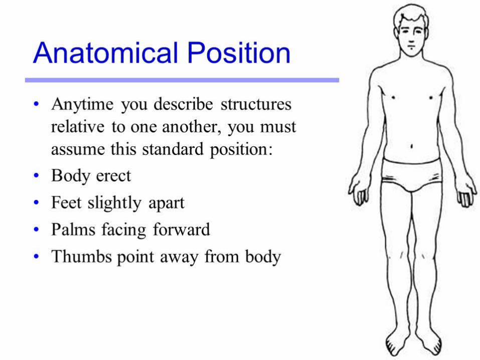
Standard Anatomical position
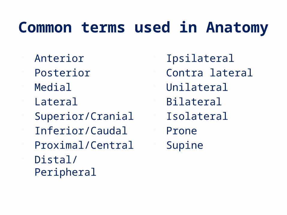
Common terms used in Anatomy
Anterior Posterior Medial Lateral Superior/Cranial Inferior/Caudal Proximal/Central Distal/Peripheral
Ipsilateral Contra lateral Unilateral Bilateral Isolateral Prone Supine

Branches Gross Anatomy – Its the study of the macroscopic
details of human body mostly through dissection
Living Anatomy – Study of the structures of live human being using techniques like palpation,auscultation,percussion etc
Microanatomy/Histology – Study of microscopic details of tissues that make the human body

Surface Anatomy – It is a branch which studies the relation between internal structures of human body with its surface.
Clinical Anatomy – It is application of anatomical knowledge to clinical practice.

Heircharcy of Cellular organisation
Cell –Smallest structural and functional unitTissue – Group of similar or dissimilar cells performing common function.
Organ – Group of similar or dissimilar tissues specific function
Organ system –Organ with blood vessels like artery, vein and capillary
Organism –Many organ systems working in harmony under control of brain.
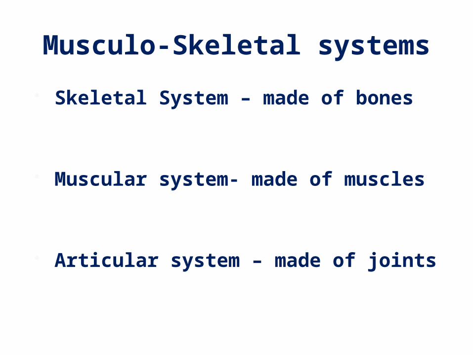
Musculo-Skeletal systems Skeletal System – made of bones
Muscular system- made of muscles
Articular system – made of joints
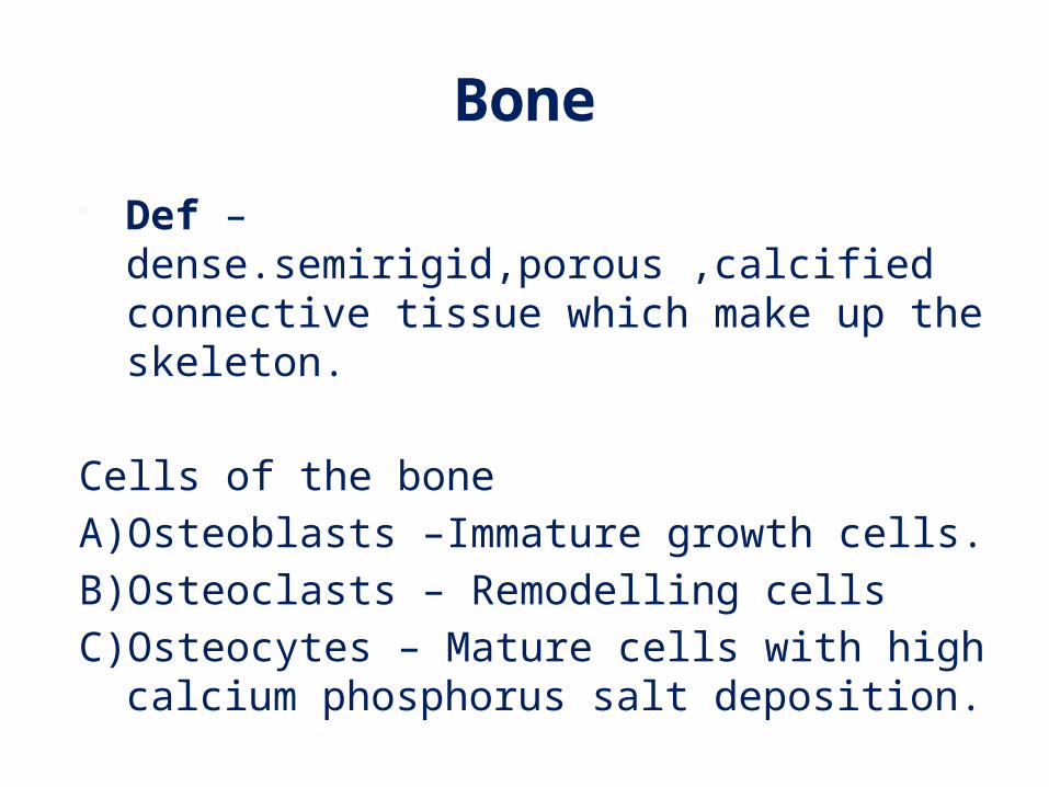
Bone Def – dense.semirigid,porous ,calcified connective
tissue which make up the skeleton.
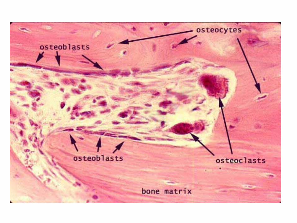
Cells of the boneA)Osteoblasts –Immature growth cells.B)Osteoclasts – Remodelling cellsC)Osteocytes – Mature cells with high calcium
phosphorus salt deposition.
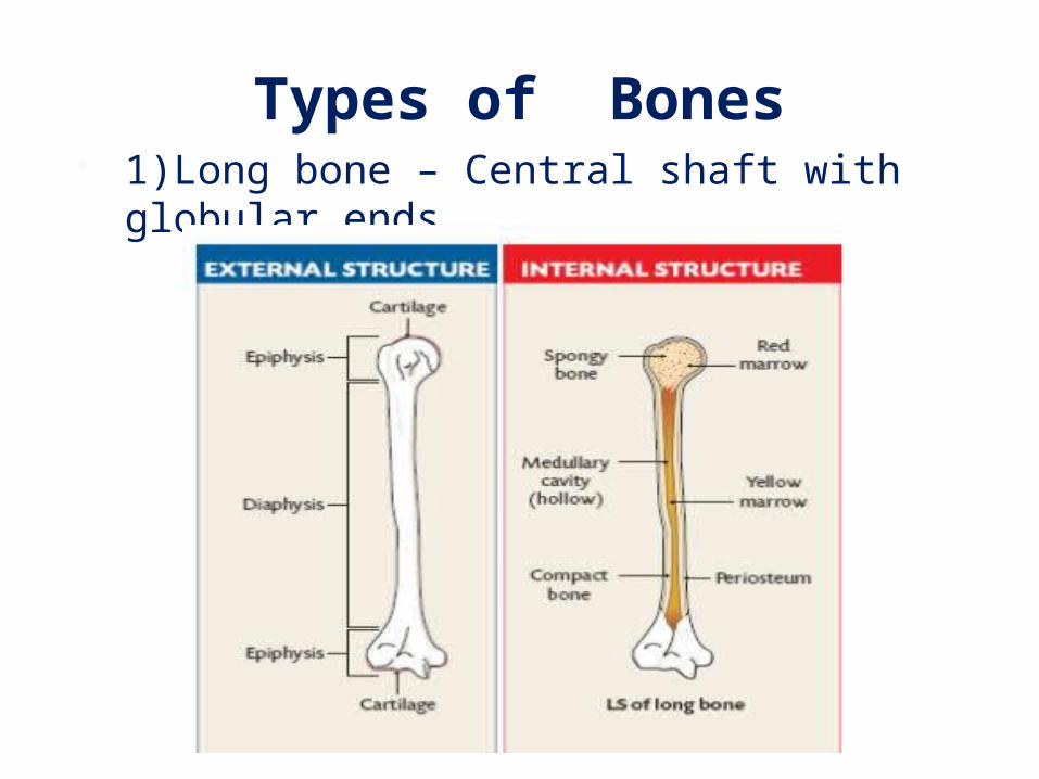
Types of Bones 1)Long bone – Central shaft with globular ends

Types of Bones 2) Short bones – Small and thick bones

Types of Bones 3) Flat bones – Two dimensional and plate like

Types of Bones4)Irregular bone – No geometric shape

Types of Bones5) Sesamoid bone – Bones shaped like sesame
developing in tendons or capsule

Skeletal systemSkeletal numbersAxial skeleton -80 Appendicular skeleton-126total bones -206Engage in voluntary movements-177
Axial skeleton – bones along the central axis includes Skull bones 28 (facial 14,auditory 6 ,cranial 8) Neck bone 1 (hyoid) Bones of spine(26 vertebral units) Flat bones of thorax (Ribs 24 ,sternum 1) Total – 80 bones
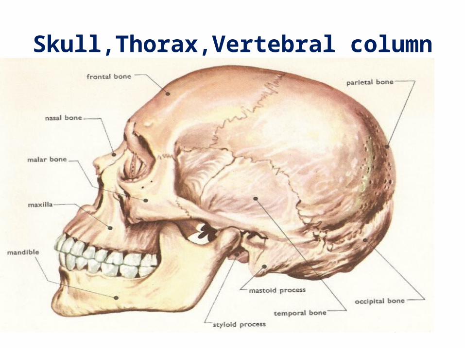
Skull,Thorax,Vertebral column
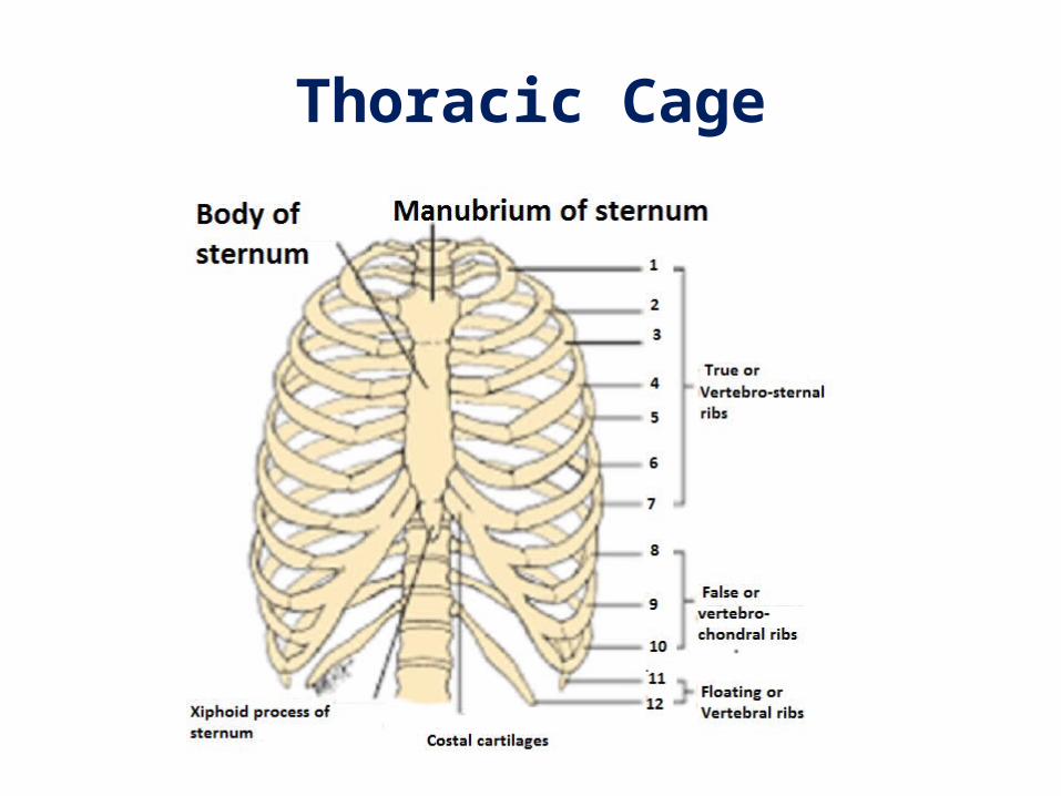
Thoracic Cage

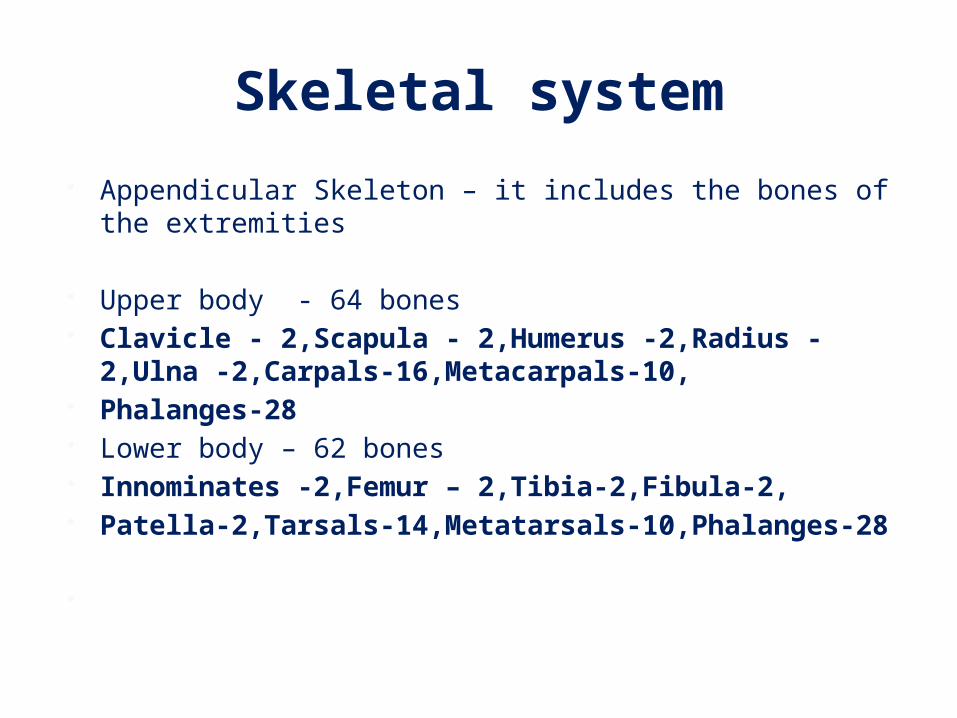
Skeletal system Appendicular Skeleton – it includes the bones of the extremities
Upper body - 64 bones Clavicle - 2,Scapula - 2,Humerus -2,Radius -2,Ulna -
2,Carpals-16,Metacarpals-10, Phalanges-28 Lower body – 62 bones Innominates -2,Femur – 2,Tibia-2,Fibula-2, Patella-2,Tarsals-14,Metatarsals-10,Phalanges-28
Total -126 bones

Clavicle

Scapula

Humerus

Radius-Ulna

Carpals,Metacarpals & Phalanges

Innominate

Femur and Patella

Tibia-Fibula
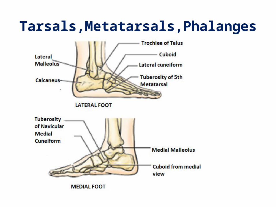
Tarsals,Metatarsals,Phalanges

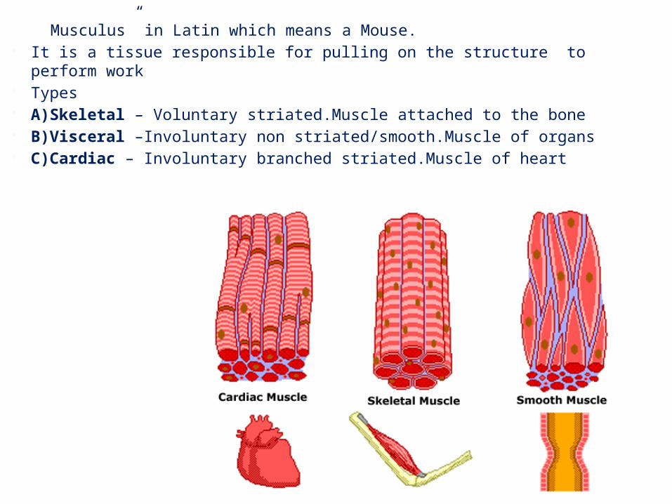
Musculus” in Latin which means a Mouse. It is a tissue responsible for pulling on the structure to perform work Types A)Skeletal – Voluntary striated.Muscle attached to the bone B)Visceral –Involuntary non striated/smooth.Muscle of organs C)Cardiac – Involuntary branched striated.Muscle of heart

TYPES OF SKELETAL MUSCLE FIBERS
Characteristics TYPE 1-Slow twitch TYPE 2A - Fast Intermediate twitch
TYPE 2B – Fast-Fastest twitch
Diameter Small Moderate LargeFatigue Low Moderate HighForce Generation
Low Moderate High
Mitochondrion High Low AbsentEnergy System Aerobic Anaerobic lactic
acidAnaerobic-Cp-ATP
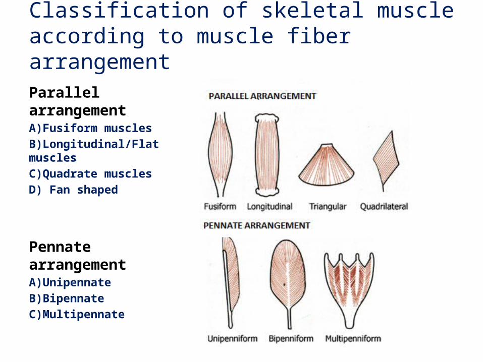
Classification of skeletal muscle according to muscle fiber arrangementParallel arrangementA)Fusiform musclesB)Longitudinal/Flat musclesC)Quadrate musclesD) Fan shaped
Pennate arrangementA)UnipennateB)BipennateC)Multipennate
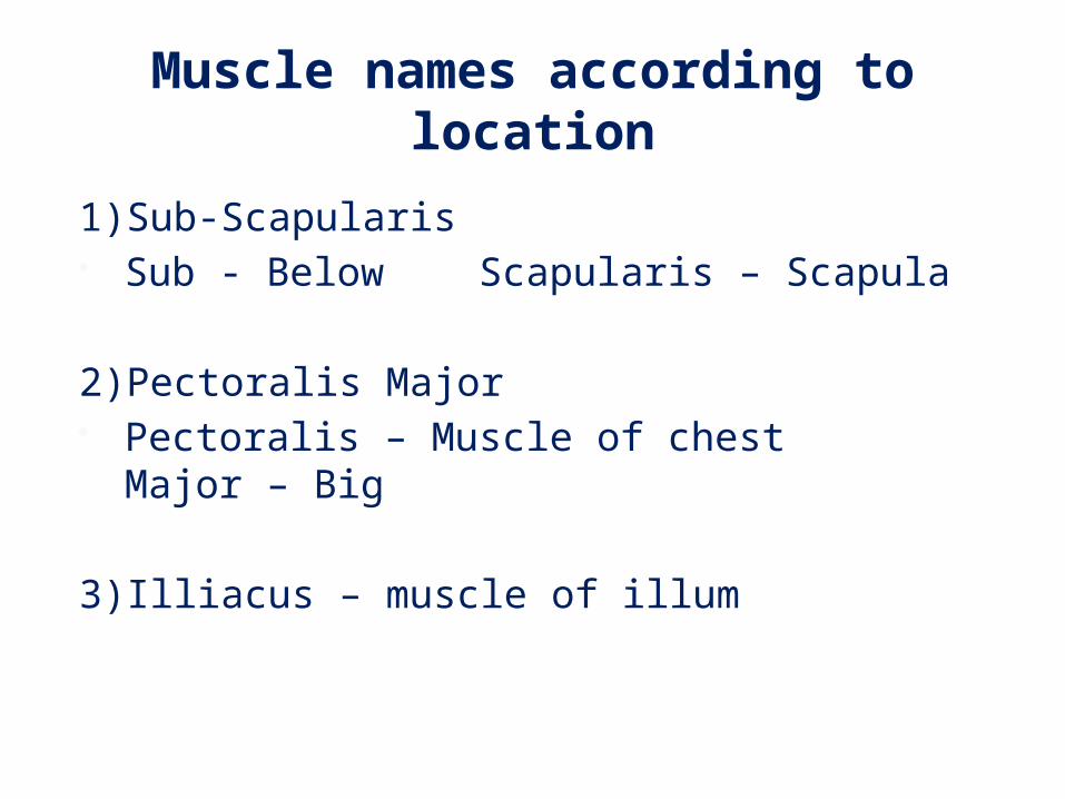
Muscle names according to location
1)Sub-Scapularis Sub - Below Scapularis – Scapula
2)Pectoralis Major Pectoralis – Muscle of chest Major – Big
3)Illiacus – muscle of illum

Muscle names according to shape
Trapezius – Kite shaped Rhomboids – Rhombus shaped
Muscle names according to role
Erector Spinae – Keep spine erect Levator Scapulae – elevates scapula

Functional Classification of muscles
A)Prime movers/Agonist/Primary muscles B)Synergists/Assistors/Secondary muscles C)Stabilizors/Fixators D)Antagonists
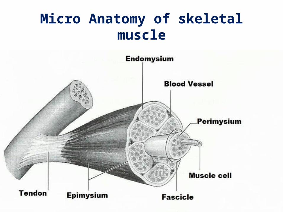
Micro Anatomy of skeletal muscle
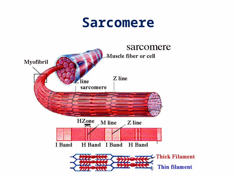
Sarcomere
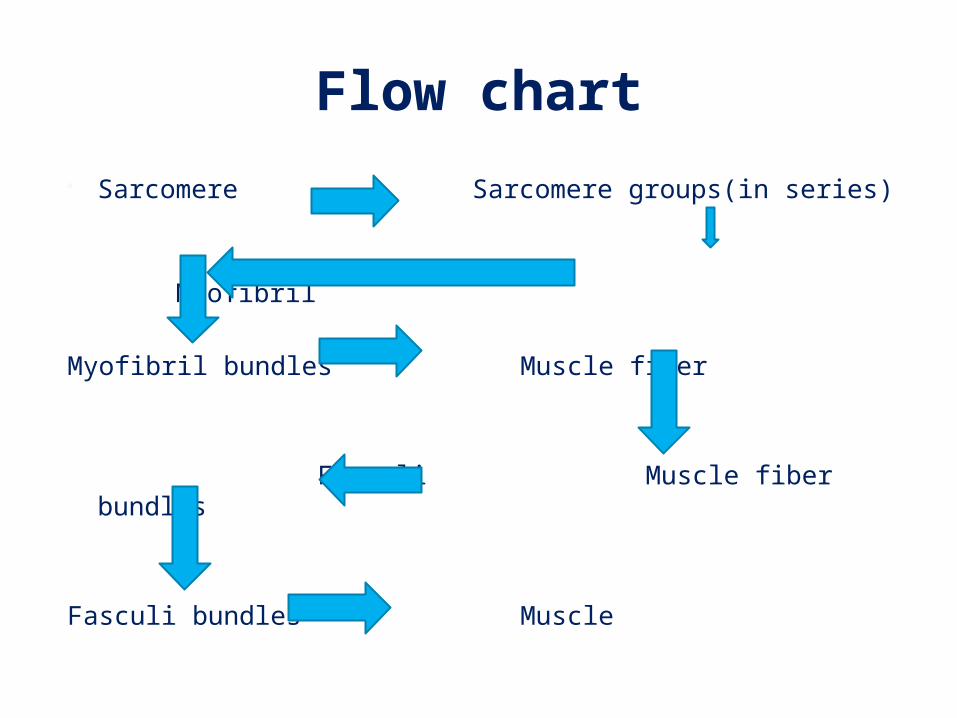
Flow chart Sarcomere Sarcomere groups(in series)
Myofibril Myofibril bundles Muscle fiber
Fasculi Muscle fiber bundles
Fasculi bundles Muscle

Sarcoplasmic reticulum is a fibrous fluid network which has calcium ion
Proteins within sarcomere Myosin (thick filament) Actin (thin filament)
Proteins within Actin filament Troponin – which is a calcium binder Tropomyosin –Its bound to troponin to form a
complex

Cross Bridge theory or sliding filament theory of muscle
contraction During muscle contraction Signal from brain reaches muscle via nerves Calcium released from sarcoplasmic reticulum deep into
muscle Calcium binds to troponin Troponin pull tropomyosin to expose binding site on actin Myosin heads attach to binding site and walk past closer to
the end on actin by a series of attachment and detachment using ATP
This looks like Cross Bridging or the filament looks to slide over each other
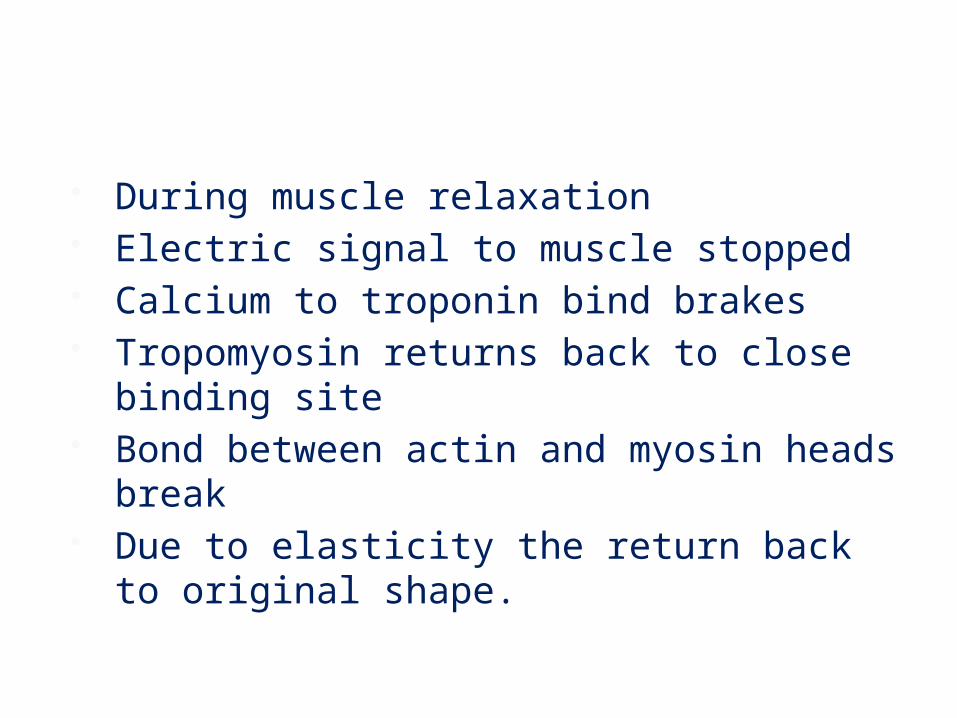
During muscle relaxation Electric signal to muscle stopped Calcium to troponin bind brakes Tropomyosin returns back to close binding site Bond between actin and myosin heads break Due to elasticity the return back to original shape.
Properties of muscle 1)Excitability/Irritability 2)Contractility 3)Extensibility 4)Elasticity 5)Tonicity

Types of contractions 1)Isotonic A)Concentric B)Eccentric
2)Isometric
3)Isokinetic

Anatomic adaptations in muscles
1)Hypertrophy
2)Hyperplasia
3)Atrophy
4)Dystrophy

Nervous system Central nervous system – It consist of brain and
spinal cord.It is the primary site for receiving sensory stimuli and center from which signals are sent for motor response
Peripheral nervous system – These are the nerves associate with the brain and spinal cord which allow the brain and spinal cord to communicate with rest of the body
Autonomic nervous system – This system consist of the sympathetic and parasympathetic pathways which help the body to regulate the subconcious drives like heart rate ,breathing , muscular tone etc .

Neuro-Muscular control The information received and sent by the nerves are in
the form of electric energy called “Nerve Impulse”. The end where the nerve communicates with the
muscle is called neuromuscular junction.Here the nerve endings release excitatory chemical called Acetylcholine.
A nerve which reaches the muscle divides into branches and goes deeper into muscle.Individual branches control few muscle fibers within its capacity.

The branch of nerve and its associated muscle fibers is called a “Motor Unit”.
Large motor units contain about 1,000 muscle fibers and is used for strength
Small motor units contain less than 50 muscle fibers and are used for accuracy of movement
Medium motor units contain about 100 to 400 muscle fibers and are used for activities that require moderate strength and accuracy.

Properties of motor units All or none principle Selective recruitment Post activation potentiation

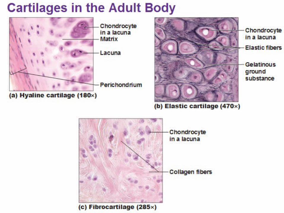
Connective tissues 1)Cartilage –These are made of chondrocytes and the spaces in
between filled with collagen and elastin fibers Types A)Hyaline cartilage – Semirigid moderately dense cartilage
having proportionate collagen and elastin.Eg articular cartilage at ends of bones
B)Fibrous cartilage –Semirigid and dense cartilage with more collagen fibers than elastin
Eg joint at the pubis C)Elastic cartilage – Flexible and thin cartilage made of more
elastin than collagen Eg)pinna of ears

Ligaments –Fibrous connective tissue which joints bone to bone
Tendons – Fibrous connective tissue which connects muscle to bone
Fascia – Thin transparent membrane which covers muscle or muscle groups.
Proprioceptors 1)Golgi tendon organs – located at the ends of
tendons which detect and inhibit excess muscular tension
2)Muscle spindle fibers – They lie parallel to muscle fibers and inhibit excessive or rapid stretching of muscles