Anatomical insights into disrupted small-world networks in
9
Anatomical insights into disrupted small-world networks in schizophrenia Qifeng Wang a , Tung-Ping Su b, c , Yuan Zhou d , Kun-Hsien Chou e , I-Yun Chen e , Tianzi Jiang a, f, g, ⁎, Ching-Po Lin e, ⁎⁎ a LIAMA Center for Computational Medicine, National Laboratory of Pattern Recognition, Institute of Automation, the Chinese Academy of Sciences, Beijing, China b Department of Psychiatry, Taipei Veterans General Hospital, Taipei, Taiwan c Department of Psychiatry, Collage of Medicine, National Yang-Ming University, Taipei, Taiwan d Key Laboratory of Behavioral Science, Institute of Psychology, Chinese Academy of Sciences, Beijing, China e Brain Connectivity Laboratory, Institute of Neuroscience, National Yang-Ming University, Taipei, Taiwan f Key Laboratory for NeuroInformation of Ministry of Education, School of Life Science and Technology, University of Electronic Science and Technology of China, Chengdu, China g The Queensland Brain Institute, The University of Queensland, Brisbane, Australia abstract article info Article history: Received 17 March 2011 Revised 13 September 2011 Accepted 15 September 2011 Available online 22 September 2011 Keywords: Anatomical connectivity Diffusion tensor imaging Graph theory Small-world Schizophrenia Schizophrenia is characterized by lowered efficiency in distributed information processing, as indicated by research that identified a disrupted small-world functional network. However, whether the dysconnection manifested by the disrupted small-world functional network is reflected in underlying anatomical disruption in schizophrenia remains unresolved. This study examined the topological properties of human brain ana- tomical networks derived from diffusion tensor imaging in patients with schizophrenia and in healthy con- trols. We constructed the weighted brain anatomical network for each of 79 schizophrenia patients and for 96 age and gender matched healthy subjects using diffusion tensor tractography and calculated the topolog- ical properties of the networks using a graph theoretical method. The topological properties of the patients' anatomical networks were altered, in that global efficiency decreased but local efficiency remained unchanged. The deleterious effects of schizophrenia on network performance appear to be localized as re- duced regional efficiency in hubs such as the frontal associative cortices, the paralimbic/limbic regions and a subcortical structure (the left putamen). Additionally, scores on the Positive and Negative Symptom Scale correlated negatively with efficient network properties in schizophrenia. These findings suggest that complex brain network analysis may potentially be used to detect an imaging biomarker for schizophrenia. © 2011 Elsevier Inc. All rights reserved. Introduction Current pathophysiological theories about schizophrenia highlight the role of dysconnectivity; that is, the complex clinical presentations (positive symptoms, negative symptoms and cognition decline) of schizophrenia arise from abnormality in inter-regional interactions rather than from abnormality in a specific focal region (Andreasen et al., 1999; Bullmore et al., 1997; Friston, 1998; Stephan et al., 2006). The dysconnection concept can also be described as an alteration in the efficiency of information exchange between separate neuronal networks in the human brain (Boutillier et al., 2008), which functions as a large, sparse, and complex network with highly efficient informa- tion processing between spatially distinct brain areas (Sporns et al., 2004). Network analysis derived from graph theory has successfully characterized the underlying architectures of such a network (Stam and Reijneveld, 2007) and has found so-called “small-world” proper- ties in the brain (Bullmore and Sporns, 2009; Salvador et al., 2005; Sporns et al., 2004). Small-world properties enable the cortical net- work to process global and local information with maximal efficiency at minimal cost (Achard and Bullmore, 2007). Using functional neuro- imaging techniques, researchers have found that small-world proper- ties are disrupted in patients with schizophrenia (Bassett et al., 2009; Liu et al., 2008; Micheloyannis et al., 2006). These findings provide fur- ther evidence for the dysconnection hypothesis of schizophrenia. The anatomical network in the multimodal cortices was found to be disor- ganized in schizophrenia when researchers considered inter-regional covariation between gray matter volumes as a proxy for connectivity (Bassett et al., 2008). This study hinted at a structural basis for the dis- rupted functional network but only provided indirect anatomical evi- dence because the connectivity matrix was estimated based on inter- regional correlations across subjects. Thus, a crucial question that re- mains unresolved is whether the dysconnection manifested by the disrupted small-world functional network in schizophrenia is the re- sult of underlying structural disruptions. NeuroImage 59 (2012) 1085–1093 ⁎ Correspondence to: T. Jiang, LIAMA Center for Computational Medicine, National Laboratory of Pattern Recognition, Institute of Automation, the Chinese Academy of Sciences, Beijing 100190, PR China. Fax: + 86 10 62551993. ⁎⁎ Correspondence to: C.-P. Lin, 155 Li-Nong St., Sec. 2, Peitou, Taipei, 112, Taiwan. Fax: +886 2 28262285. E-mail addresses: [email protected] (T. Jiang), [email protected] (C.-P. Lin). 1053-8119/$ – see front matter © 2011 Elsevier Inc. All rights reserved. doi:10.1016/j.neuroimage.2011.09.035 Contents lists available at SciVerse ScienceDirect NeuroImage journal homepage: www.elsevier.com/locate/ynimg
Transcript of Anatomical insights into disrupted small-world networks in

NeuroImage 59 (2012) 1085–1093
Contents lists available at SciVerse ScienceDirect
NeuroImage
j ourna l homepage: www.e lsev ie r .com/ locate /yn img
Anatomical insights into disrupted small-world networks in schizophrenia
Qifeng Wang a, Tung-Ping Su b,c, Yuan Zhou d, Kun-Hsien Chou e, I-Yun Chen e,Tianzi Jiang a,f,g,⁎, Ching-Po Lin e,⁎⁎a LIAMA Center for Computational Medicine, National Laboratory of Pattern Recognition, Institute of Automation, the Chinese Academy of Sciences, Beijing, Chinab Department of Psychiatry, Taipei Veterans General Hospital, Taipei, Taiwanc Department of Psychiatry, Collage of Medicine, National Yang-Ming University, Taipei, Taiwand Key Laboratory of Behavioral Science, Institute of Psychology, Chinese Academy of Sciences, Beijing, Chinae Brain Connectivity Laboratory, Institute of Neuroscience, National Yang-Ming University, Taipei, Taiwanf Key Laboratory for NeuroInformation of Ministry of Education, School of Life Science and Technology, University of Electronic Science and Technology of China, Chengdu, Chinag The Queensland Brain Institute, The University of Queensland, Brisbane, Australia
⁎ Correspondence to: T. Jiang, LIAMA Center for ComLaboratory of Pattern Recognition, Institute of AutomaSciences, Beijing 100190, PR China. Fax: +86 10 62551⁎⁎ Correspondence to: C.-P. Lin, 155 Li-Nong St., Sec.
Fax: +886 2 28262285.E-mail addresses: [email protected] (T. Jiang), chi
1053-8119/$ – see front matter © 2011 Elsevier Inc. Alldoi:10.1016/j.neuroimage.2011.09.035
a b s t r a c t
a r t i c l e i n f oArticle history:Received 17 March 2011Revised 13 September 2011Accepted 15 September 2011Available online 22 September 2011
Keywords:Anatomical connectivityDiffusion tensor imagingGraph theorySmall-worldSchizophrenia
Schizophrenia is characterized by lowered efficiency in distributed information processing, as indicated byresearch that identified a disrupted small-world functional network. However, whether the dysconnectionmanifested by the disrupted small-world functional network is reflected in underlying anatomical disruptionin schizophrenia remains unresolved. This study examined the topological properties of human brain ana-tomical networks derived from diffusion tensor imaging in patients with schizophrenia and in healthy con-trols. We constructed the weighted brain anatomical network for each of 79 schizophrenia patients and for96 age and gender matched healthy subjects using diffusion tensor tractography and calculated the topolog-ical properties of the networks using a graph theoretical method. The topological properties of the patients'anatomical networks were altered, in that global efficiency decreased but local efficiency remainedunchanged. The deleterious effects of schizophrenia on network performance appear to be localized as re-duced regional efficiency in hubs such as the frontal associative cortices, the paralimbic/limbic regions anda subcortical structure (the left putamen). Additionally, scores on the Positive and Negative Symptom Scalecorrelated negatively with efficient network properties in schizophrenia. These findings suggest that complexbrain network analysis may potentially be used to detect an imaging biomarker for schizophrenia.
putational Medicine, Nationaltion, the Chinese Academy of993.2, Peitou, Taipei, 112, Taiwan.
[email protected] (C.-P. Lin).
rights reserved.
© 2011 Elsevier Inc. All rights reserved.
Introduction
Current pathophysiological theories about schizophrenia highlightthe role of dysconnectivity; that is, the complex clinical presentations(positive symptoms, negative symptoms and cognition decline) ofschizophrenia arise from abnormality in inter-regional interactionsrather than from abnormality in a specific focal region (Andreasen etal., 1999; Bullmore et al., 1997; Friston, 1998; Stephan et al., 2006).The dysconnection concept can also be described as an alteration inthe efficiency of information exchange between separate neuronalnetworks in the human brain (Boutillier et al., 2008), which functionsas a large, sparse, and complex network with highly efficient informa-tion processing between spatially distinct brain areas (Sporns et al.,
2004). Network analysis derived from graph theory has successfullycharacterized the underlying architectures of such a network (Stamand Reijneveld, 2007) and has found so-called “small-world” proper-ties in the brain (Bullmore and Sporns, 2009; Salvador et al., 2005;Sporns et al., 2004). Small-world properties enable the cortical net-work to process global and local information with maximal efficiencyat minimal cost (Achard and Bullmore, 2007). Using functional neuro-imaging techniques, researchers have found that small-world proper-ties are disrupted in patients with schizophrenia (Bassett et al., 2009;Liu et al., 2008;Micheloyannis et al., 2006). Thesefindings provide fur-ther evidence for the dysconnection hypothesis of schizophrenia. Theanatomical network in the multimodal cortices was found to be disor-ganized in schizophrenia when researchers considered inter-regionalcovariation between gray matter volumes as a proxy for connectivity(Bassett et al., 2008). This study hinted at a structural basis for the dis-rupted functional network but only provided indirect anatomical evi-dence because the connectivity matrix was estimated based on inter-regional correlations across subjects. Thus, a crucial question that re-mains unresolved is whether the dysconnection manifested by thedisrupted small-world functional network in schizophrenia is the re-sult of underlying structural disruptions.

Table 1Demographic and clinical details (mean±SD).
NC (N=96) SCZ (N=79) P-value
Gender (male/female) 48/48 39/40 0.934a
Age (years) 37±13 37±11 0.986b
Brain size (voxels) 174,650±16,660 172,158±15,355 0.309b
Duration (years) ____ 10±9 ____PANSS_p ____ 13±5 ____PANSS_n ____ 14±4 ____PANSS_t ____ 59±16 ____
Abbreviations: SD, standard deviation; NC, normal controls; SCZ, schizophrenia;PANSS, Positive and Negative Symptom Scale; PANSS_p, positive scale score obtainedfrom PANSS; PANSS_n, negative scale score obtained from PANSS; PANSS_t, PANSStotal score.
a The p value was obtained using the Pearson Chi-square.b The p value was obtained by a two-tailed two-sample t-test.
1086 Q. Wang et al. / NeuroImage 59 (2012) 1085–1093
Because diffusion MRI is advantageous for visualizing brain ana-tomical connectivity in vivo, it has been increasingly used to investi-gate the intrinsic topological properties of the human brainnetwork. This method facilitates our understanding of brain organiza-tion and of its complex cooperating working mechanisms at the mac-roscopic scale. Hagmann et al. confirmed a small-world topologywhich is connected by dense cortico-cortical axonal pathways in thehuman cortical network (Hagmann et al., 2007), and further foundthe structural core of the anatomical network (Hagmann et al.,2008). Since these early studies, researchers using diffusion tensorimaging (DTI) have consistently found small-world topology in ana-tomical networks connected by white matter fibers in healthy volun-teers (Gong et al., 2009a; Iturria-Medina et al., 2008; Li et al., 2009).These studies indicate that using DTI to investigate anatomical net-works is feasible in disease conditions such as schizophrenia. Sku-dlarski et al. constructed separate functional and anatomicalconnectivity maps for 27 schizophrenia patients and 27 normal con-trols and reported reduced coherence between functional and struc-tural modalities in patients compared with normal controls(Skudlarski et al., 2010). In addition, they also reported a significantcorrelation between brain connectivity and the positive symptomscore on the Positive and Negative Symptom Scale (PANSS). However,this study did not mention any analysis of the network topologicalproperties. Zalesky et al. constructed binary structural networks for74 chronic schizophrenia patients and 32 control subjects using a de-terministic white matter tracking algorithm, and reported wide-spread dysconnectivity in the white matter connectionalarchitecture in the patients group (Zalesky et al., 2010). They foundthat intellectual performance was associated with brain efficiency inthe control subjects but not in the patients. However, this study didnot examine the relationship between clinical variables and topolog-ical properties. In another study, van den Heuvel et al. utilized DTI andthe magnetic transfer imaging (MTR) technique to construct weight-ed structural networks for 40 schizophrenia patients and 40 healthycontrols and reported a reduced global efficiency of the frontal, tem-poral, and occipital brain regions in schizophrenia (van den Heuvel etal., 2010). They also examined the correlation between the PANSS andnetwork properties but found no significant results. The results ofthese recent studies indicated that the structural brain networkrevealed by DTI is disrupted in schizophrenia. However, independentstudies with larger samples are necessary to verify this inference. Ad-ditionally, correlations between clinical findings and disrupted struc-tural networks in schizophrenia remain inconsistent (Skudlarski etal., 2010; van den Heuvel et al., 2010) and require furtherinvestigations.
Our goal for this study was to use DTI to detect abnormalities inthe global architecture of the anatomical connectivity patterns inschizophrenia from the perspective of the brain network. Our focuswas on network efficiency, which is defined to measure how well in-formation propagates over the network (Latora and Marchiori, 2003).Furthermore, we attempted to find clinical correlations to the abnor-malities we found in the topology of the anatomical network in pa-tients with schizophrenia.
Materials and methods
Subjects
Some of the subjects included in this work were used in a previousstudy (Bai et al., 2009). However, in the current study additional sub-jects who had the same MRI scan parameters were recruited. Werecruited 79 patients from the Psychiatric Department Outpatient Clin-ic and Day Care Unit at the Taipei Veterans General Hospital who metthe Diagnostic and Statistical Manual of Mental Disorders, Fourth Edi-tion (DSM-IV) criteria for schizophrenia. Subjects were excluded ifthey had other Axis I psychiatric diagnoses, serious neurologic or
endocrine disorders, any medical condition or treatment known to af-fect the brain, alcohol/substance misuse related disorders, or mentalretardation as defined by DSM-IV criteria. Before scanning, the patients'schizophrenia symptoms were rated by trained and experienced psy-chiatrists using PANSS (Kay et al., 1987) to rate the severity of psycho-pathology and the 17-item Hamilton Depression Rating Scale (HDRS)(Hamilton, 1960) to evaluate the depressive symptoms.
We recruited 96 healthy subjects (normal control group) via ad-vertisement. The healthy subjects were interviewed using the Mini-International Neuropsychiatric Interview to confirm that they hadno previous history of neurologic or psychiatric illness, and all hadnormal brain structure confirmed by MRI scans. The exclusion criteriawere the same as those for the patient group.
The two groups were significantly similar in terms of gender com-position, age, and brain size (Table 1).
Ethics statement
All procedures were approved by the institutional review board ofTaipei Veterans General Hospital. Written informed consent wasobtained from all subjects.
MRI data acquisition
All MR examinations were performed on a 1.5 T MR system (Ex-cite II; GE Medical Systems, Milwaukee, WI, USA) equipped with an8-channel head coil in TPE-VGH. To diminish motion artifacts gener-ated during the scan, the subject's head was immobilized with cush-ions inside the coil after the alignment. One hundred twenty-fourcontiguous axial T1-weighted images (slice thickness=1.5 mm)were acquired parallel to the anterior–posterior commissure (AC-PC) through the whole head by applying a three-dimensional fluid-attenuated inversion-recovery fast spoiled-gradient recalled echo(FLAIRFSPGR) acquisition sequence (TR=8.548 ms, TE=1.836 ms,TI=400 ms, flip angle=15°, field of view=26×26 cm, matrixsize=256×256) to aid in the localization of differences in fractionalanisotropy (Madden et al., 2004). Fourteen diffusion tensor imagingvolumes were obtained for each subject, including thirteen volumeswith diffusion gradients applied along thirteen non-collinear direc-tions (b=900 s/mm2) and one volume without diffusion weighting(b=0). In order to obtain coverage of the entire brain, each volumeconsisted of 70 contiguous axial slices (slice thickness=2.2 mm) ac-quired parallel to the AC-PC by using a single shot spin-echo echo pla-nar imaging (EPI) sequence (TR=17,000 ms, TE=68.9 ms, numberof excitation=6, field of view=26 cm2, matrix size=128×128).

1087Q. Wang et al. / NeuroImage 59 (2012) 1085–1093
Preprocessing
For the diffusion-weighted images, simple head motion and eddycurrent distortion were corrected using FMRIB's Diffusion Toolbox (FSL4.1; http://www.fmrib.ox.ac.uk/fsl). After correction, the six indepen-dent components of the diffusion tensor, the corresponding FA and ei-genvectors were calculated using an in-house software named DTITracking (http://www.ccm.org.cn/). The T1-weighted image of each sub-ject was co-registered to the subject's non-diffusion-weighted image(b=0 s/mm2) using the SPM2 package (http://www.fil.ion.ucl.ac.uk/spm), resulting in a co-registered T1 image in native DTI space.
Construction of weighted brain anatomical connectivity networks
Definition of network nodeWe used the automated anatomical labeling (AAL) template
(Tzourio-Mazoyer et al., 2002) available with the MRIcro software(http://www.sph.sc.edu/comd/rorden/mricro.html) to segment theentire cerebral cortex of each subject into 90 regions (45 for eachhemisphere) with the cerebellum excluded. Each brain region repre-sented a node of the final anatomical network. Each individual co-registered T1 image was normalized to the SPM2 T1 template in Mon-treal Neurological Institute (MNI) space. The resulting transformationmatrix was then inverted and further used to warp the AAL templatefrom the MNI space to the diffusion MRI native space, in which thediscrete labeling values were preserved, using a nearest-neighbor in-terpolation method.
Definition of network edgeWe implemented a deterministic streamline tracking algorithm
(Mori et al., 1999) to perform fiber tracking. The tracking procedureended at voxels with an FA value of less than 0.15 or when theangle between adjacent steps was greater than 45° (Li et al.,2009; Thottakara et al., 2006). Two AAL regions could be consideredto be connected if the two end points of the reconstructed fiberbundles were located in these two regions (Gong et al., 2009a; Liet al., 2009).
The inverse of the number (Nij) of connections between the tworegions was employed to weigh the edge:
Wij ¼1Nij
if Nij≥T� �
0 otherwise:
8<:
Under this definition, wij is thought to be inversely related to thedegree to which nodes i and j are connected anatomically; that is,the larger thewij (corresponding to a smaller Nij), the weaker the con-nectivity strength between these two nodes. In other words, wij isproportional to the distance between two brain regions. However,note that distance here does not correspond to the physical distancebetween cortical regions in Euclidean space but to a quantificationof the connectivity strength. Two regions that are linked by fewerfiber bundles are considered distant and correspond to a higher wij,which is similar to the definition used in previous studies (Achardand Bullmore, 2007; Gong et al., 2009b). Because false-positive con-nections could result from using this, we utilized a series of thresholdvalues (T, from 1 to 10) for the number of existing fibers to examinethe robustness of our construction method, that is, two regions linkedwith fewer than T fibers were treated as disconnected. The selectedthreshold values maintained the average size of the largest connectedcomponent at 90 across all subjects, meaning that the networks of themajority of subjects were fully connected at each threshold value(Table S1 in the Supplementary materials).
Graph theoretical analyses of the weighted network properties
Network properties definitionThe topological properties utilized in our current study can be de-
scribed as follows:
G is used to denote the whole weighted network; whereas Gi is de-fined as the subgraph of node i, which is constructed by its directneighbor nodes and the edges linking these neighbor nodes.N is used to represent the total number of nodes in the network.
The cost of a connection edge is proportional to its distance(weight), and the overall cost of a network is defined as the sum ofthe weights:
cost ¼ ∑i≠j∈G
wij :
The shortest path length between nodes i and j, Lij, is defined asthe length of a path from node i to node j in which the sum of theweights of its constituent edges is minimized.
The mean shortest path length of node i is computed as the averageshortest path length between node i and all other nodes in the network:
Li ¼1
N−1∑
j∈G;i≠jLij :
The characteristic path length of the network is the average of theshortest path lengths across all nodes:
Lp ¼ 1N∑i∈G
Li:
The global efficiency of the network, Eglob, is defined as the inverseof the harmonic mean of the shortest path lengths between each pairof nodes:
Eglob ¼1
N N−1ð Þ ∑i;j∈G;i≠j
1Lij
Eglob measures the global efficiency of parallel information transfer inthe network (Latora and Marchiori, 2001).
The regional efficiency of node i, Ereg(i), i=1,2,…, 90, is defined asthe inverse of the harmonic mean of Lij between node i and all othernodes in the network (Achard and Bullmore, 2007):
Ereg ið Þ ¼1
N−1∑
i;j∈G;i≠j
1Lij
:
This measure quantifies the importance of each node for commu-nication within the network (Gong et al., 2009b).
The local efficiency of node i, Eloc(i), i=1,2,…, 90, is calculated asthe global efficiency of Gi:
Eloc ið Þ ¼ Eglob Gið Þ:
The local efficiency measures the fault tolerance of the network,indicating how well the information is communicated between theneighbors of a given node when the node is removed (Achard andBullmore, 2007).
The local efficiency of the entire network, Eloc, is defined as the av-erage of the local efficiencies across all nodes in the network:
Eloc ¼1N∑i∈G
Eloc ið Þ:
1088 Q. Wang et al. / NeuroImage 59 (2012) 1085–1093
Evaluation of the small-world propertiesThe “small-world” concept, in which networks are characterized
by a characteristic path length L less than that in a regular latticeand a clustering coefficient C greater than that in a random graph,was originally proposed in a study of binary networks (Watts andStrogatz, 1998). However, weighted networks have been demonstrat-ed to be better suited for characterizing real world network topology,and Eglob and Eloc have been found to be suitable scales for character-izing the “small-world” properties of weighted networks (Achard andBullmore, 2007; Latora and Marchiori, 2001). Fundamentally, 1/Lmeasures the efficiency of a sequential system, whereas E measuresthe efficiency of parallel systems, in which all the nodes in the net-works concurrently exchange packets of information. Characteristicpath length is hypersensitive to local changes, whereas efficiency ismore robust to link lesions or malfunctions. For example, in the caseof a fully connected network, if one node in the network becomes iso-lated as the result of some accident, the mean shortest path length ofthe whole network tends to infinity, whereas the mean global effi-ciency is only slightly diminished (Achard and Bullmore, 2007). Ingeneral, the network would still work well even though localizeddamage has occurred; hence efficiency is more appropriate for mea-suring the performance of a parallel system, such as the humanbrain. Practically speaking, a weighted network can be categorizedas small-world if Eglob is slightly less than, but Eloc is much greaterthan, a matched random network (Achard and Bullmore, 2007;Gong et al., 2009b). Here we followed the procedure used by Gonget al. (2009b) to evaluate the small-world properties. Specifically,we generated 100 random networks for each subject's networkusing a Markov-chain algorithm while retaining the weight of eachedge in order to preserve the weight distribution of the entire net-work. Then we calculated Eglob and Eloc across these random networksand compared them with the properties of the real networks (seeresults).
Statistical analyses
A general linear model (GLM) was used to test for differences inthe network properties (Eglob, Eloc and regional efficiency) betweenthe schizophrenic patients and the healthy subjects. The gender, age
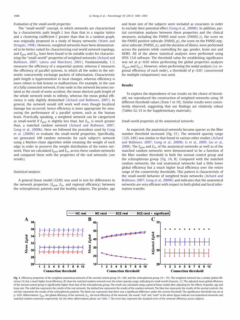
Fig. 1. Efficiency properties of the weighted anatomical network of the normal control group (N=ciency (A) but amuch higher local efficiency (B) than thematched randomnetwork over the entirof the normal control group is significantly higher than that of the schizophrenia group. The resultbrain size. The solid line represents the results of the real network; the dashed line represents the rred line represents the results of the schizophrenia patients. The black star represents that there wpb0.05. Abbreviations: Eglob, the global efficiency of the network; Eloc, the local efficiency of the nematched random networks respectively; for the other abbreviations please see Table 1. The error
and brain size of the subjects were included as covariates in orderto exclude their potential effect (Gong et al., 2009b). In addition, par-tial correlation analyses between these properties and the clinicalmeasures, including the PANSS total score (PANSS_t), the score onthe PANSS positive subscale (PANSS_p), the score on the PANSS neg-ative subscale (PANSS_n), and the duration of illness, were performedacross the patients while controlling for age, gender, brain size andHDRS. All of the above statistical analyses were performed usingSPSS 13.0 software. The threshold value for establishing significancewas set at pb0.05 when performing the global properties analyses(Eglob and Eloc). However, when performing the nodal analysis (i.e. re-gional efficiency of each node), a threshold of pb0.01 (uncorrectedfor multiple comparisons) was used.
Results
To explore the dependence of our results on the choice of thresh-old, we reproduced the construction of weighted networks using 10different threshold values (from 1 to 10). Similar results were consis-tently observed, suggesting that our findings are relatively robust(Tables S2, S3 in the Supplementary materials).
Small-world properties of the anatomical networks
As expected, the anatomical networks became sparser as the fibernumber threshold increased (Fig. S1). The network sparsity range(12%–24%) was similar to that found in various other studies (Achardand Bullmore, 2007; Gong et al., 2009b; Li et al., 2009; Liu et al.,2008). The Eglob and Eloc of the anatomical networks as well as of thematched random networks were demonstrated to be a function ofthe fiber number threshold in both the normal control group andthe schizophrenia group (Fig. 1A, B). Compared with the matchedrandom networks, the real anatomical networks had a little lowerglobal efficiency but a much higher local efficiency over the entirerange of the connectivity thresholds. This pattern is characteristic ofthe small-world behavior of weighted brain networks (Achard andBullmore, 2007; Gong et al., 2009b) and indicates that the anatomicalnetworks are very efficient with respect to both global and local infor-mation transfer.
96) and the schizophrenia group (N=79). The weighted network has a similar global effi-e sparsity range, indicating its small-world character. (C) The adjustedmean global efficiencywas calculated using a general linear model after adjusting for the effects of gender, age andesults of the randomnetwork. The blue line represents the results of the normal controls; theas a significant difference under the current threshold. The significance threshold was set at
twork; thewords “real” and “rand” in the above figure indicate real anatomical networks andbars represent the standard error of the network efficiency across subjects.
1089Q. Wang et al. / NeuroImage 59 (2012) 1085–1093
Altered global topological properties of the anatomical networks inschizophrenia
Using a GLM statistical analysis, we found that, after factoring outthe effects of age, gender and brain size, Eglob was significantly largerin the healthy group than in the schizophrenia patients over the en-tire threshold range (Fig. 1C, and Table S2 in the Supplementary ma-terials). No significant difference in Eloc was found between the twogroups, although Eloc showed a trend toward a decrease in the schizo-phrenia group compared to the normal group. We further examinedthe mean edge number and mean cost of the networks in both thetwo groups, without detecting any significant group differences.
Regional efficiency differences of individual nodes
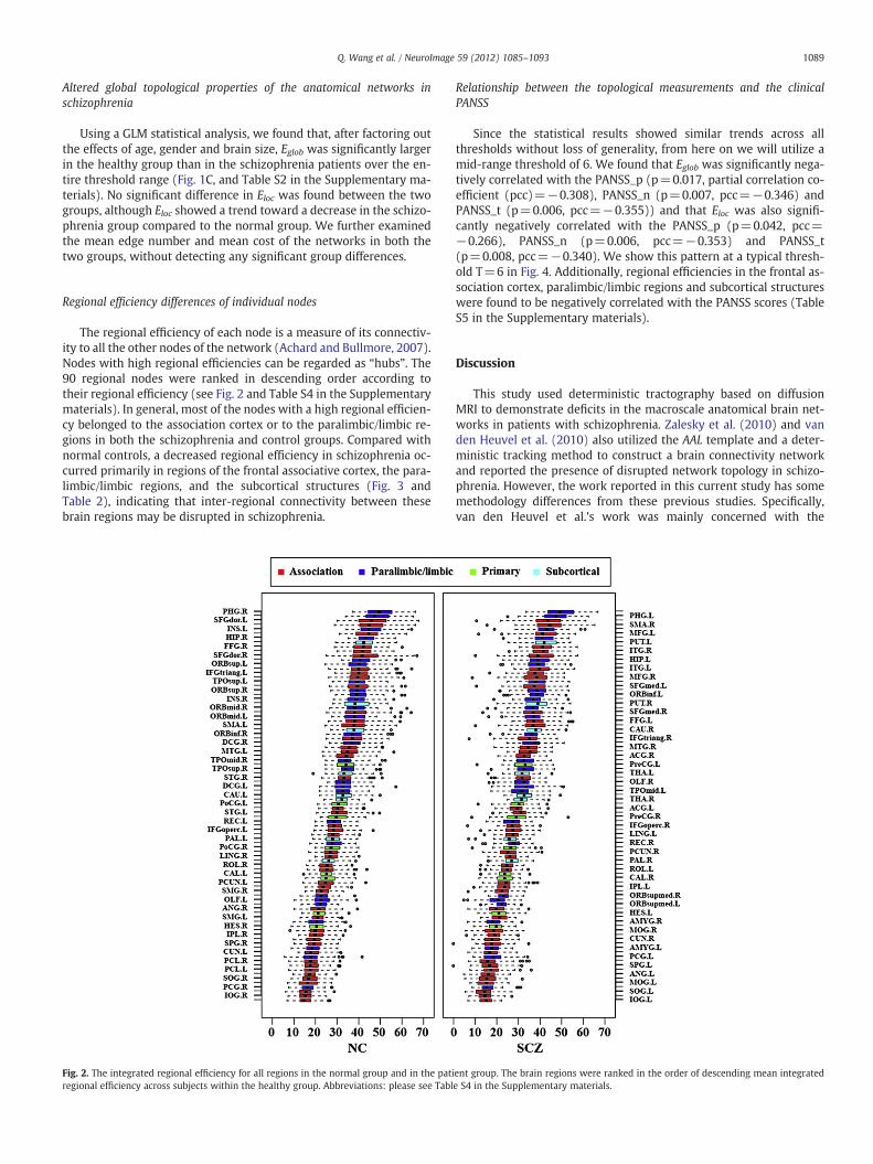
The regional efficiency of each node is a measure of its connectiv-ity to all the other nodes of the network (Achard and Bullmore, 2007).Nodes with high regional efficiencies can be regarded as “hubs”. The90 regional nodes were ranked in descending order according totheir regional efficiency (see Fig. 2 and Table S4 in the Supplementarymaterials). In general, most of the nodes with a high regional efficien-cy belonged to the association cortex or to the paralimbic/limbic re-gions in both the schizophrenia and control groups. Compared withnormal controls, a decreased regional efficiency in schizophrenia oc-curred primarily in regions of the frontal associative cortex, the para-limbic/limbic regions, and the subcortical structures (Fig. 3 andTable 2), indicating that inter-regional connectivity between thesebrain regions may be disrupted in schizophrenia.
Fig. 2. The integrated regional efficiency for all regions in the normal group and in the patiregional efficiency across subjects within the healthy group. Abbreviations: please see Tabl
Relationship between the topological measurements and the clinicalPANSS
Since the statistical results showed similar trends across allthresholds without loss of generality, from here on we will utilize amid-range threshold of 6. We found that Eglob was significantly nega-tively correlated with the PANSS_p (p=0.017, partial correlation co-efficient (pcc)=−0.308), PANSS_n (p=0.007, pcc=−0.346) andPANSS_t (p=0.006, pcc=−0.355)) and that Eloc was also signifi-cantly negatively correlated with the PANSS_p (p=0.042, pcc=−0.266), PANSS_n (p=0.006, pcc=−0.353) and PANSS_t(p=0.008, pcc=−0.340). We show this pattern at a typical thresh-old T=6 in Fig. 4. Additionally, regional efficiencies in the frontal as-sociation cortex, paralimbic/limbic regions and subcortical structureswere found to be negatively correlated with the PANSS scores (TableS5 in the Supplementary materials).
Discussion
This study used deterministic tractography based on diffusionMRI to demonstrate deficits in the macroscale anatomical brain net-works in patients with schizophrenia. Zalesky et al. (2010) and vanden Heuvel et al. (2010) also utilized the AAL template and a deter-ministic tracking method to construct a brain connectivity networkand reported the presence of disrupted network topology in schizo-phrenia. However, the work reported in this current study has somemethodology differences from these previous studies. Specifically,van den Heuvel et al.'s work was mainly concerned with the
ent group. The brain regions were ranked in the order of descending mean integratede S4 in the Supplementary materials.
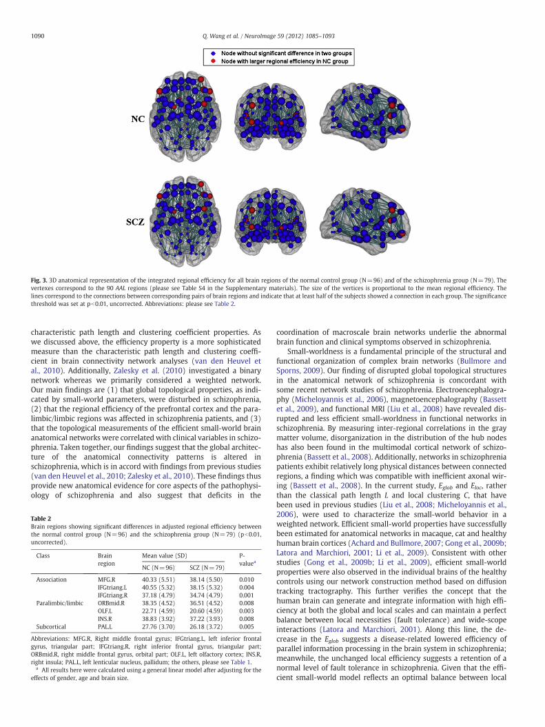
Fig. 3. 3D anatomical representation of the integrated regional efficiency for all brain regions of the normal control group (N=96) and of the schizophrenia group (N=79). Thevertexes correspond to the 90 AAL regions (please see Table S4 in the Supplementary materials). The size of the vertices is proportional to the mean regional efficiency. Thelines correspond to the connections between corresponding pairs of brain regions and indicate that at least half of the subjects showed a connection in each group. The significancethreshold was set at pb0.01, uncorrected. Abbreviations: please see Table 2.
1090 Q. Wang et al. / NeuroImage 59 (2012) 1085–1093
characteristic path length and clustering coefficient properties. Aswe discussed above, the efficiency property is a more sophisticatedmeasure than the characteristic path length and clustering coeffi-cient in brain connectivity network analyses (van den Heuvel etal., 2010). Additionally, Zalesky et al. (2010) investigated a binarynetwork whereas we primarily considered a weighted network.Our main findings are (1) that global topological properties, as indi-cated by small-world parameters, were disturbed in schizophrenia,(2) that the regional efficiency of the prefrontal cortex and the para-limbic/limbic regions was affected in schizophrenia patients, and (3)that the topological measurements of the efficient small-world brainanatomical networks were correlated with clinical variables in schizo-phrenia. Taken together, our findings suggest that the global architec-ture of the anatomical connectivity patterns is altered inschizophrenia, which is in accord with findings from previous studies(van den Heuvel et al., 2010; Zalesky et al., 2010). These findings thusprovide new anatomical evidence for core aspects of the pathophysi-ology of schizophrenia and also suggest that deficits in the
Table 2Brain regions showing significant differences in adjusted regional efficiency betweenthe normal control group (N=96) and the schizophrenia group (N=79) (pb0.01,uncorrected).
Class Brainregion
Mean value (SD) P-valuea
NC (N=96) SCZ (N=79)
Association MFG.R 40.33 (5.51) 38.14 (5.50) 0.010IFGtriang.L 40.55 (5.32) 38.15 (5.32) 0.004IFGtriang.R 37.18 (4.79) 34.74 (4.79) 0.001
Paralimbic/limbic ORBmid.R 38.35 (4.52) 36.51 (4.52) 0.008OLF.L 22.71 (4.59) 20.60 (4.59) 0.003INS.R 38.83 (3.92) 37.22 (3.93) 0.008
Subcortical PAL.L 27.76 (3.70) 26.18 (3.72) 0.005
Abbreviations: MFG.R, Right middle frontal gyrus; IFGtriang.L, left inferior frontalgyrus, triangular part; IFGtriang.R, right inferior frontal gyrus, triangular part;ORBmid.R, right middle frontal gyrus, orbital part; OLF.L, left olfactory cortex; INS.R,right insula; PAL.L, left lenticular nucleus, pallidum; the others, please see Table 1.
a All results here were calculated using a general linear model after adjusting for theeffects of gender, age and brain size.
coordination of macroscale brain networks underlie the abnormalbrain function and clinical symptoms observed in schizophrenia.
Small-worldness is a fundamental principle of the structural andfunctional organization of complex brain networks (Bullmore andSporns, 2009). Our finding of disrupted global topological structuresin the anatomical network of schizophrenia is concordant withsome recent network studies of schizophrenia. Electroencephalogra-phy (Micheloyannis et al., 2006), magnetoencephalography (Bassettet al., 2009), and functional MRI (Liu et al., 2008) have revealed dis-rupted and less efficient small-worldness in functional networks inschizophrenia. By measuring inter-regional correlations in the graymatter volume, disorganization in the distribution of the hub nodeshas also been found in the multimodal cortical network of schizo-phrenia (Bassett et al., 2008). Additionally, networks in schizophreniapatients exhibit relatively long physical distances between connectedregions, a finding which was compatible with inefficient axonal wir-ing (Bassett et al., 2008). In the current study, Eglob and Eloc, ratherthan the classical path length L and local clustering C, that havebeen used in previous studies (Liu et al., 2008; Micheloyannis et al.,2006), were used to characterize the small-world behavior in aweighted network. Efficient small-world properties have successfullybeen estimated for anatomical networks in macaque, cat and healthyhuman brain cortices (Achard and Bullmore, 2007; Gong et al., 2009b;Latora and Marchiori, 2001; Li et al., 2009). Consistent with otherstudies (Gong et al., 2009b; Li et al., 2009), efficient small-worldproperties were also observed in the individual brains of the healthycontrols using our network construction method based on diffusiontracking tractography. This further verifies the concept that thehuman brain can generate and integrate information with high effi-ciency at both the global and local scales and can maintain a perfectbalance between local necessities (fault tolerance) and wide-scopeinteractions (Latora and Marchiori, 2001). Along this line, the de-crease in the Eglob suggests a disease-related lowered efficiency ofparallel information processing in the brain system in schizophrenia;meanwhile, the unchanged local efficiency suggests a retention of anormal level of fault tolerance in schizophrenia. Given that the effi-cient small-world model reflects an optimal balance between local
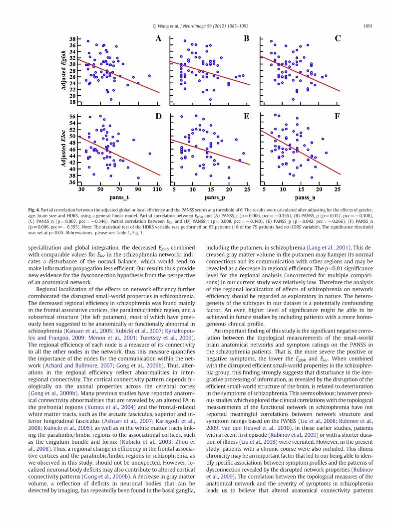
Fig. 4. Partial correlation between the adjusted global or local efficiency and the PANSS scores at a threshold of 6. The results were calculated after adjusting for the effects of gender,age, brain size and HDRS, using a general linear model. Partial correlation between Eglob and (A) PANSS_t (p=0.006, pcc=−0.355), (B) PANSS_p (p=0.017, pcc=−0.308),(C) PANSS_n (p=0.007, pcc=−0.346). Partial correlation between Eloc and (D) PANSS_t (p=0.008, pcc=−0.340), (E) PANSS_p (p=0.042, pcc=−0.266), (F) PANSS_n(p=0.006, pcc=−0.353). Note: The statistical test of the HDRS variable was performed on 63 patients (16 of the 79 patients had no HDRS variable). The significance thresholdwas set at pb0.05. Abbreviations: please see Table 1, Fig. 1.
1091Q. Wang et al. / NeuroImage 59 (2012) 1085–1093
specialization and global integration, the decreased Eglob combinedwith comparable values for Eloc in the schizophrenia networks indi-cates a disturbance of the normal balance, which would tend tomake information propagation less efficient. Our results thus providenew evidence for the dysconnection hypothesis from the perspectiveof an anatomical network.
Regional localization of the effects on network efficiency furthercorroborated the disrupted small-world properties in schizophrenia.The decreased regional efficiency in schizophrenia was found mainlyin the frontal associative cortices, the paralimbic/limbic region, and asubcortical structure (the left putamen), most of which have previ-ously been suggested to be anatomically or functionally abnormal inschizophrenia (Kanaan et al., 2005; Kubicki et al., 2007; Kyriakopou-los and Frangou, 2009; Menon et al., 2001; Turetsky et al., 2009).The regional efficiency of each node is a measure of its connectivityto all the other nodes in the network, thus this measure quantifiesthe importance of the nodes for the communication within the net-work (Achard and Bullmore, 2007; Gong et al., 2009b). Thus, alter-ations in the regional efficiency reflect abnormalities in inter-regional connectivity. The cortical connectivity pattern depends bi-ologically on the axonal properties across the cerebral cortex(Gong et al., 2009b). Many previous studies have reported anatom-ical connectivity abnormalities that are revealed by an altered FA inthe prefrontal regions (Kumra et al., 2004) and the frontal-relatedwhite matter tracts, such as the arcuate fasciculus, superior and in-ferior longitudinal fasciculus (Ashtari et al., 2007; Karlsgodt et al.,2008; Kubicki et al., 2005), as well as in the white matter tracts link-ing the paralimbic/limbic regions to the associational cortices, suchas the cingulum bundle and fornix (Kubicki et al., 2003; Zhou etal., 2008). Thus, a regional change in efficiency in the frontal associa-tive cortices and the paralimbic/limbic regions in schizophrenia, aswe observed in this study, should not be unexpected. However, lo-calized neuronal body deficits may also contribute to altered corticalconnectivity patterns (Gong et al., 2009b). A decrease in gray mattervolume, a reflection of deficits in neuronal bodies that can bedetected by imaging, has repeatedly been found in the basal ganglia,
including the putamen, in schizophrenia (Lang et al., 2001). This de-creased gray matter volume in the putamen may hamper its normalconnections and its communication with other regions and may berevealed as a decrease in regional efficiency. The pb0.01 significancelevel for the regional analysis (uncorrected for multiple compari-sons) in our current study was relatively low. Therefore the analysisof the regional localization of effects of schizophrenia on networkefficiency should be regarded as exploratory in nature. The hetero-geneity of the subtypes in our dataset is a potentially confoundingfactor. An even higher level of significance might be able to beachieved in future studies by including patients with a more homo-geneous clinical profile.
An important finding of this study is the significant negative corre-lation between the topological measurements of the small-worldbrain anatomical networks and symptom ratings on the PANSS inthe schizophrenia patients. That is, the more severe the positive ornegative symptoms, the lower the Eglob and Eloc. When combinedwith the disrupted efficient small-world properties in the schizophre-nia group, this finding strongly suggests that disturbance in the inte-grative processing of information, as revealed by the disruption of theefficient small-world structure of the brain, is related to deteriorationin the symptoms of schizophrenia. This seemsobvious; however previ-ous studieswhich explored the clinical correlationswith the topologicalmeasurements of the functional network in schizophrenia have notreported meaningful correlations between network structure andsymptom ratings based on the PANSS (Liu et al., 2008; Rubinov et al.,2009; van den Heuvel et al., 2010). In these earlier studies, patientswith a recent first episode (Rubinov et al., 2009) or with a shorter dura-tion of illness (Liu et al., 2008) were recruited. However, in the presentstudy, patients with a chronic course were also included. This illnesschronicitymay be an important factor that led to our being able to iden-tify specific associations between symptom profiles and the patterns ofdysconnection revealed by the disrupted network properties (Rubinovet al., 2009). The correlation between the topological measures of theanatomical network and the severity of symptoms in schizophrenialeads us to believe that altered anatomical connectivity patterns
1092 Q. Wang et al. / NeuroImage 59 (2012) 1085–1093
underlie the abnormal brain function and clinical symptoms observedin schizophrenia. No significant correlations were found between thenetwork properties and the duration of illness in this study. This maysuggest that the duration of illness is independent of the symptom pro-files, a finding which is consistent with clinical observations (Loebel etal., 1992).
As a preliminary exploration of the anatomical basis for disruptionin the brain small-world network in schizophrenia, the current studyenrolled a relatively large sample of patients in order to obtain robustand stable findings. However, the large sample size also may have in-troduced some potentially confounding factors that need to be con-sidered, such as the effects of medication dosage. In terms ofnetwork properties, although one study reported that pharmacologi-cal blockade of dopamine D2 receptors by a single dose of sulpiride400 mg was acutely associated with impaired global and local effi-ciency of the networks (Achard and Bullmore, 2007), other studieshave not found significant correlations between antipsychotic medi-cation dosages and network measurements (Liu et al., 2008; Miche-loyannis et al., 2006) or else have found a significantly positivecorrelation (Rubinov et al., 2009) which was independent of the se-verity of symptoms in schizophrenia. These clinical studies suggestthat medication is unlikely to be a confounding factor and may,on the contrary, exert a ‘normalizing’ influence and thus argueagainst a direct treatment effect accounting for the between-group differences. Additionally, subtype heterogeneity may be an-other potentially confounding factor. Although more definitiveconclusions will require unmedicated patients with a homoge-neous clinical profile, our stable findings that the efficient small-world properties in the anatomical network were disrupted anddeteriorated with a deterioration of symptoms in schizophrenialead us to believe that anatomical network analysis may be ableto play a vital role in improving our understanding of the patho-physiology of schizophrenia and aid researchers in detecting imag-ing biomarkers for schizophrenia.
Methodological considerations
In our current study, a deterministic tractography method and theAAL template were used for network construction. The AAL templateprimarily masks gray matter, but a deterministic tractography algo-rithm is not applicable in graymatter regions at low FA values. Actually,as some previous articles (Gong et al., 2009a; Li et al., 2009)mentioned,there is a risk that some of the fiber tracts reconstructed using ourmethod may not belong to the specific AAL region if an AAL mask con-tained too many white matter voxels that are not truly adjacent to thecortex. As a potential solution for this issue, Gong et al. (2009a) re-moved white matter voxels in the raw AAL cortical mask if no corticalvoxels existed within their 2-mm cubic neighborhood. However, theirmethod could lead to another risk, that of excluding the fiber tractsthat actually belong to the specific AAL region (Li et al., 2009). In the cur-rent study, we utilized tracking parameters that were similar to thoseused in some previous studies (Li et al., 2009; Thottakara et al., 2006);that is, an FA threshold of 0.3 was used for selecting seed voxels to re-construct fiber tracts originating from different Brodmann areas.According to Li et al. (2009), the FA threshold we used is basicallyvalid for limiting the number of false-positive connections. Fig. S2 inthe Supplementary materials shows six well-known white matterfiber tracts from four subjects (2 schizophrenia patients and 2 normalcontrols) randomly selected from our dataset. These characteristicfiber bundles may indicate that the tracking results are basically credi-ble. A probabilistic tractography method may be a better choice forour future work, as this method is advantageous for overcoming thefiber crossing problem (Behrens et al., 2003, 2007; Lazar and Alexander,2005). However, in our current study, the DTI images were obtainedfrom a 1.5-T MRI scanner using 13 non-collinear diffusion encoding
directions; a single tensor model and deterministic tracking methodmight be the optimal choice.
That the Eglob did not change much as the threshold increased andthe networks got sparser (Fig. 1A), while the Eloc became even higher(Fig. 1B) seems counterintuitive. We think this result is rational dueto the use of a dissimilarity link weight in the current study. Specifi-cally, as the threshold increased, the edges with higher weights (cor-responding to a lower number of fiber bundles between nodes and alonger distance) were removed first; these edges were usually not onthe shortest pathway between regions, hence the elimination of these“ineffective” edges contributed little to the Eglob but enhanced the Elocof the whole network. However, the efficiency would decrease any-way as the threshold continued to increase. Fig. S3 shows the entirevariation trend ofEglob andEloc over a larger threshold range (from 1to 50). Interestingly, as the weighted network degenerated to a bina-ry network, Eglob andEloc both decreased monotonously as the thresh-old increased (Fig. S4 in Supplementary materials).
Additionally, regions on the AAL template differ in size, which mayhave a confounding effect on the link weight of the network nodes.Furthermore, the definition of the template (i.e. the inclusion and ex-clusion of some brain regions) could also affect the statistical results.However, understanding how to assess and control these influenceremains an open question. In the current study, we are mainly con-cerned with differences in network attributes between healthy andschizophrenia groups. To test the between-group region size differ-ences, we performed a two-tailed t-test on each pair of correspondingbrain region and found no significant result (pb0.05). This result sug-gests that region sizes have equivalent influences in both the healthyand schizophrenia groups and, therefore, contribute little to the be-tween-group differences we found in the current study. Nevertheless,further investigations will be necessary to better address the method-ological limitations of our current study.
Conclusions
Our findings provide direct evidence for a disrupted anatomicalnetwork in schizophrenia and suggest a structural basis for the dys-connection manifested by disrupted small-world functional net-works. Correlations between topological measures of efficient small-world properties and the severity of symptoms in schizophrenialead us to believe that altered global architecture of the anatomicalconnectivity patterns underlie the abnormal brain function and clini-cal symptoms observed in schizophrenia.
Funding
This work was supported by the National Key Basic Research andDevelopment Program (973) (Grant Number 2011CB707800) toDr Jiang; the Natural Science Foundation of China (Grant Number30730035 to Dr Jiang, Grant Number 30900487 to Dr. Zhou); Ministryof Economic Affairs (Grant Number 98-EC-17-A-19-S2-0103); NationalHealth Research Institute (Grant Number NHRI-EX98-9813EC); andNa-tional Science Council (Grant Numbers 98-2517-S-004-001-MY3 and98-2627-B-010-008)
Acknowledgments
The authors acknowledge MR support from the MRI Core Labora-tory, NYMU and support from the Ministry of Education (Aim forthe Top University Plan). The authors thank Drs. Edmund F. andRhoda E. Perozzi for English editing assistance and discussions. Theauthors thank Dr. Yong Liu for his valuable suggestions on the revisedmanuscript. The authors are grateful to Professor Godfrey Pearlson forhis constructive comments and suggestions.
1093Q. Wang et al. / NeuroImage 59 (2012) 1085–1093
Appendix A. Supplementary data
Supplementary data to this article can be found online at doi:10.1016/j.neuroimage.2011.09.035.
References
Achard, S., Bullmore, E., 2007. Efficiency and cost of economical brain functional networks.PLoS Comput. Biol. 3, e17.
Andreasen, N.C., Nopoulos, P., O'Leary, D.S., Miller, D.D., Wassink, T., Flaum, M., 1999.Defining the phenotype of schizophrenia: cognitive dysmetria and its neuralmechanisms. Biol. Psychiatry 46, 908–920.
Ashtari, M., Cottone, J., Ardekani, B.A., Cervellione, K., Szeszko, P.R., Wu, J., Chen, S.,Kumra, S., 2007. Disruption of white matter integrity in the inferior longitudinalfasciculus in adolescents with schizophrenia as revealed by fiber tractography.Arch. Gen. Psychiatry 64, 1270–1280.
Bai, Y.M., Chou, K.H., Lin, C.P., Chen, I.Y., Li, C.T., Yang, K.C., Chou, Y.H., Su, T.P., 2009.White matter abnormalities in schizophrenia patients with tardive dyskinesia: adiffusion tensor image study. Schizophr. Res. 109, 167–181.
Bassett, D.S., Bullmore, E., Verchinski, B.A., Mattay, V.S., Weinberger, D.R., Meyer-Lindenberg,A., 2008. Hierarchical organization of human cortical networks in health and schizophre-nia. J. Neurosci. 28, 9239–9248.
Bassett, D.S., Bullmore, E.T.,Meyer-Lindenberg, A., Apud, J.A.,Weinberger, D.R., Coppola, R.,2009. Cognitive fitness of cost-efficient brain functional networks. Proc. Natl. Acad.Sci. U. S. A. 106, 11747–11752.
Behrens, T.E., Johansen-Berg, H., Woolrich, M.W., Smith, S.M., Wheeler-Kingshott, C.A.,Boulby, P.A., Barker, G.J., Sillery, E.L., Sheehan, K., Ciccarelli, O., Thompson, A.J.,Brady, J.M., Matthews, P.M., 2003. Non-invasive mapping of connections betweenhuman thalamus and cortex using diffusion imaging. Nat. Neurosci. 6, 750–757.
Behrens, T.E., Berg, H.J., Jbabdi, S., Rushworth, M.F., Woolrich, M.W., 2007. Probabilistic diffu-sion tractography with multiple fibre orientations: what can we gain? NeuroImage 34,144–155.
Boutillier, A.L., Macedo, C.E., Angst, M.J., Sandner, G., 2008. How can we justify the useof lower animal models to understand the pathophysiology of schizophrenia? Ad-vances in Cognitive Neurodynamics. Springer, pp. 577–582.
Bullmore, E., Sporns, O., 2009. Complex brain networks: graph theoretical analysis ofstructural and functional systems. Nat. Rev. Neurosci. 10, 186–198.
Bullmore, E.T., Frangou, S., Murray, R.M., 1997. The dysplastic net hypothesis: an inte-gration of developmental and dysconnectivity theories of schizophrenia. Schi-zophr. Res. 28, 143–156.
Friston, K.J., 1998. The disconnection hypothesis. Schizophr. Res. 30, 115–125.Gong, G., He, Y., Concha, L., Lebel, C., Gross, D.W., Evans, A.C., Beaulieu, C., 2009a. Mapping
anatomical connectivity patterns of human cerebral cortex using in vivo diffusion ten-sor imaging tractography. Cereb. Cortex 19, 524–536.
Gong, G., Rosa-Neto, P., Carbonell, F., Chen, Z.J., He, Y., Evans, A.C., 2009b. Age- and gender-related differences in the cortical anatomical network. J. Neurosci. 29, 15684–15693.
Hagmann, P., Kurant, M., Gigandet, X., Thiran, P., Wedeen, V.J., Meuli, R., Thiran, J.P., 2007.Mapping humanwhole-brain structural networks with diffusionMRI. PLoS One 2, e597.
Hagmann, P., Cammoun, L., Gigandet, X., Meuli, R., Honey, C.J., Wedeen, V.J., Sporns, O.,2008. Mapping the structural core of human cerebral cortex. PLoS Biol. 6, e159.
Hamilton,M., 1960. A rating scale for depression. J. Neurol. Neurosurg. Psychiatry 23, 56–62.Iturria-Medina, Y., Sotero, R.C., Canales-Rodriguez, E.J., Aleman-Gomez, Y.,Melie-Garcia, L.,
2008. Studying the human brain anatomical network via diffusion-weightedMRI andGraph Theory. NeuroImage 40, 1064–1076.
Kanaan, R.A., Kim, J.S., Kaufmann, W.E., Pearlson, G.D., Barker, G.J., McGuire, P.K., 2005.Diffusion tensor imaging in schizophrenia. Biol. Psychiatry 58, 921–929.
Karlsgodt, K.H., van Erp, T.G., Poldrack, R.A., Bearden, C.E., Nuechterlein, K.H., Cannon,T.D., 2008. Diffusion tensor imaging of the superior longitudinal fasciculus andworking memory in recent-onset schizophrenia. Biol. Psychiatry 63, 512–518.
Kay, S.R., Flszbein, A., Opler, L.A., 1987. The positive and negative syndrome scale(PANSS) for schizophrenia. Schizophr. Bull. 13, 261–276.
Kubicki, M., Westin, C.F., Nestor, P.G., Wible, C.G., Frumin, M., Maier, S.E., Kikinis, R., Jolesz,F.A., McCarley, R.W., Shenton, M.E., 2003. Cingulate fasciculus integrity disruption inschizophrenia: a magnetic resonance diffusion tensor imaging study. Biol. Psychiatry54, 1171–1180.
Kubicki, M., Park, H., Westin, C.F., Nestor, P.G., Mulkern, R.V., Maier, S.E., Niznikiewicz, M.,Connor, E.E., Levitt, J.J., Frumin, M., Kikinis, R., Jolesz, F.A., McCarley, R.W., Shenton,M.E., 2005. DTI and MTR abnormalities in schizophrenia: analysis of white matter in-tegrity. NeuroImage 26, 1109–1118.
Kubicki, M., McCarley, R., Westin, C.F., Park, H.J., Maier, S., Kikinis, R., Jolesz, F.A., Shenton,M.E., 2007. A review of diffusion tensor imaging studies in schizophrenia. J. Psychiatr.Res. 41, 15–30.
Kumra, S., Ashtari, M., McMeniman, M., Vogel, J., Augustin, R., Becker, D.E., Nakayama, E.,Gyato, K., Kane, J.M., Lim, K., Szeszko, P., 2004. Reduced frontal white matter integrityin early-onset schizophrenia: a preliminary study. Biol. Psychiatry 55, 1138–1145.
Kyriakopoulos, M., Frangou, S., 2009. Recent diffusion tensor imaging findings in earlystages of schizophrenia. Curr. Opin. Psychiatry 22, 168–176.
Lang, D.J., Kopala, L.C., Vandorpe, R.A., Rui, Q., Smith, G.N., Goghari, V.M., Honer, W.G.,2001. An MRI study of basal ganglia volumes in first-episode schizophrenia pa-tients treated with risperidone. Am. J. Psychiatry 158, 625–631.
Latora, V., Marchiori, M., 2001. Efficient behavior of small-world networks. Phys. Rev.Lett. 87, 198701.
Latora, V., Marchiori, M., 2003. Economic small-world behavior in weighted networks.Eur. Phys. J. B 32, 249–263.
Lazar, M., Alexander, A.L., 2005. Bootstrap white matter tractography (BOOT-TRAC).NeuroImage 24, 524–532.
Li, Y., Liu, Y., Li, J., Qin, W., Li, K., Yu, C., Jiang, T., 2009. Brain anatomical network andintelligence. PLoS Comput. Biol. 5, e1000395.
Liu, Y., Liang, M., Zhou, Y., He, Y., Hao, Y., Song, M., Yu, C., Liu, H., Liu, Z., Jiang, T., 2008.Disrupted small-world networks in schizophrenia. Brain 131, 945–961.
Loebel, A.D., Lieberman, J.A., Alvir, J.M., Mayerhoff, D.I., Geisler, S.H., Szymanski, S.R.,1992. Duration of psychosis and outcome in first-episode schizophrenia. Am. J.Psychiatry 149, 1183–1188.
Madden, D.J., Whiting, W.L., Huettel, S.A., White, L.E., MacFall, J.R., Provenzale, J.M.,2004. Diffusion tensor imaging of adult age differences in cerebral white matter:relation to response time. NeuroImage 21, 1174–1181.
Menon, V., Anagnoson, R.T., Glover, G.H., Pfefferbaum, A., 2001. Functional magneticresonance imaging evidence for disrupted basal ganglia function in schizophrenia.Am. J. Psychiatry 158, 646–649.
Micheloyannis, S., Pachou, E., Stam, C.J., Breakspear, M., Bitsios, P., Vourkas, M., Erimaki,S., Zervakis, M., 2006. Small-world networks and disturbed functional connectivityin schizophrenia. Schizophr. Res. 87, 60–66.
Mori, S., Crain, B.J., Chacko, V.P., van Zijl, P.C., 1999. Three-dimensional tracking of ax-onal projections in the brain by magnetic resonance imaging. Ann. Neurol. 45,265–269.
Rubinov,M., Knock, S.A., Stam, C.J., Micheloyannis, S., Harris, A.W.,Williams, L.M., Breakspear,M., 2009. Small-world properties of nonlinear brain activity in schizophrenia. Hum. BrainMapp. 30, 403–416.
Salvador, R., Suckling, J., Coleman, M.R., Pickard, J.D., Menon, D., Bullmore, E., 2005.Neurophysiological architecture of functional magnetic resonance images ofhuman brain. Cereb. Cortex 15, 1332–1342.
Skudlarski, P., Jagannathan, K., Anderson, K., Stevens, M.C., Calhoun, V.D., Skudlarska,B.A., Pearlson, G., 2010. Brain connectivity is not only lower but different in schizo-phrenia: a combined anatomical and functional approach. Biol. Psychiatry 68,61–69.
Sporns, O., Chialvo, D.R., Kaiser, M., Hilgetag, C.C., 2004. Organization, development andfunction of complex brain networks. Trends Cogn. Sci. 8, 418–425.
Stam, C.J., Reijneveld, J.C., 2007. Graph theoretical analysis of complex networks in thebrain. Nonlinear Biomed. Phys. 1, 3.
Stephan, K.E., Baldeweg, T., Friston, K.J., 2006. Synaptic plasticity and dysconnection inschizophrenia. Biol. Psychiatry 59, 929–939.
Thottakara, P., Lazar, M., Johnson, S.C., Alexander, A.L., 2006. Application of Brodmann'sarea templates for ROI selection in white matter tractography studies. NeuroImage29, 868–878.
Turetsky, B.I., Hahn, C.G., Borgmann-Winter, K., Moberg, P.J., 2009. Scents and nonsense:olfactory dysfunction in schizophrenia. Schizophr. Bull. 35, 1117–1131.
Tzourio-Mazoyer, N., Landeau, B., Papathanassiou, D., Crivello, F., Etard, O., Delcroix, N.,Mazoyer, B., Joliot, M., 2002. Automated anatomical labeling of activations in SPMusing a macroscopic anatomical parcellation of the MNI MRI single-subject brain.NeuroImage 15, 273–289.
van den Heuvel, M.P., Mandl, R.C., Stam, C.J., Kahn, R.S., Hulshoff Pol, H.E., 2010. Aberrantfrontal and temporal complex network structure in schizophrenia: a graph theoreticalanalysis. J. Neurosci. 30, 15915–15926.
Watts, D.J., Strogatz, S.H., 1998. Collective dynamics of ‘small-world’ networks. Nature393, 440–442.
Zalesky, A., Fornito, A., Seal, M.L., Cocchi, L., Westin, C.F., Bullmore, E.T., Egan, G.F., Pan-telis, C., 2010. Disrupted axonal fiber connectivity in schizophrenia. Biol. Psychiatry69, 80–89.
Zhou, Y., Shu, N., Liu, Y., Song, M., Hao, Y., Liu, H., Yu, C., Liu, Z., Jiang, T., 2008. Alteredresting-state functional connectivity and anatomical connectivity of hippocampusin schizophrenia. Schizophr. Res. 100, 120–132.