A new Australian species of Luffa (Cucurbitaceae) - Pensoft Publishers

International Journal of Research Studies in Biosciences (IJRSB)
Volume 3, Issue 6, June 2015, PP 85-91
ISSN 2349-0357 (Print) & ISSN 2349-0365 (Online)
www.arcjournals.org
©ARC Page | 85
Anatomical Diversity among Certain Genera of Family
Cucurbitaceae
Ismail A. Mohammed
Department of Botany and Agricultural Biotechnology, Faculty of
Agriculture, University of Khartoum, Khartoum North,
Shambat, Sudan. [email protected]
Abdel Gabbar. N. Guma
Department of Biology, Faculty of Education, University of Khartoum,
Khartoum, Sudan.
Abstract: The anatomical characters of the Cucrbitaceae having long attracted the attention of botanists. In
order to understand the evolution of anatomical diversity, anatomical features of five genera in the family
Cucurbitaceae, (Colocynthis, Cucumis, Cucurbita, Citrullus, and Luffa) were investigated. Similarities in the
distribution, tissue differentiation and number of layers of cells and tissues in the root, stem and leaf transverse
sections were similar in all genera. However, there were variations in the vascular bundles in the roots where
they were bicollateral in Cucumis sativus and Luffa aegyptiaca, while it consisted of four radial arms of
primary xylem alternating with four arms of primary phloem in the other species.
The Trichomes were multicellular, glandular and non-glandular types with a preponderance of non-glandular
types with various shapes. The variation in number of tiers within these trichomes was taxonomically
significance.
Keywords: Diversity, Cucurbitaceae, Taxonomy, Anatomy.
1. INTRODUCTION
Cucurbits belong to the family Cucurbitaceae and consist of about 130 genera and 800 species,
according to the last taxonomic treatment of Jeffrey [1]. The family Cucurbitaceae belongs to the
order Cucurbitales, class Magnoliopsida (subclass Rosidae). The family Cucurbitaceae is one of the
most important in the Angiosperm taxa. This is especially so in Sudan where members of the family
are used as fruits (water melon), food (pumpkin), soup condiment (egusi), vegetables (Fluted
pumpkin), sponge (Luffa spp), water reservoirs (Lageneria spp) and as stew condiment (snake
tomatoes) [2, 3]. Reference [4] attributed a lot of medicinal potency to the species of the family
Cucurbitaceae. The most important cultivated genera are Cucurbita L., Cucumis L., Citrullus L.,
Colocynthis Mill, Lagenaria L., Luffa L., [5] Coccinia (Wight & Arn.), Corallocarpus (Welw),
Ctenolepis (Hook), Kedrostis Medik. and Momordica L., [6, 7,8].
Literature searches revealed that the scientific importance and implications of anatomical features in
different groups of plants have been indicated by different authors. These families include
Dioscoreaceae, where certain anatomical features were used in the characterization of D. alata (L.)
and D. smilacifolia L. [9]. In Costaceae, where differences in features of vegetative anatomy
suggested a separate specific status for C. afer and C. lucanusianus as opposed to the conspecific
treatment given to them by previous researchers [10]. In Leguminosae-Caesalpinoideae, where the
nature of unicellular and multicellular trichomes are described in certain species of Senna (Tourn ex
Mill) and S. hirsuta was reported to be diagonostic in acquisition of these two types of trichomes [11].
Other studies of interest relating to anatomy of different angiospermous groups could be found in
Curcurbitaceae [12], Dicotyledon plants as a whole [13] Leguminosae-Papilionoideae [14].
The objective of this investigation is to describe the root, stem and leaf anatomical characters of the
Cucurbit species and to assess the relevance of and the extent to which anatomical features could be
Ismail A. Mohammed
& Abdel Gabbar. N. Guma
International Journal of Research Studies in Biosciences (IJRSB) Page | 86
utilized in the biosystematic deliniation of the Cucurbit species in view of their perceived similarities
in structural and reproductive biology.
2. MATERIALS AND METHODS
In order to study the variation of internal structural in the leaves, stems and roots of some cucurbits,
microtome study was undertaken and microscopic anatomical observations were carried out following
the method suggested by [15], [16], [17] and [18].
2.1. Plant Materials
Nine species of the family Cucurbitaceae were collected from different places in Khartoum State
(Table 1). About 2.5 cm long parts were prepared using vegetative parts of roots, stems and leaves
of the plant material.
2.2. Killing and Fixing
The specimens were killed and fixed in formalin acetic acid (FAA) for at least 24 hours (FAA:
10:50:5:35 proportion of formalin, alcohol, acetic acid and water) which is represented by the
previous formula according to [16]. The plant specimens were washed in distilled water three times
allowing a time of 20 minutes for each wash.
2.3. Dehydration
After killing and fixing the plant specimens in FAA solution, they were transferred to a series of
different alcoholic concentrations (50 %, 70 %, 90 %, and 95%) and then were left overnight or more
in each concentration.
2.4. Clearing
The plant specimens were cleared using two mixtures, with different ratios for different times:
mixture I composed of absolute alcohol : cedar wood oil (100 : 0 for 24hr, 50 : 50 for 3hr, 25 : 75 for
3 hr and 0 : 100 for 24 hr) respectively, and mixture II composed of cedar wood oil : xylene (100 : 0
for 24hr, 50 : 50 for 3hr, 25 : 75 for 3 hr and 100 for 24 hr), respectively.
2.5. Embedding
Next to the clearing process, the plant specimens were embedded in wax with melting point of 60 º C.
The plant specimens were placed in closed vials containing 1:1 xylene and melted wax and put in the
oven for 45 minutes. The wax was later replaced by pure wax twice after 45 minutes. The vials were
left open to get rid of the xylene vapour. The specimens were transferred from the vials to the mold
containing pure melted wax. Each specimen was pressed gently against the peripheral part of the
mold. The wax was left to consolidate.
2.6. Sectioning
The paraffin embedded specimens were sectioned with the help of a rotary microtome. The thickness
of the stem sections was 14 micrometer (µm) while the leaves and the roots were 9 and 12 µm thick,
respectively. The ribbons were mounted on slides flooded with distilled water and placed on a hot
plate to flatten the sections.
2.7. Staining
The double staining process was employed for this purpose. The steps of this process included
dewaxing, rehydration, staining, and dehydration of material. In the dewaxing process the slides were
passed twice through xylene, each for 3-5 minutes. Rehydation was carried by passing the dewaxed
slides through a series of different concentrations of ethanol in this order: absolute, absolute, 95%,
90%, 70% and 50% and were then immersed in safranine, each for 2-3 minutes. Dehydration was
carried out by passing the slides through different concentrations of ethanol (50%, 70%, 90%, 95%
and absolute) each for 2-3 minutes. The slides were then immersed in fast green stain for one and a
half minutes. The slides were then cleaned by passing through absolute ethanol and xylene.
2.8. Mounting
The material was mounted in D.P.X and covered with cover slips. The slides were kept in an oven at
60 ºC and left for 3 days, before examining under a light microscope at x10.

Anatomical Diversity among Certain Genera of Family Cucurbitaceae
International Journal of Research Studies in Biosciences (IJRSB) Page | 87
Table1. Plant accessions used in this study.
No Species Distribution and description
1 Colocynthis vulgaris Wild. Nile banks, wadis and valleys in Khartoum State. Fruit
edible by camels; the pulp is extracted and used in medicine.
2 Ctenolepis cerasiformis Wild. Nile banks, Khartoum State
3 Cucumis melo var flexuous Widespread. Cultivated, Faculty of Agric, Dept of
Horticulture. Fruits edible as salad or pickled
4 Cucumis sativus Widespread. Cultivated, Faculty of Agric, Dept of
Horticulture. Fruits edible as salad or pickled
5 Cucumis melo var reticullatus Widespread. Cultivated, Faculty of Agric, Dept of
Horticulture. The fresh flesh of the fruit is eaten as dessert.
6 Luffa aegyptiaca Wild. Widespread in Khartoum State. Used as bath sponges
and for cleaning purposes.
7 Cucurbita pepo Widespread. Cultivated, Faculty of Agric, Dept of
Horticulture. The immature fruits eaten as a fresh vegetable;
mature fruits used for baking, making jam and for pies or
forage for livestock.
8 Citrullus lanatus Cultivated. Widespread in Khartoum State. Edible. Dried
parched seeds are chewed.
9 Cucurbita moschata Widespread. Cultivated, Faculty of Agric, Dept of
Horticulture. The immature fruits eaten as a fresh vegetable;
mature fruits used for baking, making jam and for pies or
forage for livestock.
3. RESULTS AND DISCUSSION
The anatomical study features of the roots, stems and leaves of the some Cucurbit species have been
presented in photomicrographs of the transverse sections of these parts. Trichome differences in the
taxa were remarkable. Using the most comprehensive trichome terminology [19], [20] there were two
basic hair types (glandular and non – glandular) and many variations among the species under study.
All species showed multicellular and non-glandular trichomes with various shapes (Fig. 1). Reference
[21] found glandular trichomes with various tiers in Momordica sp, Lageneria and Telfairia sp. The
glandular trichomes have been implicated for storage [19]. Reference [22] pointed that the non-
glandular trichomes were useful as defense organs for the plants that posses them.
Fig1. Leaf surface of Cucumis melo L. (x4). Acuminate trichome (the nonglandular multicellular trichomes).
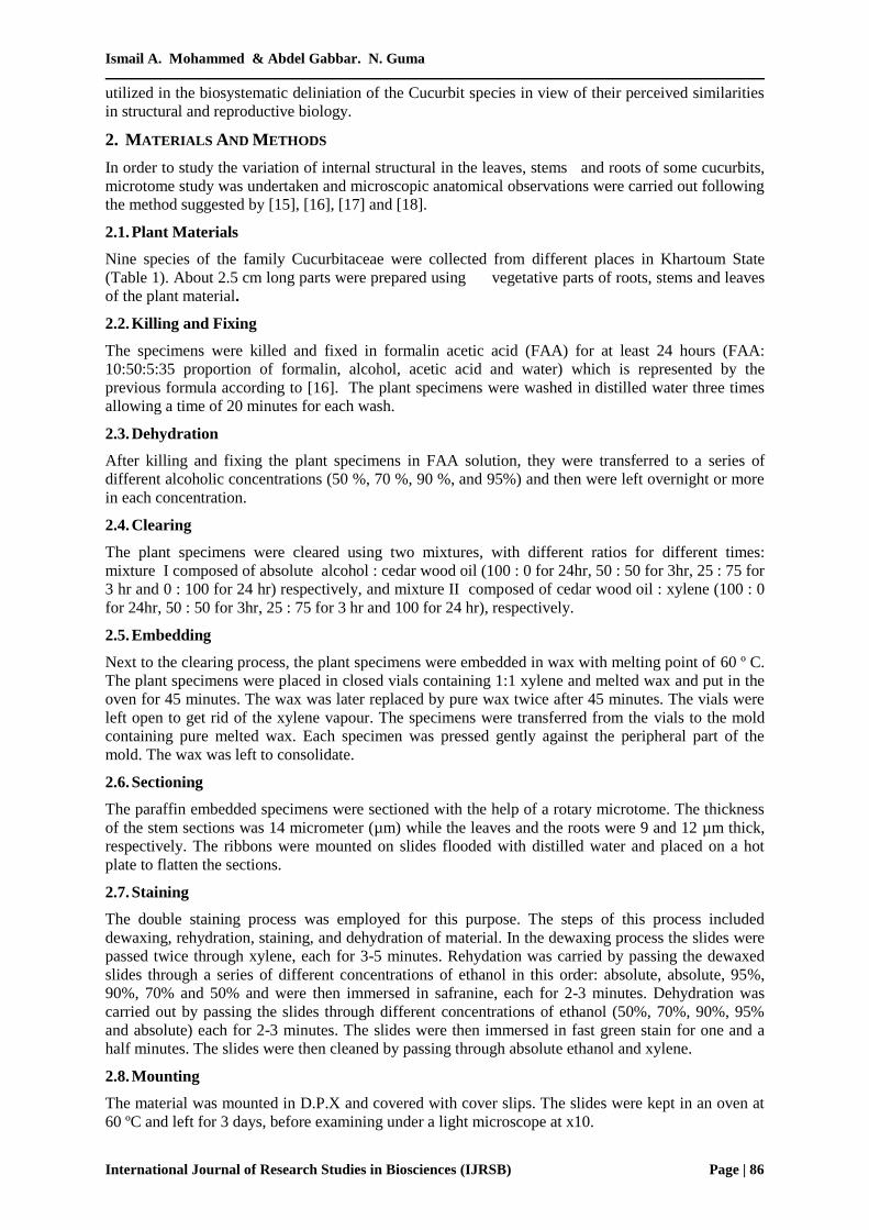
The roots usually consist of epidermis, cortex, endodermis and vascular bundles in most of the genera
of the family Cucurbitaceae. Reference [23] reported that the structure and development of the
Cucurbita pepo root is typical of dicotyledonous roots with limited secondary growth. Epidermis of
the root of most genera is one cell thick. The root cortex collenchyma is 2 – 4 cells thick and cortex
parenchyma is 5–7 cells thick in most of the genera. The endodermal and pericyclic layers are
inconspicuous. The root vascular bundles consist of four radial arms of primary xylem alternating
with four zones of primary phloem in most species except Cucumis sativus and Luffa aegyptiaca
where the vascular bundles are bicollateral and arranged in a ring (Figs. 2, A and B ).
Ismail A. Mohammed
& Abdel Gabbar. N. Guma
International Journal of Research Studies in Biosciences (IJRSB) Page | 88
Reference [24] found that the root epidermal layer of the Hibiscus rosa sinensis and Abelmoschus
esculenta studied that the epidermal cells are in the form of short chains (kioned) small and numerous
in Hibiscus rosa sinensis while they are of long chains, big and numerous in Abelmoschus esculenta .
Similarly the cortex tissue shows the presence of small-sized parenchyma cells in Hibiscus rosa
sinensis while in Abelmoschus esculenta the parenchyma cells are bigger in size. Both taxa show
presence of angular collenchyma. The xylem vessels are numerous, circular in shape and are radially
grouped in Hibiscus rosa sinensis while they are few and cuboidal in shape in Abelmoschus esculenta.
Reference [25] reported the following information on the root anatomy of Cucurbits. The meristem
consists of about seven layers of cambium-like cells, the number diminishing towards the periphery of
the root apex. The tissue directly in the center forms the vascular cylinder, while adjacent cells form
the cortical region. Approximate delimitation of the cortex and vascular cylinder is possible, because
the cells of the innermost layer of the cortex continue to divide tangentially for a longer period of time
than the adjacent cells of the vascular cylinder.
Fig2. (A): T. S of Root of Cucurbita moschata L. (X4).In general view with primary structure; Epidermis (E),
Cortex (C), Parenchyma (Par), Endodermis (En), Xylem (X), Phloem (Ph) and Pith (Pi). (B): T. S of Root of
Luffa aegyptiaca Mill. (X4). Vascular tissue (bicollateral bundle) and lateral root.
The stem epidermis of Cucurbits is regular, with thin cells and scattered collenchyma cells in most of
the species except Cucurbita moschata (Fig. 3A) where there are no scattered collenchyma. Stem
cortex collenchyma is 4 - 6 cells thick in Cucurbita moschata while it consists of a narrow band of
sclarenchyma cells in most of the other species eg. Colocynthis vulgaris (Fig.3D). Stem vascular
bundles in most of species are arranged in two rings. The outer ring is composed of the often smaller
bundles which are located at the angles of the stem. The inner ring contains the often larger bundles
which alternate with those of the outer ring as in Colocynthis vulgaris (Fig.3C). However, in
Cucurbita moschata the vascular bundles are arranged in one ring (Fig. 3B). The basic number of
bundles is ten, each cycle consisting of five. Stem vascular bundles are bicollateral (Fig. 3A) have
also been observed in Cucurbitaceae, Solanaceae and Asteraceae [26], [27], [28].
Leaves usually have a single layer of epidermal cells on both the upper and lower surfaces in all
species with hairs present on both surfaces. Collenchyma tissues are seen in the median line of the
upper surface of the leaf midrib. The thickness of the palisade parenchyma is not uniform in the
different species. The spongy parenchyma consists of 2 to 6 layers of cells. This observation is in line
with the work of reference [12], [10], [29] who used both the root and leaf anatomical features in the
family Cucurbitaceae and Dioscoraceae in establishing relationship among taxa. The vascular bundles
are bicollateral and arranged in different ways depending on the genera. The genera Cucurbita and
Citrullus had seven bundles, the largest being the undermost and the six smaller ones lying above or
on each side. The genus Cucumis has three bundles, which are arranged in a straight line, from above
downwards; the uppermost bundle is the smallest, while the lowest is the largest. The genus Luffa has
four bundles, being distinguished from the genus Cucumis by an additional small bundle near the
upper part of the central bundle (Figs. 4 A and B).
B A

Anatomical Diversity among Certain Genera of Family Cucurbitaceae
International Journal of Research Studies in Biosciences (IJRSB) Page | 89
Fig2. (A). and (D). T. S of Stem of Cucurbita moschata L. and Colocynthis vulgaris Schrad. (X4).In general
view with primary structure; Epidermis (E), Chollenchyma (Ch), Parenchyma (Par), Outer phloem (OPh),
Outer cambium (OCa) Xylem (X),Inner cambium (ICa), Inner phloem (IPh), and Pith (Pi). (B). T. S of Stem of
Cucurbita moschata L. (X4).Vascular tissue (bicollateral bundle) are arranged in one ring. (C). T. S of Stem of
Colocynthis vulgaris Schrad. (X4). Vascular tissue (bicollateral bundle) are arranged in two rings.
Various investigations have been made on the anatomy of Cucurbits, most of them prior to 1940 [30].
Although differences exist between Cucurbit species, Reference [31] had shown that, in general, there
was considerable similarity among them.
There are some reports by [32], [33] on epidermal and vegetative characteristics of the three species
Cucurbita moschata, Cucurbita maxima and Cucurbita pepo which agree with the results of this
study.
C D
A B

Ismail A. Mohammed
& Abdel Gabbar. N. Guma
International Journal of Research Studies in Biosciences (IJRSB) Page | 90
Fig4. (A). T. S of Leaf of Cucumis sativus L. (X4). .In general view with primary structure; Upper epidermis
(UE), Trichome (Tri), Palisade cells (PC), Spongy cells (SpC), Chollenchyma (Ch), Lower epidermis (LE) and
Xylem (X). (B). T. S of Leaf of Luffa aegyptiaca Mill. (X4).Vascular tissue (bicollateral bundle).
4. CONCLUSION
The data from these studies has provided further guidance for the taxonomic delimitation of species of
Cucurbita, which are found in Sudan. It is hoped that field officers, botanists and farmers will find
these information helpful in distinguishing the species. This study is therefore based on the principles
that root, stem and leaf anatomy have played a major role in the identification, characterization and
delimitation of plants. Hence the need to incorporate information from root, stem and leaf anatomy
with data derived from other botanical disciplines remains vital when formulating conclusions on the
systematics of the taxa investigated.
REFERENCES
[1]. Jeffrey C. A new system of Cucurbitaceae. Bot. Zhurn. 2005; 90:pp.332–335.
[2]. Olorode, Taxonomy of West African Flowering Plants. London Longman Inc.Pandey, B. P. 2006,
A Book Text of Botany. Angiosperm Taxonomy, Anatomy Embryology and Economic Botany.
New Delhi-India. S. Chand And Co. Ltd.
[3]. Gill, L. S., Taxonomy of Flowering Plants. Book House Onitsha. African Pep Publishers, (1988).
[4]. Gill, L.S. Ethnobotanical Uses Of Plants In Nigeria. Benin Uniben Press. (1992).
[5]. Whitaker, T. W. and Davis, G. N., Cucurbits. Interscience Publishers, Inc., New York. (1962).
[6]. Meeuse, A.D.J., The Cucurbitaceae of southern Africa. Bothalia 1962, 8: pp.1-111.
[7]. Jeffrey, C., Further Notes on Cucurbitaceae 3. Kew Bulletin, 1975, 30: pp. 485-491.
[8]. Jeffrey, C., Cucurbitaceae. Flora Zambesiaca 1978, 4: pp.414-499.
[9]. Edeoga, H.O., Anatomical studies on the roots of some Dioscorea L. species (Dioscoreaceae).
Afr. J. Root Tuber. Crop. 5 (1): 33-37 (2002).
[10]. Edeoga, H.O and Okoli, B.E., Anatomy and systematics in the Costus afer C. lucanusianus
complex (Costaceae). Acta phytotax. Geobot. 48: pp.151-158 (1997).
[11]. Edeoga, H.O and Osawe, P.J., Cuticular studies of some Nigerian species of Senna Tourn. Ex
Mill. (Syn Casia Tourn. Ex. L): Leguminosae Caesalpinoideae. Acta Phytotax Geobot 47:pp.41-
46 (1996).
[12]. Okoli, B.E., Anatomical studies in the leaf and probract of Telferia Hooker (Curcurbitaceae).
Feddes Repert. 98: pp. 231-236 (1987).
[13]. Metcalfe, C.R. and Chalk, L., Anatomy of the Dicotyledons. Vol 1 and 2 Clarendon Press.
Oxford. pp.1067-1074 (1950)
[14]. Mbagwu, F.N. and Edeoga, H.O., Anatomical studies on the root of some Vigna savi species
(Leguminosae-Papilionoideae) Agricultural Journal 1 (1): pp.8-10 (2006).
A B
Anatomical Diversity among Certain Genera of Family Cucurbitaceae
International Journal of Research Studies in Biosciences (IJRSB) Page | 91
[15]. Metcalfe, C.R. Anatomy of the Dicotyledons, 2: pp. 965-978. Oxford Clarendon Press, U.K.
(1950).
[16]. Sass, J. E., Microbotanical Techniques, third edition. Lowa University, Ames. U.S.A. 1958.
[17]. Esau, K., Anatomy of Seed Plants: 2-324. John Wiley & Sons, Inc. U.S.A. 1959.
[18]. Creedy, J., A Laboratory Manual for Schools and Colleges. Heinemann Educational books Ltd.
22 Bedford Squares. London. (1977).
[19]. Green, B.O., Taxonomy and Ethnobotanical Studies In Family Apocynaceae Ph.D Thesis.
University Of Port Harcourt. (1995).
[20]. Payne, W.W., Stomatal patterns in embryophytes. Their evolution, ontogeny and Inter prefatio.
Taxon. 1979, 28:pp.117-132.
[21]. Green, O. and Horsefall, U., Taxonomic and biosystematic study in certain members of family
cucurbitaceae in Niger Delta. Euro. Jour. of Sci. Res.Vol.20 No.1 pp.6-13 (2008).
[22]. Pandey, B. P., A Book Text of Botany. Angiosperm Taxonomy, Anatomy Embryology and
Economic Botany. New Delhi-India. S. Chand And Co. Ltd. 2006.
[23]. Esau, K., Anatomy of Seed Plants. Wiley & Sons, New York. 1977.
[24]. Nwachukwu C.U., Mbagwu F.N. and Jane Ijeoma. Iwu., Anatomical features of the roots and
leaves of Hibiscus Rosa- Sinensis and Abelmoschus esculenta. New York Science Journal.(1):
pp.27-32 (2008).
[25]. Whitaker, T. W. and Davis, G. N., Cucurbits: Botany, Cultivation and Utilization. New York :
Interscience , 1962, 250pp.
[26]. Esau, K., O caule: estágio primário de crescimento. In: Edgard Blücher, Eds. Anatomia das
plantas com sementes. São Paulo, 1974, pp.160-185.
[27]. Mauseth, J.D., Plant Anatomy. Menlo Park: Benjamin Cummings, 1988, 568 p.
[28]. Fahn, A., Plant Anatomy. 4th Ed. New York: Pergamon, 1990, 588p.
[29]. Edeoga H.O and Okoli B.E., Midrib anatomy and systematics in dioscoreae. J. Econ. Tax. Bot,
23:1 – 5 (2001).
[30]. Zimmerman, A., Die Cucurbitaceen. Beitrage zur Anatomie, Physiolgie, Morphologie, Biologie,
Pathologie and Systematik. G. Fischer, Berlin, 1922, 204 pp.
[31]. Holroyd, R., Morphology and physiology of the axis in Cucurbitaceae. Bot. Gaz., 1924, 78, pp.1-
45.
[32]. Agbagwa, I.O. and Ndukwu, B.C., Epidermal micro-morphology of Cucurbita L. species in
Nigeria. J.Appl. Sci. Environ. Mgt. 5(2):pp.59-64 (2001).
[33]. Agbagwa, I.O. and Ndukwu, B.C., Cucurbita L.species in Nigeria: under-exploited food and
vegetable crops. Niger Delta Biologia, 4(2): pp.11-15 (2004).