Analyst - Pompeu Fabra University
24
Mammalian protein glycosylation – structure versus function S. Defaus, ab P. Gupta, b D. Andreu a and R. Guti ´ errez-Gallego * abc Carbohydrates fulfil many common as well as extremely important functions in nature. They show a variety of molecular displays – e.g., free mono-, oligo-, and polysaccharides, glycolipids, proteoglycans, glycoproteins, etc. – with particular roles and localizations in living organisms. Structure-specific peculiarities are so many and diverse that it becomes virtually impossible to cover them all from an analytical perspective. Hence this manuscript, focused on mammalian glycosylation, rather than a complete list of analytical descriptors or recognized functions for carbohydrate structures, comprehensively reviews three central issues in current glycoscience, namely (i) structural analysis of glycoprotein glycans, covering both classical and novel approaches for teasing out the structural puzzle as well as potential pitfalls of these processes; (ii) an overview of functions attributed to carbohydrates, covering from monosaccharide to complex, well-defined epitopes and full glycans, including post- glycosylational modifications, and (iii) recent technical advances allowing structural identification of glycoprotein glycans with simultaneous assignation of biological functions. 1. Introduction The world of carbohydrates is extremely complex, rendering it both fascinating and challenging to those facing the task of unraveling their structural features. The term carbohydrate spans many different disciplines from large-scale industrial applications to ne-tuned biomedical uses, and the science of carbohydrates has experienced ups and downs over the last few decades in terms of attention paid, importance attributed, and level of understanding reached. Currently, the eld of carbohy- drate (bio)chemistry is enjoying renewed interest at both basic and applied (biomedical, pharmaceutical) levels, as clearly evi- denced by the >500 reviews on the subject over the past 18 months. Most efforts are devoted to the study of carbohy- drate-mediated biomolecular interactions and glycoprotein engineering but the structural analysis of carbohydrates, in all Sira Defaus received her B.S. (2008) and M.Sc. (2009) degrees from the Department of Organic Chemistry at the University of Barcelona. Later she joined the Bioanalysis and Analytical Services Research Group, and the Protein Chemistry and Pro- teomics laboratory at the Bar- celona Biomedical Research Park (PRBB) to obtain her PhD degree. During her study she investigated the structure– activity relationships of glycans and glycopeptides and developed analytical tools for protein detection in biological sources. Her research interests are peptide chemistry and glycobiology. Preeti Gupta obtained her M.Sc. degree in Biotechnology and M.Tech degree in Biochemical Engineering from Banaras Hindu University, India. In 2012 she joined the Bio-analysis Group at the Institut Hospital del Mar d'Investigacions M` ediques (IMIM), Barcelona through the Agency for Manage- ment of University and Research Grants (AGAUR) offered by the Government of Catalonia, Spain to pursue her PhD. She is interested in characterizing protein glycosylation, glycan structure and glycoprotein identication and quantication in plasma samples using HPLC and MALDI-TOF and LC-MS. a Department of Experimental and Health Sciences, Pompeu Fabra University, Barcelona Biomedical Research Park, 08003 Barcelona, Spain. E-mail: [email protected] b Bio-analysis Group, Neuroscience Research Program, IMIM-Parc Salut Mar, Barcelona Biomedical Research Park, 08003 Barcelona, Spain c Anapharm Biotech, 08038 Barcelona, Spain Cite this: Analyst, 2014, 139, 2944 Received 4th December 2013 Accepted 6th March 2014 DOI: 10.1039/c3an02245e www.rsc.org/analyst 2944 | Analyst, 2014, 139, 2944–2967 This journal is © The Royal Society of Chemistry 2014 Analyst CRITICAL REVIEW Published on 06 March 2014. Downloaded by Biblioteca de la Universitat Pompeu Fabra on 27/05/2016 10:32:57. View Article Online View Journal | View Issue
Transcript of Analyst - Pompeu Fabra University

Analyst
CRITICAL REVIEW
Publ
ishe
d on
06
Mar
ch 2
014.
Dow
nloa
ded
by B
iblio
teca
de
la U
nive
rsita
t Pom
peu
Fabr
a on
27/
05/2
016
10:3
2:57
.
View Article OnlineView Journal | View Issue
Mammalian prot
S(fCBBSttcPdi
activity relationships of glycans ananalytical tools for protein detecresearch interests are peptide chem
aDepartment of Experimental and Healt
Barcelona Biomedical Research Park,
[email protected] Group, Neuroscience Research P
Biomedical Research Park, 08003 BarcelonacAnapharm Biotech, 08038 Barcelona, Spain
Cite this: Analyst, 2014, 139, 2944
Received 4th December 2013Accepted 6th March 2014
DOI: 10.1039/c3an02245e
www.rsc.org/analyst
2944 | Analyst, 2014, 139, 2944–2967
ein glycosylation – structureversus function
S. Defaus,ab P. Gupta,b D. Andreua and R. Gutierrez-Gallego*abc
Carbohydrates fulfil many common as well as extremely important functions in nature. They show a variety
of molecular displays – e.g., free mono-, oligo-, and polysaccharides, glycolipids, proteoglycans,
glycoproteins, etc. – with particular roles and localizations in living organisms. Structure-specific
peculiarities are so many and diverse that it becomes virtually impossible to cover them all from an
analytical perspective. Hence this manuscript, focused on mammalian glycosylation, rather than a
complete list of analytical descriptors or recognized functions for carbohydrate structures,
comprehensively reviews three central issues in current glycoscience, namely (i) structural analysis of
glycoprotein glycans, covering both classical and novel approaches for teasing out the structural puzzle
as well as potential pitfalls of these processes; (ii) an overview of functions attributed to carbohydrates,
covering from monosaccharide to complex, well-defined epitopes and full glycans, including post-
glycosylational modifications, and (iii) recent technical advances allowing structural identification of
glycoprotein glycans with simultaneous assignation of biological functions.
1. Introduction
The world of carbohydrates is extremely complex, rendering itboth fascinating and challenging to those facing the task ofunraveling their structural features. The term carbohydrate
ira Defaus received her B.S.2008) and M.Sc. (2009) degreesrom the Department of Organichemistry at the University ofarcelona. Later she joined theioanalysis and Analyticalervices Research Group, andhe Protein Chemistry and Pro-eomics laboratory at the Bar-elona Biomedical Researchark (PRBB) to obtain her PhDegree. During her study shenvestigated the structure–d glycopeptides and developedtion in biological sources. Heristry and glycobiology.
h Sciences, Pompeu Fabra University,
08003 Barcelona, Spain. E-mail:
rogram, IMIM-Parc Salut Mar, Barcelona
, Spain
spans many different disciplines from large-scale industrialapplications to ne-tuned biomedical uses, and the science ofcarbohydrates has experienced ups and downs over the last fewdecades in terms of attention paid, importance attributed, andlevel of understanding reached. Currently, the eld of carbohy-drate (bio)chemistry is enjoying renewed interest at both basicand applied (biomedical, pharmaceutical) levels, as clearly evi-denced by the >500 reviews on the subject over the past18 months. Most efforts are devoted to the study of carbohy-drate-mediated biomolecular interactions and glycoproteinengineering but the structural analysis of carbohydrates, in all
Preeti Gupta obtained her M.Sc.degree in Biotechnology andM.Tech degree in BiochemicalEngineering from BanarasHindu University, India. In 2012she joined the Bio-analysisGroup at the Institut Hospitaldel Mar d'InvestigacionsMediques (IMIM), Barcelonathrough the Agency for Manage-ment of University and ResearchGrants (AGAUR) offered by theGovernment of Catalonia, Spain
to pursue her PhD. She is interested in characterizing proteinglycosylation, glycan structure and glycoprotein identication andquantication in plasma samples using HPLC and MALDI-TOFand LC-MS.
This journal is © The Royal Society of Chemistry 2014
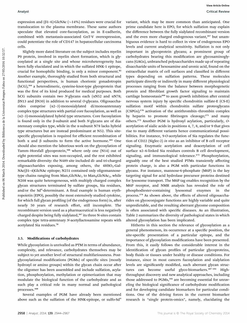
Table 1 Different types of glycosylation. The letters in the sequencecorrespond to the 1-letter annotation of amino acidsa
Linkage Type Sequence
Man–a-Trp C-Mannosylation W–X–X–WGlcNAc–b-Asn N-Glycosylation N–X–(S/T) (X s P)
N–X–C, N–G, N–X– (rare)
Critical Review Analyst
Publ
ishe
d on
06
Mar
ch 2
014.
Dow
nloa
ded
by B
iblio
teca
de
la U
nive
rsita
t Pom
peu
Fabr
a on
27/
05/2
016
10:3
2:57
. View Article Online
its aspects, remains the basis of nearly all the developments ofrecent times. The goal of this review is to highlight relevantaspects of structural analysis of carbohydrates with focus onmammalian protein glycosylation and insights into its rele-vance. The nal section deals with recent advances paving theway towards structural analysis within actual biological settings,ideally, without any external interference.
GalNAc–a-Ser/Thr O-Glycosylation Various ppGalNAcTact concertedly
GlcNAc–b-Ser/Thr Any S or TGlcNAc–a-Thr T (near P residues)Glc–a-Tyr GYG (glycogenin)Glc–b-Ser C–X–S–X–P–CGlc–b-Asn N–X–(S/T)Gal–Thr G–X–T (X ¼ A, R, P, hP, S)Gal–b-Hyl X–Hyl–GFuc–a-Ser/Thr C–X–X–G–G–(S/T)–C
X–X–X–X–(S/T)Man–a-Ser/Thr I–X–P–T–(P/X)–T–X–P–
X–X–X–X–P–T–X–(T/X)–X–XMan–a-1-P–Ser S rich domainsXyl–b-Ser –G–S–G–
(near acidic residues)
a X may be any amino acid.
2. Structural analysis of glycoproteinglycans
Glycoproteins are fundamental in most important biologicalprocesses including fertilization, immune response, inamma-tion, viral replication, parasite infection, cell growth, cell–celladhesion, or glycoproteins clearance. Whereas protein synthesisfollows a well-dened, genetically encoded linear process,glycosylation is a non-template-driven, secondary gene eventinitiated during protein synthesis and involving a large collec-tion of redundant and overlapping enzymes (glycosidases andglycosyltransferases) partially compartmentalized throughoutthe endoplasmic reticulum (ER) and the Golgi system.1 Variouscompeting reactions in the processing pathways, plus the needfor enzyme, acceptor and substrate concurrence, as well as otherphysiological factors contribute to glycan microheterogeneity,i.e., glycoprotein isoforms resulting from different glycans at agiven site. This heterogeneity may be relatively simple, such asfor RNAse B,2 or rather complex as in the case of CD59 where atleast 123 different desialylated glycan variants have been iden-tied at a single site.3 Thus, carbohydrate diversity and conse-quent complexity arises from several factors. Firstly, from thestructural variety at the monosaccharide level, where multidi-rectional combinations of different monosaccharide buildingblocks, linkages,4 anomericity, and branching generate a vastnumber of complex glycan structures (polysaccharides) that canbe further modied by sulfation, acetylation, methylation,
David Andreu studied chemistryat the University of Barcelona,where he obtained his PhD(1981, with E. Giralt). Aerfurther training in peptidechemistry during a postdoc withBruce Merrield at RockefellerUniversity (1982–1985), in 1985he returned to Spain as Asso-ciate Professor of organic chem-istry at the University ofBarcelona. In 2001 he wasappointed Professor of Chem-
istry at the Pompeu Fabra University where he heads the ProteinChemistry and Proteomics laboratory at the Barcelona BiomedicalResearch Park. In addition to glycan–protein interactions, hisother scientic endeavours include cell-penetrating and antimi-crobial peptides, and peptide-based synthetic vaccines.
This journal is © The Royal Society of Chemistry 2014
phosphorylation, etc., and linked covalently to aglycones such aspeptides (in different ways) or lipids forming the correspondingglycoconjugates (see Table 1). Secondly, from the inuence ofthe peptide sequence in determining potential glycosylationsites, the effect of the 3D protein display in subsequent glycanprocessing events, and the spatial distribution or multivalentpresentation leading to the avidity principle.5 Thirdly, frommicroheterogeneity and macroheterogeneity phenomenainherent to carbohydrate chemistry resulting from the fact thatin an individual glycoprotein a specic glycosylation site is notalways associated with the same glycan structure and that not allN-glycan sequons are necessarily glycosylated.
Ricardo Gutierrez-Gallegostudied chemistry at UtrechtUniversity (NL) and obtained aPhD in 2001 (with J. Vliegen-thart and J. Kamerling). Subse-quently, he joined the IMIM-Parc de Salut Mar where hedeveloped the analytical meth-odology program for (glyco)protein hormone detection in thecontext of antidoping policies.Simultaneously, in 2001, he wasappointed assistant professor of
chemistry at Pompeu Fabra University where he expanded hisresearch on the structure–function relationship of protein glyco-sylation with special emphasis on glycan-mediated biomolecularinteractions. In 2013 he amplied his scope of activities joiningAnapharm Biotech to provide analytical support in drug-develop-ment programs.
Analyst, 2014, 139, 2944–2967 | 2945

Fig. 1 As an example in the structural elucidation of glycoproteins an N-glycan in the human chorionic gonadotropin (hCG) glycoprotein a-chain is shown. Elements to be specified are listed on the right and some of them displayed. The shaded part represents the epitope potentiallyrecognized in a carbohydrate-driven interaction.
Analyst Critical Review
Publ
ishe
d on
06
Mar
ch 2
014.
Dow
nloa
ded
by B
iblio
teca
de
la U
nive
rsita
t Pom
peu
Fabr
a on
27/
05/2
016
10:3
2:57
. View Article Online
Eventually, such diversity gives rise to a set of glycoforms, inboth soluble and membrane-anchored forms that are asessential to life as a genetic code, and constitute an evolutionaryconserved feature of all living cells.6 The identication of thenumber, structure, and function of glycans in a particular bio-logical context, initiated decades before the “omics” boom, wasrecently termed glycomics, and substantial progress has beenmade in understanding how glycans are directly involved inalmost every biological process or human disease.7 Still, theglycome is far more complex than the genome, transcriptome,or proteome, due to a much more dynamic character that variesconsiderably not only with the cell or tissue type, but also withthe developmental stage,8 metabolic state, or changes such asdisease,9 aging,10,11 environmental factors,12 or evolution.13,14 Forinstance, epigenetic regulation may induce novel glycan struc-tures that make the organism tter in a specic environmentwithout altering genetic information.15 It is therefore of utmostimportance to know what carbohydrate structures decoratewhich glycoproteins under particular conditions.
Even for dedicated specialists analysis of protein glycosylationremains an extremely challenging task due to many differentphysical parameters that must be established before a structuralcharacterization can be considered complete (Fig. 1). As a conse-quence, there is no single analytical method capable of providingall the necessary information for fast and reliable identicationand quantication of a particular structure, let alone to alsoestablish its particular functionality. Rather, a multidimensionalapproach involving several orthogonal, physical, chemical, andbiochemical techniques as depicted in Scheme 1 is required.
In the following pages an overview is provided of the tech-niques employed in structural analysis of protein glycosylation,their shortcomings and particular virtues, and the latest trendsin this eld.
2946 | Analyst, 2014, 139, 2944–2967
2.1. Analyzing glycosylation
Over the last 2–3 decades the continuous renement ofanalytical tools has greatly facilitated glycan analysis; numerousreviews16–21 and papers cover the main technologies routinelyused today for N- and O-linked glycan analysis, includingcapillary electrophoresis (CE),22–24 liquid chromatography(LC),25,26 mass spectrometry (MS)27–30 and microarray-based31–35
approaches to glycomics and glycoproteomics.28,36,37 It isimportant to stress that in all these techniques a compromiseexists between analytical sensitivity and the degree of structuraldetail provided. None of these tools, or any other for thatmatter, can single-handedly reveal all the features (see Fig. 1)necessary for full characterization. Hence, an unambiguouslystructural analysis must be conducted at different levels,namely intact glycoprotein, glycopeptides and released glycans,and in each case the most appropriate technique for decipher-ing that part of the puzzle must be chosen. This, in turn, entailsanother compromise between the degree of informationobtained vs. the amount of (puried) material required.
2.1.1. Analysis of intact glycoproteins. In the rst evalua-tion of protein glycosylation it is recommended to assess themicroheterogeneity at the glycoprotein level as it provides anexcellent starting point. Quite oen this is done by means ofconventional sodium dodecyl sulfate polyacrylamide gel elec-trophoresis (SDS-PAGE) and comparing the resulting bands to aprotein molecular weight standard (Fig. 5). Such evaluation,when conducted with non-specic staining techniques usingcoomassie, silver, or Pro-Q emerald dyes, should provide anunbiased view of the glycoform distribution. Alternatively, thedetection could be performed through specic biomolecularrecognition (using lectins, antibodies, etc.) at much bettersensitivity than the non-specic staining. However, one shouldbear in mind that such biorecognition may be biased towards
This journal is © The Royal Society of Chemistry 2014

Scheme 1 Different levels of glycan analysis include compositional and detailed glycan structure, glycan affinity and specificity, glycoformprofiling, site-specific analysis and 3D structural and topological studies. Moreover, determination of carbohydrate-binding protein (CBP)structures and characterization of glycan–CBP recognition and complex formation are required, particularly in biological contexts. Advancedglyco-informatic resources are essential for analytical data collection, annotation, and analysis of the large-scale data generated.
Critical Review Analyst
Publ
ishe
d on
06
Mar
ch 2
014.
Dow
nloa
ded
by B
iblio
teca
de
la U
nive
rsita
t Pom
peu
Fabr
a on
27/
05/2
016
10:3
2:57
. View Article Online
particular glycoforms because of steric effects or other factorshampering interactions with other glycoforms. For its part, SDS-PAGE does not provide an accurate molecular weight determi-nation as separation is governed by the hydrodynamic volumeof the migrating species. One alternative technique is gel-based
This journal is © The Royal Society of Chemistry 2014
isoelectric focusing (IEF) (Fig. 2). This technique gained muchmomentum in the early days of proteomics as part of the two-dimensional gel-electrophoretic sample preparation andprovides a rough charge distribution of the glycoprotein. Giventhe limited number of charged modications of amino acid
Analyst, 2014, 139, 2944–2967 | 2947

Fig. 2 Isoelectric focusing profiles of an endogenous erythropoietin(EPO) standard (left lane), EPO from a human urinary sample (centerlane) and a recombinant erythropoietin mixture (right lane) composedof Eprex (3 N-glycans and migrating just below pI 6) and Darbepoetinalpha (5 N-glycans and migrating just above pI 2). On the right thecrystal structure of erythropoietin (1BUY) and its glycosylation sites areshown.
Analyst Critical Review
Publ
ishe
d on
06
Mar
ch 2
014.
Dow
nloa
ded
by B
iblio
teca
de
la U
nive
rsita
t Pom
peu
Fabr
a on
27/
05/2
016
10:3
2:57
. View Article Online
residues, the IEF prole usually provides a reliable sketch of thedegree of sialylation, sulfation, phosphorylation and/or glu-curonidation of the glycoprotein.
Similar information can also be obtained from the massspectrum of the intact glycoprotein. Matrix Assisted LaserDesorption Ionization Time Of Flight (MALDI-TOF) massspectrometry is particularly suited for this purpose, beingcapable of handling complex mixtures and fairly tolerant ofimpurities, aside from detergents, which produce signicantion suppression.38 Fig. 3 compares the MALDI-TOF spectra of aglycoprotein (rAT-III) and a non-glycosylated protein (rGH) andshows how the peak width provides information on theheterogeneity of the protein, and the peak number on theprevalent glycoforms. Depending on the purity of the glyco-protein, on its structural complexity, and on instrument reso-lution, information on microheterogeneity can be quiteexhaustive.
A subsequent step in the structural interrogation, still at theglycoprotein level, concerns the evaluation of the mono-saccharide residues present. One may distinguish differentlevels of analysis, all requiring the chemical hydrolysis of theglycoprotein. Typically, a rst level of analysis addresses sialicacid (Sia) residues. The relevance of Sia was acknowledgednearly six decades ago39 and, at the time, a specic colorimetricprotocol named Bial's reaction and based on orcinol wasemployed for its detection.With time, the number of residues inthe Sia family has increased and currently more than 50 struc-turally different sialic acid residues,40with a variety of associatedfunctions,41 are known. Even though the analysis of Sia has beenpursued through many different approaches, it was selectiveconjugation of the released a-keto-acids with ortho-diamines to
2948 | Analyst, 2014, 139, 2944–2967
form quinoxaline derivatives that allowed both sensitive andspecic analysis by liquid chromatography of this family ofcompounds. In particular, 1,2-diamino-4,5-methylenedioxy-benzene (DMB) has found widespread use due to its uorescentproperties (ex: 373 nm, em: 448 nm),42 which grants the protocola yet unmatched sensitivity, and also because the mild acidconditions required to release Sia residues do not cause migra-tion of the labile acetyl at C–O7.43 For example, the detection ofN-glycolyl neuraminic acid in erythropoietin – found at pico-molar concentrations in human specimens – is an unambiguousevidence for a doping violation that could be established by thisprotocol44 but not withMS analysis of the same sample (personalcommunication). Hence, obtaining a complete picture in termsof Sia speciation will usually require the DMB protocol, thoughcare must be exercised as, in addition to the release-relateddegradation, other a-keto acids or 1,2-diketones in biologicalsamples, e.g., a-ketoglutaric, pyruvic or p-hydroxyphenyl-pyruvicacids, can interfere. In such cases, hyphenation of liquid chro-matography with uorescence detection (LC-FLD) to massspectrometry is possibly the best solution.
The next level of analysis, namely determining all mono-saccharides present in a glycoprotein, usually requires a strongacid (e.g., 1 M HCl in methanol, 65 �C, overnight, in the pres-ence of an internal standard) to hydrolyze all glycosidic linkages– except that between the rst GlcNAc residue and Asn in N-linked glycosylation – and convert glycosidic acetals into thecorresponding methylglycosides. The procedure, however, willalso irretrievably cleave most post-glycosylational modica-tions. Following neutralization and evaporation, free hydroxylsare further derivatized with trimethylsilyl (or analogous) func-tionalities for both qualitative (four characteristic peaks foreach monosaccharide) and quantitative evaluation using gaschromatography ame ionization detection (GC-FID) or gaschromatography mass spectrometry (GC-MS) and comparisonwith a standard monosaccharide mix.45,46 Alternatively,following acid release, monosaccharides may be separatedchromatographically by high performance anion exchangechromatography (HPAEC) (CarboPack PA-100) and detected bypulsed amperometric detection (PAD).47 The latter protocoloffers the advantage of a single peak per monosaccharide and ofdirect analysis without derivatization, but the basic LC condi-tions may induce C2-epimerization in GlcNAc to yield Man-NAc,48 or peeling reactions where some monosaccharides aredegraded from the reducing end. Altogether, monosaccharideanalysis offers the possibility of identifying which type ofglycosylation is present; Man and GalNAc being representativeof N- and O-glycosylation, respectively. Furthermore, the stoi-chiometry of the different sugars allows an educated guess onthe type of N-linked glycans present by considering the ratiobetween the distinct monosaccharides with respect to Man. Asimilar approach can be employed to estimate substitutionproles (i.e. a/b1–2,3,4,6) in glycans. In this case, carbohydratesand other functional moieties are permethylated using theHakomori protocol,49 subsequently the monosaccharides arereleased by acid hydrolysis (leaving the methyl-ether bondsintact) and the resulting hydrolyzed monomers are reduced andacetylated to give volatile, partially methylated alditol acetates
This journal is © The Royal Society of Chemistry 2014

Fig. 3 Top: MALDI-TOF spectrum of recombinant human growth hormone (not glycosylated); bottom: recombinant human antithrombin-III(tri/tetra glycosylated). Microheterogeneity due to glycosylation can be clearly appreciated from the peak width.
Critical Review Analyst
Publ
ishe
d on
06
Mar
ch 2
014.
Dow
nloa
ded
by B
iblio
teca
de
la U
nive
rsita
t Pom
peu
Fabr
a on
27/
05/2
016
10:3
2:57
. View Article Online
(PMAA), again analyzed by GC-MS. This procedure providesunambiguous information on the linkage pattern as well as thering size of the corresponding sugar, but it is important toemphasize that it is unable to distinguish between a 4-O-linkedaldopyranose and a 5-O-linked aldofuranose.
A more recent development to assess glycosylation at theglycoprotein level consists in the interrogation of a particularglycoprotein or complex biological sample through a lectinarray. Even though lectins have long been recognized as tools inthe study of glycosylation, their systematic application in arrayformat to detect the glycotopes in a given sample is relativelynew.50–53 Even if the information obtained cannot be comparedwith a thorough structural analysis (vide infra), it has theadvantage of analyzing a crude biological sample, e.g. thecellular glycome,54 without too much manipulation, and hasdemonstrated its value in assessing glycosylation changes incancer cells on the basis of a direct or an antibody-assistedevanescent-eld uorescence detection scanner.55,56 In addi-tion, combination with antibodies allows the changes inglycosylation to be pinpointed to specic proteins,57 adding onemore level of specicity to the analysis. Hence, dynamic gly-come analysis can be undertaken by means of differentiallylabeled CBPs.58 Even so, there are several drawbacks to lectinarrays. For one, while current plant lectin-based arrays32 covermost monosaccharides in the mammalian glycome, mamma-lian lectins would obviously provide a more representative gly-coprole. Also, one should not ignore that most lectins arepromiscuous to a certain degree and that this behavior,different for each lectin and with different affinities for differentsugars, will complicate glycome readout. Ultimately, itappears that lectin–carbohydrate interactions are not always
This journal is © The Royal Society of Chemistry 2014
straightforward and that glycoclusters, of either homo- or het-erogenic nature, will strongly inuence the interaction, and inthis case, the analytical data.5,59 The latter phenomenon appearsto be, at least in part, responsible for the ne-tuning of bio-logical communication processes and will as such be verydifficult to interpret in terms of precise structural entities.
2.1.2. Analysis of glycoprotein glycans. Evaluation of intactglycans almost inevitably requires their release from the peptidebackbone. While some high resolution approaches, in partic-ular those based on MS, are capable of addressing micro-heterogeneity at the intact glycoprotein level, this is restricted tothose entities with a very limited number of glycans and gly-coforms such as apolipoprotein C3.60 For more complex enti-ties, separation of carbohydrate from the protein backbone isneeded. Since, in this process, both the site-specicity and theprotein origin of the glycans are lost, it is crucial to ensure themaximum degree of protein purity before the procedure isinitiated. Deglycosylation can be achieved by either chemical orbiological means, each with their respective dis/advantages.
The most widely used chemical method is hydrazinolysis, aprocedure that releases the two major classes of glycans (Fig. 4)yet requires highly skilled staff and strict conditions for success,and is invariably accompanied by side-reactions and byprod-ucts. In addition, re-acetylation is necessary to avoid N-glycansbeing lost during the process but may also induce O-acetylation.Selective and sequential release of oligosaccharides is achievedby mild hydrazinolysis of O-linked oligosaccharides at 60 �C,followed by that of N-linked oligosaccharides at 95 �C, but theremay be a signicant overlap between both processes dependingon the protein and the degree of glycosylation. In addition, theprocedure will destroy the protein backbone so that if both
Analyst, 2014, 139, 2944–2967 | 2949

Fig. 4 Release of carbohydrates from the protein backbone following hydrazine treatment. An undesired side effect is protein destruction asindicated in the bottom right.
Analyst Critical Review
Publ
ishe
d on
06
Mar
ch 2
014.
Dow
nloa
ded
by B
iblio
teca
de
la U
nive
rsita
t Pom
peu
Fabr
a on
27/
05/2
016
10:3
2:57
. View Article Online
glycans and protein sequence are to be investigated, hydrazi-nolysis is not the method of choice. Another chemical proce-dure, i.e., alkaline b-elimination (0.05 to 0.1 M NaOH or KOH,60 �C, 12 h), can be applied for O-linked carbohydrates attachedto Ser or Thr (except those at the carboxy-terminus), but not to
2950 | Analyst, 2014, 139, 2944–2967
Tyr, hydroxy-Pro or hydroxy-Lys. In this case, N-linkedcarbohydrates are unaffected. To prevent base-catalyzed peeling(vide supra), sugars must be immediately captured,61 thealkaline solution carefully prepared,62 or a reducing agent (e.g. 1M NaBH4) added which forms an alditol that precludes
This journal is © The Royal Society of Chemistry 2014

Fig. 5 SDS-PAGE and MALDI-TOF-MS analysis of human anti-thrombin III (AT-III) after conventional PNGaseF de-N-glycosylation.From the number of bands in the SDS-PAGE and peaks in the MALDI-TOF spectrum it is evident that deglycosylation is inefficient and doesnot reach completion.
Critical Review Analyst
Publ
ishe
d on
06
Mar
ch 2
014.
Dow
nloa
ded
by B
iblio
teca
de
la U
nive
rsita
t Pom
peu
Fabr
a on
27/
05/2
016
10:3
2:57
. View Article Online
reducing-end derivatization. If tagging is intended, it is bestperformed during release.63 As with hydrazinolysis, the proteinbackbone is destroyed in the process.
The only strategy that preserves both protein and carbohy-drate is enzymatic deglycosylation, which has been successfullydeveloped for N-linked glycans using several endoglycosidases.For mammalian glycoproteins, peptide-N4-(N-acetyl-beta-glu-cosaminyl)asparagine amidase (PNGase F) is the enzyme ofchoice; it liberates nearly all N-linked carbohydrates understandard conditions (e.g., phosphate buffer, 50 mM pH 7.3, 16h, 37 �C). Nevertheless, the efficiency of this procedure needs tobe checked to ensure a correct assessment of the subsequentanalysis. One example where conventional conditions do notresult in full release is human antithrombin III (AT-III) (Fig. 5),where only partial release of the four N-linked structures of thea-variant is achieved if the procedure is not optimized. For plantor invertebrate glycosylations, PNGase A is the preferred choice.In contrast to PNGase F, this enzyme, although of poorer overallefficiency, is capable of releasing a1–3-fucose-bearing corestructures. Other endoglycanases (endo F1 to F3 or endo H) canbe employed to release the carbohydrate chains, except for Asn-bound GlcNAc, as the enzymes specically target chitobioseunits. In sum, the full repertoire of N-glycans can be released byenzymatic means but caution is still advisable. Co- and post-release glycosylaminemodication to functionalities other thanC1-hydroxyl, such as urea,64 glycerol65 or thiol66 or incompleteconversion in the presence of ammoniummay obscure the nalanalysis. For O-glycans, one single deglycosylation enzyme hasbeen identied thus far,67 and its activity is restricted to T- andTn-antigenic structures on Ser or Thr. As such, its main appli-cation is in unveiling the presence of either epitope, withoutproviding further evidence on the presence of other O-glycans.
This journal is © The Royal Society of Chemistry 2014
Once the release from the protein has been completed, thecarbohydrates must be puried from the protein and buffercomponents prior to analysis. Separation into simpler glycanmixtures, a discipline in itself, can signicantly facilitatesubsequent analyses, but loss of low-abundance glycans mayinevitably bias structural identication. On average, everyseparation step may cause a 10–50% loss of starting material.Generally, separation of the mixture is done by ltration,CE,23,68 HPAEC,26 or high-performance liquid chromatography(HPLC). The latter is one of the most versatile, as separationcan be based on charge (weak or strong cation or anionexchange), hydrophobicity69 or hydrophilic interaction (HILIC),and can be performed on either conventional, micro- ornanosized platforms.25 Separation is typically performed onnormal-phase (NP), but reverse-phase (RP) analysis is alsopossible aer permethylation, as demonstrated recently in acomparative study of RP-LC-electrospray (ESI-MS), RP-LC-MALDI-MS, and MALDI-MS70 using model proteins as well ashuman blood serum. This study concluded that, for complexsamples such as serum, RP-LC-ESI-MS yielded the condentdetection of more and lower-abundance glycans, and alsopermitted the separation of several structural isomers. Anothertype of derivatization, i.e., selective incorporation of a reportergroup at the reducing end of every glycan, is one major stepforward in the eld of carbohydrate proling. Research at theOxford Glycobiology Institute pioneered this approach forcomprehensive glycosylation analysis when the starting mate-rial is scarce.71,72 In this approximation, carbohydrates arelabeled with uorescent 2-amino benzamide (2AB), proled byboth weak anion exchange (WAX) and NP (nowadays HILIC)HPLC, and elution times standardized against a partial acidhydrolysate of 2AB-labeled dextran. The resulting glucose unit(GU) values73,74 allow a preliminary assignment that can thenbe corroborated by targeted and sequential exoglycosidasedigestions, followed by another round of HPLC proling.Subsequent glycan trimming is of particular interest as notonly does it provide conrmation of structural assignments, itsimplies the glycan pool, ultimately contributing to unveilingepitopes that are obscured in the overall microheterogeneity.75
Fluorescence labeling at the reducing end is not restricted to2AB as several other tags are described76 and not only reducessample requirement to the low femtomole level of individualstructures,77 it also allows accurate relative and absolutequantitation of the glycans present in a given glycoprotein.78 Ithas become one of the standard techniques in carbohydrateproling,71,79 which can be amplied with internal standards ifa different uorescent tag is used for dextran and the sample,and can be easily extended with back-end MS evaluation whenthe material is not required for further exoglycosidase treat-ment.80 Despite these advantages, it is a laborious approachthat requires considerable care, especially during the 2ABlabeling (in 30% acetic acid, 65 �C) to avoid desialylation thatmay confound structural assignments.78 Automated samplepreparation, i.e. both the uorescence labeling and the post-release and post-labeling purication steps, greatly reducesanalysis variability, providing robust and reliable glycomicsdata.16,19,81
Analyst, 2014, 139, 2944–2967 | 2951

Analyst Critical Review
Publ
ishe
d on
06
Mar
ch 2
014.
Dow
nloa
ded
by B
iblio
teca
de
la U
nive
rsita
t Pom
peu
Fabr
a on
27/
05/2
016
10:3
2:57
. View Article Online
Arguably one of the more powerful and versatile analyticaltechniques for all sorts of compounds, including carbohy-drates, MS has become the cutting-edge technology for gly-comics, linking mass with composition and providing precisecharacterization of complex structures. A wide range of MSequipments are available for glycan analysis. The introductionof MALDI-TOF instruments allowed rapid and straightforwardevaluation of complex mixtures82,83 and was a giant leapforward in MS evaluation of carbohydrates, hitherto restrictedto the cumbersome, low-sensitivity fast atom bombardmentmass spectrometry (FAB-MS). Improved analyses are madepossible through combination with well-known derivatizationstrategies (e.g. permethylation or peracetylation) that reducepolarity and improve sensitivity by either MALDI-TOF-MS84 orLC-ESI-MS.69 Isotope-based differential derivatization proto-cols, e.g., using CH3I and CD3I, allow exact determination ofthe number of free hydroxyls in a given structure, from whichvaluable information on carbohydrate composition can beinferred.85–87 When glycan sequencing is the goal, analysesmust include tandem MS experiments where structure-revealing ions are obtained by a combination of ion activation/fragmentation strategies such as collision-induced dissocia-tion (CID), electron transfer dissociation (ETD) or electroncapture dissociation (ECD). For instance, a recent studyinvolving a series of oligosaccharide-derived oxonium frag-ment ions generated by CID enabled simultaneous charac-terization of IgG glycoforms at both Fc and Fab glycosylationsites by combining multiple reaction monitoring (MRM) MSwith energy-resolved structural analysis.88 In another study,CID-MS was employed to selectively monitor the generation ofa m/z 284.053 fragment, consistent with GlcNAc phosphory-lation in a mouse brain dataset, and unveiled this new post-glycosylational modication.89 Within the last few years, ECDand ETD have enabled the assignment of O-GlcNAc sites at theproteomic scale and greatly facilitated protein-specic studiesof single O-GlcNAc events. Particularly, ETD has been used toidentify O-GlcNAc sites and PTMs such as phosphorylationand Arg methylation, on host cell factor C1 (HCF-1), a chro-matin-associated protein involved in transcriptional regulationand cell proliferation, and one of the most highly O-GlcNAc-rich proteins found in cells.90 Although MS is clearly indis-pensable in glycomic analysis, some techniques still presentlimitations such as susceptibility to salts, difficult assignmentof isomeric and isobaric monosaccharides – even though theevolution of ion-mobility strategies are addressing this –,91
complicated behaviour of acyl groups on glycans, and ioniza-tion efficiency dependence. Moreover, interpretation of MSn
fragmentation datasets remains a limiting factor with regardto throughput, user-dependent variability in discriminationand/or interpretation and complete identication of allglycoforms.
When sample complexity is limited to only a few glycanstructures, the analytical technique of choice is nuclearmagnetic resonance (NMR), the only one providing both qual-itative and quantitative information on the glycan withoutbeing destructive. While mono-dimensional (1H, 15N, 13C)experiments readily provide information on structural reporter
2952 | Analyst, 2014, 139, 2944–2967
groups,92,93 multi-dimensional, both homo- and heteronuclearexperiments yield information on the spatial orientation of theglycotope. Moreover, NMR may provide unambiguous infor-mation on the presence and position of post-glycosylationalmodications such as sulfation, methylation, acetylation orphosphorylation.94 Ironically for a technique that had provencrucial in the early development of the carbohydrate eld, NMRhad gradually lost inuence due to the oen prohibitiveamounts of natural material required. Nevertheless, recentdevelopments enabling analysis of picomoles95 may rein-vigorate a technique which in fact has never lost its appeal forthe analysis of carbohydrate biological interactions96,97 or theeffect of changes in glycosylation.98,99
Altogether glycomics can be addressed through a variety ofstrategies and technologies that turn out to be orthogonalrather than parallel. While all of them rapidly generate verylarge amounts of data, differences between platforms can turndata analysis into a complex, time-consuming task requiringbio-informatics tools and databases to facilitate data processingand interpretation. Most of these glycoinformatic tools haveparticular focuses, e.g., data from HPLC,74 MS,100 NMR ormicroarray101,102 experiments. Initiatives for cross-linking datafrom different techniques and integrating multiple datasets areprospering and extremely useful,103,104 although in the use ofdatabase search outputs critical interrogation is advisable.Additionally, the eld of glycobiology would greatly benet froma single glycan structural annotation, easy and of worldwideaccess, and supported by public agencies such as NCBI or EMBL.Limited public initiatives in this regard (e.g. Consortium forFunctional Glycomics – http://www.functionalglycomics.org/static/consortium/consortium.shtml) are at risk of being overshadowedby commercial enterprises (e.g. Waters & NIBRT – http://www.waters.com/waters/promotionDetail.htm?id¼134654015),most likely with ensuing limitations in accessing data, let aloneseeking a universal output.
2.1.3. Analysis of site-specic glycosylation. With theincreasing awareness of the importance of site specicglycosylation much effort is invested in addressing the gly-coforms, enrichment of glycopeptides, and evaluating glycansat their site of attachment.36,37 The prerequisite of preservingthe peptide backbone eliminates the possibility of quantita-tive glycan proling through the 2AB protocol, or any otherprocedure involving tagging of the reducing end. Further-more, the analytical strategy is limited to MS as the onlytechnique capable of differentiating peptide and carbohydratesequences. However, a main drawback of glycopeptide MSanalysis is that glycosidic bonds are less stable than amidebonds, so that predominant cleavage of the former leads todeglycosylated peptides with no information on the attach-ment site. The problem has been solved by simply varying thecollision energy, so that fragmentation is selectively directedto either carbohydrate or peptide, and information on eitherpart is obtained.105 Another useful approach, requiring asabove no hardware modication, is switching between highand low cone voltage during the LC-MS analysis. Whereashigh voltage promotes glycan fragmentation, low voltageproduces intact glycopeptides that are identied through
This journal is © The Royal Society of Chemistry 2014

Fig. 6 Glycans participate in multiple mechanisms of cellular regula-tion. The general functions of glycans span from nascent proteinfolding and intracellular trafficking to roles in extracellular compart-ments such as cell–cell communication, providing specific receptorsfor noxious agents, protecting frommicroorganisms and antibodies orregulating myriad receptor–ligand interactions.
Critical Review Analyst
Publ
ishe
d on
06
Mar
ch 2
014.
Dow
nloa
ded
by B
iblio
teca
de
la U
nive
rsita
t Pom
peu
Fabr
a on
27/
05/2
016
10:3
2:57
. View Article Online
accurate mass measurements and signal intensity.106 Whenapplied to complex mixtures, deconvolution of the data is ofthe utmost importance for precise identication and quanti-cation of singular glycopeptides. A signicant advancementin the analysis of labile posttranslational modications,including glycans, has resulted from the implementation ofECD or ETD. In these experiments, electron transfer from aradical anion to the peptide backbone results in preferentialcleavage of the N–Ca bond, hence preserving the modicationand allowing reliable analysis of both permanent107 andtransient glycosylation.90 This high-accuracy mass spectro-metric characterization combined with a strategy based on“lter aided sample preparation” (FASP) technology andmulti-lectin affinity enrichment recently allowed the charac-terization of more than 5500 new glycosylation sites, con-rming 74% of known sites in different mouse tissues andrevealing their topological organization.108 Still anotherstrategy, named “in-gel non-specic proteolysis for elucidatingglycoproteins” (INPEG), includes gel-based separation andsubsequent digestion with a protease cocktail. With thereduced sample complexity afforded by SDS-PAGE and thehelp of a soware package (Glycopeptide Finder), complexsamples such as crude bovine milk or human serum can beevaluated.109 It seems clear that standardized analysis proto-cols79,110 as well as dedicated soware applications100 will benecessary to accurately and reproducibly assess glycosylationat the glycopeptide level, and to extract biologically relevantconclusions, e.g., differentiation between hepatic and livercell-surface gamma-glutamyl peptidases,111 site-specic alter-ation of haptoglobin glycosylation related to hepatocarcinomaand liver cirrhosis,112 or how a particular congenital glycosyl-ation disorder (CDG-Id) is associated with site-specic glycandeciencies.113
3. Functional analysis of glycans
The chemical and biological diversity of carbohydrates givesrise to a structural complexity that underlies their functionalvariety. Thus, glycosylation is not only important for proteinfolding and stability114,115 but also plays important roles invarious biological processes and recognition events (Fig. 6).These roles may be unrelated to the close structural environ-ment where glycosylation occurs or, in contrast, very stringentin terms of glycotope structure and protein localization. Also,the functions exerted are very diverse including: (i) structural,organizational and stabilizing roles, (ii) protective or barrierfunctions, (iii) provision of specic receptors for microorgan-isms, toxins or antibodies to attack, shield or lure, (iv) modu-lation of protein functions in a glycosylation-dependentmanner, (v) intra- and intercellular trafficking roles, and (vi)mediation of cell–matrix or cell–cell interactions.116,117 There-fore, no particular function can or should be attributed to agiven oligosaccharide, so that general statements on thesubject are practically impossible. The only common generalprinciple emerging from the numerous functions is thatglycans generate important functional diversity required forthe development, differentiation, and crosstalk in complex
This journal is © The Royal Society of Chemistry 2014
organisms as well as for their interactions with other organ-isms in the environment.
In the following sections, functions attributed to carbohy-drates are reviewed through studies going from the smallestentity to larger glycosidic structures and nally including post-glycosylational modications (see Fig. 1 and 7).
3.1. Glycosyltransferases
The majority of proteins synthesized in the rough ER undergoglycosylation and the carbohydrate chains attached to thesetarget proteins serve a variety of structural and functional rolesin membrane-anchored and secreted proteins. Glycosylationincreases proteome diversity, because almost every aspect ofglycosylation can be modied, including glycan composition,structure, bond and length.
The cellular glycome assembly i.e. the biosynthesis ofdisaccharides, oligosaccharides and polysaccharides, involvesthe action of hundreds of different glycosyltransferases (GTs),the enzymes that catalyze the regio- and stereospecic transferof sugar moieties from activated donor molecules to a varietyof acceptor biomolecules including glycans, lipids, peptides,and small molecules forming glycosidic bonds.118 The complexglycans synthesized by these mammalian GTs are known toplay crucial roles in cell–cell, cell–matrix and cell–pathogeninteractions, which impact growth and development, infectionand immunity, signaling, malignancy, and metabolic disor-ders. For instance, congenital disorders of glycosylation (CDG)are genetic diseases causing defects in the synthesis or theattachment of the glycan moiety of glycoproteins and glyco-lipids. Of the more than 40 CDG reported in humans, some80% affect the nervous system and no effective treatment isknown for any of these disorders.
Analyst, 2014, 139, 2944–2967 | 2953

Fig. 7 Different levels at which carbohydrates contribute to glyco-conjugate heterogeneity: i.e. by occupancy, themonosaccharides thatbuild-up the structure, the specific epitopes composed of themonosaccharides, and ultimately, the non-carbohydrate substituents.
Analyst Critical Review
Publ
ishe
d on
06
Mar
ch 2
014.
Dow
nloa
ded
by B
iblio
teca
de
la U
nive
rsita
t Pom
peu
Fabr
a on
27/
05/2
016
10:3
2:57
. View Article Online
Given their importance in both normal development andpathological conditions, GTs are targets for inhibition andspecic small-molecule inhibitors have long been sought tomanipulate their activity in cells and to determine the func-tional roles of glycans. Although recent, structural, kinetic andinhibitor studies have provided important information aboutthe evolution and reaction mechanism of GTs,119 virtuallynothing is known about their donor and acceptor specicity.Therefore, functional characterization remains the greatestchallenge in the GT eld as there is presently no easy way toassign functions to the many uncharacterized GTs.
3.2. Carbohydrate determinants (glycotopes)
While the complexity and diversity of the totality of glycanstructures in an organism is almost impossible to calculate,some 7000 glycan determinants (glycotopes) recognized byCBPs including lectins, receptors, toxins, antibodies, andenzymes have been reckoned for the human glycome.120 Thisvalue is probably underestimated but it provides an idea of thedimension generated by the approximately 700 proteins thatmake up the mammalian glycan repertoire, and sets theboundaries for glycan–CBP interaction studies121 where the useof lectins, receptors, antibodies, enzymes, and glycan micro-array technologies is crucial for elucidating carbohydrate-specic functions.
3.2.1. Monosaccharide constituents. In this text, “mono-saccharide” refers to the simplest form of a sugar, found eitheras a stand-alone residue or as a terminal or internal part of apolysaccharide. Sialic acids (Sias) are a family of nine-carbonsugars typically attached to the outermost ends of glyco-conjugate chains as well as on secreted glycoproteins. The highprevalence of Sias terminating glycan extensions suggests that
2954 | Analyst, 2014, 139, 2944–2967
their predominant function is modulating interactions with theenvironment. For example, receptor 2B4 of human natural killer(NK) cells has sialic acid residues on both N- and O-linkedglycans. Removal of predicted 2B4 N-glycosylation sitesdecreases binding to its ligand CD48 suggesting that N-linkedsugars are essential for binding, yet sialylation of 2B4 has anegative impact on ligand binding and 2B4-mediated NK cellcytotoxicity.122 Similarly, Sias on human corticosteroid-bindingglobulin (CBG) N-glycans were shown to modulate its function,specically by restricting the binding of CBG to its receptorthrough steric and/or electrostatic means. Removal of CBGNeuAc residues, or the entire N-glycan, increased cAMPproduction signicantly, which was used to evaluate the CBG–receptor interaction.123
O-Glycosylation of the Notch extracellular domains inepidermal growth factor (EGF)-like repeats is essential foractivity, and tissue-specic alterations in the glycan structuresare known to regulate activity. As such, O-fucose and O-glucose-initiated glycans modulate Notch signaling events critical to cellfate determination and tissue development. More specically,O-fucose-initiated glycans modulate the strength of Notchbinding to DSL Notch ligands, while O-glucose-initiated glycansfacilitate juxta-membrane cleavage, generating the substrate forintramembrane cleavage and Notch activation.124,125 Moreover,increasing both sialylation and terminal a1–3-linked fucosyla-tion in N-glycans could lead to suppression of EGF receptor(EGFR) dimerization and activation in lung cancer cells, whichcould in turn affect the metastatic ability of cancer cells, EGFR-mediated signaling, and cellular behavior. In particular, the Siaand Fuc residues in the Asn420 N-glycan could be critical ininhibiting EGFR dimerization and phosphorylation. Incontrast, core fucosylation would promote EGFR dimerizationand phosphorylation.126
Another prominent example of O-glycosylation is the intra-cellular modication of cytoplasmic and nuclear proteins withO-linked-N-acetylglucosamine (O-GlcNAc) that regulates basicand multiple cellular functions such as transcription andtranslation, neuronal function, nutrient sensing, cell cycle,and stress. Moreover, it is involved in the etiology of diabetesand neurodegeneration.127 Indeed, CREB, a central transcrip-tion factor in the brain, is highly O-GlcNAc monoglycosylated inneurons and inuences gene expression by inhibiting bothbasal and activity-induced CREB-mediated transcription,neuronal function regulation and long-term memory.128 One ofthe earliest examples of O-GlcNAc modication was found over25 years ago in nuclear proteins,129 and since then numerousstudies have suggested the existence of dynamic interactionnetworks, whereby O-GlcNAc simultaneously senses andmodulates metabolic ow through essential pathways. Forinstance, histones are modied with O-GlcNAc within thenucleosomal core in vivo. In particular, histone H2B is GlcNA-cylated at S112, and this PTM facilitates K120 mono-ubiquitination, presumably for transcriptional activation and isresponsive to serum glucose levels and/or cellular energy statesin certain cell types.130 Moreover, histone O-GlcNAcylation levelschange during mitosis and with heat shock showing that O-GlcNAc cycles dynamically on histones and can be considered
This journal is © The Royal Society of Chemistry 2014

Critical Review Analyst
Publ
ishe
d on
06
Mar
ch 2
014.
Dow
nloa
ded
by B
iblio
teca
de
la U
nive
rsita
t Pom
peu
Fabr
a on
27/
05/2
016
10:3
2:57
. View Article Online
part of the histone code.131 This modication is not conned tothe nuclear environment as demonstrated by the dynamicinduction of O-GlcNAc at Ser529 of phosphofructokinase 1(PFK1) in response to hypoxia. Here the modication inhibitsPFK1 activity and redirects the glucose-ux from glycolysisthrough the pentose phosphate pathway (PPP), thereby confer-ring a selective growth advantage to cancer cells. This wasconrmed by blocking glycosylation of PFK1 in cancer cellsresulting in reduced proliferation in vitro and impaired tumorformation in vivo.132 Extracellular O-GlcNAcylation of secretedand membrane glycoproteins also occurs and mediates cell–cellor cell–matrix interactions at the cell surface.133 Several recentreviews on O-GlcNAcylation have been published providingmore details and studies on different aspects of this PTM134,135
but it is certainly worth mentioning that modulation of thesecellular processes by O-GlcNAcylation involves a very extensivecross-talk with phosphorylation136 and that combinations ofboth, i.e. O-GlcNAc-6-phosphate have been proposed recently asa novel PTM of mammalian proteins with a variety of possiblecellular functions.89
3.2.2. Oligosaccharides. As described above, glycans aremostly constituted by multiple monosaccharides and biologicalactivity may be traced to single building blocks. However, withtime, evidence has accumulated that in carbohydrate-mediatedinteractions larger entities (di to hexasaccharides) add yetanother level of complexity. Thus, CBPs may recognize complexand relatively large structures that may be either linear orbranched homo- or heteropolymeric in nature. One of the veryrst examples in this context are the ABO(H) major blood groupantigens,137,138 where the absence (O) or presence of an a-Gal (B)or a-GalNAc (A) on Fuc(a1–2)Gal is of paramount importance.Of similar size is the Sda-antigen, comprising a Neu5Ac(a2–3)[GalNAc(b1–4)]Gal(b1–R) trisaccharide, expressed in a donor-specic manner in males, and with no particular functionhitherto attributed.139 More recently, this glycotope has beencoined as a potential biomarker for colon cancer and itsabsence related to downregulation of b-1,4-N-acetylgalactosa-minyltransferase II (b4GalNAcT-II).140 In close relationship toSda-downregulation stands upregulation of sialyl Lewisx
expression, as a-1,3-fucosyltransferase activity directlycompetes with b4GalNAcT-II for the acceptor substrate. TheLewis type carbohydrate sequences (Lewisa, Lewisb, Lewisx,Lewisy, sulfo-Lewisa, and pseudo-Lewisy antigens) are expressedonmany human glycoproteins and have been assigned amyriadof functions. Just to cite a recent description, terminal Lewisx
and Lewisy antigens have been reported to be abundantlyexpressed on N-glycans in human seminal plasma glycoproteinsand to bind specically with the lectin domains of DC-SIGN inboth male and female to maintain immune homeostasis.141 Thesialyl Lewisx moiety is also of utmost importance in the inter-action between P-selectin glycoprotein ligand 1 (PSGL-1) and P-selectin during the initial phases of inammatory response.142
While this interaction is promoted by the N-glycan in PSGL-1, incombination with upstream tyrosine sulfation, P-selectin itselfis also functionally glycosylated. On a broader scope, thespecic N-glycosylation status of a particular endothelialadhesion molecule (P/E-selectins, ICAM-1, VCAM-1, or
This journal is © The Royal Society of Chemistry 2014
PECAM-1) may regulate protein function during inammation,affecting both leukocyte capturing and endothelial signallingfunctions. Adhesion molecule N-glycosylation is a dynamicprocess regulated during inammation by mechanisms thatoperate in parallel, but independent of up-regulation of proteinexpression, and only under those conditions where the appro-priate adhesion molecule protein and the corresponding N-glycan are expressed will efficient leukocyte adhesion be ach-ieved.143 For example, the presence of polysialic acid, longchains of a2–8-linked sialic acid residues, on neural cell adhe-sion molecules (NCAMs) has been demonstrated to decreasecell adhesion, and it is critical for a variety of processesincluding brain development; synaptic plasticity; axon guidanceand path-nding; neurite outgrowth; and general cell migra-tion.144 Another unique carbohydrate structure characteristi-cally expressed on a series of cell adhesion molecules (L1,myelin associated glycoprotein, TAG1, P0, etc.) is the humannatural killer (HNK-1) epitope. Initially targeted by an antibodyraised to natural killer cells, the epitope was soon recognized toconsist of a sulfated trisaccharide, SO4-3GlcA(b1–3)Gal(b1–4)GlcNAc(b1–R), that is expressed in a spatio-temporally regulatedmanner during the development and maintenance of theperipheral nervous system. Particularly, the single glycan moietycontained in P0 plays an important role in cell–cell adhesion.82
Finally, in the phenomenon of carbohydrate-mediated bio-logical recognition, an extra level of complexity can be addedwhen the carbohydrate binding event is potentiated by amultivalent expression of glycotopes that result in stronger CBPrecognition. This phenomenon has been extensively studiedunder laboratory conditions,59,145,146 much less in biologicalsettings. A clear example is a recent study on the requirementsfor neuronal interactions and subsequent axon growth, whereclustered presentation of N-glycans with N-acetyllactosamine(LacNAc) epitopes at branch ends of neural cell adhesionmolecule L1 is required for neuronal galectin-4/L1 binding.Impairing the maturation of these epitopes precludes Gal-4/L1association resulting in a failure of L1 membrane cluster orga-nization, required for proper axon growth.147
The above mentioned examples are merely a glimpse ofrecent descriptive studies and illustrate the increasing rele-vance of glycotopes. One hopes that the information ow willgrow exponentially to meet the vast challenge posed by glyco-mics and to establish a comprehensive functional appreciationof the human glycome.
3.3. Glycosylation site occupancy
Glycosylation impacts signicantly on the physico-chemicalproperties of the glycoprotein and may thereby exert inuenceon its viability and activity. These effects are apparently inde-pendent of the structural modication but the modication perse is necessary. One of the better known examples in this respectis the folding of the nascent polypeptide chain where the mono-glucosylated oligomannose structure serves as an anchor pointfor the chaperone-assisted event.1 Persistent failure to foldproperly, possibly due to the absence of the carbohydrate chain,ultimately results in lysosomal targeting. The common
Analyst, 2014, 139, 2944–2967 | 2955

Analyst Critical Review
Publ
ishe
d on
06
Mar
ch 2
014.
Dow
nloa
ded
by B
iblio
teca
de
la U
nive
rsita
t Pom
peu
Fabr
a on
27/
05/2
016
10:3
2:57
. View Article Online
approach to site occupancy issues involves studying the bio-logical role aer (enzymatic or chemical) glycan removal, orupon inhibition of glycosylation, alteration of oligosaccharideprocessing, or elimination of specic glycosylation sites. Theconsequences of altering, decreasing or abrogating glycan siteoccupancy are variable and unpredictable, ranging from nearlyundetectable to decreased protein function, production level,stability, or even complete loss of function; in tune with this, thefunctional interpretation of the absence/defects in glycosylationis not always straightforward.
Several recent reports stress the need of glycosylation forviability; for example, in human chymotrypsin C (CTRC) it isrequired for efficient folding and secretion. Elimination of N-glycosylation by mutation of the single glycan (N52S) reducedCTRC secretion by about 10-fold.148 Similarly, OATP1B1, anorganic anion transporting polypeptide expressed in the humanliver and containing three N-linked glycans, underwentdramatically decreased expression and was retained within theendoplasmic reticulum when all three sites were mutated toGln.149 A further example is BRI2, a type-II transmembraneprotein where inhibition of its single N-glycosylation reducedcell surface trafficking and led to intracellular accumulation,although the mutation did not affect cleavage by furin orADAM10.150
For glycoproteins whose glycosylation is relevant for func-tionality, different functional levels can be attained, as shownby numerous reports in recent literature. For instance, blockingthe glycosylation of the hepatocyte growth factor receptor (c-Met), a transmembrane tyrosine kinase, attenuates c-Metfunction through inhibiting its cell membrane targeting.151
CREB-H, a liver-abundant bZIP transcription factor, requires N-glycosylation at three sites in its luminal C-terminal domain foroptimal activation.152 Another example is glycoprotein KCC4, aK+Cl� co-transporter isoform involved in maintaining proteinstability, regulation of cell volume, anchorage-independent cellgrowth, tumor formation, and lung colonization by tumors.Deglycosylated KCC4 forms decrease tumor formation and lungcolonization in mice. Also, site-directed mutagenesis on thefour putative N-glycosylation sites established that KCC4 local-ization to the cell surface depends on the central N331 and N344sites.153 This example serves to introduce a next level of func-tionality, namely when glycosylation of one or more, but not all,sites is required for proper functioning. While this type of studyis much more informative, it is also harder to perform asmultiple mutant strains must be produced or, alternatively,selective deglycosylation must be achieved. An example can befound in human acetylcholinesterase (AChET), with threeputative N-glycosylation sites that are very important for main-taining the catalytically active conformation. MutantsAChEN381Q
T , AChEN495QT and AChEN296Q/N381Q/N495Q
T , particularlythe former, showed a dramatic decrease in enzymatic activitycompared with AChEWT
T . In contrast, glycan removal did notchange the sedimentation properties or proportions of AChE,indicating that N-linked glycosylation does not affect oligo-merization.154 Similarly, human serum carnosinase CN-1,involved in diabetic nephropathy, contains three potential N-glycosylation sites which, if deleted, result in impaired protein
2956 | Analyst, 2014, 139, 2944–2967
secretion; enzyme activity, for its part, is already reduced whentwo sites are deleted.155 Finally, myeloperoxidase, a lysosomalprotein of neutrophils with ve N-glycans (N323, N355, N391,N483, and N729), undergoes signicant loss of activity upondeglycosylation at N355.156
The N-glycosylation cases described above need to becompleted with a few equally important examples of O-glyco-sylation. In addition to the well-established protective role of O-glycosylation in mucins, several more specic functions havebeen recently discovered. Thus, both regulated and aberrantglycosylation modulate the electrical signaling of the IKschannel, a macromolecular complex composed of a pore-forming a-(KCNQ1)-subunit and a modulatory b-(KCNE1)-subunit that is crucial for repolarization of the cardiac actionpotential.157 Moreover, O-glycosylation at Thr-7 in the KCNE1subunit is essential for proper biosynthesis and trafficking ofthe complex.158 Similar examples are two-pore-domain potas-sium (K(2P)) channels, where disruption of glycosylationreduced current through decreasing the number of channels onthe cell surface and hence inuencing cellular depolarization.159
3.4. Site-specic glycosylation
As described in the previous section, the sole occupation of oneor multiple glycosylation sites may affect glycoprotein func-tionality. It is being increasingly recognized that the selectiveand specic glycosylation of particular domains, amongmultiple potential sites, may be key in the regulation of theprotein function. Many examples can already be found whereglycosylation is impeded through mutations or eliminated aerexpression yielding a change in functioning. However, exampleswhere glycosylation has been characterized at the structurallevel and the function studied are still scarce, and it is evenmore difficult to nd structural studies at the site-specic level.In the following section, we review the state of the knowledgewith several examples.
The human protein disulde isomerase family A member 2(PDIA2), an ER enzyme involved in protein folding and matu-ration, contains three N-glycans, one of which modulates PDIA2homodimer formation and subsequent chaperoning activity.When devoid of carbohydrate, dimerization was highly efficientand vice versa.160 The precise glycan structure of PDIA2 has notyet been elucidated but it is plausible that a decrease in glycancomplexity accelerates protein folding as required. Similarly,upon investigating the role of glycosylation in E-cadherin (fourN-glycans) and cancer, the N633 glycan is shown to be requiredfor proper folding, trafficking, and expression whereas otherglycans are related to stability of adheren junctions. Further-more, the presence of (a1–6)-fucosylation on Asn-linked GlcNAcpromotes cell–cell adhesion in both cancer and downstreamsignaling pathways.161 Another study involved the melanocortin1 receptor, the main determinant of skin pigmentation andphototype, which is N-glycosylated at N15 and N29. Mutagen-esis and proteolytic studies showed that the N15-bound glycanwas not essential while the N29-linked counterpart was cruciallyinvolved in ligand binding and normal cell surface expres-sion.162 In a recent paper on the tumor-associated antigen
This journal is © The Royal Society of Chemistry 2014

Table 2 Examples of glycosylation changes in disease context
Protein/substrate Alteration Related disease Ref.
AMPA receptor GluR2 subunit Altered N-linked glycosylation suggestsabnormal trafficking of AMPA receptorsfrom the ER to the synaptic membrane
Schizophrenia 175
Amyloid-beta (Abeta) peptides The sulfated galactose moiety of sulfatides isessential for Abeta peptide clearance. Adeciency of sulfatides in conjunction withceramide elevation is associated with ADpathology and is present by the very earliestclinical stage of AD
Alzheimer's disease (AD) 176 and 177
Haptoglobin (Hp) Unusual hyper-fucosylated site specicglycoforms of Hp
Liver cirrhosis and hepatocellularcarcinoma (HCC)
112
Heparan sulfate (HS) N-Sulfation and 2-O-sulfation vs. lipoproteinbinding. Binding and uptake of lipoproteinsdepends on the degree of sulfation of thechains. Clearance appears to depend on N-sulfation based on loss of inhibitory activityof N-desulfated
Hepatic clearance of triglyceride-richlipoproteins
178
Human serum andcerebrospinal uid (CSF)proteins
Tetraantennary tetrasialylated glycan with apolylactosamine extension shows a 2-foldincrease in patient sera. Triantennarytrisialylated glycan containing the sLex
epitope is signicantly increased. Levels ofbisecting and sialylated glycans in thecerebrospinal uid show a generaldownregulation
Schizophrenia 179
Leukemia inhibitory factor(LIF)
Mannose phosphorylation of LIF mediatesits internalization thereby reducingextracellular levels and stimulatingembryonic stem cell differentiation
Leukemia 180
Lymphoblasts, glycoproteinsand gangliosides
Enhanced expression of 9-O-acetylatedsialoglycoproteins and 9-O-acetylateddisialoganglioside on lymphoblasts
Childhood acute lymphoblasticleukemia (ALL)
181
Mucosal addressin celladhesion molecule 1(MAdCAM-1)
Sulfation of MAdCAM-1 protein with L-selectin ligand carbohydrates (6-sulfo sialylLewis X-capped O-glycans) regulates UCdisease activity
Ulcerative colitis (UC) disease 182
Sialyl-Le(x)-positive mucins Decrease of O-acetylation contributes tocolon carcinoma-associated overexpressionof sialyl-Lex
Colorectal carcinoma 183
Sulfated mucins Cystic brosis mucins contain a higherproportion of sialylated and sulfated O-glycans compared with non-pathogenicmucins
Cystic brosis (CF) 83
Thyroglobulin antibody (TgAb) HT patients have signicantly lower corefucose content on TgAb. Increasing trend ofsialylation was found in PTC sera. In allpatients, the sialic acid content and TgAbIgG levels showed negative correlation
Thyroid diseases: Hashimoto'sthyroiditis (HT), Graves' disease (GD),papillary thyroid carcinoma (PTC),and PTC with histologicallymphocytic thyroiditis (PTC-T)
184
a-Dystroglycan (a-DG) O-Mannosyl phosphorylation of a-DG playscritical roles in the pathogenesis ofdystroglycanopathy and is a key determinantof a-DG functional expression as a lamininreceptor in normal tissues and cells. T192/M mutation caused deciencies in a-DGglycosylation and a marked reduction in itsability to bind extracellular-matrixcomponents
Limb-girdle and congenital musculardystrophy; and muscle–eye–braindisease
94,185 and 186
Critical Review Analyst
Publ
ishe
d on
06
Mar
ch 2
014.
Dow
nloa
ded
by B
iblio
teca
de
la U
nive
rsita
t Pom
peu
Fabr
a on
27/
05/2
016
10:3
2:57
. View Article Online
CD147 (N-glycans at N44, N152, and N186), enzymatic degly-cosylation and permethylation followed by high-resolution MSanalysis revealed the presence of Man3 to Man7 structures andbarely processed bi-antennary N-glycans in which core-
This journal is © The Royal Society of Chemistry 2014
fucosylated Man3 accounted for �30% of the structures. Allglycans were found to stabilize tertiary and quaternary struc-tures and to maintain the active conformation essential forCD147 activity.163 In addition, N152 was crucial for cell-surface
Analyst, 2014, 139, 2944–2967 | 2957
Analyst Critical Review
Publ
ishe
d on
06
Mar
ch 2
014.
Dow
nloa
ded
by B
iblio
teca
de
la U
nive
rsita
t Pom
peu
Fabr
a on
27/
05/2
016
10:3
2:57
. View Article Online
expression and (b1–6)-GlcNAc (�14%) residues were crucial fortranslocation to the plasma membrane. These same authorsspeculate that elevated core-fucosylation, as in E-cadherin,combined with metastasis-associated GnT-V overexpression,could potentiate the role of CD147 in hepatocellular carcinomacells.
Slightly more dated literature on the subject includes myelinP0 protein, involved in myelin sheet formation, which is gly-cosylated at a single site and whose microheterogeneity hasbeen fully elucidated and in which the sulfated HNK-1 epitope,crucial for homophilic binding, is only a minor component.82
Another example, thoroughly studied from both structural andfunctional perspectives, is human chorionic gonadotropin(hCG),164 a heterodimeric, cysteine-knot-type glycoprotein thatwas the rst of its kind produced for medical purposes. BothhCG subunits contain two N-glycans each (aN52 and aN78;bN13 and bN30) in addition to several O-glycans. Oligosaccha-rides comprise (a2–3)-monosialylated di/monoantennarycomplex type structures with partial core fucosylation, as well as(a2–3)-monosialylated hybrid type structures. Core fucosylationis found only in the b-subunit and both N-glycans are of dia-ntennary complex type, while in the a-subunit N78 lacks hybridtype structures but are instead predominant at N52. This site-specic glycosylation is required for efficient recombination ofboth a and b subunits to form the active hormone.165 Oneshould also mention the laborious work on the glycosylation ofTamm–Horsfall glycoprotein,166 where only one (N14) out ofeight potential sites was non-occupied, and the rest exhibitedremarkable diversity: the N489 site included di- and tri-chargedoligosaccharides exposing, among others, the 4HSO3-Gal-NAc(b1–4)GlcNAc epitope; N251 contained only oligomannose-type chains ranging from Man5GlcNAc2 to Man8GlcNAc2, whileN208 was quite heterogeneous, with multiply charged complexglycan structures terminated by sulfate groups, Sia residues,and/or the Sda-determinant. A nal example is human eryth-ropoietin (EPO), possibly the most extensively studied cytokine,for which full glycan proling (of the endogenous form) is, aernearly 50 years of research effort, still incomplete. Therecombinant version used as a pharmaceutical is less negativelycharged despite being fully sialylated,167 its three N-sites containcomplex type tetra-antennary N-acetyllactosamine repeats withacetylated Sia residues.78
3.5. Modications of carbohydrates
While glycosylation is unrivalled as PTM in terms of abundance,complexity, and relevance, carbohydrates themselves may besubject to yet another level of structural multifariousness. Post-glycosylational modications (PGMs) of specic sites (mostlyhydroxyl or amino groups) within the glycan chain occur aerthe oligomer has been assembled and include sulfation, acyla-tion, phosphorylation, methylation or epimerization that maymodulate the biological function of the carbohydrate and assuch play a critical role in many normal and pathologicalprocesses.168
Several examples of PGM have already been mentionedabove such as the sulfation of the HNK-epitope, or sulfo-Sda
2958 | Analyst, 2014, 139, 2944–2967
variant, which may be more common than anticipated. Oneprime candidate here is EPO, for which sulfation may explainthe difference between the fully sialylated recombinant versionand the even more charged endogenous variant,167 but unam-biguous evidence is hard to collect in view of endogenous EPOlevels and current analytical sensitivity. Sulfation is not onlyimportant in glycoprotein glycans; a prominent group ofcarbohydrates bearing this modication are glycosaminogly-cans (GAGs), unbranched polysaccharides made up of repeatingdisaccharide units of hexosamine and uronic acid, found on theextracellular matrix of cell surfaces and classied in differenttypes depending on sulfation patterns. These moleculesparticipate directly or indirectly in many different physiologicalprocesses ranging from the balance between morphogeneticprotein and broblast growth factor signaling to maintaincartilage homeostasis169 to axon growth inhibition aer centralnervous system injury by specic chondroitin sulfate-E (CS-E)sulfation motif within chondroitin sulfate proteoglycans(CSPGs);170 activation of the antithrombin–thrombin complexby heparin to promote brinogen cleavage;171 and manyothers.172 Another PGM is hydroxyl acylation, particularly, O-acetylation of sialic acids in positions 4, 7, 8, and/or 9 that givesrise to many different variants hence communicational possi-bilities. For instance, 9-O-acetylation of Sia regulates the func-tion of CD22 (Siglec-2) in vivo as an inhibitor of B cell receptorsignaling. Enzymatic acetylation and deacetylation of cellsurface a2–6-linked Sia residues controls B cell development,signaling, and immunological tolerance.173 Phosphorylation,arguably one of the best studied PTMs transiently affectingprotein charge, is also a PGM with particular functions inglycans. For instance, mannose-6-phosphate (M6P) is the keytargeting signal for acid hydrolase precursor proteins destinedfor lysosome transport. The M6P tag enables recognition by theM6P receptor, and NMR analysis has revealed the role ofphosphodiester-containing lysosomal enzymes in theprocess.174 As shown above, the effects of altered oligosaccha-rides on glycoconjugate functions are highly variable and quiteunpredictable, and the resulting aberrant glycome compositionis oen associated with specic diseases. As an illustration,Table 2 summarizes the diversity of pathological states in whichaltered glycosylation has been implicated.
Hitherto in this section the relevance of glycosylation as ageneral phenomenon, its occurrence at a specic position, thesite-specic presentation of a particular epitope, and theimportance of glycosylation modications have been presented.From this, it easily follows the considerable interest in theidentication of glycan proles of particular glycoproteins,body uids or tissues under healthy or disease conditions. Forinstance, since in most cancers fucosylation and sialylationlevels are signicantly modied, such aberrant glycan struc-tures can become useful glyco-biomarkers.187–191 High-throughput discovery and new analytical approaches, includingthose addressed to PGMs,192 are becoming essential for unrav-eling the biological signicance of carbohydrate modicationand for developing candidate biomarkers for particular condi-tions. One of the driving forces in the current biomarkerresearch is “single protein-omics”, namely, elucidating the
This journal is © The Royal Society of Chemistry 2014

Critical Review Analyst
Publ
ishe
d on
06
Mar
ch 2
014.
Dow
nloa
ded
by B
iblio
teca
de
la U
nive
rsita
t Pom
peu
Fabr
a on
27/
05/2
016
10:3
2:57
. View Article Online
association between disease and site-specic glycoform variantsof a protein rather than full-proteome coverage. The followingsection summarizes the state of the art in this eld.
4. Simultaneous structural andfunctional analysis of glycans
As described in the previous part, glycoproteins are funda-mental in many important biological processes and it is quiteclear that no single function can be attributed to a particularoligosaccharide. Likewise, there is no single method thatroutinely provides all the information required for fast andreliable identication and quantication of a particular struc-ture, let alone its particular functionality. One must also bear inmind that, from a biological point of view, identifying thecarbohydrate binding entity is as important as deciphering thecognate sugar epitope. The vast majority of structural studiesconducted today are performed within the constraints dictatedby either physiological or technical boundaries. In an idealsituation, the analysis of biological interactions with glycopro-tein participation and directly triggering a physiologicalresponse would be performed in situ, in real time, and withoutexternal intervention (Fig. 8).
However, this goal is as yet unattainable and state-of-the-artapproaches still require the use of chemical and/or biologicallabeling strategies or the analysis under in vitro conditionswhere the biological context is greatly reduced to the cellularlevel. In the following sections, the latest scientic achieve-ments, focused on the biological functionality, are summarized,oen also referring to analytical innovations not includedhitherto in this review. Thus, henceforth we will reviewapproaches to (i) investigating glycosylation diversity under
Fig. 8 An ideal analytical setup for monitoring carbohydrate-drivenbiological functionality. In a productive scenario (green circle), specificbinding of a particular carbohydrate epitope and its cognate CBP(purple) triggers further interaction (blue and red circles). In anunproductive scenario (red circle), in contrast, the lack of a sulfatedGlcA in the carbohydrate epitope or a non-matching CBP structureprecludes binding and subsequent interaction.
This journal is © The Royal Society of Chemistry 2014
physiological conditions, (ii) biological interactions promotedby a particular glycotope, or (iii) introducing well-denedglycosylation by biological (vs. chemical) means to explore itsfunctionality.
4.1. Analysis through biomolecular interactions
There seems to be little doubt that MS-based applications, withtheir excellent trade-off between analytical capacity, informa-tion ow, and sensitivity, will remain a key tool in glyco-proteomics studies.28,36,37 However, these approaches have as aninherent downside the non-natural environment in whichanalysis takes place, and the fact that comparative analysis (i.e.glycosylation vs. pathological state or glycan interactions withdifferent binding partners) always requires independent runs.In this regard, array-type experiments, either with a singlespecimen analyzed over a panel of glycans or lectins, or thereverse format where a panel of glycans or lectins is run over asingle sample, constitute interesting alternatives for functionalglycomics studies.
One of the classical approaches, i.e. use of lectin histo-chemistry to map the presence and localization of reactiveglycan epitopes, as well as detect subtle glycosylation alterationsthat attend both transformation to malignancy and tumorprogression in cells and tissues, has regained prominence oflate.193,194 This technique relies on the readout of biomolecularinteractions with surface-exposed carbohydrates, can beemployed directly on complex tissue specimens, and is ofparticular value in extracellular explorations. As such, it hasbeen used exhaustively in the evaluation of gametes. Forinstance, experiments on xed sections of the adult murinetestis and epididymis revealed that Leydig cells react specicallywith SNA or CD22 lectins, both recognizing a2–6-linked Sia; andthat the same sugar, but a2–3-linked, allowed differentiationbetween basal (no Sia) and apical (MAA lectin staining) cells ofthe epididymis.195 Also, the application of human galectins astools for glycophenotyping has been demonstrated by a detailedcomparison of their staining properties in the different layers ofthe zona pellucida extracellular matrix using confocal laserscanning microscopy.196 In the reverse situation, carbohydratebinding molecules are xed to a solid support and samples areinterrogated.32,54 One noticeable contribution in this context isthe development of a dual-color ratiometric readout, similar tothat used in gene microarrays. Briey, two differentially labeledsamples mixed in equal amounts compete for lectin binding,allowing the detection of subtle differences in glycosylationexpression among many samples by comparing them with acommon reference. The versatility, consistency, reproducibilityand sensitivity of this approach is nicely illustrated by itsapplication in the comparison of whole mammalian glycomesand the examination of dynamic glycosylation changes uponcell differentiation.58 Another level of specicity can be added tolectin microarrays by combining them with protein specicantibodies in a sandwich-type approach.57 This has beensuccessfully applied to establish the prevalence and carriers ofparticular glycosylation patterns in pancreatic cancer.56 In theseassays, issues such as orientation, native multimeric quaternary
Analyst, 2014, 139, 2944–2967 | 2959

Analyst Critical Review
Publ
ishe
d on
06
Mar
ch 2
014.
Dow
nloa
ded
by B
iblio
teca
de
la U
nive
rsita
t Pom
peu
Fabr
a on
27/
05/2
016
10:3
2:57
. View Article Online
structure, clustering, and metal ion requirements inuence therecognition and binding ability of lectins and should not beignored. Other aspects to consider when working with lectinmicroarrays are overlapping substrate specicities, whichcomplicate relative quantication, or the detrimental effect ofwashing steps on weak-to-moderate interactions. Currentchallenges in the lectin microarray eld are expanding the lec-tin repertoire to include non-plant (and novel plant) lectins,developing recombinant lectins, as well as introducing affinityrather than just specicity parameters in the read-out.
Attention must also be paid to CBPs from a functionalperspective. In many situations, the binding partners forparticular glycans are not known and, for simplicity, are regar-ded as constants rather than variables in most studies.102 In anycase, CBP study has become a discipline on its own, mainlyfueled by efforts from the Consortium for Functional Glycomics(CFG) (http://www.functionalglycomics.org/static/consortium/consortium.shtml). The generation of mammalian glycanarrays (currently with more than 650 structures) has greatlyenabled the systematic study of carbohydrate–protein-bindinginteractions, despite the intrinsic limitations discussed below.The CFG has developed databases containing not only struc-tures but also experimental data generated with their micro-arrays. Although microarrays do not reveal glycosylationpatterns, they constitute a powerful technology to characterizeCBP binding specicities.31 As an example, both carbohydratemicroarray and computational modeling approaches have beenused for the rapid screening of glycosaminoglycan (GAG)interactions with proteins and multimeric protein complexes.Novel interactions between a specic sulfated epitope, CS-E,and the neurotrophin family of growth factors have beenidentied with these methods, providing unique molecular-level insights into the diverse biological functions of GAGs.197 Inanother recent study, glycosphingolipids (GSLs) extracted frombovine brain gangliosides and uorescently labeled were boundto a microarray for subsequent interrogation by biologicallyrelevant CBPs like cholera toxin, antibodies and sera fromindividuals with Lyme disease.198 More recently, glyco-nanoparticles (GNPs) in microarray format have been used tostudy glycan–lectin interactions. The GNPs were made byconjugating carbohydrate ligands on silica nanoparticles andmicroarrays were generated by conventional photocouplingchemistry. They were then probed with uorescein-labeled lec-tins and with uorescein-doped silica nanoparticles.199 Theabove studies are representative of glycofunctional approachesbut several aspects of glycan array design and biomolecularinteraction assay should not be overlooked.34 In addition to thelimitations in oligosaccharide synthesis200 or in glycan isolationfrom natural sources, aspects such as oligosaccharidedensity,201 spacing, and orientation achieved upon immobili-zation, as well as the nature, exibility and length of the linkerare key parameters in optimizing array strategies. Indeed, sincemany CBPs achieve their specicity and affinity throughmultivalent interactions with glycans,202 glycoarrays should aimat faithful replication of multivalent sugar display, and atcapturing the physiological avidity of such interactions in asnative-like fashion as possible. For instance, a bead-modied
2960 | Analyst, 2014, 139, 2944–2967
surface providing multivalency (i.e. the cluster effect) was usedfor probing carbohydrate–protein interactions mimicking acellular environment.203 These clusters can be of identical ormixed sugar composition to assess both valency and hetero-cluster effects.5 Also, a new class of end-functionalized polymersmimicking the multivalent architecture of chondroitin sulfate(CS) proteoglycans have been designed, providing insights intohowmultivalency within and between GAG chains enhances theavidity, specicity and cooperativity of GAG–proteininteractions.204
Carbohydrates immobilized to solid surfaces have also beenemployed in slightly different formats and with various otherobjectives, such as affinity-based systems to detect specic CBPstructures or to ascertain other interaction characteristics. Forinstance, a novel glycan-affinity method combining proteolyticdigestion of protein–glycan complexes and mass spectrometry(CREDEX-MS, “Carbohydrate REcognition Domain EXcisionMass Spectrometry”) has proven useful in the structural de-nition of CBPs of two human galectins with lactose as thebinding partner.205
An extremely valuable biophysical tool for carbohydrate–protein interaction studies is surface plasmon resonance (SPR).In studies aimed at detecting multiple sclerosis autoantibodiesin sera, the glycopeptide antigen CSF114(Glc) was immobilizedat the 3D-sensor surface and real-time specic autoantibodydetection was achieved.206 SPR is particularly valuable because itallows simultaneous monitoring of several surfaces (a limitedform of arraying) and because non-specic binding can bediscarded by using a non-glycosylated peptide as a referencesurface. A rened version of SPR, Au nano-island based local-ized surface plasmon resonance, has been used to characterizethe specic recognition between concavalin A (Con A) andmannose.207 The exquisite potential of SPR for analyzingcarbohydrate binding proteins was also highlighted in a studyof galactose-specic Erythrina crista-galli agglutinin binding toseveral galactoside-epitopes exposed in a well-dened manner86
at the 3D-sensor surface.208 Results complemented those ofCREDEX-MS, demonstrating that the combination of bothtechniques can provide good insights into CBPs in varioussettings. Also, apparent differences in the binding preference ofcarbohydrate ligands have been observed by quantitative SPRanalysis, suggesting that glycan presentation and the confor-mational space it occupies plays an important role in binding,regardless of affinity.209
4.2. Glycoengineering (genetic, chemoenzymatic, chemical)
One alternative approach to assess the importance of glycosyl-ation is selective modication of the carbohydrate decorationand study of its effects. In this context, gene targeting useshomologous recombination to change an endogenous gene.The method can be used to delete a gene, remove exons, add agene, and introduce point mutations. Indeed, gene targetinghas been widely used in glycomics research by removing(“knocking out”) or adding (“knocking in”) specic mutationsof interest to a variety of models. The regulation of genes whoseprotein products are involved in glycan synthesis and glycan–
This journal is © The Royal Society of Chemistry 2014
Critical Review Analyst
Publ
ishe
d on
06
Mar
ch 2
014.
Dow
nloa
ded
by B
iblio
teca
de
la U
nive
rsita
t Pom
peu
Fabr
a on
27/
05/2
016
10:3
2:57
. View Article Online
protein interaction provides insights into glycan structuraldiversity and function in complex biological systems.210
Recently, a strategy for developing cell lines that producesimplied homogenous O-glycan structures and thus interro-gating the human O-glycoproteome has been presented. Named‘Simple Cell’, it uses zinc-nger nuclease (ZFN)-based genetargeting of COSMC gene to glycoengineer stable human celllines displaying only truncated Tn and STn O-glycans. Morethan 100 O-glycoproteins and up to 350 glycosylation sites,including a previously unidentied linkage to tyrosine, wereelucidated by this approach.211 The strategy has been used toanalyze the function of a single GalNAc-transferase (GalNAc-T)isoform and its role in congenital diseases and disorders.212
An attractive approach toward predened glycoforms is invitro chemoenzymatic glycosylation, i.e., remodeling natural orrecombinant glycoproteins by addition of sugar units throughsequential glycosyltransferase-catalyzed reactions, or by endo-glycosidase-catalyzed transglycosylation and en bloc transfer ofpre-assembled large oligosaccharides to the protein in a singlestep under the catalysis of an endo-b-N-acetylglucosaminidase(ENGase).213 Alternatively, site-specic glycosylation can beachieved by chemoselective ligation of proteins to appropriatelytagged glycans. For instance, cysteine residues in the proteincan be reacted with a thiol-reactive group pre-installed in thesugar moiety to give a disulde or thioether-linked glyco-conjugate. Other strategies involve ligation (oxime, hydrazone)between amino and carbonyl groups, or azide–alkyne cycload-ditions under mild, bio-compatible conditions. For instance,using recently developed chemoenzymatic strategies, N-glycanscontaining core-fucose substitution and/or bisecting GlcNAcwith otherwise ordinary complex-type antennae terminated ina2–3- or a2–6-linked sialic acid were synthesized and neo-glycoproteins produced. With these ultra-dened entities in vivobio-distribution was assessed showing that core substitutionsalter glycan ligand properties through conformational changeswhich act as molecular switches for target affinity and inuenceglycoprotein-mediated cell binding and serum clearance.214,215
4.3. Evaluating glycosylation within the biological context
As mentioned above, evaluating glycosylation within a givenbiological context and without external manipulation iscomplicated; hence strategies with minimal impact on thesystem are actively sought. One approach in this direction isselective targeting, under physiological conditions, of particularglycans that can be subsequently evaluated. A recent studyemploys an engineered b-1,4-galactosyltransferase to speci-cally transfer a keto-Gal functionality to O-GlcNAc-modiedproteins. The ketone moiety was subsequently reacted withvarious aminooxy-functionalized polyethylene glycol tags ofdened mass and the resulting samples were analyzed by gel-based methods. In this way, a direct read-out of O-GlcNAcstoichiometry vs. state (e.g. mono-, di-, tri-, etc.) was possible,with insights into the complex interplay between O-GlcNAcglycosylation and phosphorylation.216 Another chemoenzymaticstrategy enabled rapid, sensitive and selective detection of the(Fuc(a1–2)Gal) disaccharide motif involved in processes such as
This journal is © The Royal Society of Chemistry 2014
learning and memory, inammation, asthma, and tumorigen-esis. By exploiting the restricted substrate tolerance of a bloodgroup A GalNAc-transferase, the disaccharide is targeted withazido-functionalized UDP–GalNAc that is later captured fromthe complex sample mixture. This labeling strategy provides avariety of different enrichment strategies and imaging read-outsfor a variety of Fuc(a1–2)Gal motifs.217 Broader applicationswould of course require a supply of such restricted enzymes,currently unavailable at a larger scale. In another example ofazide–alkyne chemoselective (“click chemistry”) conjugation,218
by introducing tetraacetylated N-azidoacetyl-D-mannosamine inthe Sia biosynthetic pathway; mature glycoproteins containingazido-Sia were produced and targeted at the cellular level bycapture with a biotinylated alkyne reagent and subsequent MSevaluation. Sias have also been targeted by periodate oxidation,in which vicinal hydroxyls in a cis conguration (present only interminal Man, Gal(NAc) or non O-acetylated Sia residues) areconverted to aldehydes. Subsequent oxime ligation with ami-nooxy-biotin labels glycoprotein subpopulations with highefficiency and cell viability, aer which samples can be evalu-ated by MS.219 The authors use ultra-mild conditions to assessonly Sia and target terminal GalNAc through an enzymaticprotocol to allow differentiation between Sia-containing anddecient cells.
Another elegant example of glycome comparison, based onstable isotope labeling with amino acids in cell culture (SILAC),labels amine-containing monosaccharides in cells using 14N or15N glutamine as the sole nitrogen source. Named IsotopicDetection of Aminosugars With Glutamine (IDAWG),220 thetechnique shows great promise for analyzing glycome dynamicsunder different conditions. However, interpretation of the datamay not be straightforward as the protocol targets ManNAc,GlcNAc, GalNAc and NeuAc simultaneously, setting an equationwith at least four variables added to the intrinsic micro-heterogeneity of glycosylation. On the backip is the fact thatthe proteome may be targeted in a synchronized fashion. Arather different approach was chosen in a recent study wherecell surface amino groups were reacted with unsaturated alde-hyde yielding dihydropyridines products without affecting cellviability and simultaneously introducing the Hilyte Fluor 750tag to perform noninvasive whole body uorescence imaging.Examples included labeling of colon and gastric cancer celllines in BALB/c nude mice to monitor tumor metastasis.221
5. Outlook
In this review we have not aimed at an exhaustive examinationof all aspects related to a greater or lesser extent to proteinglycosylation. Rather, we sought to provide a taste of some ofthe disciplines involving glycoscience that will landmark thefuture. One of the major challenges that glycoscience facedsince its very beginning remains: handling the glycoproteome atthe endogenous level, addressing its complexity in an auto-mated high-throughput mode, and analyzing glycoproteins incomplex samples with simultaneous characterization of boththe glycan moieties and the corresponding protein carriers.Novel instrumental developments, such as ion mobility mass
Analyst, 2014, 139, 2944–2967 | 2961

Analyst Critical Review
Publ
ishe
d on
06
Mar
ch 2
014.
Dow
nloa
ded
by B
iblio
teca
de
la U
nive
rsita
t Pom
peu
Fabr
a on
27/
05/2
016
10:3
2:57
. View Article Online
spectrometry, to name only one, or the intelligent hyphenationof orthogonal existing techniques such as combining front-endbiomolecular interaction analysis with in-line mass spectro-metric evaluation, will be required to meet this challenge whichwill always constitute the rst step in understanding the bio-logical function of a glycoconjugate. In this respect, integrationof glycomics with other –omics elds such as genomics, epi-genomics, transcriptomics, proteomics, and metabolomics222
will certainly rank glycomics according to its merits. Currentefforts towards systems glycobiology modelling, i.e. couplingbiochemical knowledge and mathematics into in silico modelsof the cellular glycosylation system, will no doubt be decisive inthis respect.223 Evidently, a broad picture of how glycosylation isregulated through omics-data acquisition and systematic inte-gration will be an enormously valuable asset to gain under-standing of glycan functions as well as to develop clinicaldiagnostics and glyco-biomarker discoveries.190 Such systems-level studies will help establish novel quantitative and mecha-nistic links between gene expression, protein expression,enzyme activity, carbohydrate structure and glycoconjugatefunction.
Abbreviations
2AB
2962 | Analyst
2-Amino benzamide
AChE Acetylcholinesterase AD Alzheimer disease aDG Alpha dystroglycan ALL Acute lymphoblastic leukemia Arg Arginine Asn Asparagine AT Antithrombin CBPs Carbohydrate binding proteins CD147 Cluster of differentiation 147 CD22 Cluster of differentiation 22 CDG Congenital disorder of glycosylation CE Capillary electrophoresis CF Cystic brosis CFG Consortium for Functional Glycomics CID Collision-induced dissociation CREDEX Carbohydrate REcognition Domain EXcision CS-E Chondroitin sulfate E CSF Cerebrospinal uid CSPG Chondroitin sulfate proteoglycans CTRC Chymotrypsin C DC-SIGN Dendritic cell-specic intercellular adhesionmolecule-3-grabbing non-integrin
DMB 1,2-Diamino-4,5-methylenedioxybenzene DTT Dithiothreitol ECD Electron capture dissociation EGFR Epidermal growth factor receptor EMBL European Molecular Biology Laboratory EPO Erythropoietin ER Endoplasmic reticulum ESI Electrospray ionization ETD Electron transfer dissociation FAB Fast atom bombardment, 2014, 139, 2944–2967
FASP
Filter aided sample preparation FID Flame ionization detector Fuc Fucose GAGs Glycosaminoglycans Gal Galactose GalNAc N-Acetylgalactosamine GC Gas chromatography GCMS Gas chromatography mass spectrometry GH Growth hormone Glc Glucose GlcA Glucuronic acid GlcNAc N-Acetylglucosamine Gln Glutamine Glu Glutamic acid GnT-V N-Acetylglucosaminyl transferase V GT Glycosyltransferase GU Glucose unit HCC Hepatocellular carcinoma HCD Higher-energy collisional dissociation HCl Hydrochloric acid HILIC Hydrophilic interaction liquid chromatography HNK-1 Human natural killer epitope 1 Hp Haptoglobin HPAEC High performance anion exchangechromatography
HPAEC High pH anion exchange chromatography HPLC High performance liquid chromatography HS Heparin sulfate HSA Human serum albumin ICAM Inter cellular adhesion molecule IDAWG Isotopic detection of aminosugars with glutamine IEF Isoelectric focusing INPEG In-gel non-specic proteolysis for elucidatingglycoproteins
ITC Isothermal titration calorimetry LC Liquid chromatography LC-FLC Liquid chromatography with post-columnuorescence derivatization
Lewis A Galb1–3(Fuca1–4)GlcNAc Lewis B (Fuca1–2)Galb1–3(Fuca1–4)GlcNAc Lewis X Galb1–4(Fuca1–3)GlcNAc Lewis Y (Fuca1–2)Galb1–4(Fuca1–3)GlcNAc LIF Laser-induced uorescence LIF Leukemia inhibitory factor Lys Lysine M Molar – mole per liter M6P Mannose-6-phosphate MAA Maackia amurensis agglutinin MALDI-TOFMatrix assisted laser desorption ionization time ofight
Man
Mannose ManNAc N-Acetylmannosamine mM Millimolar MRM Multiple reaction monitoring MS Mass spectrometry NCAM Neural cell adhesion molecule NCBI National Center for Biotechnology Information NeuAc N-Acetylneuraminic acid NIBRT National institute for bioprocessing and trainingThis journal is © The Royal Society of Chemistry 2014

Critical Review Analyst
Publ
ishe
d on
06
Mar
ch 2
014.
Dow
nloa
ded
by B
iblio
teca
de
la U
nive
rsita
t Pom
peu
Fabr
a on
27/
05/2
016
10:3
2:57
. View Article Online
NK
This journal is ©
Natural killer
nm Nanometer NMR Nuclear magnetic resonance NP Normal-phase OXM Oxyntomodulin PAD Pulsed amperometric detection PDIA Protein disulde isomerase PECAM Platelet endothelial cell adhesion molecule PFK Phosphofructokinase PGMs Post-glycosylational modications PMAA Partially methylated alditol acetate PNGase F Peptide-N4-(N-acetyl-beta-glucosaminyl)asparagine amidase
Pro Proline PTC Papillary thyroid carcinoma PTM Post-translational modication Q-TOF Quadrupole time-of-ight RP Reverse-phase SDS-PAGE Sodium dodecyl sulfate polyacrylamide gelelectrophoresis
Ser Serine Sia Sialic acid Sialyl-Lewis XNeu5Aca2–3Galb1–4(Fuca1–3)GlcNAc
SNA
Sambucus nigra agglutinin SPR Surface plasmon resonance S-Tn Neu5Aca2–6GalNAca1-O-Ser/Thr T antigen Galb1–3-GalNAca-O-Ser/Thr Thr Threonine Tn antigen GalNAca1-O-Ser/Thr Tyr Tyrosine UC Ulcerative colitis UPLC Ultra performance liquid chromatography VCAM Vascular cell adhesion molecule WAX Weak anion exchange Xyl Xylose ZFN Zinc-nger nucleaseReferences
1 K. W. Moremen, M. Tiemeyer and A. V. Nairn, Nat. Rev. Mol.Cell Biol., 2012, 13, 448–462.
2 H. C. Joao and R. A. Dwek, Eur. J. Biochem., 1993, 218, 239–244.
3 P. M. Rudd, B. P. Morgan, M. R. Wormald, D. J. Harvey,C. W. van den Berg, S. J. Davis, M. A. Ferguson andR. A. Dwek, J. Biol. Chem., 1997, 272, 7229–7244.
4 R. G. Spiro, Glycobiology, 2002, 12, 43R–56R.5 M. Gomez-Garcıa, J. M. Benito, D. Rodrıguez-Lucena,J. X. Yu, K. Chmurski, M. C. Ortiz, R. Gutierrez-Gallego,A. Maestre, J. Defaye and J. M. Garcıa Fernandez, J. Am.Chem. Soc., 2005, 127, 7970–7971.
6 A. Varki, Cold Spring Harbor Perspect. Biol., 2011, 3, 1–14.7 G. W. Hart and R. J. Copeland, Cell, 2010, 143, 672–676.8 R. S. Haltiwanger and J. B. Lowe, Annu. Rev. Biochem., 2004,73, 491–537.
9 K. Ohtsubo and J. D. Marth, Cell, 2006, 126, 855–867.
The Royal Society of Chemistry 2014
10 L. R. Ruhaak, H. W. Uh, M. Beekman, C. A. Koeleman,C. H. Hokke, R. G. Westendorp, M. Wuhrer,J. J. Houwing-Duistermaat, P. E. Slagboom andA. M. Deelder, PLoS One, 2010, 5, e12566.
11 M. Pucic, A. Muzinic, M. Novokmet, M. Skledar, N. Pivac,G. Lauc and O. Gornik, Glycobiology, 2012, 22, 975–982.
12 J. Wang, C. I. Balog, K. Stavenhagen, C. A. Koeleman,H. U. Scherer, M. H. Selman, A. M. Deelder,T. W. Huizinga, R. E. Toes and M. Wuhrer, Mol. Cell.Proteomics, 2011, 10, M110.
13 S. A. Springer and P. Gagneux, J. Biol. Chem., 2013, 288,6904–6911.
14 G. Lauc and V. Zoldos, Mol. BioSyst., 2010, 6, 2373–2379.
15 V. Zoldos, M. Novokmet, I. Beceheli and G. Lauc,Glycoconjugate J., 2013, 30, 41–50.
16 T. Tharmalingam, B. Adamczyk, M. A. Doherty, L. Royle andP. M. Rudd, Glycoconjugate J., 2013, 30, 137–146.
17 J. F. Rakus and L. K. Mahal, Annu. Rev. Anal. Chem., 2011, 4,367–392.
18 D. Vanderschaeghe, N. Festjens, J. Delanghe andN. Callewaert, Biol. Chem., 2010, 391, 149–161.
19 K. Marino, J. Bones, J. J. Kattla and P. M. Rudd, Nat. Chem.Biol., 2010, 6, 713–723.
20 M. Pabst and F. Altmann, Proteomics, 2011, 11, 631–643.21 K. Yamada and K. Kakehi, J. Pharm. Biomed. Anal., 2011, 55,
702–727.22 J. Zaia, Methods Mol. Biol., 2013, 984, 13–25.23 Y. Mechref, Electrophoresis, 2011, 32, 3467–3481.24 D. Vanderschaeghe, A. Guttman and N. Callewaert,Methods
Mol. Biol., 2013, 919, 87–96.25 S. Hua, C. Lebrilla and H. J. An, Bioanalysis, 2011, 3, 2573–
2585.26 J. L. Behan and K. D. Smith, Biomed. Chromatogr., 2011, 25,
39–46.27 M. Wuhrer, Glycoconjugate J., 2013, 30, 11–22.28 I. M. Lazar, W. Lee and A. C. Lazar, Electrophoresis, 2013, 34,
113–125.29 J. Zaia, OMICS: J. Integr. Biol., 2010, 14, 401–418.30 N. Leymarie and J. Zaia, Anal. Chem., 2012, 84, 3040–3048.31 D. F. Smith and R. D. Cummings, Mol. Cell. Proteomics,
2013, 12, 902–912.32 G. Gupta, A. Surolia and S. G. Sampathkumar, OMICS:
J. Integr. Biol., 2010, 14, 419–436.33 D. F. Smith, X. Song and R. D. Cummings, Methods
Enzymol., 2010, 480, 417–444.34 S. Park, J. C. Gildersleeve, O. Blixt and I. Shin, Chem. Soc.
Rev., 2012, 42, 4310–4326.35 C. D. Rillahan and J. C. Paulson, Annu. Rev. Biochem., 2011,
80, 797–823.36 Y. Pasing, A. Sickmann and U. Lewandrowski, Biol. Chem.,
2012, 393, 249–258.37 J. Nilsson, A. Halim, A. Grahn and G. Larson, Glycoconjugate
J., 2013, 30, 119–136.38 C. Reichel, Drug Test. Anal., 2010, 2, 603–619.39 H. J. Muller-Eberhard and H. G. Kunkel, J. Exp. Med., 1956,
104, 253–269.
Analyst, 2014, 139, 2944–2967 | 2963

Analyst Critical Review
Publ
ishe
d on
06
Mar
ch 2
014.
Dow
nloa
ded
by B
iblio
teca
de
la U
nive
rsita
t Pom
peu
Fabr
a on
27/
05/2
016
10:3
2:57
. View Article Online
40 H. Nie, Y. Li and X. L. Sun, J. Proteomics, 2012, 75, 3098–3112.
41 A. Varki and P. Gagneux, Ann. N. Y. Acad. Sci., 2012, 1253,16–36.
42 K. Hayakawa, C. De Felice, T. Watanabe, T. Tanaka,K. Iinuma, K. Nihei, S. Higuchi, T. Ezoe, I. Hibi andK. Kurosawa, J. Chromatogr., 1993, 620, 25–31.
43 R. Schauer, G. V. Srinivasan, D. Wiper, B. Kniep andR. Schwartz-Albiez, Adv. Exp. Med. Biol., 2011, 705, 525–548.
44 J. Mallorquı, E. Llop, C. de Bolos, R. Gutierrez-Gallego,J. Segura and J. A. Pascual, J. Chromatogr. B: Anal. Technol.Biomed. Life Sci., 2010, 878, 2117–2122.
45 J. Kamerling and J. Vliegenthart, Carbohydrates, Walter deGruyter, Berlin, 1989, pp. 176–263.
46 O. M. Grace, A. Dzajic, A. K. Jager, N. T. Nyberg, A. Onderand N. Rønsted, Phytochemistry, 2013, 93, 79–87.
47 G. Palmieri, M. Balestrieri, J. Peter-Katalinic, G. Pohlentz,M. Rossi, I. Fiume and G. Pocsfalvi, J. Proteome Res., 2013,12, 2779–2790.
48 T. Toida, I. R. Vlahov, A. E. Smith, R. E. Hileman andR. J. Linhardt, J. Carbohydr. Chem., 1996, 15, 351–360.
49 S. Hakomori, J. Biochem., 1964, 55, 205–208.50 R. Rosenfeld, H. Bangio, G. J. Gerwig, R. Rosenberg,
R. Aloni, Y. Cohen, Y. Amor, I. Plaschkes, J. P. Kamerlingand R. B. Maya, J. Biochem. Biophys. Methods, 2007, 70,415–426.
51 J. Hirabayashi, M. Yamada, A. Kuno and H. Tateno, Chem.Soc. Rev., 2013, 42, 4443–4458.
52 A. Kuno, A. Matsuda, Y. Ikehara, H. Narimatsu andJ. Hirabayashi, Methods Enzymol., 2010, 478, 165–179.
53 A. Kuno, N. Uchiyama, S. Koseki-Kuno, Y. Ebe,S. Takashima, M. Yamada and J. Hirabayashi, Nat.Methods, 2005, 2, 851–856.
54 H. Tateno, A. Kuno, Y. Itakura and J. Hirabayashi, MethodsEnzymol., 2010, 478, 181–195.
55 H. Narimatsu, H. Sawaki, A. Kuno, H. Kaji, H. Ito andY. Ikehara, FEBS J., 2010, 277, 95–105.
56 T. Yue, I. J. Goldstein, M. A. Hollingsworth, K. Kaul,R. E. Brand and B. B. Haab, Mol. Cell. Proteomics, 2009, 8,1697–1707.
57 B. B. Haab, Expert Rev. Proteomics, 2010, 7, 9–11.58 K. T. Pilobello, D. E. Slawek and L. K. Mahal, Proc. Natl.
Acad. Sci. U. S. A., 2007, 104, 11534–11539.59 J. L. Jimenez Blanco, M. C. Ortiz and J. M. Garcıa
Fernandez, Chem. Soc. Rev., 2013, 42, 4518–4531.60 W. Jian, R. W. Edom, D. Wang, N. Weng and S. W. Zhang,
Anal. Chem., 2013, 85, 2867–2874.61 N. G. Karlsson and N. H. Packer, Anal. Biochem., 2002, 305,
173–185.62 R. P. Kozak, L. Royle, R. A. Gardner, D. L. Fernandes and
M. Wuhrer, Anal. Biochem., 2012, 423, 119–128.63 A. H. Merry, D. C. Neville, L. Royle, B. Matthews,
D. J. Harvey, R. A. Dwek and P. M. Rudd, Anal. Biochem.,2002, 304, 91–99.
64 L. A. Omtvedt, L. Royle, G. Husby, K. Sletten, C. Radcliffe,R. A. Dwek, P. M. Rudd and D. J. Harvey, Rapid Commun.Mass Spectrom., 2004, 18, 2357–2359.
2964 | Analyst, 2014, 139, 2944–2967
65 R. B. Trimble, P. H. Atkinson, A. L. Tarentino,T. H. Plummer, Jr, F. Maley and K. B. Tomer, J. Biol.Chem., 1986, 261, 12000–12005.
66 D. J. Harvey and P. M. Rudd, J. Mass Spectrom., 2010, 45,815–819.
67 L. V. Johnson and G. S. Hageman, Exp. Eye Res., 1987, 44,553–565.
68 J. Schwarzer, E. Rapp, R. Hennig, Y. Genzel, I. Jordan,V. Sandig and U. Reichl, Vaccine, 2009, 27, 4325–4336.
69 I. Ritamo, J. Rabina, S. Natunen and L. Valmu, Anal.Bioanal. Chem., 2013, 405, 2469–2480.
70 Y. Hu and Y. Mechref, Electrophoresis, 2012, 33, 1768–1777.71 P. M. Rudd, G. R. Guile, B. Kuster, D. J. Harvey,
G. Opdenakker and R. A. Dwek, Nature, 1997, 388, 205–207.72 G. R. Guile, P. M. Rudd, D. R. Wing, S. B. Prime and
R. A. Dwek, Anal. Biochem., 1996, 240, 210–226.73 M. P. Campbell, L. Royle, C. M. Radcliffe, R. A. Dwek and
P. M. Rudd, Bioinformatics, 2008, 24, 1214–1216.74 N. V. Artemenko, M. P. Campbell and P. M. Rudd,
J. Proteome Res., 2010, 9, 2037–2041.75 C. Reichel, Drug Test. Anal., 2011, 3, 883–891.76 K. R. Anumula, Anal. Biochem., 2006, 350, 1–23.77 R. Gutierrez-Gallego, S. R. Haseley, V. F. van Miegem,
J. F. Vliegenthart and J. P. Kamerling, Glycobiology, 2004,14, 373–386.
78 E. Llop, R. Gutierrez-Gallego, V. Belalcazar, G. J. Gerwig,J. P. Kamerling, J. Segura and J. A. Pascual, Proteomics,2007, 7, 4278–4291.
79 P. H. Jensen, N. G. Karlsson, D. Kolarich and N. H. Packer,Nat. Protoc., 2012, 7, 1299–1310.
80 A. R. de Boer, C. H. Hokke, A. M. Deelder and M. Wuhrer,Glycoconjugate J., 2008, 25, 75–84.
81 M. Pucic, A. Knezevic, J. Vidic, B. Adamczyk, M. Novokmet,O. Polasek, O. Gornik, S. Supraha-Goreta, M. R. Wormald,I. Redzic, H. Campbell, A. Wright, N. D. Hastie,J. F. Wilson, I. Rudan, M. Wuhrer, P. M. Rudd, D. Josicand G. Lauc, Mol. Cell. Proteomics, 2011, 10, M111.
82 R. Gutierrez-Gallego, J. L. Blanco, C. W. Thijssen-vanZuylen, C. H. Gotfredsen, H. Voshol, J. O. Duus,M. Schachner and J. F. Vliegenthart, J. Biol. Chem., 2001,276, 30834–30844.
83 B. Xia, J. A. Royall, G. Damera, G. P. Sachdev andR. D. Cummings, Glycobiology, 2005, 15, 747–775.
84 Z. Lin and D. M. Lubman, Methods Mol. Biol., 2013, 1007,289–300.
85 F. Tousi, J. Bones, W. S. Hancock and M. Hincapie, Anal.Chem., 2013, 85, 8421–8428.
86 C. Jimenez-Castells, B. de la Torre, D. Andreu andR. Gutierrez-Gallego, Glycoconjugate J., 2008, 25, 879–887.
87 I. Breloy, Methods Mol. Biol., 2012, 842, 165–177.88 A. Toyama, H. Nakagawa, K. Matsuda, T. A. Sato,
Y. Nakamura and K. Ueda, Anal. Chem., 2012, 84, 9655–9662.
89 H. Hahne and B. Kuster, Mol. Cell. Proteomics, 2012, 11,1063–1069.
90 S. A. Myers, S. Daou, E. B. Affar and A. L. Burlingame,Proteomics, 2013, 13, 982–991.
This journal is © The Royal Society of Chemistry 2014
Critical Review Analyst
Publ
ishe
d on
06
Mar
ch 2
014.
Dow
nloa
ded
by B
iblio
teca
de
la U
nive
rsita
t Pom
peu
Fabr
a on
27/
05/2
016
10:3
2:57
. View Article Online
91 K. Pagel and D. J. Harvey, Anal. Chem., 2013, 85, 5138–5145.92 V. H. Pomin, Anal. Bioanal. Chem., 2013, 405, 3035–3048.93 J. F. Vliegenthart and J. P. Kamerling, 1H NMR Structural-
Reporter-Group Concepts in Carbohydrate Analysis, Elsevier,2007, pp. 133–191.
94 T. Yoshida-Moriguchi, L. Yu, S. H. Stalnaker, S. Davis,S. Kunz, M. Madson, M. B. Oldstone, H. Schachter,L. Wells and K. P. Campbell, Science, 2010, 327, 88–92.
95 M. Fellenberg, A. Coksezen and B. Meyer, Angew. Chem., Int.Ed., 2010, 49, 2630–2633.
96 A. Arda, P. Blasco, S. D. Varon, V. Schubert, S. Andre,M. Bruix, F. J. Canada, H. J. Gabius, C. Unverzagt andJ. Jimenez-Barbero, J. Am. Chem. Soc., 2013, 135, 2667–2675.
97 R. E. Lehotzky, C. L. Partch, S. Mukherjee, H. L. Cash,W. E. Goldman, K. H. Gardner and L. V. Hooper, Proc.Natl. Acad. Sci. U. S. A., 2010, 107, 7722–7727.
98 H. Ghasriani, P. J. Belcourt, S. Sauve, D. J. Hodgson,D. Brochu, M. Gilbert and Y. Aubin, J. Biol. Chem., 2013,288, 247–254.
99 A. W. Barb and J. H. Prestegard, Nat. Chem. Biol., 2011, 7,147–153.
100 C. L. Woodin, M. Maxon and H. Desaire, Analyst, 2013, 138,2793–2803.
101 A. Porter, T. Yue, L. Heeringa, S. Day, E. Suh and B. B. Haab,Glycobiology, 2010, 20, 369–380.
102 D. Kletter, S. Singh, M. Bern and B. B. Haab, Mol. Cell.Proteomics, 2013, 12, 1026–1035.
103 S. V. Bennun, K. J. Yarema, M. J. Betenbaugh andF. J. Krambeck, PLoS Comput. Biol., 2013, 9, e1002813.
104 C. W. der Lieth, A. A. Freire, D. Blank, M. P. Campbell,A. Ceroni, D. R. Damerell, A. Dell, R. A. Dwek, B. Ernst,R. Fogh, M. Frank, H. Geyer, R. Geyer, M. J. Harrison,K. Henrick, S. Herget, W. E. Hull, J. Ionides, H. J. Joshi,J. P. Kamerling, B. R. Leeang, T. Lutteke, M. Lundborg,K. Maass, A. Merry, R. Ranzinger, J. Rosen, L. Royle,P. M. Rudd, S. Schloissnig, R. Stenutz, W. F. Vranken,G. Widmalm and S. M. Haslam, Glycobiology, 2011, 21,493–502.
105 M. Vila-Perello, R. Gutierrez-Gallego and D. Andreu,ChemBioChem, 2005, 6, 1831–1838.
106 M. M. Ivancic, H. S. Gadgil, H. B. Halsall andM. J. Treuheit,Anal. Biochem., 2010, 400, 25–32.
107 W. R. Alley, Jr, Y. Mechref and M. V. Novotny, RapidCommun. Mass Spectrom., 2009, 23, 161–170.
108 D. F. Zielinska, F. Gnad, J. R. Wisniewski and M. Mann,Cell, 2010, 141, 897–907.
109 C. C. Nwosu, J. Huang, D. L. Aldredge, J. S. Strum, S. Hua,R. R. Seipert and C. B. Lebrilla, Anal. Chem., 2013, 85,956–963.
110 D. Kolarich, P. H. Jensen, F. Altmann and N. H. Packer, Nat.Protoc., 2012, 7, 1285–1298.
111 M. B. West, Z. M. Segu, C. L. Feasley, P. Kang, I. Klouckova,C. Li, M. V. Novotny, C. M. West, Y. Mechref andM. H. Hanigan, J. Biol. Chem., 2010, 285, 29511–29524.
112 P. Pompach, Z. Brnakova, M. Sanda, J. Wu, N. Edwardsand R. Goldman, Mol. Cell. Proteomics, 2013, 12, 1281–1293.
This journal is © The Royal Society of Chemistry 2014
113 U. M. Bailey, M. F. Jamaluddin and B. L. Schulz, J. ProteomeRes., 2012, 11, 5376–5383.
114 M. M. Chen, A. I. Bartlett, P. S. Nerenberg, C. T. Friel,C. P. Hackenberger, C. M. Stultz, S. E. Radford andB. Imperiali, Proc. Natl. Acad. Sci. U. S. A., 2010, 107,22528–22533.
115 D. D. Banks, J. Mol. Biol., 2011, 412, 536–550.116 A. Varki, Glycobiology, 1993, 3, 97–130.117 R. A. Dwek, Chem. Rev., 1996, 96, 683–720.118 L. L. Lairson, B. Henrissat, G. J. Davies and S. G. Withers,
Annu. Rev. Biochem., 2008, 77, 521–555.119 C. Breton, S. Fournel-Gigleux and M. M. Palcic, Curr. Opin.
Struct. Biol., 2012, 22, 540–549.120 R. D. Cummings, Mol. BioSyst., 2009, 5, 1087–1104.121 J. C. Paulson, O. Blixt and B. E. Collins, Nat. Chem. Biol.,
2006, 2, 238–248.122 S. Margraf-Schonfeld, C. Bohm and C. Watzl, J. Biol. Chem.,
2011, 286, 24142–24149.123 Z. Sumer-Bayraktar, D. Kolarich, M. P. Campbell, S. Ali,
N. H. Packer and M. Thaysen-Andersen, Mol. Cell.Proteomics, 2011, 10, M111.
124 P. Stanley and T. Okajima, Curr. Top. Dev. Biol., 2010, 92,131–164.
125 N. A. Rana and R. S. Haltiwanger, Curr. Opin. Struct. Biol.,2011, 21, 583–589.
126 Y. C. Liu, H. Y. Yen, C. Y. Chen, C. H. Chen, P. F. Cheng,Y. H. Juan, C. H. Chen, K. H. Khoo, C. J. Yu, P. C. Yang,T. L. Hsu and C. H. Wong, Proc. Natl. Acad. Sci. U. S. A.,2011, 108, 11332–11337.
127 G. W. Hart, M. P. Housley and C. Slawson, Nature, 2007,446, 1017–1022.
128 J. E. Rexach, P. M. Clark, D. E. Mason, R. L. Neve,E. C. Peters and L. C. Hsieh-Wilson, Nat. Chem. Biol.,2012, 8, 253–261.
129 G. D. Holt, C. M. Snow, A. Senior, R. S. Haltiwanger,L. Gerace and G. W. Hart, J. Cell Biol., 1987, 104, 1157–1164.
130 R. Fujiki, W. Hashiba, H. Sekine, A. Yokoyama,T. Chikanishi, S. Ito, Y. Imai, J. Kim, H. H. He,K. Igarashi, J. Kanno, F. Ohtake, H. Kitagawa,R. G. Roeder, M. Brown and S. Kato, Nature, 2011, 480,557–560.
131 K. Sakabe, Z. Wang and G. W. Hart, Proc. Natl. Acad. Sci. U.S. A., 2010, 107, 19915–19920.
132 W. Yi, P. M. Clark, D. E. Mason, M. C. Keenan, C. Hill,W. A. Goddard, III, E. C. Peters, E. M. Driggers andL. C. Hsieh-Wilson, Science, 2012, 337, 975–980.
133 Y. Sakaidani, T. Nomura, A. Matsuura, M. Ito, E. Suzuki,K. Murakami, D. Nadano, T. Matsuda, K. Furukawa andT. Okajima, Nat. Commun., 2011, 2, 583.
134 K. T. Schjoldager and H. Clausen, Biochim. Biophys. Acta,2012, 1820, 2079–2094.
135 J. A. Hanover, M. W. Krause and D. C. Love, Nat. Rev. Mol.Cell Biol., 2012, 13, 312–321.
136 G. W. Hart, C. Slawson, G. Ramirez-Correa and O. Lagerlof,Annu. Rev. Biochem., 2011, 80, 825–858.
137 W. M. Watkins and W. T. Morgan, Nature, 1952, 169, 825–826.
Analyst, 2014, 139, 2944–2967 | 2965

Analyst Critical Review
Publ
ishe
d on
06
Mar
ch 2
014.
Dow
nloa
ded
by B
iblio
teca
de
la U
nive
rsita
t Pom
peu
Fabr
a on
27/
05/2
016
10:3
2:57
. View Article Online
138 W. T. Morgan and W. M. Watkins, Glycoconjugate J., 2000,17, 501–530.
139 J. J. van Rooijen, J. P. Kamerling and J. F. Vliegenthart,Glycobiology, 1998, 8, 1065–1075.
140 N. Malagolini, D. Santini, M. Chiricolo and F. Dall'Olio,Glycobiology, 2007, 17, 688–697.
141 G. F. Clark, P. Grassi, P. C. Pang, M. Panico, D. Lafrenz,E. Z. Drobnis, M. R. Baldwin, H. R. Morris, S. M. Haslam,S. Schedin-Weiss, W. Sun and A. Dell, Mol. Cell.Proteomics, 2012, 11, M111.
142 T. Pouyani and B. Seed, Cell, 1995, 83, 333–343.143 D. W. Scott and R. P. Patel, Glycobiology, 2013, 23, 622–
633.144 K. J. Colley, Adv. Exp. Med. Biol., 2010, 663, 111–126.145 M. Gomez-Garcia, J. M. Benito, R. Gutierrez-Gallego,
A. Maestre, C. O. Mellet, J. M. Fernandez and J. L. Blanco,Org. Biomol. Chem., 2010, 8, 1849–1860.
146 P. V. Murphy, S. Andre and H. J. Gabius, Molecules, 2013,18, 4026–4053.
147 S. Velasco, N. Dıez-Revuelta, T. Hernandez-Iglesias,H. Kaltner, S. Andre, H. J. Gabius and J. Abad-Rodrıguez,J. Neurochem., 2013, 125, 49–62.
148 M. Bence andM. Sahin-Toth, FEBS J., 2011, 278, 4338–4350.149 J. Yao, W. Hong, J. Huang, K. Zhan, H. Huang andM. Hong,
PLoS One, 2012, 7, e52563.150 M. Tsachaki, D. Serlidaki, A. Fetani, V. Zarkou, I. Rozani,
J. Ghiso and S. Ehimiopoulos, Glycobiology, 2011, 21,1382–1388.
151 R. Chen, J. Li, C. Feng, S. Chen, Y. Liu, C. Duan, H. Li,X. Xia, T. He, M. Wei and R. Dai, J. Cell. Biochem., 2012,114, 816–822.
152 C. P. Chan, T. Y. Mak, K. T. Chin, I. O. Ng and D. Y. Jin, J.Cell Sci., 2010, 123, 1438–1448.
153 T. Y. Weng, W. T. Chiu, H. S. Liu, H. C. Cheng, M. R. Shen,D. B. Mount and C. Y. Chou, Biochim. Biophys. Acta, 2013,1833, 1133–1146.
154 V. P. Chen, R. C. Choi, W. K. Chan, K. W. Leung, A. J. Guo,G. K. Chan, W. K. Luk and K. W. Tsim, J. Biol. Chem., 2011,286, 32948–32961.
155 E. Riedl, H. Koeppel, F. Pster, V. Peters, S. Sauerhoefer,P. Sternik, P. Brinkkoetter, H. Zentgraf, G. Navis,R. H. Henning, B. J. Van Den, S. J. Bakker, B. Janssen,F. J. van der Woude and B. A. Yard, Diabetes, 2010, 59,1984–1990.
156 P. Van Antwerpen, M. C. Slomianny, K. Z. Boudjeltia,C. Delporte, V. Faid, D. Calay, A. Rousseau,N. Moguilevsky, M. Raes, L. Vanhamme, P. G. Furtmuller,C. Obinger, M. Vanhaeverbeek, J. Neve andJ. C. Michalski, J. Biol. Chem., 2010, 285, 16351–16359.
157 M. L. Montpetit, P. J. Stocker, T. A. Schwetz, J. M. Harper,S. A. Norring, L. Schaffer, S. J. North, J. Jang-Lee,T. Gilmartin, S. R. Head, S. M. Haslam, A. Dell,J. D. Marth and E. S. Bennett, Proc. Natl. Acad. Sci. U. S.A., 2009, 106, 16517–16522.
158 K. D. Chandrasekhar, A. Lvov, C. Terrenoire, G. Y. Gao,R. S. Kass and W. R. Kobertz, J. Physiol., 2011, 589, 3721–3730.
2966 | Analyst, 2014, 139, 2944–2967
159 A. Mant, S. Williams, L. Roncoroni, E. Lowry, D. Johnsonand I. O'Kelly, J. Biol. Chem., 2013, 288, 3251–3264.
160 A. K. Walker, K. Y. Soo, V. Levina, G. H. Talbo andJ. D. Atkin, FEBS J., 2013, 280, 233–243.
161 S. S. Pinho, R. Seruca, F. Gartner, Y. Yamaguchi, J. Gu,N. Taniguchi and C. A. Reis, Cell. Mol. Life Sci., 2011, 68,1011–1020.
162 C. Herraiz, B. L. Sanchez-Laorden, C. Jimenez-Cervantesand J. C. Garcıa-Borron, Pigm. Cell Melanoma Res., 2011,24, 479–489.
163 W. Huang, W. J. Luo, P. Zhu, J. Tang, X. L. Yu, H. Y. Cui,B. Wang, Y. Zhang, J. L. Jiang and Z. N. Chen, Biochem. J.,2013, 449, 437–448.
164 L. A. Cole, Reprod. Biol. Endocrinol., 2009, 7, 8.165 D. L. Blithe andR. K. Iles, Endocrinology, 1995, 136, 903–910.166 J. J. van Rooijen, A. F. Voskamp, J. P. Kamerling and
J. F. Vliegenthart, Glycobiology, 1999, 9, 21–30.167 V. Belalcazar, R. Gutierrez-Gallego, E. Llop, J. Segura and
J. A. Pascual, Electrophoresis, 2006, 27, 4387–4395.168 S. M. Muthana, C. T. Campbell and J. C. Gildersleeve, ACS
Chem. Biol., 2012, 7, 31–43.169 S. Otsuki, S. R. Hanson, S. Miyaki, S. P. Grogan,
M. Kinoshita, H. Asahara, C. H. Wong and M. K. Lotz,Proc. Natl. Acad. Sci. U. S. A., 2010, 107, 10202–10207.
170 J. M. Brown, J. Xia, B. Zhuang, K. S. Cho, C. J. Rogers,C. I. Gama, M. Rawat, S. E. Tully, N. Uetani, D. E. Mason,M. L. Tremblay, E. C. Peters, O. Habuchi, D. F. Chen andL. C. Hsieh-Wilson, Proc. Natl. Acad. Sci. U. S. A., 2012,109, 4768–4773.
171 I. Martınez-Martınez, J. Navarro-Fernandez, A. Ostergaard,R. Gutierrez-Gallego, J. Padilla, N. Bohdan, A. Minano,C. Pascual, C. Martınez, M. E. de la Morena-Barrio,S. Aguila, S. Pedersen, S. R. Kristensen, V. Vicente andJ. Corral, Blood, 2012, 120, 900–904.
172 J. R. Bishop, M. Schuksz and J. D. Esko, Nature, 2007, 446,1030–1037.
173 A. Cariappa, H. Takematsu, H. Liu, S. Diaz, K. Haider,C. Boboila, G. Kalloo, M. Connole, H. N. Shi, N. Varki,A. Varki and S. Pillai, J. Exp. Med., 2009, 206, 125–138.
174 L. J. Olson, F. C. Peterson, A. Castonguay, R. N. Bohnsack,M. Kudo, R. R. Gotschall, W. M. Caneld, B. F. Volkmanand N. M. Dahms, Proc. Natl. Acad. Sci. U. S. A., 2010, 107,12493–12498.
175 J. Tucholski, M. S. Simmons, A. L. Pinner, V. Haroutunian,R. E. McCullumsmith and J. H. Meador-Woodruff,Schizophr. Res., 2013, 146, 177–183.
176 Y. Zeng and X. Han, J. Neurochem., 2008, 106, 1275–1286.177 X. Han, M. Holtzman, D. W. McKeel, Jr, J. Kelley and
J. C. Morris, J. Neurochem., 2002, 82, 809–818.178 K. I. Stanford, L. Wang, J. Castagnola, D. Song, J. R. Bishop,
J. R. Brown, R. Lawrence, X. Bai, H. Habuchi, M. Tanaka,W. V. Cardoso, K. Kimata and J. D. Esko, J. Biol. Chem.,2010, 285, 286–294.
179 J. L. Stanta, R. Saldova, W. B. Struwe, J. C. Byrne,F. M. Leweke, M. Rothermund, H. Rahmoune, Y. Levin,P. C. Guest, S. Bahn and P. M. Rudd, J. Proteome Res.,2010, 9, 4476–4489.
This journal is © The Royal Society of Chemistry 2014

Critical Review Analyst
Publ
ishe
d on
06
Mar
ch 2
014.
Dow
nloa
ded
by B
iblio
teca
de
la U
nive
rsita
t Pom
peu
Fabr
a on
27/
05/2
016
10:3
2:57
. View Article Online
180 J. Barnes, J. M. Lim, A. Godard, F. Blanchard, L. Wells andR. Steet, J. Biol. Chem., 2011, 286, 24855–24864.
181 C. Mandal, C. Mandal, S. Chandra, R. Schauer andC. Mandal, Glycobiology, 2012, 22, 70–83.
182 M. Kobayashi, H. Hoshino, J. Masumoto, M. Fukushima,K. Suzawa, S. Kageyama, M. Suzuki, H. Ohtani,M. Fukuda and J. Nakayama, Inammatory Bowel Dis.,2009, 15, 697–706.
183 B. Mann, E. Klussmann, V. Vandamme-Feldhaus,M. Iwersen, M. L. Hanski, E. O. Riecken, H. J. Buhr,R. Schauer, Y. S. Kim and C. Hanski, Int. J. Cancer, 1997,72, 258–264.
184 L. Zhao, M. Liu, Y. Gao, Y. Huang, G. Lu, Y. Gao, X. Guo andB. Shi, Eur. J. Endocrinol., 2013, 168, 585–592.
185 A. Kuga, M. Kanagawa, A. Sudo, Y. M. Chan, M. Tajiri,H. Manya, Y. Kikkawa, M. Nomizu, K. Kobayashi,T. Endo, Q. L. Lu, Y. Wada and T. Toda, J. Biol. Chem.,2012, 287, 9560–9567.
186 Y. Hara, B. Balci-Hayta, T. Yoshida-Moriguchi,M. Kanagawa, D. B. Beltran-Valero, H. Gundesli, T. Willer,J. S. Satz, R. W. Crawford, S. J. Burden, S. Kunz,M. B. Oldstone, A. Accardi, B. Talim, F. Muntoni,H. Topaloglu, P. Dincer and K. P. Campbell, N. Engl. J.Med., 2011, 364, 939–946.
187 B. Adamczyk, T. Tharmalingam and P. M. Rudd, Biochim.Biophys. Acta, 2012, 1820, 1347–1353.
188 K. B. Chandler and R. Goldman,Mol. Cell. Proteomics, 2013,12, 836–845.
189 Y. Mechref, Y. Hu, A. Garcia and A. Hussein, Electrophoresis,2012, 33, 1755–1767.
190 T. Angata, R. Fujinawa, A. Kurimoto, K. Nakajima, M. Kato,S. Takamatsu, H. Korekane, C. X. Gao, K. Ohtsubo,S. Kitazume and N. Taniguchi, Ann. N. Y. Acad. Sci., 2012,1253, 159–169.
191 H. J. An and C. B. Lebrilla, Methods Mol. Biol., 2010, 600,199–213.
192 H. Yu and X. Chen, Org. Biomol. Chem., 2007, 5, 865–872.193 H. J. Gabius, S. Andre, J. Jimenez-Barbero, A. Romero and
D. Solıs, Trends Biochem. Sci., 2011, 36, 298–313.194 S. A. Brooks and D. M. Hall, Methods Mol. Biol., 2012, 878,
31–50.195 M. Lohr, H. Kaltner, R. Schwartz-Albiez, F. Sinowatz and
H. J. Gabius, Anat., Histol., Embryol., 2010, 39, 481–493.196 F. A. Habermann, S. Andre, H. Kaltner, D. Kubler,
F. Sinowatz and H. J. Gabius, Histochem. Cell Biol., 2011,135, 539–552.
197 C. J. Rogers, P. M. Clark, S. E. Tully, R. Abrol, K. C. Garcia,W. A. Goddard, III and L. C. Hsieh-Wilson, Proc. Natl. Acad.Sci. U. S. A., 2011, 108, 9747–9752.
198 X. Song, Y. Lasanajak, B. Xia, J. Heimburg-Molinaro,J. M. Rhea, H. Ju, C. Zhao, R. J. Molinaro,R. D. Cummings and D. F. Smith, Nat. Methods, 2011, 8,85–90.
199 Q. Tong, X. Wang, H. Wang, T. Kubo and M. Yan, Anal.Chem., 2012, 84, 3049–3052.
200 B. Lepenies, J. Yin and P. H. Seeberger, Curr. Opin. Chem.Biol., 2010, 14, 404–411.
This journal is © The Royal Society of Chemistry 2014
201 T. K. Dam and C. F. Brewer, Glycobiology, 2010, 20, 270–279.202 T. K. Dam and C. F. Brewer, Adv. Carbohydr. Chem.
Biochem., 2010, 63, 139–164.203 G. Simone, P. Neuzil, G. Perozziello, M. Francardi,
N. Malara, E. Di Fabrizio and A. Manz, Lab Chip, 2012,12, 1500–1507.
204 S. G. Lee, J. M. Brown, C. J. Rogers, J. B. Matson,C. Krishnamurthy, M. Rawat and L. C. Hsieh-Wilson,Chem. Sci., 2010, 1, 322–325.
205 A. Moise, S. Andre, F. Eggers, M. Krzeminski, M. Przybylskiand H. J. Gabius, J. Am. Chem. Soc., 2011, 133, 14844–14847.
206 F. Real-Fernandez, I. Passalacqua, E. Peroni, M. Chelli,F. Lolli, A. M. Papini and P. Rovero, Sensors, 2012, 12,5596–5607.
207 G. Bellapadrona, A. B. Tesler, D. Grunstein, L. H. Hossain,R. Kikkeri, P. H. Seeberger, A. Vaskevich and I. Rubinstein,Anal. Chem., 2012, 84, 232–240.
208 C. Jimenez-Castells, S. Defaus, A. Moise, M. Przbylski,D. Andreu and R. Gutierrez-Gallego, Anal. Chem., 2012,84, 6515–6520.
209 S. H. Millen, D. M. Lewallen, A. B. Herr, S. S. Iyer andA. A. Weiss, Biochemistry, 2010, 49, 5954–5967.
210 J. L. Baker, E. Celik and M. P. Delisa, Trends Biotechnol.,2013, 31, 313–323.
211 C. Steento, S. Y. Vakhrushev, M. B. Vester-Christensen,K. T. Schjoldager, Y. Kong, E. P. Bennett, U. Mandel,H. Wandall, S. B. Levery and H. Clausen, Nat. Methods,2011, 8, 977–982.
212 K. T. Schjoldager, S. Y. Vakhrushev, Y. Kong, C. Steento,A. S. Nudelman, N. B. Pedersen, H. H. Wandall,U. Mandel, E. P. Bennett, S. B. Levery and H. Clausen,Proc. Natl. Acad. Sci. U. S. A., 2012, 109, 9893–9898.
213 L. X. Wang and J. V. Lomino, ACS Chem. Biol., 2012, 7, 110–122.
214 S. Andre, T. Kozar, R. Schuberth, C. Unverzagt, S. Kojimaand H. J. Gabius, Biochemistry, 2007, 46, 6984–6995.
215 S. Andre, T. Kozar, S. Kojima, C. Unverzagt and H. J. Gabius,Biol. Chem., 2009, 390, 557–565.
216 J. E. Rexach, C. J. Rogers, S. H. Yu, J. Tao, Y. E. Sun andL. C. Hsieh-Wilson, Nat. Chem. Biol., 2010, 6, 645–651.
217 J. L. Chaubard, C. Krishnamurthy, W. Yi, D. F. Smith andL. C. Hsieh-Wilson, J. Am. Chem. Soc., 2012, 134, 4489–4492.
218 L. Yang, J. O. Nyalwidhe, S. Guo, R. R. Drake andO. J. Semmes, Mol. Cell. Proteomics, 2011, 10, M110.
219 T. N. Ramya, E. Weerapana, B. F. Cravatt and J. C. Paulson,Glycobiology, 2013, 23, 211–221.
220 R. Orlando, J. M. Lim, J. A. Atwood, III, P. M. Angel,M. Fang, K. Aoki, G. Alvarez-Manilla, K. W. Moremen,W. S. York, M. Tiemeyer, M. Pierce, S. Dalton andL. Wells, J. Proteome Res., 2009, 8, 3816–3823.
221 K. Tanaka, K. Moriwaki, S. Yokoi, K. Koyama, E. Miyoshiand K. Fukase, Bioorg. Med. Chem., 2013, 21, 1074–1077.
222 V. Zoldos, T. Horvat and G. Lauc, Curr. Opin. Chem. Biol.,2012, 17, 34–40.
223 S. Neelamegham and G. Liu, Glycobiology, 2011, 21, 1541–1553.
Analyst, 2014, 139, 2944–2967 | 2967