An Update on Aptamer-Based Multiplex System … update on aptamer...Elham Masoudipour et al. 2011...
17
An Update on Aptamer-Based Multiplex System Approaches for the Detection of Common Foodborne Pathogens Omar Mukama 1,2 & Jean Paul Sinumvayo 1 & Muhammad Shamoon 3 & Muhammad Shoaib 3 & Henriette Mushimiyimana 1 & Waseem Safdar 3 & Leo Bemena 1 & Peter Rwibasira 2 & Samson Mugisha 1 & Zhouping Wang 3 Received: 16 September 2016 /Accepted: 11 January 2017 # Springer Science+Business Media New York 2017 Abstract Foodborne ailments constitute a public health chal- lenge and pose an incredible economic burden in healthcare system around the globe. This dilemma has urged authorities and other entities working in field of food quality control and supply chain to play a pivotal role in ensuring food safety. Analytical strategies have been developed using numerous systematic evolution of ligands by exponential enrichment (SELEX) methods to assure food safety. High-affinity and high-sensitivity ssDNA and RNA aptamers against pathogens have emerged as a novel strategy, as compared to the more resource-demanding and complicated biochemical test-based approaches. Thus, this review aims to focus on some methods used in the selection of specific bare, modified, and conjugat- ed aptamers and on the further analysis of selected aptamers using flow cytometer or post-SELEX modifications for en- hanced detection of frequently diagnosed foodborne bacteria such as Bacillus sp., Campylobacter jejuni, Escherichia sp., Salmonella sp., Staphylococcus aureus, Shigella sp., Listeria monocytogenes, and Streptococcus pyogenes and/or targeting their cell components towards attaining fast, sensitive, and selective methods for the detection of pathogens in food(s) or other sources. Keywords Aptamers . Modified SELEX . Foodborne pathogens . Food safety Abbreviations FAM 6-Carboxyfluorescein ATCC American Type Culture Collection FluMag- SELEX Aptamer fluorescent labeling and magnetic beads immobilization AM-ECL Aptamer-magnetic bead- electrochemiluminescence CE Capillary electrophoresis–SELEX CDC Centers for Disease Control and Prevention dPa Deoxyribonucleoside triphosphate 4-propynylpyrrole-2-carbaldehyde Px Diol-modified 2-nitro-4-propynylpyrrole DCE DNA capture element ELASA Enzyme-linked aptamer sedimentation assay ECDC European Centre for Disease Prevention and Control FITC Fluorescein isothiocyannate FRET Fluorescence resonance energy transfer FACS Fluorescence-activated cell sorting HTS High-throughpuT-SELEX Ds Hydrophobic base 7-(2-thienyl) imidazo[4, 5-b]pyridine MREs Molecular recognition elements NECEEM Non-equilibrium capillary electrophoresis of equilibrium mixtures OMPs Outer membrane proteins QD Quantum dot * Omar Mukama [email protected] * Zhouping Wang [email protected] 1 Key Laboratory of Carbohydrate Chemistry and Biotechnology, School of Biotechnology, Jiangnan University, Wuxi 214122, People’ s Republic of China 2 Department of Applied Biology, College of Science and Technology University of Rwanda, Avenue de l’armée, P.O. Box: 3900, Kigali, Rwanda 3 State Key Laboratory of Food Science and Technology, School of Food Science and Technology, Synergetic Innovation Center of Food Safety and Nutrition, Jiangnan University, Wuxi 214122, People’ s Republic of China Food Anal. Methods DOI 10.1007/s12161-017-0814-5
Transcript of An Update on Aptamer-Based Multiplex System … update on aptamer...Elham Masoudipour et al. 2011...

An Update on Aptamer-Based Multiplex System Approachesfor the Detection of Common Foodborne Pathogens
Omar Mukama1,2 & Jean Paul Sinumvayo1 & Muhammad Shamoon3&
Muhammad Shoaib3& Henriette Mushimiyimana1 & Waseem Safdar3 & Leo Bemena1 &
Peter Rwibasira2 & Samson Mugisha1 & Zhouping Wang3
Received: 16 September 2016 /Accepted: 11 January 2017# Springer Science+Business Media New York 2017
Abstract Foodborne ailments constitute a public health chal-lenge and pose an incredible economic burden in healthcaresystem around the globe. This dilemma has urged authoritiesand other entities working in field of food quality control andsupply chain to play a pivotal role in ensuring food safety.Analytical strategies have been developed using numeroussystematic evolution of ligands by exponential enrichment(SELEX) methods to assure food safety. High-affinity andhigh-sensitivity ssDNA and RNA aptamers against pathogenshave emerged as a novel strategy, as compared to the moreresource-demanding and complicated biochemical test-basedapproaches. Thus, this review aims to focus on some methodsused in the selection of specific bare, modified, and conjugat-ed aptamers and on the further analysis of selected aptamersusing flow cytometer or post-SELEX modifications for en-hanced detection of frequently diagnosed foodborne bacteriasuch as Bacillus sp., Campylobacter jejuni, Escherichia sp.,Salmonella sp., Staphylococcus aureus, Shigella sp., Listeriamonocytogenes, and Streptococcus pyogenes and/or targeting
their cell components towards attaining fast, sensitive, andselective methods for the detection of pathogens in food(s)or other sources.
Keywords Aptamers .Modified SELEX . Foodbornepathogens . Food safety
AbbreviationsFAM 6-CarboxyfluoresceinATCC American Type Culture CollectionFluMag-SELEX
Aptamer fluorescent labeling and magneticbeads immobilization
AM-ECL Aptamer-magnetic bead-electrochemiluminescence
CE Capillary electrophoresis–SELEXCDC Centers for Disease Control and PreventiondPa Deoxyribonucleoside triphosphate
4-propynylpyrrole-2-carbaldehydePx Diol-modified 2-nitro-4-propynylpyrroleDCE DNA capture elementELASA Enzyme-linked aptamer sedimentation assayECDC European Centre for Disease Prevention
and ControlFITC Fluorescein isothiocyannateFRET Fluorescence resonance energy transferFACS Fluorescence-activated cell sortingHTS High-throughpuT-SELEXDs Hydrophobic base 7-(2-thienyl) imidazo[4,
5-b]pyridineMREs Molecular recognition elementsNECEEM Non-equilibrium capillary electrophoresis
of equilibrium mixturesOMPs Outer membrane proteinsQD Quantum dot
* Omar [email protected]
* Zhouping [email protected]
1 Key Laboratory of Carbohydrate Chemistry and Biotechnology,School of Biotechnology, Jiangnan University, Wuxi 214122,People’s Republic of China
2 Department of Applied Biology, College of Science and TechnologyUniversity of Rwanda, Avenue de l’armée, P.O. Box: 3900,Kigali, Rwanda
3 State Key Laboratory of Food Science and Technology, School ofFood Science and Technology, Synergetic Innovation Center of FoodSafety and Nutrition, Jiangnan University, Wuxi 214122, People’sRepublic of China
Food Anal. MethodsDOI 10.1007/s12161-017-0814-5

SELEXp Systematic evolution of ligands byexponential enrichment
SEA Staphylococcus aureus enterotoxin ASEB Staphylococcus aureus enterotoxin BSERS Surface-enhanced Raman spectroscopyTECS-SELEX Target expressed on cell surface-SELEXUCNPs Up conversion nanoparticlesROX X-rhodamine
Introduction
Foodborne pathogens are life threating to a community ingeneral and particularly to people susceptible to disease.Common causative agents include bacteria, viruses, parasites,and other microbes. Pathogenic microorganisms easily findtheir way into a food and ultimately into consumer, especiallyin an unhygienic environment in a food chain. Poor conditionsin a food production chain are a major source of unsafe food,often with a considerable load of pathogens which could causesevere illness. Recent US Centers for Disease Control andPrevention reports show that 2.2 million people die everyyear, of which 1.9 million are children due to pathogenic bac-teria: specifically, Bacillus sp. (cereus, thuringiensis, andanthracis), Campylobacter sp., Escherichia sp., Listeria sp.,Salmonella sp., and Staphylococcus sp. are associated withfoodborne and waterborne disease outbreaks (Control CfD2013; Sett 2012). Therefore, to ensure food safety for con-sumers, a strict and reliable quality control and assurance ap-proach must be implemented. To combat the ever-increasingsuccession of unsafe food norms, several analysis-based foodsafety measures have been developed such as immunologicalmethods (conventional ELISA), immunomagneticelectrochemiluminescence assay, time-resolved fluorescenceassay, rolling circle amplification, and strand displacementamplification. Despite all these innovations, many challengesexist in developing sensitive and reliable analytical methodswith fast detection of specific food hazard(s) (Amaya-Gonzalez et al. 2013; Hayat et al. 2013; Litao Yang 2007;Velusamy et al. 2010). The ineffectiveness of non-nucleic acidbased techniques is associated with complicated handling pro-cedures, strict laboratory protocols, high cost, and significantsusceptibility to change in conditions (Tombelli et al. 2007).Aptamers and their biosensor-derived techniques rapidly de-tect food contaminants at extremely low levels yielding apromising pathogen detection approach (McKeague andDerosa 2012; McKeague et al. 2014; Xu et al. 2011).Aptamer-based biosensors play a pivotal role to lessen thisapprehension, since they are ideal candidates to improve test-ing. Use of aptamers in foodborne pathogen detection wasfirst reported in 1990 (Tuerk 1990a), since then numerousbiosensors have been designed for different targets fromagri-food, environmental, pharmaceutical, and therapeutical
origin (Beier et al. 2014a; Scognamiglio et al. 2014; Thieland Giangrande 2009). However, still few putative techniquesin detection of parasites (Giardia lamblia, Taenia saginata,Taenia solium, Entamoeba histolytica, CyclosporaCayetanensis, Pseudo terranova spp., Sarcocystissuihominis), fungi (yeasts, molds), and viruses (Hepatitis A,Avian influenza, Rotovirus) and some bacteria (only M-type11 Streptococcus pyogenes) are implemented.
In vitro selection and production attempts of bare and con-jugated aptamer biosensors range from microbial detection tometabolic pathway study towards therapeutic usage (Biniet al. 2011). They are developed using systematic evolutionof ligands by exponential enrichment (SELEX) protocol andbind to the target by inducing the duplexes dissociation andsingle strand DNA/RNA folding in a unique 3-D structure.This is achieved in two different ways namely specific ligandsand/or through Watson–Crick complementary strand binding(Marty and Hayat 2014). They possess high affinity and spec-ificity binding to numerous target molecules namely inorganicions (Zn2+), small molecules (biotin),organic dyes (malachitegreen), nucleotides (ATP), amino acids (citrulline, arginine),neutral disaccharides aminoglycoside antibiotics, proteins,large glycoproteins (CD4), viruses, anthrax spores (ZhijunGuoa 2011), cells (Drabovich 2009), toxins, drugs, low-molecular-weight ligands (Subramanian Viswanathan 2008),environmental pollutants, and disease biomarkers (Mei 2013).
Cell-SELEX provides flexibility in the detection of variouscell types (bacteria, viruses, cancer cells) and is now used inthe identification of the unknown cells’ surface biomarkers(Kim et al. 2013c). Recently, modified SELEX protocolsbased on traditional SELEX method have significantly im-proved to advance the application of aptamer technology invarious fields (Xu et al. 2011). SELEX process is modified toobtain aptamers with specific features towards definite uses(Stoltenburg et al. 2007). Chemical modifications of aptamersdepend on their applications, primarily to increase their stabil-ity against biological fluids, introducing reporter moleculesand/or functional groups and immobilization (Stoltenburget al. 2005b). The immobilization of modified aptamers onthe surface of biosensors through various fixation methodssuch as direct attachment (a biosensor’s gold electrodes bindto thiol-modified aptamers), covalent binding (amino group-modified aptamers react covalently with different chemicalgroups (amino, carboxyl, hydroxyl, etc.), and biocoatings (av-idin-coated biosensor bind with high specificity and affinity tobiotin-labeled aptamers) exert impacts on the properties ofbiosensors which, in turn, enhance the performance ofaptasensors (Zhou et al. 2011). These oligonucleotides areable to form a specific hairpin loop allowing them to recognizea variety of targets even for closely related pathogenic bacteria(Table 1). Though all improvements, aptamer technology stillneeds refinements to obtain aptamers with target specificityand strong binding properties in different environmental
Food Anal. Methods

Tab
le1
Recentreport(s)on
emerging
foodbornepathogensdetectionby
aptamers
Causativ
eagents
Aptam
era
Sam
ple(s)
Sensitiv
ityb
Targetingpart
Ref.
Bacteria
Escherichia
coliO157:H7
ssDNA/FITC-ssD
NA-PDAc
Feces
104 ∼10
8LPSbinding
Soetal.2
008;
Wenhe
WuandWeiYe2012
Staphylococcus
aureus
FAM-ssD
NA
Food,w
ater
104 ∼10
6Cellsurface-associatedproteins,
teichoicacid
recognition
Abbaspour
etal.2015;
Baumstum
mleretal.2
014;
Chang
etal.2013a;M
oonetal.2015
Yersinia
pestis
ssDNA
––
LOSandF1
antig
entargeting
Fernandez
2006;R
adi2
011
Vibrio
chorela
ssDNA
Purecultu
re4.2×10
2Cellsurface
antig
ensbinding
Zhang
etal.2015
Shigella
sonnei
ssDNA
Strain
109
IpaH
protein
Elham
Masoudipour
etal.2011
Shigella
dysenteriae
FAM-ssD
NA
Strain
102 ∼10
7 (Kd=23.47±2.48
nM)Wholecell
Duanetal.2013b
Salmonella
enteritid
isPo
lyvalent
ssDNA
Strain
108
Aptam
er–cellepitope
interactions
Parketal.2014a;W
eiWu2015
Salmonella
typhimurium
-ssD
NApolymer
Au@
AgN
Ps-apt
1-target-apt
2-ROXcomplex
Strains
−10–10
6
−15
Wholecell
Duanetal.2016;
Yuanetal.2
014
Listeria
monocytogenes
ssDNA/ssD
NA-digoxigenin
ATCCpurecultu
re10
2 ∼10
7
(Kd=48.74±3.11
nM)
Lipopolysaccharide,lip
oprotein
orouter
mem
braneproteins
Duanetal.2013a;L
iuetal.2014;
Suhand
Jaykus
2013
Pseudom
onas
aeroginosa
FITC/QD-ssD
NA
Water
1–10
3Whole-cellrecognitio
nKim
etal.2013a;W
angetal.2011;
Wangetal.2
012
Cam
pylobacter
jejuni
FAM-ssD
NAssDNA-Biotin
-qPC
RStrains
Kd=292.8±53.1
nM1.1
log 1
0/300
μL
Whole-celld
etectio
nSy
stem
Dwivedietal.2010a;S
uhetal.2014b
Streptococcuspyogenes
FAM-ssD
NA
Strains
Kd=7±1nM
M-typesurfaceproteins
Ham
ulaetal.2016
Parasites
Cryptosporidium
parvum
ssDNA
Spiked
freshfruits
150–800oocysts
Oocystd
etection
Iqbaletal.2
015
Anisakisspp.
ssDNA
Fish,fish-derivedproducts
––
Baird
etal.2
014;
Mossalietal.2010
Pseudoterranova
spp.
ssDNA
Fish,fish-derivedproducts
–Oocysts
Mossalietal.2
010
Viruses
HepatitisC(H
CV)
ssRNA,ssD
NA
Kd =142–224nM
Viralnonstructuralp
roteins(core
antigen)HCVNS5
Bprotein
KotaroFu
kuda
2003;L
eeetal.2015
HepatitisB(H
BC)
ssRNA
–--
Pproteins(capsid)
Fengetal.2011
Norovirus
ssDNA
–Low
picomolar
range
Syntheticcapsidsandcapsid
proteinVP1
Beier
etal.2014b;G
iamberardinoetal.2013
Enterovirus
ssDNA
Lettuce
3.2–4.4
Virus-likeparticles(G
IandgG
II)
Escudero-Abarcaetal.2
014
aTy
pe+label
bcfu/mL
cUnpolym
erized
vesicles
aremadefrom
polydiacetylene(PDA)
Food Anal. Methods

conditions like pH variation, nuclease presence, salt concen-trations, and temperature (Tombelli et al. 2007; Tibor Hianik2007). Hence, this review aims to cover comprehensively cur-rent implemented bare and conjugated aptamer-based multi-plex techniques in detection of food pathogens. The putativeaptamers will be discussed in the context of their selectionthrough modified whole-cell SELEX, proceeding to the modeof action and culminating with their advantages towards moreaccurate and reliable bioanalysis.
General Principle and Modified SELEXfor Foodborne Pathogens
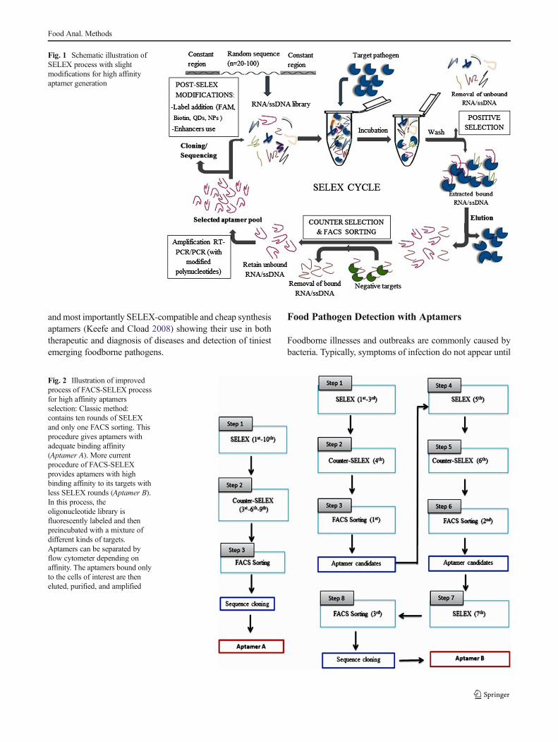
Aptamers against specific targets are selected through a pro-cess termed as SELEX (Tuerk 1990b) which is based onchemically synthesized oligonucleotide forming a combinato-rial library. The latter consists of multitude of single-strandedDNA (ssDNA) or RNA fragments with different sequences(~1014–1016) (Marty and Hayat 2014). This selection processfor emerging pathogenic microorganisms has two criticalsteps: (1) targets screening and (2) separation of the bindingsequences. Briefly, the selected aptamers are initially incubat-ed with the target(s) of interest (i.e., pathogens) followed byseparating the high-affinity aptamers from that of unboundedtarget(s). Next, the eluted aptamers are amplified using poly-merase chain reaction (PCR) to get an enriched pool for moreselection rounds. These aptamers are cloned, sequenced, char-acterized, and may be manipulated to achieve aptamerswith desired characteristics (Fig. 1) (McKeague andGiamberardino 2011; McKeague and Giamberardino 2011;Wang et al. 2012).
The ultimate goal of modifications is to select aptamerswith specific properties such as efficiency, rapidity, and lesslabor. Based on aptamer properties and screening methods, theSELEX process has been improved to get high-affinity andselectivity aptamers via improving selection, amplification,and pool enrichment monitoring (Kim and Gu 2014). Otherchanges include target immobilization and nucleic acid libraryexpension (McKeague and Derosa 2012). For oligonucleotidestability and resistance against nuclease enhancement, severalSELEX protocols have been developed such as genomicSELEX or cDNA–SELEX (Zimmermann et al. 2010), cova-lent SELEX (Kopylov and Spiridonova 2000; Spiridonova2014; Spiridonova et al. 2014), deconvolution SELEX or sub-tractive SELEX (Torres-Chavolla and Alocilja 2009), photoSELEX (Cho et al. 2004), and multistage SELEX (modifiedchimeric SELEX) (Wu and Curran 1999) for rationalization ofstarting pool. Counter SELEX or negative SELEX protocol inaptamer is for selectivity enhancement (Xi et al. 2014). Fortarget applicability, expression cassette (Martell et al. 2002),TECS-SELEX (Ohuchi et al. 2006), whole-bacteria SELEX(Torres-Chavolla and Alocilja 2009), mirror-image SELEX
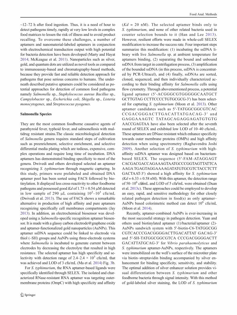
(Klussmann et al. 1996), blended SELEX (Kulbachinskiy2007), toggle SELEX target-switching (Hamula et al. 2006),whole-cell SELEX (Park et al. 2014b), complex targetSELEX (Chen 2007) are more promising. Current emergedtechniques such as HTS or automated SELEX (Huennigeret al. 2014; Lu et al. 2014), Non– or NECEEM–SELEX(Ashley et al. 2012; Yun et al. 2014), CE-SELEX (Kasaharaet al. 2013), FluMag-SELEX (Stoltenburg et al. 2005a),microfluidic SELEX (Lin et al. 2014), and in silico SELEX(Nonaka et al. 2013; Savory et al. 2013) have improved effi-ciency of designed aptamers in both binding affinity and highthroughput in tertiary structure prediction, thermodynamic,and aptamer–target model (Das et al. 2010). Despite all theseenumerated SELEX techniques, some whole-cell-selectedaptamer candidates would have high affinity but poor speci-ficity (Hamula et al. 2008). Thus, in most current used tech-niques for pathogens detection, accurate counter selection andFACS sorting (FACS-SELEX) steps are crucial for effectiveaptamer candidates (Fig. 2) where bound sequences are per-fectly separated from unbound ones. Moon and others report-ed that the use of cell sorting followed by FAM-labeling ofaptamer candidates improved the affinity up to a Kd of3.49 ± 1.43 nM (Moon et al. 2014).
SELEX protocol modification has been acquired also inoptimizing PCR amplification towards precise number ofrounds franking primer sequences to the high-affinity se-quences of oligonucleotides. In this novel in vitro selection,fixing primer sequences allows to restrain a double-strandedstructure of oligonucleotides. It allows primerless selection toprevent the primer binding sequences to incorporate into thetarget oligonucleotide-binding motif (Florian Jarosch 2006).Additionally, the use of unnatural bases (library modification)using expanded genetic alphabet of oligonucleotides contrib-uted to a >100-fold affinity DNA aptamers as compared to thenatural ones. Kimoto and colleagues have designed a DNAaptamer with four natural nucleotides (dNTPs) and unnaturalnucleotides (dDsTPs, dPx, and dPaTP) recognizing the humanproteins such as signal protein vascular endothelial cellgrowth factor-165 (VEGF-165) and interleukins(interferon-γ (IFN-γ) with binding affinity of Kd values of0.65 pM and 0.038 nM, respectively) (Kimoto et al. 2013).In addition, the use of IsoG, IsoC, and other unnatural basepairs combined to the natural ones functions as third base pair.These modifications are prominent to high efficiency and se-lectivity in new modified SELEX process through hydrogenbonding principle and their inotropic properties towardsaptamers (Fig. 2); however, to date, few reports have beenapplied to affinity selection methods. Furthermore, polynucle-otide library with modified polynucleotides such as 2′-aminopyrimidines, 2′-fluoro pyrimidines, 2′-O-methyl nucleotides,boranophosphateinternucleotide linkages, 5′-modified pyrim-idines, 4′-thio pyrimidines, phosphorothioate inter-nucleotidelinkages providing high stable and affinity, nuclease resistant,
Food Anal. Methods
and most importantly SELEX-compatible and cheap synthesisaptamers (Keefe and Cload 2008) showing their use in boththerapeutic and diagnosis of diseases and detection of tiniestemerging foodborne pathogens.
Food Pathogen Detection with Aptamers
Foodborne illnesses and outbreaks are commonly caused bybacteria. Typically, symptoms of infection do not appear until
Fig. 2 Illustration of improvedprocess of FACS-SELEX processfor high affinity aptamersselection: Classic method:contains ten rounds of SELEXand only one FACS sorting. Thisprocedure gives aptamers withadequate binding affinity(Aptamer A). More currentprocedure of FACS-SELEXprovides aptamers with highbinding affinity to its targets withless SELEX rounds (Aptamer B).In this process, theoligonucleotide library isfluorescently labeled and thenpreincubated with a mixture ofdifferent kinds of targets.Aptamers can be separated byflow cytometer depending onaffinity. The aptamers bound onlyto the cells of interest are theneluted, purified, and amplified
Fig. 1 Schematic illustration ofSELEX process with slightmodifications for high affinityaptamer generation
Food Anal. Methods
~12–72 h after food ingestion. Thus, it is a need of hour todetect pathogens timely, rapidly at very low levels in complexfood matrices to lessen the risk of illness and to avoid productrecalling. To overcome these challenges, a number ofaptamers and nanomaterial-labeled aptamers in conjunctionwith electrochemical transduction output with high potentialfor bacteria detection have been developed (Marty and Hayat2014; McKeague et al. 2011). Nanoparticles such as silver,gold, and quantum dots are utilized as novel tools as comparedto immunoassay-based and chromatography-based methods,because they provide fast and reliable detection approach forpathogens that pose serious concerns to humans. The under-neath described putative aptamers could be considered as po-tential approaches for detection of common food pathogensnamely Salmonella sp., Staphylococcus aureus Bacillus sp.,Campylobacter sp., Escherichia coli, Shigella sp., Listeriamonocytogenes, and Streptococcus pyogenes.
Salmonella Species
They are the most common foodborne causative agents ofparathyroid fever, typhoid fever, and salmonellosis with mul-tidrug resistant strains.The classic microbiological detectionof Salmonella genera requires standard steps of cultivationsuch as preenrichment, selective enrichment, and selectivedifferential media plating which are tedious, expensive, com-plex operations and require long time of incubation. DNAaptamers has demonstrated binding specificity to most of thegenera. Dwivedi and others developed selected an aptamerrecognizing S. typhimurium through magnetic capturing. Inthis study, primers were prelabeled and obtained DNAaptamer pool has been sorted using FACS followed by bio-tinylation. It displayed less cross-reactivity to other foodbornepathogens and possessed goodKd of 1.73 ± 0.54 μMdetectedin low sample of 290 μL containing 102–103 cfu/mL(Dwivedi et al. 2013). The use of FACS shows a remarkablealternative in production of high affinity and pure aptamersrecognizing specifically cell membranes compartments (Jay2013). In addition, an electrochemical biosensor was devel-oped using a Salmonella-specific recognition aptamer biosen-sor. It is made with a glassy carbon electrode@graphene oxideand aptamer-functionalized gold nanoparticles (AuNPs). Thisaptamer ssDNA sequence could be linked to electrode viathiol (–SH) groups and AuNPs using three-electrode systemswhere Salmonella is incubated to generate current betweenelectrodes by decreasing the electrolyte that resulted in highresistance. The selected aptamer has high specificity and se-lectivity with detection range of 2.4–2.4 × 10³ cfu/mL thatwas achieved and LOD of 3 cfu/mL (Ma et al. 2014) Fig. 3b.
For S. typhimurium, the RNA aptamer-based ligands werespecifically identified through SELEX. The isolated and char-acterized RNase-resistant RNA aptamer was targeting outer-membrane proteins (OmpC) with high specificity and affinity
(Kd = 20 nM). The selected aptamer binds only toS. typhimurium, and none of other related bacteria used incounter selection bounds to it (Han and Lee 2013).However, resilient efforts were made in whole-cell SELEXmodification to increase the success rate. Four important stepssummarize this modification: (1) incubating the ssDNA li-brary with live Salmonella sp. at ambient temperature foraptamers binding, (2) separating the bound and unboundssDNA from target in centrifugation process, (3) amplificationof the bounded ssDNA (in this process, ssDNA is concentrat-ed by PCR-Ultracel), and (4) finally, ssDNAs are sorted,cloned, sequenced, and then individually characterized ac-cording to their binding affinity for Salmonella cells usingflow cytometry. Through abovementioned process, a potentialligand aptamer (5′-ACGGGCGTGGGGGCAATGCTGCTTGTAG CCTTCCCCTGTGCGCG-3′) has been select-ed for capturing S. typhimurium (Moon et al. 2013). Otheraptamer candidates such as 5′-TATGGCGGCGTCACCCGACGGGACTTGACATTATGACAG-3 ′ andGAGGAAAGTC TATAGCAGAGGAGATGTGTGAACCGAGTAA have also been selected after the seventhround of SELEX and exhibited low LOD of 10–40 cfu/mL.These aptamers are DNase resistant which enhance specificitytowards outer membrane proteins (OMPs) and high affinitydetection when using spectrometry (Raghavendra Joshi2009). Another selection of S. typhimurium with high-affinity ssDNA aptamer was designed based on bacterium-based SELEX. The sequence (5′-FAM-ATAGGAGTCACGACGACCAGAAAGTAATGCCCGGTAGTTATTCAAAGATGAGTAGGAAAAGATATGTGCGTCTACCTCTTGACTAAT-3′) showed a high affinity for S. typhimurium(Kd = 6.33 ± 0.58 nM). With this aptamer, the detection rangeof 50–106 cfum̸L and LOD of 5 cfu m̸L were obtained (Duanet al. 2013c). These approaches could be employed to developan easy, rapid, and sensitive methodology for other closelyrelated pathogen detection in food(s) as only aptamer–AuNPs based colorimetric method can detect 106 cfu/mL(Moon et al. 2014).
Recently, aptamer-combined AuNPs is ever-increasing inthe most successful strategy in pathogen detection. Yuan andothers used biotinylated aptamer (1)/bacterial/aptamer (2)-AuNPs sandwich system with 5′-biotin-C6-TATGGCGGCGTCACCCGACGGGGACTTGACATTAT GACAG-3′and 5′-SH-TATGGCGGCGTCA CCCGACGGGGACTTGACATTATGCAG-3′ for Vibrio parahaemolyticus andS. typhimurium aptamer-AuNPs, respectively. The aptamerswere immobilized on the well’s surface of the microtiter platevia biotin–streptavidin binding accompanied by silver en-hancement for binding specificity, sensitivity, and stability.The optimal addition of silver enhancer solution provides vi-sual differentiation between S. typhimurium and otherfoodborne bacteria through signal intensity. With this methodof gold-labeled silver staining, the LOD of S. typhimurium
Food Anal. Methods

reached 7 cfu/mL (Yuan et al. 2014) which depicts the pro-spective utilization of combinatorial techniques in improvingthe microbial visual detection (Fig. 3d).
Few years back, we used aptasensors and dual fluores-cence from green QDs and red QDs nanoparticles as do-nors and amorphous carbon nanotubes as acceptors couldgive the LOD of 25 and 35 cfu/mL in food sample forVibrio parahaemolyticus and S. typhimurium, respectively(Duan et al. 2013d). Interestingly, this method providedimproved simultaneous and visual detection of pathogensbased on QDs fluorescence which is linearly proportionalto the target concentration. Furthermore, a high affinityaptamer (5′-ATGGACGAATATCGTCTCCCAGTGAATTCAGTCGGACAGCG-3 ′) has been screened usingwhole-cell SELEX conjugated carbon nanomaterialst o d e t e c t t h e S . p a r a t y p h i A and a c h i e v e dKd = 47 ± 3 nM. Add i t i ona l l y, a SWCNTs@DNAzyme- aptamer combination was designed for this
pathogen and this complex has self-aggregation inthe presence of specific target. A linear range of103-107 cfu/mL was attained with a LOD of 103cfu/mL(Ming Yang 2013). More currently, a remarkable combi-natorial technique of S. typhimurium detection has beenpublished. Here, A Surface-enhanced Raman spectrosco-py (SERS) substrate bearing Au@AgNPs combined with5′-SH-aptamer 1 for target cells capture followed byaptamer 2 modification using X-rhodamine (ROX) (rec-ognition element and SERS reporter). S. typhimurium spe-cifically bind with the aptamers to form Au@AgNPs-aptamer1-target-aptamer2-ROX sandwich-like complexes.This tremendous technique results a rapid and high sensi-tive detection with a linear range of 15–1.5 × 10⁶ cfu/mLwith a LOD 15 cfu/mL(Duan et al. 2016). Though manytechniques have been discussed, this technique promisespotential use in foodborne pathogens detection in terms ofLOD and feasibility.
Fig. 3 Aptamer enhancers: Aptamer-functionalized gold nanoparticle(AuNP). AuNPs possess a strong surface plasmon resonance band,which has strong distance-dependent properties. When AuNPs bind totargeted sites and come into close proximity with other AuNPs, theychange the refractive index of the surrounding medium, resulting in acolor change from red to purple. a Graphene oxide-SELEX: ThessDNA library is preincubated with AuNPs followed by adsoption onGO via to π–π bonding. b. The DNA capture element (DCE) systemquenching and dequenching. This process consists of fluorophore (e.g.,QDs, FAM,)-labeled starting ssDNA library through covalent linkage atthe 3′ end and a bead covalently linked to the 5′ end, and the
complementary strand aptamer has a quencher covalently linked to the5′ end. The two strands of DNA are annealed together, and the quencherquenches the fluorescence of the fluorophore. The presence of targetstriggers the DCE system fluctuation resulting in the double strandsdenaturation which gives fluorescencer. c Illustration of capture aptamerand UV light requirement to immobilize the capture aptamer. Colloidalgold or quantum dot is used as reporter conjugate. d The aptamermultiplex system may involve digoxigenin-5′ reporter aptamer-3′-biotin–streptavidin colloidal gold or Q-dot conjugates, amino-captureaptamers immobilized with UV light for colorimetric observation. Theintensity of the dots increases proportionally with targets
Food Anal. Methods

Staphylococcus aureus
It is a pathogen commonly found in food. It secretes a mixtureof enterotoxins known as staphylococcal enterotoxins A and B(SEA and SEB). (Bruno et al. 2010; Bruno JG 2002; TombelliS 2007). For these toxins, aptamer targeting SEB1 has beenselected for SEB (DeGrasse 2012b). Though its high affinityand selectivity together with other several ssDNA aptamers toSEB were described, their aptamer sequences are not imple-mented, limiting their utilization. For S. aureus SEA, a methodbased on primarily antibody dependent was replaced byaptamers that bind to target with high affinity and selectivity.Those aptamers were selected and generated in vitro by a 12-round selection process based on magnetic separation(Kd = 48.57 ± 6.52 nM). This aptamer shows more reliableresults consistent with previously reported S. aureus SEA de-tection having a LOD of 8.7 × 10⁻³ μg/mL (Huang et al. 2014)with low cost and high affinity compared to its antibody-basedcounterpart which is challenging and laborious.
For whole-cell-based SELEX, 107 S. aureus have been in-cubated for aptamer selection followed by isolation of boundaptamers. In the counter-selection process, aptamers with spe-cific binding were incubated with structurally related negativetargets (108 S. epidermidis) and highly specific aptamers wereamplified and purified. The selected ssDNAwas again incubat-ed with new S. aureus to start next SELEX round(s) and aftereight rounds cloned and sequenced. Likewise, a panel ofssDNA aptamer specific to S.aureus was obtained by a wholebacterium-based SELEX and applied to probing S. aureus.Here, 11 sequences from different families were selected forfurther characterization by confocal imaging and flow cytom-etry. The Kd values obtained for a single candidate aptamerwere in the nanomolar range and showed a high specificity(Cao et al. 2009). Recently, combinatorial assays for simulta-neous detection of three pathogenic bacteria was developedusing multicolor up conversion nanoparticles (UCNPs) as lu-minescence labels coupled with aptamers as the molecular rec-ognition elements (Wu et al. 2014). Other simple and fast meth-od based on a polydimethylsiloxane (PDMS)/paper/glass hy-brid microfluidic system integrated with aptamer-functionalized graphene oxide nanobiosensors for simple,one-step, multiplexed pathogen detection has been developed.This method has been simultaneously used in detection of twoinfectious pathogens S. aureus and S.enterica (Zuo P 2013 ).Chang et al. performed two ssDNA aptamers with high affin-ities and specific against S. aureus (Kd = 35 and 129 nM). ThisKd was improved to 3.03 and 9.9 nM which demonstrated thedetection of single S. aureus cell in 1.5 h and promised theconstructed aptamer to be used in food safety for S. aureusdetection (Chang et al. 2013b). Recently, chemically modifiedaptamer-functionalized grapheme oxide combinatorial tech-nique has been developed able to detect a single colony-forming units per milliliter. In this technique, graphene works
as a transducer layer of aptasensor while the aptamer is a sens-ing body. The modified graphene showed less noise comparedto non-modified in detection of challenging strains likeS. aureus (Hernández et al. 2014). This study demonstrated ahigh sensitivity of combination than either bare-used aptamersor nanomaterials in recognizing S. aureus. This techniquepromises potential use in foodborne pathogen detection.Though its high affinity and selectivity together with otherseveral ssDNA aptamers to SEBwere described, however, theiraptamer sequences are not implemented in lieu or as alternativeof antibodies, limiting their utilization (DeGrasse 2012a).
Bacillus Species
Bacillus anthracis is a rod-shaped, Gram-positive bacteriaable to form endospores. It causes disease to both humanand livestock owing to its immune system escaping mecha-nism and causes high fatality rate derived from its high load infood such as hot chocolate, rice, mashed potatoes, and turkeyand meat loafs (Drobniewski 1993; King et al. 2007).Etiologically, it is known as major cause of anthrax in live-stock and toxicity in human acquired from its membrane sur-face protein capsule namely poly-D-gamma-glutamic acid andlethal factor endopeptidase that hydrolyze mitogen-activatedprotein kinase kinases (virulence part). Recent novel strategiesare able to differentiate between the Bacillus strains althoughsome setbacks occur in detection of B. anthracis especiallyphenotypic and genotypic resemblance within bacillus generathus high possibility of getting the false results. For example,B.thuringiensis and B.cereus are closely related withB. anthracis with only two dissimilarities: (1) two virulenceplasmids in B. anthracis and insecticidal toxins coding plas-mids in B. thuringiensis (Warda et al. 2016). Gram-positivebacteria B. cereus is responsible for causing severe nausea,vomiting, diarrhea, and other similar foodborne illness thathas the threshold for onset the disease(s) is >106 B. cereus/gin food produce (Helgason E 2000), and yet, no putativeaptamer-conjugated essay was reported. As B. thuringiensisis also injurious to human, a DNA aptamer 5′-CATCCGTCACACCTGCTCTGGCCACTAACATGGGGACCAGGTGGTGTTGGCTCCCGTATC-3′ was specific forB. thuringiensis whereby combination of 60 base aptamer tofluorescent zinc sulfide-capped cadmium selenide quantumdots (QD) detect B. thuringiensis (B. cereus closely relatedbacteria). During this testing, several controls with non-functionalized QDs and without spores were verified and val-idated to measure QDs specificity to the spores and the fluo-rescence attained the LOD of 103–104 cfu/mL (Ikanovic et al.2007; Torres-Chavolla and Alocilja 2009). For specificity pur-poses, the detection of B. globigii spores (B. subtilis vs.B. niger) can differentiate B. thuringiensis from B. globigiiat concentrations above 105 cfu/mL (Lim et al. 2010)(Fig. 3b). Additionally, the use of Raman spectroscopy
Food Anal. Methods

combined to the single-walled carbon nanotube (SWCNT)generates high affinity oligonucleotides for B. anthracis de-tection. A complementary DNA to the target DNA is added toDNA-SWCNT complex resulting hybridization (Bansal et al.2013).
To date, SELEX is the most often used process in aptamerselection for various purposes including food safety.However, during the selection process, it requires highamounts of target molecules. Therefore, as a possible alterna-tive, another sensing system called DNA capture element(DCE) is used to detect scarce pathogens (Tuerk 1990b).B. anthracis, Shiga toxin, botulinum neurotoxin (BoNT),and Francisella tularensisDCE systems have been developedfrom their aptamers (Mathew et al. 2015; Tuerk 1990b). InDCE approach, the target interacts with dsDNA and magneticseparation technology followed by its denaturation(quenching and dequenching system) resulting fluorescencesignals (Fig. 3c) (Fan et al. 2008). The resulted target-boundssDNA complex can be separated later by random ssDNAmagnetic beads. The use magnetic beads requires very smallamount of target during ssDNA/target separation and helps insignal amplification. The bacillus spores were used as targetsof DNA aptamer during screening, and 79 DNA fragmentsequences from 13 different classes of DNA aptamers wereobtained by 18 cycles (Wang et al. 2012). A DNA aptamer (5′-CATCCGTCACACCTGCTCTGGCCACTA-3′) obtainedagainst spores of B.anthracis strain has paved the way to de-velop more sophisticated aptamer-magnetic bead–electrochemiluminescence (AM-ECL) sandwich techniquewith high sensitivity of detecting <10 B. anthracis spores(Xu et al. 2011). The binding of quantum dot (QD)-labeledaptamer to B. cereus, B. anthracis spores, could be applied totarget similar pathogens (Yang et al. 2013).
Campylobacter jejuni
Campylobacter jejuni is a common food poisoning bacteriumand mainly found in raw meat, raw milk, and cross-contaminated foods. When ingested with contaminated food,it colonizes the intestine wall and produces enterotoxins.Dwivedi and others performed cell-SELEX on live C. jejuniby applying combinatorial library of FAM-labeled ssDNA onwhole-cell SELEX method. Here, ssDNA molecular recogni-tion elements (MREs) are used followed by FACS sortingtargeting C. jejuni A9a. These aptamers showed high affinityand specificity against the target with relative high dissocia-tion constant of 292.8 ± 53.1 nM (Jaykus 2010; Dwivedi et al.2010b). Furthermore, Stratis-Cullum and others performed aCE-SELEX to evaluate the aptamer specific for dead C. jejuniwith qualitative capillary electrophoresis immunoassay andthe obtained LODwas 6.3 × 10⁶ cells/mL (Stratis-Cullum et al.2009). Aptamer-linked QDs have been developed and used ina sandwich assay in conjugation with magnetic beads for the
detection of C. jejuni at levels of between 2.5 and 10–50 cfu/mL in buffer and food matrices, respectively (Bruno et al.2009). However, currently, it was proved that biotinylatedmagnetic bead-based aptamers followed by qPCR detectionof C. jejuni in sample volume range of 300 μL to 10 mL. Thiscombination targets the glyA gene and is species specific withclear exclusion of closely related genera (Suh et al. 2014b).The qPCR complemented with biotinylation properties suchas fast on rate, specificity, affinity, and nucleic acid stabiliza-tion. The principle behind consists of aptamer conjugation toits target with donor and acceptor microbeads. After interac-tion, there is fluorescent signal production which could beread by fluorescence reader. This automated biotinylated mag-netic bead qPCR-based aptamers can simultaneously detecttargets with high affinity and in a short time.
Escherichia coli
Escherichia coli have enterotoxigenic effects and potentiallycontaminate food and water (Castro-Rosas et al. 2012).Detection of E. coli O157:H7 is of high priority in food sur-veillance as this Gram-negative bacteria cause infection whichmay lead to hemorrhagic diarrhea and kidney failure (BiekeVan Dorsta 2010; McKeague and Giamberardino 2011).Sensitive methods are of great importance as the infectiousconcentration can be as low as 10–100 cfu/mL. SeveralDNA and RNA aptamer-based platforms have been devel-oped for several strains of foodborne E.coli. One example isan aptamer-modified SWCNT in both field-effect transistorsand label-free potentiometric measurements to provide rapidand reusable biosensors. For effective potentiometric aptamer-based biosensor, a FRET-based assay using a DNA aptamerfor nonpathogenic E. coli CECT 675 strain is used as a modelorganism substitute for pathogenic E. coli O157:H7. TheSWCNT works as ion–electron transducers, and aptamersare covalently immobilized on it as a biorecognition elements.Aptamers interact with target(s) with subsequent electrical po-tential changes that enables quick identification and detectionof the target. As a result, 104 cfu/mL can be detected selec-tively and this biosensor can be built and regenerated easilywith a low LOD in complex samples (Zelada-Guillén et al.2010). Furthermore, aptamer-functionalized SWCNT field-effect transistors have also been reported for the detection ofE. coliDH5α strain (So et al. 2008). However for endotoxins,producing E. coli strains, only E. coli O111:B4 endotoxin isreported. Here E. coli cells or lipopoplysaccharide (LPS) areused as target for selecting specific aptamer and showed highaffinity and selectivity when analyzed by a colorimetricenzyme-based technique (Bruno et al. 2008).
Other nanomaterials in conjunction with aptamer-basedbiosensors for E. coli detection were developed. An exampleis a Fe2O3–AuNPs electrochemical DNA biosensor. ThisDNA biosensor forms a sandwich complex made of
Food Anal. Methods

recognition element on Fe2O3@AuNPS, reporter@HRP en-zyme, and the target. After E. coli capture, the induction ofmagnetic field activates magnetic separation of complexesleading to HRP catalysis resulting in detectable signals. TheLOD of 5 cfu/mL was achieved without requiring PCR am-plification of target DNA (Li et al. 2011), though it requiredlong-time incubation period (Fig. 3a). This assay is reliable;however, its detection sensitivity is of concern in terms ofworking time. Therefore, a DNA aptamer–quantum dot lateralflow test strip can improve the detection time and providecolorimetric results for more common foodborne pathogens.At the presence of targets, the high-affinity selected aptamersstabilize nanoparticles and the latter act as signal transducerelements via their aggregation leading to pinkish visible colorchange from red (Bruno 2014).
Currently, Kim and others detected E. coli strains that usedfluorescence-tagged aptamer cocktails and found this canboost signals than single aptamers. The use of three variousaptamer cocktails, each labeled with FAM, is able to recognizeone particular bacterium and target the outer membrane com-partments. Compared to single aptamers, the cocktail en-hances sensitivity and fluorescence signal simultaneouslywith cumulative influence. This method is attractive becauseit can reduce the LOD from 6778 to 3.7 × 10² cfu/mL of singleaptamer and multiple ones, respectively (Kim et al. 2014).However, due to outer cell surface complexity, molecular levelunderstanding of how these aptamers bind on E. coli strains isstill unknown. Using, the same aptamers could bind to cellsurface components competitively followed by fluorescenceemission; therefore, two or more different aptamers could beassumed to bind non-competitively (Kim et al. 2013c).
Shigella Species
Shigella dysenteriae is a Gram-positive, facultative intracellu-lar and shigellosis, epidemic bacillary dysentery causing bacte-rium. It is the dominant cause of and poses a major publichealth problem worldwide (Kotloff et al. 1999; Niyogi et al.2005). People having lack of access to adequate clean drinkingwater, sanitation of environments, and dwelling in slums areespecially at higher risk of S. dysenteriae’s contagiousnesscausing a million of deaths each year (Sur et al. 2004). Fewtechniques have been used alongside with classic conventionalculture. Immunomagnetic detection of S. dysenteriae type 1and S. flexneri serotypes in feces has been used targetingShigella sp. O-antigen (Islam et al. 1993) and lipopolysaccha-rides (Carlin and Lindberg 1987) and PCR method withprimers targeting invasion plasmid antigen H genesequence(ipaH gene) (Thiem et al. 2004). Duan and othersused cell-SELEX to identify ssDNA MRE specific forS. dysenteriae. The best candidate MREs (CGGAACTAGCGTTTAAATG CCAGGACTGAAGTAGGCAGGG) hadd emon s t r a t e d p r o b a b l e l ow b i n d i n g a f f i n i t y
(Kd = 23.47 ± 2.48 nM) targeted bacteria. Fluorescent-basedassay detection was carried, and results showed a LOD of50 cfu/mL. This aptamer could be employed for the captureand subsequent detection of S. dysenteriae from complex sam-ple matrices and food (Duan et al. 2013b) and bears strongmerits over market-dominated antibody and PCR-based tech-nique. However, it is the only disclosed aptamer and no avail-able aptamer-combinatorial technique used in detection of thispathogen. Furthermore, other Shigella sp. like S. sonnei is cur-rently causing foodborne disease with shiga toxin production(Lamba et al. 2016). Our group developed a more simplifiedS. sonnei sandwich detection system. Selected aptamers Sp1and Sp2 were analyzed by FACS and found to reach the sen-sitivity of 5.980 ± 0.835 and 14.32 ± 2.19 nM, respectively.Then, both aptamers were used in a sandwich manner to cap-ture the target. The LOD of 30 cfu/mL were achieved (Gonget al. 2015). To date, few available combinatorial aptamers arereported. At this point, aptamer–ELISA has been used withantibody as a recognition element of the target (ElhamMasoudipour 2011). However, this combination approach istedious, expensive, and requires professionalism. Therefore, itis of utmost importance to design alternative combinatorialapproaches for improved detection of this pathogen.
Listeria Species
It is pathogenic bacteria Gram-positive, facultative anaerobes,rod-shaped bacteria. Its genus Listeria contains eight non-pathogenic species (L. marthii, L. innocua, L. welshimeri,L. seeligeri, L. grayi, L. rocourtiae, L. fleischmannii, andL. weihenstephanensis) and two pathogenic species(L. monocytogenes and L. ivanovii) which cause infection tohuman as well as animals (Kuenne 2010). Motility, catalasepositive, and virulence are its unique features; however, littleis known about its virulence. They are dispersed into the lowtemperature environs and most frequently in the soil, water,sewage, and foodstuffs (L. Somer 2003). The European Unionin collaboration with FDA has applied a zero tolerance onL. monocytogenes in ready-to-eat food products as its rate ofdeath is beyond C. botulinum and Salmonella spp. (CDC2013). Although listeriosis is rare, still serious illness predom-inantly affects sensitive populations, i.e., infants, elders, andimmunocompromised patients (Kathariou 2002). The ssDNAaptamers with binding affinity to Listeria spp. have been se-lected using whole-cell SELEX method, and aptamers withbinding affinity were analyzed by flow cytometry. A ssDNAaptamer (LM6-116) 5′-TACTCGTTATTTCGT AGCACTTTTCCCCACCACCTTGGTG-3′ binds various cell surfacecomponents (Suh et al. 2014a).
The use of SELEX to an ssDNA-conjugated biotin libraryis currently applied in aptamer selection and characterizedwith flow cytometry. The selected aptamer displayed a goodbinding affinity to L. monocytogenes.The selected aptamer 5′-
Food Anal. Methods

GAGGGAAGAAGGGCCAGCACAGATCAGATCAATCGCTCCG-3′ conjugated to magnetic beads and when usedin a combined aptamer magnetic capture (AMC)-qPCR assay:the pathogens were detected at concentrations <60 cfu/500 μLbuffer in the presence of non-Listeria mixtures bacteria with acapturing efficiency of 26–77%. This method shows a highLOD 2.7 and 4.8 log cfu/L (Suh and Jaykus 2013). In anotherSELEX-based technique, an aptamer-A8 (5 ′-ATCCATGGGGCGGAGATGAGGGGGAGGAGGGCGGGTACCC GGTTGAT- 3 ′ ) s p e c i f i c f o r i n t e r n a l i n A(L. monocytogenes invasin protein) was used in the fiber opticsensor together with antibody in a sandwich format for detec-tion of L. monocytogenes. This selected aptamer was veryselective to L. monocytogenes with quantitative detection of~103 cfu/mL (Ohk et al. 2010) which is of concern due to theuse of bare aptamer. For enhanced detection, a whole-bacterium SELEX strategy and fluorescence-based recogni-tion for rapid detection of L. monocytogenes was reported.Here , the fol lowing sequence TGGGAGCTCAGAATAAACGCTCAACTTTGTTCTTCTTGCTTTTTTTTTCTTTTTTTGTTCGACATG was modified withdigoxigenin for direct fluorescent observation after combiningwith targeted bacteria and a fluorescent microscopy was usedfor direct observation of digoxigenin–aptamer binding to thetarget L. monocytogenes with much higher specificity (LiuGuo-qing 2014). Recently, Liu and others have shown thateight rounds of selection give ssDNA molecular recognitionelements (MREs) specific for L.monocytogenes with highspecificity up to Kd value of 60.01 nM (Liu et al. 2014).Moreover, we achieved the whole cell in vitro selection onL. monocytogenes and selected aptamer showed a consider-able affinity (Kd = 48.74 ± 3.11 nM). A fluorescent bindingbioassay was developed to confirm the high specific bindingof theseMREs towards targets demonstrated a LOD of 75 cfu/mL (Duan et al. 2013a). In summary, both bare and modifiedaptamers have significantly contributed in improved detectionof pathogens and at the same time noticeably reduced the costincurred for detection method(s) when compared to antibod-ies. Despite the advantage(s) of each approach, available tech-niques for this pathogen are only fluorophore–aptamer-basedcombination rather than the use of other enhancers.
Streptococcus Species
Streptococcus species are spherical, Gram-positive bacteriabelonging to Firmicutes phylum and Lactobaliales order.Many species are facultative anaerobes, catalase-negative,not motile, and occur in chains or pairs with ability to adjustat low temperature (<7 °C). Medically, Streptococcus speciesare classified depending on their hemolytic characteristicsgroup namely alpha and beta hemolysis. They have the abilityto oxidize hemoglobin iron or destroy completely red bloodcells, respectively. Depending on antigenicity and
physiological characteristics, Streptococcus genus is groupedinto A, B, C, D, F, and G; and among them, groups AStreptococcus (GAS, with 40 antigenic species) and D(Enterococcus) can be transmitted via food. S. pyogenes pos-sess ten major M-type surface proteins (M1, M2, M3, M4,M6, M11, M12, M28, M77, and M89) constituting its majorvirulence factor (Nitzsche et al. 2015). They are able to causevarious infections to all individuals such as impetigo, pharyn-gitis, and other serious infections like necrotizing fasciitis,sepsis, and frequent GAS that may cause post-infection auto-immune diseases such as rheumatic fever, glomerulonephritis,and heart diseases which lead to high mobility and mortality(Walker et al. 2014). To date, immunodiagnosis and classicdiagnosis still dominate despite tedious procedure of stool,saliva, swab, blood, and contaminated food samples culture,depicting the need of simple and fast detection techniques ofthese pathogens (Kim et al. 2013b). Though aptamer frame-work contribution in recent year increased, apparently fewGAS species are studied intensely. It has been reported a com-binatorial technique sensor consisting of immobilization ofaptamer on SWCNTs via p–p stacking bond of ssDNAwhichis serially connected to Au electrode and piezoelectric quartzcrystal. This complex is detached when the immobilized anti-GAS aptamer react with GAS resulting frequency shift re-sponse because aptamer binding force to SWCNT is lowerthan its binding on GAS. The detected impedance spectraincreased precisely to aptamer–GAS detachment with3 × 102 to 3 × 10⁶ cfu/mL concentration and the LOD of12 cfu/mL in 40min (Shi et al. 2014). However, this techniqueobviously could not detect targets when aptamers are dis-persed or scarce since the frequency shift increased in a lowrange of aptamers concentration from 1 to 10 μM where theincrease of aptamer concentration to 12 μM lowered the fre-quency shift due to their accumulation which can hinder theGAS recognition. Currently, Hamula and others developedtargeted ten common and other ten non-common M-type S.pyogenes. Bacteria were incubated with 80 nucleotide DNAlibraries, and other process follows standard SELEX proce-dure mediated with FACS to assess the binding capacity ofaptamer pool and 5′ FAM labeling for visual detection. TheKd = 4 to Kd = 86 affinity range was achieved (Hamula et al.2011). The same group improved S. pyogenes targetingaptamers with modification in standard SELEX. Special for-ward primers are designed to contain polyadenines linked to itvia triethylene glycol spacer that plays a critical role in gelelectrophoresis separation of both primers to the PCR product.The reverse primer was fluorescently labeled with 5′ FAM toallow FACS step (Mayer et al. 2010). During incubation, se-lected aptamers were incubated with S. pyogenes, and tRNAand BSAwere added in incubation buffer to increase ssDNArecognition of cell target. The modified PCR sequences,counter-selection using non-M-type cells, and finally, FACSanalysis improved binding of M-type 11 protein of
Food Anal. Methods

S. pyogenes with a Kd = 7 ± 1 nM (Hamula et al. 2016).Though this binding affinity can be achieved byimmunoassay-based technique, this technique is less laboriousand comprehensive. Moreover, these aptamers can be fixed tosolid surface for aptamer array development used in M typingor whole-cell detection of food or clinical samples. However,to date, no research has been reported for the other predomi-nant M types of GAS, depicting the need to develop a reliabletechnique of detection with further modification(s) inaptamer–immunoassay combination where fluorescently la-beled aptamer or antibody recognize the non-variable M-typeprotein of the bacteria and/or its C-terminal residues in a sand-wich manner.
Conclusion and Future Directives
Aptamers were shown to be an attractive alternative toimmunosensors and enzyme-based sensors for microorgan-isms monitoring. Those oligonucleotides are proving to beeffective molecular recognition probes of high priority in foodand drug quality and safety testing, disease diagnosis. etc.
They are relatively stable with ease synthesis and modifica-tion. The aptamer stability is of great importance for bacteriadetection in food samples. Therefore, in near future, aptamersare promising to be antibodies other biosensors replacer indetection and diagnosis assays. Various methods based onSELEX were used for selecting aptamers with high affinityand selectivity to both small and large targets independentlyfrom their toxicity and matrix effects by influencing positivelythe performance and accuracy of test results. This reviewshowed main rapid, specific, robust, and highly sensitivemethod approaches to select aptamers for the detection ofmicroorganisms in foods. Although the aptamer-based assaysfor different targets in food are still far from guarantying com-plete food safety, yet, the aptamer preparation systems arefacilitating the development of more reliable aptamer-basedcommercial devices (i.e., biosensors). Thus, we emphasizedfrequently used combinatorial techniques in foodborne path-ogen detection. Intriguingly, all methods discussed can beused for detection of other targets by substituting the aptamer.Aptamer-conjugated nanoparticles have also showed highpreference given the role of nanoparticles in stabilization ofaptamer and provision of additional affinity with dissociation
Fig. 4 Scheme of the SELEX procedure using hydrophobic unnaturalbase pair system for high-affinity DNA aptamer generation: The initiallibrary is made of mixture of 22 different chemically synthesized DNAsublibraries, and each sublibrary contains 43 natural base-randomizedregions with 1–3 unnatural nucleotide Ds bases at predeterminedpositions, flanked by constant regions of a 25-base region composed ofa 5′ primer and a 2–3-natural base recognition tag on the 5′ end and a 27base 3′ primer region. In each selection round, the single-stranded DNAlibrary can be mixed with a target and the DNA fragments that bound tothe target proteins extracted, followed by asymmetric PCR amplification
involving the Ds–Px pair for the next round of selection. After sevenrounds of selection, the enriched DNA library is PCR amplified in thepresence of the natural dNTPs and the unnatural dPa′TP as substrates toreplace the Ds bases in the DNA fragments with the natural bases (mainlyA or T) via the Ds–Pa′ pair. This is followed by sequencing with IonTorrent PGM, and then amplified DNA fragments (chemicallysynthesized using the natural base and Ds amidite reagents) provide themost potent aptamers towards the target(s). Finally, the selected aptamerscan be optimized through doped in vitro selection to obtain the Bwinning^aptamers
Food Anal. Methods

constant ranging from nanomolar to picomolar and LOD aslow as 1–10 cfu/mL. This elucidates the aptamer–nanoparticlecombination ability in signal transduction which is character-ized by null interference (i.e., no change in surface plasmonresonance and absorption). Furthermore, functionalization ofcarbon-based nanomaterials with photochromic molecules al-lows covalent (through cyclo-addition, condensation, orradical polymerization reaction) and non-covalent (p–p stack-ing, hydrophobic interaction, or electrostatic interaction) bind-ing approach; hence, its combination with aptamer could beuseful. However, discussed post-SELEX modifications fre-quently reduce aptamer affinity to the targets. The integrationof chemically synthesized polynucleotide into the starting li-brary (Fig. 4) and nanoparticle combination (Fig. 3) increasethe aptamer stability but at an increased cost of the aptamerselection process. Hence, we propose combination of aptamercocktails with nanomaterials for sensitivity enhancement anddirect colorimetric visualization. This may prove promisingfor the development of aptamer-based techniques due to theshortage in detection of most foodborne parasites, some bac-teria (e.g. all Streptococcus sp. M types except M11), viruses,and fungi towards development of feasible strips as replacer oflateral flow immunoassay for rapid detection and user friend-liness. In future, leaping beyond the scope of ensuring thefood safety in terms of improved detection methods, optimi-zation of aptamer multiplex system, and whole-cell SELEXconditions may also be exploited in therapeutic arena like, butnot limited to, target inhibitor aptamers or carrier aptamers oftherapeutics (e.g., macular degeneration treatment) providingthe insights of aptamers usage. Thus, more refinements areneeded to increase aptamer and aptamer-functionalized nano-particles and other combinatorial forms, biostability, and bio-availability to eventually tackle existing challenges.
Compliance with Ethical Standards
Conflict of Interest Omar Mukama declares that he has no conflict ofinterest. Jean Paul Sinumvayo declares that he has no conflict of interest.Muhammad Shamoon declares that he has no conflict of interest.Muhammad Shoaib declares that he has no conflict of interest.Henriette Mushimiyimana declares that he has no conflict of interest.Waseem Safdar declares that he has no conflict of interest. Leo Bemenadeclares that he has no conflict of interest. Peter Rwibasira declares thathe has no conflict of interest. Samson Mugisha declares that he has noconflict of interest. Zhouping Wang declares that he has no conflict ofinterest.
Informed Consent Not applicable. This article does not contain anystudies with human or animal subjects.
References
Abbaspour A, Norouz-Sarvestani F, Noori A, Soltani N (2015) Aptamer-conjugated silver nanoparticles for electrochemical dual-aptamer-
based sandwich detection of Staphylococcus aureus Biosensorsand Bioelectronics 68:149–155 doi:http://dx.doi.org/10.1016/j.bios.2014.12.040
Amaya-Gonzalez S, de los Santos Alvarez N, Miranda-Ordieres AJ,Lobo-Castanon MJ (2013) Aptamer-based analysis: a promisingalternative for food safety control. Sensors 13:16292–16311.doi:10.3390/s131216292
Ashley J, Ji K, Li SFY (2012) Selection of bovine catalase aptamers usingnon-SELEX. Electrophoresis 33:2783–2789. doi:10.1002/elps.201200032
Baird FJ, Gasser RB, Jabbar A, Lopata AL (2014) Foodborne anisakiasisand allergy. Mol Cell Probes 28:167–174. doi:10.1016/j.mcp.2014.02.003
Bansal J, Singh I, Bhatnagar PK, Mathur PC (2013) DNA sequencedetection based on Raman spectroscopy using single walled carbonnanotube. J Biosci Bioeng 115:438–441. doi:10.1016/j.jbiosc.2012.11.002
Baumstummler A, Lehmann D, Janjic N, Ochsner UA (2014) Specificcapture and detection of Staphylococcus aureus with high-affinitymodified aptamers to cell surface components. Lett Appl Microbiol59:422–431. doi:10.1111/lam.12295
Beier R, Boschke E, Labudde D (2014a) New strategies for evaluationand analysis of SELEX experiments. Biomed Res Int 2014:849743.doi:10.1155/2014/849743
Beier R, Pahlke C, Quenzel P, Henseleit A, Boschke E, Cuniberti G,Labudde D (2014b) Selection of a DNA aptamer against noroviruscapsid protein VP1. FEMS Microbiol Lett 351:162–169.doi:10.1111/1574-6968.12366
BiekeVanDorsta JM, Bekaertb K,Martina ER, De CoenaW, Dubruelc P,Blusta R, Robbensa J (2010) Recent advances in recognition ele-ments of food and environmental biosensors. Biosens Bioelectron26:1178–1194
Bini A, Mascini M, Mascini M, Turner AP (2011) Selection of thrombin-binding aptamers by using computational approach for aptasensorapplication. Biosensors & bioelectronics 26:4411–4416.doi:10.1016/j.bios.2011.04.053
Bruno JGKJ (2002) Use of magnetic beads in selection and detection ofbiotoxin aptamers by electrochemiluminescence and enzymaticmethods. BioTechniques 32:175–180
Bruno J (2014) Application of DNA aptamers and quantum dots to lateralflow test strips for detection of foodborne pathogens with improvedsensitivity versus colloidal gold pathogens 3:341–355 doi:10.3390/pathogens3020341
Bruno JG, Carrillo MP, Phillips T (2008) In vitro antibacterial effects ofantilipopolysaccharide DNA aptamer-C1qrs complexes. FoliaMicrobiol 53:295–302. doi:10.1007/s12223-008-0046-6
Bruno JG, Carrillo MP, Phillips T, Andrews CJ (2010) A novel screeningmethod for competitive FRET-aptamers applied to E. coli assay devel-opment. J Fluoresc 20:1211–1223. doi:10.1007/s10895-010-0670-9
Bruno JG, Phillips T, Carrillo MP, Crowell R (2009) Plastic-adherentDNA aptamer-magnetic bead and quantum dot sandwich assay forCampylobacter detection. J Fluoresc 19:427–435. doi:10.1007/s10895-008-0429-8
Cao X et al (2009) Combining use of a panel of ssDNA aptamers in thedetection of Staphylococcus aureus. Nucleic Acids Res 37:4621–4628. doi:10.1093/nar/gkp489
Carlin NI, Lindberg AA (1987) Monoclonal antibodies specific forShigella flexneri lipopolysaccharides: clones binding to type IV, V,and VI antigens, group 3,4 antigen, and an epitope common to allShigella flexneri and Shigella dysenteriae type 1 stains. InfectImmun 55:1412–1420
Castro-Rosas J, Cerna-Cortes JF, Mendez-Reyes E, Lopez-Hernandez D,Gomez-Aldapa CA, Estrada-Garcia T (2012) Presence of faecal co-liforms, Escherichia coli and diarrheagenic E. coli pathotypes inready-to-eat salads, from an area where crops are irrigated with
Food Anal. Methods

untreated sewage water. Int J Food Microbiol 156:176–180.doi:10.1016/j.ijfoodmicro.2012.03.025
CDC E (2013) Surveillance for foodborne disease outbreaks—UnitedStates, 1998–2008. Morb Mortal Wkly Rep 62
Chang Y-C, Yang C-Y, Sun R-L, Cheng Y-F, Kao W-C, Yang P-C(2013b) Rapid single cell detection of Staphylococcus aureus byaptamer-conjugated gold nanoparticles. Scientific Reports 3:1863.doi:10.1038/srep01863
Chang YC, Yang CY, Sun RL, Cheng YF, Kao WC, Yang PC (2013a)Rapid single cell detection of Staphylococcus aureus by aptamer-conjugated gold nanoparticles. Sci Rep 3:1863. doi:10.1038/srep01863
Chen CK (2007) Complex SELEX against target mixture: stochasticcomputer model, simulation, and analysis. Comput Methods ProgBiomed 87:189–200. doi:10.1016/j.cmpb.2007.05.008
Cho S, Lee SH, Chung WJ, Kim YK, Lee YS, Kim BG (2004)Microbead-based affinity chromatography chip using RNA aptamermodified with photocleavable linker. Electrophoresis 25:3730–3739. doi:10.1002/elps.200406103
Control CfD (2013) Surveillance for foodborne disease outbreaks—United States, 1998–2008 vol 60.
Das R, Karanicolas J, Baker D (2010) Atomic accuracy in predicting anddesigning noncanonical RNA structure. Nat Methods 7:291–294.doi:10.1038/nmeth.1433
DeGrasse JA (2012a) A single-stranded DNA aptamer that selectivelybinds to Staphylococcus aureus enterotoxin B. PLoS One 7:e33410. doi:10.1371/journal.pone.0033410
DeGrasse JA (2012b) A single-stranded DNA aptamer that selectivelybinds to Staphylococcus aureus enterotoxin B. PLoS One 7:7.doi:10.1371/journal.pone.0033410
Drabovich A (2009) Aptamers in bioanalysis. John Wiley & Sons, Inc.Drobniewski FA (1993) Bacillus cereus and related species. Clin
Microbiol Rev 6:324–338Duan N, Ding X, He L, Wu S, Wei Y, Wang Z (2013a) Selection, iden-
tification and application of a DNA aptamer against Listeriamonocytogenes. Food Control 33:239–243. doi:10.1016/j.foodcont.2013.03.011
Duan N, Ding X, Wu S, Xia Y, Ma X, Wang Z, Chen J (2013b) In vitroselection of a DNA aptamer targeted against Shigella dysenteriae. JMicrobiol Methods 94:170–174. doi:10.1016/j.mimet.2013.06.016
Duan N, Wu S, Chen X, Huang Y, Xia Y, Ma X, Wang Z (2013c)Selection and characterization of aptamers against Salmonellatyphimurium using whole-bacterium Systemic Evolution ofLigands by Exponential Enrichment (SELEX). J Agric FoodChem 61:3229–3234. doi:10.1021/jf400767d
Duan N, Chang B, Zhang H, Wang Z, Wu S (2016) Salmonellatymphimurium detection using a surface-enhanced raman scatteringbased aptasensor. Int J Food Microbiol 218:38–43. doi:10.1016/j.ijfoodmicro.2015.11.006
Duan N et al (2013d) A dual-color flow cytometry protocol for the simul-taneous detection of Vibrio parahaemolyticus and Salmonellatyphimurium using aptamer conjugated quantum dots as labels.Anal Chim Acta 804:151–158. doi:10.1016/j.aca.2013.09.047
Dwivedi HP, Smiley RD, Jaykus LA (2013) Selection of DNA aptamers forcapture and detection of Salmonella typhimurium using a whole-cellSELEX approach in conjunction with cell sorting. Appl MicrobiolBiotechnol 97:3677–3686. doi:10.1007/s00253-013-4766-4
Dwivedi H, Smiley RD, Jaykus L-A (2010a) Selection and characteriza-tion of DNA aptamers with binding selectivity to Campylobacterjejuni using whole-cell SELEX. Appl Microbiol Biotechnol 87:2323–2334. doi:10.1007/s00253-010-2728-7
Dwivedi HP, Smiley RD, Jaykus L-A (2010b) Selection and characteri-zation of DNA aptamers with binding selectivity to Campylobacterjejuni using whole-cell SELEX. Appl Microbiol Biotechnol 87:2323–2334. doi:10.1007/s00253-010-2728-7
ElhamMasoudipour SLM, Basiri M (2011) Specific detection of Shigellasonnei by enzyme-linked aptamer sedimentation assay. Progress inBiological Sciences 1:11–15
Elham Masoudipour, Seyed Latif Mousavi, Basiri M (2011) Specificdetection of Shigella sonnei by enzyme-linked aptamer sedimenta-tion assay Progress in Biological Sciences 1
Escudero-Abarca BI, Suh SH, Moore MD, Dwivedi HP, Jaykus LA(2014) Selection, characterization and application of nucleic acidaptamers for the capture and detection of human norovirus strains.PLoS One 9:106805. doi:10.1371/journal.pone.0106805
Fan M, McBurnett SR, Andrews CJ, Allman AM, Bruno JG, Kiel JL(2008) Aptamer selection express: a novel method for rapid single-step selection and sensing of aptamers. Journal of BiomolecularTechniques : JBT 19:311–319
Feng H, Beck J, NassalM, HuKH (2011) A SELEX-screened aptamer ofhuman hepatitis B virus RNA encapsidation signal suppresses viralreplication. PLoS One 6:27862. doi:10.1371/journal.pone.0027862
Fernandez PP (2006) Selection of an aptamer against surface exposedtargets on yersinia pestis. Oklahoma State University, Research
Florian Jarosch KB, Klussmann S (2006) In vitro selection using a dualRNA library that allows primerless selection. Nucleic Acids Res 34:86
Giamberardino A et al (2013) Ultrasensitive norovirus detection usingDNA aptasensor technology. PLoS One 8:e79087. doi:10.1371/journal.pone.0079087
Gong W, Duan N, Wu S, Huang Y, Chen X, Wang Z (2015) Selection,identification, and application of dual DNA aptamers againstShigella sonnei. Anal Methods 7:3625–3631. doi:10.1039/C5AY00214A
Hamula C, Guthrie J, Zhang H, Li X, Le X (2006) Selection and analyt-ical applications of aptamers. TrAC Trends Anal Chem 25:681–691.doi:10.1016/j.trac.2006.05.007
Hamula CL, Le XC, Li XF (2011) DNA aptamers binding to multipleprevalent M-types of Streptococcus pyogenes. Anal Chem 83:3640–3647. doi:10.1021/ac200575e
Hamula CLA, Peng H, Wang Z, Tyrrell GJ, Li X-F, Le XC (2016) Animproved SELEX technique for selection of DNA aptamers bindingto M-type 11 of Streptococcus pyogenes. Methods 97:51–57.doi:10.1016/j.ymeth.2015.12.005
Hamula CL, Zhang H, Guan LL, Li XF, Le XC (2008) Selection ofaptamers against live bacterial cells. Anal Chem 80:7812–7819.doi:10.1021/ac801272s
Han SR, Lee SW (2013) In vitro selection of RNA aptamer specific toSalmonella typhimurium. J Microbiol Biotechnol 23:878–884.doi:10.4014/jmb.1212.12033
Hayat A, Andreescu S, Marty JL (2013) Design of PEG-aptamer twopiece macromolecules as convenient and integrated sensing plat-form: application to the label free detection of small size molecules.Biosensors & bioelectronics 45:168–173. doi:10.1016/j.bios.2013.01.059
Helgason EOO, Caugant D et al (2000) Bacillus anthracis, Bacillus cere-us and Bacillus thuringiensis—one species on the basis of geneticevidence. Appl Environ Microbiol 66:2627–2630
Hernández R, Vallés C, Benito AM, Maser WK, Xavier Rius F, Riu J(2014) Graphene-based potentiometric biosensor for the immediatedetection of living bacteria. Biosens Bioelectron 54:553–557.doi:10.1016/j.bios.2013.11.053
Huang YK, Chen XJ, Xia Y, Wu SJ, Duan N, Ma XY, Wang ZP (2014)Selection, identification and application of a DNA aptamer againstStaphylococcus aureus enterotoxin a. Anal Methods 6:690–697.doi:10.1039/c3ay41576g
Huenniger T,Wessels H, Fischer C, Paschke-Kratzin A, FischerM (2014)Just in time-selection: a rapid semiautomated SELEX of DNAaptamers using magnetic separation and BEAMing. Anal Chem86:10940–10947. doi:10.1021/ac503261b
Food Anal. Methods

Ikanovic M et al (2007) Fluorescence assay based on aptamer-quantumdot binding to Bacillus thuringiensis spores. J Fluoresc 17:193–199.doi:10.1007/s10895-007-0158-4
Iqbal A, Labib M, Muharemagic D, Sattar S, Dixon BR, Berezovski MV(2015) Detection of Cryptosporidium parvum oocysts on fresh pro-duce using DNA aptamers. PLoS One 10:0137455. doi:10.1371/journal.pone.0137455
Islam D, Tzipori S, Islam M, Lindberg AA (1993) Rapid detection ofShigella dysenteriae and Shigella flexneri in faeces by animmunomagnetic assay with monoclonal antibodies European. jour-nal of clinical microbiology & infectious diseases : official publica-tion of the European Society of Clinical Microbiology 12:25–32
Jay HPDRDSL-A (2013) Selection of DNA aptamers for capture anddetection of SalmonellaTyphimurium using a whole-cell SELEXapproach in conjunction with cell sorting. Appl MicrobiolBiotechnol 97:677–3686
Jaykus HPDRDSL-A (2010) Selection and characterization of DNAaptamers with binding selectivity toCampylobacter jejuni usingwhole-cell SELEX. Appl Microbiol Biotechnol 87:2323–2334
Kasahara Y, Irisawa Y, Ozaki H, Obika S, Kuwahara M (2013) 2′,4′-BNA/LNA aptamers: CE-SELEX using a DNA-based library offull-length 2′-O,4′-C-methylene-bridged/linked bicyclic ribonucleo-tides. Bioorg Med Chem Lett 23:1288–1292. doi:10.1016/j.bmcl.2012.12.093
Kathariou S, Food Prot J (2002) Listeria monocytogenesvirulence andpathogenicity, a food safety perspective. J Food Prot 65:1811–1829
Keefe AD, Cload ST (2008) SELEX with modified nucleotides. CurrOpin Chem Biol 12:448–456. doi:10.1016/j.cbpa.2008.06.028
Kim YS, Gu MB (2014) Advances in aptamer screening and small mol-ecule aptasensors. Adv Biochem Eng Biotechnol 140:29–67.doi:10.1007/10_2013_225
Kim MA, Jeon HS, Shin SY, Baik BJ, Yang YM, Lee KY, Kim JG(2013b) Rapid detection of S. mutans surface antigen I/II using asensitive monoclonal anti-Ag I/II antibody by ELISA. Monoclonalantibodies in immunodiagnosis and immunotherapy 32:336–340.doi:10.1089/mab.2013.0017
Kim YS, Song MY, Jurng J, Kim BC (2013c) Isolation and characteriza-tion of DNA aptamers againstEscherichia coli using a bacterial cell-systematic evolution of ligands by exponential enrichment ap-proach. Anal Biochem 436:22–28. doi:10.1016/j.ab.2013.01.014
Kim L, Yu H-W, Kim Y-H, Kim I, Jang A (2013a) Potential offluorophore labeled aptamers for Pseudomonas Aeruginosa detec-tion in drinking water. J Korean Soc Appl Biol Chem 56:165–171.doi:10.1007/s13765-013-3019-7
Kim YS, Chung J, SongMY, Jurng J, Kim BC (2014) Aptamer cocktails:enhancement of sensing signals compared to single use of aptamersfor detection of bacteria. Biosens Bioelectron 54:195–198.doi:10.1016/j.bios.2013.11.003
Kimoto M, Yamashige R, Matsunaga K, Yokoyama S, Hirao I (2013)Generation of high-affinity DNA aptamers using an expanded ge-netic alphabet. Nat Biotechnol 31:453–457. doi:10.1038/nbt.2556
King NJ, Whyte R, Hudson JA (2007) Presence and significance ofBacillus cereus in dehydrated potato products. J Food Prot 70:514–520
Klussmann S, Nolte A, Bald R, Erdmann VA, Furste JP (1996) Mirror-image RNA that binds D-adenosine. Nat Biotechnol 14:1112–1115.doi:10.1038/nbt0996-1112
Kopylov AM, Spiridonova VA (2000) Combinatorial chemistry of nucleicacids: SELEX. Mol Biol 34:940–954. doi:10.1023/a:1026696330245
Kotaro Fukuda DV, Sekiya S, Hwang J, Kakiuchi N, Taira K,Shimotohno K, Kumar PKR, Nishikawa S (2003) Isolation andcharacterization of RNA aptamers specific for hepatitis C virus non-structural proteins 3 protease issue. Eur J Biochem 267:3685–3694
Kotloff KL, Winickoff JP, Ivanoff B, Clemens JD, Swerdlow DL,Sansonetti PJ et al (1999) Global burden of Shigella infections:
implications for vaccine development and implementation of controlstrategies. Bull World Health Organ 77:651–666
Kuenne C, Vogets S, Pischimarov J, Oehm S, Goesmann A, Daniel R,Hain T, Chakraborty T (2010) Comparative analysis of plasmids inthe genus Listeria. PLoS One 5:12511
Kulbachinskiy AV (2007) Methods for selection of aptamers to proteinta rge t s . Biochem Mosc 72:1505–1518 . doi :10 .1134/s000629790713007x
Lamba K et al (2016) Shiga toxin 1-producing Shigella sonnei infections,California, United States, 2014-2015. Emerg Infect Dis 22:679–686.doi:10.3201/eid2204.151825
Lee CH, Lee SH, Kim JH, Noh YH, Noh GJ, Lee SW (2015)Pharmacokinetics of a cholesterol-conjugated aptamer against thehepatitis C virus (HCV) NS5B. Protein Mol Ther Nucleic Acids 4:e254. doi:10.1038/mtna.2015.30
Li K, Lai Y, Zhang W, Jin L (2011) Fe2O3@Au core/shell nanoparticle-based electrochemical DNA biosensor for Escherichia coli detec-tion. Talanta 84:607–613. doi:10.1016/j.talanta.2010.12.042
Lim YC, Kouzani AZ, Duan W (2010) Aptasensors: a review. J BiomedNanotechnol 6:93–105
Lin H, Zhang W, Jia S, Guan Z, Yang CJ, Zhu Z (2014) Microfluidicapproaches to rapid and efficient aptamer selection Biomicrofluidics8 doi:10.1063/1.4890542
Litao Yang CWF, Cho EJ, Ellington AD (2007) Real-time rolling circleamplification for protein detection. Anal Chem 79:3320–3329
Liu Guo-qing LY-Q, Chao G, Xiao-feng Y, Ming Z, Kai Z, Xuejiao C, YiY (2014) In vitro selection of DNA aptamers and fluorescence-based recognition for rapid detection Listeria monocytogenes. JIntegr Agric 13:1121–1129
Liu G-Q et al (2014) In vitro selection of DNA aptamers and fluorescence-based recognition for rapid detection Listeria monocytogenes. J IntegrAgric 13:1121–1129. doi:10.1016/s2095-3119(14)60766-8
Lu E, Elizondo-Riojas M-A, Chang JT, Volk DE (2014) Aptaligner: au-tomated software for aligning pseudorandom DNA X-aptamersfrom next-generation sequencing data. Biochemistry 53:3523–3525. doi:10.1021/bi500443e
Ma X, Jiang Y, Jia F, Yu Y, Chen J, Wang Z (2014) An aptamer-basedelectrochemical biosensor for the detection of Salmonella. JMicrobiol Methods 98:94–98. doi:10.1016/j.mimet.2014.01.003
Martell RE, Nevins JR, Sullenger BA (2002) Optimizing aptamer activityfor gene therapy applications using expression cassette SELEX.MolTher 6:30–34. doi:10.1006/mthe.2002.0624
Marty JL Hayat A (2014) Aptamer based electrochemical sensors foremerging environmental pollutants Front Chem 2
Mathew A, Maekawa T, Sakthikumar D (2015) Aptamers in targetednanotherapy Current Topics in Medicinal Chemistry 15:1102–1114
Mayer G, Ahmed M-SL, Dolf A, Endl E, Knolle PA, Famulok M (2010)Fluorescence-activated cell sorting for aptamer SELEX with cellmixtures. Nat Protocols 5:1993–2004
McKeagueM, Derosa MC (2012) Challenges and opportunities for smallmolecule aptamer development. Journal of nucleic acids 2012:748–913. doi:10.1155/2012/748913
McKeague M, Giamberardino A (2011) Advances in aptamer-basedbiosensors for food safety. Somerset (Ed.) edn. InTech,
McKeagueM, Giamberardino A, DeRosa CM (2011) Advances in aptamer-based biosensors for food safety. In: Somerset V (ed). CarletonUniversity, Department of Chemistry Ottawa, Ontario,canada,
McKeague M, Velu R, Hill K, Bardoczy V, Meszaros T, MC DR (2014)Selection and characterization of a novel DNA aptamer for label-free fluorescence biosensing of ochratoxin A. Toxins 6:2435–2452.doi:10.3390/toxins6082435
Mei ZM, Chu HQ, Chen W, Xue F, Liu J, Xu HN, Zhang R, Zheng L(2013) Ultrasensitive one-step rapid visual detection of bisphenol Ain water samples by label-free aptasensor. Biosensors & bioelectron-ics 39:26–30
Food Anal. Methods

MingYang ZP, NingY, ChenY, ZhouQ, Deng L (2013) Highly specific andcost-efficient detection of Salmonella Paratyphi A combining aptamerswith single-walled carbon. Nanotubes Sensors 13:6865–6881
Moon J, Kim G, Lee S, Park S (2013) Identification of SalmonellaTyphimurium-specific DNA aptamers developed using whole-cellSELEX and FACS analysis. J Microbiol Methods 95:162–166.doi:10.1016/j.mimet.2013.08.005
Moon J, Kim G, Park S (2014) Development of ssDNA aptamers for thecapture and detection of Salmonella typhimurium. Anal Methods 6:7442–7448. doi:10.1039/c4ay01035c
Moon J, Kim G, Park SB, Lim J, Mo C (2015) Comparison of whole-cellSELEX methods for the identification of Staphylococcus aureus-spe-cific DNA aptamers. Sensors 15:8884–8897. doi:10.3390/s150408884
Mossali C et al (2010) Sensitive detection and quantification of anisakidparasite residues in food products. Foodborne Pathog Dis 7:391–397. doi:10.1089/fpd.2009.0428
Nitzsche R, Rosenheinrich M, Kreikemeyer B, Oehmcke-Hecht S (2015)Streptococcus pyogenes triggers activation of the human contactsystem by streptokinase. Infect Immun 83:3035–3042.doi:10.1128/IAI.00180-15
Niyogi, SK, (2005) Shigellosis J Microbiol: 133–143Nonaka Y, Yoshida W, Abe K, Ferri S, Schulze H, Bachmann TT,
Ikebukuro K (2013) Affinity improvement of a VEGF aptamer byin silico maturation for a sensitive VEGF-detection system. AnalChem 85:1132–1137. doi:10.1021/ac303023d
Ohk SH, Koo OK, Sen T, Yamamoto CM, Bhunia AK (2010) Antibody-aptamer functionalized fibre-optic biosensor for specific detection ofListeria monocytogenes from food. J Appl Microbiol 109:808–817.doi:10.1111/j.1365-2672.2010.04709.x
Ohuchi SP, Ohtsu T, Nakamura Y (2006) Selection of RNA aptamersagainst recombinant transforming growth factor-beta type III recep-tor displayed on cell surface. Biochimie 88:897–904. doi:10.1016/j.biochi.2006.02.004
Park H-C, Baig I, Lee S-C, Moon J-Y, Yoon M-Y (2014a) Developmentof ssDNA aptamers for the sensitive detection of Salmonellatyphimurium and Salmonella enteritidis. Appl BiochemBiotechnol 174:793–802. doi:10.1007/s12010-014-1103-z
Park H-C, Baig IA, Lee S-C,Moon J-Y,YoonM-Y (2014b)Development ofssDNA aptamers for the sensitive detection of Salmonella typhimuriumand Salmonella enteritidis. Appl Biochem Biotechnol 174:793–802.doi:10.1007/s12010-014-1103-z
Radi A-E (2011) Electrochemical aptamer-based biosensors. RecentAdvances and Perspect ives Internat ional Journal ofElectrochemistry 2011:1–17. doi:10.4061/2011/863196
Raghavendra Joshi HJ, Hari P, Dwivedi TMA, Kumar S, Jayku L-A,Schefers J, Sreevatsan S (2009) Selection, characterization, and ap-plication of DNA aptamers for the capture and detectionofSalmonella entericaserovars. Mol Cell Probes 23:20–28
Savory N et al (2013) In silico maturation of binding-specificity of DNAaptamers against Proteus mirabilis. Biotechnol Bioeng 110:2573–2580. doi:10.1002/bit.24922
Scognamiglio V, Arduini F, Palleschi G, Rea G (2014) Biosensing tech-nology for sustainable food safety. TrAC Trends Anal Chem 62:1–10. doi:10.1016/j.trac.2014.07.007
Sett A (2012) Aptasensors in health. Environment and Food SafetyMonitoring Open Journal of Applied Biosensor 01:9–19.doi:10.4236/ojab.2012.12002
Shi X, He F, Lian Y, Yan D, Zhang X (2014) A new aptamer/SWNTsIDE-SPQC sensor for rapid and specific detection of Group AStreptococcus. Sensors and Actuators B: Chemical 198:431–437.doi:10.1016/j.snb.2014.03.067
So HM et al. (2008) Detection and titer estimation of Escherichia coliusing aptamer-functionalized single-walled carbon-nanotube field-effect transistors Small 4:197–201
Somer L, Kashi Y (2003) A PCR method based on 16S rRNA sequencefor simultaneous detection of the genus listeria and the speciesListeria monocytogenesin food products. J Food Prot 66:1658–1665
Spiridonova LN (2014) Introgression of nuclear and mitochondrial DNAmarkers of Mus musculus musculus to aboriginal populations of wildmice from Central Asia (M-m. wagneri) and South Siberia (M-m.gansuensis). Mol Biol 48:75–83. doi:10.1134/s0026893314010142
Spiridonova VA, Levashov PA, Ovchinnikova ED, Afanasieva OI, GlinkinaKA, Adamova IY, Pokrovsky SN (2014) DNA aptamer-based sorbentsfor binding human IgE. Russian Journal of Bioorganic Chemistry 40:151–154. doi:10.1134/s1068162014020125
Stoltenburg R, Reinemann C, Strehlitz B (2005b) FluMag-SELEX as anadvantageous method for DNA aptamer selection. Anal BioanalChem 383:83–91. doi:10.1007/s00216-005-3388-9
Stoltenburg R, Reinemann C, Strehlitz B (2005a) FluMag-SELEX as anadvantageous method for DNA aptamer selection. Anal BioanalChem 383:83–91. doi:10.1007/s00216-005-3388-9
Stoltenburg R, Reinemann C, Strehlitz B (2007) SELE—a(r)evolutionary method to generate high-affinity nucleic acid li-gands. Biomol Eng 24:381–403. doi:10.1016/j.bioeng.2007.06.001
Stratis-Cullum DN, McMasters S, Pellegrino PM (2009) Evaluation ofrelative aptamer binding to campylobacter jejuni bacteria using af-finity probe capillary electrophoresis. Anal Lett 42:2389–2402.doi:10.1080/00032710903137376
Subramanian Viswanathan JR (2008) Nanomaterials in electrochemicalbiosensors for food analysis—a review food and nutrition sciences58:157–164
Suh SH, Jaykus L-A (2013) Nucleic acid aptamers for capture and detec-tion of Listeria spp. J Biotechnol 167:454–461. doi:10.1016/j.jbiotec.2013.07.027
Suh SH, Dwivedi HP, Jaykus L-A (2014b) Development and evaluationof aptamer magnetic capture assay in conjunction with real-timePCR for detection of Campylobacter jejuni. LWT Food SciTechnol 56:256–260. doi:10.1016/j.lwt.2013.12.012
Suh SH, Dwivedi HP, Choi SJ, Jaykus LA (2014a) Selection and charac-terization of DNA aptamers specific for Listeria species. AnalBiochem 459:39–45. doi:10.1016/j.ab.2014.05.006
Sur DS, Ramamurthy T, Deen J, Bhattarcharya SK et al (2004)Shigellosis: challenges and management issues. Indian J Med 120:454–462
Thiel KW, Giangrande PH (2009) Therapeutic applications of DNAand RNA aptamers. Oligonucleotides 19:209–222. doi:10.1089/oli.2009.0199
ThiemVD et al (2004) Detection of Shigella by a PCR assay targeting theipaH gene suggests increased prevalence of shigellosis in NhaTrang, Vietnam. Journal of Clinical Microbiology 42:2031–2035.doi:10.1128/JCM.42.5.2031-2035.2004
Tibor Hianik VO, Sonlajtnerova M, Grman I (2007) Influence of ionicstrength, pH and aptamer configuration for binding affinity tothrombin. bielectrochemistry 70:127–133
Tombelli SMM, Mascini M (2007) Aptamers-based assays for diagnos-tics, environmental and food analysis. Biomol Eng 24:191–200
Tombelli S, Minunni M, Mascini M (2007) Aptamers-based assays fordiagnostics, environmental and food analysis. Biomol Eng 24:191–200. doi:10.1016/j.bioeng.2007.03.003
Torres-Chavolla E, Alocilja EC (2009) Aptasensors for detection of mi-crobial and viral pathogens. Biosensors & bioelectronics 24:3175–3182. doi:10.1016/j.bios.2008.11.010
Tuerk CALG (1990a) Systematic evolution of ligands by exponentialenrichment: RNA ligands to bacteriophage T4 DNA polymerase.BioTechniques. doi:10.2144/000113786
Tuerk CALG (1990b) Systematic evolution of ligands by exponentialenrichment: RNA ligands to bacteriophage T4 DNA polymerase.BioTechniques 249:505–510. doi:10.2144/000113786
Velusamy V, Arshak K, Korostynska O, Oliwa K, Adley C (2010) Anoverview of foodborne pathogen detection: in the perspective of
Food Anal. Methods

biosensors. Biotechnol Adv 28:232–254. doi:10.1016/j.biotechadv.2009.12.004
Walker MJ et al (2014) Disease manifestations and pathogenic mecha-nisms of Group A Streptococcus. Clin Microbiol Rev 27:264–301.doi:10.1128/CMR.00101-13
Wang Y-X, Ye Z-Z, Si C-Y, Ying Y-B (2012) Application ofaptamer based biosensors for detection of pathogenic microor-ganisms. Chin J Anal Chem 40:634–642. doi:10.1016/s1872-2040(11)60542-2
Wang KY, Zeng YL, Yang XY, LiWB, Lan XP (2011) Utility of aptamer-fluorescence in situ hybridization for rapid detection ofPseudomonas aeruginosa. Eur J Clin Microbiol Infect Dis 30:273–278. doi:10.1007/s10096-010-1074-0
Warda AK et al (2016) Linking Bacillus cereus genotypes and car-bohydrate utilization capacity. PLoS One 11:e0156796.doi:10.1371/journal.pone.0156796
Wei Wu ZF, Zhaoa S, Lua X, Yua L, Meia T, Zeng L (2015) A simpleaptamer biosensor for Salmonellae enteritidis based onfluorescence-switch signaling graphene oxide. RSC Adv 4:22009–22012. doi:10.1039/b000000x
Wenhe Wu JZ, Zheng M, Zhong Y, Yang J, Zhao Y, Wu W, Wei YeJW, Wang Q, Lu J (2012) An aptamer-based biosensor forcolorimetric detection of Escherichia coli O157:H7. PLoSOne 7:9. doi:10.1371/journal.pone.0048999.g002
Wu LH, Curran JF (1999) An allosteric synthetic DNA. Nucleic AcidsResearch 27:1512–1516. doi:10.1093/nar/27.6.1512
Wu S, Duan N, Shi Z, Fang C, Wang Z (2014) Simultaneous aptasensorfor multiplex pathogenic bacteria detection based on multicolorupconversion nanoparticles labels. Anal Chem 86:3100–3107.doi:10.1021/ac404205c
Xi Z, Huang R, Deng Y, He N (2014) Progress in selection and biomed-ical applications of aptamers. J Biomed Nanotechnol 10:3043–3062
Xu D-M, Wu M, Zou Y, Zhang Q, Wu C-C, Zhou Y, Liu X-J (2011)Application of aptamers in food safety. Chin J Anal Chem 39:925–933. doi:10.1016/s1872-2040(10)60447-1
YangX-H, KongW-J, YangM-H, ZhaoM,Ouyang Z (2013) Applicationof aptamer identification technology in rapid analysis of myco-toxins. Chin J Anal Chem 41:297–306. doi:10.1016/s1872-2040(13)60630-1
Yuan J, Tao Z, Yu Y, Ma X, Xia Y, Wang L, Wang Z (2014) A visualdetection method for Salmonella Typhimurium based on aptamerrecognition and nanogold labeling. Food Control 37:188–192.doi:10.1016/j.foodcont.2013.09.046
Yun W, Li H, Chen S, Tu D, Xie W, Huang Y (2014) Aptamer-basedrapid visual biosensing of melamine in whole milk. Eur Food ResTechnol 238:989–995. doi:10.1007/s00217-014-2166-3
Zelada-Guillén GA, Bhosale SV, Riu J, Rius FX (2010) Real-time poten-tiometric detection of bacteria in complex samples. Anal Chem 82:9254–9260. doi:10.1021/ac101739b
Zhang X, Du X-J, Guan C, Li P, ZhengW-J, Wang S (2015) Detection ofVibrio cholerae by isothermal cross-priming amplification com-bined with nucleic acid detection strip analysis. Mol Cell Probes29:208–214. doi:10.1016/j.mcp.2015.05.001
Zhijun Guoa JR, Wanga J, Wanga E (2011) Single-walled carbon nano-tubes based quenching of free FAM-aptamer for selective determi-nation of ochratoxin A. Talanta 85:2517–2521
Zhou L, Wang M-H, Wang J-P, Ye Z-Z (2011) Application of biosensorsurface immobilization methods for aptamer Chinese. J Anal Chem39:432–438. doi:10.1016/s1872-2040(10)60429-x
Zimmermann B, Bilusic I, Lorenz C, Schroeder R (2010) GenomicSELEX: a discovery tool for genomic aptamers. Methods( S a n D i e g o , C a l i f ) 5 2 : 1 2 5 –1 32 . d o i : 1 0 . 1 0 1 6 / j .ymeth.2010.06.004
Zuo PLX, Dominguez DC, Ye BC (2013) A PDMS/paper/glass hybridmicrofluidic biochip integrated with aptamer-functionalizedgraphene oxide nano-biosensors for one-step multiplexed pathogendetection. Biosensor Bioelectron 13:3921–3928
Food Anal. Methods