
Bacterial Negativity and Reactivation (Relapse) of Lepromatous ...

1 N ' I' Kl\ \, IO NAL J O t 'RN \1. OF L EI'ROSY VolulI1e 33, N umber :1 Printed ;1/ U .S.A .
SESSION 4-12 MAY 1965
Dr. Binford. I would like to introduce Dr. Robert E. Stowell, Scientific Director of the Armed Forces Institute of Pathology, who has given us much support in our leprosy program. Dr. Stowell will introduce the next speaker.
Dr. Stowell. In March 1963 it was my pleasure to travel in South India, where I visited the Christian Medi cal College in Vellore. 1 was very much imprcssed with the activities in the Department of Pathology there and the new professor of pathology, who had been chosen to be the future chairman of the department. I also went
14 miles away to the Scheiffelin Leprosy Research Sanatorium, where, again, I was much impressed by the work I saw, as well as by thei r medical superintendent, their pathologist and their chief investigator. Now all of these people, as you might guess, were one person, Dr. C. K. Job, who is our speaker this afternoon. He visited us when hc was in the States, and spent two months at the Armed Forces Institute of Pathology in April and May 1964. We are glad to have him back again at this time and to speak to us this afternoon on "An outline of the pathology of leprosy."
An Outline of the Pathology of Leprosy
C. K. Job, B.Sc. , M.D.l
The subject assigned to me is not easy to cover within the time available. I shall try therefore to lay before you simply a bare outline of the morbid anatomy of leprosy. Leprosy is considered an infectious disease, although it has not fulfilled all of Koch's postulates. Do we have enough evidence to say that the leprosy bacillus in the form known to us is infective? The portal of entry of the infecting organism in the human body is still not known definitely. The three possible paths of entry are through the skin , the respiratory passages, and the gas tro-intestinal tract. It is probable that in most cases infection is contracted through the skin.
Leprosy is essentially a disease of the peripheral nerves. Some patients are seen with involvement of peripheral nerves only, but no case is yet reported in which the nerve tissue is not involved at all.
' Medi ca l SlIperintendent, SchicITclin Lcprosy R c· sea rch San atori um , Karigiri P .O., I'ia Ka tpadi , N . Arent Di stri ct , SOllth India.
Leprosy is manifested in a variety of forms. It may be limited to a small hypopigmen,ted patch in the skin or it may be a generalized sys temi c disease. It may cause no discomfort at a ll , or may induce varying degrees of pain , deformity and disfigurement.
THE INDETERMINATE FORM OF LEPROSY
The most common initial symptom in leprosy is an arca of numbness or a hypopigmented patch in the skin ( Fig. 1 ). Biopsy of the skin in this area shows pcrivascular infiltration with small round cells, chiefly in the corium ( Fig. 2 ) . There is also obvious perin eural inflammation ( Fig. 3 ). The Schwann cells are numerous and appear swollen. Careful search in properly stained sections reveals acid-fast bacilli inside the nerve bundles. This is call cd the indeterminate lesion in leprosy. The indeterminate lesion is most probably the primary lesion and can be compared to the
533

534 Internat'ional Journal of L eprosy 1965
primary complex in tuberculosis . It may pass off unnoticed or may remain as a smouldering infection for some time, with ultimate healing, or , may pass on into the other forms of the disease.
THE 'TUBERCULOID TYPE The tuberculoid type of leprosy is essen
tially a locali zed disease involving a sin gle nerve trunk or a small area in the skin. The lesion is well ci rcumscribed, with a clearly defin ed border ( Fig. 4 ). The maximum ti ssue reaction is seen at thc cdge of the lesion. There is considerable ti ssue resistance, and the disease is largely self-limited.
FIG, L Indeterminate leprosy, A hypopigmented patch on the right arm .
In many instances it heals without any treatment. Biopsy of the lesion shows patchy atrophy of the epidermis. There is a severe inHammatory reaction, the inflammatory cells reaching up to the epidermis. They consist mostly of epithelioid cells, with, in addition , Langhans' giant eells and collections of lymphocytes ( Fig. 5) . The
FIG, 2, Indeterminate leprosy. Focal collections of lymphocytes in the corium, especiall y around skin appendages.
FIG. 3. Indeterminate leprosy. Small nerve bund le in the corium showing perineurial inBammation with lymphocytes.

33, 3 (Pt. 2) Job: Pathology of Leprosy 535
nerve bundles are most severely infiltrated. There is marked perineural thickening and intraneural inRltration with epithelioid cells and lymphocytes (Fig. 6 ) . In some there is total destruction of the nerve tissue, with caseous necrosis. The inflamma-
FIG. 4. Tuberculoid leprosy. A well defin ed raised patch on the cheek with a clear-cu t border .
FIG . 5. Tuberculoid leprosy. Dense collection of epithelioid cells and giant cells in the corium hugging the epidermis.
tion of the nerv may be of the Schwann cells or the axons or both, and therefore the nerve bundle itself is invaded by the inflammatory cells. Evidence now available suggests that the inflammation may b e represented largely by the Schwann cells.
In some patients, for some unknown reason, the bacilli in the original lesion proliferate and become so numerous as to be demons'trable in routine smears taken from the lesion. This phase may be fol lowed by spread through the lymph and blood streams, causing many similar lesions of varying size in different parts of the body. Histologically these lesions are not greatly different from the classic tuberculous picture. There is atrophy of the epidermis. Dense collections of epithelioid cells and giant cells forming tubercles are seen. The inflammatory reaction in these cases is marked by a pronounced increase in cellularity and tissue edema. Unlike the picture in the classic tuberculoid lesions, many intracellular organisms are present.
During this phase microgranulomata are found in other parts of the body. These are composed mostly of epithelioid cells. They have been demonstrated in the liver, lymph nodes, and testis. Bacilli are present in these lesions also, but are seen only after prolonged and careful search.
The tuberculoid type of leprosy compares closely with reinfection or postprimary tuberculosis, in which the body possesses a certain capacity to limit the disease and take care of it tlU'ough its own defense mechanisms. There is considerable tissue immunity, as proven by the natural history of this form of the disease.
THE BORDERLINE FORM
In the borderline group of cases, even though there is an attempt to localize the disease, it is highly inadequate. Therefore the disease spreads extensively in the skin, nerves, and other parts of the body. In many cases a large portion of the skin and most of the peripheral nerves are affected. There is a border to these lesions, but it merges gradually with the surrounding skin . The lesions are chiefly in the form of plaques, which are generalized and symmetric (Fig. 7). The peripheral nerve

536 lntemalional Journal of Leprosy 1965
FIG. 7. Borderline leprosy. Symmetric raised patches with indefinite borders over the buttocks, a very large raised patch over the back, and man y small raised patches of various sizes all over the body.
F IG. G. Tuberculoid leprosy. A nerve bundle in the corium infiltrated by epithelioid cells and giant cells and almost entirely destroyed.
FIG. 8. Lepromatous leprosy. Nodular lesions over the face and diffuse infiltration of the entire skin.

33, 3 (Pt. 2) Job: Pathology of Leprosy 537
trunks, i.e., posterior tibial, lateral popliteal, radial, median , uln ar, and facial , are infected.
Biopsy of the skin lesion shows an atrophic epidermis with dense collections of inflammatory cells consisting of a mixture of epithelioid cells and collections of foamy macrophages. The small nerve bundles in the skin also show a varied picture, some wtih perineural and others with intraneural inflammation. In an occasional case the intraneural inflammatory reaction may be so severe as to induce caseous necrosis.
Microgranulomata, consisting chiefly of epithelioid cells, are noted in the liver lymph nodes, and testi s. Acid-fast bacill i also may be seen in these lesions.
The borderline type of leprosy may be compared to the fibrocaseous form of tuberculosis. Although there is some ti ssue resistance and an attempt to locali ze the disease, the effort by the defense mechanism is inadequate, with the result that the disease spreads locally through the lymphatics and blood stream. The inflammatory reaction is so intense and widespread that the most severe forms of deformity are seen in this type of the disease.
THE LEPROMATOUS TYPE
The lepromatous type of case is the one in which the bacilli have found the best host. There is no obvious injury to the hos~ or the parasite for a significantly long perIod. Once the initial infection is established, the patient does not show any apparent defense reaction to bacilli until the disease is fully established. The organism spreads and affects every part of the skin and ( Fig. 8) almost all the peripheral nerves. Except for some numbness and tingling in the extremities, the patient is apparently normal and does not complain .
Section of the skin shows an atrophic, Battened epidermis, with a clear area separating a sheet of macrophages. There are rows and rows of bacillus-fill ed sacs piled one over the other ( Fig. 9). The small nerve bundles are surrounded by foamy macrophages and there is perineural thickenin~ (Fig. 10 ) . The Schwann cells appear swollen and are often distended with bacilli. Strangely enough, the Schwann cells
FIG. 9. Lepromatous leprosy. Flattened epidermis separated by a clear area from a sheet of macrophages in the corium.
that have engulfed the bacilli seem none the worse for it. The bacilli seem to proliferate inside them, and apparently by sheer increase in their number the Schwann cells are disrupted . The organisms thus released may be taken up by other Schwann cells or macrophages. The nerve tissue thus destroyed is replaced by fibrou s tissue, giving rise to an onion peel appearance. In the course of time there is hyalinization, and the entire nerve bundle is replaced by hyalinized fibrous tissue.
Paralysis of nerves is slow in appearance and insidious, except when there is a sudden exacerbation of the disease or occurrence of erythema nodosum.
The lepromatous type of leprosy is comparable to the forms of childhood tuberculosis in which there is little resistance to the multiplication of bacilli and to extension of the disease. The infection marches on to involve the entire organs of attack, which in leprosy are the peripheral nerves and skin.
LEPROSY OF THE RETICULOENDOTHELIAL SYSTEM
In lepromatous leprosy the reticulo-endothelial system is extensively involved. The lymph nodes contain large collections of

538 International Journal of Leprosy 1965
foam y macrophages filled with bacilli (Fig. 11 ). Sometimes a large portion of a lymph node may be replaced by lepromatous granulomata. The inguinal, supratrochlear, axillary, and cervical lymph nodes are commonly involved. Occasionally lesions are seen in the external and internal iliac nodes.
The bone marrow shows infiltration with bacillus-filled macrophages. The spleen contains granulomatous lesions consisting of bacillus-fill ed macrophages. The Kupffcr cells of the liver are prominent and contain bacilli. In addition there are many microgranulomata composed of foamy
FIG. 11. Lepromatous ly mph a d e niti s. Lymph node ill f i I tra ted with foamy macrophages.
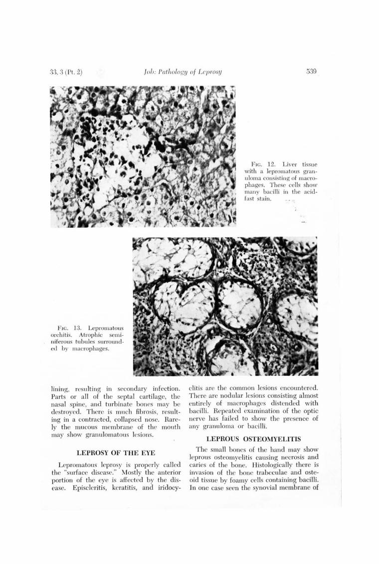
macrophages ( Fig. 12 ) . The granulomata in these lesions are growing ones, and the bacilli in them are morphologically the same as those seen in the skin .
LEPROSY OF THE NOSE
The mucous membrane of the upper respiratory tract is a very common site of inflammation. The subepithelial tissue is infiltrated with foamy macrophages and bacilli inside these cells are very well preserved. Often the infiltration of the mucous membrane may be so massive as to destroy the mucous glands. There is crust form ation and ulceration of the epithelial
FIG. 10. Lepromatous leprosy. Macrophages surrOllnding cross sections of the nerve twigs. The nerve bundles themselves are not infiltrated by the inflammatory cells.
.'3.'3, :3 (Pt. :2) JoT): Pathology of Lcpl'oslj 539
FI G. 13. Lepromatous orchitis. Atrophic seminiferous tubules surrounded by macrophages.
lining, resulting in secondary infection. Parts or all of the septal cartilage, the nasal spine, and turbinate bones may be destroyed. There is much fibrosis, resulting in a contracted, collapsed nose. Rarely the mucous membrane of the mouth may show granulomatous lesions.
LEPROSY OF THE EYE
Lepromatous leprosy is properly called the "surface disease." Mostly the anterior portion of the eye is affected by the disease. Episcleritis, keratitis, and iridocy-
FIG. 12. Liver tissue with a lepromatous granuloma consis ting of macrophages. These cells show many baci lli in the acidfast stain.
eli tis are the common lesions encountered. There are nodular lesions consisting almost entirely of macrophages distended with bacilli . Repeated examination of the optic nerve has failed to show the presence of any granuloma or bacilli.
LEPROUS OSTEOMYELITIS
The small bones of the hand may show leprous os teomyelitis causing necrosis and caries of the bone. Histologically there is in vasion of the bone trabeculae and osteoid tissue by foamy cells containing bacilli. In one case seen the synovial membrane of

.540 Tn/ el'll(l/i())Io l jOlll'll(ll of Le}iro,l'!I
an adjacenl joint was invaded by leprous gran u lomata.
LEPHOUS OHCIIITIS
The testis is a ffected in leprosy with destruction of the parenchyma, resultin g in loss of function. There is infiltration of the seminiferous tubules and interstitial tissue by hacillus-filled macrophagcs, plasma cells, and lymphocytes (Fig. 13 ). The end resu lt is atrophy and hya lini zation of the seminiferolls tubules. In a small proportion of the patients the interstitial cells arc preserved and apparently hyperplasti c. Gynecomastia is noted in 11 p er cent of the patients ( Fig. 14 ).
FIG. 14. Lepromatous leprosy. Gynecomastia.
ERYTHEMA NODOSUM
No discussion on the pathology of leprosy is complete without mentioning erythema nodosum. early 30 per cent of the lepromatolls cases develop painful , tender, and erythematous nodules in the skin ( Fig. 15 ). They are transient and appear almost a lways during the course of anti leprosy treatment. Often they are accompanied by fever, polyarthralgia, and polyneuritis. The course may be mild or severe. Histologically the lesion shows the basic lepromatous granuloma infiltrated b y polymorphonu-
FIG. 15. Erythema nodosum leprosum. Nodular eruption on the face and trunk.
clear leucocytes . The subcutaneous tissue may show microabscesses, and there may be vasculiti s. The nodule may ulcerate and discharge many granular acid-fast bacilli . No other organisms are isolated in smears or in cultures from these exudates, un less they are secondarily infected. Pati ents pron e to get erythema nodosum lesions tend to develop more severe deformiti es and develop them earlier in the course of the disease. They are also less amenable to antileprosy treatment.
GEOGRAPHIC PATHOLOGY
The incidence of the different fOlms of leprosy in the various countries of the world is worth mention. The lepromatous leprosy rate seems to decrease with increase in the color of the skin. It is 5-8 p er cent in Africa, 13-15 per cent in India, 40-50 per cent in China, and 70-80 p er cent in the western countries. It would b e in teresting to know the in cidence of lepromatous leprosy in colored p eople in the United States and the white p eople in Africa and India. The difference may be one simply of different strains of leprosy

33, 3 (PL. 2) Slic/){Ird: ~1. Icprau & TClII/Jcrallirc to/' Gruu; th 541
baci ll i. It is now wull knO\vn that tuberde baci Iii in India arc much less virulent than tubercle bacilli in Grcat Britain.
Leprosy is a fascinating disease. It prcsents several unsolved problems and therefore offers a great challenge to the medical scientist.
Dr. Binford. Thank you very much, Dr. Job, for this brief but compact story of the pathology of leprosy. We havc to apologizc for giving you such a large assignment for 20 minutes, but you accomplished it well and I am sure that those of the audience who have not had the privilege of studying this disease gained much by it.
Cultivation of M. leprae (Cont'd)
Chairman : R. J. W. Rees
D r. Binford. Dr. Rees will now take the chair. You do not nced any introduction to him . He has been with us at our previous meetings and is well known to all of you who are working with mycobactcrial diseases. H e comes from the National Institute for Medical Research at Mill Hill in London .
Dr. Rees. We are beginning another section on the cultiva tion problems with M. lepme, and are now getting deeper into the subject. Itis a particu larly great honor for me to introduce the first speaker, Dr.
Charles C. Shepard. H e truly has contributed more significantly than anyone else to advancement in the scientific field of leprosy, sincc Hansen first recognized the leprosy bacillus, by demonstrating to the world that M. leprae can be cultivated in the foot pad of mice. Many of us now are following in Dr. Shepard's foot steps. Dr. Shepard is Chief of the Special Projects Unit, Virology Section , Communicable Disease Center, United States Public Health Service, Atlanta, Georgia. His subject is "Stabi lity of M ycobactel'ium lepme and temperature optimum for growth."
Stability of Mycobacterium, leprae and
Temperature Optimum for Growth
Charles C. Shepard, M.D.1
Two general areas will be discussed that need to be considered in attempts to cultivate Mycobacterium lepme : first, the stabi li ty of M. leprae at diffcrent temperatures , and second the tempera ture opt:mll m of it·s growlh.
'Chi ef. Special !'miens H llit , Virolog-y Section . C(1ll1l1lllllirahle Di sease Center. U nit ed Stales Public I-Ica llh Serl'ice, ,\tianla , Ceorgia , 30,333.
The stability of the organism is a mattel of special concern in leprosy research. Many of the research laboratories are located far from the sources of suppJy in the important leprosy endemic areas, so that clinical material ml1st be shipped long distanc'('s at considerable tn1llhk.
In the J'('slllts to be descl'ilwd, viability was tested by the ability of M. leprae to multiply in mice. Such multiplication is,


![OFFl AND COUNCILLORS - ILSLila.ilsl.br/pdfs/v31n4a11.pdf · INTERNATIONAL LEPROSY ASSOCIATION 6 l-lillercst Avenue, Pinner, "Jiidc] lcsl'x, England OFFl C~R8 AND COUNCILLORS OFFICERS](https://static.fdocuments.us/doc/165x107/5e048c6a47bcff7c7117d65c/offl-and-councillors-international-leprosy-association-6-l-lillercst-avenue-pinner.jpg)


