An Introduction to Regenerative Injection Therapy (RIT) in ...
71
Transcript of An Introduction to Regenerative Injection Therapy (RIT) in ...


An Introduction to Regenerative Injection Therapy (RIT) in Interventional Regenerative Orthopedic
Medicine (IROM) …an Interventional Radiologist’s perspective
DL Harshfield M.D., M.S.
Board certified Radiologist with specialty training in NMSK, Ultrasound, Interventional Radiology and Cellular Medicine Director of the College of Integrative Medicine- coimed.org
Published By:
MedCrave Group LLC
Date: December 07, 2016

Perineural Injection Therapy (PIT- subcutaneous D5W)
Prolotherapy (Subcutaneous To Deep- Intralesional)
Cellular Therapy Interventional Regenerative Orthopedic Medicine (IROM)
Prolotherapy, PRP and Stem Cells
Prolotherapy
Arachidonic Acid
Neurons from Embryonic Stem Cells Replace Damaged Neurons, “Directly” Rewiring the Brain
Indirect” Matrix Effects May Supersede “Direct” Cellular Effects in Non-Embryonic Regenerative Medicine
TNTs Transfer Cellular Compartments, Such As Endoplasmic Reticulum (ER), Mitochondria, Golgi and Endosomes
Stem Cell Recruitment of Newly Formed Host Cells via a Successful Seduction? Filling the Gap between Neurogenic Niche and Injured Brain Site
A Hyaluronan-Based Injectable Hydrogel Improves the Survival and Integration of Stem Cell Progeny following Transplantation Brian
Case Studies
Standard Procedure Note for Shoulder Regenerative Injection Therapy (RIT)Impression
Case Presentations
Right Shoulder pain and decreased range of motion with ultrasound guided PRP
Right Anterior Shoulder pain with suspected Biceps Brachii Tendon injuryElbow
Distal Biceps Tendon treated with Platelet Rich Plasma
Knee
Knee with Regenerative Injection Therapy (RIT) Using Intra-Articular Hypertonic Dextrose
Knee with PRP therapy with 5-year follow-up
ACL Tear Treated with RIT
Bucket Handle Tear of the Medial Meniscus Treated with RIT
Patellofemoral Glide Pathology (Patellar Tracking Dysfunction) Treated with RIT in Conjunction with Rigorous Physical Therapy
Lumbar Spine
Hamstring Insertional Tear Treated with PRP
Contents
17
7
17
17
1818
19
21
22
22
23
23
28
30
32
36
37
38
38
44
44
51
53
56
Low Back and SI Joint Pain Treated with Caudal Epidural Buffered 5% Dextrose
Superior Cluneal Nerve Entrapment induced Low Back Pain and “Iliolumbar Syndrome”
Conclusion
Final Overview for Patients and Integrative Medical Physicians
Summary of the FDA Stance on Platelet-Rich Plasma
Appendix 2
References
Contents
59
60
6263
63
64
67

An Introduction to Regenerative Injection Therapy (RIT) in Interventional Regenerative Orthopedic Medicine (IROM)
1
Regenerative Injection Therapy (RIT) is a nonsurgical, percutaneous approach to pain and healing of orthopedic injuries that is very different from traditional invasive orthopedic approaches. This text provides the reader with the author’s combined clinical approach and experience from an interventional radiologist viewpoint. Several of the topics presented are discussed elsewhere in this edition and the reader can refer to those respective manuscripts for further reference and basic science. Regenerative injection therapy (RIT) encompasses various procedures that utilize several potential restorative and regenerative compounds, depending on the diagnosis and objective of the treatment (Table 1). These will be addressed briefly in our discussion followed by a presentation of various case observations with the intent of providing the reader with a working approach to such patients. Although there is certainly much to be studied to further delineate cause and effect and to explain these observations, the intent is to share one practitioners experience using RIT approaches.
RIT includes the following procedures:
i. Perineural Injection Therapy (Lyftogt PIT- subcutaneous injection of buffered D5W with ½ inch, 27 ga. needles)
ii. Prolotherapy (subcutaneous to deep- intralesional with longer needles)
iii. Cellular Therapy (cellular products administered with image guidance to area of injury)
Imaging-guided regenerative injection procedures are fast becoming requested and preferred, and not just when status quo physical and pharmaco therapy or surgery fails to provide adequate pain relief or to restore function. The intent is not necessarily curative, and injections alone may not alleviate pain permanently or completely; but the aim is to help patients achieve enough pain relief so that they can undergo physical therapy, reduce the administration of systemic pain medications, and return to activities of daily life. In addition, imaging-guided therapy can be a diagnostic tool, as the response to these injections, even if temporary, can often provide the Integrative Medicine physician with a better idea as to what is the pain generator.
Practitioners of regenerative medicine must communicate an overview of regenerative injection therapy
(RIT) in sync with patient’s understanding of their existing health care regimen (making clear that RIT is
‘in addition to’, not ‘instead of’ the patient’s existing and evolving ‘patient specific’ integrative health care
regimen).
Perineural Injection Therapy (PIT- subcutaneous D5W)“New Science” of Lyftogt Perineural Injection Therapy.
The latest revelation is that neuropathic pain is simply caused by low tissue energy levels! It now appears that neuroglycopenia (low sugar levels detected by the nerves lying in the tissues) is the cause of neuropathic pain. And neuropathic pain is therefore a signal to the spinal cord and brain of low glucose levels in involved tissues. Neuropathic pain is to be differentiated from nociceptive pain, which is not associated with neurogenic inflammation.
Inflammation produces pain, stiffness, discomfort and distress depending on the severity. Pain can vary from a constant steady ache, to throbbing, pulsating or a stabbing sensation. Pain is a very individual experience and the only person who can describe it properly is the one who is feeling it. Pain, whether acute or chronic, is stored in the spinal cord, not in the brain, and with excessive narcotics and opioids the pain memory is “hard wired” often persisting long after the actual cause is gone.
As opposed to neuropathic pain, specific receptors are stimulated for nociceptive pain. These receptors sense changes in temperature, vibration, stretch, and chemicals which damaged cells release. “Nociceptive” means causing or reacting to pain - the cause of the pain comes from outside the nervous system, and the nervous system reacts to it. “Non-nociceptive” (neuropathic) means the pain comes from within the nervous system itself.
The instigators of neuropathic pain appear to be the TRPV1 receptors, thought to represent calcium ion channels, which induce neural inflammation in the environment of acidity and low tissue glucose levels (as opposed to nociceptive pain which is caused by acidosis and low oxygen levels).
The above diagram (Figure 1) demonstrates how Perineural Injection Therapy (PIT) of the left shoulder administers buffered Dextrose 5% in sterile water (D5W) in subcutaneous near nerve injections. PIT appears to be having an antagonist effect blocking neuropathic pain and reducing neurogenic inflammation, often producing immediate, sometimes permanent pain relief with repeated treatments. PIT involves injecting sterile buffered dextrose (neutral pH sugar solution) around the subcutaneous nerves, utilizing only 27-30 gauge, 1/2” needles, 1/4” deep to the skin surface.
The above Power Doppler Ultrasound images Figures 2a & 2b show that PIT utilizing buffered D5W to treat neuropathic pain, creates a regional hyperemia accompanied by an almost instant analgesia. Power Doppler evaluation documents an induced epidermal, subcutaneous and dermal vasodilatation (hyperemia) within minutes of the injection. D5W injection (PIT) causes instant vasodilatation of epidermal arterioles, which has been attributed not only to needle micro-trauma (histamine effect), but also to a Calcitonin Gene Related Peptide (CRGP) effect (that is not altered by blocking the histamine effect). CGRP is produced in both peripheral and central neurons. It is a

An Introduction to Regenerative Injection Therapy (RIT) in Interventional Regenerative Orthopedic Medicine (IROM)
2
potent neuro-peptide vasodilator and can function in the transmission of pain. CGRP receptors are found throughout the body, suggesting that the protein embedded within the cell membrane may modulate a variety of physiological functions in all major systems (e.g., respiratory, endocrine, gastrointestinal, immune, and cardiovascular).
Figure 1: Perineural Injection Therapy.
The increased Power Doppler (PD) flow in the upper right image (Figure 2b) is due to vasodilatation, as opposed to neovascularization. Neovascularization takes much more time. And although angiogenesis and/or vasculogenesis (neovascularization) would have a very similar PD appearance to the above right Power Doppler image, true neovascularization would also have been present on the pre-injection upper left Power Doppler image (Figure 2a). And if neovascularization had been present on the pre-PIT images that would have indicated pre-existing high levels of “neurogenic inflammation”. Neurogenic inflammation is induced by high levels of both Substance P (SP) and CGRP and is associated with neuropathic pain and collagenolysis and other degenerative signs- none of which are seen on the initial Power Doppler image in this patient (Figure 2a).
Figure2a: Power Doppler immediately prior to PIT.
Pain has become the most common reason patients seek medical attention. Narcotics, opioids, NSAID’s and steroids only mask pain without treating the cause, often
to the detriment of the patient and delaying the appropriate diagnosis and treatment. The current hypothesis for the effectiveness of PIT (perineural injection therapy) is that the primary cause of neuropathic pain is the depolarization (firing) of unmyelinated pain fibers due to “insufficiency of energy” (low glucose-glycopenia) in their neighborhood, which is rapidly calmed by buffered, neutral pH, 5% Dextrose. The 5% buffered dextrose is iso-osmolar (near identical concentration) and neutral pH (not acidic) to the nerve cells, thereby repolarizing (stabilizing) them.
Figure 2b: Power Doppler 3 minutes post PIT.
In medicine, we must resist the tendency to confuse “association” with “causality”. For instance, pain can be “associated” with physical injury, but pain is not necessarily “caused” by injury, per se. As an example, ischemic pain of cardiac disease is not posttraumatic in nature, instead being a consequence of acidosis and hypoxia on the nociceptors in the heart muscle (similar to the cause of claudication and rest pain in the lower extremities from peripheral artery disease). Thus, although decreased with lidocaine (anesthetic), ischemic pain is not alleviated by glucose (analgesic). But the recent discovery that most neuropathic pain is simply caused by low energy levels in tissues provides the explanation for the remarkable effectiveness of Dr. Lyftogt’s Perineural Injection Therapy [1].
Understandably, clinicians may question the paucity of peer reviewed literature on Dextrose injection therapy. But informed health decisions can be based on good science rather than evidence based medicine (EBM) alone. The call for randomized controlled trials (RCT’s) to prove the efficacy of buffered sugar water (PIT) brings to mind the British Medical Journal’s tongue in cheek article- “Parachute use to prevent death and major trauma related to gravitational challenge: systematic review of randomized controlled trials”.
The conclusion of the spoof was: “As with many interventions intended to prevent ill health, the effectiveness of parachutes has not been subjected to rigorous evaluation by using randomized controlled trials. Advocates of evidence based medicine have criticized the adoption of interventions

An Introduction to Regenerative Injection Therapy (RIT) in Interventional Regenerative Orthopedic Medicine (IROM)
3
evaluated by using only observational data. We think that everyone might benefit if the most radical protagonists of evidence based medicine organized and participated in a double blind, randomized, placebo controlled, and crossover trial of the parachute”.
The relevance to parachute use is that individuals jumping from aircraft without the help of a parachute are likely to have a higher prevalence of morbidity while individuals who use parachutes are likely to have less morbidity.
The above review of the necessity of RCT’s on parachute use analogous to the opinion that neither are RCT’s required to address whether buffered D5W relieves pain- it does, and without appreciable risk. So, under this circumstance, common sense might be applied when considering the potential risks and benefits of Dextrose interventions. And the timeliness could not be more appropriate, particularly in the face of the growing safety issues with narcotics, opioids, NSAID’s, steroids and ineffective surgery. Observational studies have been performed, clinical trials are ongoing and that should do for now. And the following case reports should provide sufficient evidence not only of the safety but of the efficacy of PIT. After thousands of patients, there have been no reported severe adverse events (SAE’s) associated with RIT utilizing the combination of PIT and PRP therapy.
Perineural Injection Therapy (PIT) is based on the observations and teachings of John Lyftogt that buffered D5W can alleviate neuropathic pain. Another recent revelation is that D5W (5% dextrose water) obtained in the United States arrives in an acidic form with a pH ranging from 3.5 to 6.9. TRPV1 pain receptors activate at pH lower than 6.5, thus in order to optimize perineural injection therapy and maximize the pain fiber membrane stabilizing effect of the D5W one must buffer with 8.4% Sodium Bicarbonate to bring the pH up to neutral (7.2). In addition, sterile water in bottles obtained in the U.S. also are acidic, with a pH 5.5; a fact which must be taken into consideration when diluting dextrose with sterile water.
These concepts are not exactly new, being over 150 years old. Hilton’s law 1863: The nerve trunk supplying a joint also supplies the overlying skin and the muscles that move the joint.
Buffered Dextrose 5% (D5W) in sterile water in subcutaneous near nerve injections are predicted to facilitate the potassium channel and have a TRPV1 antagonist effect blocking neuropathic pain and reducing neurogenic inflammation, producing cure with repeated treatments. PIT involves injecting sterile buffered dextrose (neutral pH sugar solution) around the subcutaneous nerves with only 27- gauge 1/2” needles 1/4” deep, blocking neuropathic pain and reducing neurogenic inflammation, producing cure with repeated treatments.
The PIT utilized buffered D5W treats neuropathic pain with
resulting instant analgesia and also induces hyperemia within minutes of the injection. D5W injection causes instant vasodilatation of epidermal arterioles, clearly a Calcitonin Gene Related Peptide (CRGP) effect. CGRP is produced in both peripheral and central neurons. It is a potent peptide vasodilator and can function in the transmission of pain. CGRP receptors are found throughout the body, suggesting that the protein may modulate a variety of physiological functions in all major systems (e.g., respiratory, endocrine, gastrointestinal, immune, and cardiovascular).
There is mounting evidence to suggest that CGRP is beneficial in preventing the development of hypertension and cardiovascular disease. CGRP expression in keratinocytes is substantially increased in certain human chronic pain conditions and animal models of induced chronic pain conditions, whereas the alpha-CGRP containing peptidergic innervations are decreased in painful skin sites. CGRP is thought to play a role in cardiovascular homeostasis and nociception, as well as neuropathic pain.
CGRP is in a positive feedback loop with Vascular Endothelial Growth Factor (VEGF), which is also stimulated by Hypoxia Inducible Factor (HIF). VEGF is in a positive feedback loop with MMP1. This vasodilatation is not thought to be due to a decreased sympathetic tone as the autonomic nervous system responds to BP, cardiac load and heat loss/gain, not to local noxious tissue stimuli. And there are no nociceptors in the autonomic nervous system.
In fact, pre-treatment hypervascularity is due to neovascularization, usually considered to be an accurate sign of chronic tendon disease (Alfredson 2005), but when absent, the cause of pain is usually neuropathic. It may be that subcutaneous injection itself could be the noxious stimulus for acute vasodilatation reaction- so called flare reaction mediated by histamine, and lasting 8 to 12 hours. However, even after histamine block, there is still hyperemia after needling procedures.
There may be no way to separate the traumatic and mediator induced effects of PIT on cutaneous blood flow (CBF). Laser Doppler velocimetry (LDV) studies have shown the CGRP action on the cutaneous microvasculature is more sustained and not affected by histamine block. Older individuals have a more attenuated (diminished) response as determined by response onset, the peak reaction, the magnitude of the reaction and the clearance of the injectate. The CBF response to needling actually increases from childhood to early adulthood, plateauing after age 60. Skin reactivity has been shown to be markedly dependent on age, but not on sex or in patients with atopic diatheses (dermatitis, etc.).
Skin pharmacology was further advanced with the discovery that topical administration of EMLA (prilocaine and lidocaine) blunted the “wheal”, but not the “flare” effects of histamine on the cutaneous microvasculature. This means that the “flare” (hyperemic) component is mediated more by

An Introduction to Regenerative Injection Therapy (RIT) in Interventional Regenerative Orthopedic Medicine (IROM)
4
neurogenic mechanisms than directly by histamine effects. Additionally, NSAID’s blunt the hyperemia seen after needle sticks, thus the reason for stopping them several days (or longer) prior to regenerative injection therapies in which ablation of neuropathic pain is the goal.
The fact that a similar post injection hyperemia occurs after cellular therapy injections as well as “dry needling” would favor needle puncture and the resultant “micro hemorrhaging” to be the instigator of this phenomenon. Patients often report immediate pain relief most likely the result of the buffered Dextrose on the neuropathic pain, with a day or two of dull achiness which corresponds with Power Doppler demonstrable hyperemia- essentially each needle stick creates a “mini PRP” treatment.
Of course, the ion-channel involved is TRPV1. Up regulation of TRPV1 results in the release of the pro inflammatory neuro peptides CGRP and SP causing neurogenic inflammation? Glucose is thought to down regulate TRPV1 through an allosteric modulation effect reducing SP and CGRP levels and hence decreasing neurogenic inflammation. Glucose also binds to tandem pore K+ channels resulting in neuronal inhibition, blocking spike formation and resulting in analgesia.
Pain is the most common reason for patients to seek medical attention. The current understanding of the primary cause of pain is the depolarization (firing) of unmyelinated pain fibers due to “insufficiency of energy” (low glucose) in their neighborhood, which is rapidly calmed by buffered, neutral pH, 5% Dextrose. The 5% buffered dextrose is iso-osmolar (near identical concentration) and neutral pH (not acidic) to the nerve cells, thereby repolarizing (stabilizing) them.
In medicine, we must resist the tendency to confuse “association” with “causality”. For instance, pain can be “associated” with physical injury, but pain is not necessarily “caused” by injury, per se. For instance, ischemic pain of cardiac disease is not post traumatic in nature, instead being a consequence of acidosis and hypoxia on the nociceptors in the heart muscle. Thus, although decreased with lidocaine, ischemic pain is not alleviated by glucose. But the recent discovery that neuropathic pain is simply caused by low energy levels in tissues provides the explanation for the remarkable effectiveness of Lyftogt Perineural Injection Therapy [2].
Fat suppressed, water weighted MRI sequences reveal several of the subcutaneous perineural injections of .25 to .5 ccs of buffered D5% Dextrose (white arrows in the above images, Figures 3a & 3b are coronal, Figure 3c sagittal). PIT can be a stand-alone “superficial” procedure to treat regional pain or can be is utilized in conjunction with “deeper” cellular injection therapies where the goal is to reconstitute injured tissue as well as achieve immediate pain control.
Figure 3a: Lyftogt Perineural Injection Therapy (coronal).
Figure 3b: Lyftogt Perineural Injection Therapy (coronal).
Figure 3c: Lyftogt Perineural Injection Therapy (sagittal).

An Introduction to Regenerative Injection Therapy (RIT) in Interventional Regenerative Orthopedic Medicine (IROM)
5
Typically, cellular therapy alone does not produce pain relief for several days, thus PIT is performed initially for immediate pain relief to allow improved range of motion that will enhance the outcome of the cellular therapy. Generally, the cellular injectate is a more localized injection that is applied “deep” or intra articular, and the buffered glucose is administered superficially and more regionally.
The peripheral most pain receptors lie just beneath the skin, thus can be successfully and safely treated utilizing buffered D5W administered with a ½ inch, 27-30 gauge needles. The “named” nerves that are memorized in medical school are in general “hollow tubes”, each day transporting fluid (axoplasmic flow) at rates of 40 cm per day peripherally, and 30 cm per day back toward the spinal cord.
But it is the “nerves to those nerves”, the nervi nervorum, that lie within the epineurium surrounding those relatively large tubes that are responsible for generating pain signals, not the nerves themselves, but so called sensocrine nerves. These strange pseudo-unipolar neurons curiously conduct action potentials in both directions and teleologically they were originally endocrine cells that have developed neurological function (thus termed sensocrine nerves).
Nociceptive pain, neuropathic pain, anesthesia and analgesiaNociceptive pain (induced by acidosis and low O2) is not the same as neuropathic pain (caused by acidosis and low glucose), and hence does not generally respond to perineural glucose injection (PIT). However, particularly in patients with lower extremity ischemic pain, there is third space acidosis with both low oxygen levels as well as low glucose. Thus, for instance in patients with peripheral arterial disease (PAD) and rest pain, dextrose injection therapy may provide relief of the pain caused by glycopenia.
But, add to PIT the technique of ultrasound guided cellular injection along the below knee trifurcation vessels and beneath any non-healing wound. The combination therapy completes the lower extremity rescue by the ensuing increased vascularity. Ultimately, with the patient able to begin walking, the invariably concomitant venous insuffiency is addressed by activation of the “muscle pump” of the lower extremity. In other words, peripheral vascular disease is actually a combination of many of the 11 systems that make up the human body, not the least of which is the arterial and venous system and peripheral nervous system.
Lidocaine, which blocks sodium channels, inhibits the “upward” stroke of nerve depolarization potentials, thus inducing anesthesia- thereby blocking nociceptive pain (along with motor and sensory function). Glucose, however, does not induce anesthesia or alleviate nociceptive pain, instead functioning by inducing analgesia (pain relief without blocking the motor or sensory function) thereby specifically blocking neuropathic pain- neuralgia.
Nervous tissue is characterized by high lipid and protein content, but it does not contain large amounts of saccharides. However, most nerves, both within the central (CNS) as well as peripheral nervous systems (PNS), are highly dependent on glucose for energy. This energy is produced in the mitochondria with each neuron containing hundreds or thousands of these power plants. These mitochondria use oxygen to extract energy from glucose and fats and to produce molecules of the energy-storage compound adenosine triphosphate (ATP) via the Krebs cycle. These ATP molecules are the “currency of exchange” then used to “pay for” the various chemical reactions that take place within the neuron.
Nerve cells require oxygen and glucose to transmit impulses and neurotransmitters. When neurons fire, they consume oxygen. Lack of glucose and oxygen (as in patients with neurovascular disease) deplete the cellular energy stores required to maintain electrical potentials and ion gradients. After administration of cellular regenerative therapy, despite the obstruction of the major “named” arteries, nearby capillaries become able to dilate in response to the oxygen deficit, thereby infuse the previously ischemic region with oxygen rich blood.
Neurons are just like other cell types, particularly regarding their need to use ATP to maintain their structures and stay alive. Nerve cells conduct signals electrically down their length. The membrane of neurons has many embedded proteins which require ATP for energy to operate. These proteins actively transport K- and Na- in and out of the cells membranes. This active transporting of charged particles creates an electrical current. But this is not like the electrical current in electrical wires, instead a voltage difference- potential. The “charge” within each nerve depends on an imbalance of Na and K ions across the plasma membrane - and this imbalance sets up a voltage difference.
When the nerve “fires”, tiny channels across the membrane open and the voltage difference (along with osmosis) cause the ions to flow to opposite sides of the membrane. This basically flips the voltage, which triggers the next set of channels down the line to open. The process repeats itself, like a row of dominoes, down the length of the axon until it hits the terminus (end). There, it causes the release of neurotransmitters, which flow across the synapse and affect the next neuron- and so on.
TRPV1 receptors are up-regulated (increased in number) within the membrane of the unmyelinated C fibers (pain fibers) by decreased sugar levels (glycopenia) in the surrounding tissues, and the TRPV1 capsaicin sensitive channels, of which there are 28 distinct types in mammals, are the key pain signal generators within these so called “sensocrine” nerves.
In addition to low glucose levels, TRPV1 receptors activate at pH less than 6.5, and commercially available D5W

An Introduction to Regenerative Injection Therapy (RIT) in Interventional Regenerative Orthopedic Medicine (IROM)
6
is acidic, averaging a pH of 4.5 (3.5 to 6.7) thus less effective at relieving pain unless buffered with 8.4% sodium bicarbonate (½ cc of 8.4% sodium bicarbonate will buffer 100cc’s of acidic D5W to pH of 7.0- pH scale ranges from 0 to 14 and a pH of 7 is neutral).
It is estimated that there are approximately 1 to 2 million pain generators in the human body, and to make things even more challenging, all respond differently. But neuropathic pain almost universally responds to subcutaneous (shallow) perineural buffered 5% dextrose injection therapy.
Analgesics decrease pain signals, enhancing musculoskeletal function, while anesthesia induces numbness, decreasing musculoskeletal function.
Peripheral nerve blocks (PNB), for instance, utilize local anesthetics. LA agents work by temporarily binding to sodium channels, thus rendering them inactive. PNB involves the injection of LA near a nerve or nerve group with the intention of attenuating or inhibiting the conduction of sensory, motor, or autonomic impulses to the brain. Any tissue that is dependent on sodium channel for its function, such as the cardiovascular system and CNS, are therefore susceptible to systemic toxicity from LA. The following attributes of the nerve anatomy and physiology allow PNB to be performed in a safe and reproducible fashion.
Although Lidocaine can induce both anesthesia as well as analgesia, it has an acidic pH that not only activates TRPV1 pain receptors, but its amide chemical construct (procaine is an ester, but acts similarly) induces crystalline formation. The crystals can infarct tissues into which they are injected, and when injected into a joint, lidocaine has been shown to be toxic to chondrocytes.
Long acting steroids are also crystalline, and the small crystals form even larger “snow balls”, which also infarct tissues- believe it or not, the primary mechanism of their “pain relieving” action. The steroid induced nerve infarction temporarily “relieves” pain, which invariably returns in 4 to 6 weeks when the more resilient nerves regenerate. However, the background tissue within and surrounding the injection site does not recover, in fact the collagen within tendons and ligaments is disrupted and disorganized and inhibited from healing.
Surprisingly, there is no literature that supports the use of steroids for the multitude of MSK applications, and the dose of steroids (if they even work) is also problematic. Humans obviously have endogenous steroids, but at physiologic nanogram (billionth of a gram) levels. To put this in perspective, if a nanogram were the thickness of a match book, a milligram would be the height of the Empire State Building.
Buffered 5% Dextrose (D5W) provides analgesia by opening potassium channels in the Nervi Nervorum, pseudo-unipolar unmyelinated C fibers, causing inhibition
of the pain impulses. Potassium channels have a receptor for glucose. Thus, increased glucose allows potassium to flow out of those channels, repolarizing the cells membrane and increasing the threshold for pain (decreasing the likelihood of depolarizing). Since low glucose (glycopenia) excites TRPV1 calcium ion channels which detect low energy levels in the tissues they monitor, the administration of buffered glucose also down regulates those TRPV1 receptors, further increasing the threshold for pain.
Lidocaine is a sodium channel blocker, therefore only affecting the depolarizing sodium based “upstroke” of the depolarization/re-polarization of the cells. Dextrose is a much more effective analgesic than lidocaine, acting on the potassium based “down stroke” of re-polarization to stabilize the cell membrane. Dextrose acts almost immediately, with the effect lasting 3 to 4 weeks, while Lidocaine takes 20 minutes to affect analgesia/anesthesia, with a very short half-life (therefore short time of effect). Thus, when treating neuropathic pain, there is no need to mix lidocaine with buffered 5% dextrose; in fact, it’s counterproductive.
As an aside, sterile normal 0.9% saline (Normal Saline) has also been utilized in prolotherapy, but salt tends to accelerate and enhance electrical impulses while dextrose is preferred because it acts as an insulator, slowing pain impulses. Also, adequately buffered D5W is painless when injected into “normal”, non-painful areas with 27-30 gauge needles. When injected into a palpable pain locus, there is a temporary burning sensation when it is injected. However, if there is a burning sensation when injected into a normal site, it is still too acidic and needs to be further buffered and pH paper can help determine if the D5W is adequately neutralized.
And for those diabetics concerned about “sugar” injection therapy, there is no appreciable effect on blood glucose levels after perineural dextrose injection therapy. And finally, NSAID’s do not interfere with D5W injection therapy, although patients generally no longer need them after treatment.
And as for imaging pain; at present, we cannot. Radiologists are only just now even considering the importance of doing so. Most remain focused on imaging supine, non-weight bearing and motionless patients to ascertain static structure without understanding what is important for MSK diagnosis- i.e. assessing kinematics (motion) and the presence of absence of basic function and documenting dysfunction.
Thus, static anatomy without kinematic assessment will not reveal how a given structure effects function or allows us to detect the “Holy Grail of MSK imaging”- abnormal movement.
So, the fact remains, we can image broken bones and disc herniations that can cause pain, but we cannot directly image pain in humans. Researchers have taken a rat,

An Introduction to Regenerative Injection Therapy (RIT) in Interventional Regenerative Orthopedic Medicine (IROM)
7
injured its foot, injected it with radioactive manganese and with nuclear imaging scanners mapped the nerve pathway that carries the information of that injury to the spinal cord and brain. But thus far, we haven’t managed to convince human subjects to submit to the same toxic procedure.
Typically, cellular therapy (PRP or stem cells) alone does not produce pain relief for several days, and patients may have increased pain for several days after the procedure. Thus, PIT is performed initially for immediate pain relief, which allows improved range of motion that will enhance the outcome of any needed cellular therapy, particularly in conjunction with Physical Therapy regimens.
Often, patients only require PIT (Figures 3a-3c) in conjunction with patient specific Physical Therapy. When PIT is insufficient by itself, Prolotherapy applied to deeper ligamentous, tendinous and capsular insertions is then administered. And when necessary, cellular therapy is applied in a more localized injection that is “deep” or intra articular, in conjunction with the previously administered buffered glucose (PIT and/or prolotherapy) which is applied regionally and more superficially.
Prolotherapy (Subcutaneous To Deep- Intralesional)Prolotherapy: Basic Science, Clinical Studies and TechniqueSpecifically, prolotherapy involves the injection of substances that pro-liferate collagen tissues, which are the building blocks of ligaments and tendons. Not every treatment that involves the injection of dextrose is prolotherapy and prolotherapy does not always use dextrose. Prolotherapy (growth factor or growth factor stimulation injection) raises growth factor levels to increase effectiveness to promote tissue repair or growth. Prolotherapy involves placement by needle of a solution that raises growth factor activity enough to stimulate cell growth or cell production of collagen or matrix. Although inflammatory prolotherapy has been used for many years, non-inflammatory prolotherapy methods are rapidly expanding.
Growth factors are complex proteins (polypeptides), and their beneficial effects on human ligament, tendon, cartilage, and bone are under intense investigation. Prolotherapy may utilize inflammatory or non-inflammatory mechanisms.
There are two general approaches to proliferation therapy. Physicians tend to combine aspects of both methods. The first, known as the Hackett method, is based on the approach of George Hackett with subsequent refinements made primarily by Drs. Gustaff Hemwall and Gerald Montgomery. The West Coast method, popularized by physicians in this region, was promoted by Dorman, Ongley, and others. The comparisons result from direct observation of techniques used by Hemwall, Montgomery, and Ongley
and the author’s personal experience.
In the Hackett method, dextrose is used as the proliferant in the majority of cases. Cellular disruption is minimal and nerve damage has not been reported. This method is slower to perform, but is easier to teach and is uniform in distribution of solution. In contrast, the West Coast approach utilizes phenol 1.25%, glycerine 12.5%, and dextrose (D-glucose in water) 12.5%. The needles are generally larger, and needle movements are more rapid and difficult to learn. In addition to needle insertion and injection method, other considerations include proper patient selection, timing, proliferant solution choice and preparation, identification of injection sites, sedation, positioning and anesthesia issues, post procedure care, and complications.
Cellular Therapy Interventional Regenerative Orthopedic Medicine (IROM)IROM (interventional regenerative orthopedic medicine) takes RIT (regenerative injection therapy) to another level of healing that is revolutionary in the medical world and is fast becoming the preferred integrative intervention of choice. IROM uses image guidance along with fluoroscopic and ultrasound stress testing correlated with Physical Therapy examination to diagnose areas of weakness often missed on a MRI and other static imaging modalities. In addition to regional PIT and Prolotherapy, the trained IROM clinician can also introduce cellular products (platelets, stem cells or amniotic tissues) precisely into the offending tissues.
IROM involves having patients initially undergo clinical and imaging evaluation in conjunction with physical therapy that is often can often improve function for the patient. In addition, particularly when treating cardiovascular and pulmonary disease, hyperbaric oxygen therapy (HBOT) accelerates the total healing process by feeding the newly implanted cells and by helping repair and strengthen the torn tendon. IROM provides faster recovery and quicker return to athletic participation. And the addition of hyperbaric oxygen therapy further reduces inflammation and further improves and accelerates the healing.
With the proper patient selection and appropriate injection techniques, IROM has the potential to reduce swelling and improve connective integrity by injecting regenerative and platelet growth factors directly into the affected area, improving the blood supply around the injury. Of the many cellular therapies (Bone Marrow Aspirate Concentrate (BMAC), Mesenchymal stem cells (MSC’s from fat graft and stromal vascular fraction- SVF) and amniotic tissues) Platelet Rich Plasma (PRP) is currently the most ubiquitous.
Musculoskeletal practitioners began using PRP for tendinopathy in the early 1990s. These early practitioners were primarily trained in the use of prolotherapy. The popularity of PRP grew as physicians began to see clinical

An Introduction to Regenerative Injection Therapy (RIT) in Interventional Regenerative Orthopedic Medicine (IROM)
8
results in concentrating a patient’s own blood factors. The PRP procedure is significantly more complex and requires additional equipment to perform successfully, but many practitioners have seen a relatively more robust response, fewer treatments and improved tissue health compared to prolotherapy.
The current 6 point guideline focuses on general principles of RIT utilizing in part or together PIT, prolotherapy and cellular therapy with applications including musculoskeletal care, as well as pulmonary, cardiovascular and cosmetology applications.
Platelet Rich Plasma (PRP) ApplicationsFrom an Interventional Radiologist’s perspective, one of the most common omissions in RIT is the absence of correlation of patients’ physical, clinical and laboratory findings with prior imaging studies; performing appropriate up-to-date imaging studies when necessary to arrive at the correct diagnosis and to determine appropriate therapy. And it is essential that the patient understand the findings, as the patient with no confidence in the diagnosis will have no confidence in the therapy.
R’s of correlative imaging
Recent MRI unless contraindicated, imaging the area(s) of concern (based on both clinician and patient input).
Regional MRI of the portion of the spine that could serve as a potential “proximal” instigator (co-conspirator) of patient’s more distal symptomatic area (i.e. Cervical spine MRI for patients with upper extremity symptoms, Lumbar MRI for pelvic and lower extremity symptoms, etc.).
Review all existing clinical notes and prior imaging studies and correlate with updated imaging studies.
Reassure the patient that their direct participation and understanding is key if successful regenerative therapy is to be achieved. The patient’s ideas, concerns and expectations for their personalized regenerative medicine therapy are addressed.

An Introduction to Regenerative Injection Therapy (RIT) in Interventional Regenerative Orthopedic Medicine (IROM)
9Prior to the availability of these RIT protocols Conventional Medicine recommended first line therapies such as
a. Relative rest.
b. Appropriate bracing and kinesiotaping.
c. Evaluation of core stabilization.
d. Reintegration of kinetic chain mechanics.
e. Chiropractic and/or Physical/manual therapy- with or without eccentric loading protocol.
After determining the appropriate diagnosis and patient acceptable therapy, the physician qualified and certified in Interventional Regenerative Orthopedic Medicine (IROM) treats the patient with buffered Dextrose for instantaneous pain relief along with a derivative of their own cells (autologous) or amniotic tissue (heterologous) for regeneration of injured tissues, as indicated by their diagnosis.
The success of RIT (regenerative injection therapies) is proving that inflammation plays as important a role in osteoarthritis pain as structural issues. The two-dimensional preoccupation with pharmaceutical and surgical intervention is currently being reconsidered (expensive, dangerous, ineffective orthopedic surgery versus much less expensive, safe and effective orthopedic medicine). For instance, recent literature has shown that arthroscopic resection of a torn meniscus has no added benefit over sham surgery to relieve knee catching or occasional locking. These findings bring into question whether mechanical symptoms are caused by a degenerative meniscus tear and should prompt caution in using patients’ self-report of these symptoms as an indication for APM (arthroscopic partial meniscectomy).
Osteoarthritis, the most common articular disease in humankind, is associated with defects in articular cartilage but we now know that it is primarily caused by hypermobility (ligament laxity) and has significant effects on the quality of life (QOL) of patients, especially the elderly. For this reason, the effects of osteoarthritis and related therapeutic interventions on the QOL and patients’ functions have been assessed in different regenerative medicine studies.
There are different methods used for alleviating the symptoms of patients with knee osteoarthritis (OA), including various medications and supplements (NSAIDs, glucosamine, and chondroitin- sulfate), intra-articular injections (glucocorticoids, hyaluronic acid), physical agents (prescription of appropriate braces, shoes and insoles, exercise therapy, laser therapy, application of heat and cold modalities, etc.), and surgical interventions.
Although some of these treatments have had short- and mid-term effects on improving patients’ functions and decreasing the level of disability, there remain controversial results about
their effects on decreasing the amount of articular damage and slowing the rate of disease progression. It seems that existing treatments cannot change the pathophysiology or prognosis of the disease.
Ongoing studies are focusing on modern therapeutic methods stimulating cartilage healing process and improving its damage, including application of matrix metalloproteinase inhibitors, gene therapy, cytokinase inhibitors, PRP, amniotic tissues, stem cells, and growth factors. Growth factor effects have been evaluated extensively both in vivo and in vitro.
Known platelet growth factors stimulate the healing process and lead to partial modification of the damaged tissue. Platelet rich plasma (PRP), with higher platelet concentrations than the mean blood measures, is one of the sources for growth factors. In most studies, the effective platelet concentrations have been between 3 and 7 times the normal average measures, depending on the kind of application (skin, hair, musculoskeletal, etc.). By activation of the platelets, different growth factors available in alpha and dense granules initiate the healing chain. This chain includes three steps of inflammation, proliferation, and remodeling.
Pain- Inflammation versus structural pathologyOsteoarthritis (OA) is a heterogeneous and multi-factorial disease characterized primarily by the progressive loss of hyaline articular cartilage precipitated by hypermobility (ligament insufficiency). But OA is no longer considered strictly a “non-inflammatory” condition, as inflammation associated with synovitis or effusion appears to play as great a role in OA pain as inability to dissipate mechanical load. And there is an association between baseline presence of synovitis or effusion and pain threshold years later (a marker of central pain sensitization), but not between baseline presence of bone marrow lesions (BML’s- a marker of mechanical load) and chronic, persistent knee pain.
There are several mechanisms by which pain occurs in knee OA, with different pathologic structures contributing to pain through different mechanisms. Early targeting of inflammation (rather than focusing solely on structural abnormalities) can reduce sensitization (which contributes to more severe, persistent and progressive pain in OA.
Importantly, imaging changes may not adequately reflect the molecular, hormonal or cellular mechanisms (cellular and humoral immunology) that mediate the augmentation of neuronal sensitization by synovitis. However, the MOST subjects (funded by the National Institutes of Health) showed that imaging evidence of persistence of synovitis and effusions was correlated with increased pain sensitivity. On the other hand, resolution of those features on MRI is not necessarily associated with improvement in knee pain.

An Introduction to Regenerative Injection Therapy (RIT) in Interventional Regenerative Orthopedic Medicine (IROM)
10
Thus, once sensitization has occurred, just focusing on surgically changing the structural problems even when it results in decreased imaging evidence of inflammation might not result in ablation of the pain.
The gut microbiome regulates not only the local intestinal immune system but also the host’s adaptive immune responses. It is therefore not surprising that non-GI autoimmune disorders like rheumatoid arthritis (RA) have also been linked to the gut microbiome. A study comparing the intestinal microbiota of patients with recent-onset RA and fibromyalgia observed substantial differences between the two, with the authors concluding that these results supported the microbiome’s potential role in development of RA. One theory holds that the microbiome may interact with predisposing genetic factors to trigger peripheral or axial arthritis. The autoimmune disorder spondyloarthritis has also shown a common bacterial profile to another inflammatory disorder, IBD. Early-stage research is looking at the efficacy of probiotic interventions for conditions such as these.
Regenerative cellular therapies appear to alter the neurologic processing of nociceptive signals that occur in OA might potentially prevent the progressive worsening of pain in knee OA. For instance, Ozone can be anti-inflammatory and is an antioxidant. An ozone injection into the knee, which inhibits prostaglandins and cytokines and reduces oxidative stress, decreases pain accompanying knee osteoarthritis.
Although intra-articular corticosteroid injection and exercise have been considered standard of care in recommendations and guidelines for knee pain, studies do not support the superiority of intra- articular injection of corticosteroid compared with regenerative injection therapies coupled with an exercise intervention.
Ongoing joint inflammation appears to affect long-term pain sensitivity, thus earlier and more aggressive treatment of joint inflammation with regenerative therapies in OA may decrease chronicity of knee pain. The implications are that we need to non-surgically address synovitis and effusion to prevent increased pain sensitization. It appears that OA is not just a wear-and-tear process, as there is a previously overlooked inflammatory component to OA to the associated pain, rather than pain solely being due to mechanical factors. For this reason, regenerative cellular medicine therapies, including PRP, are replacing pharmaceutical and surgical interventions.
Previous studies of bioactive molecules in platelet-rich plasma (PRP) have documented growth factor concentrations that promote tissue healing. However, the effects of leukocytes and inflammatory molecules in PRP have not been defined. Growth factor and catabolic cytokine concentrations are influenced by the cellular composition of
PRP. Platelets increase anabolic signaling and, in contrast, leukocytes increase catabolic signaling molecules. Thus, platelet- rich plasma products should be analyzed for content of platelets and leukocytes, as both can influence the biologic effects of PRP.
Preferable are PRP techniques that allow obtaining “non-red” platelet rich plasma with point of care therapy. “Non-red” PRP is a platelet rich plasma preparation that contains highly concentrated platelet growth factors with essentially no red blood cells- therefore no post injection “flare” or chondrocyte toxicity. We utilize systems in which PRP can be processed with or without neutrophil granulocytes. The platelet and growth factor concentrations are 7 to 9 X baseline. The collected PRP sample is approximately 6mL from a 60mL sample of anti-coagulated whole blood. This “non-red” PRP solution meets most clinical demands in a medium that is injectable through small bore needles and suitable for tissue restoration and active wound repair.
“Non-Red” PRP provides high concentrations of platelet & growth factors in a pure plasma suspension. It has a no red blood cells that tend to increase PRP viscosity making it more difficult to inject. In addition, red blood cells in PRP have also been reported to cause pain due to a “flare” phenomenon and intra-articular blood/heme is counterproductive to healing. Further, the polymorphonuclear cell family (PMNs) includes the body’s most abundantly occurring granulocyte known as neutrophils. These granulocytes, in abundant and sustained doses release cytokines, which can amplify inflammatory reactions by several other cell types. This inflammation may be needed for active wound repair, but in some cases, is undesirable. “Non-red” PRP preparations can be processed with or without neutrophil granulocytes, providing physicians with their choice of active components needed for the care of their carefully selected patients.
CytokinesCytokines play a main role in the natural or innate response by means of the direct action of mechanisms against the invading agent during the early stages of the infection, or by means of immune-modulatory mechanisms which activate Natural Killer cells and mono-cytes-macrophages; which then induce the release of cytokines.
Despite the increase in the use of the PRP for local tissue healing and regeneration in human and animal studies, little is currently known about the bimolecular characteristics of PRP in terms of the cytokine-release kinetics per different activation protocols. It is important to determine the method of local application as well as to evaluate the effectiveness of the procedure. The concentration of cytokines released from PRP varied over time and was influenced by various activation protocols. The effect of the activation was shown to be dependent on the preparation method as well as on the type of cytokine and, accordingly, proper PRP components

An Introduction to Regenerative Injection Therapy (RIT) in Interventional Regenerative Orthopedic Medicine (IROM)
11
with activation methods should be selected by considering their bimolecular characteristics and patient indications.
PRP- to activate or notNon-activated PRP becomes activated when exposed to collagen immediately after injection into the body. There is ongoing debate as to whether and how to activate PRP prior to injection. The activation method or the thrombin dose affects the cytokine-release kinetics of PRPs and must be considered when interpreting the results of PRP studies.
When calcium chloride is added exogenously to PRP, after injection of the activated PRP results in a low level of thrombin being formed endogenously. The endogenous thrombin formation allows a slower GF release over a longer period than when thrombin is added the PRP exogenously. On the other hand, thrombin causes a rapid aggregation of platelets and an excessive condensing of the fibrin matrix with a rapid activation of the platelets. A low dose of thrombin has been shown to increase the migration and the number of mesenchymal progenitor cells derived from bone marrow, whereas high concentrations have been demonstrated to have limited effects on the proliferation of osteoblasts and alveolar bone cell, suggesting that the thrombin dose plays a role in the GF-release kinetics of the PRP preparations.
The effect of the activation depends not only on the preparation method but on the type of cytokine being assessed: Ca-only activation has a significant effect on the double spine (DS) PRP preparation (VEGF, FGF, and IL-1βv concentrations). Whereas Ca/thrombin activation has significant effects on both single spin (SS) and DS PRP preparations (PDGF-BB and VEGF concentrations sustainably and TGF and FGF concentrations shortly).
These observations may be because the biological activity of the platelets is sensitive to any kind of process-related stress and that more platelets are activated during the process with the DS method.
These results are also consistent with the findings that the individual dynamics of the GF release depend exclusively on the type of GF rather than on the preparation method. They also demonstrate that TGF-β1 and bFGF are promptly released within 24 hours of exogenous activation whereas the GF release of the PDGF-BB and the VEGF are more dependent on the technique that is used.
In terms of the catabolic cytokines, Ca or thrombin activation sustainably increases the IL-1β concentration to a level ten times higher than that of control, but does not increase the MMP-9 release over time. These results suggest that the mechanisms underlying the synthesis, release, and/or degradation of IL-1β differ from those of MMP-9 and that the release of IL-1β may be a result of some de novo synthesis by leukocytes or platelets following PRP activation with a
low concentration of Ca or thrombin.
Thus, PRP has distinct characteristics that reflect specific mixtures of bioactive molecules, and the regenerative potency of PRP may depend on the GF levels. Cytokine content is observed to be different between the SS and DS methods. A low dose of thrombin/calcium activation causes an overall increase in cytokine content over a period of seven days when compared with a calcium-only supplement or non-activated preparations, and the effect of the activation depends not only on the preparation method but also on the type of cytokine.
Ca-only activation has a significant effect on DS PRP preparation while a low dose of exogenous thrombin with calcium activation has significant effects on both SS and DS PRP preparations. Low dose of thrombin activation is recommended for enhancing growth factor contents of PRPs. Physicians and researchers should interpret results of clinical or laboratory tests of PRP in the context of their activation protocols.
The PDGF-BB, TGF-β1, VEGF, and FGF concentrations in PRP are known to play a crucial role in cell proliferation, chemotaxis, cell differentiation, and angiogenesisa. PDGF is a powerful mitogen for fibroblasts and smooth
muscle cells.
b. TGF-β stimulates the proliferation of undifferentiated mesenchymal stem cells and the chemotaxis of endothelial cells and angiogenesis.
c. VEGF stimulates endothelial cell mitogenesis and cell migration, and
d. FGF plays a key role in the proliferation and differentiation of a wide variety of cells and tissues.
Conversely
a. IL-1β and MMP-9 are catabolic cytokines that are known to play roles in inflammation or matrix degradation.
b. Interleukin-1β is a primary cytokine during inflammation and matrix degradation, and it is a common target to reduce inflammation by manipulating IL-1ra.
c. MMP-9 is known to degrade collagen and other extracellular matrix molecules and has been implicated as a predictor of poor healing.
Anti-inflammatory (anabolic) cytokines include
a. IL-4 (Interleuken-4)
b. IL-5
c. IL-10
d. IL-13

An Introduction to Regenerative Injection Therapy (RIT) in Interventional Regenerative Orthopedic Medicine (IROM)
12e. TGF-B (tissue growth factor-B)
If any antigen gets through the chemical and physical barriers of the first line of defense of the body, innate immunity provides a second line of defense. The innate immunity system provides the initial non-specific defense barrier.
In this type of response cytokines play a very important role, both directly (for example, blocking viral replication by the interferons) and by means of different immune-modulatory mechanisms that trigger the inflammatory response, produce and elevation on the body temperature, activate Natural Killer cells and macrophages, etc.
Those cytokines that have a role in this step are mainly produced by monocytes-macrophages and other non-immunological cells, such as fibroblasts and endothelial cells. Variations in formulations used to activate platelet-rich plasmas (PRPs) result in differences in biological activity of the platelet, which poses methodological challenges to investigators. The hypothesis is that PRP preparations are different in terms of their quality of cytokine content because of the differences in activation protocols.
PRP preparations can be classified per preparation protocols as single- or double- spin methods. Single-spin (SS) methods generally result in a lower concentration of platelets and white blood cells (leukocyte-poor PRP), and double-spin (DS) methods result in a higher concentration of platelets and white blood cells (leukocyte-rich PRP), so there are variable effects due to different activation protocols on cytokine-release kinetics in SS and DS PRP preparations.
Bone Marrow Aspirate Concentrate (BMAC)Studies comparing the cellular and cytokine characteristics of platelet rich plasma (PRP) and bone marrow concentrate (BMC) found that the BMC had markedly increased leukocytes: Neutrophils up by 19x, mono-cytes by 11 x and lymphocytes by 7x.
Although there were differences in the nucleated cells between PRP systems, most demonstrate equal colony forming unit assays (CFU’s). Many cytokines, such as TGF-β1, PDGF, Il-1β, IL-8, and IL-1ra are present in elevated concentrations in the BMC compared to PRP.
Bone marrow-derived biologics contain clinically relevant concentrations of IL-1ra. This Il-1 inhibitor is one particular component of autologous conditioned serum (ACS) thought to underpin its clinical effectiveness. Because the creation of ACS is restricted in parts of the United States, it is helpful for clinicians to know that BMC can be a potential source of IL-1ra.
Bone Marrow Stem Cell Derived Paracrine Factors for Regenerative Medicine “Direct” Cellular Effects versus “Indirect” Paracrine Effects.
During the past several years, there has been intense research in the field of bone marrow-derived stem cell (BMSC) therapy to facilitate its translation into clinical setting. Evidence showing that administration of BMSC-derived conditioned media (BMSC-CM- essentially matrix and scaffolding) can recapitulate the beneficial effects observed from bone marrow stem cell therapy (BMSC therapy). BMSCs produce a wide range of cytokines and chemokines that have shown extensive therapeutic potential. These paracrine mechanisms could be as diverse as stimulating receptor- mediated survival pathways, inducing stem cell homing and differentiation or regulating the anti- inflammatory effects in wounded areas.
The mechanisms by which stem cells have been thought to function and reverse the effects of cell death include differentiation, cell fusion- direct effects. However, stem cell solutions may in fact utilize “indirect” means by secretion of cytokines or paracrine effects. There is a recent and rapid shift of interest from BMSC (stem cells) to BMSC-CM (condition media- i.e. acellular matrix and scaffolding) to alleviate many logistical and technical issues regarding cell therapy and evaluates its future potential as an effective regenerative therapy.
Most of the prior research has been focused on the “direct” ability of stem cells to differentiate within injured areas, however more recent research suggests other “indirect” mechanisms may be more therapeutically relevant. Better understanding of the “indirect” paracrine mechanisms, mediated by stem cells, is essential as cellular regenerative therapy is demonstrating clinical importance.
Cardiovascular applications of cellular therapies have shown that the frequency of stem cell engraftment and the number of newly generated cardiomyocytes or vascular cells are too insignificant to represent the remarkable cardiac functional improvement attributed to fusion or differentiation alone. In addition, in vivo transplanted cells are exposed to local immune cells and soluble mediators, which influence the cells behavior in an unpredictable manner in the microenvironment. Thus, it is necessary to further understand the potential benefits of maximizing the paracrine effects for regenerative therapy.
BMSCs include many populations of progenitor cells: hematopoietic stem cells (HSC), mesenchymal stem cells or stromal cells (MSC), side population cells, and multipotent adult progenitor cells. BMSCs can be aspirated, and the entire mononuclear cell fraction containing a heterogeneous mix of progenitor and inflammatory cells is obtained through density-gradient centrifugation. MSCs are multipotent and can differentiate into multiple lineages, including fibroblasts, osteoblasts, chondroblasts, and adipocytes.
Since research has shown that stem cell differentiation and fusion alone cannot account for regenerative function, evidence for an “a cellular” pathway of tissue repair is

An Introduction to Regenerative Injection Therapy (RIT) in Interventional Regenerative Orthopedic Medicine (IROM)
13becoming increasingly explored. The debate surrounding fusion and differentiation is of little importance now because the number of reported cardiomyocytes derived from exogenous stem cells is too low to account for the impressive enhancement of physiological function. The proposal is now that stem cell transplantation produces therapeutic effects “indirectly” by means of the endogenous repair mechanisms induced by secretions from the BMSCs that account for functional benefits of cell therapy.
Human studies of adult cell therapy following acute myocardial infarction have shown an overall improvement of cardiac function. Myocardial and vascular regeneration have been initially proposed as mechanisms of stem cell action. However, in many cases, the frequency of stem cell engraftment and the number of newly generated cardiomyocytes and vascular cells, either by Trans differentiation or cell fusion, appear too low to explain the significant cardiac improvement described. Accordingly, an alternative hypothesis has been advanced: the transplanted stem cells release soluble factors that, acting in a paracrine fashion, contribute to cardiac repair and regeneration. Indeed, cytokines and growth factors can induce cytoprotection and neovascularization. It has also been postulated that paracrine factors may mediate endogenous regeneration via activation of resident cardiac stem cells. Furthermore, cardiac remodeling, contractility, and metabolism may also be influenced in a paracrine fashion. This article reviews the potential paracrine mechanisms involved in adult stem cell signaling and therapy.
Immune system overviewThe organs of the immune system include:
a. Bone Marrow
b. Tonsils and Adenoids
c. Thymus
d. Lymph nodes
e. Spleen
f. Peyer’s Patches
g. Appendix
There are two basic defense systems in humans- innate and adaptive.
I. Innate Defenses
A. Surface barriers
a. Skin
b. Mucous membranes
c. Saliva
d. Flushing action of urination and tears
e. Stomach acid
B. Internal defenses
a. Phagocytes
b. Neutrophils
c. Monocytes
d. Eosinophils
e. Basophils
f. Mast cells
g. Dendritic cells
h. Fever
i. Natural Killer cells
j. Antimicrobial proteins
k. Inflammation
II. Adaptive defenses
A. Humeral immunity- B cells
i. Antigen exposure
ii. Lymphoblasts
iii. Plasma Cells
iv. Antibodies
v. Competent Cascade
B. Cellular immunity- T cells
i. Suppressor T cells
ii. Helper T cells
iii. Cytotoxic T cells
International human cell atlas initiativeAn ambitious global initiative to create a Human Cell Atlas - a description of every cell in the human body as a reference map to accelerate progress in biomedical science - was discussed at an International meeting in London on 13-14 October. Ultimately, the Human Cell Atlas will revolutionize how doctors and researchers understand, diagnose and treat disease (Figure 4).

An Introduction to Regenerative Injection Therapy (RIT) in Interventional Regenerative Orthopedic Medicine (IROM)
14
Figure 4: International human cell atlas initiative
The first project of its kind, and as ambitious in scope as the Human Genome Project - which catalogued the first full human DNA sequence - the Human Cell Atlas aims to
chart the types and properties of all human cells, across all tissues and organs, to build a reference map of the healthy human body.
The meeting, convened by the Broad Institute of MIT and Harvard, Well come Trust Sanger Institute and Well come Trust, brings international experts together to decide on the elements of the first phase of the Human Cell Atlas initiative.
By making the Atlas freely available to scientists all over the world, scientists hope to transform research into our understanding of human development and the progression of diseases such as asthma, Alzheimer’s disease and cancer. In the future, the reference map could also point the way to new diagnostic tools and treatments.
The human body is made of trillions of cells - the fundamental units of life – which divide, grow and acquire distinct functions in the embryo, eventually leading to different cell types (such as skin cells, neurons or fat cells) that form the various tissues of the body. These tissues come together to form organs such as the lungs and the brain. Of note, only 10% of those cells are “us”; a majority is the communalistic bacteria and fungi that make up our Microbiome- gut based immune system.
Previous knowledge of cells has come from looking at them under a microscope, or more recently by analyzing clumps of hundreds or thousands of cells and finding the average properties. However, to see the true picture for every cell type, it is necessary to first separate the cells and then find out what molecules are produced in each. These molecules include sets of RNA messages, called the transcriptome, which help give each cell its own identity and distinguish it from the many other cell types found in the body.
A few years ago, measuring this complex and extensive information would have been impossible, but recent technological advances in the field of single-cell genomics can separate individual cells from different tissues and organs, and measure the transcriptome or other important molecules from each of them.
The phase 1 pilot projects already underway and being discussed this week have been organized through close coordination with research partners around the world. These are designed to generate insights into efficient and effective sampling and analysis strategies - which will inform the full-scale effort.
These projects include surveys of
a. The human immune system at an extremely high level of resolution;
b. Diverse cells found in different parts of the brain and nervous system;
c. Epithelial tissue, which lines the inside and outside of organs and is a protective layer in blood vessels; and

An Introduction to Regenerative Injection Therapy (RIT) in Interventional Regenerative Orthopedic Medicine (IROM)
15
d. Tumors from cancer patients studied at single-cell resolution, including malignant cells, surrounding normal cells and immune cells.
Cytokine-release kineticsStudies are underway to evaluate the cytokine-release kinetics of platelet-rich plasma (PRP) per different activation protocols. One such study assessed two manual preparation procedures (single- spin (SS) at 900 g for five
minutes; double-spin (DS) at 900 g for five minutes and then 1500 g for 15 minutes) were performed. Both preparations were tested for platelet activation by one of three activation protocols:
a. No activation.
b. Activation with calcium (Ca) only.
c. Calcium with a low dose (50 IU per 1 ml PRP) of thrombin.
Each preparation was divided into four aliquots and incubated for one hour, 24 hours, 72 hours, and seven days. The cytokine-release kinetics were evaluated by assessing PDGF, TGF, VEGF, FGF, IL-1, and MMP-9 concentrations with bead-based sandwich immunoassay.
The concentration of cytokine released from PRP varied over time and was influenced by various activation protocols:
a. Ca-only activation had a significant effect on the DS PRPs (where the VEGF, FGF, and IL-1 concentrations were sustained).
b. Ca/thrombin activation had effects on both SS and DS PRPs (where the PDGF and VEGF concentrations were sustained and the TGF and FGF concentrations were short).
The IL-1 content showed a significant increase with Ca-only or Ca/thrombin activation while these activations did not increase the MMP-9 concentration.
The SS and DS methods differed in their effect on cytokine release, and this effect varied among the cytokines analyzed. In addition, low dose of thrombin/calcium activation increased the overall cytokine release of the PRP preparations over seven days, relative to that with a calcium-only supplement or non-activation.
a. The PRP preparations showed different qualities in terms of their cytokine-release kinetics depending on activation protocols used.
b. The SS (single spin) and DS (double spin) methods differed in their effect on cytokine release, and this effect varied among the cytokines analyzed.
c. A low dose of thrombin/calcium activation increased the overall cytokine release of the PRP preparations over seven days, relative to that with a calcium-only supplement or non- activation.
Pro and Anti-inflammatory Effects
d. The immune system typically makes cytokines when it is fighting off an antigen or infection.
e. In people with autoimmune diseases, the immune system produces these cytokines mistakenly, and they wind up damaging the person’s own cells.
f. If any antigen gets through the chemical and physical barriers of the first line of defense of the body, innate immunity provides a second line of defense.
g. The innate immunity system provides the initial non-specific defense barrier.
h. In this type of response cytokines play a very important role, both directly (for example, blocking viral replication by the interferons) and by means of different immune-modulatory mechanisms that trigger the inflammatory response, produce and elevation on the body temperature, activate NK cells and macrophages, etc.

An Introduction to Regenerative Injection Therapy (RIT) in Interventional Regenerative Orthopedic Medicine (IROM)
16
Pro and Anti-inflammatory Effects
i. Those cytokines that have a role in this step are mainly produced by mono-cytes- macrophages and other non-immunological cells, such as fibroblasts and endothelial cells.
j. Variations in formulations used to activate platelet-rich plasmas (PRPs) result in differences in biological activity of the platelet, which poses methodological challenges to investigators.
k. The hypothesis is that PRP preparations are different in terms of their quality of cytokine content because of the differences in activation protocols.
l. PRP preparations can be classified per preparation protocols as single- or double- spin methods.
m. Single-spin (SS) methods generally result in a lower concentration of platelets and white blood cells (leukocyte-poor PRP), and double-spin (DS) methods result in a higher concentration of platelets and white blood cells (leukocyte-rich PRP), so there are variable effects due to different activation protocols on cytokine-release kinetics in SS and DS PRP preparations.
PRP regenerative injection therapy stimulates musculoskeletal healing in a similar manner as 5% Dextrose Perineural Injection Therapy, but also provides growth factors to the tissue directly (like “adding fertilizer”). PRP growth factors and cytokines aid in wound healing and tissue repair while inhibiting inflammatory processes in osteoarthritis and are useful in conjunction with the pain relief provided by subcutaneous Dextrose Perineural Injection Therapy (PIT).
The physiology and time course of wound healing is via the wound healing cascade of intricate process of three overlapping phases of healing: inflammation, proliferation, and remodeling. The chronicity of the patient’s symptoms helps in deciding whether to use only dextrose therapy for pain control or to add cellular solutions. For instance, chronic inflammation tends to deplete the local repair mechanism
(endogenous stem cells and background matrix) thus favoring the importance of adding repair cells and matrix (i.e. CD34 hematopoietic stem cells contained in PRP, stem cells in BMAC, MSC or scaffolding in amnion) to that area.
In regenerative medicine, we utilize buffered glucose for both regenerative and analgesic properties. However, platelet by-products containing factors physiologically involved in wound healing; have also successfully been used for the topical therapy of various clinical conditions since it produces an improvement in tissue repair as well as analgesic effects of PRP.
Measurement of endocannabinoids and related compounds in PRP reveal the presence of a significant amount of anandamide, 2-arachidonoylglycerol, palmitoylethanolamide, and oleoylethanolamide. Investigation of the activity of PRP on the keratinocyte cell line NCTC2544 in physiological and inflammatory conditions showed that, under inflammatory conditions, PRP induced in a statistically significant manner the production of these compounds by the cells suggesting that PRP might induce the production of these analgesic mediators particularly in the physiologically inflamed wounded tissue.
Studies in a mouse model of acute inflammatory pain induced by formalin injection demonstrated a potent anti-nociceptive effect against both early and late nocifensive responses. This effect was observed following injection of
a. Total PRP.
b. Lipids extracted from PRP and,
c. Endocannabinoid-enriched lipid fraction of PRP.
In all conditions, antagonists of endocannabinoid CB1 and CB2 receptors abrogates the anti- nociceptive effects strongly suggesting for this preparation a peripheral mechanism of action. PRP and PRP lipid extract exert a potent anti-nociceptive activity linked, at least in part, to their endocannabinoids and related compound content, and to their capability of elevating the levels of these lipid mediators in cells.
PRP- how much and how often?To date, there is no consensus on the number of injections, the most effective platelet concentration, injection intervals, and the length of long-term effects of PRP treatment in musculoskeletal disorders. In different study protocols, average series of injections is two to three at two to six-week intervals. Because the inflammatory process and patient symptoms usually subside in 2 weeks, incorporating a series of 2 injections with 4-week interval will allow to enough time to pass to alleviate patient symptoms. Thus, 2 injections of PRP with 4-week interval improve pain, stiffness, and functional capacity. Improvements in QOL (both PCS and MCS) are meaningful after injections. These changes are more significant in physical domains.

An Introduction to Regenerative Injection Therapy (RIT) in Interventional Regenerative Orthopedic Medicine (IROM)
17
PRP injection is an alternative therapy in selected patients in lieu of current surgical and nonsurgical pharmacologic treatments of pain and osteoarthritis.
Results are diminished in patients with age over 75 years, history of diabetes mellitus, immunosuppressive and collagen vascular disorders. An exclusion criterion includes history or presence of cancer or malignant disorders within the last 5 years. Relative exclusion criteria include any infection or active wounds (unless performing procedure for wound care), recent history of severe trauma, autoimmune and platelet disorders, treatment with anticoagulant and anti platelet medications 10 days before injection, use of NSAIDs 2 days before injection, history of articular injections of corticosteroids during previous 3 weeks or use of systemic corticosteroids 2 weeks before PRP injections, hemoglobin measures of less than 12 g/dL and platelet counts of less than 150,000 per microliter, history of vasovagal shock, pregnancy, or breastfeeding.
Stem cells versus MatrixThis introductory information (highlighted in gray) is an excerpt from an AAOM presentation by good friend and colleague Dr. Andrew Kochan, putting into perspective the history and development of regenerative medicine from “non-cellular” prolotherapy to cellular therapy.
Prolotherapy, PRP and Stem CellsAndrew Kochan, AAOM
History of regenerative medicine
a. The invention of the syringe 1832 by Dr. Zopher Jayne in France,
b. Treatment of hernias from 1835 to time of Civil War when it became popular in the US to the 1950’s.
c. 1st prolotherapy article in JAMA published on Sept. 23, 1927 by Dr. Louis W. Schultz MD/ DDS on treatment of TMJ with Sylnasol which was Psyllium Seed Oil which is no long available.
Developers of prolotherapya. Dr. George Hackett did research on animals and
treatment of people and wrote many articles which were incorporated in the Hackett, Hemwall and Montgomery Text. Dr. Hackett coined the term “prolotherapy” and used Sylnasol as his prolotherapy injectate.
b. Miln Ongley developed P2G which was a combination of glucose, glycerine and phenol.
c. This was later refined to just Glucose which worked almost as well as P2G.
d. P2G is still used by some Prolotherapists on patients who are not responding to dextrose.
Prolotherapy researcha. Much research has been done by members of the AAOM and others using glucose/dextrose as the proliferant and it is no longer “experimental”. Per Dr. Dean Reeves there is Level 1 evidence in 5 areas (rotator cuff tendinopathy, Knee OA, Hand arthritis, OSD and lateral epicondylosis) and 5 other areas of level 2 evidence (SI joint, LBP, Achilles Tendinosis, Groin Pain/ Adductor strain and ACL laxity)
PRP (platelet rich plasma)a. PRP was first used by cardiac surgeons to help healing
of the sternum after open heart surgery. It was later picked up by dental surgeons and used for mixing with bone for dental implants and reconstruction of the mandible.
b. The healing properties of PRP were noted by the prolotherapy community and we started using it for healing about 15 yrs. ago. It has since grown into a big industry and much research is being done to validate its use.
Stem cell therapya. Use of Stem Cells from bone marrow, adipose, and
umbilical cords has become popular.
b. Initially some physicians were using stem cells from aborted fetuses for IV and intrathecal administration for many different purposes.
c. This source of stem cells proved ethically difficult.
d. Since the advent of techniques to harvest adult stem cells from bone marrow and adipose tissue and fetal cells from umbilical cords the use of stem cells has become popular and relatively common and accepted (except by the FDA).
Prolotherapy
DefinitionProlotherapy is injection in which primary intent is to repair connective tissue (that is, ligament, tendon or cartilage). The term Proli is Latin for “to grow.”
Perineural Subcutaneous Injection (PSI) DefinitionInjection close to subcutaneous (under the skin) nerves to restore their normal function, not to grow new tissue. There is another type of inflammation that has been recognized, and that is called neuropathic inflammation. This type of inflammation is produced by special small sensory nerves that are protein producing (“peptidergic”). These nerves normally produce proteins that can be either healing or damaging. When nerve produce damaging proteins, it is deemed “neuropathic inflammation” (see below). There are

An Introduction to Regenerative Injection Therapy (RIT) in Interventional Regenerative Orthopedic Medicine (IROM)
18many scientific articles published each month on this type of inflammation. Dextrose injection in low concentration (5%) reduces neuropathic inflammation. This does not stimulate AA inflammation; the primary intent perineural injection therapy (PIT) is to treat nerves, not ligaments, tendons, or cartilage. The primary intent of PIT is NOT to grow new tissue.
The ongoing understanding of “non-cellular” Prolotherapy (as contrasted to perineural injection therapy-PIT) is that the injectate is intended to induce proliferation of tissues at the site of injection-an “indirect” effect. We must now reconsider the prevailing “understanding” of cellular injection therapy that the cells themselves are inducing a cytologic repair- i.e. a “direct effect”, more so than simply stimulating the local immune system and background matrix to effect repairs indirectly.
For instance, “non-cellular” prolotherapy utilizing Dextrose injection (12.5% to 25% concentration) stimulates a brief AA (arachidonic acid) pathway inflammation. AA inflammation is the type of inflammation to which most doctors are referring when using that term. After an injury, the body uses primarily AA inflammation to try to repair the damage. With “non-cellular” prolotherapy, aside from the needle induced microtrauma (and accompanying microhemorrhage), there is no significant additional damage. Because there is no stretching or macro tearing of fibers, the body is stimulated indirectly to begin the repair process, which allows the structure to become stronger and tighter (rather than first becoming weaker and looser as has been presumed to occur when cellular therapy is administered).
Arachidonic AcidArachidonic acid is present in phospholipids, abundant in the brain muscles and liver. AA helps with cellular signaling as a second messenger involved in signaling enzymes and is a key inflammatory intermediate and can act as a vasodilator. AA is released from a phospholipid molecule by phospholipase A2 (PLA2) and is regulated by phosphorylation and calcium. There are 3 different PLA2 types:
a. Secreted from venoms of bees and wasps
b. Cytosolic-cell signaling- inflammation
c. Lipoprotein associated- atherosclerosis
A. Biologic effects of arachidonic acid
a. They are made-
i. After physical activity
b. To promote growth
i. Inflammation or immunity after intake of toxic compounds and pathogens
ii. Messengers in central nervous system
c. During inflammatory cascade-
i. Halt cell damage and promote cell repair by conversion to omega-6 eicosanoids
There are other solutions that stimulate the arachidonic acid type of inflammation, such as phenol, and they are also termed prolotherapy. However, when cells are obtained from the patient (i.e. PRP, BMAC, mesenchymal stem cells) or from another human (amniotic tissues) and then re-injected, that cellular solution is thought to induce a “biologic repair.” The theory that f cellular therapies (versus non-cellular prolotherapy) has been via “direct” repair may be changing. And the new theory is that the effects are primarily “indirect” when using tissue from living (biologic) sources such as injection of whole blood, stem cells, platelet rich plasma injection and amniotic tissues.
Neurons from Embryonic Stem Cells Replace Damaged Neurons, “Directly” Rewiring the BrainAbstract of Transplanted embryonic neurons integrate into adult neocortical circuits The ability of the adult mammalian brain to compensate for neuronal loss caused by injury or disease is very limited. Transplantation aims to replace lost neurons, but the extent to which new neurons can integrate into existing circuits is unknown. Here, using chronic in vivo two-photon imaging, we show that embryonic neurons transplanted into the visual cortex of adult mice mature into bona fide pyramidal cells with selective pruning of basal dendrites, achieving adult-like densities of dendritic spines and axonal boutons within 4–8 weeks. Monosynaptic tracing experiments reveal that grafted neurons receive area-specific, afferent inputs matching those of pyramidal neurons in the normal visual cortex, including topographically organized geniculo-cortical connections.
Furthermore, stimulus-selective responses refine over the course of many weeks and finally become indistinguishable from those of host neurons. Thus, grafted neurons can integrate with great specificity into neocortical circuits that normally never incorporate new neurons in the adult brain.
As shown in this in vivo two-photon image, neuronal transplants (blue) connect with host neurons (yellow) in the adult mouse brain in a highly specific manner, rebuilding neural networks lost upon injury. (Credit: Sofia Grade/LMU/Helmholtz Zentrum München)
Embryonic neural stem cells transplanted into damaged areas of the visual cortex of adult mice could differentiate into pyramidal cells - forming normal synaptic connections, responding to visual stimuli, and integrating into neural networks - researchers at LMU Munich, the Max Planck Institute for Neurobiology in Martinsried and the Helmholtz Zentrum München have demonstrated.

An Introduction to Regenerative Injection Therapy (RIT) in Interventional Regenerative Orthopedic Medicine (IROM)
19
The adult human brain has very little ability to compensate for nerve-cell loss, so biomedical researchers and clinicians are exploring the possibility of using transplanted nerve cells to replace neurons that have been irreparably damaged because of trauma or disease, leading to a lifelong neurological deficit. After 2–3 months, the “direct” effects of the embryonic stem cells became apparent. The transplanted embryonic neurons were fully integrated in the brain, showing functional properties indistinguishable from the original neurons.
“Indirect” Matrix Effects May Supersede “Direct” Cellular Effects in Non-Embryonic Regenerative MedicineAs opposed to the “direct” effects of embryonic stem cells in the CNS of mice (transplanted embryonic neurons integrate directly into adult neocortical circuits), the adult stem cells (and other biologic medicine therapies- PRP, MSC’s and anionic tissues) in humans may act in an “indirect” manner. The literature is growing in support of the concept that the scaffolding and matrix contained within our regenerative cellular medicine solutions (not necessarily the “cells” themselves) may be the unheralded heroes of our procedures- particularly those involving the central nervous system (CNS).
The Orthopedic Surgical literature initially supported utilizing the platelet poor plasma (PPP) rather than the platelet rich plasma (PRP) for orthopedic cellular therapies, citing the importance of the growth factors and matrix contained within the PPP, and fearful that PRP potentially contained counterproductive WBC’s. The success of PPP in many orthopedic applications speaks to the efficacy of relatively “non-cellular” matrix in healing.
Thus, not unlike ‘non-cellular” prolotherapy, cellular therapy may also effect healing indirectly by stimulating the patient’s
existing back ground immune system (regenerative inflammatory processes). And further, this burgeoning “scaffold-centric” theory points toward the concept that the cells contained in the “cellular” injectate do not appear to be, in and of themselves, directly reconstituting the damaged or injured tissues.
It appears that the injected “cells” are acting more as “go-between” construction engineers. These cellular based therapies act via cytokinetic signaling to call into action the patients’ background immune systems to induce autologous cellular migration to the injury site and utilize preexisting “self” as well as “provided” matrix/scaffolding contained within autologous (patients’) and heterologous (amniotic tissues) to effect repair. In fact, especially regarding amniotic tissues, the regenerative capabilities may be due to abundant hyaluronic acid and collagen matrix in amnion more so than accompanying stem cells.
To date, there have been two widely-held views on how stem cells may work to provide potential treatments for brain damage caused by injury or neurodegenerative disorders:
a. One school of thought is that stem cells implanted into the brain “directly” replace dead or dying cells.
b. The other more recent view is that transplanted stem cells secrete growth factors that “indirectly” rescue the injured tissue.
Researchers have shown that the transplanted stem cells create a neurovascular matrix that bridges the long-distance gap between the region in the brain where host neural stem cells arise and the site of injury. This pathway, or “biobridge,” ferries the newly emerging host cells to the specific place in the brain in need of repair, helping promote functional recovery from traumatic brain injury (TBI).
So, it may be short sighted to focus on the presence or absence of stem cells to underestimate or berate amniotic products. And despite the status of stem cells contained within the amniotic tissues, cellular medicine therapists have been having a difficult time explaining the successful clinical outcomes associated with administration of those amniotic tissues. It is beginning to appear that the essence of the amniotic procedures may be in their “indirect” effects on the recipients’ repair processes and immune systems.
Based on recent research in cellular therapy to treat traumatic brain injury (TBI), transplanted stem cells create a neurovascular matrix that bridges the long-distance gap between the region in the brain where host neural stem cells arise and the site of injury. This pathway, or “biobridge,” ferries the newly emerging host cells to the specific place in the brain in need of repair, helping promote functional recovery from traumatic brain injury. Biomaterial gel made up of both synthetic and natural sources has the potential to spur the growth of a patient’s own neural stem cells in the body, structurally repairing the brain injury site.

An Introduction to Regenerative Injection Therapy (RIT) in Interventional Regenerative Orthopedic Medicine (IROM)
20

An Introduction to Regenerative Injection Therapy (RIT) in Interventional Regenerative Orthopedic Medicine (IROM)
21
There are recent revelations in the “indirect” effects of stem cell and cellular biologic therapies. For instance, mesenchymal stem cells reside in many parts of the human body. Bone marrow derived MSC’s (BMSC’s) live near hematopoietic stem cells within the highly vascular bone marrow. BMSC’s are the stem cells are responsible for making red and white blood cells, and are intimately tied to the body’s immune system.
When tissues in the body are damaged, the damaged/dying cells release a certain pattern of cytokines into the bloodstream. This signal (communication) is sensed by the MSC’s in the bone marrow, inducing migration to the damaged tissue. The damaged tissue also produces special chemoattractant chemicals, providing a navigation signal to MSC’s, and other chemicals that help them to snag onto the damaged cells (a sort of lifeline). The MSC’s then engraft (attach to) the tissue and begin proliferating (dividing), and forming in groups termed “niches”.
Changing is the thought that engraftment and differentiation is the main way that bone-marrow derived MSC’s effect their therapeutic effect. It is being proven that very few new cells are derived from MSC’s- very little “direct” effects. Rather it turns out that MSC’s function “indirectly” to secrete cytokines, in a precise pattern and sequence, to essentially orchestrate damage control and healing. It has also been noted that some cell-to-cell (CTC) communications take place by secretion of cytokines in the spaces between cells. When cells are in close proximity the signals are deemed “paracrine”. Communication taking place within cells themselves is termed “autocrine”.
This past year two publications that extend this concept by suggesting it is not just cytokines that are shared. In a paper entitled Human Mesenchymal Stem Cells Reprogram Adult Cardiomyocytes Toward a Progenitor-Like State Through Partial Cell Fusion and Mitochondria Transfer the authors present evidence that MSC’s can share more than just chemicals with damaged cells, they can fuse with them and inject entire organelles (cell organs) into them- essentially an “organelle transplant”.
The key organelle in such inter-cellular exchanges is the mitochondria- the engines of cells which take fuel and turn it into usable energy. Dying cells run out of energy, and mitochondrial damage can be the harbinger of cell death. Based on the findings in this paper, it appears that MSC’s are “indirectly” resuscitating cells on the critical list by injecting fresh mitochondria into them- not “directly” rebuilding them. This process is an engine rebuild. What is also news is that the MSC’s were also reprogramming the patient’s endogenous cells backward to become more like progenitor cells, in other words sharing some of their “stemness” with them. So, these cells could then divide into more new cells of various types.
A second recently published paper is entitled Tunneling-nanotube: A new way of cell-cell communication. Tunneling-nanotubes (TNT’s) are a kind of cell-cell communication exhibited by certain cells (e.g. brain cells, and possibly skin cells) under stress. The hypothesis is that damaged cells use TNT’s as a conduit to transfer materials and energy from healthy to unhealthy cells.
TNTs Transfer Cellular Compartments, Such As Endoplasmic Reticulum (ER), Mitochondria, Golgi and EndosomesInterestingly, the exogenous stem cell does not initiate the “conversation”; it is always the damaged endogenous cell that extends TNT to an exogenous healthy cell. The exogenous cells and matrix introduced into patients are not directly controlling the milieu, instead functioning as perfect “organelle donors” and scaffolding, respectively. Not unlike the “refreshment cart” at the golf course, no exchange is made unless the player initiates it from the cart attendee. And to take the metaphor one step further, it would only seem logical that there be some form of remuneration for what is requested from the refreshment cart- unless it is an open bar. Maybe (at no “charge”) nature will care for weakened immune systems and outnumbered and aging stem cells that are simply willing and able to ask for help.
The two major schools of thought in stem cell repair mechanism primarily support the concept of direct “cell replacement” and indirect bystander effects of “trophic factor secretion”. The existing metaphors for exogenous stem cells may oversimplify the ways in which they accomplish their purpose. On the one hand, stem cells resemble self-propelled crash carts that contain many different instruments, medicines, transplant organs, sensors, and devices to effect resuscitation and repair. And, as Dr. Arnold Caplan now believes, the acronym MSC may not stand for “mesenchymal stem cell”, so much as “medicinal storage cell”.
Exogenous stem cells appear to be able to perform long

An Introduction to Regenerative Injection Therapy (RIT) in Interventional Regenerative Orthopedic Medicine (IROM)
22term rehab and “geriatrics” as well. One thing is becoming clear, however. We must rethink the previous belief that non-embryonic, cellular based therapies bring about repair by “direct” effects. Instead, non-embryonic biologic therapies appear to be based more on “indirect” effects at the behest of the endogenous cells, and primarily dependent on the introduced (exogenous) matrix in concert with endogenous scaffolding- not at the sole discretion of exogenous cells per se.
Stem Cell Recruitment of Newly Formed Host Cells via a Successful Seduction? Filling the Gap between Neurogenic Niche and Injured Brain Site
Abstract Here, we report that a unique mechanism of action exerted by stem cells in the repair of the traumatically injured brain involves their ability to harness a biobridge between neurogenic niche and injured brain site. This biobridge, visualized immunohistochemical and laser captured, corresponded to an area between the neurogenic sub-ventricular zone and the injured cortex. That the biobridge expressed high levels of extracellular matrix metalloproteinase characterized initially by a stream of transplanted stem cells, but subsequently contained only few to non- detectable grafts and overgrown by newly formed host cells, implicates a novel property of stem cells. The transplanted stem cells manifest themselves as pathways for trafficking the migration of host neurogenic cells, but once this biobridge is formed between the neurogenic site and the injured brain site, the grafted cells disappear and relinquish their task to the host neurogenic cells.
Our findings reveal that long-distance migration of host cells from the neurogenic niche to the injured brain site can be achieved through transplanted stem cells serving as biobridge for initiation of endogenous repair mechanisms. This is the first report of a stem cell-paved “biobridge”. Indeed, to date the two major schools of discipline in stem cell repair mechanism primarily support the concept of “cell replacement” and bystander effects of “trophic factor secretion”. The present novel observations of a stem cell seducing a host cell to engage in brain repair advances basic science concepts on stem cell biology and extracellular matrix, as well as provokes translational research on propagating this stem cell-paved biobridge beyond cell replacement and trophic factor secretion for the treatment of traumatic brain injury and other neurological disorders.
Scaffolds improve stem cell transplantation to the CNSMost studied focused on the transplantation of stem
cells in the central nervous system have demonstrated a cell survival of 2%-8%. Because of such poor viability, biomaterial and scaffold-based approaches have become attractive in recent years.
However, the selection of biomaterial is critical for successful cell transplantation. We have previously discussed the importance of selection of biomaterial for scaffold development, and a new study takes these concepts further by testing biomaterial-specific transplantation of cells into multiple tissues of injured mice.
The paper, titled A Hyaluronan-Based Injectable Hydrogel Improves the Survival and Integration of Stem Cell Progeny following Transplantation was published in Stem Cell Reports last week.
The authors used a previously developed Hyaluronan (HA) and methylcellulose (MC) (HAMC) hydrogel, which they injected, alongside target cells as a blend, into the brain or sub-retinal space of adult CD10 mice.
Cell transplantation in the central nervous system(CNS) requires exogenous cells to survive and integrate into the neural circuitry, thereby restoring function. The three major barriers to successful cell transplantation in adult tissue are distribution, survival, and integration of donor cells. The co- dependency of cell survival and cell integration on transplantation efficacy has been described.
A Hyaluronan-Based Injectable Hydrogel Improves the Survival and Integration of Stem Cell Progeny following Transplantation Brian
SummaryThe utility of stem cells and their progeny in adult transplantation models has been limited by poor survival and integration. We designed an injectable and bioresorbable hydrogel blend of Hyaluronan and methylcellulose (HAMC) and tested it with two cell types in two animal models, thereby gaining an understanding of its general applicability for enhanced cell distribution, survival, integration, and functional repair relative to conventional cell delivery in saline. HAMC improves cell survival and integration of retinal stem cell (RSC)-derived rods in the retina. The pro-survival mechanism of HAMC is ascribed to the interaction of the CD44 receptor with HA. Transient disruption of the retinal outer limiting membrane, combined with HAMC delivery, results in significantly improved rod survival and visual function. HAMC also improves the distribution, viability, and functional repair of neural stem and progenitor cells (NSCs). The HAMC delivery system improves cell transplantation efficacy in two CNS models, suggesting broad applicability.

An Introduction to Regenerative Injection Therapy (RIT) in Interventional Regenerative Orthopedic Medicine (IROM)
23
DiscussionIn this study, we showed that HAMC delivery is critical to both the survival of transplanted RSC- derived rods in retinal dysfunction and NSCs in the stroke-injured brain, demonstrating HAMC’s broad applicability with multiple cell types in multiple tissues.
Compared to cells transplanted in buffered saline, cells in HAMC were better distributed in the tissue and promoted cell survival and integration-key components for improved behavioral recovery- reflecting remarkable material properties.
The enhanced distribution observed for NSCs delivered in HAMC (versus aCSF) in the brain echoes those observed of RSCs in the retina. This improved cell distribution correlates with greater behavioral recovery, likely reflecting greater cell survival and host tissue integration as a result of improved tissue interaction.
Given the dual functions of CD44 in survival and migration, HAMC may also contribute a pro- migratory effect that dramatically increases RSC-derived rod photoreceptor cell survival in HAMC+AAA treatment in vivo.
We attribute the anti-inflammatory and pro-survival effects to HA, which is supported by previous studies in the brain (Wang et al., 2013) and spinal cord (Austin et al., 2012) for inflammation. Previously, immature post-mitotic rod photoreceptors isolated from the early post-natal mouse eye at P4 showed an optimum ability to migrate and integrate into adult host retina (MacLaren et al., 2006).
Using an injectable hydrogel delivery strategy to interrogate the mechanism of cell survival, we revealed factors important for successful cell transplantation in both retina and brain. Materials that can address transplantation barriers in a multifaceted approach, as shown with HAMC, will find utility in future cell therapies.
Further understanding the interplay of cell survival and integration signals will lead to new designs of clinically relevant strategies for treating CNS diseases for which no regenerative strategies exist.
Case StudiesShoulder
Treatment of shoulder injury with regenerative injection therapy (RIT): PRP and buffered 5% dextrose (D5W) injection therapy should be considered for shoulder pain and rotator cuff tears or tendinopathies before operative intervention, especially where surgical necessity is unclear.
“Traditional” (non-cellular) treatment for torn rotator cuff includes:
a. Rest
b. Heat or cold to the sore area
c. Medicines that reduce pain and swelling
d. Electrical stimulation of muscles and nerves
e. Ultrasound
f. Cortisone injection
g. Surgery
Research shows 57% failure rate in large rotator cuff repairs. Even after surgery, physicians are exploring a non-surgical alternative in situations where the surgery was not successful, especially for high performance athletes. In a recent article, researchers noted that even after surgical intervention, residual defects or “retears” often recur in the tendon. But Platelet-rich plasma therapy has been shown to enhance the rotator cuff tendon healing. Intratendinous injection of PRP can modify the natural history of the rotator cuff tendinopathy compared with medical and physical therapies.
Rotator cuff tears and shoulder dysfunction: Challenging for the primary care, chiropractic, orthopedic, pain, and sports medicine physician; especially when accompanied by adhesive capsulitis and/or compensatory cardiothoracic sprain/strain/dysfunction.
In addition to x-rays and MRI, we advocate utilizing high resolution ultrasound not only to diagnose cuff pathology but to guide regenerative injection therapy. Blind injections are not 100% accurate, with studies showing that the accuracy is 76% when utilizing the posterior approach and 69% via anteromedial approaches (Henkus, 2006).
And there is only a 66% correlation between the injector’s confidence in being in the subacromial bursa and accuracy as confirmed on post-injection MRI scanning. An accurate injection is associated with pain reduction with VAS (visual analogue score) and function returning to pre-injury levels at six to eight weeks in most patients.
Narido et al. 2004 performed a randomised trial of 41 patients between blind and ultrasound-guided injections. They showed a significant improvement of the ultrasound-guided injections compared to the blind injections at five weeks.
Standard Procedure Note for Shoulder Regenerative Injection Therapy (RIT)Ultrasound Guided Shoulder Joint Prp, Quadralateral Space and Regional Trigger Point Perineural Regenerative Injection Therapy
Patient Name:
Date of Service:

An Introduction to Regenerative Injection Therapy (RIT) in Interventional Regenerative Orthopedic Medicine (IROM)
24
Chart #:
Date of Birth:
Referring Physician: Dr.
Interventional list: Dr.
Procedure noteAs needed, lidocaine-based numbing cream can be applied liberally to the skin overlying the shoulder 30 minutes prior to the procedure.
A time out is called to make certain the appropriate procedure is applied to the appropriate location in the correct patient.
The patient has completed all necessary pre-procedure paperwork, patient registry documents, as well as a consent form acknowledging potential risks and benefits of the procedure, as well as any potential risks or sequela of not having the procedure (Figure 5).
The upper left image Figure 5a shows the expected pathways of the left supraclavicular nerves which are susceptible to “chronic constrictive injuries” (CCI’s). The upper middle diagram (Figure 5b) depicts the dermal plexus and epidermal ramifications demonstrating that fat contains more nerves than any other tissue in the human body. 5% buffered DEXTROSE injected subcutaneously selectively blocks neuropathic pain (dextrose is analgesic, not anesthetics like lidocaine) and can be introduced along symptomatic nerves and trigger points. The upper right image (Figure 5c) depicts the sites into which PRP is injected within and along the glenohumeral joint, with specific attention to the inferior glenohumeral (IGHL) and other key static stabilizers of the shoulder joint- ligament laxity is the primary non-traumatic cause of premature joint degeneration.
Figure 5a: Left supraclavicular nerves.
Sterile preparation is performed utilizing ChloraPrep (2% chlorhexidine gluconate {CHC} and 70% isopropyl alcohol {IPA}). As for preparation of the injected solutions and conforming to appropriate injection techniques, the federal guidelines are followed as issued in their recommendations for aseptic versus sterile procedures.
Figure 5b: Plexus and epidermal ramifications
Figure 5c: Glenohumeral joint
For trigger point injections, 10 cc syringes are filled with anesthetic solution comprised of:
a. cc 1% lidocaine
b. 2.0 cc 8.5% bicarbonate
c. 7.0 cc 5% dextrose (buffered D5W)
For perineural injection therapy (PIT), 10 or 20 cc syringes are filled with isotonic buffered dextrose solution comprised of:
a. 10.0 cc 5% dextrose (D5W) buffered with sodium bicarbonate to an approximate pH of 7.2
Fat suppressed, coronal, water weighted MRI sequences

An Introduction to Regenerative Injection Therapy (RIT) in Interventional Regenerative Orthopedic Medicine (IROM)
25
of the left shoulder reveal examples of the multiple trigger point injections of .25 to .5 ccs of buffered 5% Dextrose (white arrows in the upper three images- Figure 6a & 6b coronals, Figure 6c modified sagittal).
Figure 6a: perineural injection therapy of coronals
Figure 6b: Perineural injection therapy of coronals.
Figure 6c: Modified sagittal.
The Trigger Point (with buffered, dilute lidocaine) and Perineural Injection Therapy (PIT) are utilized in conjunction with the localized Prolotherapy injection technique of buffered 5% Dextrose into and around the quadralateral space, defined in abovc diagrams (Figures 7a&b). Figure 7c depicts the “in-line” technique in whicn the needle is oriented and introduced parralel to the long axis of the ultrasound probe to improve visulaization. The upper right ultrasound image (Figure 7b) reveals the structures deep to the ultrasound probe shown in the upper left image (Figure 7c).
Figures 7e- 7g are consecutive, coronal, fat suppressed MRI images of the left shoulder. Figure 7e is performed prior to ultrasound guided Dextrose Prolotherapy injection. Figures 7f &7g are consecutive, coronal, fat suppressed MRI images performed immediately after injection, revealing increased fluid signal within and around the quadrilateral space representing the buffered Dextrose Prolotherapy injection (PIT is subcutaneous, Prolotherapy is deeper, including intra- articular).
Figure 7a
The patients experience almost immediate relief of symptoms, with marked improved range of motion after RIT.
To create an acid free (buffered) glucose solution:
a. Add 0.5cc of 8.4% Sodium Bicarbonate solution (comes in a small vial, measuring a pH of 7.0 to 8.5) to 100 cc bag of D5W (pH of 3.5 to 6.5- average of 4.5).
b. Add 1.5cc of 8.4% Sodium Bicarbonate solution (comes in a small vial, measuring a pH of 7.0 to 8.5) to 250 cc bag of D5W (pH of 3.5 to 6.5-k average of 4.5).
c. Add 5.0 cc of 8.4% Sodium Bicarbonate solution (comes in a small vial, measuring a pH of 7.0 to 8.5) to 1000 cc bag of D5W (pH of 3.5 to 6.5-k average of 4.5).

An Introduction to Regenerative Injection Therapy (RIT) in Interventional Regenerative Orthopedic Medicine (IROM)
26
Figure 7b: Conventional radiograph of the left shoulder (Fig.7a) for correlation with a diagram from the literature (Fig 7b) depicting the key MSK and neurovascular structures about the shoulder.
Figure 7c
After preparing the buffered Dextrose solution, draw a few drops of the de-acidified glucose solution and inject along a pH paper strip to allow testing for pH to confirm the solution is in the optimum pH range between 7.0 and 7.4.
The dextrose and non-cellular solutions can be placed in a warmer prior to use, to maximize patient comfort. No skin anesthesia is needed when performing strictly PIT
(perineural injection therapy), as the 27 and 30 gauge needles are virtually painless. When performing deeper prolotherapy, the skin markings denoting the key needle insertion locations can be anesthetized utilizing the anesthetic (trigger point) solution injected via a 27ga, ½ inch needle, applied in a superficial/intradermal fashion (like a TB skin test, with the needle bevel facing upward, the skin anesthetic is an intradermal injection, and when placed correctly, the injection produces a pale elevation of the skin {a wheal} 3 to 5 mm in diameter.).
Figure 7d
When inserting the needle through the skin wheals for deeper injections with higher gauge, longer needles utilized

An Introduction to Regenerative Injection Therapy (RIT) in Interventional Regenerative Orthopedic Medicine (IROM)
27for Prolotherapy technique, entry is deliberate and smooth, at the appropriate angle, and if an obstruction is met the needle is retracted slightly and aim adjusted appropriately. The torn or diseased portions of the static and dynamic joint stabilizers can be injected. However, specific attention should be taken to “tap” the needle tip on the bone while injecting at the sites of the ligament and tendon insertions. This technique assures that neovascularization will be induced in which innervations and blood flow is increased by neo-neural and neo-vasculature in-growth arising from the bone insertion growing into the pathologic ligaments and tendons.
Figure 7e: Consecutive, coronal, fat suppressed MRI images of the left shoulder
Figure 7f: Consecutive, coronal, fat suppressed MRI images of the left shoulder
Figure 7g: Consecutive, coronal, fat suppressed MRI images of the left shoulder
Palpation of the key surface features of the shoulder allows identification of the humeral head and neck, coracoid process, glenoid fossa and blade of the scapula, clavicle and acromioclavicular joint, as well as active and passive trigger points. With a marking pen, the key surface features can be marked on the skin to aid in the injection process. Image guidance (Fluoroscopy and Ultrasound) can be utilized if a more precise technique is needed for diagnosis and injection guidance.
Prolotherapy technique utilizing a 25-27-gauge, 2 ½ inch needle, a 2-3 cc aliquot of the mildly hypertonic, de acidified (buffered), 5% dextrose solution can be infused primarily outside of (as opposed to into) the glenohumeral joint space, to include the osseous attachments of the glenoid fibrocartilaginous labrum.
Intra-articular and paracapsular injection of PRP can be performed either anteriorly, utilizing the portal between the anterior fibrocartilaginous labrum and underlying anterior humeral head or posteriorly between the fibrocartilaginous labrum and posterior humeral head, while avoiding damage to the fibro and hyaline cartilage.
Subsequently, injection within and along the anterior fascicle of the inferior glenohumeral ligament (IGHL- one of the key static stabilizers of the shoulder) is performed, to include the glenoid and humeral osseous attachments of the IGHL with care to not injure the fibro or hyaline cartilage near the needle tip, but to gently perform tapping of the needle tip on the bone while injecting the ligament and tendon insertions.

An Introduction to Regenerative Injection Therapy (RIT) in Interventional Regenerative Orthopedic Medicine (IROM)
28
Vertical approach shoulder injection is performed with the needle entering just medial to the acromioclavicular joint, with the needle tip directed vertically downward and advanced caudally to land directly on the superior tubercle of the osseous glenoid labrum. Injection is performed by tapping the needle tip along the posterosuperior and antero superior fibrocartilaginous labrum (the region of most SLAP lesions). The needle tip is then retracted 2-3 mm to the level of the supraspinatus tendon of the rotator cuff and injection performed at that depth (ultrasound guidance may be needed). Slight, further needle retraction of 2-3mm allows injection into the subacromial/subdeltoid bursal space, which can be monitored under ultrasound as well. Finally, the needle is retracted and angled lateral to allow infusion into the acromioclavicular joint and paraosseous AC joint tissues. Utilizing this vertical approach, four major structures/compartments can be addressed via one needle placement.
From a posterior approach, under ultrasound guidance, infusion can be performed placing the needle tip beneath the posterior fibrocartilaginous labrum, to include a posterior intracapsular injection as well as into the posterior fascicle of the IGHL (“tapping” on both the glenoid and humeral insertions). As the needle is retracted and redirected the infraspinatus and teres minor contributions to the rotator cuff can be injected as indicated, to include redirection of the needle into the quadrilateral space.
Perineural trigger point injections are performed in areas of palpable tenderness along the para articular musculotendinous anatomy as needed. Perineural Injection Therapy (PIT) is performed into subcutaneous space superficial to the deltoid muscle overlying the posterior,
superolateral humeral head in patients with supraspinatus and infraspinatus tendon pathology.
Impressiona. Shoulder joint, quadrilateral space, trigger point
and perineural regenerative injection therapy (RIT) performed with ultrasound guidance as described above.
b. The patient is referred back to the treating physician for follow up functional rehabilitation and specific joint manipulation to help restore normal joint function.
c. If functional impairment remains or returns after rehab, the patient can be re-treated with another round of regenerative injection therapy as indicated.
d. No immediate complications encountered after today’s procedure, and the patient was discharged to home, with post procedure instruction sheet. Please see above report for additional and pertinent negative findings.
Case PresentationsRight supraspinatus tendon tear involving the insertion of the supraspinatus seen on the pre-PRP injection image (upper left (Figure 8a - T1W coronal image). PIT was not administered in this patient, and she described an immediate aching pain followed by discomfort for the next 5 days following ultrasound guided injection of “red” PRP (containing RBC’s). The next MRI (Figure 8b) performed 4 months later, shows interval “filling in” of the full thickness tendon defect indicating remodeling and reconstitution of the normal longitudinal collagenous architecture of the supraspinatus tendon insertion.
Figure 8a: T1W coronal image

An Introduction to Regenerative Injection Therapy (RIT) in Interventional Regenerative Orthopedic Medicine (IROM)
29
Figure 8b
Figure 8: 48-year-old female with MRI revealing a right “rotator cuff tear” after a motor vehicle collision (MVC).
We no longer perform regenerative injection therapies on a “stand alone” basis. Our preferred RIT therapy now consists of injection of both “non-red” platelet-rich plasma (PRP) administered in conjunction with PIT, but only in “addition to” and not “instead of” appropriate pre and post procedure
Chiropractic and /or physical rehabilitation therapy. This Integrative approach has been shown not only to reduce pain almost immediately, but to also improve recovery, restore function and achieve sustained relief in patients with rotator cuff (and many other MSK) pathologies (Figure 9).
Figure 9: Pre-injection, modified sagittal T2-weighted MRI image of the left shoulder (figure 9a) showing partial thickness bursal surface and interstitial supraspinatus tendon tear corroborated with grayscale ultrasound image (figure 9b) showing bursal and articular surface partial thickness tendon erosions and interstitial tear accompanied by peritendinous inflammation. RIT utilizing both PIT and “non- red” PRP resulted in immediate pain relief and, in conjunction with appropriate Physical Therapy, achieved continued reduction in pain and improved range of motion over the next 4 weeks. MRI image taken 6 months after RIT (Figure9c) shows healing of the bursal surface defect of the rotator cuff tendon, corroborated by the ultrasound appearance (figure 9d) of improved longitudinal collagenous architecture of the tendon with reduced peritendinous inflammation.

An Introduction to Regenerative Injection Therapy (RIT) in Interventional Regenerative Orthopedic Medicine (IROM)
30
The reverse is also true. For example, patients from sports medicine clinics who are not responding positively to standard physical therapy and corticosteroid injections alone, often respond to the addition of regional PIT accompanied by Platelet Rich Plasma injection at the site of the rotator cuff tear and surrounding tendon. Often, a single session of Regenerative Injection Therapy (RIT as combination of PIT and PRP) in conjunction with patient specific Physical Therapy can result in safe and significant improvement of pain and function, as well as MRI documented improved connective tissue recovery in these “difficult to treat” patients.
Initial coronal, consecutive 2.0 mm, fat suppressed MRI images of the right shoulder demonstrates an intermediate to high grade, predominantly bursal surface and interstitial, partial thickness tear (Figures 10a- 10c). Corroborative grayscale ultrasound images (Figures 10d & 10e) show the disrupted collagenous architecture of the rotator cuff tendon accompanied by peritendinous fluid. RIT therapy utilizing both PIT and “non-red” PRP resulted in bursal surface hyperemia within 5 minutes of the injection procedure (Figure 10f Power Doppler image), and the patient experienced near immediate pain relief and improved range of motion.
Figure 10a Figure 10b Figure 10c
Figure 10d Figure 10e Figure 10f
Subsequent consecutive coronal T2W MRI images (Figures 11a- 11c) performed 6 weeks after RIT (combined regional PIT and intralesional “non-red” PRP) and Physical Therapy reveal interval decreased inflammation and although persistent interstitial derangement along the anterior 1/3rd of the supraspinatus tendon (Figure 11a), there has been a return toward the normal longitudinal collagenous architecture of the remaining supraspinatus tendon (Figure’s 11b & 11c). Corroborative gray scale ultrasound (Figures 11d & 11e) confirms partial healing of the initial intermediate to high-grade, partial thickness, insertional and interstitial tendon tear. Interval remodeling of the bursal surface of the tendon is apparent on ultrasound, as well.
Right Shoulder pain and decreased range of motion with ultrasound guided PRP.59-year-old gentleman with chronic right shoulder pain, undergoing ultrasound guided PRP injection therapy. No prior MRI imaging. Coronal oriented Power Doppler ultrasound image of the right supraspinatus tendon (Figure 12a- suboptimal due to utilizing an early prototype, hand held ultrasound system) before PRP (without accompanying PIT or Dextrose Prolotherapy) revealing absence of Power Doppler demonstrable blood flow within the partial thickness insertional tendon tear. Just 5 minutes after PRP injection there is demonstrable vasodilation indicting increased blood flow within and around the diseased tendon (Figure 12b).

An Introduction to Regenerative Injection Therapy (RIT) in Interventional Regenerative Orthopedic Medicine (IROM)
31
Figure 11a Figure 11b Figure 11c
Figure 11: Subsequent consecutive coronal T2W MRI.
Figure 12: 59-year-old gentleman with chronic right shoulder pain, undergoing ultrasound guided PRP injection therapy. No prior MRI imaging.
Figure 11d Figure 11e
Figure 12a Figure 12b
The observation of immediate post RIT vasodilation on Power Doppler ultrasound is consistent and reproducible. When associated with buffered Dextrose administration, there is an accompanying reduction in pain along with a concomitant increase in range of motion. Pain relief is certainly important in the short term, but the long term benefit is derived from the restoration of joint mobility. Collagen is laid down along lines of stress, thus restoration
of “normal” movement is required to reconstitute injured and diseased connective tissues. Simply immobilizing a joint may relieve pain, but the joint will never heal properly. Kinematic ultrasound and fluoroscopic assessment of movement (not just motion) is revealing the importance of restoring the axis of rotation via static stability (a function of ligaments) as well as facilitating dynamic stability (a function of the musculotendinous units).

An Introduction to Regenerative Injection Therapy (RIT) in Interventional Regenerative Orthopedic Medicine (IROM)
32
This phenomenon led to the hypothesis that chronic, osteoarthritic joint pain may be caused by third space acidosis with a combination of neuropathic pain (glycopenia) as well as nociceptive pain caused by lack of flood flow (ischemic hypoxia). Many patients, despite having severe joint pain, demonstrate no Power Doppler evidence of hyperemia (increased blood flow) on initial pretreatment ultrasound- ischemia? Chest pain that occurs in heart attack victims (cardiac ischemia) and patients with leg pain in peripheral arterial disease (claudication) is considered “nociceptive”, not neuropathic. Our working hypothesis is that patients with pain from osteoarthritis often is a combination of ischemic, nociceptive pain as well as neuropathic pain because of acidosis with a combination of both hypoxia and low oxygen levels, respectively. This would explain the success when utilizing RIT with a combination of Dextrose and PRP. The regenerative injection therapy (RIT) induces an immediate increased blood flow from vasodilatation, while the PRP facilitates a longer-term neovascularization (angio and vasculogenesis), thereby facilitating restoration and repair of the injected hard and soft tissues.
Right Anterior Shoulder pain with suspected Biceps Brachii Tendon injury27-year-old linebacker with post traumatic anterior right shoulder pain and weakness with decreased range of motion.
Long axis (sagittal) gray scale ultrasound image (Figure 13a) with medial subluxation of the vertical and arcuate portions of the proximal biceps tendon. Corroborative short axis (transverse) gray scale ultrasound image (Figure 13b) with medial tendon subluxation and peritendinous fluid distension of the bicipital sheath.
Upper left (Figure 13c), short axis (transverse) Power Doppler ultrasound prior to RIT showing medial subluxation of the vertical and arcuate portions of the proximal biceps tendon, with no appreciable Power Doppler hyperemia within the large field of view. Upper right image (Figure 13d) obtained 3 minutes post RIT therapy with combined PIT around the joint, and ultrasound guided deeper insertion of the Dextrose utilizing the 27 ga. ½ inch needle to bath the bicipital tendon sheath followed by ultrasound guided “non-red” PRP injected directly into and along the biceps tendon, showing immediate vasodilatation corresponding to the patient’s immediate relief of pain.
Upper left longitudinal, Power Doppler image (Figure 13e) performed 1 week after RIT reveals persistent hyperemia along with amorphous, indistinct fascial planes, but with marked improvement in the level of pain. Upper middle longitudinal Power Doppler image (Figure 13f) performed 4 weeks post RIT reveals no appreciable hyperemia and with restoration of the fascial planes associated with complete
resolution of symptoms. Upper right transverse image (Figure 14) further confirms that there has been interval resolution of the fluid distention of the sheath, repositioning of the bicipital tendon within the intertubercular groove, and reconstitution of the transverse humeral ligament and key static stabilizers of the bicipital tendon.
Left shoulder pain after lifting injury treated with RIT.27-year-old wine sommelier with left shoulder pain from lifting a case of wine followed by exquisite point tenderness over the central band of the left deltoid muscle corresponding to site of steroid injection performed two weeks earlier.
Figure 14a is a longitudinal Power Doppler image revealing a solitary, well circumscribed, hypoechoic, 12 mm lesion arising deep within the dermis at the junction with the underlying deltoid muscle consistent with small injection abscess/granuloma. Low aggressive ultrasound appearance of the lesion, being oval and well circumscribed (round and poorly circumscribed being more aggressive US features). However, intralesional hyperemia is demonstrated with surrounding increased flow within the adjacent dermis extending to the epidermis. Notice the forked appearance of the vessels branching immediately below the epidermis.
The upper second image Figure 14b shows the anechoic dextrose flowing from the needle tip that was placed directly into the pain inducing lesion.
The third image (Figure 14c) performed 3 minutes after intralesional injection of buffered 5% Dextrose reveals diminished intralesional blood flow and increasing perilesional blood flow, with near complete relief of symptoms.
The upper 4th image (Figure 14d) was obtained 10 minutes after the Dextrose injection and revealed no intralesional flow and with progression of the vasodilation manifested as increased Doppler flow within both the dermis and the epidermis (skin).
The upper far right image was performed when the patient returned 2 days later for a complete left shoulder RIT (PIT and PRP), revealing a persistent dermal hyperemia, resolved epidermal (skin) blood flow, without return of the intralesional blood flow and the lesion was no longer painful to palpation. The patient ultimately responded to the general RIT, and returned to normal activities within the month.
ElbowRegenerative Injection Therapy (RIT) with right elbow treated with 5% buffered Dextrose (D5W) Prolotherapy into and along the joint as well as regional PI
Initial coronal, fat suppressed, T2W MRI image, (Figure

An Introduction to Regenerative Injection Therapy (RIT) in Interventional Regenerative Orthopedic Medicine (IROM)
33
15a- upper left) reveals subluxation of the radial head with high grade insertional tear and inflammation of the common extensor tendon and underlying radial collateral ligament complex. Upper middle coronal 7 MHz ultrasound image (Figure 15b) corroborates the MRI findings of outward distension of the abnormal extensor tendon/radial collateral ligament complex associated with a large joint effusion. The
upper right 18 MHz, higher resolution coronal ultrasound image (Figure 15c) performed with compression to displace the effusion reveals amorphous fascial planes along with disrupted collagenous architecture of the common extensor tendon and radial collateral ligamentous static stabilization complex.
Figure 13a Figure 13b
Figure 13: a. Long axis (sagittal) gray scale ultrasound image; b. Corroborative short axis (transverse) gray scale ultrasound image
Figure 13c Figure 13d
Figure 13e Figure 13f Figure 13g

An Introduction to Regenerative Injection Therapy (RIT) in Interventional Regenerative Orthopedic Medicine (IROM)
34
Figure 14a Figure 14b Figure 14c Figure 14d Figure 14e
Figure 14: Left shoulder pain after lifting injury treated with RIT.
Figure 15a Figure 15b Figure 15c
Figure 15: Regenerative Injection Therapy (RIT) with right elbow treated with 5% buffered Dextrose (D5W).
Figure 15d Figure 15e Figure 15f
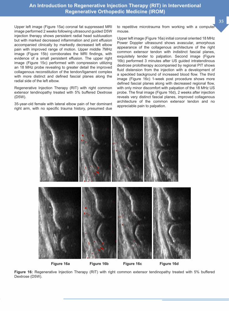
An Introduction to Regenerative Injection Therapy (RIT) in Interventional Regenerative Orthopedic Medicine (IROM)
35Upper left image (Figure 15a) coronal fat suppressed MRI image performed 2 weeks following ultrasound guided D5W injection therapy shows persistent radial head subluxation but with marked decreased inflammation and joint effusion accompanied clinically by markedly decreased left elbow pain with improved range of motion. Upper middle 7MHz image (Figure 15b) corroborates the MRI findings, with evidence of a small persistent effusion. The upper right image (Figure 15c) performed with compression utilizing an 18 MHz probe revealing to greater detail the improved collagenous reconstitution of the tendon/ligament complex with more distinct and defined fascial planes along the radial side of the left elbow.
Regenerative Injection Therapy (RIT) with right common extensor tendinopathy treated with 5% buffered Dextrose (D5W).
35-year-old female with lateral elbow pain of her dominant right arm, with no specific trauma history, presumed due
to repetitive microtrauma from working with a computer mouse.
Upper left image (Figure 16a) initial coronal oriented 18 MHz Power Doppler ultrasound shows avascular, amorphous appearance of the collagenous architecture of the right common extensor tendon with indistinct fascial planes, exquisitely tender to palpation. Second image (Figure 16b) performed 3 minutes after US guided intratendinous dextrose prolotherapy accompanied by regional PIT shows fluid distension from the injection with a development of a speckled background of increased blood flow. The third image (Figure 16c) 1-week post procedure shows more defined fascial planes along with decreased regional flow, with only minor discomfort with palpation of the 18 MHz US probe. The final image (Figure 16d), 2 weeks after injection reveals very distinct fascial planes, improved collagenous architecture of the common extensor tendon and no appreciable pain to palpation.
Figure 16a Figure 16b Figure 16c Figure 16d
Figure 16: Regenerative Injection Therapy (RIT) with right common extensor tendinopathy treated with 5% buffered Dextrose (D5W).

An Introduction to Regenerative Injection Therapy (RIT) in Interventional Regenerative Orthopedic Medicine (IROM)
36
Distal Biceps Tendon treated with Platelet Rich Plasma45-year-old neurologist injured left elbow while skiing presenting with pain and MRI showing high grade, partial thickness, and insertional distal biceps brachii tendon tear, with images left to right representing distal to proximal consecutive transverse MRI images (below).
Collage of transverse T2 weighted MRI images from 3 separate MRI studies over time showing regeneration and repair of the torn distal biceps brachii tendon (Figure 17a).
Treatment of partial distal biceps brachii tears often begins conservatively with splinting and physical therapy. Without knowledge of regeneration injection therapy (RIT), or due to patients’ common misunderstanding of the importance of avoidance of non-steroidal therapy and steroid injections, patients may eventually seek surgical repair for pain relief and to regain strength. However, repairs performed after 4 weeks from the time of injury may require use of a tendon graft because of muscle atrophy, loss of tendon length, and obliteration of the biceps tunnel, with high rate of neurovascular complications. Further, biomechanical studies have shown that surgical repair often does not return the short and long heads of the distal biceps brachii tendon to its native footprint, resulting in loss of supination torque hindering the restoration of biceps function- in this case, RIT (PIT, Prolotherapy and PRP) coupled with patient
specific Physical Therapy resulted in complete recovery.
Phase I: Inflammatory phase of healingThe initial pretreatment set of 5 images (Figure 17b) represents the earliest, immediate (1-week post injury), depicting near complete disruption of the expected “dark” smooth tubular appearance of the distal biceps tendon due to the high-grade tear involving the insertion point upon the radial tuberosity (far left image of the collage).
a. There is fluid signal in the antecubital fossa due to both vascular congestion (venous)and localized inflammation (extracellular fluid within the “3rd space”).
b. The injured but intact bicipital tendon aponeurosis is preventing complete musculotendinous retraction of the distal biceps tendon (not demonstrated, being superficial to this field of view).
Phase II: Repair/Proliferation phase of healinga. The second series of 5 images (Figure 17c) represent
the MRI appearance 2 weeks post RIT with both PIT and PRP injection (3 weeks post injury), and are just beginning to show reorganization, but without identifiable “dark” defined tendon morphology. The injection procedure was performed via a dorsal approach with wrist pronation, bringing the radial tuberosity insertion site of the distal biceps brachii tendon into a shallow, safe (compared to ventral antecubital fossa) approach.
Figure 17: 45-year-old neurologist injured left elbow while skiing presenting with pain and MRI.

An Introduction to Regenerative Injection Therapy (RIT) in Interventional Regenerative Orthopedic Medicine (IROM)
37
Figure 17: 45-year-old neurologist injured left elbow while skiing presenting with pain and MRI.
b. The fluid signals in these MRI images is a manifestation of both the inflammatory/reparative process (extra cellular fluid) accompanied by venous vascular congestion (intravascular blood).
c. Also, note the subtle muscle volume loss (atrophy) compared to the initial series only 3 weeks earlier, as the patient was utilizing a sling to immobilize the elbow during much of the day, and sarcopenia can occur rapidly. For this reason, we recommend a week to 10 days of relative rest after RIT (advising the patients-“if it hurts when you do that, don’t do that”), but initiating Physical Therapy by no later than 12 days after injection.
Phase III: Remodeling-consolidation/maturationa. The third MRI series of 5 images (Figure 17c) obtained
at 9 months post injection, reveal reconstitution of the now “dark”, tubular, functioning distal biceps brachii tendon.
b. The “dark” hypo intensity of the tendon is indicative of “new” type I collagen, and if we were to image the patient’s contra lateral intact biceps tendon it would probably appear “gray” intermediate in intensity, as most 45 year olds demonstrate type II and III collagen as a manifestation of the normal aging process.
c. The fluid signal in these images represents slow moving venous blood within the intact, normal veins and arteries occupying the antecubital fossa. The “dark” flow void signal can be seen as a manifestation of fast moving blood within the normal arterial structures in the region, the “bright” signal within the slower flowing veins.
KneeRight Knee pain after valgus rotatory stress injury treated with PRPUpper left, sagittal T1W image Figure 18a shows anterior cruciate ligament tear, with disruption of the normal longitudinal fiber orientation consistent with a high grade, partial thickness femoral insertion and interstitial ACL tear.
The top right sagittal T1W image (Figure 18b) from MRI examination 28 days after injection of PRP into the ACL sheath reveals reconstitution of the anterior cruciate ligament fibers in comparison to the prior tear and disruption of the longitudinal collagenous architecture of the ACL with disorientation of the fascicles (top of the left image- Figure 18a) seen on the initial study (28 days earlier). The patient underwent a very intense and sophisticated regimen of Manual Therapy, and returned to play within weeks of injury.
Tear and RIT repair of the medial collateral ligament complexUpper left coronal fat suppressed MRI image of the left knee prior to RIT (Figure 19a) reveals increased fluid signal and lamellation of the medial collateral ligament complex consistent with intermediate grade, partial thickness tear of the MCL. Upper middle image (Figure 19b) performed 2 weeks after ultrasound guided RIT with combined PIT and PRP reveals resolving MCL inflammation and improved function with resolution of pain. Upper right image (Figure 19c) performed 3 months after RIT reveals reconstitution of the MCL architecture as well as decreased extension

An Introduction to Regenerative Injection Therapy (RIT) in Interventional Regenerative Orthopedic Medicine (IROM)
38
of the fluid signal along the pes anserine complex (semitendinosus, gracilis and sartorius tendons attachment site along the anteromedial proximal tibial metaphysis). The
patient was pain free, with normal range of motion for age, having resumed normal activities of daily life.
Figure 18a Figure 18b
Figure 18: Right Knee pain after valgus rotatory stress injury treated with PRP.
Figure 19: 55-year-old gentleman with valgus stress injury of the left knee.
Knee with Regenerative Injection Therapy (RIT) Using Intra-Articular Hypertonic DextroseInitial fat suppressed MRI image (Figure 20a) reveals high grade partial thickness to complete femoral insertional and interstitial ACL tear (with permission from Dr. Brian Shiple). Upper right image (Figure 20b) performed after 4 monthly treatments with D25 to joint and D20 to ACL origin/ insertion w/ blind technique, reveals reconstitution of the ACL and decreased effusion and joint inflammation.
Above left image (Figure 20c) and above right image (Figure 20d) depict surface marking and correlative underlying anatomy of the knee. The upper left image depicts appropriate needle placement to access the ACL sheath. The ACL is “intra-articular” but is “extrasynovial”. That is, simple injection into the knee joint will not treat the ACL. Thus, the needle placement via the “eye” of the knee
provides best access to achieve a true intra ACL sheath administration of RIT.
Knee with PRP therapy with 5-year follow-upPreviously shown ACL tear in 18-year-old high school football player with 5-year follow-up:
The top right image from MRI examination 28 days later (Figure 21b) documents reconstitution of the anterior cruciate ligament fibers in comparison to the initial MRI revealing the tear and disruption of the longitudinal collagenous architecture of the ACL with disorientation of the fascicles seen on the previous study (28 days earlier).
Five year follow up after playing 4 years of college footballThe patient has played football for 4 years following a single regenerative injection therapy, but with intensive and

An Introduction to Regenerative Injection Therapy (RIT) in Interventional Regenerative Orthopedic Medicine (IROM)
39
specific Physical Therapy. The upper left and right sagittal MRI images (Figure 21c & d) reveal laxity and amorphous collagenous pattern of the ACL, although the patient was asymptomatic.
Sequential, consecutive proton density weighted, sagittal MRI images of the ACL reveal intact but wavy, redundant longitudinal collagenous architecture of both the anteromedial and posterolateral bundles of the ACL (upper 5 images- Figure 21e).
The redundancy and laxity of the anterior cruciate ligament
is accompanied by laxity of the posterior cruciate ligament (upper 2 images- Figures 21f & g) serving as indirect evidence of ACL insufficiency, although the patient was asymptomatic and playing at full speed. These “bothersome” imaging findings after RIT in clinically intact patients/players begs the question as to whether re- reinjection might be preventative regarding re-injury and to reduce the risk of osteoarthritis; as we know that ligaments are the static stabilizers of joints, and ligamentous instability is the culprit in OA (osteoarthritis).
Figure 20a Figure 20b
Figure 20: 35-year-old female martial arts artist with high grade, partial right ACL tear.
Figure 20c Figure 20d

An Introduction to Regenerative Injection Therapy (RIT) in Interventional Regenerative Orthopedic Medicine (IROM)
40
Figure 21a Figure 21b
Figure 21c Figure 21d
Figure 21e
Figure 21f Figure 21g
A longitudinal axis of femur is drawn before (dark) and after (lighter) the 20º knee flexion. Two points on the longitudinal axis are selected (A and B), where the axis is crossing the articular surface before and after the 20º displacement (flexion). Two more points A1 and B1 are selected on each longitudinal axis with an equal distance from point A and B. Lines are drawn between A and B, between A1 and B1. These lines are the serial displacements of each point.
The perpendicular bisectors are drawn from the center of the two serial displacement lines. Where these two perpendicular bisectors cross each other we find the centrode for this particular degree of knee displacement.
The articular surface of Femur is not circular and its shape together with the influence of connective tissues (particularly ligamentous static stabilizers) makes the center for successive positions change during knee motion. Frankel et al. describe in their study how they transposed 6 to 8 lateral knee roentgenograms from maximum extension to 90º of flexion in a non-weight bearing position. The study was done on the knee as if it was a planar mechanism, although they clearly describe their understanding of the importance of the knee’s conjunct and adjunct rotations. However, they noticed in comparing normal with pathological knees that the conjunct rotations were significantly reduced Figure 21: Knee with PRP therapy.

An Introduction to Regenerative Injection Therapy (RIT) in Interventional Regenerative Orthopedic Medicine (IROM)
41
The above illustrations (Figure 22) are drawings of a knee joint in the lateral view where the first image (bones in brown) is in neutral extension, and the lighter image representing
the femur in 20º of flexion. In all images the tibia is perfectly transposed, with a superimposed lighter image of the femur in 20º of flexion.
Figure 22: With permission from Ola Grimsby.
A longitudinal axis of femur is drawn before (dark) and after (lighter) the 20º knee flexion. Two points on the longitudinal axis are selected (A and B), where the axis is crossing the articular surface before and after the 20º displacement (flexion). Two more points A1 and B1 are selected on each longitudinal axis with an equal distance from point A and B. Lines are drawn between A and B, between A1 and B1. These lines are the serial displacements of each point.
The perpendicular bisectors are drawn from the center of the two serial displacement lines. Where these two perpendicular bisectors cross each other we find the centrode for this particular degree of knee displacement.
The articular surface of Femur is not circular and its shape together with the influence of connective tissues (particularly ligamentous static stabilizers) makes the center for successive positions change during knee motion. Frankel et al. describe in their study how they transposed 6 to 8 lateral knee roentgenograms from maximum extension to 90º of flexion in a non-weight bearing position. The study was done on the knee as if it was a planar mechanism, although they clearly describe their understanding of the importance of the knee’s conjunct and adjunct rotations. However, they noticed in comparing normal with pathological knees that the conjunct rotations were significantly reduced
in the presence of pathology, like lesions of the menisci and disruption of the static stabilizers of the knee (i.e. the cruciate and collateral ligaments).
Figure 23 demonstrates a knee flexed in 4 intervals where 0º has no velocity. At 10º the centrode is optimally positioned, and the surface velocity is parallel to the joint plane (red arrow). However, at 15º of flexion the centrode is positioned posterior to a perpendicular relationship to a point on the joint plane line. Thus, the angle between the dotted line and the joint plane is >90º, exposing the articular surface to compressive friction and compressive forces. The pathologic “migrating” axis of rotation is a function of ligamentous insufficiency, and the instability results in accelerated degeneration of the hypermobile joint. RIT coupled with patient specific Physical Therapy can reconstitute ligamentous integrity and reestablish a more optimal axis of rotation far better than surgical procedures.
The above diagram (Figure 24) reveals the importance of having intact static stabilizers (ligamentous integrity) which will maintain a normal axis of rotation, resulting in the optimal amount of vertical load stress and translational friction stress necessary for cartilage (and joint) health- physiologic loading.
Figure 21: Knee with PRP therapy.

An Introduction to Regenerative Injection Therapy (RIT) in Interventional Regenerative Orthopedic Medicine (IROM)
42
Figure 23: With permission from Ola Grimsby.
Putting a joint at rest may relieve pain, but the joint will wither. Further, taping a “weak” ankle only puts at risk the other two joints in the ipsilateral lower extremity. Articular under load succeeds primarily in deleteriously reducing the cellular activity, causing shrinking of the matrix, reduced joint stability and secondary absorption of the tissue.
On the other hand, putting a joint with loss of ligamentous stability under normal stress results in overloading of the cartilage lining the joint. During abnormal static compression and hypermobility, there is an even more severe reduction in the cellular synthesis. As cells die, the reduction in matrix and joint dysfunction cause permanent degeneration- osteoarthritis is caused by ligament laxity.
ACL Tear Treated with RITUpper left sagittal MRI image (Figure 25a) shows interstitial tear and attenuation (thinning) of the anteromedial and posterolateral bundles of the ACL. Upper right sagittal MRI image (Figure 25b) is performed 3 months following RIT with a combination of PIT and PRP injection and patient specific physical rehab. In addition to resolving clinical symptoms, the anterior cruciate ligament demonstrates thickening with reconstitution of the previously torn bundles along the femoral attachment to the posterior aspect of the medial surface of the lateral femoral condyle.
ACL tear treated with RITTransverse MRI images 6 months apart, revealing thickening
and reconstitution of the anterior cruciate ligament (top right image- Figure 26b) compared to the appearance of the interstitial tear of the ACL on the initial post trauma MRI study (top left image- Figure 26a). After RIT with PIT and PRP, anterior cruciate ligament demonstrates a thickened, more normal appearance of the femoral insertion along the posterior aspect of the medial surface of the lateral femoral condyle. Evidence of reconstitution of the longitudinal collagenous architecture of the anteromedial and posterolateral bundles of the anterior cruciate ligament were accompanied by complete return to normal activities 6 months after initial injury.
Bucket Handle Tear of the Medial Meniscus Treated with RIT1st MRI-The upper 4 images (Figure 27a & 27b- upper first and second- are sagittal fat suppressed MRI images, Figure 27c & 27d- upper third and fourth- are coronal fat suppressed MRI images) performed immediately after the first valgus stress injury reveals the complex, bucket handle tear of the left medial meniscus associated with meniscocapsular junction injury. Under the close scrutiny of his Physical Therapist, the patient’s symptoms resolved within weeks of the RIT, returning to full court basketball within 6 months of the injury.
2nd MRI-These upper 4 images from a repeat MRI after a second valgus stress injury of the same (left) knee 6 months later (Figure 28a- 28d corresponding MRI images to

An Introduction to Regenerative Injection Therapy (RIT) in Interventional Regenerative Orthopedic Medicine (IROM)
43
the first study). Evidence of interval “fusion” to reconstitute a relatively functional medial meniscus from the original fragmented bucket handle tear, but with interval traumatic
MCL sprain, fortunately without re-injury of the medial meniscus. RIT and Physical Therapy again allowed the patient to return to play 6 months after second injury.
Figure 24: With permission from Ola Grimsby.
Figure 25: ACL tear treated with RIT.
Figure 25a Figure 25b
Oblique flap tear of the medial meniscus, with chondromalacia from meniscal insufficiency, patellar glide pathology and medical collateral ligament injury treated with RITUpper left image obtained on 10-21-10 (Figure 29), coronal
fat suppressed image revealing an oblique flap tear of the medial meniscus with inflammation/sprain of the overlying medial collateral ligament complex. Upper right image (Figure 30) performed 3 months after RIT reveals interval decreased conspicuity of the amorphous and linear intrameniscal hyperintensity and inflammation along the

An Introduction to Regenerative Injection Therapy (RIT) in Interventional Regenerative Orthopedic Medicine (IROM)
44
posterior horn/root junction compared to prior MRI of 10-21-10, now demonstrating equivocal extension to the inferior articular surface. Now, without evidence of definite meniscal macro tear, and with decrease in the degree of Fairbanks
changes of meniscal insufficiency (when present includes: squaring of the medial femoral condyle, marginal ridging, narrowing of the joint space and pseudo extrusion of the meniscus). The patient had resumed triathletic competition.
Figure 26: ACL tear treated with RIT.
Figure 27: Bucket handle tear of the medial meniscus treated with RIT.
Figure 28
Figure 29 Figure 30

An Introduction to Regenerative Injection Therapy (RIT) in Interventional Regenerative Orthopedic Medicine (IROM)
45
Multitude of a series of knee injuries in a world class triathlete with repair successfully treated with RIT and Physical Therapy
In addition to the ultrasound guided RIT, PRP injection was also performed directly into the cruciate and collateral ligamentous complexes to rejuvenate the static stabilizers of the joint and to optimize the axis of rotation. The above
coronal MRI images (Figure 31a- 31d- yellow arrows) document the remodeling of the localized area of Class III/IV chondromalacia involving the posteromedial weight bearing surface of the medial femoral condyle over the long course of therapy. The surface features of the hyaline cartilage become smoother and more uniform and the subchondral osteoedema-like signal diminishes, with the knee joint becoming clinically more stable.
Figure 31: Chondromalacia from medial meniscal insufficiency.
Tear and PRP repair of the medial collateral ligament complex
Above coronal fat suppressed MRI images (Figures 32a- 32c) documents the decreasing fluid signal and lamellation of the medial collateral ligament complex consistent with continued resolution of the MCL inflammation/sprain. In addition, decreased extension of the fluid signal along the pes anserine complex (semitendinosus, gracilis and sartorius tendons attachment site along the anteromedial proximal tibial metaphysis).
Patellar glide pathology requiring combined surgical plica resection in conjunction with RIT
Knee hemarthrosis from original injury (upper left image- Figure 33a), treated with RIT, including intra-articular and intra-ACL sheath PRP. Second MRI 4days later following interval arthroscopic plica resection (upper middle image- Figure 33b) revealing decreased joint effusion and decreased inflammation of the medial patellofemoral ligament (key static stabilizer of the patellar glide mechanism). Third
MRI 2½ months later (Figure 33c) in conjunction with improved clinical symptoms shows continued decrease in joint effusion and tightening of the peripatellar retinacula and capsular stabilization mechanism (improving patellar tracking).
Transverse MRI images (top images normal, complete field of view, bottom images magnified) show progressive remodeling of the Class IV chondromalacia involving the femoral notch comparing the initial study 10-17-10 (upper left images- Figure 34a) with the 4 days post RIT study of 10-21-10 (upper middle images- Figure 34b), and with the third MRI performed 2 ½ months later on 01-13-11 (upper right images-
Figure 34c). There is evidence of interval remodeling and “filling in” of the longitudinal cartilage fissure involving the femoral notch, with persistent subchondral osteoedema like signal (representing ongoing “regenerative” inflammation). The patient’s clinical symptoms mirrored the MRI improvement.
Figure 32: Coronal fat suppressed MRI.

An Introduction to Regenerative Injection Therapy (RIT) in Interventional Regenerative Orthopedic Medicine (IROM)
46
Figure 33: Knee hemarthrosis from original injury.
Figure 34: Transverse MRI.
Lateral collateral ligamentous complex injury treated with RIT
Upper left coronal, fat suppressed MRI image Figure 35a shows LCL tear with marrow edema consistent with accompanying micro fracture (bone bruise) of the lateral compartment of the knee.
With the patient’s clinical symptoms resolving, 6 weeks after the RIT (upper right image-Figure 35b) MRI reveals interval decreased bone marrow signal indicative of healing of the micro fracture accompanied by decreased T2 signal with return to normal lamellation (layering) of the lateral collateral ligament complex. In this clinical context, these MRI findings are consistent with interval collagen
reorganization and “regenerative” inflammation of the LCL proper and healing of the bone bruise (micro fracture) of the lateral compartment of the knee following RIT and Physical Therapy.
Acute onset of medial knee pain without recent trauma successfully treated with RIT
45-year-old female with diffuse bilateral knee pain, recently growing much worse on the left. Remote history of left patella dislocated in 1993 with two subsequent prior knee surgeries, also several years ago. Acute onset of left knee pain without recent injury. The patient also had mid back pain radiating into the left subscapular region, along with bilateral leg pain with numbness.

An Introduction to Regenerative Injection Therapy (RIT) in Interventional Regenerative Orthopedic Medicine (IROM)
47
Figure 35: Upper left coronal, fat suppressed MRI.
The initial diagnostic, coronal, fat suppressed MRI of the right knee (upper left image- Figure 36a) in this clinical context (middle aged female without recent trauma history) and MRI appearance is consistent with spontaneous osteonecrosis of the knee (SONK-diffuse marrow edema). Immediately following the initial MRI, the patient underwent targeted RIT with a combination of RIT and platelet rich plasma (PRP) injection, with upper right image (Figure 36b) performed 6 weeks later revealing improved MRI appearance accompanied by resolution of patient’s knee and upper back symptoms (in conjunction with a remarkable “body wide” Physical Therapy regimen).
6 weeks after RIT therapy (Figure 36b), the patient was clinically cleared to return to activities of daily life without restrictions. Although the MRI revealed interval decreased marrow signal, there was interval increase in the amount of circumferential subcutaneous edema overlying the knee. In the clinical context of improving symptoms, the MRI findings are consistent with “regenerative” inflammation, with intact of the underlying collateral ligamentous complexes. The marked improved MRI appearance of the marrow signal is consistent with interval resolution of the suspected spontaneous osteonecrosis (SONK).
Figure 36: The initial diagnostic, coronal, fat suppressed MRI of the right knee.

An Introduction to Regenerative Injection Therapy (RIT) in Interventional Regenerative Orthopedic Medicine (IROM)
4867-year-old gentleman with chronic knee pain and painful scar from remote open knee surgery for repair of a medial collateral ligament tear treated with PIT
The upper left image Figure 37a shows skin thickening corresponding to the epidermal surgical scar, with no appreciable Power Doppler flow. Upper right image (Figure 37b) obtained 5 minutes after regional PIT and direct injection of buffered 5% Dextrose into the scar reveals hyperemia within both the scar (epidermal) and underlying dermis. The patient had complete relief of pain, and after 3 more sessions returned to normal activities of daily life for a 67-year-old golfer. The superficial scar was apparently the pain generator, not underlying spinal degeneration or deeper sequela of prior surgical fusion procedure.
Medial collateral ligamentous complex injury treated with RIT
Teenage football player with valgus rotatory stress injury, not treated with RIT initially, followed by re injury that was treated with RIT.
Upper left coronal, fat suppressed MRI image (Figure 38a) revealing consequences of initial valgus stress injury of the left knee in 2008; intermediate grade MCL sprain with traction related femoral insertional micro fracture.
Upper middle image (Figure 38b) one year later, immediately after a second valgus stress injury, reveals intermediate to high grade recurrent MCL sprain with more extensive trabecular micro fracture related to traction stress on the femoral insertional site of the MCL.
The upper right image (Figure 38c) 4 weeks later, after PIT injection, reveals reconstitution of the medial collateral ligament complex, with resolving underlying traction related trabecular micro fracture. This young football player had returned to play two weeks earlier in his state final football game which was only two weeks after his most recent injury/RIT injection. Although the MRI was not “normal”, the player’s “trainers” determined he was ready to return to play clinically, and he rushed for a state record for a playoff game.
Patellar tendon tear- jumper’s knee treated with RIT
Upper left and second proton density sagittal MRI images (Figures 39a & 39b) reveals hypertrophic, intermediate grade, partial thickness, insertional tear involving the teno osseous junction of the patellar tendon with the lower pole of the patella (so-called “jumper’s knee” or inferior patellar apicitis).
Third image (Figure 39c) Power Doppler ultrasound image revealing hyperemia within and along the patellar tendon insertion extending into the underlying deep infrapatellar Hoffa fat pad. This in consistent with neovascularity associated with chronic tendinopathy.
The 4th image (Figure 37d) obtained 5 minutes after RIT
therapy reveals slightly increased Power Doppler flow, associated with partial relief of pain. The RIT therapy consisted of combined regional PIT and intralesional PRP.
Upper left and middle MRI images (Figures 40a & b) performed 4 weeks after PIT and intra- tendinous and peripatellar tendon injection of PRP reveals persistent “abnormal” MRI appearance including interval increased intra and peritendinous fluid signal despite resolution of symptoms.
The 3rd image (Figure 40c) reveals persistent Power Doppler evidence of hyperemia, again, with no pain to palpation with the ultrasound probe.
This case points out the fact that “the pictures aren’t the patient”. The post RIT imaging findings appear to be revealing not “degenerative” but “regenerative” (good) inflammation, as the patient reported resolution of symptoms and had returned to playing basketball. The case makes the important point that imaging studies must be viewed in context with the patient’s clinical symptoms, as the imaging studies may not return to a completely “normal” appearance for months (even years) after the RIT, despite resolution of the patient’s symptoms.
5 years after RIT, there has been complete healing of the patellar tendon, with return to normal of the MRI appearance (Figures 41a & 41b) and Power Doppler Ultrasound (Figure 41c) appearance revealing restoration of the normal “cable-like” longitudinal collagenous architecture and absence of hyperemia, correlating with the patient reporting no recurrent symptoms.
Chronic anterior knee pain with patellar tendinopathy treated with RIT
a. Initial sagittal fat suppressed proton density weighted MRI (Figure 42a) reveals abnormal increased signal with amorphous appearance of the patellar tendon.
b. Upper right MRI image (Figure 42b) performed 4 weeks after RIT. Despite resolution of the patient’s anterior knee pain and patellar tendinopathy symptoms, there has been interval increase in the MRI fluid signal along the prepatellar superficial subcutis space and essentially unchanged insertional signal involving the teno-osseous junctions of the quadriceps and patellar tendons with the superior and inferior poles of the patella, respectively. This case is yet another example of why MRI cannot be utilized without clinical correlation to determine healing.
c. Although the patient had been clinically cleared to resume normal activity, the MRI paradoxically appears “worse” based on the increased fluid signal which following RIT is not due to prepatellar “degenerative inflammation” but due to “regenerative inflammation”, which can be seen extending inferiorly along the infrapatellar superficial subcutis space, not extending into the deep infrapatellar Hoffa fat space.

An Introduction to Regenerative Injection Therapy (RIT) in Interventional Regenerative Orthopedic Medicine (IROM)
49
Figure 37: Repair of a medial collateral ligament tear treated with PIT.
Figure 38: Upper left coronal, fat suppressed.
Figure 39: Upper left and second proton density sagittal MRI.

An Introduction to Regenerative Injection Therapy (RIT) in Interventional Regenerative Orthopedic Medicine (IROM)
50
Figure 40: Persistent Power Doppler evidence of hyperaemia.
Figure 41: Power Doppler Ultrasound.
Figure 42: Initial sagittal fat suppressed proton density weighted.

An Introduction to Regenerative Injection Therapy (RIT) in Interventional Regenerative Orthopedic Medicine (IROM)
51Patellofemoral Glide Pathology (Patellar Tracking Dysfunction) Treated with RIT in Conjunction with Rigorous Physical Therapy32-year-old female college basketball coach, with history of borderline patella alta, and history of ACL injury when playing high school basketball as a teenager.
The following MRI images for this patient are all performed on an Upright MRI machine, allowing imaging while weight bearing and performing flexion and extension maneuvers.
The upper consecutive, sagittal, proton density MRI images reveal borderline patella alta (Figure 43a), which predisposes to patellar glide pathology. The sagittal images revealed hypertrophy of the patellar tendon (Figure 43b), with knee joint effusion and fluid distension of the deep infrapatellar bursa (Figure 43c).
In the upright, standing, weight bearing position (top two images- Figures 43d & 43e), there is sulcus retention
insufficiency with moderate lateral patellar subluxation, without tilt. Also noted is thinning and chondromalacia of the hyaline cartilage covering the apposing articular surfaces of the lateral patellar facet and lateral femoral trochlear ridge. Traction related enthesophyte along the insertion site of the thickened lateral peripatellar retinaculum. This constellation of imaging features consistent with sequela of chronic excessive lateral pressure (ELP), exacerbated by borderline patella Alta.
RIT performed, including PIT and intra-articular and intra-cruciate PRP. Immediate following RIT, upright, weight-bearing, kinematic transverse, proton density MRI images performed, documenting patellar repositioning from 0 (Figure 43f) to 15 degrees (Figure 43g) to 30 degrees (Figure 43h) of flexion. As is typical in patients with high riding patella, when the knee is in full extension (0 degrees of flexion- Figure 43f), the patella alta means the patella is positioned within a cephalad, shallow portion of the femoral notch, predisposing to lateral tracking and subluxation.
Figure 43a Figure 43b Figure 43c
Figure 43d Figure 43e

An Introduction to Regenerative Injection Therapy (RIT) in Interventional Regenerative Orthopedic Medicine (IROM)
52
Figure 43f Figure 43g Figure 43h
Figure 43: The upper consecutive, sagittal, proton density MRI.
Figure 44a Figure 44b
Figure 44c Figure 44d Figure 44e

An Introduction to Regenerative Injection Therapy (RIT) in Interventional Regenerative Orthopedic Medicine (IROM)
53
Figure 44f Figure 44g
Figure 44: Conventional radiographs revealed asymmetric lumbosacral and thoracolumbar transitional anatomy.
Patellar tracking is defined as the motion of the patella relative to the femur or femoral groove throughout knee flexion and extension. Abnormalities of tracking (mal tracking) are thought to relate to many disorders of the patellofemoral joint, predisposing to osteoarthritis of the knee. The patellar glide may be difficult to observe and classify when using only supine, non-weight bearing imaging studies. The reporting of patellar tracking is affected significantly by basic definitions of coordinate systems and reference points. The method of muscle loading, range, and direction of knee motion, uses of static or dynamic measurement techniques and tibial rotation also affect the results obtained.
There is general agreement that the patella translates medially in early knee flexion (as in this patient) and then translates back laterally with extension. Regarding patellar tilt, results are less consistent, especially in vivo and the results for patellar rotation are highly variable. In the current upright kinematic study of this patient, there is lateral subluxation of the patella in static upright extension and in 0-degree flexion images, with medial translation (normal repositioning) of the patella into the femoral notch occurring with 15 and 30 degrees of knee flexion.
Lumbar SpineLumbar Spine treated with Platelet Rich Plasma
28-year-old tennis professional presents with history of chronic low back pain recently made worse following a fall while playing tennis.
Conventional radiographs revealed asymmetric lumbosacral and thoracolumbar transitional anatomy (Figures 44a & 44b) accompanied by sigmoid configure scoliosis and pelvic tilt associated with relative shortening of the right lower extremity.
Initial MRI (upper left image- Figure 44c) did not reveal (except in retrospect) the occult right L5 laminar fracture, easily seen on the CT (upper middle image- Figure 44d) performed to guide needle placement for the PRP injection. CT depicts the occult right L5 laminar fracture, and documents needle placement (upper right image- Figure 44e) allowing precise injection of the PRP solution. The laminar fracture was the etiology of the patient’s acute symptoms, which remitted within days of the PRP injection procedure.
However, the patient’s chronic symptoms were due to SI joint stress created by the asymmetric transitional lumbosacral anatomy. Upper left image (Figure 44f) depicts the right sacroiliac joint, with the upper right image (Figure 44g) depicting the left sacroiliac joint, documenting placement of the 25 gauge spinal needles to allow precise injection of the PRP solution into both the capsulosynovial lined (anteroinferior) and predominantly collagenous/ligamentous (posterosuperior) portions of the SI joints. After relentless Physical Therapy focusing on recovering SI joint stability, the patient returned to playing tennis.
Lumbar Spine treated with Platelet Rich Plasma
58-year-old pain medicine physician with history of acute onset LBP after jogging, without radiculopathy.
Upper left image (Figure 45a) is a lateral localizer from a CT study of the spine. The upper middle image is a sagittal T1W MRI image (Figure 45b) and the upper right image is a sagittal T2W MRI image (Figure 45c).
All three sagittal L spine images (Figures 45a- 45c) reveal hyperdynamic curvature of the spinal axis as a manifestation of hyperlordosis and the two coronal images (Figures 45d & e) document the presence of a shallow dextroconvex rotoscoliosis.

An Introduction to Regenerative Injection Therapy (RIT) in Interventional Regenerative Orthopedic Medicine (IROM)
54Pelvic tilt also noted on the coronal images (Figures 45d & e), consistent with leg length discrepancy, with the right leg being functionally/effectively shorter.
L4-5
Figure 45f is a collage, comprised of two transverse T2W MRI (top row), two CT images (middle row) with 5 consecutive sagittal T2W MRI images (bottom row).
Selective injection of platelet rich plasma (PRP) is performed along the left L4 lamina and left L4 pars interarticularis (Figure 45g) and within the effusion contained within the hypertrophied left L4-5 facet joint (Figure 45h). Injection of PRP was also performed within and along the L3-4 and L4-5 posterior interspinous ligaments and spaces, and along the rightward L4 lamina, pars interarticularis and L4-5 facet joint (not shown).
L5-S1
Figure 45i is a collage, comprised of one transverse T2W
MRI image (top row, left image), one CT image (top row, right image) with 5 consecutive sagittal T2W MRI images (bottom row).
Right L5 pars fracture (Figure 45i) with fragmentation and vacuum phenomenon of the hypertrophied right L5-S1 facet with evidence of partial fusion of the left L5-S1 facet (upper middle and right CT images- Figures 45j & k).
Selective injection of platelet rich plasma (PRP) is performed along the right L5 lamina, right L5 pars interarticularis and within the effusion contained within the fragmented/hypertrophied right L5-S1 facet joint (upper 2 CT transverse images- Figures 45l & m). Injection of PRP was also performed within and along the L5-S1 posterior interspinous ligaments and space, and along the leftward L5 lamina, pars interarticularis and left L5-S1 partially fused facet joint (not shown). Two days following the PRP injection on Friday night, the physician was nearly pain free, and able to go to Church on Sunday and return to work on Monday (the third day post injection).
Figure 45a Figure 45b Figure 45c
Figure 15a Figure 15b Figure 15c
Figure 45d Figure 45e

An Introduction to Regenerative Injection Therapy (RIT) in Interventional Regenerative Orthopedic Medicine (IROM)
55
Figure 45f Figure 45g Figure 45h
Figure 45i Figure 45j Figure 45k
Figure 45l Figure 45m
Figure 45: Lumbar Spine treated with Platelet Rich Plasma.

An Introduction to Regenerative Injection Therapy (RIT) in Interventional Regenerative Orthopedic Medicine (IROM)
56
Hamstring Insertional Tear Treated with PRP22-year-old female Olympic pole vaulter with chronic ischial tuberosity pain with acute exacerbation after a national track meet.
Transverse (Figure 46a) and coronal (Figure 46b) fat suppression MRI images reveal an abnormal appearance of the left hamstring complex. Insertional and interstitial
tendinopathy/tear involving the tenoosseous junction of the left hamstring complex, including from the most medial to lateral, the biceps femoris, semitendinosus, and semimembranosus insertions. No muscular atrophy is appreciated. The patient was able to return to pole vaulting two months after receiving PRP injection coupled with intensive and patient specific Physical Therapy.
Figure 46a Figure 46b
Figure 46: Transverse and coronal fat suppression MRI.
Hamstring myotendinous junction tear
15-year-old football player with acute onset posterior thigh pain.
Upper 4 image collage (Figure 47a) consists from top row, left to right, coronal STIR and T2W images, with bottom row, left to right, transverse STIR and proton density images, performed 08-07-08.
The images reveal a Grade I insertional tear accompanied by a Grade II myotendinous junction tear of the right hamstring complex.
Figure 47b is a collage with single row of transverse CT images obtained to rule out an avulsion fracture of the right hamstring insertion. Prior to performing the diagnostic portion of the study, a needle was placed for PRP injection and is within the field of view, performed 08-07-08.
Figures 47c coronal STIR image performed one week after the PRP injection revealed remodeling of the myotendinous junction tear, with the patient reporting improving symptoms. Figure 47d performed the second week post injection (one week after Figure 47c) reveals continued healing of the tear involving the myotendinous junction with decreased surrounding fluid signal.
Figure 47e transverse and coronal STIR images performed one day from Figure 47d MRI due to reinjury from starting back playing before the hamstring was ready. The MRI performed the day before the injury described “improved MRI appearance”, which compelled the patient’s football coach to encourage the patient to play in the weekly game. The patient was only moderately better clinically, but the coaching staff used the “optimistic” MRI findings to compel the patient to play. Figure 47f transverse and coronal STIR images performed one week from Figure 47e (and only 3 weeks from the PRP injection) reveal the hamstring tear has an “improved” MRI, although not verbalized in the report, and the patient sat out the game that week.
Figure 47g, from left to right, coronal and transverse STIR images performed one week from Figure 47f (and 4 weeks from PRP injection) reveals continuing MRI appearance, but the patient had remained symptomatic and continue to sit out of full contact football. Figure 47h, from left to right, coronal and transverse STIR images performed one week from Figure 475g (and 5 weeks from PRP injection) reveals interval worsening of the MRI appearance. The patient described having to undertake a long drive to visit a recruiting college that exacerbated the hamstring symptoms.

An Introduction to Regenerative Injection Therapy (RIT) in Interventional Regenerative Orthopedic Medicine (IROM)
57
Figure 47i, from left to right, coronal and transverse STIR images performed one week from Figure 47h (and 6 weeks from PRP injection) reveals improved MRI appearance, and the patient had remarkably improved symptoms due to a change from the high school trainers to a world class Manual Therapist who recommended to the parents (and against the coaching staff’s wishes) that the patient should continue to sit out of full contact football.
Figure 47k, from left to right, coronal and transverse STIR images performed two weeks from Figure 47j (and 8 weeks from PRP injection) reveals continued improved MRI appearance but without MRI resolution of the myoedema and interstitial fluid signal. Despite the MRI not appearing completely “normal”, and the patient had complete resolution of symptoms and the patient was allowed to resume full contact football based on the clinical opinion of the Manual Therapist.
Figure 47l, from left to right, coronal and transverse STIR images performed one week from Figure 47k (and 9 weeks from PRP injection) reveals continued improved MRI appearance but again without complete MRI resolution of the myoedema and interstitial fluid signal. This is another case demonstrating that despite the MRI not returning to completely “normal”, decisions on when to allow patients with MSK injuries to return to full activities must be made on a clinical basis by an experienced Physical Therapist willing not to be swayed by outside influences.
It is notable that neither MRI nor ultrasound is highly sensitive in defining an abnormality of the hamstring as the responsible pain generator in many patients. In fact, MR studies and ultrasound studies are often interpreted as “normal”. This underscores the importance of having a skilled clinician for patient assessment, referral to ultrasound imaging, and referral for imaging-guided injection.
Figure 47a
Figure 47b

An Introduction to Regenerative Injection Therapy (RIT) in Interventional Regenerative Orthopedic Medicine (IROM)
58
Figure 47c Figure 47d
Figure 47e Figure 47f
Figure 47g Figure 47h
Figure 47i Figure 47j

An Introduction to Regenerative Injection Therapy (RIT) in Interventional Regenerative Orthopedic Medicine (IROM)
59
Figure 47: Hamstring myotendinous junction tear.
Figure 47k Figure 47l
Low Back and SI Joint Pain Treated with Caudal Epidural Buffered 5% DextroseFigure 48a is a diagrammatic representation to be correlated with Figure 48b depicting the actual surface anatomy of the needed to perform a caudal injection utilizing the sacral hiatus.
Rapid analgesic effect was achieved with buffered 5% Dextrose (glucose) instillation into the caudal epidural space (Figure 48c). Caudal Epidural is performed utilizing a 2 ½ inch, 25-gauge needle with fluoroscopic or ultrasound guidance if the sacral hiatus is not readily palpable. After
the needle is place, injection 10-20 cc of Dextrose 5% in Sterile Water (neutral pH D5W) into the sacral hiatus can result in almost instant and near complete analgesia of low back and lower limb pain. 10 cc’s flows from the sacral hiatus (S5) to the Lumbosacral junction (L5-S1). With 20 cc’s, the dextrose baths the epidural space to the L3-4 level. Whether injecting 10 or 20 cc’s, the Dextrose typically flows around the perineural sheaths emanating through the neural foramina along the anterior surface of the sacrum. Treatment- deemed a “sweet caudal”- can be given weekly, and then reduced to biweekly, then monthly; on average, six treatments may be needed for cure, as was the case in this patient.
Figure 48a Figure 48b
Figure 48c
Figure 48: (a) is a diagrammatic representation to be correlated with (b) depicting the actual surface anatomy of the needed to perform a caudal injection utilizing the sacral hiatus.

An Introduction to Regenerative Injection Therapy (RIT) in Interventional Regenerative Orthopedic Medicine (IROM)
60
Superior Cluneal Nerve Entrapment induced Low Back Pain and “Iliolumbar Syndrome”25-year-old female patient presented with clinical history of “Low Back Pain” recalcitrant to multiple steroid injection and RF ablation procedures. Clinical exam revealed marked sensitivity to light palpation over the medial aspects of the sacroiliac joints. Due to failure of the multiple prior facet and SI joint injection and ablation procedures, RIT with combination buffered Dextrose and PRP was performed. The patient’s acute symptoms were immediately improved with PIT along the distribution of the superior cluneal nerve. And PRP injection into the SI joints and L4-5 and L5-S1 facets provided stabilization such that the patient’s chronic symptoms abated after 6 months.
This scenario has come to be deemed “Pseudo Sciatica”, and when recognized and appropriately treated can literally change lives. The anatomic explanation is based on the fibro-osseous tunnel of the medial crest of the iliac wing (two finger breaths- 8 cm.- off midline and above the posterior superior iliac spine- PSIS) through which the medial branch of the superior cluneal nerve becomes entrapped.
The medial branch of the superior cluneal nerve crosses over the left iliac crest through a fibro- osseous tunnel, within which it can become entrapped. Nerves are not elastic, and if fact has 70% as much collagen as ligaments, thus when stretched while entrapped can tear. There are two osteofibrous tunnels through which cluneal plexus and the T10 iliohypogastric nerve(s) can become entrapped along their respective grooves over iliac crest.
Figures 49b & 49c are diagramatic and 47d demonstrates marking the skin surface of a patient with cluneal nerve entrapment syndrome, so-called pseudosciatica. PIT can be performed utilzing palpation and marking techniques, althought the patients will often “guide” the injectionist by their positive response to focal palpation for active trigger points. Ultrasound can also be of benefit, especially in larger patients. In this patient, PIT alone resulted in complete resolution of pain and stiffness after Physical Therapy.
Occasionally, an isolated classic supperior cluneal nerve entrapment syndrome occurs, but more often there is an accompanying inferior cluneal and/or posterior femoral cutaneous nerve entrapment with associated groin pain (Figures 49e-49h).
Figure 49a
Figure 49b Figure 49c Figure 49d

An Introduction to Regenerative Injection Therapy (RIT) in Interventional Regenerative Orthopedic Medicine (IROM)
61
Figure 49e Figure 49f Figure 49g Figure 49h
Figure 49: Diagrammatic and demonstrates marking the skin surface of a patient with cluneal nerve entrapment syndrome.
a. Pain relief when treated with pit typically lasts 1-4 weeks, then may recur but usually not as severe.
b. Patient will have good days and bad days (almost a toggle switch, on-off effect)
c. Monitor for pain that has recurred versus areas that were inititally missed.
d. Monitor for new areas since the prior treatment.
e. Exercise to tolerance.
76-year-old gentleman with failed low back surgery and persistent low back pain specifically corresponding to a large midline surgical scar. Imaging findings in this clinical context favors TLF (thoracolumbar fascia) syndrome.
The upper left image Figure 50a shows irregular skin thickening corresponding to the epidermal surgical scar, with low level Power Doppler flow within the epidermal layer. The marked hypoechoic appearance below epidermal scar is due to “resorption” of the ultrasound beam by the dermal scar tissue below the skin level. A “dark” appearance on gray scale ultrasound is created by acceleration (fluid), reflection (large acoustic impedance mismatch), refraction (bending of the beam around curved surfaces) and resorption of the beam (as in this case, the ultrasound energy is absorbed by scar tissue).
Upper right image (Figure 48b) obtained 5 minutes after regional PIT and direct injection of buffered 5% Dextrose into the epiderma and dermal scar reveals hyperemia within both the epidermal and underlying dermal layers. The patient had complete relief of pain, and after 3 more sessions returned to normal activities of daily life for a 67-year-old golfer. The superficial scar was apparently
the pain generator, not underlying spinal degeneration or deeper sequela of prior surgical fusion procedure.
Notice the injection marks in the cheek (upper right) and not in the naso-labial folds. By augmenting the cheeks, the nasolabial folds are “pulled out” while creating a higher, more glamorous “cheek bone.” Injecting platelet rich fibrin matrix, a different technique than Juvederm must be used.
Also, notice that she has a gull shaped brow (in the after photo on the right) from the filler.
This woman only asked to have treated the naso-labial folds by injecting those and did not even notice or know that all the rest could treat
a. Straighten and narrow the nose,
b. Augment the cheeks,
c. Augment the brow,
d. Straighten her mouth,
e. Take the tired out of her eyes by filling the tear troughs, and
f. Finally, as a side-effect of all the rest, make her naso-labial folds disappear.
Communicate with the Patient and Key Health Care Givers
Ask about the patient triad of
a. Ideas about the “correctness” of the working diagnosis.
b. Concerns about the proposed therapy.
c. Expectations of the short and long term for the proposed health care plan.
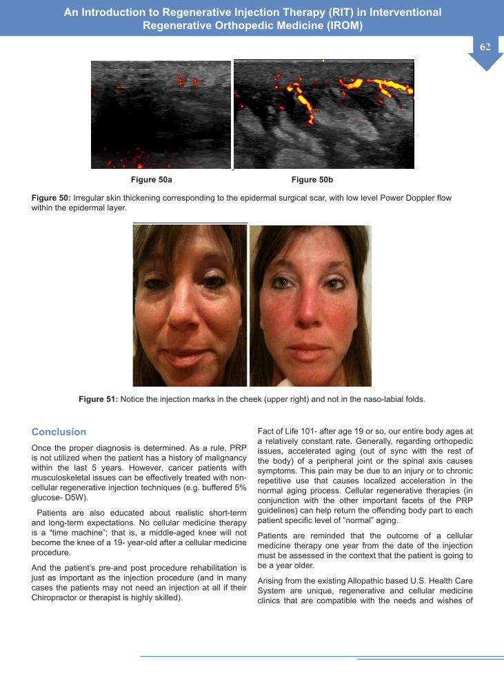
An Introduction to Regenerative Injection Therapy (RIT) in Interventional Regenerative Orthopedic Medicine (IROM)
62
Figure 50a Figure 50b
Figure 50: Irregular skin thickening corresponding to the epidermal surgical scar, with low level Power Doppler flow within the epidermal layer.
Figure 51: Notice the injection marks in the cheek (upper right) and not in the naso-labial folds.
ConclusionOnce the proper diagnosis is determined. As a rule, PRP is not utilized when the patient has a history of malignancy within the last 5 years. However, cancer patients with musculoskeletal issues can be effectively treated with non-cellular regenerative injection techniques (e.g. buffered 5% glucose- D5W).
Patients are also educated about realistic short-term and long-term expectations. No cellular medicine therapy is a “time machine”; that is, a middle-aged knee will not become the knee of a 19- year-old after a cellular medicine procedure.
And the patient’s pre-and post procedure rehabilitation is just as important as the injection procedure (and in many cases the patients may not need an injection at all if their Chiropractor or therapist is highly skilled).
Fact of Life 101- after age 19 or so, our entire body ages at a relatively constant rate. Generally, regarding orthopedic issues, accelerated aging (out of sync with the rest of the body) of a peripheral joint or the spinal axis causes symptoms. This pain may be due to an injury or to chronic repetitive use that causes localized acceleration in the normal aging process. Cellular regenerative therapies (in conjunction with the other important facets of the PRP guidelines) can help return the offending body part to each patient specific level of “normal” aging.
Patients are reminded that the outcome of a cellular medicine therapy one year from the date of the injection must be assessed in the context that the patient is going to be a year older.
Arising from the existing Allopathic based U.S. Health Care System are unique, regenerative and cellular medicine clinics that are compatible with the needs and wishes of

An Introduction to Regenerative Injection Therapy (RIT) in Interventional Regenerative Orthopedic Medicine (IROM)
63humankind- the change we have been waiting for. These centers are designed not only to encourage but to facilitate human interaction. There are “great rooms” in each clinic within which patients undergo rehab and treatments, able to interact and commiserate with one another. Americans had lost track of their history and with their human nature. Quite simply, people need a health care system that allows them to care for themselves and each other in a better way and to promote “Good” in society.
These “Medical Malls” are very colloquial, each being quite different and unique to their community. And the patients at each facility truly feel like family- a tribe. These centers are truly community medicine at its finest, with the open-air atmosphere allowing patients to share experiences and encouragement. Patients don’t feel isolated or “on their own”, instead empowered by the group “tribal’ atmosphere and Integrative approach to their health care, which includes cellular therapy, proper nutrition, exercise, life style changes and not least of all- human kindness. The human body has a heretofore untapped enormous power for healing. Humans contain a potential resource for immunotherapy to fight cancer, as well as regenerative and recuperative powers to overcome MSK and chronic pain issues.
Because the injection of PRP induces local inflammation (regenerative inflammation), pain should be expected after the procedure. Also, the use of non-red blood cell containing platelet rich plasma causes much less post procedural discomfort (ask your practitioner about their PRP preparation).
Nonsteroidal anti-inflammatory drugs (NSAID’s) are also avoided 2 weeks prior and several weeks after the procedure to not inhibit the effects of growth factors and the “regenerative” inflammatory healing response. This is the reason that NSAID’s are strongly discouraged, as they function to inhibit inflammation, but to heal, humans need to go through a short term “regenerative” inflammatory response to jump start the healing process.
Patients are generally misinformed, believing that NSAID’s are part of the healing process. They are mistaking the pain reduction afforded by these anti-inflammatory drugs with healing. Although NSAID’s do reduce the pain from the inflammation that accompanies arthritis, they only mask the symptoms without treating the underlying cause- instability.
When there is insufficiency of the ligaments (the static stabilizers of our joints) the hypermobile joint begins to degenerate at an accelerated rate. The pain from the ensuing arthritis caused by ligament instability is accompanied by cartilage loss and bone erosions (“bone on bone” appearance on radiographic studies). By “tightening” the ligaments with cellular medicine therapies, the joints function more normally, relieving pain and allowing the joint to return to the normal rate of aging.
Although the follow-up protocol may vary, patient specific physical therapy is recommended prior to the procedure and reinstituted 10 to 14 days after the procedure, being considered an integral component to improve the long-term success of the procedure.
Final Overview for Patients and Integrative Medical PhysiciansWith increased utilization of platelet-rich plasma (PRP), U.S. practitioners must understand the Food and Drug Administration (FDA) regulatory role and stance on PRP. Blood products such as PRP fall under the purview of FDA’s Center for Biologics Evaluation and Research (CBER). CBER regulates human cells, tissues, and cellular and tissue-based products. The regulatory process for these products is described in the FDA’s 21 CFR 1271 of the Code of Regulations. Under these regulations, certain products including blood products such as PRP are exempt and therefore do not follow the FDA’s traditional regulatory pathway that includes animal studies and clinical trials.
Platelet-rich preparations received FDA approval to be used to mix with bone graft materials to enhance intraoperative bone graft handling properties in orthopedic surgical procedures. The use of PRP outside this setting, for example, an office injection, is considered “off label.” However, clinicians can use a product off-label if certain responsibilities are met. There are organizations committed to the education, training and credentialing of physicians (AAOM) and for certifying clinics (ICMS) to see that those responsibilities are met.
Summary of the FDA Stance on Platelet-Rich Plasmaa. Currently, there are several systems for obtaining PRP
that have received 510(k) clearances which indicates that a given preparation system is substantial equivalent to a predicate device in that it is safe and capable of producing PRP.
b. Per the FDA, PRP’s intended use is in an operative setting to mix with bone graft materials to enhance bone graft handling properties. Use outside this setting is off label.
c. Clinicians using PRP off label (office injections) as part of good medical practice and in the best interest of the patient have the responsibility to be well informed on the product and to keep records of its use; however, they do not require oversight from the FDA.
d. The language in 21 CFR 1271 regarding the manipulation of cells has impacted the use of cultured stem cells, causing concern for some over activated PRP.
e. To date, the FDA has not attempted to regulate activated

An Introduction to Regenerative Injection Therapy (RIT) in Interventional Regenerative Orthopedic Medicine (IROM)
64
PRP. Clinicians using activated PRP should be mindful of these concerns and continue to stay informed.
Our centers utilize Laminar Flow Hoods that are HEPA or ULPA-filtered positive-pressure environments designed for applications that require a sterile work space. These Clean Rooms utilize a powerful fan to draw in ambient air and purify it with high-quality filtration media to provide exceptionally clean, purified airflow inside the hood. These bench top systems are used for a wide selection of applications, including Medical Applications like Stem Cell Therapy Processes.
Once the patients undergo clinical and imaging evaluation, are deemed to be appropriate patients, and agree with the method of therapy and condition to be treated, informed ICMS (International Cellular Medicine Society) approved consent forms are signed.
Physician qualifications in our centers include credentialing by the American Association of Orthopedic Medicine (AAOM) for regenerative injection therapy (RIT) as well as certification by the International Cellular Medicine Society (ICMS) to perform cellular medicine therapies. Our Medical Director serves on the Board of Directors for the AAOM as well as the ICMS, as well as serving on the Institutional Review Board (IRB) of the ICMS.
The International Cellular Medical Society (ICMS) asserts that a need exists to create standards for cellular and biologic therapy protocols, preparations, techniques and tracking. We believe that physician-led organizations will serve the needs and interests of both patients and physicians toward achieving the best outcomes. To advance PRP, autologous cellular medicine and amniotic tissue therapies, we have developed these guidelines to assist physicians in performing safe therapies, promote patient education, encourage robust clinical research and begin to define the scope and anticipated effects of these procedures.
Regarding basic Physician Training, our Medical Directors have additional training in NMSK (neuro-musculoskeletal system), Ultrasound and Interventional Regenerative Orthopedic Medicine (IROM). This assures that our physicians have been instructed in obtaining a proper history, performing a detailed neuromusculoskeletal examination, considering appropriate differential diagnoses, understanding the usual treatments for the common diagnoses and having the ability to consider alternative, complimentary or advanced treatment options.
Regarding PRP training, our Medical Directors attend and administer international training courses on the preparation and use of PRP, including the appropriate indications and contraindications as well as recognition and management of untoward outcomes and use of proper pain management strategies for peri and post procedural pain control.
Regarding injection guidance training, our Medical Directors have training and expertise in the appropriate choice and use of guidance technology (i.e. Ultrasound, CT, fluoroscopy, etc.) through either extensive residency training, fellowship training, post-graduate continuing medical education or clinical proctoring (peer to peer training)
Our clinics provide the ICMS a self-assessment of compliance to the ICMS clinical, laboratory and patient record standards.
a. Board certified Radiologist with specialty training in NMSK, Ultrasound, Interventional Radiology and Cellular Medicine
b. Director of the College of Integrative Medicine- coimed.org
c. Director of the Arkansas Institute of Regenerative Medicine (AIRM)
d. Member Board of Directors International Society for Cellular Medicine (ICMS)
e. Chairman of the Institutional Review Board (IRB) of the ICMS
f. Board Member American Association of Orthopedic Medicine (AAOM)
g. Editor of AAOM e-Journal
Appendix 2Perineural injection therapy (PIT- subcutaneous D5W) a concise history
Perineural Injection Therapy (PIT) evolved from several fortuitous observations using glucose injections for the treatment of chronic injuries in sports medicine, dating back to 2003. The first observation was that subcutaneous injections of hypertonic Dextrose (20%) with lidocaine (0.1%) were effective in treating recalcitrant Achilles tendinopathy [1,2]. The treatment was then named ‘subcutaneous prolotherapy’ as it was postulated that subcutaneous hypertonic Dextrose caused a pro-inflammatory effect resulting in proliferation and healing. Further prospective Practice Based Evidence studies on Chronic Exertional Compartment Syndrome (CECS) [3], chronic Knee, Shoulder and Elbow pain [4] and Recalcitrant Lumbago [5] showed similar results.
These promising results were followed by a RCT on subcutaneous hypertonic Dextrose injections for Achilles tendinopathy [6] published in the Br J Sports medicine in 2009.
In 2005 and at the recommendation of Professor H Alfredson from the University of Umea, Sweden, an international expert on Achilles tendinopathy, the concentration of Dextrose was increased to 30% for one year and 40%

An Introduction to Regenerative Injection Therapy (RIT) in Interventional Regenerative Orthopedic Medicine (IROM)
65
for a further six months. He postulated that the effect of hypertonic Dextrose in subcutaneous injections was due to a sclerosing effect of neo-vessels, followed by elimination of pain and inflammation.
Clinical outcomes remained the same irrespective of varying Dextrose concentrations2 and the sclerosing hypothesis could not be supported by outcomes.
In 2008 I postulated that subcutaneous Dextrose effectively targets subcutaneous nerves involved with tissue maintenance and renewal and repair following injury [7]. At this point, I proposed a name change from Subcutaneous Prolotherapy to Neural Prolotherapy.
The author subsequently did a trial (unpublished) on Achilles tendinopathy with Dextrose 15% and Dextrose 10% all with the same outcomes as earlier published results. These results led to the question, what is the optimal concentration of subcutaneous Dextrose for a therapeutic effect? Traditional prolotherapy has not published studies comparing different concentrations of Dextrose versus outcome.
In 2011 Professor Dean Reeves and myself published a chapter on Prolotherapy and Neural Prolotherapy in Waldman: Pain Management [8]
In January 2010 I consulted Professor Dean Reeves who is familiar with all published articles on Prolotherapy. He recalled a Dr Gale Borden, an orthopaedic surgeon had used Dextrose 5%/Lidocaine injections for some patients who were extremely sensitive to hypertonic Dextrose. The results were not known.
I proposed treating my own chronic shin splints with subcutaneous Dextrose 5%, without Lidocaine. To my complete and utter surprise subcutaneous D5W targeting the saphenous nerves resulted in instant complete analgesia lasting 48 hours. Repeated treatments over the next four weeks resulted in complete healing of this chronic running injury.
The instant analgesic effect of subcutaneous D5W has been repeated since by hundreds of Doctors on thousands of patients worldwide, with an analgesia response rate of over 90% lasting between 4 hours and 4 days. Repeat weekly treatments will result in full recovery from pain and disability in over 80% of patients.
The first RCT study on end stage chronic low back pain by Drs Liza Smigel and Dean Reeves with caudal epidurals using D5W has been completed. It is expected to be published later this year.
An earlier RCT in Korea also showed beneficial effects of D5W on trigger points [9].
These extraordinary analgesic and regenerative effects
of isotonic Dextrose targeting peripheral small fibers are in need of an explanation. For over 50 years the diabetic literature has raised the question: Are there glucosensing neurons?[10].
It has been known for a long time that nerve terminals metabolize significant amounts of glucose to guarantee adequate energy levels for maintaining a membrane potential of -70/90 mV and supporting axoplasmic flow. Hence sensing ambulatory glucose fluctuations are important for tissue homeostasis.
Glucosensing neurons are specialized cells that use glucose as a signaling molecule to alter their action potential frequency in response to variations in ambient glucose levels10. Most research in this field has focused on brain metabolism and appetite, showing that Glucose-excited neurons increase their activity when glucose levels rise and Glucose -inhibited neurons decrease their activity. More recently Burdakov et al. [11] looked at glucose inhibition. They comment: “Glucose-inhibited neurons orchestrate behavior and metabolism according to body energy levels, but how glucose inhibits these cells is unknown.
We studied glucose inhibition of orexin/hypocretin neurons, (which are known capsaicin sensitive small neurons)”. They continue: “Here we demonstrate that their inhibition by glucose is mediated by ion channels not previously implicated in central or peripheral glucose sensing: tandem-pore K+
(K2P or TREK) channels. Importantly, we show that this electrical mechanism is sufficiently sensitive to encode variations in glucose levels reflecting those occurring physiologically between normal meals. These results reveal an unexpected energy-sensing pathway in neurons that regulate states of consciousness and energy balance”. Effectively Glucose opens K2P channels, increasing outflow of K+ resulting in repolarisation and hyperpolarisation of the cell membrane.
The concept of treating pain with a drug opening K2P channels, resulting in repolarisation and glucose inhibition of nociceptors has already been established. The drug is called Flupirtine; FDA approved and is an aminopyridine functioning as a novel centrally acting non-opioid analgesic. It first became available in Europe in 1984.
A further literature search for glucosensing unmyelated C fibers detecting glycopenia/hypoglycemia revealed an unexpected in-vitro study by MacIver et al. [12]. An in-vitro corneal nerve preparation was used. The cornea, nasal mucosa, tympanic membrane, dental pulp and glans penis are exclusively innervated by capsaicin sensitive C- fibers responsible for neuropathic pain and neurogenic inflammation.

An Introduction to Regenerative Injection Therapy (RIT) in Interventional Regenerative Orthopedic Medicine (IROM)
66The study on hypoglycemia/glycopenia showed an increase in firing rate in C fibers of 653% within 10 minutes of the onset of glycopenia/hypoglycemia. After restoring glucose levels C- fiber firing rates returned to normal resting rates within 20 minutes. These results suggest extreme C-fiber sensitivity to ambulatory changes in glucose levels. Glycopenia/hypoglycemia is a potent stimulus for C-fiber firing, likewise restoring glucose levels allows for return to normal resting firing rates in C-fibers.
The outstanding question at this point is how do neurons become glycopenic? I propose that the role of the specific neuronal Glucose Transporter 3 (GLUT 3) [13] is critical. I am hypothesizing that tissue damage; degeneration or dysfunction is associated with impaired GLUT 3 function, possibly in response to tissue acidosis. It has been well known that the inflammatory soup following injury is acidic. Acidosis is detected by Acid Sensing Ion Channels (ASIC) and the Transient Receptor Potential Vanilloid 1(TRPV1) ion channel.
The TRPV1 ion channel is exclusively expressed on capsaicin sensitive C fibers. A pH below 6.5 results in up-regulation of TRPV1, triggering neurogenic inflammation and neuropathic pain [14]. Alan Fein, Ph.D. Professor of Cell Biology, University of Connecticut details the importance of nociceptive ion channels TRPV1-4, TRPA1 and TRPM8 and acidity in his Internet book on Nociceptors and the experience of pain.
The role of ion channels in pain is extensively discussed in more detail in Ramin Raouf’s excellent overview of Pain and Channelopathies [15]. The authors conclude: “Understanding the mechanistic basis of ion channel malfunction in terms of trafficking, localization, biophysics, and consequences for neurotransmission is a potential route to new pain therapies”.
TRPV1 upregulation results in a release of Calcium Gene Related Peptide (CGRP) and Substance P (SP), both proinflammatory neuropeptides. CGRP causes vasodilatation of pre-capillary arterioles and SP causes vasodilatation of post-capillary venules as well as increased vascular permeability, the first steps in a neurogenic inflammation reaction [16-18].
There are an increasing number of articles raising the critical issue of energy metabolism in injury and degeneration. Concerns have been expressed about injecting healthy energy demanding PRP and stem cells in intervertebral disc degeneration, an area with assumed energy deficits [19]. The authors comment: “Successful repair requires that the disc cells remain viable and active; they therefore need an adequate supply of nutrients. However, as the disc degenerates, the nutrient supply decreases, thereby limiting cell activity and viability. Current biologic approaches might place additional demands on an already precarious nutrient supply”.
It has been the clinical experience of many that a combination of PIT and PRP is highly effective, suggesting a synergistic effect of Dextrose and PRP.
We have decided on a name change from Neural Prolotherapy to Perineural Injection Treatment (PIT) reflecting the changing rationale for the effect of isotonic Dextrose in response to recent scientific insights. The D5W analgesic effect is instant, compelling and undeniable. The subsequent D5W effect on tissue repair and renewal has been confirmed in Practice Based Evidence studies and RCTs. In an area of medicine where there is very little to offer to those suffering intolerable and intractable pain, PIT is welcomed by patients and Doctors alike. It is an effective and harmless treatment, which meets with our Hippocratic oath: relieve suffering and do no harm.
Another recent revelation is that D5W (5% dextrose water) obtained in the United States arrives in an acidic form with a pH ranging from 3.5 to 6.5. The TRPV1 ion channel is activated at pH lower than 6.5, thus to optimize perineural injection therapy and maximize the pain fiber membrane stabilizing effect of the D5W one must buffer with 8.4% Sodium Bicarbonate to bring the pH up to neutral (7.4). In addition, sterile water in bottles obtained in the U.S. also are acidic, with a pH 5.5; a fact which must be taken into consideration when diluting dextrose with sterile water.
The PIT utilized buffered D5W treats neuropathic pain with resulting instant analgesia and also induces hyperemia within minutes of the injection. D5W injection causes instant vasodilatation of epidermal arterioles, clearly a Calcitonin Gene Related Peptide (CRGP) effect. The increase power Doppler flow in the upper right image is not due to neovascularization. Usually neovascularization takes much more time. And although it would appear very similar to the above right ultrasound (Power Doppler) image, true neovascularization would have been present in the pre-injection upper left image and would have indicated pre-existing high levels of “neurogenic inflammation” which is induced by high levels of both Substance P (SP) and CGRP and is associated with neuropathic pain and collagenolysis and other degenerative signs not seen on the initial US image.
Fat suppressed, water weighted MRI sequences reveal several of the subcutaneous perineural injections of .25 to .5 ccs of buffered D5% Dextrose (white arrows in the above images). PIT can be a stand-alone procedure to treat regional pain or can be utilized in conjunction with cellular injection therapies where the goal is to reconstitute injured tissue (PRP) as well as achieve near immediate pain control (PIT).
Pre-and post PRP injections also show the rapid post injection hyperemia, slightly more pronounced in degree compared to the hyperemia after Dextrose injection therapy. The above left image shows the subcutaneous

An Introduction to Regenerative Injection Therapy (RIT) in Interventional Regenerative Orthopedic Medicine (IROM)
67space overlying a fractured left frontal sinus in a soccer player. The dot dash pattern along the lower images is created by the mesh that was utilized to surgically address the compressed and comminuted fracture. Low grade flow is seen initially, with marked hyperemia within two minutes of the PRP injection.
References1. Lyftogt J (2005) Prolotherapy and Achilles tendinopathy:
a prospective pilot study of an old treatment. Australasian Journal of Mus Med1-4.
2. Lyftogt J (2007) Subcutaneous prolotherapy for Achilles tendinopathy. The best solution? AJMM 1-3.
3. Lyftogt J (2006) Chronic Exertional Compartment Syndrome and Prolotherapy AJMM.
4. Lyftogt J (2007) Subcutaneous prolotherapy treatment for refractory knee, shoulder and lateral elbow pain. AJMM 12(2): 110-112.
5. Lyftogt J (2008) Prolotherapy for Recalcitrant Lumbago. AJMM 13(1): 18-20.
6. Yelland MJ, Sweeting KR, Lyftogt JA, Scuffham PA, Evans KE, et al. (2009) Prolotherapy injections and eccentric loading exercises for painful Achilles tendinosis. Br J Sports Med 45(5):421-428.
7. Lyftogt J (2008) Pain Conundrums: Which Hypothesis? Central Nervous System Sensitization versus Peripheral Nervous System Autonomy. AJMM 13(2): 72-74.
8. K Dean Reeves, J Lyftogt (2011) Waldman: Pain Management. (2nd edn), Prolotherapy, Elsevier.
9. Kim MY, Na YM, Moon JH (1997) Comparison on Treatment Effects of Dextrose Water, Saline and Lidocaine for Trigger Point Injection. J Korean Acad Rehabil Med 21(5): 967-973.
10. Levin BE, Routh VH, Kang L, Sanders NM, Dunn-Meynell AA (2004) Neuronal Glucosensing. What Do We Know After 50 Years? Diabetes 53(10): 2521-2528.
11. Burdakov D, Jensen LT, Alexopoulos H, Williams RH, Fearon IM, et al. (2006) Tandem-Pore K Channels Mediate Inhibition of Orexin Neurons by Glucose. Neuron 50(5): 711-722.
12. MacIver MB, Tanelian LD (1992) Activation of C-fibers by Metabolic Perturbations Associated with Tourniquet Ischaemia. Anesthesiology 76(4): 617-623.
13. Zao FQ, Keating AF (2007) Functional Properties and Genomics of Glucose Transporters. Current Genomics 8(2): 113-128.
14. Fein A (2012) Nociceptors and the Experience of Pain. University of Connecticut Health Center, Farmington, pp.1-153.
15. Raouf R, Quick K, Wood JN (2010) Pain as a channelopathy. J Clin Invest 120(11): 3745-3752.
16. Brain SD, Moore PK (1999) Pain and Neurogenic Inflammation. Birkhauser Verlag, p. 1-4.
17. Malmberg AB, Bley KR (2005) Turning up the Heat on Pain: TRPV1 receptors in Pain and Inflammation. Birkhauser Verlag.
18. Jancso G (2009) Neurogenic Inflammation in Health and Disease. Elsevier.
19. Huang YC, Urban JPG, Luk KDK (2014) Intervertebral disc regeneration: do nutrients lead the way? Nat Rev Rheumatol 561–566.
![REGENERATIVE BRAKING SYSTEM IN ELECTRIC VEHICLES · REGENERATIVE BRAKING SYSTEM IN ELECTRIC VEHICLES ... REGENERATIVE BRAKING SYSTEM ... Regenerative action during braking[9].](https://static.fdocuments.us/doc/165x107/5adccef67f8b9a1a088c7cf0/regenerative-braking-system-in-electric-vehicles-braking-system-in-electric-vehicles.jpg)