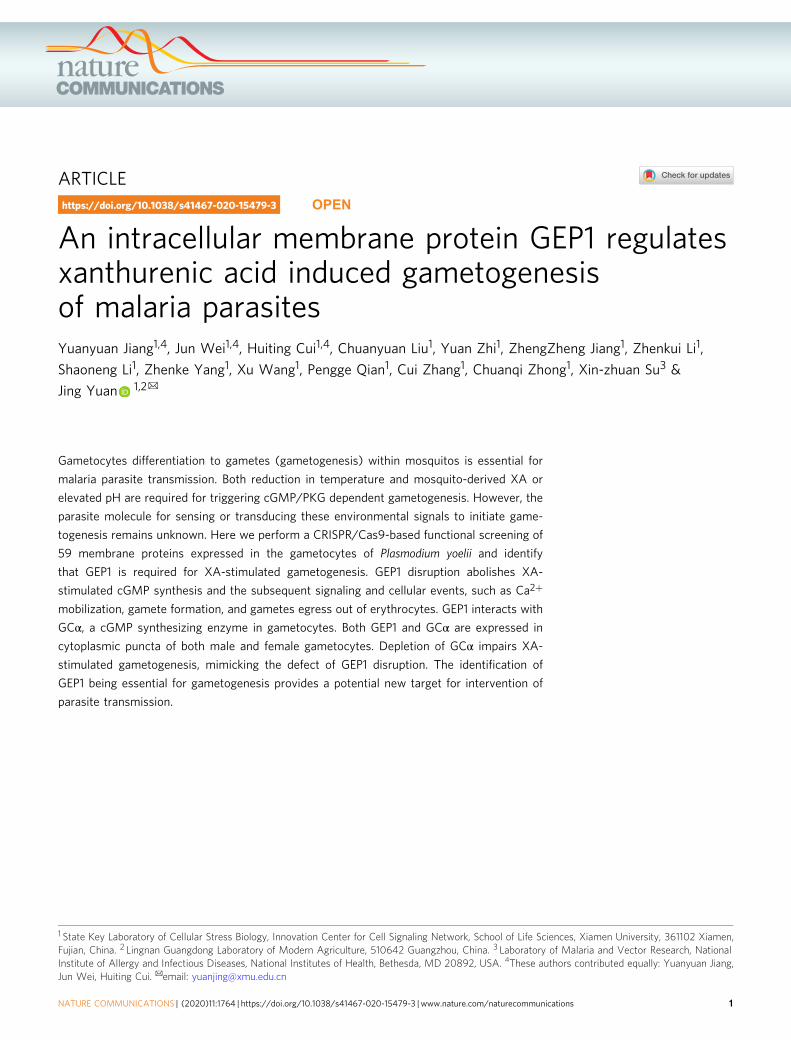
An intracellular membrane protein GEP1 regulates ...
15
ARTICLE An intracellular membrane protein GEP1 regulates xanthurenic acid induced gametogenesis of malaria parasites Yuanyuan Jiang 1,4 , Jun Wei 1,4 , Huiting Cui 1,4 , Chuanyuan Liu 1 , Yuan Zhi 1 , ZhengZheng Jiang 1 , Zhenkui Li 1 , Shaoneng Li 1 , Zhenke Yang 1 , Xu Wang 1 , Pengge Qian 1 , Cui Zhang 1 , Chuanqi Zhong 1 , Xin-zhuan Su 3 & Jing Yuan 1,2 ✉ Gametocytes differentiation to gametes (gametogenesis) within mosquitos is essential for malaria parasite transmission. Both reduction in temperature and mosquito-derived XA or elevated pH are required for triggering cGMP/PKG dependent gametogenesis. However, the parasite molecule for sensing or transducing these environmental signals to initiate game- togenesis remains unknown. Here we perform a CRISPR/Cas9-based functional screening of 59 membrane proteins expressed in the gametocytes of Plasmodium yoelii and identify that GEP1 is required for XA-stimulated gametogenesis. GEP1 disruption abolishes XA- stimulated cGMP synthesis and the subsequent signaling and cellular events, such as Ca 2+ mobilization, gamete formation, and gametes egress out of erythrocytes. GEP1 interacts with GCα, a cGMP synthesizing enzyme in gametocytes. Both GEP1 and GCα are expressed in cytoplasmic puncta of both male and female gametocytes. Depletion of GCα impairs XA- stimulated gametogenesis, mimicking the defect of GEP1 disruption. The identification of GEP1 being essential for gametogenesis provides a potential new target for intervention of parasite transmission. https://doi.org/10.1038/s41467-020-15479-3 OPEN 1 State Key Laboratory of Cellular Stress Biology, Innovation Center for Cell Signaling Network, School of Life Sciences, Xiamen University, 361102 Xiamen, Fujian, China. 2 Lingnan Guangdong Laboratory of Modern Agriculture, 510642 Guangzhou, China. 3 Laboratory of Malaria and Vector Research, National Institute of Allergy and Infectious Diseases, National Institutes of Health, Bethesda, MD 20892, USA. 4 These authors contributed equally: Yuanyuan Jiang, Jun Wei, Huiting Cui. ✉ email: [email protected] NATURE COMMUNICATIONS | (2020)11:1764 | https://doi.org/10.1038/s41467-020-15479-3 | www.nature.com/naturecommunications 1 1234567890():,;
Transcript of An intracellular membrane protein GEP1 regulates ...
ARTICLE
An intracellular membrane protein GEP1 regulatesxanthurenic acid induced gametogenesisof malaria parasitesYuanyuan Jiang1,4, Jun Wei1,4, Huiting Cui1,4, Chuanyuan Liu1, Yuan Zhi1, ZhengZheng Jiang1, Zhenkui Li1,
Shaoneng Li1, Zhenke Yang1, Xu Wang1, Pengge Qian1, Cui Zhang1, Chuanqi Zhong1, Xin-zhuan Su3 &
Jing Yuan 1,2✉
Gametocytes differentiation to gametes (gametogenesis) within mosquitos is essential for
malaria parasite transmission. Both reduction in temperature and mosquito-derived XA or
elevated pH are required for triggering cGMP/PKG dependent gametogenesis. However, the
parasite molecule for sensing or transducing these environmental signals to initiate game-
togenesis remains unknown. Here we perform a CRISPR/Cas9-based functional screening of
59 membrane proteins expressed in the gametocytes of Plasmodium yoelii and identify
that GEP1 is required for XA-stimulated gametogenesis. GEP1 disruption abolishes XA-
stimulated cGMP synthesis and the subsequent signaling and cellular events, such as Ca2+
mobilization, gamete formation, and gametes egress out of erythrocytes. GEP1 interacts with
GCα, a cGMP synthesizing enzyme in gametocytes. Both GEP1 and GCα are expressed in
cytoplasmic puncta of both male and female gametocytes. Depletion of GCα impairs XA-
stimulated gametogenesis, mimicking the defect of GEP1 disruption. The identification of
GEP1 being essential for gametogenesis provides a potential new target for intervention of
parasite transmission.
https://doi.org/10.1038/s41467-020-15479-3 OPEN
1 State Key Laboratory of Cellular Stress Biology, Innovation Center for Cell Signaling Network, School of Life Sciences, Xiamen University, 361102 Xiamen,Fujian, China. 2 Lingnan Guangdong Laboratory of Modern Agriculture, 510642 Guangzhou, China. 3 Laboratory of Malaria and Vector Research, NationalInstitute of Allergy and Infectious Diseases, National Institutes of Health, Bethesda, MD 20892, USA. 4These authors contributed equally: Yuanyuan Jiang,Jun Wei, Huiting Cui. ✉email: [email protected]
NATURE COMMUNICATIONS | (2020) 11:1764 | https://doi.org/10.1038/s41467-020-15479-3 |www.nature.com/naturecommunications 1
1234
5678
90():,;

Male and female gametocytes are sexual precursor cellsessential for malaria parasite transmission. Within10–15 min after being taken up by a mosquito, game-
tocytes differentiate into gametes in mosquito midgut, a processknown as gametogenesis. A female gametocyte forms a roundedfemale gamete, whereas a male gametocyte undergoes threemitotic divisions, assembles eight intracytoplasmic axonemes, andproduces eight flagellated male gametes1. Both male and femalegametes egress from their residing erythrocytes via an inside-outmechanism, during which the parasitophorus vacuole membrane(PVM) ruptures prior to the opening of the erythrocyte mem-brane (EM)2. After the release from erythrocytes, the male andfemale gametes fertilize to produce zygotes and then the motileookinetes that penetrate mosquito midgut wall to develop intooocysts each containing thousands of sporozoites. The spor-ozoites then migrate to mosquito salivary glands and are injectedinto a new host when the mosquito bites again.
Gametogenesis is triggered by two stimuli, a drop in tem-perature of approximately 5 °C3,4 and the presence of xanthurenicacid (XA) that is a metabolite of tryptophan from mosquito5,6.An additional signal reported to induce gametogenesis is anincrease in pH from 7.4 to 84. Since the groundbreaking discoveryof XA as a trigger for Plasmodium gametogenesis in mosquitoes,studies have shown that XA can enhance parasite guanylyl cyclase(GC) activity on gametocyte membrane fraction, leading toincreased level of second messenger 3’−5’-cyclic guanosinemonophosphate (cGMP)7. Two integral membrane GC proteins(GCα and GCβ) are found in Plasmodium parasites. GCα hasbeen implicated to be responsible for cGMP synthesis duringgametogenesis because disruption of GCβ has no effect on XA-induced gametogenesis8–10. The increased level of cGMP activatescGMP-dependent protein kinase G (PKG) that functions as amaster regulator of the downstream signaling events duringgametogenesis11. Inhibition of PKG using Compound 2 (C2)prevented gametocytes rounding up, gamete formation of bothsexes, and gametes egress from erythrocytes in P. falciparum andP. berghei11,12. PKG-dependent Ca2+ mobilization was alsoobserved in the cytosol of P. falciparum and P. berghei gameto-cytes 10–15 s after addition of XA13,14. PKG activates thesynthesis of inositol (1,4,5)-trisphosphate (IP3) via phosphoino-sitide metabolism and triggers cytosolic mobilization of Ca2+ thatlikely originates from the endoplasmic reticulum15. Unfortu-nately, the molecule(s) responsible for sensing XA or transducingthe XA-stimulated signal to activate the cGMP-PKG signalingremain unknown.
Membrane proteins are known to play critical roles in sensing,transporting, and/or transducing environmental signals to initiatecellular responses. To identify potential molecules involved insensing or transducing XA signal during gametogenesis, weperform CRISPR/Cas9-mediated genetic deletion screens of 59candidate genes encoding integral membrane proteins expressedin gametocytes of the rodent malaria parasite P. yoelii. Weidentify a multiple-spanning membrane protein GEP1 (gameto-genesis essential protein 1) that was essential for XA-stimulatedgametogenesis. Disruption of GEP1 completely abolishes XA-stimulated gametogenesis of both sexes. Parasites deficient ofGEP1 show no synthesis of XA-stimulated cGMP and nodownstream cellular and signaling events such as Ca2+ mobili-zation, parasite egress out of PVM and EM, genome replicationand axoneme assembly in male gametocytes, and release oftranslational repression in female gametocytes. GEP1 interactswith GCα in gametocytes, and GCα depletion also impairs XA-stimulated gametogenesis, mimicking the effects of GEP1 dis-ruption. This study identifies a molecule essential for the initia-tion of gametogenesis and a potential target for blocking parasitetransmission.
ResultsGEP1 is essential for XA-stimulated gametogenesis. To identifymembrane proteins critical in sensing XA or transducing XA-induced signal during gametogenesis, we identified 59 P. yoeliigenes that are expressed in gametocytes and encode proteins with1 to 22 predicted transmembrane domains (TMs) from thePlasmoDB database (Supplementary Table 1). We designed singleguide RNA (sgRNA) to disrupt each of these genes usingCRISPR/Cas9 methods16,17 and were able to successfully knock-out (KO) 45 (76%) of the genes in the P. yoelii 17XNL strain,obtaining at least two cloned lines for each mutant (Supple-mentary Fig. 1a, c, d, i). The remaining 14 genes (24%) wererefractory to repeated deletion attempts using three independentsgRNA sequences, suggesting their essential roles for asexualblood-stage growth.
The 45 gene deletion mutants proliferated asexually in mouseblood normally and were able to produce both male and femalegametocytes although the gametocytemia level varied among thesemutants (Supplementary Fig. 2, Supplementary Fig. 3a). Next wemeasured the gametogenesis of male gametocyte by countingexflagellation centers (ECs) formed in vitro after stimulation with50 μM XA at 22 °C. Only one mutant (PY17X_1116300 disruption)showed complete deficiency in EC formation and male gameterelease (Fig. 1a–c). The PY17X_1116300 gene contains four exons(Fig. 1d) encoding a putative amino acid transporter protein that isessential for gametogenesis; we therefore name the gene gep1 forgametogenesis essential protein 1. As controls, disruption of P. yoeliicdpk4 or map2 also caused defect in EC formation (Fig. 1a),confirming the phenotypes observed in P. berghei13,18. Conse-quently, the Δgep1mutant parasite produced no ookinete in in vitroculture (Supplementary Fig. 3b), oocyst in Anopheles stephensimidgut (Fig. 1f), or sporozoite in mosquito salivary gland(Supplementary Fig. 3c).
To further confirm the phenotype of Δgep1, we generated threeadditional gep1 mutant parasites (Δgep1n, Δgep1fl, and Δgep1mS-carlet) (Fig. 1d, Supplementary Fig. 1c–e). The Δgep1n parasitehad a 464 bp deletion at the 5’ coding region, causing a frameshiftfor the remaining coding region. The Δgep1fl parasite had thewhole gep1 coding region deleted, and the Δgep1mScarlet parasitehad its gep1 coding regions replaced with a gene encoding redflorescent protein mScarlet. These mutations were confirmed byPCR and DNA sequencing (Supplementary Fig. 1j, k), and themutant parasites displayed developmental phenotypes similar tothose of Δgep1 in both mouse and mosquito stages (Fig. 1e, f,Supplementary Fig. 3a–c). We also reintroduced the 558 bpdeleted segment plus a sextuple HA epitope (6HA) into the Δgep1parasite to rescue the gene function using Cas9-mediatedhomologous replacement (Fig. 1d, Supplementary Fig. 1b, j).Two clones of the rescued parasite (Δgep1/gep1::6HAc1 andΔgep1/gep1::6HAc2) showed expression of the GEP1::6HAprotein in both Western blotting and immunofluorescenceanalysis (IFA) (Supplementary Fig. 3d, e). Importantly, bothclones produced wild type (WT) levels of EC in vitro (Fig. 1e) andmidgut oocyst in mosquitoes (Fig. 1f). The GEP1 protein is well-conserved among P. yoelii, P. berghei, and the human P.falciparum parasites (Supplementary Fig. 4), suggesting conservedfunction. Deletion of P. berghei gep1 gene (PBANKA_1115100)resulted in parasite clones that failed to form XA-stimulated ECsin vitro and midgut oocyst in mosquitoes (Supplementary Fig. 1l,m, Supplementary Fig. 3f–h). Together, these results demonstratethat GEP1 depletion completely block male gametogenesis andmosquito transmission of malaria parasites.
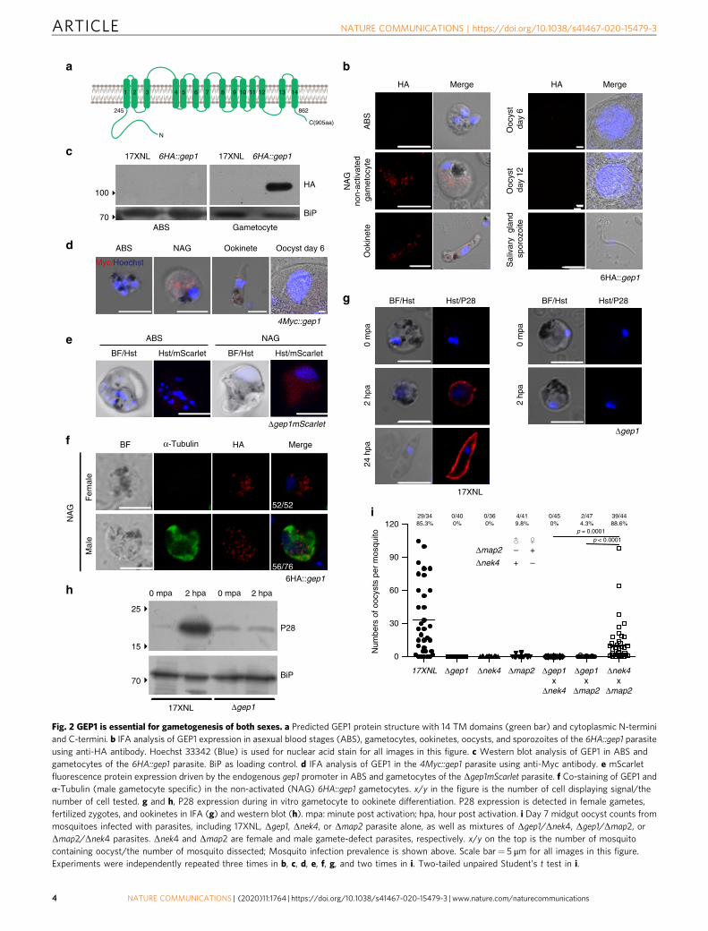
GEP1 is expressed in cytosol puncta of gametocytes. GEP1 is aPlasmodium-specific protein with 905 residues and 14 predicted
ARTICLE NATURE COMMUNICATIONS | https://doi.org/10.1038/s41467-020-15479-3
2 NATURE COMMUNICATIONS | (2020) 11:1764 | https://doi.org/10.1038/s41467-020-15479-3 | www.nature.com/naturecommunications

TMs (Fig. 2a). Previous transcriptomic study indicated the gep1gene is transcribed in gametocytes and ookinetes, but not asexualblood stages of P. falciparum and P. berghei19,20. To investigateprotein expression and localization, we tagged the endogenousGEP1 with 6HA at N-terminus (Supplementary Fig. 1g, j), gen-erating 6HA::gep1 parasite that had normal developmentthroughout the life cycle (Supplementary Fig. 5a). The GEP1
protein is expressed in gametocytes and ookinetes, but not inasexual blood stages and other mosquito stages of the 6HA::gep1parasite (Fig. 2b, c). We also tagged the GEP1 protein withquadruple Myc (4Myc) (Supplementary Fig. 1j, SupplementaryFig. 5b) and observed similar expression pattern in the 4Myc::gep1parasite (Fig. 2d). In addition, mScarlet fluorescent signals drivenby the endogenous gep1 promoter were detected only in
a
17XNL exon1 e2 e3 e45′UTR
S1: Δgep1 exon1 e2 e35′UTR
3′UTR
3′UTR
3′UTRexon1 e2 e3 e45′UTR 6HAComplementedS2: S1/gep1::6HA
exon15′UTR e2 e3 e4 3′UTRS3: Δgep1n
5′UTR 3′UTRS4: Δgep1fl
mScarlet5′UTR 3′UTRS5: Δgep1mScarlet
b d
c f
17XNLΔPY17X_1116300
(Δgep1)
n = 22 n = 22 n = 22 n = 21 n = 21 n = 26 n = 26e
17XNL
Δgep1
0
40
20
60
Exf
lage
llatio
n ce
nter
s pe
r fie
ld
17XNL S1 c1 c2 S3 S4 S5S2
0
100
200
300
400
17XNL S1 c1 c2 S3 S4 S5S2
20/2387%
0/400%
41/4395.3%
24/2596%
0/290%
0/300%
0/300%
Num
bers
of o
ocys
ts p
er m
osqu
ito
p < 0.0001
p < 0.0001
p < 0.0001
p < 0.0001
p < 0.0001
p < 0.0001
Δcdp
k4Δm
ap2
17XN
L
Δnek
4Δ1
2434
00Δ1
4315
00Δ0
9187
00Δ1
4339
00Δ0
5246
00Δ0
6058
00Δ0
6174
00Δ1
1286
00Δ1
3139
00Δ0
7024
00Δ0
9335
00Δ0
3094
00Δ0
9363
00Δ1
4364
00Δ0
8237
00Δ0
8203
00Δ1
1163
00Δ0
9174
00Δ0
6094
00Δ1
4463
00Δ1
3155
00Δ0
9049
00Δ0
4034
00Δ1
3587
00Δ0
9286
00Δ0
6108
00Δ1
0196
00Δ1
0316
00Δ0
9299
00Δ1
0185
00Δ0
7033
00Δ1
2406
00Δ1
4411
00Δ1
3673
00Δ1
4242
00Δ1
1052
00Δ1
3152
00Δ1
3394
00Δ1
3428
00Δ1
3661
00Δ1
4633
00
Δ080
9500
Δ113
8200
Δ061
9700
Δ091
6600
200E
xfla
gella
tion
rate
nor
mol
ized
to w
idet
ype
%
0
50
100
150
P < 0.0001P < 0.0001
P = 0.0492
P = 0.4365P = 0.0269
P = 0.7556
P = 0.0060P = 0.2606
P = 0.0080
P = 0.0075P = 0.0325
P = 0.1968
P = 0.0158P = 0.0288
P = 0.1458
P = 0.1348P = 0.9328
P = 0.1201
P = 0.1590P < 0.0001
P = 0.5110
P = 0.0282P = 0.8786
P = 0.0015
P = 0.0546P < 0.0001
P = 0.0332
P = 0.0431P = 0.0004
P = 0.0527
P = 0.5524P = 0.0364
P = 0.3394
P < 0.0001P < 0.0001
P = 0.7514
P = 0.2991P = 0.5686
P = 0.0286
P < 0.0001P = 0.0409
P = 0.0002
P = 0.0017P < 0.0001
P = 0.0303
P = 0.1649P = 0.0033
P = 0.0157
Xanthurenic acid induced gametogenesis
Fig. 1 Membrane proteins screening identified gep1 essential for gametogenesis. a In vitro XA stimulated exflagellation rates for P. yoelii 17XNL wild type(WT) and 45 mutant strains each with a specific gene disruption. The exflagellation rate of each mutant was normalized with that of WT parallelly testedeach time. The numbers for the gene name are the gene IDs derived in PlasmoDB. Data are shown as mean ± SD from n= 3 independent experiments forstrains except n= 5 for Δ1315200, Δ1339400, Δ1342800, Δ1366100 and Δ1463300, and n= 6 for Δ1240600. b Representative images of XA stimulatedexflagellation centers (ECs, white arrows) under light microscope (10×). Scale bar= 20 μm. c Images of the exflagellated male gametes (Black arrow) afterGiemsa staining under light microscope (100×). Scale bar= 5 μm. d Diagrams of WT gep1 gene structure and various mutants: S1 (Δgep1), deletion inC-terminus; S2 (Δgep1/gep1::6HA), reconstructed gep1 with a 6HA tag; S3 (Δgep1n), deletion in N-terminus; S4 (Δgep1fl), deletion of the full coding region;S5 (Δgep1mScarlet), coding region replaced with mScarlet gene. e XA-stimulated EC counts from WT and the gep1 mutants. c1 and c2 are two clones of S2parasite. n is the numbers of microscopic fields counted (40×). f Oocyst counts from WT and the gep1 mutants. Oocysts are counted from the mosquitomidguts 7 days post blood feeding. x/y on the top is the number of mosquito containing oocyst/the number of mosquito dissected; the percentage numberis the mosquito infection prevalence. Experiments were independently repeated six times in b, and three times in c, e, and f. Two-tailed unpaired Student’st test was applied in a, e, and f. Source data of a, e, and f are provided as a Source Data file.
NATURE COMMUNICATIONS | https://doi.org/10.1038/s41467-020-15479-3 ARTICLE
NATURE COMMUNICATIONS | (2020) 11:1764 | https://doi.org/10.1038/s41467-020-15479-3 |www.nature.com/naturecommunications 3
N
C(905aa)
1 2 3 4 5 6 7 8 9 10 11 12 13 14
245 862
AB
SO
okin
ete
Ooc
yst
day
6O
ocys
t d
ay 1
2 S
aliv
ary
gla
nd
spo
rozo
ite
BF HA Mergeα-Tubulin
Fem
ale
M
ale
HA Merge
0 mpa
17XNL Δgep1
P28
BiP
15
70
25
HA Merge
0 m
pa 2
hpa
24
hpa
0 m
pa 2
hpa
17XNL
Δgep1
2 hpa 0 mpa 2 hpa
6HA::gep1
6HA::gep1
BF/Hst Hst/P28 BF/Hst Hst/P28
Hst/mScarlet
ABS NAG
Hst/mScarlet
Δgep1mScarlet
BF/Hst BF/Hst
a b
c
d
e
g
f
h
ABS Gametocyte
100
70
HA
BiP
17XNL 6HA::gep1 17XNL 6HA::gep1
4Myc::gep1
Oocyst day 6ABS NAG Ookinete
Myc/Hoechst
i
NA
G
NA
Gno
n-ac
tivat
ed g
amet
ocyt
e
52/52
56/76
0
30
60
90
120
17XNL
29/34
Δgep1
0/40
Δnek4
0/36
Δmap2
4/41
Δgep1x
Δnek4
0/45
Δgep1x
Δmap2
2/47 39/44
Δnek4x
Δmap2
Δmap2 – +
Δnek4 + –
Num
bers
of o
ocys
ts p
er m
osqu
ito 85.3% 0% 0% 9.8% 0% 4.3% 88.6%
p < 0.0001
p = 0.0001
Fig. 2 GEP1 is essential for gametogenesis of both sexes. a Predicted GEP1 protein structure with 14 TM domains (green bar) and cytoplasmic N-terminiand C-termini. b IFA analysis of GEP1 expression in asexual blood stages (ABS), gametocytes, ookinetes, oocysts, and sporozoites of the 6HA::gep1 parasiteusing anti-HA antibody. Hoechst 33342 (Blue) is used for nuclear acid stain for all images in this figure. c Western blot analysis of GEP1 in ABS andgametocytes of the 6HA::gep1 parasite. BiP as loading control. d IFA analysis of GEP1 in the 4Myc::gep1 parasite using anti-Myc antibody. e mScarletfluorescence protein expression driven by the endogenous gep1 promoter in ABS and gametocytes of the Δgep1mScarlet parasite. f Co-staining of GEP1 andα-Tubulin (male gametocyte specific) in the non-activated (NAG) 6HA::gep1 gametocytes. x/y in the figure is the number of cell displaying signal/thenumber of cell tested. g and h, P28 expression during in vitro gametocyte to ookinete differentiation. P28 expression is detected in female gametes,fertilized zygotes, and ookinetes in IFA (g) and western blot (h). mpa: minute post activation; hpa, hour post activation. i Day 7 midgut oocyst counts frommosquitoes infected with parasites, including 17XNL, Δgep1, Δnek4, or Δmap2 parasite alone, as well as mixtures of Δgep1/Δnek4, Δgep1/Δmap2, orΔmap2/Δnek4 parasites. Δnek4 and Δmap2 are female and male gamete-defect parasites, respectively. x/y on the top is the number of mosquitocontaining oocyst/the number of mosquito dissected; Mosquito infection prevalence is shown above. Scale bar= 5 μm for all images in this figure.Experiments were independently repeated three times in b, c, d, e, f, g, and two times in i. Two-tailed unpaired Student’s t test in i.
ARTICLE NATURE COMMUNICATIONS | https://doi.org/10.1038/s41467-020-15479-3
4 NATURE COMMUNICATIONS | (2020) 11:1764 | https://doi.org/10.1038/s41467-020-15479-3 | www.nature.com/naturecommunications

gametocytes, but not in asexual blood stages of the Δgep1mScarletparasite (Fig. 2e). Co-staining 6HA::gep1 gametocytes with anti-α-Tubulin (male gametocyte specific) and anti-HA antibodyshowed that GEP1 was expressed in both male and femalegametocytes (Fig. 2f). Interestingly, GEP1 is not expressed inplasma membrane, but in punctate dots in the cytoplasm ofgametocytes and ookinetes (Fig. 2b, d, f).
GEP1 regulates both male and female gametogenesis. BecauseGEP1 is expressed in both male and female gametocytes, weasked whether GEP1 also regulates the gametogenesis of femalegametocytes. P28 protein, a marker for female gamete21, isexpressed in female gametes, fertilized zygotes, and ookinetes of17XNL parasite, but not in the Δgep1 parasite 2 h after XA-stimulation (Fig. 2g, h), indicating that GEP1 depletion also causedefect in female gametogenesis. We next performed geneticcrosses between Δgep1 and Δmap2 (male gamete-deficient) orΔnek4 (female gamete-deficient) parasites22,23 (SupplementaryFig. 1j, k). No midgut oocyst was observed in mosquitoes fromthe Δgep1 × Δmap2 or Δgep1 × Δnek4 cross day 7 post infection(pi), whereas the Δmap2 × Δnek4 cross produced slightly feweroocysts than the WT parasite (Fig. 2i), suggesting no functionalmale and female gametes in the Δgep1 parasite. Together, theseresults demonstrate that GEP1 is essential for both male andfemale gametogenesis.
The purified Δgep1 gametocytes had morphology indistin-guishable from that of WT 17XNL parasite (SupplementaryFig. 6a); however, whether GEP1 depletion causes gametocytedeath or affects the fitness of gametocytes remains to bedetermined. We analyzed cell viability by Trypan blue exclusionassay. No gametocyte of WT or Δgep1 parasites were stained byTrypan blue (Supplementary Fig. 6b). As a control, bothgametocytes were stained after heating the parasites at 60 °C for5 min. In addition, staining with propidium iodide (PI) alsoindicated that the Δgep1 gametocytes are viable (SupplementaryFig. 6c). To further confirm the observations, we disrupted theendogenous gep1 in a P. yoelii reporter strain DFsc7 thatexpressed GFP and mCherry in male and female gametocytes,respectively24 (Supplementary Fig. 1j, Supplementary Fig. 6d, e).The expressions of fluorescent proteins in both male and femalegametocytes were comparable with those of the parental parasite(Supplementary Fig. 6f, g). These results suggest that GEP1-depleted gametocytes are viable, but lost the ability to producefunctional male and female gametes.
GEP1 depletion blocks PKG-mediated signaling. Upon stimu-lation, male gametocytes undergo tubulin polymerization intomicrotubules and three rounds of genome replication, resulting inrelease of eight flagellated gametes within 10–15 min25. The lackof exflagellation suggests defect in either axoneme assembly oregress from erythrocyte of the Δgep1 male gametes. Typicalcytosolic distribution of α-Tubulin was observed in male game-tocytes of WT, Δgep1, and Δmap2 parasites before XA stimula-tion (Fig. 3a). Assembled axonemes were formed and coiledaround the nucleus of WT and Δmap2 gametocytes 8 min postXA stimulation, but axoneme formation was not observed in theΔgep1 parasite (Fig. 3a). By 15 min, WT gametocytes releasedflagellated male gametes, but not Δmap2 and Δgep1 gametocytes(Fig. 3a). Strikingly, α-Tubulin remained in cytosol of the Δgep1male gametocytes (Fig. 3a). We also analyzed the genome repli-cation in stimulated male gametocytes. Flow cytometry analysis ofDNA content in Hoechst-stained gametocytes showed thatfluorescence increased (from 8.4% to 28.5%) in WT, but not inthe Δgep1 parasites (from 8.4% to 7.6%) after XA stimulation(Fig. 3b). As reported for P. berghei13,22, no genome replication
occurs in the Δcdpk4 parasite (Fig. 3b, Supplementary Fig. 1j, k).These results show no axoneme assembly or mitotic division inthe stimulated Δgep1 male gametocytes.
Differentiation of male and female gametes result in sequentialrupture of PVM and EM for escaping from erythrocytes2,26.TER119 is a plasma membrane protein of mouseerythrocytes27,28, and anti-TER119 antibody showed no EMstaining for stimulated WTmale and female gametocytes (Fig. 3c).In contrast, intact EM was observed for the Δgep1 gametocytes30 min post stimulation (Fig. 3c), indicating that GEP1 depletionaffects EM lysis.
XA triggers a cytosolic Ca2+ mobilization event within 10–15 spost stimulation of gametocytes13, which is essential for gametesformation and EM rupture11,13. We next examined XA-stimulated Ca2+ mobilization in the Δgep1 gametocytes usingFluo-8 probe as described29–31. Fluo-8 did not affect thegametogenesis since WT gametocytes pre-loaded with Fluo-8could form XA-stimulated ECs (Supplementary Fig. 7a) andresponded to A23187, a Ca2+ ionophore13, in a dose-dependentmanner using flow cytometry (Supplementary Fig. 7b). Asexpected, XA triggered a sharp increase in cytosolic Ca2+ signalin WT gametocytes, reaching maximal levels 10–15 s poststimulation, which resembled the observations in P. bergheiusing luminescence-based GFP::Aequorin sensor13,15. However,no Ca2+ response was detected in XA stimulated Δgep1gametocytes (Fig. 3d). Ca2+ mobilization occurred in theΔmap2 gametocytes as MAP2 functions downstream of Ca2+
signal18,22 (Fig. 3d).Different from Ca2+-dependent EM rupture, PVM rupture is
controlled by a Ca2+-independent mechanism2. To study PVMlysis, a parasite line sep1::4Myc was generated by C-terminallytagging a PVM protein SEP1 with 4Myc27,28 (SupplementaryFig. 1j). This parasite line developed normally throughout the lifecycle (Supplementary Fig. 5e), indicating intact protein functionof SEP1::4Myc. We next deleted the gep1 gene in the sep1::4Mycparasite, generating sep1::4Myc/Δgep1 mutant (SupplementaryFig. 1j). IFA showed lysis of Sep1::4Myc-labeled PVM in thesep1::4Myc gametocytes (Fig. 3e), while intact PVM wasmaintained in the sep1::4Myc/Δgep1 gametocytes 8 min post XAstimulation (Fig. 3e), indicating no PVM lysis in stimulated Δgep1gametocytes. Together, these results suggest that GEP1 functionsupstream of PKG in XA-stimulated signaling cascade (Fig. 3f).
Impaired cGMP synthesis in GEP1 deficient parasite. BecausecGMP is the direct upstream signal activating PKG in XA-stimulated gametogenesis7,11,13,15, we examined intracellularcGMP synthesis during gametogenesis. Purified gametocytes werestimulated with XA for 2 min, and cGMP levels were measuredusing an enzyme immunoassay7,32. Strikingly, XA induced asignificant increase in cGMP level in WT gametocytes (Fig. 4a),consistent with previous observation in P. falciparum7. In con-trast, the Δgep1 gametocytes failed to increase cGMP in responseto XA stimulation (Fig. 4a). As a control, cGMP responseoccurred in Δmap2 gametocytes because MAP2 functionsdownstream of both cGMP and Ca2+ signaling18,22. These resultsindicate that GEP1 regulates cGMP level, the most upstreamintracellular signal known in Plasmodium gametogenesis.
cGMP level is tightly regulated by the opposing actions ofcGMP-synthesizing GC and cGMP-hydrolyzing phosphodiester-ase (PDE)10,11,33. Inhibition of PDE activity by specific inhibitorZaprinast (Zap) has been shown to trigger P. falciparumgametogenesis in the absence of XA11,33. Indeed, treatment ofWT gametocytes with 100 μM Zap also induced EC countscomparable to those induced by 50 μM XA (Fig. 4b), andgametogenesis stimulated by either XA or Zap could be blocked
NATURE COMMUNICATIONS | https://doi.org/10.1038/s41467-020-15479-3 ARTICLE
NATURE COMMUNICATIONS | (2020) 11:1764 | https://doi.org/10.1038/s41467-020-15479-3 |www.nature.com/naturecommunications 5

by a Plasmodium PKG protein inhibitor C2 (Fig. 4b), consistentwith the established cGMP-PKG signal cascade ofgametogenesis14,15. In contrast, the Δgep1 gametocytes failed toform ECs after treatment with Zap (Fig. 4b). No EC wereobserved in the control Δmap2 gametocytes treated in either XAor Zap (Fig. 4b). Consistently, we examined the intracellularcGMP level in gametocytes treated with Zap for 2 min anddetected significant increase in both WT and Δmap2 gametocytes,but not in the Δgep1 gametocytes (Fig. 4c). Together, these resultssuggest that the GC activity for cGMP synthesis is impaired, andtherefore no elevation of cGMP in the Δgep1 gametocytes afterXA stimulation or Zap inhibition of PDE activity. In addition toXA and Zap, increasing pH from 7.4 to 8.0 has been reported toinduce gametogenesis although the underlying mechanism is notclear2,4. Treating WT gametocytes with pH 8.0 at 22 °C indeed
induced comparable number of ECs to those induced by XA orZap (Fig. 4b), and gametogenesis could be blocked by C2treatment (Fig. 4b)15, indicating that the signaling stimulated bypH 8.0 is also cGMP/PKG-dependent. However, pH 8.0treatment could not induce gametogenesis of the Δgep1gametocytes, further suggesting impaired activity of cGMPsynthesis in GEP1 deficient parasite (Fig. 4d).
GEP1 interacts and co-localizes with GCα. We next carried outimmunoprecipitation and mass spectrometry experiments toidentify molecules that may interact with GEP1 in gametocytes.By comparison of peptide signals (hits) between WT and 6HA::gep1 gametocyte samples from three biological replicates, weobtained 308 proteins that might interact with GEP1 (Supple-mentary Table 2), including GCα protein that is the enzyme
sep1::4Myc sep1::4Myc/Δgep1
Hoechst α-Tubulin HoechstTer119
17XNL
1.80%
4.64%
7.61%
8.41%
28.5%
8.43%
200K
400K
600K
800K
1M
200K
400K
600K
800K
1M
103 104 105 106 103 104 105 106 103 104 105 106
17XNL Δgep1 Δcdpk4
0 m
pa8
mpa
Hoechst
SS
C-H
Time/s
FIT
C-A
0 30 60 90 120180
200
220
240
DMSO 100 μM XA
330
350
370
390
0 30 60 90 120
DMSO 100 μM XA
0 30 60 90 120180
200
220
240
DMSO 100 μM XA
Δgep1
Δmap2
17XNL Δgep1
0 m
pa30
mpa
0 m
pa8
mpa
17XNL Δgep1 Δmap20
mpa
8 m
pa15
mpa
HoechstSep1
a c e
b
d f
PVM rupture
Ca2+
XA & 22°C
DNA replicationAxoneme assembly
CDPK4
Axoneme motility
Map2
P28 proteintranslation
GEP1
PKG ( + )
EM rupture( + )
( )
( )
EMPVMPPM
cGMP
74/75
92/95
84/89
69/69
86/86
87/87
72/72
81/87
20/50
80/80
46/86
80/80
80/80
53/59
47/60
58/58
53/53
Fig. 3 GEP1 acts upstream of PKG in the cGMP-PKG-Ca2+ signaling cascade. a α-Tubulin expression and distribution in differentiating male gametocytesfrom 17XNL, Δgep1 and Δmap2 parasites after XA stimulation. mpa: minute post XA activation. b Flow cytometry analysis of genomic DNA content in XA-stimulated male gametocytes of 17XNL, Δgep1 and Δcdpk4 parasites. The parasites were fixed with 4% paraformaldehyde at indicated time and stainedwith Hoechst. c Representative images of gametocytes stained by anti-mouse TER119 antibody 0 and 30min post XA stimulation (mpa). d Flow cytometrydetection of cytosolic Ca2+ in gametocytes using Fluo-8 probe. Purified gametocytes were preloaded with Fluo-8, and signals were collected 30 s beforeaddition of XA or DSMO. Black arrows indicate the time for DMSO or XA addition. e Representative IFA images of the sep1::4Myc and sep1::4Myc/Δgep1gametocytes stained by anti-Myc antibody. f Proposed location of GEP1 in the XA-PKG-Ca2+ signal cascade of gametogenesis. GEP1 depletion causesdefect in both Ca2+-dependent and Ca2+-independent cellular events of gametogenesis. EM: erythrocyte membrane, PVM: parasitophorus vacuolemembrane, PPM: parasite plasma membrane. x/y in a, c, and e are the number of cell displaying representative signal/the number of cell analyzed. Scalebar= 5 μm for all images in this figure. All experiments in this figure were repeated three times independently with similar results.
ARTICLE NATURE COMMUNICATIONS | https://doi.org/10.1038/s41467-020-15479-3
6 NATURE COMMUNICATIONS | (2020) 11:1764 | https://doi.org/10.1038/s41467-020-15479-3 | www.nature.com/naturecommunications

presumably responsible for cGMP synthesis during gameto-genesis (Fig. 5a, b)8–10. The P. yoelii GCα is a large protein (3850amino acids) with 22 TMs distributed in an N-terminal P4-ATPase-like domain (ALD) and a C-terminal guanylate cyclasedomain (GCD)34,35. To study the expression of GCα in game-tocytes, we generated two parasite lines (gcα::6HA and gcα::4-Myc) with endogenous GCα C-terminally tagged with 6HA and4Myc, respectively (Supplementary Fig. 1j). These parasitesdeveloped normally in mouse and mosquito hosts (Supple-mentary Fig. 5c, d). Similar to GEP1, GCα was also expressed ascytoplasmic puncta in both male and female gametocytes of thegcα::6HA and gcα::4Myc parasites (Supplementary Fig. 8a). Tofurther confirm the interaction between GEP1 and GCα, wegenerated a doubly tagged parasite line, 4Myc::gep1/gcα::6HA(DTS1), by tagging the endogenous GEP1 with 4Myc in thegcα::6HA parasite (Supplementary Fig. 1j, SupplementaryFig. 5f–h). Results from immunoprecipitation using anti-Mycantibody indicated that GCα interacted with GEP1 in cell lysateof the DTS1 gametocytes (Fig. 5c). We next generated anotherindependent doubly tagged parasite, 6HA::gep1/gcα::4Myc(DTS2) by tagging GCα with 4Myc in the 6HA::gep1 parasite(Supplementary Fig. 1j, Supplementary Fig. 5f–h) and detectedsimilar interaction between GEP1 and GCα (Fig. 5d). As a
control, no interaction between GEP1 and GCβ was detected ingametocytes of the 4Myc::gep1/gcβ::6HA (DTS3) parasite (Sup-plementary Fig. 8b). These data demonstrate that GEP1 inter-acts with GCα in gametocytes. In addition, IFA results from theDTS1 parasite showed that GEP1 and GCα are co-localized atcytosolic puncta in non-activated gametocytes (Fig. 5e, f).Together, these data suggest that GEP1 co-localizes and binds toGCα in gametocytes.
GCα depletion causes defect in XA-stimulated gametogenesis.GCα has been implicated in cGMP synthesis during gameto-genesis8–10; however, there has been no direct evidence to supportthe speculation. We attempted to disrupt the gcα gene but failedto obtain a GCα mutant parasite, indicating an essential functionin asexual blood stage development, as reported in P. falciparumand P. berghei previously10. We used a promoter swap methoddescribed previously36 to replace 1322 bp of endogenous gcαpromoter region with that (1626 bp) of sera1 gene(PY17X_0305700) (Fig. 6a, Supplementary Fig. 1h), whose tran-scripts are expressed in asexual stages, but absent in gametocytesand mosquito stages37. In this editing, a 6HA tag was inserted inframe at the N-terminus of the GCα coding sequence. Correctmodification in two parasite clones of the resulting mutant
0
30
60
Exf
lage
llatio
n ce
nter
s pe
r fie
ld
90
120
Δmap2Δgep117XNL
XA
C2
ZappH8.0
+
–
––
–
–
––
+
+
––
–
–
+–
–
+
+–
–
–
–+
–
+
–+
–
–
+–
–
–
–+
–
–
+–
––+–
17XNL Δmap2Δgep1
Ctl XA Ctl XA Ctl XA
a
b
c
0
500
1000
1500
17XNL Δ map2Δ gep1
Ctl Zap Ctl Zap Ctl Zap
d
GEP1
PKG C2
GTPcGMPGMP
Zap PDE
pH 8.0 & 22°Cor
XA & 22°C
Gametogenesis
guanylate cyclaseGC
p = 0.0099 p = 0.0453
p = 0.7546
n = 12 n = 12 n = 12 n = 10 n = 12 n = 12 n = 12 n = 12 n = 12 n = 12 n = 12
p = 0.0880
p = 0.0237
Ace
tyla
ted
cGM
P (
pg/m
l)
Ace
tyla
ted
cGM
P (
pg/m
l)
0
1000
2000
3000
4000
p = 0.00039
p = 0.0011
p = 0.1399
p = 0.0044
p = 0.0010
p = 0.6964
p = 0.1683
p = 0.0022
p = 0.8188
Fig. 4 Impaired activity of cGMP synthesis in GEP1 deficient gametocytes. a Enzyme immunoassay detecting intracellular cGMP level in XA-stimulatedgametocytes of the 17XNL, Δgep1, and Δmap2 parasites. Cells were incubated with 100 μM XA at 22 °C for 2 min before assay. Ctl are control groupswithout XA stimulation. b Exflagellation center counts of 17XNL, Δgep1, and Δmap2 parasites after treatment with XA (100 μM), Zaprinast (Zap, 100 μM),or pH 8.0 alone at 22 °C, or at the presence of compound 2 (C2, 5 μM). n is the numbers of microscopic fields counted (40×). c Enzyme immunoassaydetecting intracellular cGMP level in Zap-treated gametocytes of the 17XNL, Δgep1, and Δmap2 parasites. Cells were incubated with 100 μM Zap at 22 °Cfor 2 min before assay. Ctl are control groups without Zap stimulation. d Proposed role of GEP1 in regulating cGMP synthesis activity of guanylyl cyclase ingametogenesis. All source data are provided as a Source Data file. Experiments in a, b, and c were repeated three times independently. Data are shown asmean ± SD; two-tailed unpaired Student’s t test.
NATURE COMMUNICATIONS | https://doi.org/10.1038/s41467-020-15479-3 ARTICLE
NATURE COMMUNICATIONS | (2020) 11:1764 | https://doi.org/10.1038/s41467-020-15479-3 |www.nature.com/naturecommunications 7

parasite gcαkd was confirmed by PCR (Supplementary Fig. 1j).The promoter replacement allowed expression of the GCα proteinin asexual blood stages at a level comparable with that of anotherparallelly modified parasite 6HA::gcα (Supplementary Fig. 1j), butsignificantly reduced GCα protein expression in gametocytes(Fig. 6b, c). Notably, the gcαkd parasite completely lost the abilityto synthesize cGMP and form ECs after XA stimulation in vitro(Fig. 6d, e). In mosquitos fed with gcαkd parasite-infected mouse
blood, no oocyst was detected in mosquito midgut (Fig. 6f). Theseresults support that GCα is the GC responsible for XA-stimulatedcGMP synthesis in gametogenesis (Fig. 6g). In addition, thephenotype caused by GCα knockdown in gametocytes resemblesthat of GEP1 defect.
Compared to the expression of GCα in both male and femalegametocytes, GCβ expression was detected in gcβ::6HA femalegametocytes only8 (Supplementary Fig. 8a, lower panel). In
DTS1 NAG
BF HA MergeMyc
Inte
nsity
0
Distance [nm]
Inte
nsity
GCα GEP1
c
d
DTS1: 4Myc::gep1/gc�::6HA
Top 10 GEP1 interacting proteins detected via IP-Mass spectrum
Gene_ID
PY17X_1347900 21
Unique peptide
Conserved plasmodium protein, unknown function
Description
PY17X_1226000 19 Tyrosine--tRNA ligase, putativePY17X_0911700 15
PY17X_1109100 15 Conserved protein, unknown functionPY17X_0404000 13 HAD superfamily protein, putativePY17X_1114400 10 Deoxyribodipyrimidine photo-lyase, putativePY17X_0807500 10 Conserved plasmodium protein, unknown functionPY17X_0922400 9 Conserved plasmodium protein, unknown functionPY17X_0706700 9 Conserved plasmodium protein, unknown functionPY17X_1221300 9 Oocyst capsule protein cap380, putative
a
b
Guanylyl cyclase alpha
400 600 800 1000 1200 1400
0
1000
2000
3000
4000
5000
m/z
Inte
nsity
A K E D A H D S I D F A L S M L Q V S S H I K
y11 y10 y9 y8 y7 y6 y5 y4 y3y19
b7 b9 b10 b11 b12 b13 b14 b15 b17
e
0306090
120
0 500 1000 1500 2000 2500
0
30
60
90
120
f
BiP
Myc(GEP1)
HA(GCα)170
100
70
gc�::6HADTS1
17XNL
Input IP: Myc
170
100
70
DTS2: 6HA::gep1/gc�::4Myc
6HA::gep1DTS2
17XNL
BiP
HA(GEP1)
Myc(GCα)
Input IP: Myc
500 1000 1500 2000 2500
gc�::6HADTS1
17XNL
6HA::gep1DTS2
17XNL
0.0
0.2
0.4
0.6
0.8
Pea
rson
coe
ffici
ent
GCα/GEP1
n = 10
Fig. 5 GEP1 interacts with GCα in gametocytes. a Top 10 GEP1 interacting proteins in the gametocytes of the 6HA::gep1 parasite detected byimmunoprecipitation and mass spectrometry (MS), including guanylyl cyclase α (GCα) with 15 peptides detected. b MS2 spectrum of a representativepeptide of the GCα protein. c Co-immunoprecipitation of Myc::GEP1 and GCα::HA proteins in gametocytes of the double tagged parasite 4Myc::gep1/gcα::6HA (DTS1). IP-Myc, anti-Myc antibody was used. d Co-immunoprecipitation of HA::GEP1 and GCα::Myc proteins in gametocytes of the double taggedparasite 6HA::gep1/gcα:: 4Myc (DTS2). IP-Myc, anti-Myc antibody was used. e Two-colored IFA of GEP1 and GCα proteins in the DTS1 gametocytes usinganti-HA (GCα) and anti-Myc (GEP1) antibodies (left panel). Cross sections (white dash line) of the cells show the co-localization of GEP1 and GCα (rightpanel). Scale bar= 5 μm. f Pearson coefficient analysis for GEP1 and GCα co-localization shown in e, data are shown as mean ± SD from n= 10 cellsmeasured. Experiments in c, d, and e were repeated three times independently with similar results.
ARTICLE NATURE COMMUNICATIONS | https://doi.org/10.1038/s41467-020-15479-3
8 NATURE COMMUNICATIONS | (2020) 11:1764 | https://doi.org/10.1038/s41467-020-15479-3 | www.nature.com/naturecommunications

addition, GCβ depletion had no effect on XA-stimulated elevationof cGMP (Supplementary Fig. 8c) and in vitro EC formation(Supplementary Fig. 8d) in gametocytes of the Δgcβ parasite8, inagreement with previous reports in P. falciparum and P.berghei9,33. These results exclude the involvement of GCβ inXA-stimulated cGMP signaling and gametogenesis.
GEP1 depletion has no effect on GCα expression and locali-zation. As GCα and GEP1 interacted with each other and func-tioned upstream of cGMP signaling, we investigated whether GEP1depletion would affect the expression and cellular localization ofGCα in gametocytes. We deleted gep1 gene in the gcα::6HA parasite,generating a gcα::6HA/Δgep1 mutant parasite (SupplementaryFig. 1j, Supplementary Fig. 5i, j). GEP1 depletion had no effect ongcα mRNA level or GCα protein abundance in gametocytes of thegcα::6HA/Δgep1 parasite compared to the parental parasite (Fig. 7a,b). As a control, depletion of CDPK4 had no effect on both mRNAand protein level of GCα either because CDPK4 functions down-stream of cGMP signal (Fig. 7a, b). In addition, XA stimulation hadno effect on protein abundance of both GEP1 and GCα in game-tocytes of the DTS1 parasite (Fig. 7c).
Next, we investigated the effect of XA stimulation in cellularlocalization of GEP1 and GCα proteins in gametocytes of the6HA::gep1 or gcα::6HA parasite, respectively. Two minutes postXA stimulation, both GEP1 and GCα were expressed ascytoplasmic puncta in activated female gametocytes (Fig. 7d, e).Even 8 min post XA stimulation, both GEP1 and GCα stillmaintained in cytoplasmic puncta in activated female gameto-cytes (Supplementary Fig. 9a, b). Strikingly, both proteins wereredistributed from cytoplasm to the cell periphery of activatedmale gametocytes 2 min post XA stimulation (Fig. 7d, e). Wefurther investigated the localization of both GEP1 and GCα inactivated gametocytes of the DTS1 parasite. Two color IFA resultsindicate that GEP1 and GCα were co-localized in cytoplasm ofactivated female gametocytes but in cell periphery of activatedmale gametocytes 2 min post XA stimulation (SupplementaryFig. 9c, d), repeating the results from single color IFA. Inactivated male gametocytes, eight axonemes are assembled in thecytoplasm and coiled around the enlarged nucleus containingoctaploid genome, likely pushing the cytosolic puncta to cellperiphery. However, no redistribution of GCα was detected fromcytoplasm to cell periphery in the stimulated gcα::6HA/Δgep1male gametocytes (Fig. 7e), which could be explained by no
17XNL
6HA::gc�
gc�kd c1
gc�kd c2
Asexal blood stage
17XNL
6HA::gc�
gc�kd c1
gc�kd c2
BiP
Gametocyte
HA
170
70
Psera1::6HA::gc�(gcαkd)
6HA::gc� 6HA gc� CDS
17XNL
gc�
gc� CDS 3′UTR
3′UTR
6HA
sera1
gc� CDS 3′UTR
a
b
d
GEP1
cGMPGTP
PKG
Gametogenesis
GCα
XA & 22°C
EMPVMPPM
17XNL
6HA::g
c�
gc�k
d c1
gc�k
d c2
num
bers
of o
ocys
ts p
er m
osqu
ito
0
100
200
300
400
500
0
20
40
60
80
100
Exf
lage
llatio
n ra
te (
%)
17XNL
6HA::g
c�
gc�k
d c1
gc�k
d c2
e
f g
0
200
400
600
800
Ace
tyla
ted
cGM
P (
pg/m
l)
17XNL gc�kd c1Ctl XA Ctl XA
p < 0.0001
p < 0.0001
27/3284.4%
26/2796.3%
0/470%
0/330%
p = 0.0001c
p = 0.0055 p = 0.7913
p = 0.0037
gc�
GC
α/B
iP (
AB
S)
6HA::g
c�
gc�k
d c1
gc�k
d c2
p = 0.4595
p = 0.6111
6HA::g
c�
gc�k
d c1
gc�k
d c2
p = 0.0200
p = 0.0125
0.0
0.5
1.0
1.5
GC
α/B
iP (
G)
0.0
0.5
1.0
1.5
2.0
p < 0.0001
Fig. 6 GCα knockdown in gametocytes results in gametogenesis defect. a Diagram showing a promoter swap strategy to knockdown gcα expression ingametocytes, generating HA-tagged gcαkd mutant with endogenous gcα promoter replaced with the sera1 promoter. b Western blotting of GCα expressionin asexual blood stages and gametocytes of the gcαkd parasite. The 6HA::gcα as a control. c Quantitative analysis of GCα protein expression in b. dIntracellular cGMP level in XA-stimulated gametocytes of the 17XNL and gcαkd parasites. Cells were incubated with 100 μM XA at 22 °C for 2 min beforeassay. Ctl are control groups without XA stimulation. e In vitro exflagellation rates for 17XNL, 6HA::gcα, and two clones of the gcαkd parasite after XAstimulation. f Day 7 midgut oocyst counts in mosquitos infected with 17XNL, 6HA::gcα, and two clones of the gcαkd parasites. Mosquito infectionprevalence is shown above. g A proposed model of GEP1/GCα interaction essential for XA-stimulated cGMP synthesis and gametogenesis. Experimentswere independently repeated three times in b, d, e, and f. Data are shown as mean ± SD in c, d, and e. Two-tailed unpaired Student’s t test in c, d, e, and f.Source data of c, d, e, and f are provided as a Source Data file.
NATURE COMMUNICATIONS | https://doi.org/10.1038/s41467-020-15479-3 ARTICLE
NATURE COMMUNICATIONS | (2020) 11:1764 | https://doi.org/10.1038/s41467-020-15479-3 |www.nature.com/naturecommunications 9

initiation of gametogenesis caused by GEP1 depletion. To furtherconfirm the observations above, we treated the gametocytes withPKG inhibitor C2 to block the initiation of XA-stimulatedgametogenesis. Indeed, no redistribution of either GEP1 or GCαwas observed from cytoplasm to the cell periphery in thestimulated male gametocytes of the 6HA::gep1 and gcα::6HAparasite respectively (Fig. 7f). Together, these results indicate thatGEP1 does not regulate the expression level and localization ofGCα in non-activated male and female gametocytes, but affectsthe localizations of GCα in XA activated male gametocytes.
XA stimulation likely enhances the GEP1/GCα interaction.Lastly we asked whether XA stimulation could enhance theinteraction between GEP1 and GCα in gametocytes. ProximityLigation Assay (PLA) is a homogeneous immunohistochemicaltool that couples the specificity of ELISA with the sensitivity of
PCR, which allows in situ detection of endogenous proteinsinteraction with high specificity and sensitivity38,39. We per-formed the PLA to investigate the protein interaction in bothnon-activated gametocytes and activated gametocytes 2 min postXA stimulation. Robust PLA signals were detected in cytoplasmof the non-activated gametocytes of DTS1 parasite when bothanti-Myc and anti-HA primary antibodies were present (Fig. 8a),indicative of GEP1 and GCα interaction. As a control, no PLAsignal was detected in gametocytes of the single tagged gcα::6HAparasite. 2 min post XA stimulation, the PLA signals weredetected in cytoplasm of activated female gametocytes but in cellperiphery of activated male gametocytes (Fig. 8a), which is con-sistent with the protein localization in IFA analysis (Fig. 7d, e,Supplementary Fig. 9c). Quantifying the number of PLA signaldots in each cells of gametocytes showed no difference betweennon-activated and activated gametocytes (Fig. 8b). However, thefluorescence intensity of PLA signal in the XA-activated
e
17XN
L
gc�:
:6H
Agc
�::6
HA
/�ge
p1gc
�::6
HA
/�cd
pk4
HA
BiP
170
70
gcα
18s
17XNL
+– +–
Δgep1
+–
Δcdpk4kb
0.20.1
0.20.1
Myc
BiP
HA
170
100
70
17XNL DTS1
Ctl XA Ctl XA
da
b
c
DTS1: 4Myc::gep1/gc�::6HA
f
gc�:
:6H
A6H
A::g
ep1
XA + C2gc�::6HA gc�::6HA/Δgep1
6HA::gep1
Fem
ale
gam
etoc
yte
Mal
e ga
met
ocyt
e
Fem
ale
Mal
eF
emal
eM
ale
NA
G
Fem
ale
gam
etoc
yte
Mal
e ga
met
ocyt
e
AG
(2 m
in)
NA
GA
G(2
min
)
NA
GA
G(2
min
)N
AG
AG
(2 m
in)
α-Tubulin HA(GEP1) Merge
α-Tubulin HA(GCα) Merge α-Tubulin HA(GCα) Merge α-Tubulin HA Merge
73/73
56/56
152/207
143/180
132/132
198/269
78/78
66/66
75/85
83/98
98/98
90/90
95/95
103/103
137/137
102/116
Fig. 7 GCα expression and localization in the GEP1-depleted gametocytes. a RT-PCR analysis of gcα transcript in gametocytes of the 17XNL, Δgep1, andΔcdpk4 parasites. b Western blotting detecting GCα protein in gametocytes of the 17XNL, gcα::6HA, gcα::6HA/Δgep1, and gcα::6HA/Δcdpk4 parasites.c Western blotting detecting GEP1 (Myc) and GCα (HA) proteins expression in gametocytes of DTS1 parasite 2 min post XA stimulation. Ctl are controlgroups without XA stimulation. d Co-staining of GEP1 and α-Tubulin expressions in gametocytes of the 6HA::gep1 parasite 2 min post XA stimulation. NAG:non-activated, AG: XA stimulation. e Co-staining of GCα and α-Tubulin expressions in the gcα::6HA and 6HA::gcα/Δgep1 gametocytes 2 min post XAstimulation. NAG: non-activated, AG: XA stimulation. f Co-staining of α-Tubulin and HA-tagged GEP1 or GCα expressions in the 6HA::gep1 (upper panel)and gcα::6HA (lower panel) gametocytes 2min post XA stimulation plus C2 treatment. x/y in d, e, and f are the number of cell displaying representativesignal/the number of cell analyzed. Scale bar= 5 μm for all images in this figure. All experiments in this figure were repeated three times independently.
ARTICLE NATURE COMMUNICATIONS | https://doi.org/10.1038/s41467-020-15479-3
10 NATURE COMMUNICATIONS | (2020) 11:1764 | https://doi.org/10.1038/s41467-020-15479-3 | www.nature.com/naturecommunications

gametocytes is significantly higher than that of the non-activatedgametocytes (Fig. 8c), suggesting possible enhanced interactionbetween GEP1 and GCα in gametocytes after XA stimulation. Weperformed the PLA experiment in another independent doublytagged parasite DTS2 and observed the same results (Supple-mentary Fig. 10a–c).
DiscussionIt has been well-established that the XA-cGMP-PKG-Ca2+ sig-naling drives gametogenesis of Plasmodium parasites7,11,13 sincethe discovery of mosquito-derived XA as an inducer for game-togenesis more than two decades ago5,6. However, how theparasite senses external stimuli such as XA and reduction inenvironmental temperature to activate the cGMP signalingpathway remains unknown. In this study, we identified a mem-brane protein (GEP1) that responds to XA stimulation and bindsto GCα, leading to activation of cGMP-PKG-Ca2+ signalingpathway and gametogenesis after functional screening 59 genesencoding integral membrane proteins expressed in gametocytes.Using CRISPR/Cas9 method, we successfully obtained genedeletion mutant parasites for 45 out of 59 candidate genes. To thebest of our knowledge, our study is the first CRISPR/Cas9-basedgene functional screening performed in malaria parasites, and theresults from our CRISPR/Cas9-based screen largely matched theoutcomes of a recent gene disruption screening using conven-tional homologous recombination in P. berghei40. Of the 45genes, 25 orthologs of P. berghei were shown to be dispensable forasexual blood stage proliferation, 8 orthologs were resistant fordisruption, and 12 orthologs were not tested in the screening of P.berghei (Supplementary Table 1)40. For the 14 disruption-resistant genes in our hands, all of the P. berghei orthologs alsofailed deletion attempts40.
After establishing the causative relationship of GEP1 deletionand gametogenesis defect, we investigated the position whereGEP1 exerts its function in the XA-stimulated signaling cascadeduring gametogenesis. Previous studies have shown that cGMPenhances exflagellation of P. berghei and P. falciparum41,42. Inaddition, XA was shown to increase cGMP synthesis by GC fromisolated membrane preparations of P. falciparum gametocytes7,suggesting that XA-stimulated gametogenesis is mediated byelevated GC activity and cGMP synthesis. Consistent with theseobservations, we detected significant increases in cytosolic cGMPlevel in WT gametocytes 2 min after XA stimulation, but not inΔgep1 gametocytes. GEP1 depletion resulted in impaired cGMPproduction in response to XA, indicating that GEP1 locatesupstream of cGMP in the XA-cGMP-PKG-Ca2+ cascade. Com-pared with the 10–15 min required for whole process of game-togenesis, XA rapidly triggers a cytosolic Ca2+ mobilizationwithin 10–15 s post stimulation, which was also observed in otherstudies13. These results suggest that GEP1 functions at an early orinitiating step of gametogenesis. Consistently, disruption of gep1causes defects in all PKG-downstream cellular and signalingevents during gametogenesis, including Tubulin polymerizationfor axoneme assembly, genome replication in male gametocytes,release of P28 translational repression in female gametocytes,PVM and EM rupture for egressing of both male and femalegametes from erythrocytes, and Ca2+ mobilization. These resultssuggest that GEP1 functions upstream of cGMP-PKG-Ca2+
cascade in XA-stimulated gametogenesis.The cytosolic cGMP level is balanced by the activities of
cGMP-synthesizing GC and cGMP-hydrolyzing PDE10,11,33. Thatinhibition of PDE activity by inhibitor Zap could trigger game-togenesis in the absence of XA suggests the existence of low andsub-threshold endogenous cGMP level precluding PKG activationin gametocytes11,33. Strikingly, the Δgep1 gametocytes not only
a bPLA Hst Merge PLA Hst Merge
gc�:
:6H
A
DT
S1
(4M
yc::g
ep1/
gc�:
:6H
A)
0
2
4
6
8
10
Num
ber
of P
LA s
igna
ls/c
ell
n = 82 n = 80
n = 80 n = 80
0
100
200
300
NAG AG
gc�::6HA DTS1 gc�::6HA DTS1
NAG AG (2 min)NAG AG
n = 96 n = 95
Inte
nsity
mea
n va
lue/
sign
al
c
Mal
eF
emal
e
p < 0.0001
p = 0.8575
Fig. 8 XA stimulation likely enhances the interaction between GEP1 and GCα. a Proximity Ligation Assay (PLA) detecting protein interaction betweenGEP1 and GCα in DTS1 gametocytes. NAG: non-activated, AG: 2 min after XA stimulation. Activated male gametocytes were observed with enlargednucleus containing replicated genome. Scale bar= 5 μm. b Number of PLA signal dot in each cell shown in a, n is the number of cells counted. cFluorescence intensity value for each PLA signal dot shown in a. n is the number of PLA signal dot measured. Source data are provided as a Source Datafile. Experiment was repeated three times independently. Data are shown as mean ± SD; two-tailed unpaired Student’s t test.
NATURE COMMUNICATIONS | https://doi.org/10.1038/s41467-020-15479-3 ARTICLE
NATURE COMMUNICATIONS | (2020) 11:1764 | https://doi.org/10.1038/s41467-020-15479-3 |www.nature.com/naturecommunications 11

failed to initiate XA-stimulated gametogenesis, but also could notundergo Zap-induced gametogenesis. Consistently, we detectedno significant Zap-induced elevation of cytosolic cGMP level inthe Δgep1 gametocytes as seen in WT gametocytes. These resultssuggest that GEP1 is an essential component of the GC synthesismachinery, and its depletion completely impairs parasite ability tosynthesize cGMP, resulting in no accumulation of basal levelcGMP in gametocytes.
Two large guanylyl cyclases (GCα and GCβ) for cGMPsynthesis are found in Plasmodium parasites34. GCα and GCβ inP. yoelii consist of 3850 and 3015 amino acids, respectively, andboth proteins are predicted to have 22 TMs distributed in anN-terminal P4-ATPase-like domain (ALD) and a C-terminalguanylate cyclase domain (GCD). GC enzymes possessing theALD/GCD structure are observed in many protozoan species34,43.Whereas the GCD is responsible for cGMP synthesis, the func-tion of the ALD is still obscure. Both P. berghei and P. falciparumparasites without GCβ can produce functional male gametes9,10.Consistent with these reports, our study also showed deletion ofgcβ did not affect XA-stimulated cGMP elevation and malegamete formation, confirming that GCβ is not the enzyme forcGMP synthesis during gametogenesis. Using unbiased immu-noprecipitation and mass spectrometry analysis, we found thatGEP1 interacted with GCα and this interaction was confirmed byco-immunoprecipitation and co-localization analyses. Further-more, we attempted to disrupt the gcα gene, but were not able toobtain a viable mutant parasite, consistent with previous reportsin other Plasmodium species10. Alternatively, we generated amutant parasite with decreased GCα expression in gametocytes.Specific knockdown of GCα in gametocytes blocked XA-stimulated cGMP elevation and the consequent gametogenesis,mimicking the defect of GEP1 disruption. These results indicatethat GCα is the enzyme for cGMP synthesis in gametogenesis.
Interestingly, GEP1 and GCα proteins were expressed ascytoplasmic puncta in female gametocytes either before or afterXA stimulation. In the contrast, both proteins were redistributedfrom cytoplasm to the cell periphery of male gametocytes post XAstimulation. Once gametogenesis is initiated after XA stimulation,eight axonemes are assembled and coiled around the enlargednucleus containing octaploid genome18,22, possibly occupyingmost cytoplasmic space and pushing cytoplasmic vesicles,including the GEP1/GCα residing puncta or possible membranevesicle, to the periphery of the stimulated male gametocytes.Consistent with our observations, Carucci et al. also revealed thatGCα displayed a peripheral localization in the P. falciparum sti-mulated gametocytes using immunoelectron microscopy34. Inaddition, these results also suggest that GEP1 likely exerts itsfunction in controlling cGMP synthesis by directly binding GCαand regulating GCα conformation because GEP1 depletion hadno effect in the expression and cellular localization of GCα ingametocytes.
GEP1 possesses 14 predicted TM domains, encoding a possiblesodium-neurotransmitter symporter or amino acid transporterfamily protein. Three independent studies recently revealed thatthe Toxoplasma gondii, another Apicomplexan parasite, regulatesnatural egress of tachyzoites from host cell via a guanylate cyclasereceptor platform44–46. Similar to Plasmodium GCα and GCβ,T. gondii guanylate cyclase (TgGC) also possesses the atypicalALD/GCD structure. By crosslinking experiment coupled toimmunoprecipitation and mass spectrometry, 55 TgGC-interacting proteins were identified44, including a top 5th hit(TGGT1_208420) encoding a putative sodium-neurotransmittersymporter family protein. Notably, TGGT1_208420 displayssome similarity in protein sequence with GEP1. These resultssuggest the interaction between GC and sodium-neurotransmittersymporter family protein is conserved in Plasmodium and T.
gondii. Similar to P. yoelii GEP1, depletion of this protein doesnot cause tachyzoite growth defect44, suggesting a dispensablerole in asexual lytic cycle of T. gondii although its function insexual cycle is unknown. In addition, these studies also identifiedanother T. gondii GC-interacting protein UGO that is believed toact as a chaperone44. Whether the Plasmodium UGO orthologprotein (PY17X_1204500) plays a similar role in the GCmachinery remains to be determined.
Based on our results, we proposed a model for GEP1/GCαmediated cGMP signaling in XA-stimulated gametogenesis. Themembrane protein GEP1 acts as a binding partner of GCα. In theabsence of XA, GEP1 supports a functional conformation of GCαthat maintains its basal catalytic activity and synthesizes low andsub-threshold endogenous cGMP level precluding PKG activa-tion. In the presence of XA, the stimulation enhances the inter-action of GEP1/GCα, leading to enhanced GC activity of GCαand increased cGMP level for PKG activation. In the GEP1-deficient gametocytes, GCα loses catalytic activity of cGMPsynthesis and therefore fails to elevate cGMP level in response toXA, Zap treatment, or environmental pH. Currently, we couldnot exclude the possibility that there is an unknown molecule asthe XA sensor residing in cytoplasm or plasma membrane andfunctioning upstream of GEP1/GCα complex. XA-stimulatedgametocyte to gamete differentiation in the midgut is the first andessential step for mosquito transmission of malaria parasites, andelucidating the mechanisms involved may facilitate developmentof measures to block disease transmission.
MethodsAnimal usage and ethics statement. Animal experiments were performed inaccordance with the approved protocols (XMULAC20140004) by the Committeefor Care and Use of Laboratory Animals of Xiamen University. ICR mice (female, 5to 6 weeks old) were purchased and housed in the Animal Care Center of XiamenUniversity and kept at room temperature under a 12 h light/dark cycle at a con-stant relative humidity of 45%.
Mosquito maintenance. The Anopheles stephensi mosquito (strain Hor) wasreared at 28 °C, 80% relative humidity and at a 12 h light/dark cycle. Mosquitoeswere fed on a 10% sucrose solution.
Plasmid construction and parasite transfection. CRISPR/Cas9 plasmid pYCmwas used for all the genetic modifications. For gene deleting, 5’-genomic and 3’-genomic segments (400 to 700 bp) of the target genes were amplified as left andright homologous arms, respectively, using gene specific primers (SupplementaryTable 3). The PCR products were digested with appropriate restriction enzymes,and the digested products were inserted into matched restriction sites of pYCm.Oligonucleotides for sgRNAs were annealed and ligated into pYCm17. For eachdeletion modification, two sgRNAs were designed to disrupt the coding region of atarget gene (Supplementary Table 3) using the online program ZiFit47. For genetagging, a 400 to 800 bp segment from N-terminal or C-terminal of the codingregion and 400 to 800 bp sequences from 5’UTR or 3’UTR of a target gene wereamplified and fused with a DNA fragment encoding 6HA or 4Myc in frame at N-terminal or C-terminal of the gene. For each tagging modification, two sgRNAswere designed to target sites close to the C-terminal or N-terminal of the genecoding region. Infected red blood cells (iRBC) were electroporated with 5 μg cir-cular plasmid DNA using Lonza Nucleofector. Transfected parasites were imme-diately injected i.v. into a naive mouse and treated with pyrimethamine (6 μg/ml)in drinking water. Parasites with transfected plasmids usually appear 5 to 7 dayspost drug selection.
Genotype analysis of transgenic parasites. All transgenic parasites were gen-erated from P. yoelii 17XNL strain or P. berghei ANKA strain. The schematic fordifferent genetic modifications and the results of parasite transfection, singlecloning and genetic verification of modified strains are summarized in Supple-mentary Fig. 1. Blood samples from infected mice were collected from the orbitalsinus, and blood cells were lysed using 1% saponin in PBS. Parasite genomic DNAswere isolated from blood stage parasites using DNeasy Blood kits (QIAGEN). Foreach parasite, both 5’ and 3’ homologous recombination events were detected usingspecific PCR primers (Supplementary Fig. 1). PCR products from some modifiedparasites were DNA sequenced. All the primers used in this study are listed inSupplementary Table 3. Parasite clones with targeted modifications were obtainedafter limiting dilution. At least two clones for each gene-modified parasite were
ARTICLE NATURE COMMUNICATIONS | https://doi.org/10.1038/s41467-020-15479-3
12 NATURE COMMUNICATIONS | (2020) 11:1764 | https://doi.org/10.1038/s41467-020-15479-3 | www.nature.com/naturecommunications

used for phenotype analysis. Parasite growth characteristics in mouse and inmosquito for the modified parasite strains are shown in Supplementary Fig. 5.
Negative selection with 5-fluorouracil. Parasites subjected to sequential mod-ifications were negatively selected with 5-Fluorouracil (5FC, Sigma, F6627) toremove episomal plasmid. 5FC (2 mg/ml) in drinking water was provided to micein a dark bottle for 8 days with a change of drug on day 4. Clearance of episomalplasmid in parasites after negative selection was confirmed by checking the parasitesurvival after reapplying pyrimethamine pressure (6 μg/ml) in new infected mice.
Gametocyte induction. ICR mice were treated with phenylhydrazine (80 μg/gmouse body weight) through intraperitoneal injection. Three days post treatment,the mice were infected with 3.0 × 10⁶ parasites through tail vein injection. Game-tocytemia usually peaks at day 3 post infection. Male and female gametocytes werecounted via Giemsa staining of thin blood smears. Gametocytemia was calculatedas the ratio of male or female gametocyte over parasitized erythrocytes. Allexperiments were repeated three times independently.
Male gametocyte exflagellation assay. Two and a half microliters of mouse tailblood with 4–6% gametocytemia were added to 100 μl exflagellation medium(RPMI 1640 supplemented with 10% fetal calf serum and 50 μM XA, pH 7.4)containing 1 μl of 200 units/ml heparin. After 10 min of incubation at 22 °C, thenumbers of EC and RBC were counted in a hemocytometer under a light micro-scope. The percentage of RBCs containing male gametocytes was calculated fromGiemsa-stained smears, and the number of ECs per 100 male gametocytes was thencalculated as exflagellation rate. Compound 2 (5 μM) and Zaprinast (100 μM) wereadded to exflagellation medium with or without XA (for Zaprinast) to evaluatetheir effects on exflagellation.
In vitro ookinete differentiation. In vitro culture for ookinete differentiation wasprepared as described previously13. Briefly, mouse blood with 4–6% gametocytemiawas collected in heparin tubes and immediately added to ookinete culture medium(RPMI 1640 medium containing 25 mM HEPES, 10% fetal calf serum, 100 μM XA,and pH 8.0) in a blood/medium volume ratio of 1:10. The cultures were incubatedat 22 °C for 12 h to allow gametogenesis, fertilization, and ookinete differentiation.Ookinete formation was monitored by Giemsa-staining of culture smears. Ooki-nete conversion rate was calculated as the number of ookinetes (including matureand immature) per 100 female gametocytes.
Mosquito feeding and transmission assay. Thirty female mosquitoes wereallowed to feed on an anaesthetized mouse with 4–6% gametocytemia for 30 min.Mosquito midguts were dissected on day 7 post blood-feeding and stained with0.1% mercurochrome for detection of oocyst. Salivary glands from 20–30 mos-quitoes were dissected on day 14 post blood-feeding, and the number of spor-ozoites per mosquito was calculated.
Parasite genetic cross. Genetic crosses between two different parasite lines wereperformed by infecting phenylhydrazine pre-treated mice with equal numbers ofboth parasites. Day 3 pi, 30 female mosquitoes were allowed to feed on micecarrying gametocytes for 30 min. Mosquito midguts were dissected on day 7 postblood-feeding and stained with 0.1% mercurochrome for oocyst counting.
Gametocyte purification. Gametocytes were purified using the method describedpreviously48. Briefly, mice were treated with phenylhydrazine 3 days before parasiteinfection. From day 3 pi, infected mouse were treated with sulfadiazine at 20 mg/lin drinking water to eliminate asexual blood stage parasites. After 48 h treatmentwith sulfadiazine, mouse blood containing gametocytes was collected from orbitalsinus into a heparin tube. Gametocytes were separated from the uninfected ery-throcyte by centrifugation using 48% Nycodenz solution (27.6% w/v Nycodenz in5 mM Tris-HCl, 3 mM KCl, 0.3 mM EDTA, pH 7.2,) and prepared in gametocytemaintenance buffer (GMB, 137 mM NaCl, 4 mM KCl, 1 mM CaCl2, 20 mM glu-cose, 20 mM HEPES, 4 mM NaHCO3, pH 7.24–7.29, 0.1% BSA)48. Gametocyteswere harvested from the interphase and washed three times in the GMB buffer. Allthe operations were performed at 19–22 °C.
Trypan blue staining. Purified gametocytes were prepared in PBS and mixed with0.4% trypan blue solution at a 1:9 volume ratio. The mixtures were incubated atroom temperature for 5 min and examined under a light microscope.
Propidium iodide staining. Purified gametocytes were prepared in PBS andstained with Propidium iodide (PI) at a final concentration of 50 μg/ml. Themixtures were incubated at room temperature for 10 min, washed with PBS twice,and then examined under a fluorescencec microscope.
Flow cytometry analysis. For measuring DNA content in gametocytes, half ofpurified gametocytes were immediately fixed and half were transferred to
exflagellation medium for gametogenesis for 8 min before fixation. Cells were fixedin 4% paraformaldehyde (PFA) for 20 min, washed in PBS and stained withHoechst 33342 (0.5 μg/ml) for 30 min. Hoechst fluorescence signal of gametocyteswas collected using Novocyte 3130 flow cytometer. For detecting GFP andmCherry in gametocytes, the gametocytes were stained with Hoechst 33342 andwashed with PBS twice, GFP and mCherry fluorescence signal of gametocytes wascollected using BD LSR Fortessa flow cytometer. Cell gating strategies are providedin Supplementary Fig. 11.
Ca2+ mobilization assay using flow cytometry. Purified gametocytes were washedthree times with Ca2+ free buffer (CFB, 137mM NaCl, 4mM KCl, 20mM glucose,20mM HEPES, 4mM NaHCO3, pH 7.2–7.3, 0.1% BSA) and then incubated in CFBcontaining 5 μM Fluo-8 at 37 °C for 20min. Fluo-8 loaded gametocytes were washedtwice with CFB and suspended in RPMI 1640 for flow cytometer analysis. Fluo-8fluorescence signal reflecting cellular Ca2+ content in gametocytes were collectedusing BD LSR Fortessa flow cytometer. Signals were consecutively collected at 30 sbefore until 90 s post addition of XA (100 μM) or A23187 (0.1 and 1 μM). Cell gatingstrategies are provided in Supplementary Fig. 11.
Detection of cellular cGMP. The assay for measuring cGMP levels in gametocyteswas performed using a cyclic cGMP enzyme immunoassay kit (Cayman Chemical,#581021). For each test, more than 1.5 × 10⁷ gametocytes were collected andmaintained in GMB buffer on ice. After treatment with 100 μM XA or 100 μM Zapfor 2 min, cells were immediately lysed by 0.2 M cold hydrochloric acid on ice for10 min, vortexed, and passed through a 22-gauge needle. For each replicate, threeequal volumes of cell extract from each parasite preparation were parallel testedaccording to manufacturer’s instructions.
Antibodies and antiserum. The primary antibodies used were: rabbit anti-HA(Western blot, 1:1000 dilution, IFA, 1:500 dilution) and rabbit anti-Myc (Westernblot, 1:1000 dilution, IFA, 1:500 dilution) from Cell Signaling Technology; mouseanti-HA (IFA, 1:200) and mouse anti-Myc (IFA, 1:200) from Santa Cruz; mouseanti-α-Tubulin II from Sigma-Aldrich (IFA, 1:1000). The secondary antibodiesused were: goat anti-rabbit IgG HRP-conjugated and goat anti-mouse IgG HRP-conjugated secondary antibodies from Abcam (1:5000); the Alexa 555 labeled goatanti-rabbit IgG, Alexa 555 labeled goat anti-mouse IgG, and Alexa 488 labeled goatanti-mouse IgG secondary antibodies from Thermo Fisher Scientific (1:500); Alexa488 labeled anti-mouse TER-119 IgG antibody from BioLegend (IFA, 1:1000),biotinylated anti-rabbit IgG (H+L) antibody from Cell Signaling Technology (IFA,1:1000); Streptavidin-ACP from Bioscience (IFA, 1:500). The anti-sera, includingrabbit anti-Hep17 (Western blot, 1:1000), rabbit anti-P28 (Western blot, 1:1000,IFA, 1:1000), rabbit anti-BiP (Western blot, 1:1000) were prepared by immuni-zation of synthetic peptides or recombinant protein as described previously8.
Immunofluorescence assays. Purified parasites or chemical-treated parasites werefixed in 4% PFA and transferred onto a poly-L-Lysine pre-treated coverslip. Thefixed cells were permeabilized with 0.1% Triton X-100 PBS solution for 7 min,blocked in 5% BSA solution for 60 min at room temperature or 4 °C overnight, andincubated with the primary antibodies diluted in PBS with 3% BSA at 4 °C for 12 h.The coverslip was incubated with fluorescently conjugated secondary antibodies.Cells were stained with Hoechst 33342, mounted in 90% glycerol solution, andsealed with nail polish. All images were captured and processed using identicalsettings on a Zeiss LSM 780 confocal microscope.
Proximity ligtaion assay. The PLA assay detecting in situ protein interaction wasperformed using the kit (Sigma-Aldrich: DUO92008, DUO92001, DUO92005, andDUO82049). Non-activated and activated gametocytes were fixed with 4% PFA for30 min, permeabilized with 0.1% Triton X-100 for 10 min, and blocked with ablocking solution overnight at 4 °C. The primary antibodies were diluted in theDuolink Antibody Diluent, added to the cells and then incubated in a humiditychamber overnight at 4 °C. The primary antibodies were removed and the slideswere washed with Wash Buffer A twice. The PLUS and MINUS PLA probe werediluted in Duolink Antibody Diluent, added to the cells and incubated in a pre-heated humidity chamber for 1 h at 37 °C. Next, cells were washed with WashBuffer A and incubated with the ligation solution for 30 min at 37 °C. Then, cellswere washed with Wash Buffer A twice and incubated with the amplificationsolution for 100 min at 37 °C in the dark. Cells were washed with 1× Wash Buffer Btwice and 0.01× Wash Buffer B once. Finally, cells were incubated with Hoechst33342 and washed with PBS. Images were captured and processed using identicalsettings on a Zeiss LSM 780 confocal microscope.
Protein extraction and western blotting. Proteins were extracted from asexualblood parasites and gametocytes using buffer A (0.1% SDS, 1mM DTT, 50mM NaCl,20mM Tris-HCl, pH 8.0) containing protease inhibitor cocktail and PMSF. Afterultrasonication, the protein solution was kept on ice for 15min before centrifugationat 14,000 × g for 10min at 4 °C. The supernatant was lysed in Laemmli sample buffer.GEP1 protein was separated in 9% SDS-PAGE and transferred to PVDF membrane(Millipore, IPVH00010). GCα and GCβ proteins were separated in 4.5% SDS-PAGE.
NATURE COMMUNICATIONS | https://doi.org/10.1038/s41467-020-15479-3 ARTICLE
NATURE COMMUNICATIONS | (2020) 11:1764 | https://doi.org/10.1038/s41467-020-15479-3 |www.nature.com/naturecommunications 13

The membrane was blocked with TBST buffer (0.3M NaCl, 20mM Tris-HCl, 0.1%Tween 20, pH 8.0) containing 5% skim milk and incubated with primary antibodies.After incubation, the membrane was washed three times with TBST and incubatedwith HRP-conjugated secondary antibodies. The membrane was washed five times inTBST before enhanced chemiluminescence detection.
Immunoprecipitation. For immunoprecipitation analysis, 6.0 × 10⁷ gametocyteswere lysed in 1 ml protein extraction buffer A plus (0.01% SDS, 1 mM DTT, 50 mMNaCl, 20 mM Tris-HCl; pH8.0). After ultrasonication, the protein solution wasincubated on ice for 15 min before centrifugation at 14,000 × g at 4 °C for 10 min.Rabbit anti-Myc antibody (1 μg, CST, #2272 s) or Rabbit anti-HA antibody (1 μg,CST, #3724 s) was added to the supernatant, and the solution was incubated on avertical mixer at 4 °C for 15 h. After incubation, 20 μl buffer A plus pre-balancedprotein A/G beads (Pierce, #20423) was added and incubated for 5 h. The beadswere washed three times with buffer A plus before elution with Laemmli buffer.
Mass spectrometry. After immunoprecipitation as described above, proteins wereeluted twice with 0.3% SDS in 20mM Tris-HCl (pH 8.0). Eluted proteins wereprecipitated using 20% trichloroacetic acid (TCA), washed twice with 1ml coldacetone, and dried in centrifugation vacuum. The protein pellets were dissolved inbuffer containing 1% SDC, 10mM TCEP, 40mM CAA, Tris-HCl pH 8.5 and weredigested with trypsin (1:100 ratio) at 37 °C for 12–16 h after dilution with water toreduce SDS content to 0.5%. Peptides were desalted using SDB-RPS StageTips. FortimsTOF Pro, an ultra-high pression nano-flow chromatography system (EluteUHPLC, Bruker) was coupled. Liquid chromatography was performed on a reversed-phase column (40 cm× 75 μm i.d.) at 50 °C packed with Magic C18 AQ 3-μm 200-Åresin with a pulled emitter tip. The timsTOF Pro was operated in PASEF mode49.Bruker.tdf raw files were converted to mgf files with the vendor provided software.The mgf files were searched against P. yoelii 17X genome database (downloaded fromUniprot) using PEAKS Studio X (BSI, Canada). Candidate peptides of targetedproteins were systematically validated by manual inspection of spectra.
Bioinformatics analysis and tools. The genomic sequences of Plasmodium geneswere downloaded from the Plasmodium database of PlasmoDB (http://plasmodb.org). Transmembrane domains of proteins were identified using the TMHMMServer (http://www.cbs.dtu.dk/services/TMHMM/). Multiple sequence alignmentswere performed by ClustalW in MEGA7.0 [41]. Flow cytometry data were analyzedusing FlowJo v10.
Quantification and statistical analysis. Statistical analysis was performed usingGraphPad Software 8.0. Two-tailed Student’s t-test or Whiney Mann test was usedto compare differences between treated groups. P-value in each statistical analysiswas indicated within the figures.
Reporting summary. Further information on research design is available inthe Nature Research Reporting Summary linked to this article.
Data availabilityThe data supporting the findings of this study are available within the paper and itsSupplementary Information files or are available from the corresponding author onreasonable request. The source data underlying Figs. 1a, e, f, 2i, 4a–c, 5e–f, 6c–f, 8b-c andSupplementary Figs. 2a, 3a–c, f, 5a–j, 6d-e, 8c-d, 9c-d, and 10b-c are provided as a SourceData file.
Received: 1 August 2019; Accepted: 14 March 2020;
References
1. Guttery, D. S., Roques, M., Holder, A. A. & Tewari, R. Commit and transmit:molecular players in Plasmodium sexual development and zygotedifferentiation. Trends Parasitol. 31, 676–685 (2015).
2. Sologub, L. et al. Malaria proteases mediate inside-out egress of gametocytesfrom red blood cells following parasite transmission to the mosquito. CellMicrobiol. 13, 897–912 (2011).
3. Sinden, R. E. & Croll, N. A. Cytology and kinetics of microgametogenesisand fertilization in Plasmodium yoelii nigeriensis. Parasitology 70, 53–65(1975).
4. Sinden, R. E. Sexual development of malarial parasites. Adv. Parasitol. 22,153–216 (1983).
5. Billker, O. et al. Identification of xanthurenic acid as the putative inducer ofmalaria development in the mosquito. Nature 392, 289–292 (1998).
6. Garcia, G. E., Wirtz, R. A., Barr, J. R., Woolfitt, A. & Rosenberg, R.Xanthurenic acid induces gametogenesis in Plasmodium, the malaria parasite.J. Biol. Chem. 273, 12003–12005 (1998).
7. Muhia, D. K., Swales, C. A., Deng, W., Kelly, J. M. & Baker, D. A. Thegametocyte-activating factor xanthurenic acid stimulates an increase inmembrane-associated guanylyl cyclase activity in the human malaria parasitePlasmodium falciparum. Mol. Microbiol 42, 553–560 (2001).
8. Gao, H. et al. ISP1-anchored polarization of GCbeta/CDC50A complex initiatesmalaria ookinete gliding motility. Curr. Biol. 28, 2763–2776 e2766 (2018).
9. Hirai, M., Arai, M., Kawai, S. & Matsuoka, H. PbGCbeta is essential forPlasmodium ookinete motility to invade midgut cell and for successfulcompletion of parasite life cycle in mosquitoes. J. Biochem. 140, 747–757 (2006).
10. Moon, R. W. et al. A cyclic GMP signalling module that regulates glidingmotility in a malaria parasite. PLoS Pathog. 5, e1000599 (2009).
11. McRobert, L. et al. Gametogenesis in malaria parasites is mediated by thecGMP-dependent protein kinase. PLoS Biol. 6, e139 (2008).
12. Taylor, H. M. et al. The malaria parasite cyclic GMP-dependent protein kinaseplays a central role in blood-stage schizogony. Eukaryot. Cell 9, 37–45 (2010).
13. Billker, O. et al. Calcium and a calcium-dependent protein kinase regulategamete formation and mosquito transmission in a malaria parasite. Cell 117,503–514 (2004).
14. Bennink, S., Kiesow, M. J. & Pradel, G. The development of malaria parasitesin the mosquito midgut. Cell Microbiol. 18, 905–918 (2016).
15. Brochet, M. et al. Phosphoinositide metabolism links cGMP-dependentprotein kinase G to essential Ca(2)(+) signals at key decision points in the lifecycle of malaria parasites. PLoS Biol. 12, e1001806 (2014).
16. Zhang, C. et al. Efficient editing of malaria parasite genome using the CRISPR/Cas9 system. MBio 5, e01414–e01414 (2014).
17. Zhang C., et al. CRISPR/Cas9 mediated sequential editing of genes critical forookinete motility in Plasmodium yoelii. Mol. Biochem. Parasitol. (2016).
18. Rangarajan, R. et al. A mitogen-activated protein kinase regulates malegametogenesis and transmission of the malaria parasite Plasmodium berghei.EMBO Rep. 6, 464–469 (2005).
19. Lopez-Barragan, M. J. et al. Directional gene expression and antisensetranscripts in sexual and asexual stages of Plasmodium falciparum. BMCGenomics 12, 587 (2011).
20. Yeoh, L. M., Goodman, C. D., Mollard, V., McFadden, G. I. & Ralph, S. A.Comparative transcriptomics of female and male gametocytes in Plasmodiumberghei and the evolution of sex in alveolates. BMC Genomics 18, 734 (2017).
21. Grotendorst, C. A., Kumar, N., Carter, R. & Kaushal, D. C. A surface proteinexpressed during the transformation of zygotes of Plasmodium gallinaceum isa target of transmission-blocking antibodies. Infect. Immun. 45, 775–777(1984).
22. Tewari, R., Dorin, D., Moon, R., Doerig, C. & Billker, O. An atypical mitogen-activated protein kinase controls cytokinesis and flagellar motility during malegamete formation in a malaria parasite. Mol. Microbiol. 58, 1253–1263 (2005).
23. Reininger, L. et al. A NIMA-related protein kinase is essential for completionof the sexual cycle of malaria parasites. J. Biol. Chem. 280, 31957–31964(2005).
24. Liu, C., Li, Z., Jiang, Y., Cui, H. & Yuan, J. Generation of Plasmodium yoeliimalaria parasite carrying double fluorescence reporters in gametocytes. Mol.Biochem. Parasitol. 224, 37–43 (2018).
25. Sinden, R. E., Butcher, G. A., Billker, O. & Fleck, S. L. Regulation of infectivityof Plasmodium to the mosquito vector. Adv. Parasitol. 38, 53–117 (1996).
26. Rupp, I. et al. Malaria parasites form filamentous cell-to-cell connectionsduring reproduction in the mosquito midgut. Cell Res. 21, 683–696 (2011).
27. Deligianni, E. et al. A perforin-like protein mediates disruption of theerythrocyte membrane during egress of Plasmodium berghei malegametocytes. Cell Microbiol. 15, 1438–1455 (2013).
28. Kehrer, J. et al. A putative small solute transporter is responsible for thesecretion of G377 and TRAP-containing secretory vesicles duringPlasmodium Gamete Egress and sporozoite motility. PLoS Pathog. 12,e1005734 (2016).
29. Zipprer, E. M., Neggers, M., Kushwaha, A., Rayavara, K. & Desai, S. A. Akinetic fluorescence assay reveals unusual features of Ca(+)(+) uptake inPlasmodium falciparum-infected erythrocytes. Malar. J. 13, 184 (2014).
30. Gao, X., Gunalan, K., Yap, S. S. & Preiser, P. R. Triggers of key calcium signalsduring erythrocyte invasion by Plasmodium falciparum. Nat. Commun. 4,2862 (2013).
31. Singh, S., Alam, M. M., Pal-Bhowmick, I., Brzostowski, J. A. & Chitnis, C. E.Distinct external signals trigger sequential release of apical organelles duringerythrocyte invasion by malaria parasites. PLoS Pathog. 6, e1000746 (2010).
32. Lakshmanan, V. et al. Cyclic GMP balance is critical for malaria parasitetransmission from the mosquito to the mammalian host.MBio 6, e02330 (2015).
33. Taylor, C. J., McRobert, L. & Baker, D. A. Disruption of a Plasmodiumfalciparum cyclic nucleotide phosphodiesterase gene causes aberrantgametogenesis. Mol. Microbiol. 69, 110–118 (2008).
ARTICLE NATURE COMMUNICATIONS | https://doi.org/10.1038/s41467-020-15479-3
14 NATURE COMMUNICATIONS | (2020) 11:1764 | https://doi.org/10.1038/s41467-020-15479-3 | www.nature.com/naturecommunications
34. Carucci, D. J. et al. Guanylyl cyclase activity associated with putativebifunctional integral membrane proteins in Plasmodium falciparum. J. Biol.Chem. 275, 22147–22156 (2000).
35. Baker, D. A. et al. Cyclic nucleotide signalling in malaria parasites. Open Biol7, 331–339 (2017).
36. Sebastian, S. et al. A Plasmodium calcium-dependent protein kinase controlszygote development and transmission by translationally activating repressedmRNAs. Cell Host Microbe 12, 9–19 (2012).
37. Qian, P. et al. A Cas9 transgenic Plasmodium yoelii parasite for efficient geneediting. Mol. Biochem. Parasitol. 222, 21–28 (2018).
38. Bagchi, S., Fredriksson, R. & Wallen-Mackenzie, A. In Situ Proximity LigationAssay (PLA). Methods Mol. Biol. 1318, 149–159 (2015).
39. Alam, M. S. Proximity Ligation Assay (PLA). Curr. Protoc. Immunol. 123, e58(2018).
40. Bushell, E. et al. Functional profiling of a Plasmodium genome reveals anabundance of essential genes. Cell 170, 260–272 e268 (2017).
41. Kawamoto, F., Alejo-Blanco, R., Fleck, S. L., Kawamoto, Y. & Sinden, R. E.Possible roles of Ca2+ and cGMP as mediators of the exflagellation ofPlasmodium berghei and Plasmodium falciparum. Mol. Biochem. Parasitol.42, 101–108 (1990).
42. Kawamoto, F. et al. The roles of Ca2+/calmodulin- and cGMP-dependentpathways in gametogenesis of a rodent malaria parasite, Plasmodium berghei.Eur. J. Cell Biol. 60, 101–107 (1993).
43. Linder, J. U. et al. Guanylyl cyclases with the topology of mammalian adenylylcyclases and an N-terminal P-type ATPase-like domain in paramecium,tetrahymena and plasmodium. EMBO J. 18, 4222–4232 (1999).
44. Bisio, H., Lunghi, M., Brochet, M. & Soldati-Favre, D. Phosphatidic acidgoverns natural egress in Toxoplasma gondii via a guanylate cyclase receptorplatform. Nat. Microbiol. 4, 420–428 (2019).
45. Brown, K. M. & Sibley, L. D. Essential cGMP signaling in toxoplasma isinitiated by a Hybrid P-type ATPase-guanylate cyclase. Cell Host Microbe 24,804–816 e806 (2018).
46. Yang, L. et al. An apically located hybrid guanylate cyclase-ATPase is criticalfor the initiation of Ca(2+) signaling and motility in Toxoplasma gondii. J.Biol. Chem. 294, 8959–8972 (2019).
47. Wright, D. A. et al. Standardized reagents and protocols for engineering zincfinger nucleases by modular assembly. Nat. Protoc. 1, 1637–1652 (2006).
48. Raabe, A. C., Wengelnik, K., Billker, O. & Vial, H. J. Multiple roles forPlasmodium berghei phosphoinositide-specific phospholipase C inregulating gametocyte activation and differentiation. Cell Microbiol. 13,955–966 (2011).
49. Meier, F. et al. Online parallel accumulation-serial fragmentation (PASEF)with a novel trapped ion mobility mass spectrometer. Mol. Cell Proteom. 17,2534–2545 (2018).
AcknowledgementsThis work was supported by the National Natural Science Foundation of China(31772443, 31872214, 31970387), the Natural Science Foundation of Fujian Province
(2019J05010), the 111 Project sponsored by the State Bureau of Foreign Experts andMinistry of Education of China (BP2018017), and the Division of Intramural Research,National Institute of Allergy and Infectious Diseases (NIAID), National Institutes ofHealth (NIH, X-z.S.). The authors thank Bradley Otterson (NIH Library Writing Center)for manuscript editing assistance.
Author contributionsJ.Y.Y., C.H.T., W.J., L.C.Y., Z.Y., J.Z.Z., L.Z.K., L.S.N., Y.Z.K., W.X., and Q.P.G. generatedthe modified parasites, J.Y.Y. conducted the phenotype analysis, IFA assay, image ana-lysis, mosquito experiments, and performed the biochemical experiments. Z.C. per-formed the Ca2+ mobilization, Z.CQ. analyzed the MS results, J.Y.Y., C.H.T., and Y.J.analyzed the data. Y.J. and C.H.T. supervised the work. X.-z.S. and Y.J. wrote themanuscript.
Competing interestsThe authors declare no competing interests.
Additional informationSupplementary information is available for this paper at https://doi.org/10.1038/s41467-020-15479-3.
Correspondence and requests for materials should be addressed to J.Y.
Peer review information Nature Communications thanks David Baker, Gabriele Pradeland the other, anonymous, reviewer(s) for their contribution to the peer review ofthis work.
Reprints and permission information is available at http://www.nature.com/reprints
Publisher’s note Springer Nature remains neutral with regard to jurisdictional claims inpublished maps and institutional affiliations.
Open Access This article is licensed under a Creative CommonsAttribution 4.0 International License, which permits use, sharing,
adaptation, distribution and reproduction in any medium or format, as long as you giveappropriate credit to the original author(s) and the source, provide a link to the CreativeCommons license, and indicate if changes were made. The images or other third partymaterial in this article are included in the article’s Creative Commons license, unlessindicated otherwise in a credit line to the material. If material is not included in thearticle’s Creative Commons license and your intended use is not permitted by statutoryregulation or exceeds the permitted use, you will need to obtain permission directly fromthe copyright holder. To view a copy of this license, visit http://creativecommons.org/licenses/by/4.0/.
© The Author(s) 2020
NATURE COMMUNICATIONS | https://doi.org/10.1038/s41467-020-15479-3 ARTICLE
NATURE COMMUNICATIONS | (2020) 11:1764 | https://doi.org/10.1038/s41467-020-15479-3 |www.nature.com/naturecommunications 15