An Inherited Eye Defect in the Guinea Pig
10
1146 HARRY LEWIS FOUST '"Inire, J. Trans. German Ophth. Soc, Heidelberg, July 4, 1938. Arch, of Ophth., 1940, v. 23, p. 425. " Castroviejo, R. Keratoplasty. Microscopic study of the corneal grafts. Trans. Amer. Ophth. Soc, 1937, v. 35, p. 355. ,= Thomas, J. W. T. The clinical record and histology of two successful corneal grafts. Trans. Ophth. Soc. United Kingdom, 1935, v. 55, p. 114. AN INHERITED EYE DEFECT IN THE GUINEA PIG REPORT OF FURTHER ANATOMIC STUDIES HARRY LEWIS FOUST* Ames, Iowa EXPERIMENTAL RESULTS Functional-activity study of the angle of the eye. In order to determine the functional activity of the cells in the angle of the eye and in the spaces of Fontana in relationship to inert material injected into the anterior chamber, Berlin blue in aqueous solution and diluted India ink were used. The substances were forced into the anterior chamber until there was a distinct clouding. One guinea pig (no. 29) so injected with Berlin blue was destroyed in one hour, and sections were made of the an- terior segment of the eye. Particles of Berlin blue were found in the anterior chamber, the posterior chamber, and in the epithelial cells of the iris. Guinea pig no. 30 with a right-eye defect of number 3 severity, and left eye apparently normal was similarly injected and destroyed in one-and-one-half hours. Particles of Berlin blue were found in the anterior chamber, in epithelial cells of the iris, and in the cells of the walls of the spaces of Fontana of the right eye. The left eye had been removed under anes- thesia one hour after the injection. The results for it paralleled the findings in the * From the Department of Veterinary An- atomy, Iowa State College. Part I of this study, including the bibliog- raphy, appeared in the preceding (September) issue of the Journal. normal eye (no. 29) which was taken one hour after the injection. One other normal guinea pig (no. 31), injected intraocularly with Berlin blue, was destroyed after five-and-one- half hours; and two normals (nos. 436.1 and 318.1) similarly injected with India ink were destroyed at 15- and 24-hour intervals. All three showed particulate matter in the cells lining the spaces of Fontana. In numbers 31, 436 1, and 318.1 there was a slight amount of the particu- late matter in the spaces of Fontana. RESULTS OF OBSERVATIONS ON THE EYES OF NORMAL EMIIRYOS Twenty-five days. The corneal anlage was fairly distinct from the anterior epi- thelium of the lens. Cells of the anterior epithelium of the cornea and those of the epidermis were indistinctly marked and formed two to three layers. The surface cells had about the same shape as had the deeper polyhedral epithelial cells. Melanophores were confined to the chor- oid and retina. There was a depression at the level of each end of the retinal anlage, which in- dicated the location of the future fornix conjunctivae. A mesenchymal layer continuous with the sclero-choroid mesenchyme extended for some distance deeply to the corneal epithelium and projected into the space
-
Upload
harry-lewis -
Category
Documents
-
view
212 -
download
0
Transcript of An Inherited Eye Defect in the Guinea Pig

1146 H A R R Y L E W I S F O U S T
'"Inire, J. Trans. German Ophth. Soc , Heidelberg, July 4, 1938. Arch, of Ophth., 1940, v. 23, p. 425.
" Castroviejo, R. Keratoplasty. Microscopic study of the corneal grafts. Trans. Amer. Ophth. Soc, 1937, v. 35, p. 355.
,= Thomas, J. W. T. The clinical record and histology of two successful corneal grafts. Trans. Ophth. Soc. United Kingdom, 1935, v. 55, p. 114.
AN I N H E R I T E D E Y E D E F E C T I N T H E G U I N E A P I G
REPORT OF FURTHER ANATOMIC STUDIES
HARRY L E W I S F O U S T * Ames, Iowa
E X P E R I M E N T A L RESULTS
Functional-activity study of the angle of the eye. In order to determine the functional activity of the cells in the angle of the eye and in the spaces of Fontana in relationship to inert material injected into the anterior chamber, Berlin blue in aqueous solution and diluted India ink were used. The substances were forced into the anterior chamber until there was a distinct clouding.
One guinea pig (no. 29) so injected with Berlin blue was destroyed in one hour, and sections were made of the anterior segment of the eye. Particles of Berlin blue were found in the anterior chamber, the posterior chamber, and in the epithelial cells of the iris.
Guinea pig no. 30 with a right-eye defect of number 3 severity, and left eye apparently normal was similarly injected and destroyed in one-and-one-half hours. Particles of Berlin blue were found in the anterior chamber, in epithelial cells of the iris, and in the cells of the walls of the spaces of Fontana of the right eye. The left eye had been removed under anesthesia one hour after the injection. The results for it paralleled the findings in the
* From the Department of Veterinary Anatomy, Iowa State College.
Part I of this study, including the bibliography, appeared in the preceding (September) issue of the Journal.
normal eye (no. 29) which was taken one hour after the injection.
One other normal guinea pig (no. 31) , injected intraocularly with Berlin blue, was destroyed after five-and-one-half hours ; and two normals (nos. 436.1 and 318.1) similarly injected with India ink were destroyed at 15- and 24-hour intervals. All three showed particulate matter in the cells lining the spaces of Fontana. In numbers 31, 436 1, and 318.1 there was a slight amount of the particulate matter in the spaces of Fontana.
RESULTS OF OBSERVATIONS ON T H E EYES OF NORMAL EMIIRYOS
Twenty-five days. The corneal anlage was fairly distinct from the anterior epithelium of the lens. Cells of the anterior epithelium of the cornea and those of the epidermis were indistinctly marked and formed two to three layers. The surface cells had about the same shape as had the deeper polyhedral epithelial cells. Melanophores were confined to the chor-oid and retina.
There was a depression at the level of each end of the retinal anlage, which indicated the location of the future fornix conjunctivae.
A mesenchymal layer continuous with the sclero-choroid mesenchyme extended for some distance deeply to the corneal epithelium and projected into the space
INHERITED RYE DEFECT IN THE GUINEA PIG 1147
between the lens and retinal anlagen. The cell bodies stained deeply. In the deeper part of this layer there were some capillaries.
Twenty-six days. The cells of the anterior epithelium were somewhat more distinct than were those of the 25-day specimen. A few of the surface cells had become flattened and their nuclei were elongated with their longer axes parallel to the surface. The deeper cells appeared more definitely cuboidal.
The mesenchyme of the substantia propria was largely cellular, a few fibers here and there forming primitive trabecu-lae. In the majority of the sections it was four to five times as thick as the epithelial layer.
In sections of some eyes of this-aged embryo there was a distinct anterior chamber, but the posterior endothelium was not a complete layer. In its position were isolated thin polyhedral cells. There was slight hemorrhage into the anterior chamber of one eye.
The choroid and sclera were not differentiated.
Thirty-two days. The superficial layer of anterior epithelial cells was more flattened than in 26-day embryos. The epithelium of the fornix was not folded. Melanophores were not present in the conjunctival nor in the corneal epithelium, but were plentiful in the choroid and iris.
Trabeculae of the substantia propria were about one half the thickness of that of the adult. The trabeculae were distinctly fibrillar. The trabeculae of the angle of the anterior chamber were quite large and close together while as a consequence the spaces of Fontana were very small.
Forty-five days. The superficial layer of the anterior epithelial cells of the cornea contained, quite largely, flat cells with elongated nuclei. Occasionally, just
beneath this outer layer, a larger cell with a large nucleus could be seen. This type was more numerous near the limbus. A single layer of plump columnar cells intervened between the superficial layer and the substantia propria. These cells had large vesicular nuclei. There were thus but two well-defined layers in this series.
Near and at the fornix conjunctivae the surface cells became more rounded. There was some folding of the epithelial layer with ridging of the superficial part. The deeper portion of the epithelial layer at this point appeared to contain more cells. At the turn, the epithelial layer contained two or three layers of flattened cells which soon became modified to the type typical of the palpebral conjunctiva.
A few melanophores were found in the anterior epithelium at the limbus. They had the appearance of having developed in situ, since they did not appear to be related to any similar neighboring cell group.
The trabeculae of the substantia propria contained prominent nuclei and much resembled the mature type of trabeculae. At the limbus the superficial trabeculae were lacelike and fibrous. This portion appeared to be continuous with the adjacent sclera.
The posterior endothelium appeared to be quite completely developed.
A few spaces of Fontana were discernible in the superficial part of the angle of the anterior chamber, while the deeper portion was composed of quite dense fibrous tissue and was cellular.
Sixty days. The cornea as a whole resembled that of the mature eye.
The anterior epithelium was composed of four to six layers of cells, the superficial layer of which was made up of flat cells. The deeper cells resembled those of the mature epithelium. At the fornix conjunctivae the superficial cells changed

1148 H A R R Y L E W I S F O U S T
?w*- • t A W R j B ^ ' f ^ * . V»V*f t *^
» *. ■ « * ♦
a j * " . > *
., rt*.**^.
i * ' "■•ff*" *
4.K #
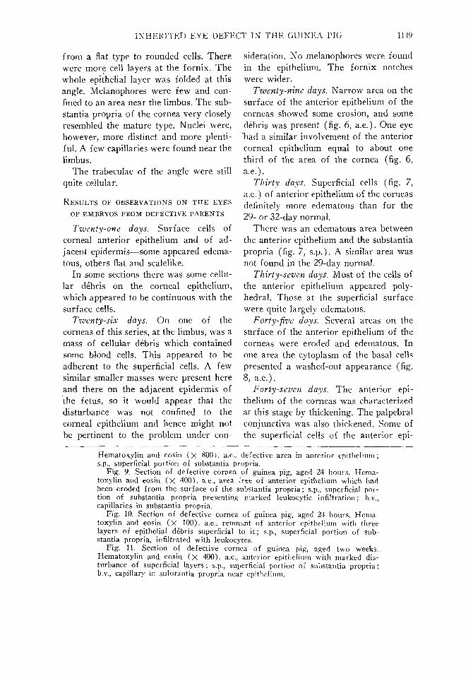
Plate 2 (Fous t ) . Fig. 6. Section of defective cornea of guinea-pig
Hematoxylin and eosin (X 800). a.e., defective area s.p., superficial portion of substantia propria.
Fig. 7. Section of defective cornea of guinea-pig Hematoxylin and eosin (X 400). a.e., defective area s.p., superficial portion of substantia propria.
Fig. 8. Section of defective cornea of guinea-pig embryo, aged 45 days
embryo, aged 29 days. in anterior epithelium;
embryo, aged 30 days. in anterior epithelium;
I N H E R I T E D E Y E D E F E C T IN T H E GUINEA PIG 1149
from a flat type to rounded cells. There were more cell layers at the fornix. The whole epithelial layer was folded at this angle. Melanophores were few and confined to an area near the limbus. The sub-stantia propria of the cornea very closely resembled the mature type. Nuclei were, however, more distinct and more plentiful. A few capillaries were found near the limbus.
The trabeculae of the angle were still quite cellular.
RESULTS OF OBSERVATIONS ON THE EYES OF EMBRYOS FROM DEFECTIVE PARENTS
Twenty-one days. Surface cells of corneal anterior epithelium and of adjacent epidermis—some appeared edema-tous, others flat and scalelike.
In some sections there was some cellular debris on the corneal epithelium, which appeared to be continuous with the surface cells.
Twenty-six days. On one of the corneas of this series, at the limbus, was a mass of cellular debris which contained some blood cells. This appeared to be adherent to the superficial cells. A few similar smaller masses were present here and there on the adjacent epidermis of the fetus, so it would appear that the disturbance was not confined to the corneal epithelium and hence might not be pertinent to the problem under con
sideration. No melanophores were found in the epithelium. The fornix notches were wider.
Twenty-nine days. Narrow area on the surface of the anterior epithelium of the corneas showed some erosion, and some debris was present (fig. 6, a.e.). One eye "had a similar involvement of the anterior corneal epithelium equal to about one third of the area of the cornea (fig. 6, a.e.).
Thirty days. Superficial cells (fig. 7, a.e.) of anterior epithelium of the corneas definitely more edematous than for the 29- or 32-day normal.
There was an edematous area between the anterior epithelium and the substantia propria (fig. 7, s.p.). A similar area was not found in the 29-day normal.
Thirty-seven days. Most of the cells of the anterior epithelium appeared polyhedral. Those at the superficial surface were quite largely edematous.
Forty-five days. Several areas on the surface of the anterior epithelium of the corneas were eroded and edematous. In one area the cytoplasm of the basal cells presented a washed-out appearance (fig. 8, a.e.).
Forty-seven days. The anterior epithelium of the corneas was characterized at this stage by thickening. The palpebral conjunctiva was also thickened. Some of the superficial cells of the anterior epi-
Hematoxylin and eosin ( x 800). a.e, defective area in anterior epithelium; s.p., superficial portion of substantia propria.
Fig. 9. Section of defective cornea of guinea pig, aged 24 hours. Hema-toxylin and eosin ( X 400). a.e., area free of anterior epithelium which had been eroded from the surface of the substantia propria; s.p., superficial portion of substantia propria presenting marked leukocytic infiltration; b.v., capillaries in substantia propria.
Fig. 10. Section of defective cornea of guinea pig, aged 24 hours. Hema-toxylin and eosin (X 100). a.e., remnant of anterior epithelium with three layers of epithelial debris superficial to i t ; s.p., superficial portion of substantia propria, infiltrated with leukocytes.
Fig. 11. Section of defective cornea of guinea pig, aged two weeks.. Hematoxylin and eosin (X 400). a.e., anterior epithelium with marked disturbance of superficial layers; s.p., superficial portion of substantia propria; b.v., capillary in substantia propria near epithelium.
1150 HARRY LEWIS FOUST
thelium were broken up, some were edematous. In some limited areas debris was present; there was also edema of the deeper adjacent epithelial cells. Some edematous cells were so swollen that their outlines were indistinct.
Sections of corneas were stained for keratin with negative results.
Fifty-five days. Near the vertex of the corneas a considerable area presented a change which was peculiar to a particular
series of embryos (B402.2E). The sub-stantia propria was edematous, nearly doubled in thickness, and the nuclei did not take the hematoxylin stain.
The posterior endothelium on some portions of the modified substantia propria was broken down into debris.
In some portions, and especially in one cornea, the anterior epithelium was greatly thickened. The superficial cells in this particular one were edematous. There
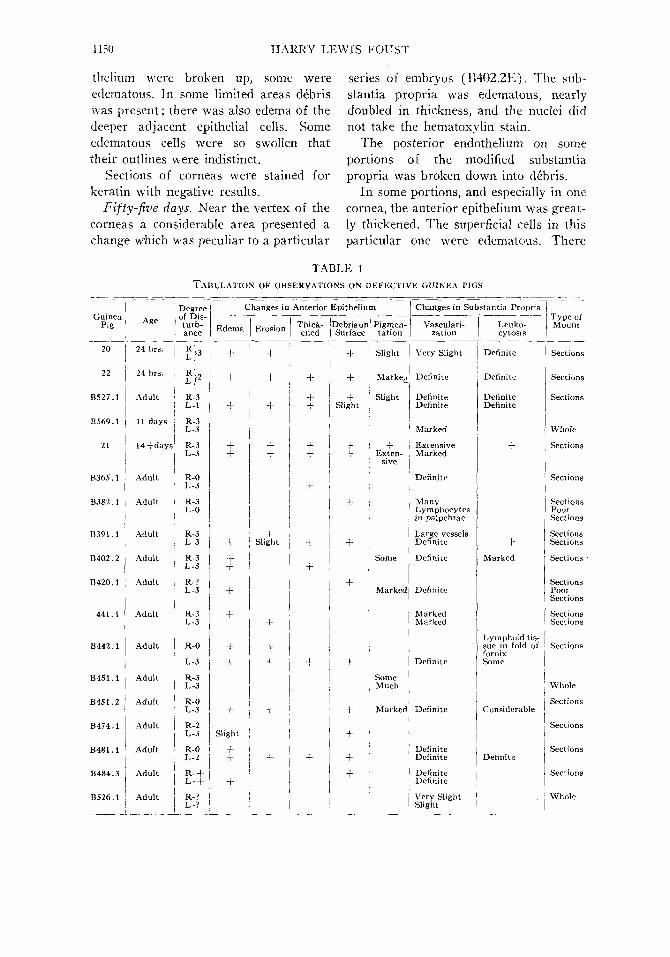
TABLE 1 TABULATION OF OBSERVATIONS ON DEFECTIVE GUINEA FIGS
Guinea Pig
20
22
B527.1
B569.1
21
B365.1
B382.1
B391.1
B402.2
B420.1
441.1
B442.1
B451.1
B451.2
B474.1
B481.1
B484.3
B526.1
Age
24 hrs.
24 hrs.
Adult
11 days
14-Mays
Adult
Adult
Adult
Adult
Adult
Adult
Adult
Adult
Adult
Adult
Adult
Adult
Adult
Degree of Disturbance
Lt ?}' R-3 L-l
R-3 L-3
R-3 L-3
R-0 L-3 R-3 L-0
R-3 L-3
R-3 L-3 R-? L-3
R-3 L-3
R-0
L-3
R-3 L-3
R-0 L-3
R-2 L-3
R-0 L-2
R- + L- + R-? L-?
Changes in
Edema
+
+
+ +
+ + +
+
+
+ +
+ Slight
+ + +
Erosion
+
+
+
+ +
+ Slight
+
+ +
+
+
Anterior Epithelium
Thickened
+ + +
+ +
+
+
+
+
+
Debris on Surface
+
+ +
Slight
+ +
+
+
+
+
+
+
+ +
Pigmentation
Slight
Marke^
Slight
+ Exten
sive
Some
Marked
Some Much
Marked
Changes in Substantia Propria
Vasculari-zation
Very Slight
Definite
Definite Definite
Marked
Extensive Marked
Definite
Many Lymphocytes in palpebrae
Large vessels Definite
Definite
Definite
Marked Marked
Definite
Definite
Definite Definite
Definite Definite
Very Slight Slight
Leuko-cytosis
Definite
Definite
Definite Definite
+
+ Marked
Lymphoid tissue in fold of fornix Some
!
Considerable '
Definite
,
Type of Mount
Sections
Sections
Sections
Whole
Sections
Sections
Sections Poor Sections
Sections Sections
Sections
Sections Poor Sections
Sections Sections
Sections
Whole
Sections
Sections
Sections
Sections
Whole

INHERITED EYE DEFECT IN THE GUINEA PIG 1151
was a bulging of the superficial part of the substantia propria into the base of the thickened, cellular mass.
Sixty days. There were changes here and there in the anterior epithelium of the corneas. In some areas the superficial cells were edematous, the cytoplasm being pale and indistinct. The nuclei of some cells were unstained with hematoxylin, in some other cells vesicular. In some areas the entire epithelial layer appeared to be edematous.
Subconjunctivally, near one fornix on the bulb, the capillaries were filled with leukocytes. The capillaries in the substantia propria near the limbus were somewhat more numerous than in the normal.
EXPERIMENT ON FUNCTIONING OF NERVI VASORUM OF THE CONJUNCTIVA
In order to determine if the vasa nervorum of the conjunctivae of the affected eyes were functioning, an experiment was conducted in which vaso-con-strictor and vaso-dilator substances (adrenalin-chloride solution, 0.1 percent; lactic acid, 1 percent) were used.
Two guinea pigs with characteristically normally functioning conjunctivae and lacrimal apparatus and typically normal exophthalmos were used as checks. One eye was used as a check while into the other was instilled the testing substances. In one case 1 drop of the adrenalin-chloride solution was instilled into the conjunctival sac and the resulting constriction of the blood vessels characterized by paling was noted at varying time intervals. The conjunctival sac of the test eye was then washed with aqueous 0.85 percent sodium-chloride solution. Then 1 drop of 1-percent lactic acid was placed in the conjunctival sac and the consequent reddening of the conjunctiva and injection of blood vessels were noted. The
procedure was reversed on the other test guinea pig.
From the results observed, it was believed that the responses, which appeared to be typical for the substances used, were evident enough to serve as tests for the particular functioning of the abnormal eyes.
Three mature guinea pigs with abnormal eyes ranging in defectiveness from moderate to severe were used. The degree of the severity of the defect was arbitrarily designated : 1, for least severe; 2, for moderate severity; and 3, for most severe. This method was used for referring to the severity of defectiveness throughout this paper. The more severely affected eye of each guinea pig was used. The gross changes in the affected eyes have been described by Whitlock (1935). In addition to the changes given by him, there was marked enophthalmos unless the palpebrae were swollen, in which case there may have been exophthalmos.
The same procedure was followed for the defective eyes as for the normals. In two cases the lactic acid was used first, while in the other case the adrenalin-chloride solution was used first. In all three of the cases studied the responses compared very favorably with those in the normal eye.
Thus it would seem that the blood-supply control was not at fault in affected animals.
DISCUSSION
In comparing the structure of the normal embryonic eyes with that of the descriptions given in the literature, there appeared to be no marked deviations. The distinctness of the anterior chamber had become quite apparent in the 26-day embryo. Harman and Prickett (1932) had described the lens vesicle as beginning to separate from the corneal anlage at 18 days.

1152 H A R R Y L E W I S F O U S T
*?>«* «* —' f -^ 'Xj
^ , - * - » * *v
\ ^
Plate 3 (Fous t ) . Fig. 12. Section of defective cornea of guinea pig, aged 2 weeks. Hematoxyiin
and eosin, understained with hematoxyiin (X 200). a.e., anterior epithelium, a cone-shaped process of epithelium which often remained after partial sloughing of the surface layers is shown; s.p., superficial portion of substantia propria; b.v., capillary.
Fig. 13. Section of defective cornea of guinea pig, adult. Hematoxyiin and eosin ( x 400). a.c., anterior epithelium, somewhat edematous and presenting

I N H E R I T E D E Y E D E F E C T IN T H E GUINEA PIG 1153
In the 25-day embryo, the anterior epithelium of the cornea was composed of two to three layers of indistinct cells. In the later stages studied there was a flattening of the surface cells but no marked increase in number until in the 60-day embryo there had developed an anterior epithelium which resembled that of the adult cornea. In the 45-day embryo, the large cells with large nuclei in the anterior epithelium were thought to be cells similar to those "ellipsoidal" and "club-shaped" cells described by Strieker (1873) and Schaffer (1922) and referred to in the literature review on the anterior corneal epithelium. Melanophores were first observed in the anterior epithelium of the cornea of embryos of 45 days. These, because they were not related to similar contiguous groups of cells, might be considered to have arisen in situ, thus adding evidence to the idea of the ecto-dermal origin of melanophores of Strong (1902), Percival and Stewart (1930), Peck (1931), Ramon-Cajal (1933), Du Shane (1938), Pillat (1933a), and Dorris (1939).
A subepithelial layer of mesenchyme was present in the cornea of the earliest embryo studied. Hensen (1866), La-guesse (1923, 1926), Studnicka (1913), Knape (1909), Hagedoorn (1937), Mann (1928), Wolf rum (1903), Rabl (1900), Grosser (1934), Ayres (1879), and Hert-wig (1899) have variously described the development of the substantia propria. The question of origin is not under consideration here so much as the changes that occurred between the early appearance of the mesenchymatous layer and the appearance of the adult type of tissue.
The substantia propria corneae was observed to change from a cellular tissue in the 25-day embryo to that of the adult type in the 45-day specimen. Some fibers had appeared in the 26-day embryo and there was from that time onward an increase of fibers and of trabeculae in the substantia propria. Laguesse (1923, 1926), Hagedoorn (1937), Knape (1909), and Hertwig (1899) have described in considerable detail the process as being substantially the same. The spaces of Fontana were small in the 32-day stage while the trabeculae were correspondingly large. This relation was maintained up to 60 days. Speciale-Cirincione (1917) has described the formation of the lacunae as having occurred by the separation of cells, of what he termed the "endothelial circle," by the penetration of liquid.
The structure of the cornea of the guinea pig was given in the earlier paper by Whitlock (1935) on the problem being considered further in this paper. The literature corresponds closely with his description. Especially should be mentioned the absence of an anterior elastic membrane as referred to by him and by Ellenberger (1906), Lightbody (1867), and Rubert (1914). The avascularity of the cornea of the guinea pig coincided with that of other animals (noted by Graves, 1936 and Frey, 1875) ; however, in some animals (according to Frey, 1875; Leydig, 1857; Lightbody, 1867; Strieker, 1873; Lodemann, 1917; Ruede-man, 1933), capillary plexuses were found at the periphery of the substantia propria of the cornea and extended cen-tripetally varying distances from the lim-
sections of processes of melanophores; s.p., superficial portion of substantia propria, edematous ; b.v., capillary.
Fig. 14. Portion of whole mount of defective cornea of a guinea pig. The cadaver had been injected intravascularly (common carotid artery) with a colored gelatin mass. Unstained (X 30). b.v., blood vessels in the substantia propria at the vertex corneae. The relation of these vessels to the vessels at the limbus is quite evident on one side of the photograph.

1154 HARRY LEWIS FOUST
bus. Whitlock (1935, figs. 1 and 2) observed the same thing in the eye of the guinea pigs of the stock studied in this work. In this connection reference is made to the literature review on the absence also of the canal of Schlemm (Swindle, 1937; Troncoso and Castro-viejo, 1936; Maggiore 1917; and Whitlock, 1935).
In the experiment in which Berlin blue in solution and diluted India ink were injected into the anterior chamber, there was no gross movement of the mass from the anterior chamber into the spaces of Fontana; thus it would appear that there was no direct communication between anterior chamber and the spaces. This would corroborate the report of Frieden-wald (1936).
In that same experiment, the phago-cytic action of the cells in the walls of the spaces of Fontana was observed. Gasteiger (1934) had found reticulo-endothelial cells in the conjunctiva, sclera, and uvea.
Some evidence of the defect being considered in this paper could be seen in embryos of 21 days. At 29 days, however, the defect was quite pronounced in the anterior epithelium (fig. 6, a.e.). The degree of change appeared to remain at about this level during the remaining portion of the gestation period as judged from the feti studied (figs. 7-8, a.e.).
Briefly summarizing "The tissue changes" as given by Whitlock (1935), there was a vascularization of the sub-stantia propria of the cornea and a variable amount of melanosis due to varying numbers of "stellate pigment cells in the deeper layers of the anterior epithelium." Varying degrees of disturbance of the anterior epithelium were described. The vascularization was often the only change in the substantia propria; however, there may have been leukocytosis or edema resulting in near obliteration of the fibers.
The further studies, that is, the observations upon the normal developing eye, the changes in the defective eye during development, and the tabulation in table 1 of further observations upon defective eyes of guinea pigs, upon which this paper has been based, add data which corroborate Whitlock's (1935) observations. The effect of the defect on the eye in the living animal is well shown in figures 1, 2, 3, and 4. The melanotic cell observed by Whitlock (1935, fig. 4) had a morphology similar to that given by Laidlaw (1932), Strong (1902), Percival and Stewart (1930), Peck (1931), Ramon-Cajal (1933), Du Shane (1938), Pillat (1933b), and Rubert (1914), who had described it as an irregularly shaped cell with dendritic processes, some of which extended into intercellular spaces.
In figure 13, a.e., may be seen sections of processes which appear to be intercellular. Schaffer (1922) and Fol (1896) too have noted this position of pigment. In the same figure there appears to be some pigment in the cytoplasm of the epithelial cells. Figure 4 of Whitlock's (1935) paper also shows pigment granules in the same locus. Pigmentation of the cornea is not uncommon (Ellen-berger, 1906; Bulliard and Champy, 1929; Lightbody, 1867; Fischer, 1905; Lode-mann, 1917; Fischel, 1919b; Duke-Elder, 1934; and Ovio, 1927), Duke-Elder (1934) and Lightbody (1867) having found it more marked near the limbus corneae. Whitlock in his figures 1 and 2 shows this pigmentation. Rubert (1914) too reports it in the guinea pig. From one defective specimen (B569.1, table 1) a drawing was made of the distribution of the pigment cells as seen from the surface of a whole mount of a cornea (fig. 5). This was taken from a very defective specimen, as reference to table 1 shows it to have been of grade 3. Along with the epithelial disturbances shown in figures 6,

INHERITED EYE DEFECT IN THE GUINEA PIG 1155
7, 8, 10, 11, 12, and 13, and those shown in the substantia propria of the same figures and in figure 9, there appears to be an increase in the melanophores. The photophobia (Whitlock, 1935) may be a cause of this proliferation of pigment (Pillat, 1933b). The local disturbance of the corneal tissues may also have been a cause of the excess pigmentation (Com-berg, 1934; Steiner, 1923).
Vascularization of the cornea appears to be a phenomenon associated with irritation (Steiner, 1923; Wintersteiner, 1909; Lightbody, 1867; Strieker, 1873; Long, Holley, and Vorwald, 1933; Julian-elle and Lamb, 1934; Julianelle and Bishop, 1936; and Julianelle, Morris, and Harrison, 1933). In the material here studied the new vessels were largely in the superficial part of the substantia propria (figs. 9, 11, 12, and 13), although occasionally there were some more deeply located (Whitlock, 1935). Vascularity according to Hirsch (1906) is only found at the vertex of the cornea in pathological conditions. That being the case, the great plexus of vessels shown in figure 14 would give an index to the intensity of the disturbances.
The most marked character of the defect and the first to appear (embryo of 21 days) was the disturbance of the anterior epithelium of the cornea. The macroscopic appearance of the eye thus affected is shown in figures 1 to 4. The opacity and the eroded surface are quite evident. The microscopic picture is shown from that of a slight disturbance in figure
13, to more marked disturbances as shown in figures 9, 10, and 11.
SUMMARY
1. Further studies upon an inherited defect of the eye of the guinea pig are here reported.
2. Observations on development of the normal guinea pig eye at intervals from the 25th day of gestation to the 60th day showed the anterior epithelium to grow from two to three cell-layers to four to six layers of cells. The development of the substantia propria corneae was found to follow the descriptions commonly given.
3. The defect was evident in the 21-day embryo as a slight epithelial disturbance which became pronounced at 29 days.
4. The findings of Whitlock (1935) of abnormal vascularization of the cornea with inflammatory changes in the substantia propria and anterior epithelium and a variable amount of melanosis in the epithelium were confirmed by additional data.
Acknowledgement is made of an allotment by the Graduate College of Iowa State College from the Rockefeller Fluid Research Fund which made possible the undertaking of the studies here reported. The writer is especially indebted to Dr. W. V. Lambert for his cooperation and to the Department of Genetics of Iowa State College for supplying the guinea pigs upon which the observations were made.