An In-Vivo Stereoscopic Imaging Device with Pan/Tilt and Integrated Lighting Peter K. Allen and...
38
An In-Vivo Stereoscopic Imaging Device with Pan/Tilt and Integrated Lighting Peter K. Allen and Dennis Fowler Departments of Computer Science & Surgery Columbia University
-
Upload
louis-fenney -
Category
Documents
-
view
219 -
download
5
Transcript of An In-Vivo Stereoscopic Imaging Device with Pan/Tilt and Integrated Lighting Peter K. Allen and...
An In-Vivo Stereoscopic Imaging Device with Pan/Tilt and
Integrated Lighting
Peter K. Allen and Dennis Fowler Departments of Computer Science & Surgery
Columbia University

Surgical Robotics: Research Goals Create simple-to-use and cost-effective surgical robots Convert more “major access” operations to “minimal access”
operations. Focus on abdominal surgery.
Reduce the invasiveness of current minimal access interventions SPA: Single Port Access for laparoscopic surgery NOTES: Natural Orifice Translumenal Endoscopic Surgery Use natural body openings with robotic platforms

Current Generation Robotic Surgery
Devices such as DaVinci®
Huge leap in robotics, but: Large footprint in the OR Cost is extremely high Requires multiple incisions Multiple assistants needed Uses traditional endoscope with limited mobility within body cavity Has not reduced the invasiveness of robotic MIS While this paradigm has been enormously
successful, and has spurred development of new methods and devices, it is ultimately limiting in what it can achieve
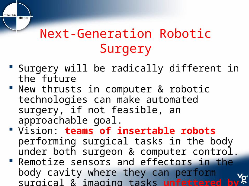
Next-Generation Robotic Surgery
Surgery will be radically different in the future New thrusts in computer & robotic technologies can make
automated surgery, if not feasible, an approachable goal. Vision: teams of insertable robots performing surgical
tasks in the body under both surgeon & computer control. Remotize sensors and effectors in the body cavity where
they can perform surgical & imaging tasks unfettered by traditional endoscopic instrument design.

Building New In-vivo Devices Current minimal access surgery adheres to
the Chopstick Paradigm: Pushing long sticks into small openings

Our Focus: New in-vivo Imaging Devices
Can we improve on the traditional laparoscope?
Laparoscope Issues: Narrow angle imaging Limited workspace Multiple incisions for camera
placements Counter intuitive motion for
control Trained assistants needed to
control the camera Multiple incisions for camera
placements Additional incisions needed
for laparoscopic instruments.

Device must be fully insertable into body cavity, leaving the insertion port free for other sensors and tooling
Device diameter must be restricted to 15 mm diameter for use with standard trocars.
Pan and Tilt degrees of freedom needed to increase internal imaging field of view
Image Zoom function required Integrated lighting Simple intuitive control interface to operator Real-time computer control of DOF’s to allow tracking
and visual servoing User friendly 2D/3D display system Low cost and possible disposal use
Columbia Imaging Device: Design Goals

Columbia Imaging Device Overview
Design 0: Paper design, 2 cameras, 5-DOF Device 0: Single camera prototype, 3-DOF, tested in
surgical trainer Device I: Single camera, pan/tilt/lighting, tested in
animals Device II: Single camera, pan/tilt/zoom, tested in
animals Device III: Stereo cameras, pan/tilt, tested in animals
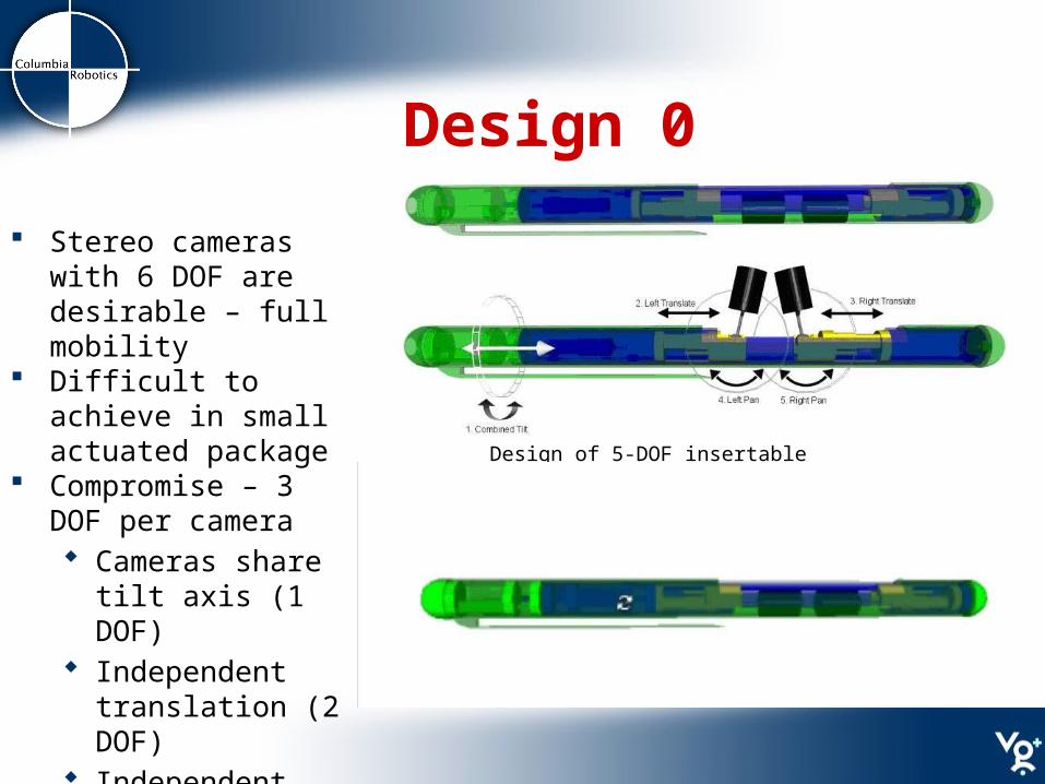
Design 0
Design of 5-DOF insertable camera device
Stereo cameras with 6 DOF are desirable – full mobility
Difficult to achieve in small actuated package
Compromise – 3 DOF per camera Cameras share tilt
axis (1 DOF) Independent
translation (2 DOF) Independent pan
(2 DOF)
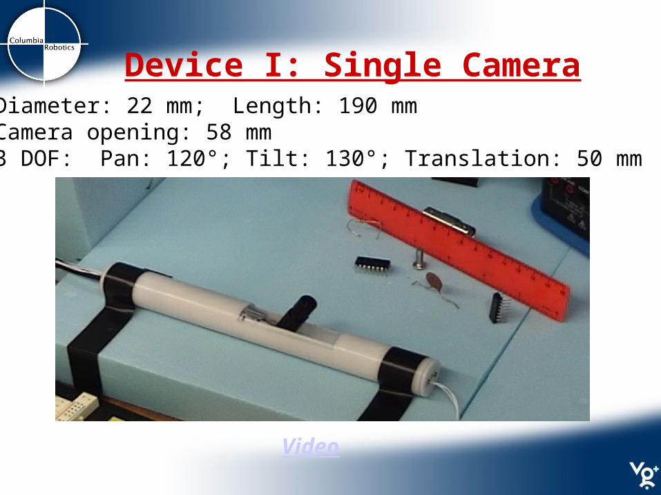
Device I: Single CameraDiameter: 22 mm; Length: 190 mmCamera opening: 58 mm3 DOF: Pan: 120°; Tilt: 130°; Translation: 50 mm
Video

Initial Testing and Validation Does new imaging device improve surgery visualization? 6 fellows & surgeons performed MISTELS* tests with
standard laparoscope and the new robotic camera 5 of 6 subjects showed no significant difference in
MISTELS task performance with the robotic camera compared to the standard laparoscope
Mean score of 999 +/- 69 using a laparoscope Mean score of 953 +/- 68 for the robotic camera:
statistically insignificant difference
*McGill Inanimate System for the Training and Evaluation of Laparoscopic Skill
Device I: Design Goals Need to reduce size to fit 12mm trocar Motors are major determinant of device size Removing a camera reduced motor count by 2 Translation DOF is least useful. Removing this also
reduced motor count by 1 Include integrated light source Make imaging head modular Tradeoff: Degrees-of-freedom for compactness

Device I: Single Camera
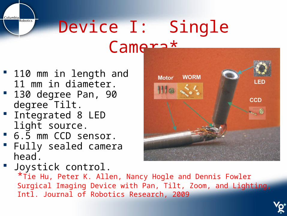
Device I: Single Camera*
110 mm in length and 11 mm in diameter.
130 degree Pan, 90 degree Tilt.
Integrated 8 LED light source. 6.5 mm CCD sensor. Fully sealed camera head. Joystick control.
*Tie Hu, Peter K. Allen, Nancy Hogle and Dennis Fowler Surgical Imaging Device with Pan, Tilt, Zoom, and Lighting, Intl. Journal of Robotics Research, 2009

LED Light Source Light-emitting diode (LED) as
a light source in laparoscopy: Lower power Higher efficiency Compact package Longer lifespan Lower cost
Luxeon portable PWT white LED(LXCL_PWT1) 2.0 X1.6 X 0.7 mm 26 lumens of light at 350 mA
8 PWT LED in a printed circuit board with 9mm diameter. 208 lumens light at 8.4 w

Lens and Camera Unit
Pin hole lens (PTS 5.0 from Universe Kogaku America) Focal length 5.0 mm. F number 4. Angle of view D-H-V(85.4-68.3-50.9 ).
6.5 mm CCD camera sensor. NET USA Inc, CSH-1.4-V4-END-R1. 450 TV lines in horizontal resolution and 420 TV lines in vertical
resolution. Fully sealed package to isolate body fluid and moisture.

Pan/Tilt Mechanism
Miniature Brushless DC motor (0513G, Faulhaber Group). 25mNm torque. 5.8 mm in diameter.
Miniature worm gear (Kleiss Gear Inc.) gear ratio 16:1.

Device II:Pan, Tilt, Zoom
Mechanical zoom: linear motion of camera head
Stepper motor drives rack and pinion mechanism
Can only achieve ~ 2x zoom

System Architecture
Mounting the Camera
Camera attached to insuflated abdominal wall Attachment methods:
Suturing: small stitch through abdomen Magnets “Fish Hook” which grabs the abdominal wall Intelligent trocar for attachment
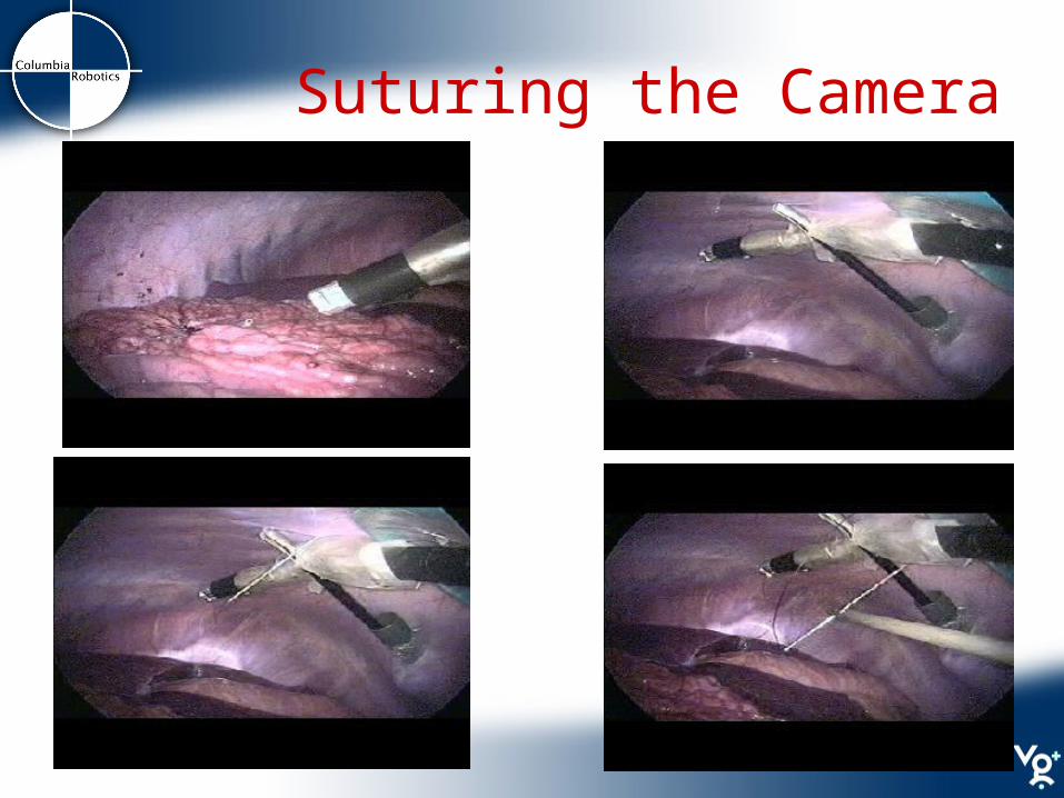
Suturing the Camera

In-Vivo Animal Experiments
Bowel Running Appendectomy
Suturing Nephrectomy
Video
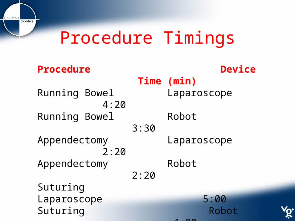
Procedure Timings
Procedure Device Time (min)Running Bowel Laparoscope 4:20Running Bowel Robot 3:30Appendectomy Laparoscope 2:20Appendectomy Robot 2:20Suturing Laparoscope 5:00Suturing Robot 4:00Nephrectomy Laparoscope 18:00Nephrectomy Robot 21:00

Intelligent Software
Position/Velocity control of axes Intuitive Joystick Control Real-Time Image Processing:
Digital Zoom Image rotation/stabilization Distortion Correction Picture-in-Picture Visual Servoing/Tracking 3D Stereo output
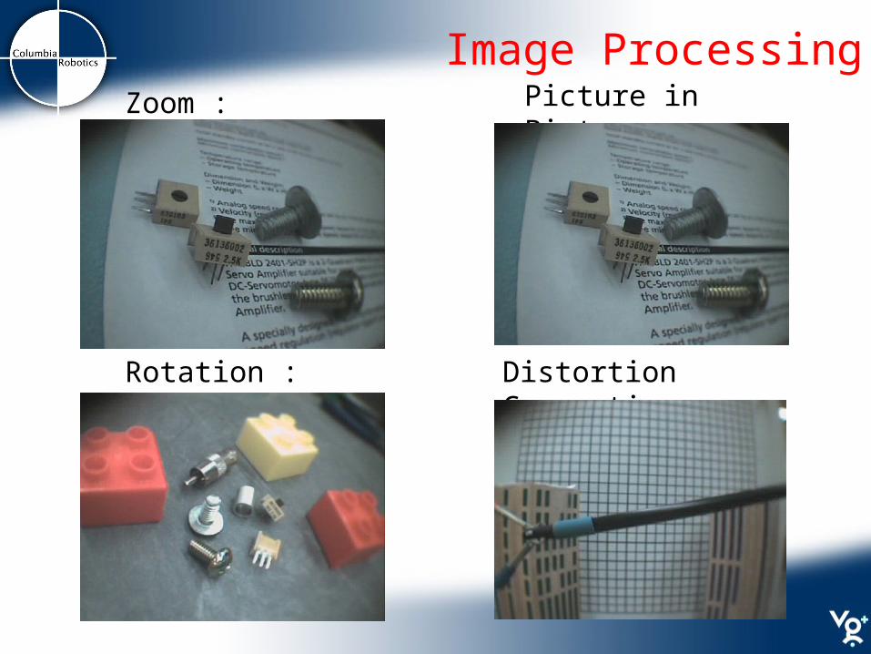
Image ProcessingZoom :
Distortion Correction :Rotation :
Picture in Picture :
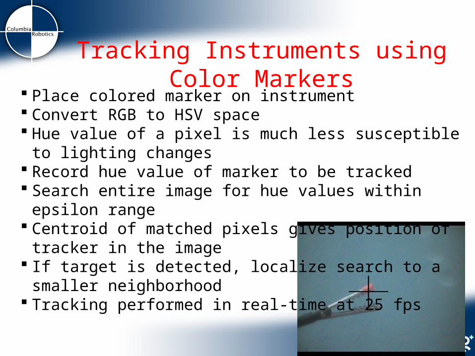
Place colored marker on instrument Convert RGB to HSV space Hue value of a pixel is much less susceptible to lighting changes Record hue value of marker to be tracked Search entire image for hue values within epsilon range Centroid of matched pixels gives position of tracker in the image If target is detected, localize search to a smaller neighborhood Tracking performed in real-time at 25 fps
Tracking Instruments using Color Markers

Visual Servoing Allows shared autonomy with surgeon The feedback from the tracker can be used to drive motors to
keep the tool in the center of the image PD controller used ( Ex , Ey ): offset error of tracker from center of image
Pan speed ( x * Ex ) – ( x * dEx/dt )
Tilt speed ( y * Ey ) – ( y * dEy/dt )
Video

Device III: Stereo Imaging*
• A stereo imaging device with similar mechanical design. • 15 mm in diameter and 120 mm in length.• 6.5mm Inter-Pupillary Distance (IPD)
*T. Hu, P. Allen,, T. Nadkarni, N. Hogle, D. Fowler, Insertable Stereoscopic 3D surgical imaging device, IEEE BIOROB 2008
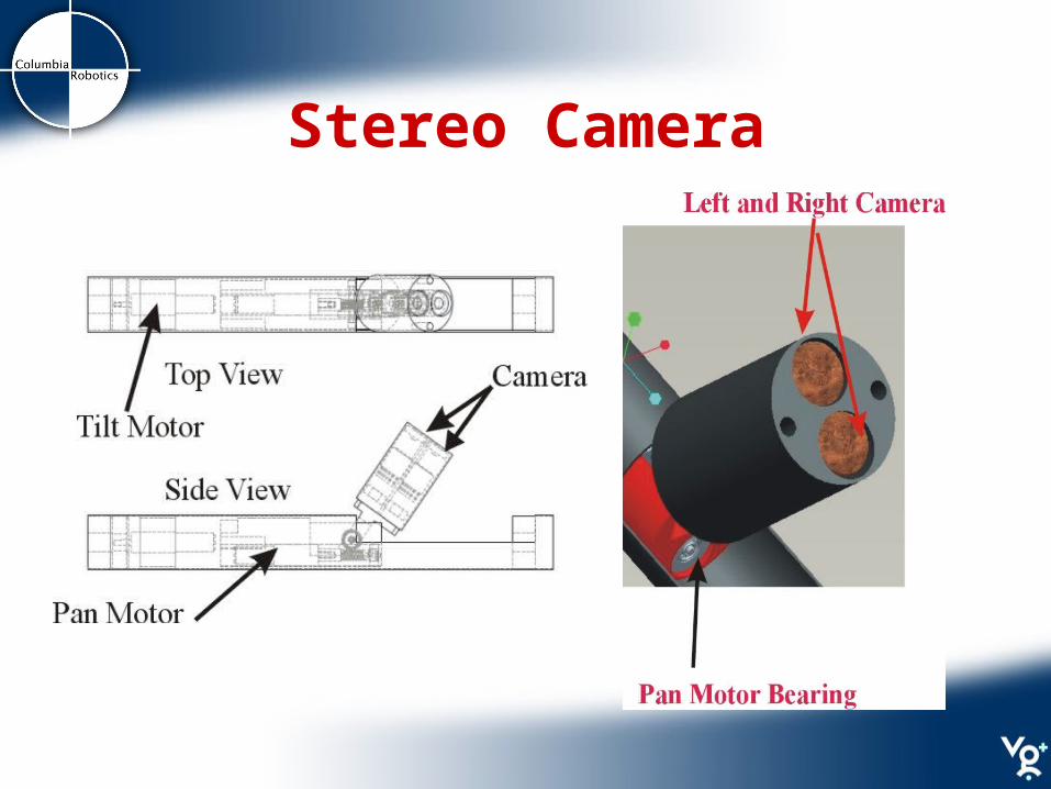
Stereo Camera

eMagin Z800 Head-Mounted VR Display- Uncomfortable
- Single User
RealD Crystal Eyes shutter glasses- Uncomfortable over longer periods
- Need to maintain Line Of Sight with
synchronizing emitter
True Vision back projected 3D display- Low incremental cost for additional users
- Bigger display size
- Passive polarization, lightweight glasses
3D Displays

Visual Servoing with StereoWhen using stereo cameras the pixel disparity Ep between
stereo images is used to damp the motors Speed Damping ( * Ep )Damping is applied to both Pan and Tilt motorsPrevents the motors from oscillating when instrument is too
close to camera

Device III: Stereo Imaging
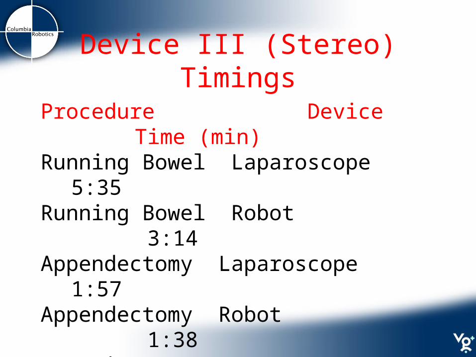
Device III (Stereo) Timings
Procedure Device Time (min)
Running Bowel Laparoscope 5:35
Running Bowel Robot 3:14
Appendectomy Laparoscope 1:57
Appendectomy Robot 1:38
Suturing Laparoscope 4:30
Suturing Robot 2:12
Nephrectomy Robot 9:59

3D Trajectory Reconstruction
The Flock of Birds (FoB) sensor can transmit the position of its sensor w.r.t. its base
Accuracy within 1.8mm Refresh rate up to 144Hz By placing an optical marker
on the FoB sensor we can track its position in the image
By tracking the sensor using stereo cameras we can compute its 3D trajectory
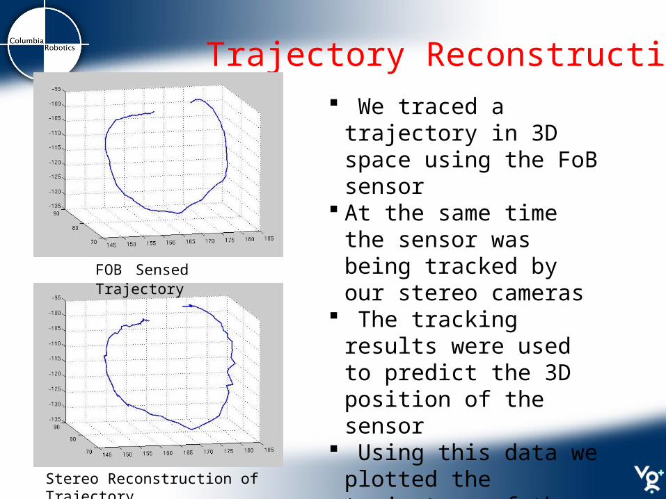
Trajectory Reconstruction We traced a trajectory in 3D
space using the FoB sensor At the same time the sensor
was being tracked by our stereo cameras
The tracking results were used to predict the 3D position of the sensor
Using this data we plotted the trajectory of the sensor
average reprojection error ~3mm
FOB Sensed Trajectory
Stereo Reconstruction of Trajectory
Recap: New device is easier and more intuitive to use than a
standard laparoscope. Insertion port available for tooling Joystick operation requires no specialized operator training. Pan/Tilt functions provide large imaging volume not
restricted by fulcrum point of standard laparoscope Time to perform procedures was better or equivalent to a
standard laparoscope. Automatic Tracking and Visual Servoing assist surgeon 3D vision system significantly improves the visualization and
depth perception of the surgeon. Trajectories can be tracked over time with 3D reconstruction Cost effective, perhaps single-use or modular replacement

Acknowledgements Austin Reiter Dennis Fowler Tie Hu Andrew Miller Tejas Nadkarni Nancy Hogle Nabil Simaan Kai Xu Roger Goldman Jienan DingThis work was supported by NIH grants 1R21EB004999-01A1
and 5R21EB007779-02