An electrochemical immunosensor based on agarose hydrogel films for rapid determination of...
6
An electrochemical immunosensor based on agarose hydrogel films for rapid determination of ractopamine Li Shen b , Pingli He a, * a National Key Laboratory of Animal Nutrition, China Agricultural University, Beijing 100094, China b College of Chemistry, Peking University, Beijing 100871, China Received 3 October 2006; accepted 23 October 2006 Available online 27 November 2006 Abstract A novel label-free electrochemical immunosensor for rapid determination of ractopamine was constructed by incorporating ractop- amine–bovine thyroglobulin antigen in agarose hydrogel films modified on a glassy carbon electrode. Cyclic voltammetry and differential pulse voltammetry were employed to investigate the electrochemical character of the immunosensor during different modified stages using a redox probe system of K 3 Fe(CN) 6 /K 4 Fe(CN) 6 . In the presence of polyclonal antibody against ractopamine, K 3 [Fe(CN) 6 ] elec- tron transfer is inhibited, presumably due to that antibody in solution could adsorb on the electrode surface modified antigen. A com- petitive immunoreaction system was applied to determinate the free ractopamine in phosphate buffer solution. The selected polyclonal antibody showed very high sensitivity and specificity for ractopamine, and was used for the detection and quantitative determination of trace amounts of ractopamine in spiked animal feeds. Ó 2006 Elsevier B.V. All rights reserved. Keywords: Ractopamine; Immunosensor; Electrochemistry; Agarose hydrogel; Feed 1. Introduction Ractopamine is a b-agonist belonging to the phenolic group. In the livestock industry, b-agonists have been used as repartitioning agents and there is a large body of research showing that b-agonists reduce carcass fat and increase muscle mass while improving growth rate and feed conversion when fed to calves [1], pigs [2], and poultry [3]. Except clenbuterol, ractopamine became the most effective b-agonist as growth promoting agent [4]. However, the presence of drug residues in animal tissues is a food safety concern. Meat products obtained from agonist-treated ani- mals may pose a potential risk for consumer health, partic- ularly in persons with muscular tremors, vomiting, nervousness, and cardiac palpitations [5]. Various analytical methods have been reported for the determination of ractopamine in animal feeds and ractop- amine residues in animal tissues. These included liquid chromatography [6–9], gas chromatography [10,11], enzyme-linked immunoassay [12–15], and surface plasmon resonance immunosensor [16]. Recently, electrochemical immunosensors have gained growing attention since they combine the high specificity of traditional immunoassay methods with the low detection limits and low expenses of electrochemical measurement system, and the fact that they are fast, accurate, simple, portable, and inexpensive. The method has been used by a number of investigators for determination of various analytes with great sensitivity and specificity [17–19]. Various types of electrodes, such as glassy carbon electrodes [20,21], gold electrodes [22], carbon paste electrodes [23], and screen-printed elec- trodes [24,25], have been used to immobilization of immunoreagent. It has been reported that the crucial aspect of electro- chemical immunosensor is the immobilization of immuno- logical sensitivity compounds on the electrodes. The biological macromolecules immobilized in films on 1388-2481/$ - see front matter Ó 2006 Elsevier B.V. All rights reserved. doi:10.1016/j.elecom.2006.10.049 * Corresponding author. E-mail address: hepl@mafic.ac.cn (P. He). www.elsevier.com/locate/elecom Electrochemistry Communications 9 (2007) 657–662
Transcript of An electrochemical immunosensor based on agarose hydrogel films for rapid determination of...
www.elsevier.com/locate/elecom
Electrochemistry Communications 9 (2007) 657–662
An electrochemical immunosensor based on agarose hydrogel filmsfor rapid determination of ractopamine
Li Shen b, Pingli He a,*
a National Key Laboratory of Animal Nutrition, China Agricultural University, Beijing 100094, Chinab College of Chemistry, Peking University, Beijing 100871, China
Received 3 October 2006; accepted 23 October 2006Available online 27 November 2006
Abstract
A novel label-free electrochemical immunosensor for rapid determination of ractopamine was constructed by incorporating ractop-amine–bovine thyroglobulin antigen in agarose hydrogel films modified on a glassy carbon electrode. Cyclic voltammetry and differentialpulse voltammetry were employed to investigate the electrochemical character of the immunosensor during different modified stagesusing a redox probe system of K3Fe(CN)6/K4Fe(CN)6. In the presence of polyclonal antibody against ractopamine, K3[Fe(CN)6] elec-tron transfer is inhibited, presumably due to that antibody in solution could adsorb on the electrode surface modified antigen. A com-petitive immunoreaction system was applied to determinate the free ractopamine in phosphate buffer solution. The selected polyclonalantibody showed very high sensitivity and specificity for ractopamine, and was used for the detection and quantitative determination oftrace amounts of ractopamine in spiked animal feeds.� 2006 Elsevier B.V. All rights reserved.
Keywords: Ractopamine; Immunosensor; Electrochemistry; Agarose hydrogel; Feed
1. Introduction
Ractopamine is a b-agonist belonging to the phenolicgroup. In the livestock industry, b-agonists have been usedas repartitioning agents and there is a large body ofresearch showing that b-agonists reduce carcass fat andincrease muscle mass while improving growth rate and feedconversion when fed to calves [1], pigs [2], and poultry [3].Except clenbuterol, ractopamine became the most effectiveb-agonist as growth promoting agent [4]. However, thepresence of drug residues in animal tissues is a food safetyconcern. Meat products obtained from agonist-treated ani-mals may pose a potential risk for consumer health, partic-ularly in persons with muscular tremors, vomiting,nervousness, and cardiac palpitations [5].
Various analytical methods have been reported for thedetermination of ractopamine in animal feeds and ractop-
1388-2481/$ - see front matter � 2006 Elsevier B.V. All rights reserved.
doi:10.1016/j.elecom.2006.10.049
* Corresponding author.E-mail address: [email protected] (P. He).
amine residues in animal tissues. These included liquidchromatography [6–9], gas chromatography [10,11],enzyme-linked immunoassay [12–15], and surface plasmonresonance immunosensor [16]. Recently, electrochemicalimmunosensors have gained growing attention since theycombine the high specificity of traditional immunoassaymethods with the low detection limits and low expensesof electrochemical measurement system, and the fact thatthey are fast, accurate, simple, portable, and inexpensive.The method has been used by a number of investigatorsfor determination of various analytes with great sensitivityand specificity [17–19]. Various types of electrodes, such asglassy carbon electrodes [20,21], gold electrodes [22],carbon paste electrodes [23], and screen-printed elec-trodes [24,25], have been used to immobilization ofimmunoreagent.
It has been reported that the crucial aspect of electro-chemical immunosensor is the immobilization of immuno-logical sensitivity compounds on the electrodes. Thebiological macromolecules immobilized in films on
658 L. Shen, P. He / Electrochemistry Communications 9 (2007) 657–662
electrode surfaces should exhibit their specific immunoac-tivity, retain their native structure and demonstrateexcellent stability. For these reasons a number of differenttype films, such as conducting polymer, titania sol–gel, andself-assemble film, have been used to immobilize immuno-genic molecule and achieve their immune response[20,26,27]. Among them, cast method has been widely usedmainly because of its simplicity and versatility, and castfilms provided a good microenvironment for the immuno-reaction between antigen immobilized on electrode and thespecific antibody in solution. For example, paraoxon anti-body was incorporated in gold nanoparticles films modifiedon glassy carbon electrodes as a label-free electrochemicalimmunosensor [28]. Natural biomacromolecule such asagarose hydrogel, because of its better biocompatibilitywith proteins, has been used to immobilize immunogenicmolecules on the electrode surface, and realized the deter-mination of ferritin in serum [21]. However, few immuno-sensors were used to detect small molecular haptenswhich are difficult to immobilize and have little effect onelectron transfer of the electrochemical mediator insolution.
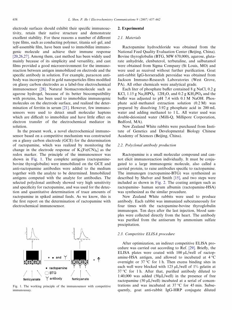
In the present work, a novel electrochemical immuno-sensor based on a competitive mechanism was constructedon a glassy carbon electrode (GCE) for the determinationof ractopamine, which was realized by monitoring thechange in the electrode response of K3[Fe(CN)6] as theredox marker. The principle of the immunosensor wasshown in Fig. 1. The complete antigens (ractopamine-bovine thyroglobulin) were immobilized on the GCE andanti-ractopamine antibodies were added to the mediumtogether with the analyte to be determined. Immobilizedantigens competed with the analyte for antibodies. Theselected polyclonal antibody showed very high sensitivityand specificity for ractopamine, and was used for the detec-tion and quantitative determination of trace amounts ofractopamine in spiked animal feeds. As we know, this isthe first report on the determination of ractopamine withelectrochemical immunosensor.
Fig. 1. The working principle of the immunosensor with competitiveimmunoassay.
2. Experimental
2.1. Materials
Ractopamine hydrochloride was obtained from theNational Feed Quality Evaluation Center (Beijing, China).Bovine thyroglobulin (BTG, MW 670,000), agarose, gluta-rate anhydride, clenbuterol, terbutaline, and salbutamolwere obtained from Sigma Company (St Louis, MO) andwere used as received without further purification. Goatanti-rabbit IgG-horseradish peroxidae was obtained fromJackson Immuno-Research Laboratories (West Grove,PA). All other chemicals were analytical grade.
Each liter of phosphate buffer contained 8 g NaCl, 0.2 gKCl, 1.15 g Na2HPO4 Æ 12H2O, and 0.2 g KH2PO4 and thebuffer was adjusted to pH 7.4 with 0.1 M NaOH. Phos-phate acid–methanol extraction solution (0.2 M) wasprepared by dissolving 3.92 g phosphate acid in 200 mLwater and adding methanol to 1 L. All water used wasdouble-deionized water (Milli-Q, Millipore Corporation,Bedford, MA).
New Zealand White rabbits were purchased from Insti-tute of Genetics and Developmental Biology ChineseAcademy of Sciences (Beijing, China).
2.2. Polyclonal antibody production
Ractopamine is a small molecular compound and can-not elicit immunoreaction individually. It must be conju-gated to a large immunogenic molecule, also called acarried protein, to raise antibodies specific to ractopamine.The immunogen (ractopamine-BTG) was synthesized asdescribed by Shelver and Smith [15], and two steps wereincluded as shown in Fig. 2. The coating antigen such asractopamine- human serum albumin (ractopamine-HSA)was synthesized as the similar procedure.
New Zealand White rabbits were used to produceantibody. Each rabbit was immunized subcutaneously forfour times with the ractopamine-bovine thyroglobulinimmunogen. Ten days after the last injection, blood sam-ples were collected directly from the heart. The antibodywas purified from the antiserum by ammonium sulfateprecipitation.
2.3. Competitive ELISA procedure
After optimization, an indirect competitive ELISA pro-cedure was carried out according to Ref. [29]. Briefly, theELISA plates were coated with 100 lL/well of ractop-amine-HSA antigen, and allowed to incubated at 4 �Covernight or 37 �C for 1 h. Then excess binding sites ineach well were blocked with 125 lL/well of 1% gelatin at37 �C for 1 h. After that, purified antibody diluted to1:40,000 was added (50lL/well) in the presence of freeractopamine (50 lL/well) incubated at a serial of concen-trations and was incubated at 37 �C for 45 min. Subse-quently, goat anti-rabbit IgG-HRP conjugate diluted
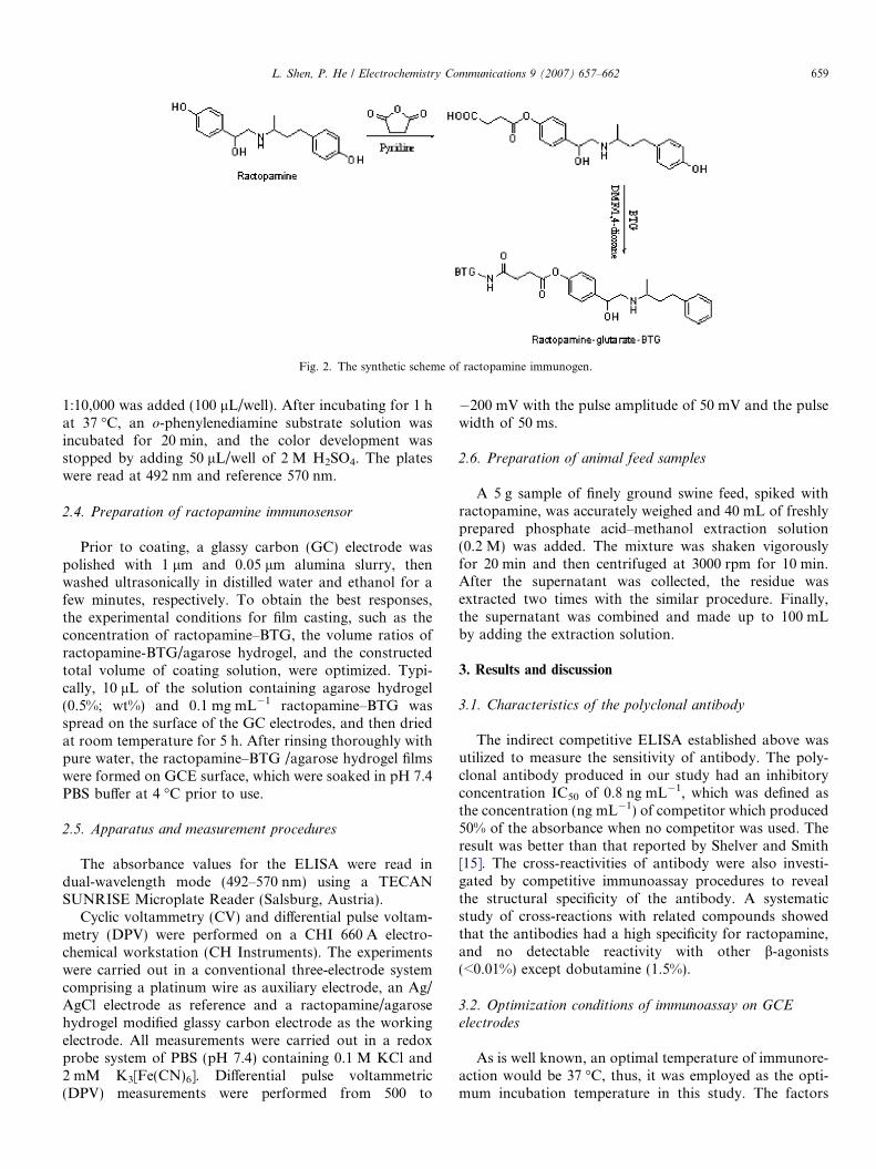
Fig. 2. The synthetic scheme of ractopamine immunogen.
L. Shen, P. He / Electrochemistry Communications 9 (2007) 657–662 659
1:10,000 was added (100 lL/well). After incubating for 1 hat 37 �C, an o-phenylenediamine substrate solution wasincubated for 20 min, and the color development wasstopped by adding 50 lL/well of 2 M H2SO4. The plateswere read at 492 nm and reference 570 nm.
2.4. Preparation of ractopamine immunosensor
Prior to coating, a glassy carbon (GC) electrode waspolished with 1 lm and 0.05 lm alumina slurry, thenwashed ultrasonically in distilled water and ethanol for afew minutes, respectively. To obtain the best responses,the experimental conditions for film casting, such as theconcentration of ractopamine–BTG, the volume ratios ofractopamine-BTG/agarose hydrogel, and the constructedtotal volume of coating solution, were optimized. Typi-cally, 10 lL of the solution containing agarose hydrogel(0.5%; wt%) and 0.1 mg mL�1 ractopamine–BTG wasspread on the surface of the GC electrodes, and then driedat room temperature for 5 h. After rinsing thoroughly withpure water, the ractopamine–BTG /agarose hydrogel filmswere formed on GCE surface, which were soaked in pH 7.4PBS buffer at 4 �C prior to use.
2.5. Apparatus and measurement procedures
The absorbance values for the ELISA were read indual-wavelength mode (492–570 nm) using a TECANSUNRISE Microplate Reader (Salsburg, Austria).
Cyclic voltammetry (CV) and differential pulse voltam-metry (DPV) were performed on a CHI 660 A electro-chemical workstation (CH Instruments). The experimentswere carried out in a conventional three-electrode systemcomprising a platinum wire as auxiliary electrode, an Ag/AgCl electrode as reference and a ractopamine/agarosehydrogel modified glassy carbon electrode as the workingelectrode. All measurements were carried out in a redoxprobe system of PBS (pH 7.4) containing 0.1 M KCl and2 mM K3[Fe(CN)6]. Differential pulse voltammetric(DPV) measurements were performed from 500 to
�200 mV with the pulse amplitude of 50 mV and the pulsewidth of 50 ms.
2.6. Preparation of animal feed samples
A 5 g sample of finely ground swine feed, spiked withractopamine, was accurately weighed and 40 mL of freshlyprepared phosphate acid–methanol extraction solution(0.2 M) was added. The mixture was shaken vigorouslyfor 20 min and then centrifuged at 3000 rpm for 10 min.After the supernatant was collected, the residue wasextracted two times with the similar procedure. Finally,the supernatant was combined and made up to 100 mLby adding the extraction solution.
3. Results and discussion
3.1. Characteristics of the polyclonal antibody
The indirect competitive ELISA established above wasutilized to measure the sensitivity of antibody. The poly-clonal antibody produced in our study had an inhibitoryconcentration IC50 of 0.8 ng mL�1, which was defined asthe concentration (ng mL�1) of competitor which produced50% of the absorbance when no competitor was used. Theresult was better than that reported by Shelver and Smith[15]. The cross-reactivities of antibody were also investi-gated by competitive immunoassay procedures to revealthe structural specificity of the antibody. A systematicstudy of cross-reactions with related compounds showedthat the antibodies had a high specificity for ractopamine,and no detectable reactivity with other b-agonists(<0.01%) except dobutamine (1.5%).
3.2. Optimization conditions of immunoassay on GCE
electrodes
As is well known, an optimal temperature of immunore-action would be 37 �C, thus, it was employed as the opti-mum incubation temperature in this study. The factors
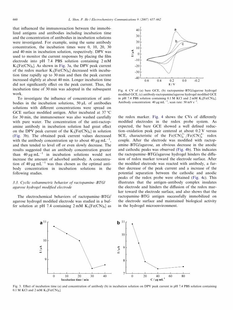
Fig. 4. CV of (a) bare GCE, (b) ractopamine–BTG/agarose hydrogelmodified GCE, (c) antibody-ractopamine/agarose hydrogel modified GCEin pH 7.4 PBS solution containing 0.1 M KCl and 2 mM K3[Fe(CN)6].Antibody concentration: 40 lg mL�1, scan rate: 50 mV s�1.
660 L. Shen, P. He / Electrochemistry Communications 9 (2007) 657–662
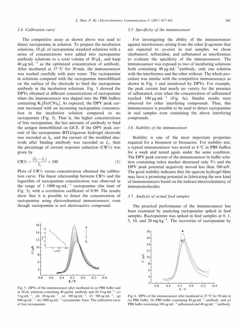
that influenced the immunoreaction between the immobi-lized antigens and antibodies including incubation timeand the concentration of antibodies in incubation solutionswere investigated. For example, using the same antibodyconcentration, the incubation times were 0, 10, 20, 30and 40 min in incubation solution, respectively. DPV wasused to monitor the current responses by placing the filmelectrode into pH 7.4 PBS solution containing 2 mMK3[Fe(CN)6]. As shown in Fig. 3a, the DPV peak currentof the redox marker K3[Fe(CN)6] decreased with incuba-tion time rapidly up to 30 min and then the peak currentincreased slightly at about 40 min. Longer incubation timedid not significantly effect on the peak current. Thus, theincubation time of 30 min was adopted in the subsequentwork.
To investigate the influence of concentration of anti-bodies in the incubation solutions, 50 lL of antibodiessolutions with different concentrations were spread onGCE surface modified antigen. After incubated at 37 �Cfor 30 min, the immunosensor was also washed carefullywith pure water. The concentration of the anti-ractop-amine antibody in incubation solution had great effecton the DPV peak current of the K3[Fe(CN)6] in solution(Fig. 3b). The obtained peak current values decreasedwith the antibody concentration up to about 40 lg mL�1,and then tended to level off or even slowly decrease. Theresults suggested that an antibody concentration greaterthan 40 lg mL�1 in incubation solutions would notincrease the amount of adsorbed antibody. A concentra-tion of 40 lg mL�1 was thus chosen as the optimal anti-body concentration in incubation solutions in thefollowing studies.
3.3. Cyclic voltammetric behavior of ractopamine–BTG/
agarose hydrogel modified electrode
The electrochemical behaviors of ractopamine–BTG/agarose hydrogel modified electrode was studied in a buf-fer solution at pH 7.4 containing 2 mM K3[Fe(CN)6] as
Fig. 3. Effect of incubation time (a) and concentration of antibody (b) in incu0.1 M KCl and 2 mM K3[Fe(CN)6].
the redox marker. Fig. 4 shows the CVs of differentlymodified electrodes in the redox probe system. Asexpected, the bare GCE showed a well defined reduc-tion–oxidation peak pair centered at about 0.2 V versusSCE, characteristic of the FeðCNÞ3�6 =FeðCNÞ4�6 redoxcouple. After the electrode was modified with ractop-amine–BTG/agarose, an obvious decrease in the anodicand cathodic peaks was observed (Fig. 4b). This indicatesthe ractopamine–BTG/agarose hydrogel hinders the diffu-sion of redox marker toward the electrode surface. Afterthe modified electrode was reacted with antibody, a fur-ther decrease of the peak current and a increase of thepotential separation between the cathodic and anodicpeaks of the redox probe were obtained (Fig. 4c). Thisillustrates that the antigen–antibody complex insulatesthe electrode and hinders the diffusion of the redox mar-ker toward the electrode surface, and also shows that theractopamine–BTG antigen successfully immobilized onthe electrode surface and maintained biological activityin the hydrogel microenvironment.
bation solution on DPV peak current in pH 7.4 PBS solution containing
L. Shen, P. He / Electrochemistry Communications 9 (2007) 657–662 661
3.4. Calibration curve
The competitive assay as shown above was used todetect ractopamine in solution. To prepare the incubationsolutions, 10 lL of ractopamine standard solutions with aseries of concentrations were added into ractopamineantibody solutions to a total volume of 50 lL, and keep40 lg mL�1 as the optimized concentration of antibody.After incubated at 37 �C for 30 min, the immunosensorwas washed carefully with pure water. The ractopaminein solutions competed with the ractopamine immoblilizedon the surface of the electrode to bind the ractopamineantibody in the incubation solutions. Fig. 5 showed theDPVs obtained at different concentrations of ractopaminewhen the immunosensor was dipped into the PBS bufferscontaining K3[Fe(CN)6]. As expected, the DPV peak cur-rent increased with an increasing ractopamine concentra-tion in the incubation solution compared to noractopamine (Fig. 5). That is, the higher concentrationsof free ractopamine, the less amounts of antibody to bindthe antigen immoblilized on GCE. If the DPV peak cur-rent of the ractopamine–BTG/agarose hydrogel electrodewas recorded as I0, and the current of the modified elec-trode after binding antibody was recorded as Ix, thenthe percentage of current response reduction (CR%) wasgiven by
CR% ¼ ðI0 � IxÞIx
� 100 ð1Þ
Plots of CR% versus concentration obtained the calibra-tion curve. The linear relationship between CR% and thelogarithm of ractopamine concentration was observed inthe range of 1–1000 ng mL�1 ractopamine (the inset ofFig. 5), with a correlation coefficient of 0.99. The resultsshow that it is possible to detect the concentration ofractopamine using electrochemical immunosensor, eventhough ractopamine is not electroactive compound.
Fig. 5. DPVs of the immunosensor after incubated in (a) PBS buffer andin 50 lL solutions containing 40 lg/mL antibody and (b) 0 ng mL�1, (c)5 ng mL�1, (d) 20 ng mL�1, (e) 100 ng mL�1, (f) 500 ng mL�1, (g)800 ng mL�1, (h) 1000 ng mL�1 ractopamine. Inset: The calibration curveof free ractopamine.
3.5. Specificity of the immunosensor
For investigating the ability of the immunosensoragainst interferences arising from the other b-agonists thatare expected to co-exist in real samples, we choseclenbuterol, terbutaline, and salbutamol as interferencesto evaluate the specificity of the immunosensor. Theimmunosensor was exposed to two of incubating solutionsboth containing 40 lg mL�1antibody, only one solutionwith the interference and the other without. The whole pro-cedure was similar with the competitive immunoassays asshown in Fig. 1 and monitored by DPVs. For example,the peak current had nearly no variety for the presenceof salbutamol, even when the concentration of salbutamolreached 100 lg mL�1 (Fig. 6c). Similar results wereobserved for other interfering compounds. Thus, thisimmunosensor is possible to be used to detect ractopaminein real samples even containing the above interferingcompounds.
3.6. Stability of the immunosensor
Stability is one of the most important propertiesrequired for a biosensor or bioreactor. For stability test,a typical immunosensor was stored at 4 �C in PBS buffersfor a week and tested again under the same condition.The DPV peak current of the immunosensor in buffer solu-tion containing redox marker decreased only 5% and theDPV peak potential negatively moved less than 100 mV.The good stability indicates that the agarose hydrogel filmsmay have a promising potential in fabricating the new kindof immunosensors based on the indirect electrochemistry ofimmunomolecules.
3.7. Analysis of actual feed samples
The practical performance of the immunosensor hasbeen examined by analyzing ractopamine spiked in feedsamples. Ractopamine was spiked in feed samples at 0, 1,5, 10, and 20 mg kg�1. The recoveries of ractopamine by
Fig. 6. DPVs of the immunosensor after incubated at 37 �C for 30 min in(a) PBS buffer, (b) PBS buffer containing 40 lg mL�1 antibody, and (c)PBS buffer containing 100 lg mL�1 salbutamol and 40 lg mL�1 antibody.
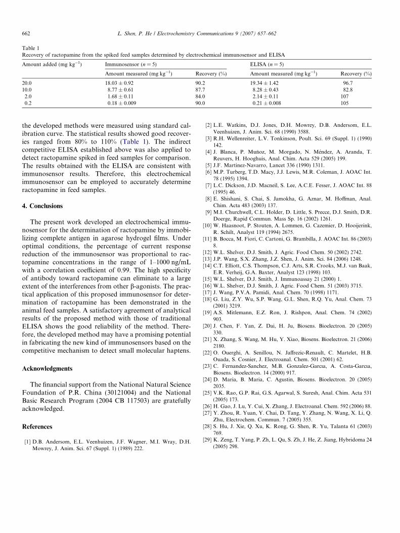
Table 1Recovery of ractopamine from the spiked feed samples determined by electrochemical immunosensor and ELISA
Amount added (mg kg�1) Immunosensor (n = 5) ELISA (n = 5)
Amount measured (mg kg�1) Recovery (%) Amount measured (mg kg�1) Recovery (%)
20.0 18.03 ± 0.92 90.2 19.34 ± 1.42 96.710.0 8.77 ± 0.61 87.7 8.28 ± 0.43 82.82.0 1.68 ± 0.11 84.0 2.14 ± 0.11 1070.2 0.18 ± 0.009 90.0 0.21 ± 0.008 105
662 L. Shen, P. He / Electrochemistry Communications 9 (2007) 657–662
the developed methods were measured using standard cal-ibration curve. The statistical results showed good recover-ies ranged from 80% to 110% (Table 1). The indirectcompetitive ELISA established above was also applied todetect ractopamine spiked in feed samples for comparison.The results obtained with the ELISA are consistent withimmunosensor results. Therefore, this electrochemicalimmunosensor can be employed to accurately determineractopamine in feed samples.
4. Conclusions
The present work developed an electrochemical immu-nosensor for the determination of ractopamine by immobi-lizing complete antigen in agarose hydrogel films. Underoptimal conditions, the percentage of current responsereduction of the immunosensor was proportional to rac-topamine concentrations in the range of 1–1000 ng/mLwith a correlation coefficient of 0.99. The high specificityof antibody toward ractopamine can eliminate to a largeextent of the interferences from other b-agonists. The prac-tical application of this proposed immunosensor for deter-mination of ractopamine has been demonstrated in theanimal feed samples. A satisfactory agreement of analyticalresults of the proposed method with those of traditionalELISA shows the good reliability of the method. There-fore, the developed method may have a promising potentialin fabricating the new kind of immunosensors based on thecompetitive mechanism to detect small molecular haptens.
Acknowledgments
The financial support from the National Natural ScienceFoundation of P.R. China (30121004) and the NationalBasic Research Program (2004 CB 117503) are gratefullyacknowledged.
References
[1] D.B. Andersom, E.L. Veenhuizen, J.F. Wagner, M.I. Wray, D.H.Mowrey, J. Anim. Sci. 67 (Suppl. 1) (1989) 222.
[2] L.E. Watkins, D.J. Jones, D.H. Mowrey, D.B. Andersom, E.L.Veenhuizen, J. Anim. Sci. 68 (1990) 3588.
[3] R.H. Wellenreiter, L.V. Tonkinson, Poult. Sci. 69 (Suppl. 1) (1990)142.
[4] J. Blanca, P. Munoz, M. Morgado, N. Mendez, A. Aranda, T.Reuvers, H. Hooghuis, Anal. Chim. Acta 529 (2005) 199.
[5] J.F. Martinez-Navarro, Lancet 336 (1990) 1311.[6] M.P. Turberg, T.D. Macy, J.J. Lewis, M.R. Coleman, J. AOAC Int.
78 (1995) 1394.[7] L.C. Dickson, J.D. Macneil, S. Lee, A.C.E. Fesser, J. AOAC Int. 88
(1995) 46.[8] E. Shishani, S. Chai, S. Jamokha, G. Aznar, M. Hoffman, Anal.
Chim. Acta 483 (2003) 137.[9] M.I. Churchwell, C.L. Holder, D. Little, S. Precce, D.J. Smith, D.R.
Doerge, Rapid Commun. Mass Sp. 16 (2002) 1261.[10] W. Haasnoot, P. Stouten, A. Lommen, G. Cazemier, D. Hooijerink,
R. Schilt, Analyst 119 (1994) 2675.[11] B. Bocca, M. Fiori, C. Cartoni, G. Brambilla, J. AOAC Int. 86 (2003)
8.[12] W.L. Shelver, D.J. Smith, J. Agric. Food Chem. 50 (2002) 2742.[13] J.P. Wang, S.X. Zhang, J.Z. Shen, J. Anim. Sci. 84 (2006) 1248.[14] C.T. Elliott, C.S. Thompson, C.J. Arts, S.R. Crooks, M.J. van Baak,
E.R. Verheij, G.A. Baxter, Analyst 123 (1998) 103.[15] W.L. Shelver, D.J. Smith, J. Immunoassay 21 (2000) 1.[16] W.L. Shelver, D.J. Smith, J. Agric. Food Chem. 51 (2003) 3715.[17] J. Wang, P.V.A. Pamidi, Anal. Chem. 70 (1998) 1171.[18] G. Liu, Z.Y. Wu, S.P. Wang, G.L. Shen, R.Q. Yu, Anal. Chem. 73
(2001) 3219.[19] A.S. Mitlemann, E.Z. Ron, J. Rishpon, Anal. Chem. 74 (2002)
903.[20] J. Chen, F. Yan, Z. Dai, H. Ju, Biosens. Bioelectron. 20 (2005)
330.[21] X. Zhang, S. Wang, M. Hu, Y. Xiao, Biosens. Bioelectron. 21 (2006)
2180.[22] O. Ouerghi, A. Senillou, N. Jaffrezic-Renault, C. Martelet, H.B.
Ouada, S. Cosnier, J. Electroanal. Chem. 501 (2001) 62.[23] C. Fernandez-Sanchez, M.B. Gonzalez-Garcıa, A. Costa-Garcıa,
Biosens. Bioelectron. 14 (2000) 917.[24] D. Maria, B. Maria, C. Agustin, Biosens. Bioelectron. 20 (2005)
2035.[25] V.K. Rao, G.P. Rai, G.S. Agarwal, S. Suresh, Anal. Chim. Acta 531
(2005) 173.[26] H. Gao, J. Lu, Y. Cui, X. Zhang, J. Electroanal. Chem. 592 (2006) 88.[27] Y. Zhou, R. Yuan, Y. Chai, D. Tang, Y. Zhang, N. Wang, X. Li, Q.
Zhu, Electrochem. Commun. 7 (2005) 355.[28] S. Hu, J. Xie, Q. Xu, K. Rong, G. Shen, R. Yu, Talanta 61 (2003)
769.[29] K. Zeng, T. Yang, P. Zh, L. Qu, S. Zh, J. He, Z. Jiang, Hybridoma 24
(2005) 298.