Heparin-Induced Thrombocytopenia · Heparin-induced thrombocytopenia • Thrombocytopenia •

LUND UNIVERSITY
PO Box 117221 00 Lund+46 46-222 00 00
An antimicrobial helix A-derived peptide of heparin cofactor II blocks endotoxinresponses in vivo.
Papareddy, Praveen; Kalle, Martina; Singh, Shalini; Mörgelin, Matthias; Schmidtchen, Artur;Malmsten, MartinPublished in:Biochimica et Biophysica Acta
DOI:10.1016/j.bbamem.2014.01.026
Published: 2014-01-01
Link to publication
Citation for published version (APA):Papareddy, P., Kalle, M., Singh, S., Mörgelin, M., Schmidtchen, A., & Malmsten, M. (2014). An antimicrobialhelix A-derived peptide of heparin cofactor II blocks endotoxin responses in vivo. Biochimica et Biophysica Acta,1838(5), 1225-1234. DOI: 10.1016/j.bbamem.2014.01.026
General rightsCopyright and moral rights for the publications made accessible in the public portal are retained by the authorsand/or other copyright owners and it is a condition of accessing publications that users recognise and abide by thelegal requirements associated with these rights.
• Users may download and print one copy of any publication from the public portal for the purpose of privatestudy or research. • You may not further distribute the material or use it for any profit-making activity or commercial gain • You may freely distribute the URL identifying the publication in the public portal

1
An antimicrobial helix A-derived peptide of heparin cofactor II blocks
endotoxin responses in vivo
Praveen Papareddy1, Martina Kalle
1, Shalini Singh
2, Matthias Mörgelin
3, Artur
Schmidtchen1,4
, and Martin Malmsten2,*
1Division of Dermatology and Venereology, Department of Clinical Sciences, Lund
University, SE-221 84 Lund, Sweden
2Department of Pharmacy, Uppsala University, SE-75123, Uppsala, Sweden
3Division of Infection Medicine, Department of Clinical Sciences, Lund University, SE-221
84 Lund, Sweden
4Lee Kong Chian School of Medicine, Nanyang Technological University, 11 Mandalay
Road, Singapore 308232
*Corresponding author. Tel: +46184714334; Fax: +46184714377; E-mail:
Key words: antimicrobial peptide, lipopolysaccharide, lipid A, membrane

2
Abstract
Host defense peptides are key components of the innate immune system, providing multi-
facetted responses to invading pathogens. Here, we describe that the peptide GKS26
(GKSRIQRLNILNAKFAFNLYRVLKDQ), corresponding to the A domain of heparin
cofactor II (HCII), ameliorates experimental septic shock. The peptide displays antimicrobial
effects through direct membrane disruption, also at physiological salt concentration and in the
presence of plasma and serum. Biophysical investigations of model lipid membranes showed
the antimicrobial action of GKS26 to be mirrored by peptide incorporation into, and
disordering of, bacterial lipid membranes. GKS26 furthermore binds extensively to bacterial
lipopolysaccharide (LPS), as well as its endotoxic lipid A moiety, and displays potent anti-
inflammatory effects, both in vitro and in vivo. Thus, for mice challenged with ip injection of
LPS, GKS26 suppresses pro-inflammatory cytokines, reduces vascular leakage and
infiltration in lung tissue, and normalizes coagulation. Together, these findings suggest that
GKS26 may be of interest for further investigations as therapeutic against severe infections
and septic shock.

3
Introduction
Antimicrobial and host defense peptides play a central role in the protection against invading
pathogens, providing antimicrobial, anti-inflammatory, as well as other effects (1,2). With
increasing resistance development against conventional antibiotics, as well as remaining
challenges in the treatment of both acute and chronic inflammation, such peptides have been
subject of considerable research interest during the last decade. As part of this, various
approaches have been employed for identification of potent, but simultaneously selective,
peptides, including, e.g., quantitative structure-activity approaches (3). Within context, we
have focused our efforts primarily on peptides derived from endogenous proteins, including
complement, coagulation, and matrix proteins, displaying potent antimicrobial effects but also
low toxicity to human cells. In addition to such directly antimicrobial effects, some of these
studies have also been demonstrated to display potent anti-inflammatory capacity, able to
counteract the multiple detrimental effects of bacterial lipopolysaccharide (LPS) from Gram-
negative bacteria, as well as related effects from lipoteichoic acid (LTA) from Gram-positive
bacteria and zymosan from fungi (4,5).
In our work with coagulation-based host defense peptides and holoproteins, we previously
investigated the serine proteinase inhibitor heparin cofactor II (HCII), and were able to
demonstrate a novel host defense function of this protein (6). Thus, compared to normal mice,
HCII-/-
knock-outs were found to be more susceptible to LPS-induced shock and invasive
infection by Pseudomonas aeruginosa. Concomitantly, a significantly increased cytokine
response was observed in HCII-/-
mice in response to infection, and HCII levels were reduced
in infected normal mice. The LPS-binding, as well as antimicrobial capacity, of activated
HCII was furthermore found to be dependent on a conformational change, exposing the
amphiphilic and positively charged A and D helices of HCII.

4
Having shown that these helices are important for the host defense function of HCII, we
hypothesized that the epitopes alone could potentially maintain some functional properties of
the holoprotein, thus offering interesting opportunities as simpler therapeutic agents. We here
describe biological activities of a helix A-derived peptide of HCII, GKS26
(GKSRIQRLNILNAKFAFNLYRVLKDQ), spanning the entire length of the helix. In doing
so, we report on its antimicrobial and anti-inflammatory properties, using a battery of assays
for broad effect characterization. In parallel, biophysical investigations provided additional
insight into the mechanisms underlying these effects. The peptide exerted potent antibacterial
and anti-inflammatory activities, and showed efficiency in a model of endotoxin-induced
shock in vivo. Taken together, the results thus demonstrate this peptide to be of potential
interest in the development of novel anti-infective and anti-inflammatory therapies.
Experimental
Chemicals. GKS26 (Table 1) was synthesized by Biopeptide Co., San Diego, USA, and was
of >95% purity, as evidenced by mass spectral analysis (MALDI-TOF Voyager). LPS and
lipid A from E. coli (0111:B4 and F583 (Rd mutant), respectively) were from Sigma (St.
Louis, USA). In LPS (>500000 EU/mg), protein and RNA content were both less than 1%.
Microorganisms. Escherichia coli (E. coli) ATCC 25922, Pseudomonas aeruginosa (P.
aeruginosa) ATCC 27853, Staphylococcus aureus (S. aureus) ATCC 29213, and Bacillus
subtilis (B. subtilis) ATCC 6633 isolates were obtained from the Department of Clinical
Bacteriology at Lund University Hospital, Sweden.

5
Scanning electron microscopy. Bacteria were grown in TH medium at 37ºC to mid-
logarithmic phase. The bacteria were washed in buffer (10 mM Tris, pH 7.4, 0.15 M NaCl, 5
mM glucose) and resuspended in the same buffer. Peptides at 30 M were incubated with S.
aureus ATCC 29213 or P. aeruginosa ATCC 27853 (1×10⁸ bacteria) for two hours in a total
volume of 50 ml in 10 mM Tris, pH 7.4, with additional 150 mM NaCl. Bacteria were fixed
and prepared for scanning electron microscopy as described previously (7). Samples were
examined with a Philips/FEI CM 100 electron microscope operated at 80 kV accelerating
voltage and images recorded with a side-mounted Olympus Veleta camera at the Core Facility
for integrated Microscopy (CFIM), Copenhagen University, Denmark.
Fluorescence microscopy. For monitoring of membrane permeabilization using the
impermeant probe FITC (Sigma-Aldrich, St. Louis, USA), E. coli ATCC 25922 bacteria
were grown to mid-logarithmic phase in TSB medium. Bacteria were washed and re-
suspended in buffer (10 mM Tris, pH 7.4, 0.15M NaCl) to yield a suspension of 1x10⁷
CFU/ml. 100 ml of the bacterial suspension was incubated with 30 μM of the respective
peptides at 37°C for 60 min. Microorganisms were then immobilized on poly (L-lysine)–
coated glass slides by incubation for 45 min at 30°C, followed by addition onto the slides of
200 ml of FITC (6 mg/ml) in buffer and incubation for 30 min at 30°C. The slides were
washed and bacteria fixed by incubation, first on ice for 15 min, then in room temperature for
45 min in 4% paraformaldehyde. The glass slides were subsequently mounted on slides using
Prolong Gold anti-fade reagent mounting medium (Invitrogen, Eugene, USA). For
fluorescence analysis, bacteria were visualized using a Nikon Eclipse TE300 (Nikon,
Melville, USA) inverted fluorescence microscope equipped with a Hamamatsu C4742-95
cooled CCD camera (Hamamatsu, Bridgewater, USA) and a Plan Apochromat ×100

6
objective (Olympus, Orangeburg, USA). Differential interference contrast (Nomarski)
imaging was used for visualization of the microbes themselves.
Liposome preparation and leakage assay. Model liposomes investigated were anionic
(DOPE/DOPG 75/25 mol/mol), frequently used as bacteria membrane models (8). DOPG
(1,2-dioleoyl-sn-Glycero-3-phosphoglycerol, monosodium salt) and DOPE (1,2-dioleoyl-sn-
Glycero-3-phosphoethanolamine) were from Avanti Polar Lipids (Alabaster, USA) and of
>99% purity. In addition, reconstituted lipid membranes were used based on polar extract of
E. coli, with a lipid composition of 67.0% phosphatidylethanolamine, 23.2%
phosphatidylglycerol, and 9.8% diphosphatidylglycerol (Avanti Polar Lipids; Alabaster,
USA). The lipid mixture was dissolved in chloroform, after which solvent was removed by
evaporation under vacuum overnight. Subsequently, 10 mM Tris buffer, pH 7.4, was added
together with 0.1 M carboxyfluorescein (CF) (Sigma, St. Louis, USA). After hydration, the
lipid mixture was subjected to eight freeze-thaw cycles (not for E. coli liposomes), consisting
of freezing in liquid nitrogen and heating to 60C. Unilamellar liposomes of about Ø140 nm
were generated by multiple extrusions (30 passages) through polycarbonate filters (pore size
100 nm) mounted in a LipoFast miniextruder (Avestin, Ottawa, Canada) at 22C. Untrapped
CF was removed by two subsequent gel filtrations (Sephadex G-50, GE Healthcare, Uppsala,
Sweden) at 22C, with Tris buffer as eluent. CF release from the liposomes was determined
by monitoring the emitted fluorescence at 520 nm from a liposome dispersion (10 M lipid in
10 mM Tris, pH 7.4). For the leakage experiment in the presence of LPS, 0.02 mg/ml LPS
was first added to the above liposome dispersion (which did not cause liposome leakage in
itself; results not shown), after which peptide was added and leakage monitored as a function
of time. An absolute leakage scale was obtained by disrupting the liposomes at the end of
each experiment through addition of 0.8 mM Triton X-100 (Sigma-Aldrich, St. Louis, USA).
7
A SPEX-fluorolog 1650 0.22-m double spectrometer (SPEX Industries, Edison, USA) was
used for the liposome leakage assay. Measurements were performed in triplicate at 37 °C.
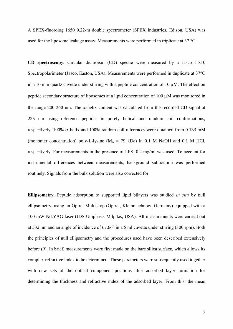
CD spectroscopy. Circular dichroism (CD) spectra were measured by a Jasco J-810
Spectropolarimeter (Jasco, Easton, USA). Measurements were performed in duplicate at 37°C
in a 10 mm quartz cuvette under stirring with a peptide concentration of 10 M. The effect on
peptide secondary structure of liposomes at a lipid concentration of 100 M was monitored in
the range 200-260 nm. The -helix content was calculated from the recorded CD signal at
225 nm using reference peptides in purely helical and random coil conformations,
respectively. 100% -helix and 100% random coil references were obtained from 0.133 mM
(monomer concentration) poly-L-lysine (Mw = 79 kDa) in 0.1 M NaOH and 0.1 M HCl,
respectively. For measurements in the presence of LPS, 0.2 mg/ml was used. To account for
instrumental differences between measurements, background subtraction was performed
routinely. Signals from the bulk solution were also corrected for.
Ellipsometry. Peptide adsorption to supported lipid bilayers was studied in situ by null
ellipsometry, using an Optrel Multiskop (Optrel, Kleinmachnow, Germany) equipped with a
100 mW Nd:YAG laser (JDS Uniphase, Milpitas, USA). All measurements were carried out
at 532 nm and an angle of incidence of 67.66° in a 5 ml cuvette under stirring (300 rpm). Both
the principles of null ellipsometry and the procedures used have been described extensively
before (9). In brief, measurements were first made on the bare silica surface, which allows its
complex refractive index to be determined. These parameters were subsequently used together
with new sets of the optical component positions after adsorbed layer formation for
determining the thickness and refractive index of the adsorbed layer. From this, the mean
8
refractive index (n) and layer thickness (d) of the adsorbed layer can be obtained. From the
thickness and refractive index the adsorbed amount () was calculated according to:
ddcdn
nn
/
)( 0-=G (1)
where n0 is the refractive index of the bulk solution, and dn/dc the refractive index increment
(0.154 cm3/g). Corrections were routinely done for changes in bulk refractive index caused by
changes in temperature and excess electrolyte concentration.
LPS-coated surfaces were obtained by adsorbing E. coli LPS to methylated silica surfaces
(surface potential -40 mV, contact angle 90° (10)) from 5 mg/ml LPS stock solution in water
at a concentration of 0.4 mg/ml over a period of 2 hours. This results in a hydrophobically
driven LPS adsorption of 1.48±0.38 mg/m2, corresponding to plateau in the LPS adsorption
isotherm under these conditions. Non-adsorbed LPS was removed by rinsing with Tris buffer
at 5 ml/min for a period of 30 minutes, followed by buffer stabilization for 20 minutes.
Peptide addition was performed at different concentrations of 0.01, 0.1, 0.5, and 1 M, and
the adsorption monitored for one hour after each addition. All measurements were performed
in at least duplicate at 25°C. For lipid A deposition, this was solubilized with 0.25 wt %
triethyl amine (TEA) under vigorous vortexing and heating to 60 0C for 10 minutes. Lipid A
was adsorbed at methylated silica surfaces for 2 hours from 5 mg/ml lipid A stock solution in
0.25 % TEA at a concentration of 0.4 mg/ml in 10 mM Tris, pH 7.4, containing 150 mM
NaCl. Non-adsorbed lipid A was subsequently removed by rinsing with same buffer at 5
ml/min for 15 minutes, followed by buffer stabilization for 20 minutes. This results in a lipid
A adsorption of 0.8±0.2 mg/m2. Peptide addition was subsequently performed to 0.01, 0.1,

9
0.5, and 1 µM, and adsorption monitored for one hour after each addition. All measurements
were performed in at least duplicate at 25°C.
Supported lipid bilayers were generated by liposome adsorption. DOPE/DOPG (75/25
mol/mol) liposomes were prepared as described above, but the dried lipid films resuspended
in Tris buffer only with no CF present. In order to avoid adsorption of peptide directly at the
silica substrate through any defects of the supported lipid layer, poly-L-lysine (Mw = 170 kDa,
Sigma-Aldrich, St. Louis, USA) was preadsorbed from water prior to lipid addition to an
amount of 0.045 ± 0.01 mg/m2, followed by removal of nonadsorbed poly-L-lysine by rinsing
with water at 5 ml/min for 20 minutes (11). Water in the cuvette was then replaced by buffer
containing also 150 mM NaCl, followed by addition of liposomes in buffer at a lipid
concentration of 20 M, and subsequently by rinsing with buffer (5 ml/min for 15 minutes)
when liposome adsorption had stabilised. The final layer formed had structural characteristics
(thickness 4±1 nm, mean refractive index 1.47±0.03), suggesting that a layer fairly close to a
complete bilayer is formed. After lipid bilayer formation, the cuvette content was replaced by
10 mM Tris buffer at a rate of 5 ml/min over a period of 30 minutes. After stabilization for 40
minutes, peptide was added to a concentration of 0.01 M, followed by three subsequent
peptide additions to 0.1 M, 0.5 M, and 1 M, in all cases monitoring the adsorption for one
hour. All measurements were made in at least duplicate at 25°C.
Dual polarization interferometry. Peptide disordering of DOPE/DOPG supported bilayers
were investigated by dual polarization interferometry (DPI), using a Farfield AnaLight 4D
(Biolin Farfield, Manchester, U.K.), operating with an alternating 632.8 nm laser beam. The
technique is based on a dual slab waveguide, consisiting of an upper sensing waveguide
(supporting the lipid bilayer) and a lower reference waveguide. The changes induced by the

10
peptide/lipid adsorption was monitored through changes in the transverse electric and
transverse magnetic modes as described previously (12). As for ellipsometry, Eq. 1 was used
for determining the mass adsorbed. Although treating phospholipids as optically isotropic
systems is a reasonably accurate approximation for unsaturated and disorganized
phospholipid bilayers, these actually display some optical birefringence, which is measurable
with the sensitive DPI technique. DPI allows two parameters to be obtained from each
measurement, e.g., for the simultaneous determination of two optical parameters of the
adsorbed layer. The latter were chosen to be the adsorbed amount and the anisotropy,
assuming the adsorbed layer thickness to be constant during the adsorption process. The
birefringence (nf), obtained from the refractive indices for the TM and TE waveguide
modes, reflects ordering of the lipid molecules in the bilayer, and decreases with increasing
disordering of the bilayer (13). Consequently, nf can be used to monitor disordering
transitions in lipid bilayers, e.g., as a result of peptide binding and incorporation, and
therefore offers a simpler alternative to, e.g., order parameter analyses in 2H-NMR
spectroscopy (14). In the present study, DOPE/DOPG liposomes (75/25 mol/mol) were
prepared as described above for ellipsometry, and the liposomes (at a lipid concentration of
0.2 mg/ml in 10 mM HEPES buffer, containing also 150 mM NaCl and 1.5 mM CaCl2) fused
to the silicon oxynitride/silicon substrate (contact angle <5°) at a flow rate of 25 L/min for 8
minutes. This resulted in bilayer formation, characterized by a refractive index of 1.47, a
thickness of 4.5±0.3 nm, and an area per molecule of 54 Å2. After bilayer formation, the
buffer was changed to 10 mM Tris buffer, allowing continous flushing (at flow rate, 50 l/
min) for 10 minutes, after which peptide was added at the desired concentration. Peptide
adsorption was monitored for one hour. Measurements were performed at least in duplicate at
25°C.

11
Radial diffusion assay. Essentially as described before (15,16), bacteria were grown to mid-
logarithmic phase in 10 ml of full-strength (3% w/v) trypticase soy broth (TSB) (Becton-
Dickinson, Cockeysville, USA) (enzymatic digest of casein (17 g/L) and soybean (3 g/L),
glucose (2.5 g/L), sodium chloride (5 g/L), and dipotassium hydrogen phosphate (2.5 g/L), pH
7.3). The microorganisms were then washed once with 10 mM Tris, pH 7.4. Subsequently, 4
x 106
bacterial colony forming units (cfu) were added to 15 ml of the underlay agarose gel,
consisting of 0.03% (w/v) TSB, 1% (w/v) low electroendosmosis type (EEO) agarose (Sigma-
Aldrich, St Louis, USA), and 0.02% (v/v) Tween 20 (Sigma-Aldrich, St Louis, USA), with or
without 150 mM NaCl. The underlay was poured into a Ø 144 mm petri dish. After agarose
solidification, 4 mm-diameter wells were punched and 6 l of test sample added to each well.
Plates were incubated at 37ºC for 3 hours to allow diffusion of the peptides. The underlay gel
was then covered with 15 ml of molten overlay (6% TSB and 1% Low-EEO agarose in
distilled H2O). Antimicrobial activity of a peptide is visualized as a zone of clearing around
each well after 18-24 hours of incubation at 37°C.
Viable-count analysis. Bacteria were grown to mid-logarithmic phase in Todd-Hewitt (TH)
medium (heart infusion (3.1 g/L), yeast-enriched peptone (20 g/L), glucose (2 g/L), sodium
chloride (2 g/L), disodium phosphate (0.4 g/L), and sodium carbonate (2.5 g/l), pH 7.8), and
then washed and diluted in 10 mM Tris, pH 7.4, 5 mM glucose, 0.15 M NaCl. Following this,
bacteria (50 µl of 2 x 10⁶ cfu/ml) were incubated at 37°C for 2 hours in the presence of
peptide at indicated concentrations in 10 mM Tris, 0.15 M NaCl, with or without 20% human
citrate-plasma (CP) or 20% serum. Serial dilutions of the incubation mixture were plated on
TH agar, followed by incubation at 37°C overnight and cfu determination.

12
LPS effects on macrophages in vitro. RAW 276.4 cells (3.5×10⁶
cells/ml) (ATCC TIB
71, American Type Culture Collection, Manassas, USA) were seeded in 96-well tissue culture
plates (Nunc) in phenol red-free Dulbecco´s modified Eagle medium (DMEM; PAA
laboratories) supplemented with 10% (v/v) heat-inactivated fetal bovine serum (FBS);
Invitrogen) and 1% (v/v) Antibiotic-Antimycotic solution (ASS; Invitrogen). After 20 h of
incubation to permit adherence, cells were stimulated with 10 ng/mL E. coli LPS (0111:B4)
(Sigma-Aldrich), and various doses of GKS26. The level of nitrite oxide (NO) in culture
supernatants was determined after 20 h incubation at 37°C and 5% CO2 using the Griess
reaction as described previously (17). In addition, RAW-BlueTM
cells (1x10⁶ cells/ml)
(InvivoGen) were seeded in phenol red-free DMEM, supplemented with 10% (v/v) heat-
inactivated FBS and 1% (v/v) AAS, and allowed to adhere before they were stimulated with
10 - 1000 ng/ml E. coli (0111:B4) LPS and 10 μM GKS26. NF-κB activation was determined
after 20 h of incubation according to manufacturers instructions (InvivoGen). Briefly,
activation of NF-κB leads to the secretion of embryonic alkaline phosphatase (SEAP) into the
cell supernatant were it was measured by mixing supernatants with a SEAP detection reagent
(Quanti-BlueTM
, InvivoGen) followed by measurement of the absorbance (A) at 600 nm.
Lactate dehydrogenase assay. RAW-BlueTM
cells (5-6×10⁶
cells/ml) were stimulated as
described for the NF-κB activation assay. Lactate dehydrogenase (LDH) release was
quantified using the TOX-7 kit (Sigma-Aldrich). After incubation overnight, 50 μl of cell
supernatant was mixed with 100 µl LDH reagent mixture, incubated for 5-10 min at RT in the
dark before absorption was measured at 490 nm. The percentage of LDH release was
calculated as follows: ((A490 sample - A490 negative control)*100)/(A490 positive control - A490
negative control). The positive control consistent of cells lysed with LDH lysis buffer for 45
min, at 37° C considered as 100 % LDH release.

13
Whole blood assay. Human lepirudin blood from healthy volunteers was diluted in PBS (1:4
v/v) before the addition of 100 ng/ml E. coli (0111:B4) LPS and 10 μM GKS26. After
incubation at 37°C overnight, 5 % CO2 in cell culture plates (Nunc), samples were centrifuged
and supernatants were collected and stored at -20°C prior to cytokine analysis.
LPS animal model. All animal experiments were approved by the Laboratory Animal Ethics
Committee of Malmö/Lund, Sweden. The animals were obtained from the animal facility at
the University of Lund, and housed under standard conditions of light and temperature with
free access to laboratory chow and water. Male C57BL/6 mice (8 weeks) were injected
intraperitoneally (ip) with 18 mg/kg E. coli 0111:B4 LPS (Sigma-Aldrich, ≈500.000
endotoxin units/mg). Thirty minutes after LPS injection, 200 or 500 μg GKS26 in 10 mM
Tris, pH 7.4, or buffer alone (control), were injected ip into the mice. The mice were
sacrificed after 20 h to determine platelet counts, cytokines, and coagulation parameter. The
number of platelets was determined using the VetScan HM5 System (TRIOLAB, Mölndal,
Sweden).
Histochemistry. Organs were collected 20 h after LPS injection and immediately fixed in 4%
formaldehyde (Histolab AB, Göteborg, Sweden) prior to paraffin embedding and sectioning.
Sections were stained with Mayers Haematoxylin (Histolab AB, Göteborg, Sweden) and
Eosin (Merck).
Cytokine assay. The levels of IL-6, IL-10, MCP-1, INF-γ, and TNF-α were assessed using
the Mouse Inflammation Kit (Becton Dickinson AB, Stockholm, Sweden), according to the
manufacturer’s instructions. The levels of IL-6, TNF-α, and IL-10 were measured in

14
supernatants of whole blood using CytoSetsTM
(Invitrogen) according to manufacture´s
instructions.
Coagulation assays. Clotting times were analyzed using an Amelung coagulometer (Lemgo,
Germany). Activated partial thromboplastin time (aPTT) was measured by incubating 50 μl
citrated mouse plasma for 1 min followed by the addition of 50 μl Dapttin (Technoclone,
Vienna Austria) for 200 seconds at 37°C. Clotting was initiated by the addition of 50 μl of
CaCl2 (30 mM). For the prothrombin time (PT), clotting was initiated by addition of 50 μl
TriniClot-PT Excel (Trinity Biotech, Wicklow, Ireland) to 50 μl of pre-warmed mouse citrate
plasma.
Results
GKS26 is antimicrobial in vitro and ex vivo. First, radial diffusion (RDA) and viable count
(VCA) assays were performed to probe direct bactericidal effects of GKS26. As shown in
Figure 1A and B, GKS26 displays potent antimicrobial effects against Gram-negative E. coli
and P. aeruginosa, as well as Gram-positive S. aureus and B. subtilis. Furthermore, Figure 1C
demonstrates similar kinetics of this effect for E. coli and P. aeruginosa. While some of the
antimicrobial activity is lost in the presence of serum, high antimicrobial activity is observed
in the presence of CP (Figure 1B). The latter is noteworthy, since binding of cationic AMPs to
net negatively charged serum proteins frequently results in considerable loss in antimicrobial
effects (8). The finding that plasma even accentuates the antimicrobial effects demonstrates
that fibrinogen and other coagulation factors may in fact contribute to bactericidal effects,
presumably through membrane interactions of highly surface active fibrinogen (18,19).
Adding to this, Figure 2 shows GKS26 to display pronounced selectivity between bacteria and
mammalian cells. Thus, for P. aeruginosa and E. coli added to human blood, the peptide is

15
able to potently kill bacteria, but at the same time not cause hemolysis much above that of the
negative control. For S. aureus, this selectivity in blood is less pronounced.
GKS26 interacts with bacterial lipid membranes. As demonstrated by peptide-induced
release of intracellular material, as well as by FITC permeation, the antimicrobial effect of
GKS26 is caused by bacterial membrane lysis (Figure 3A and B). Analogously, GKS26
causes lysis of liposomes composed of model anionic phospholipids (DOPE/DOPG), as well
as liposomes composed of E. coli lipid extract (Figure 4). As seen, the DOPE/DOPG model
liposomes behaved similarly to liposomes formed by E. coli lipid extract, demonstrating the
experimentally preferred DOPE/DOPG to be a good model for E. coli lipid membranes.
Taken together, the combined bacteria and liposome data clearly show bacterial membrane
disruption to play a key role in the antimicrobial action of GKS26.
The mechanism of lipid membrane disruption was next investigated by studies of peptide
binding, insertion, and disordering to/of supported lipid membranes. As shown in Figure 5A,
GKS26 displays concentration-dependent adsorption at the anionic DOPE/DOPG lipid
membrane, reaching high adsorption densities at concentration corresponding to maximum
membrane lysis. Thus, an adsorption density of ≈450 nmol/m2 corresponds to one peptide
molecule per 8 lipid molecules. As shown in Figure 5B, peptide binding to supported
DOPE/DOPG lipid bilayers results in a monotonous decrease in the membrane optical
anisotropy, indicating that peptide adsorption results in disordering of the lipid membrane.
Importantly, there is no threshold for membrane insertion. Instead, the DPI results clearly
reports on a continuous process, where all peptides bound to the membrane are inserted into
the membrane (as opposed to sitting on-top), throughout the binding process, undergoing
modest conformational changes in the process (Figure 5C).
16
GKS26 interacts with bacterial lipopolysaccharide and reduces LPS responses in vitro,
ex vivo, and in vivo. Moreover, GKS26 displays extensive binding to both E. coli LPS and its
endotoxic lipid A moiety (Figure 6A), causing a pronounced structure transition in the
LPS/peptide complex (Figure 6B). As shown in Figure 4, GKS26 binds preferentially to LPS
over the lipid membrane. In previous reports it has been shown that binding to LPS,
especially the lipid A moiety, is a prerequisite for anti-inflammatory effects of amphiphilic
peptides (20). Therefore, experiments were performed to investigate whether this is the case
also for GKS26. Studies using mouse macrophages revealed that GKS26 dose-dependently
reduced the LPS-induced production of nitric oxide (Figure 7A). After recognition of LPS via
the TLR4-MD2 complex, a signaling cascade is initiated leading to the activation of NF-B
and finally to cytokine production and release (21-23). In experiments using a NF-B reporter
cell line (RAW Blue cells), the data show that GKS26 significantly reduces NF-B activation
(Figure 7B). Moreover the data reveal that this inhibition depends on the ratio of LPS and
peptide, indicating that direct LPS scavenging is indeed important for this effect by GKS26.
This is further supported by data from a LDH assay, showing no toxic effects of the peptide
on RAW-Blue cells (Figure 7C), thus the reduction is at least partly due to LPS-GKS26
interactions and not due to toxicity. Moreover these LDH data are consistent with reduced
toxicity seen in blood (Figure 2B). Finally, effects of GKS26 on LPS-induced cytokine
release were determined ex vivo using a whole blood model. The data show that GKS26
reduced the pro-inflammatory cytokines TNF-α and IL-6, but had no effect on anti-
inflammatory IL-10 release (Figure 7D).
Having demonstrated potential anti-inflammatory effects of GKS26 in vitro, but also ex vivo,
a next set of experiments was performed to investigate those in an in vivo context. C57BL/6
17
mice were challenged with 18 mg/kg E. coli LPS and treated with either 200 μg of 500 μg of
GKS26. Consistent with the data of the whole blood experiments, GKS26 treatment resulted
in a dose-dependent reduction of the pro-inflammatory cytokines IL-6, TNF-, MCP-1, and
IFN-, as well as in IL-10 (Figure 7E). In parallel, peptide treatment improved the overall
status of these animals as illustrated by improved health scoring (Figure 7F).
Inflammation is an important initiator of coagulation (24). Furthermore, systemic activation
of coagulation is a compromising factor in sepsis, characterized by consumption of
coagulation factors and excessive fibrinolysis, leading to e.g. prolonged coagulation times and
thrombocytopenia in patients (25). Compatible to these symptoms are results on platelet
levels, as well as clotting times in mice challenged with LPS (Figure 7G and H). The latter
displayed prolonged clotting times and a significant reduction in platelets, in contrast to mice
treated with GKS26, which showed normalization of both the activated partial thromboplastin
time (aPTT) and the prothrombin time (PT) (Figure 7G), as well less partial recovery of
platelet numbers (Figure 7H). Examination of mouse lungs further revealed a reduction of
disease symptoms, as illustrated by reduced vascular leakage, infiltration of inflammatory and
red blood cells, and less reduction in alveolar space for peptide-treated animals compared to
LPS-treated ones (Figure 7I), effects particularly clear at the higher concentration of GKS26.
Taken together, these findings demonstrate that treatment with GKS26 dampened a range of
sepsis-associated pathologies, including the release of pro-inflammatory cytokines, vascular
leakage, excessive coagulation, as well as platelet consumption.
Discussion
With its high net charge and intermediate hydrophobicity (Table 1), GKS26 is highly
amphiphilic, thus binding readily to lipid membranes. As demonstrated by the DPI

18
experiments, peptide binding is accompanied by peptide insertion into the membrane
throughout the binding process, without a threshold density prior to insertion. While the
interplay between peptide binding and insertion is relatively sparsely investigated in literature,
these findings are in line with results found for aurein 1.2 on E. coli extract and
DMPE/DMPG bilayers (26), HCII-derived KYE28 (27), as well as peptides derived from S1
peptidases at DOPE/DOPG bilayers (28). As a result of peptide insertion into the lipid
membrane, the latter is destabilized, illustrated by peptide-induced liposome leakage.
Importantly, there is a clear correlation between peptide binding density/insertion,
destabilization of both model DOPE/DOPG liposomes and liposomes composed of E. coli
lipid extract, and bacteria lysis, the latter in turn causing bactericidal effects. Importantly,
however, GKS26 displays selectivity between bacteria and human cells, demonstrated by the
combined bactericidal and hemolysis assays on blood supplemented with bacteria. Such
selectivity has previously been observed, e.g., for highly charged and simultaneously
moderately hydrophobic peptides (29), and is due to lower peptide binding caused by the
lower charge density of the human cell membranes, combined with resistance to peptide
insertion caused by the presence of cholesterol.
LPS is a negatively charged lipopolysaccharide, anchored to the outer membrane in Gram-
negative bacteria through its hydrophobic lipid A moiety (20). Through complex formation
with lipopolysaccharide-binding protein (LBP) by its lipid A moiety, and subsequent
recognition of CD14 and the Toll-like receptor 4 (TLR4), LPS causes an up-regulation of pro-
inflammatory cytokine production (21-23). Consequently, peptides can provide anti-endotoxic
through direct binding to lipid A, precluding or inhibiting LBP binding. It has also been
found, however, that LPS aggregates are particularly potent in their endotoxic effect (30),
presumably through providing an alternate cell uptake route to the LBP/CD14 receptor one.

19
Thus, peptides can reach anti-endotoxic effects also through causing disruption of LPS
aggregates (27,31,32). Mirroring this, it was recently demonstrated that peptide-induced LPS
disaggregation is correlated to pronounced structure transition in the LPS/peptide complex
(27,28), as presently observed for GKS26.
Due to its central role for the understanding of peptides of the innate immune system, as well
as the need for novel anti-inflammatory drugs, the interaction between LPS and host defense
peptides has been previously investigated in literature. For example, Andrä et al. reported on
LPS interactions with the peptide NK-2, demonstrating both hydrophobic and electrostatic
interactions to be necessary for efficient LPS neutralization (33). Addressing the issue of
binding localization and preference, Yang et al., found the rALF-Pm3 peptide to bind to a
similar extent to lipid A and LPS, but with a higher affinity to lipid A (34). Similarly,
Brandenburg et al. demonstrated preferential binding of lactoferrin to lipid A (35). These
findings are in line with the present findings for GKS26, demonstrating a comparable
adsorption density at LPS and its lipid A moiety, despite considerably less binding sites in the
latter. Also addressing the issue of binding affinity, Singh et al. investigated the binding of
cationic peptides is stronger for LPS than for the lipid membrane, and strongest for the lipid A
moiety of LPS (27). Thus, the presence of both membrane-bound and non-adsorbed LPS will
compete successfully with the liposomes for peptide and prevent its incorporation and
disruption of the liposome membranes, in agreement with the present finding of substantially
reduced liposome leakage induction by GKS26 in the presence of LPS. While there may
certainly be a barrier function of LPS contributing to this effect as well (20), results on
antimicrobial effects for Gram-negative bacteria show that this barrier function is not
sufficient for preventing the fraction of peptides not bound to LPS to incorporate into the
bacteria membrane and disrupt these. Also in line with findings of the present investigation,

20
conformational transitions in peptide/LPS complexes has previously been correlated to anti-
inflammatory effects, e.g., for fowlicidin-1 fragments (36). In this context, it is worth noting
that peptide-induced LPS packing transitions, may also contribute to peptide anti-endotoxic
effects. For example, both are likely to facilitate phagocytosis, in analogy to size and charge
dependence of phagocytosis of other types of nanoparticular systems (37). Through this, an
alternative pathway to inflammation-triggering LPS-LBP/CD14 binding/activation is
provided. Indeed, such phagocytosis-based anti-inflammatory scavenging has been previously
observed for inflammation caused by amyloid A (38).
Given LPS-induced exposure of both helix A and D on HCII activation, it interesting to
compare the findings of the present investigation of helix A-derived GKS26 with those
obtained for the analogous helix D-derived KYE28
(KYEITTIHNLFRKLTHRLFRRNFGYTLR). As GKS26, KYE28 was previously found to
be antimicrobial against both Gram-negative and Gram-positive bacteria. Analysis of peptide
effects on liposomes and supported lipid bilayers showed that antibacterial action by KYE28
is caused by direct membrane rupture, where the peptide binds at high density to the lipid
membrane, insert into this without insertion threshold, and destabilize the membrane (27). In
addition, like GKS26, KYE28 binds extensively to both LPS and its lipid A moiety, inducing
pronounced helix formation in the peptide/LPS complex. Liposome leakage experiments in
the presence and absence of LPS furthermore showed KYE28 to bind preferentially to LPS
over the lipid membrane, again in agreement with the present results for GKS26. Also from a
biological effect perspective, GKS26 and KYE28 behave similarly, as both effectively
arrobogate adverse LPS effects.

21
Although the biological significance of the latter effects remains to be clarified, the potent
antimicrobial and anti-inflammatory effects of GKS26, as well as limited toxicity and
restoring effects on platelets, coagulation, and overall status, indicates a therapeutic potential
of this peptide, offering likely advantages compared to the HCII holoprotein related to a lower
cost of goods and increased stability. As with most macromolecular therapeutics, peptides of
this size will most likely have to be administred parenterally. With its positive charge and
intermediate hydrophobicity, even such routes may provide challenges in the form of serum
protein binding and uptake in the reticuloendothelial system (RES), resulting in shortened
bloodstream circulation time and reduced bioavailability. Various approaches can be
employed for reducing such effects, including conjugation with poly(ethylene glycol) or
incorporation of the peptide in a suitable drug carrier (39,40). In addition, for continuous
infusion, the most likely administration of peptides against severe infection and sepsis, such
serum protein scavenging does generally not affect bioavailability detrimentally as peptide is
constantly replentished.
Conclusions
The peptide GKS26, derived from the A domain of HCII, displays potent antimicrobial
effects, also at physiological salt concentration and in the presence of plasma and serum,
related to peptide binding and destabilization of bacterial lipid membranes. In addition,
GKS26 binds extensively to both LPS and its endotoxic moiety lipid A, and displays potent
anti-inflammatory effects. The latter extend also to in vivo models of mice challenged with ip
injection of LPS, where GKS26 suppresses pro-inflammatory cytokines, reduces vascular
leakage and infiltration of inflammatory cells, and normalizes coagulation times. Given this,
GKS26 may have therapeutic potential against severe infection and sepsis.

22
Acknowledgement
This work was supported by the Swedish Research Council (projects 2012-1842 and 2012-
1883) and XImmune AB. Generous access to the DPI instrumentation from Biolin Farfield is
gratefully acknowledged, as is valuable scientific discussion on the DPI results with Marcus
Swann and Usha Devi at Biolin Farfield. Björn Walse is gratefully acknowledged for help
with the HCII picture, and for valuable discussions. Finally, Lise-Britt Wahlberg and Ann-
Charlotte Strömdahl are gratefully acknowledged for technical support.

23
References
1. Hancock, R.E. and Sahl, H.G. (2006) Antimicrobial and host-defense peptides as new
anti-infective therapeutic strategies. Nat. Biotechnol. 24, 1551-1557.
2. Elsbach, P. (2003) What is the real role of antimicrobial polypeptides that can mediate
several other inflammatory responses? J. Clin. Invest. 111, 1643-1645.
3. Pasupuleti, M., Schmidtchen, A., and Malmsten, M. (2012) Antimicrobial peptides:
key components of the innate immune system. Crit. Rev. Biotechnol. 32, 143-171.
4. Papareddy, P., Rydengård, V., Pasupuleti, M., Walse, B., Mörgelin, M., Chalupka, A.,
Malmsten, M., and Schmidtchen, A. (2011) Proteolysis of human thrombin generates
novel host defense peptides. PLoS Pathogens 6, e1000857, 1-15.
5. Kasetty, G., Papareddy, P., Kalle, M., Rydengård, V., Walse, B., Svensson, B.,
Mörgelin, M., Malmsten, M., and Schmidtchen, A. (2011) The C-terminal sequence of
several human serin proteases encodes host defense functions. J. Innate Immun. 3,
471-482.
6. Kalle, M., Papareddy, P., Kasetty, G., Tollefsen, D.M., Malmsten, M., Mörgelin, M.,
and Schmidtchen, A. (2013) Proteolytic activation transforms heparin cofactor II into
a host defense molecule. J. Immunol. 190, 6303-6310.
7. Kalle, M., Papareddy, P., Kasetty, G., Mörgelin, M., Van der Plas, M.J.A., Rydengård,
V., Malmsten, M., Albiger, B., and Schmidtchen, A. (2012) Host defense peptides of
thrombin modulate inflammation and coagulation in endotoxin-mediated shock and P.
aeruginosa sepsis. PLoS One 7, e51313, 1-11.
8. Strömstedt, A.A., Ringstad, L., Schmidtchen, A., and Malmsten, M. Interaction
between amphiphilic peptides and phospholipid membranes. Curr. Opinion Colloid
Interface Sci. 15 (2010) 467-478.

24
9. Malmsten, M. (1994) Ellipsometry studies of protein layers adsorbed at hydrophobic
surfaces. J. Colloid Interface Sci. 166, 333-342.
10. Malmsten, M., Burns, N., and Veide, A. (1998) Electrostatic and hydrophobic effects
of oligopeptide insertions on protein adsorption. J. Colloid Interface Sci. 204, 104-
111.
11. Ringstad, L., Schmidtchen, A., and Malmsten, M. (2006) Effect of peptide length on
the interaction between consensus peptides and DOPC/DOPA bilayers. Langmuir 22,
5042-5050.
12. Mashagi, A., Swann, M., Popplewell, J., Textor, M., and Reimhult, E. (2008) Optical
anisotropy of supported lipid structures probed by waveguide spectroscopy and its
application to study of supported lipid bilayer formation kinetics. Anal. Chem. 80,
3666-3676.
13. Yu, L., Guo, L., Ding, J.L., Ho, B., Feng, S.-S., Popplewell, J., Swann, M., and
Wohland, T. (2009) Interaction of an artificial antimicrobial peptide with lipid
membranes. Biochim. Biophys. Acta 1788, 333-344.
14. Orädd, G., Schmidtchen, A., and Malmsten, M. (2011) Effects of peptide
hydrophobicity on its incorporation in phospholipid membranes – an NMR and
ellipsometry study. Biochim. Biophys. Acta 1808, 244-252.
15. Lehrer, R. I., Rosenman, M., Harwig, S. S., Jackson, R., and Eisenhauer, P. (1991)
Ultrasensitive assays for endogenous antimicrobial polypeptides, J. Immunol. Methods
137, 167-173.
16. Andersson, E., Rydengard, V., Sonesson, A., Mörgelin, M., Bjorck, L., and
Schmidtchen, A. (2004) Antimicrobial activities of heparin-binding peptides, Eur. J.
Biochem. 271, 1219-1226.

25
17. Pollock, J.S., Forstermann, U., Mitchell, J.A., Warner, T.D., Schmidt, H.H., Nakane,
M., and Murad, F. (1991) Purification and characterization of particulate endothelium-
derived relaxing factor synthase from cultured and native bovine aortic endothelial
cells. Proc. Natl. Acad. Sci. USA 88, 10480-10484.
18. Malmsten, M. (1994) Ellipsometry studies of protein adsorption at lipid surfaces. J.
Colloid Interface Sci. 168, 247-254.
19. Price, M.E., Cornelius, R.M., and Brash, J.L. (2001) Protein adsorption to
polyethylene glycol modified liposomes from fibrinogen solution and from plasma.
Biochim. Biophys. Acta 1512, 191-205.
20. Schmidtchen, A. and Malmsten, M. (2013) Peptide interactions with bacterial
lipopolysaccharides. Curr. Opinion Colloid Interface Sci. 18, 381-392.
21. Wright, S.D., Ramos, R.A., Tobias, P.S., Ulevitch, R.J., and Mathison, J.C. (1990)
CD14, a receptor for complexes of lipopolysaccharide (LPS) and LPS binding protein,
Science 249, 1431-1433.
22. Schumann, R.R., Leong, S.R., Flaggs, G.W., Gray, P.W., Wright, S.D., Mathison,
J.C., Tobias, P.S., and Ulevitch, R.J. (1990) Structure and function of
lipopolysaccharide binding protein. Science 249, 1429-1431.
23. Shimazu, R., Akashi, S., Ogata, H., Nagai, Y., Fukudome, K., Miyake, K., and
Kimoto, M. (1999) MD-2, a molecule that confers lipopolysaccharide responsiveness
on toll-like receptor 4. J. Exp. Med. 189, 1777-1782.
24. Esmon, C.T. The interactions between inflammation and coagulation. (2005) Br. J.
Haematol. 131, 417-430.
25. Mayeux, P.R. (1997) Pathobiology of lipopolysaccharide. J. Toxicol. Environ. Health
51, 415-435.

26
26. Lee, T.-H., Heng, C., Swann, M.J., Gehman, J.D., Separovic, F., and M.-I. Aguilar
(2010) Real-time quantitative analysis of lipid disordering by aurein 1.2 during
membrane adsorption and lysis. Biochim. Biophys. Acta 1798, 1977-1986.
27. Singh, S., Papareddy, P., Kalle, M., Schmidtchen, A., and Malmsten, M. (2013)
Importance of lipopolysaccharide aggregate disruption for the anti-endotoxic effect of
heparin cofactor II peptides. Biochim. Biophys. Acta 1828, 2709-2719.
28. Singh, S., Kasetty, G., Schmidtchen, A., and Malmsten, M. (2012) Membrane and
lipopolysaccharide interactions of C-terminal peptides from S1 peptidases. Biochim.
Biophys. Acta 1818, 2244-2251.
29. Schmidtchen, A., Ringstad, L., Kasetty, G., Mizuno, H., Rutland, M.W., and
Malmsten, M. (2011) Membrane selectivity by W-tagging of antimicrobial peptides.
Biochim. Biophys. Acta 1808, 1081-1091.
30. Mueller, M., Lindner, B., Kusumoto, S., Fukase, K., Schromm, A.B., and Seydel, U.
(2004) Aggregates are the biologically active units of endotoxin. J. Biol. Chem. 279,
26307-26313.
31. Rosenfeld, Y., Papo, N., and Shai, Y. (2006) Endotoxin (lipopolysaccharide)
neutralization by innate immunity host-defense peptides. J. Biol. Chem. 281, 1636-
1643.
32. Rosenfeld, Y., Sahl, H.-G., and Shai, Y. (2008) Parameters involved in antimicrobial
and endotoxin detoxification activities of antimicrobial peptides. Biochemistry 47,
6468-6478.
33. Andrä, J., Koch, M.H., Bartels, R., and Brandenburg, K. (2004) Biophysical
characterization of endotoxin inactivation by NK-2, an antimicrobial peptide derived
from mammalian NK-lysin. Antimicrob. Agents Chemother. 48, 1593-1599.

27
34. Yang, Y., Boze, H., Chemardin, P., Padilla, A., Moulin, G., Tassanakajon, A.,
Pugniere, M., Roquet, F., Destoumieux-Garzon, D., Gueguen, Y., Bachere, E., and
Aumelas, A. (2009) NMR structure of rALF-Pm3, an anti-lipopolysaccharide factor
from shrimp: model of the possible lipid A-binding site. Biopolymers 91, 207-220.
35. Brandenburg, K., Jürgens, G., Müller, M., Fukuoka, S., and Koch, M.H.J. (2001)
Biophysical characterization of lipopolysaccharide and lipid A inactivation by
lactoferrin, Biol. Chem. 382, 1215-1225.
36. Bhunia, A., Mohanram, H., and Bhattacharjya, S. (2009) Lipopolysaccharide bound
structures of the active fragments of fowlicidin-1, a cathelicidin family of
antimicrobial and antiendotoxic peptide from chicken, determined by transferred
nuclear Overhauser effect spectroscopy. Peptide Science 92, 9-22.
37. Sahay, G., Alakhova, D.Y., and Kabanov, A.V. (2010) Endocytosis of nanomedicines.
J. Controlled Release 145, 182-195.
38. Richman, M., Perelman, A., Gertler, A., and Rahimipour, S. (2013) Effective targeting
of A to macrophages by sonochemically prepared surface-modified protein
microspheres. Biomacromolecules 14, 110-116.
39. Veronese, F.M. and Mero, A. (2008) The impact of PEGylation on biological
therapies. Biodrugs 22, 315-329.
40. Bysell, H., Månsson, R., Hansson, P., and Malmsten, M. (2011) Microgels and
microcapsules in peptide and protein delivery. Adv. Drug Delivery Rev. 63, 1172-
1185.
41. Kyte, J., and Doolittle, R.F. (1982) A simple method for displaying the hydropathic
character of a protein. J. Mol. Biol. 157, 105-132.

28
Table 1. Sequence and key properties of GKS26.
Sequence IP1 Znet
2
(pH
7.4)
H
GKS26 GKSRIQRLNILNAKFAFNLYRVLKDQ 11.49 +5 -
0.42
1 IP:isoelectric point;
2Znet: net charge;
3H mean hydrophobicity on the Kyte-Doolittle
scale (41).
29
Figure Captions
Figure 1. Antimicrobial activity. (A) Antimicrobial activity of GKS26 (at 100 µM), as
obtained by RDA, against the indicated microbes. Bacteria (4 x 10⁶ cfu) were inoculated in
0.1% TSB agarose gel, followed by peptide addition to 4 mm-diameter wells. Clearance zones
correspond to the inhibitory effect of each peptide after incubation at 37 C for 18-24 h. (B)
Antibacterial effects of GKS26 against the indicated bacterial strains in viable count assays. 2
x 106 cfu/ml bacteria were incubated in 50 µl with peptides at the indicated concentrations in
10 mM Tris, containing 0.15 M NaCl (buffer), in 10 mM Tris, 0.15 M NaCl, pH 7.4,
containing 20% human citrate plasma (CP), or in 20% human serum (Serum). (C) Kinetics of
GKS26 killing of E. coli and P. aeruginosa at a peptide concentration of 3 µM in 10 mM Tris,
0.15 M NaCl, pH 7.4. (n=3)
Figure 2. GKS26 toxicity vs antimicrobial activity in human whole blood. The indicated
bacteria (2 x 10⁸ cfu/ml) were incubated in 50% human citrate blood with or without 60 and
120 µM of peptide, followed by determination of cfu by VCA (A) and hemolysis (B). (n=3)
Figure 3. Permeabilizing effects of GKS26 on bacteria. (A) E. coli was incubated with 30
µM peptide in 10 mM Tris, pH 7.4, and permeabilization assessed using the impermeant
probe FITC. (B) Scanning electron microscopy images of P. aeruginosa and S. aureus before
and after incubation with 30 µM peptide for 2 h at 37 °C.
Figure 4. Liposome leakage induction. Peptide-induced liposome leakage for E. coli and
DOPE/DOPG (75/25 mol/mol) liposomes, as well as leakage from DOPE/DOPG liposomes
in the presence of 0.02 mg/ml E. coli LPS. Measurements were performed in 10 mM Tris, pH
7.4. (n=3)

30
Figure 5. Peptide adsorption. (A) Peptide binding to supported DOPE/DOPG (75/25
mol/mol) bilayers. (B) Peptide-induced membrane destabilization, monitored by DPI for
DOPE/DOPG supported bilayers (75/25 mol/mol). Measurements were performed in 10 mM
Tris, pH 7.4, and data reported as reduction in nf, the lipid membrane birefringence, (due to
peptide insertion and membrane disordering) as a function of the adsorption density of the
peptide. (C) CD spectra in 10 mM Tris, pH 7.4, the absence and presence of DOPE/DOPG
liposomes (75/25 mol/mol; 100 M lipid). (n=3).
Figure 6. (A) Peptide binding to E. coli LPS and E. coli lipid A. (B) CD spectra for GKS26
with or without E. coli LPS (0.2 mg/ml). Shown also is the CD spectrum for LPS in the
absence of peptide. Measurements were performed in 10 mM Tris, pH 7.4. (n=3).
Figure 7. Effects of GKS26 on LPS-induced responses in vitro. (A) RAW 264.7
macrophages were stimulated with 10 ng/ml E. coli LPS in combination with indicated
concentrations of GKS26. Nitric oxide production was determined in the cell supernatants
using the Griess reaction (n=3, mean±SEM). (B) RAW Blue cells were stimulated with
indicated concentrations of E. coli LPS and 10 μM of GKS26. NF-B activation was
measured after 20 h (n=5). (C) LDH release in supernatants of RAW-Blue cells from the
experiments in (B) (n=3). (D) Human lepirudin-treated blood was stimulated with 100 ng/ml
E. coli LPS in combination with 10 μM GKS26. Indicated cytokines were measured in plasma
after 20 h of incubation (n=5).
Figure 8. Effects of GKS26 on LPS-induced responses in vivo. Septic shock in C57BL/6
mice was induced by intraperitoneal (ip) injection of E. coli LPS (18 mg/kg) followed by ip
31
injection of 200 or 500 μg of GKS26 or buffer. Effects were assessed 20 h post LPS-
challenge. (A) Indicated cytokines were analysed in plasma (LPS n=12, GKS26200μg n= 5,
GKS26500μg n=11). (B) Scoring of mice status 20 hours after LPS injection. (1; healthy, 4;
septic shock (most severe scoring); Control n= 6, LPS n=12, GKS26200μg n= 6, GKS26500μg
n=11). (C) Activated partial thromboplastin time (aPTT) and prothrombin time (PT) were
measured using citrate plasma (Control n= 6, LPS n=10, GKS26200μg n= 5, GKS26500μg n=11;
Median indicated). (D) Number of platelets was determined in mice 20 hours after injection
of LPS and treatment with GKS26 (Control n= 6, LPS n=12, GKS26200μg n= 5, GKS26500μg
n=11). (E) Lungs of non-treated (Control), LPS-challenged, and GKS26-treated mice were
analyzed by histology. Representative light microscopy images stained with haematoxylin-
eosin (magnification 4x) are shown.

32
Figure 1.

33
Figure 2.

34
Figure 3.

35
Figure 4.

36
Figure 5.

37
Figure 6.

38
Figure 7.

39

40
Figure 8.