An ancient family of mobile genomic islands introducing ...
37
University of Groningen An ancient family of mobile genomic islands introducing cephalosporinase and carbapenemase genes in Enterobacteriaceae Nepal, Suruchi; Bonn, Florian; Grasso, Stefano; Stobernack, Tim; de Jong, Anne; Zhou, Kai; Wedema, Ronald; Rosema, Sigrid; Becher, Dörte; Otto, Andreas Published in: Virulence DOI: 10.1080/21505594.2018.1509666 IMPORTANT NOTE: You are advised to consult the publisher's version (publisher's PDF) if you wish to cite from it. Please check the document version below. Document Version Publisher's PDF, also known as Version of record Publication date: 2018 Link to publication in University of Groningen/UMCG research database Citation for published version (APA): Nepal, S., Bonn, F., Grasso, S., Stobernack, T., de Jong, A., Zhou, K., Wedema, R., Rosema, S., Becher, D., Otto, A., Rossen, J. W., van Dijl, J. M., & Bathoorn, E. (2018). An ancient family of mobile genomic islands introducing cephalosporinase and carbapenemase genes in Enterobacteriaceae. Virulence, 9(1), 1377-1389. https://doi.org/10.1080/21505594.2018.1509666 Copyright Other than for strictly personal use, it is not permitted to download or to forward/distribute the text or part of it without the consent of the author(s) and/or copyright holder(s), unless the work is under an open content license (like Creative Commons). The publication may also be distributed here under the terms of Article 25fa of the Dutch Copyright Act, indicated by the “Taverne” license. More information can be found on the University of Groningen website: https://www.rug.nl/library/open-access/self-archiving-pure/taverne- amendment. Take-down policy If you believe that this document breaches copyright please contact us providing details, and we will remove access to the work immediately and investigate your claim. Downloaded from the University of Groningen/UMCG research database (Pure): http://www.rug.nl/research/portal. For technical reasons the number of authors shown on this cover page is limited to 10 maximum.
Transcript of An ancient family of mobile genomic islands introducing ...

University of Groningen
An ancient family of mobile genomic islands introducing cephalosporinase andcarbapenemase genes in EnterobacteriaceaeNepal, Suruchi; Bonn, Florian; Grasso, Stefano; Stobernack, Tim; de Jong, Anne; Zhou, Kai;Wedema, Ronald; Rosema, Sigrid; Becher, Dörte; Otto, AndreasPublished in:Virulence
DOI:10.1080/21505594.2018.1509666
IMPORTANT NOTE: You are advised to consult the publisher's version (publisher's PDF) if you wish to cite fromit. Please check the document version below.
Document VersionPublisher's PDF, also known as Version of record
Publication date:2018
Link to publication in University of Groningen/UMCG research database
Citation for published version (APA):Nepal, S., Bonn, F., Grasso, S., Stobernack, T., de Jong, A., Zhou, K., Wedema, R., Rosema, S., Becher,D., Otto, A., Rossen, J. W., van Dijl, J. M., & Bathoorn, E. (2018). An ancient family of mobile genomicislands introducing cephalosporinase and carbapenemase genes in Enterobacteriaceae. Virulence, 9(1),1377-1389. https://doi.org/10.1080/21505594.2018.1509666
CopyrightOther than for strictly personal use, it is not permitted to download or to forward/distribute the text or part of it without the consent of theauthor(s) and/or copyright holder(s), unless the work is under an open content license (like Creative Commons).
The publication may also be distributed here under the terms of Article 25fa of the Dutch Copyright Act, indicated by the “Taverne” license.More information can be found on the University of Groningen website: https://www.rug.nl/library/open-access/self-archiving-pure/taverne-amendment.
Take-down policyIf you believe that this document breaches copyright please contact us providing details, and we will remove access to the work immediatelyand investigate your claim.
Downloaded from the University of Groningen/UMCG research database (Pure): http://www.rug.nl/research/portal. For technical reasons thenumber of authors shown on this cover page is limited to 10 maximum.

Full Terms & Conditions of access and use can be found athttp://www.tandfonline.com/action/journalInformation?journalCode=kvir20
Virulence
ISSN: 2150-5594 (Print) 2150-5608 (Online) Journal homepage: http://www.tandfonline.com/loi/kvir20
An ancient family of mobile genomic islandsintroducing cephalosporinase and carbapenemasegenes in Enterobacteriaceae
Suruchi Nepal, Florian Bonn, Stefano Grasso, Tim Stobernack, Anne de Jong,Kai Zhou, Ronald Wedema, Sigrid Rosema, Dörte Becher, Andreas Otto, JohnW. Rossen, Jan Maarten van Dijl & Erik Bathoorn
To cite this article: Suruchi Nepal, Florian Bonn, Stefano Grasso, Tim Stobernack, Anne deJong, Kai Zhou, Ronald Wedema, Sigrid Rosema, Dörte Becher, Andreas Otto, John W. Rossen,Jan Maarten van Dijl & Erik Bathoorn (2018): An ancient family of mobile genomic islandsintroducing cephalosporinase and carbapenemase genes in Enterobacteriaceae, Virulence, DOI:10.1080/21505594.2018.1509666
To link to this article: https://doi.org/10.1080/21505594.2018.1509666
© 2018 The Author(s). This open accessarticle is distributed under a CreativeCommons Attribution (CC-BY) 4.0 license
View supplementary material
Accepted author version posted online: 13Aug 2018.
Submit your article to this journal
Article views: 48 View Crossmark data
1
Publisher: Taylor & Francis & Informa UK Limited, trading as Taylor & Francis Group
Journal: Virulence
DOI: 10.1080/21505594.2018.1509666
An ancient family of mobile genomic islands introducing cephalosporinase
and carbapenemase genes in Enterobacteriaceae
Suruchi Nepal1, Florian Bonn2,†, Stefano Grasso1, Tim Stobernack1, Anne de Jong3, Kai
Zhou1,4, Ronald Wedema1, Sigrid Rosema1, Dörte Becher2, Andreas Otto2, John W. Rossen1,
Jan Maarten van Dijl1*#, and Erik Bathoorn1*#
1University of Groningen, University Medical Center Groningen, Department of Medical
Microbiology, Hanzeplein 1, 9700 RB Groningen, the Netherlands. E-mail: [email protected];
[email protected]; [email protected]; [email protected]; [email protected];
[email protected]; [email protected]; [email protected].
2Institute for Microbiology, Ernst-Moritz-Arndt-University Greifswald, Greifswald, Germany.
E-mail: [email protected]; [email protected]; andreas.otto@uni-
greifswald.de;
3Department of Molecular Genetics, University of Groningen, Groningen Biomolecular
Sciences and Biotechnology Institute, 9747 AG Groningen, the Netherlands. E-mail:
4State Key Laboratory for Diagnosis and Treatment of Infectious Disease, The First Affiliated
Hospital, Zhejiang University, Hangzhou, China. E-mail: [email protected].
†Present Address: Institute of Biochemistry 2, Goethe University Medical School, Frankfurt,
Germany.
*These authors contributed equally to this work.

2
#Corresponding Authors.
Running title: Cephalosporinase islands in Enterobacteriaceae
Abstract
The exchange of mobile genomic islands (MGIs) between microorganisms is often mediated
by phages, which may provide benefits to the phage's host. The present study started with
the identification of Enterobacter cloacae, Klebsiella pneumoniae and Escherichia coli
isolates with exceptional cephalosporin and carbapenem resistance phenotypes from
patients in a neonatal ward. To identify possible molecular connections between these
isolates and their β-lactam resistance phenotypes, the respective bacterial genome
sequences were compared. This unveiled the existence of a family of ancient MGIs that
were probably exchanged before the species E. cloacae, K. pneumoniae and E. coli emerged
from their common ancestry. A representative MGI from E. cloacae was named MIR17-GI,
because it harbors the novel β-lactamase gene variant blaMIR17. Importantly, our
observations show that the MIR17-GI-like MGIs harbor genes associated with high-level
resistance to cephalosporins. Among them, MIR17-GI stands out because MIR17 also
displays carbapenemase activity. As shown by mass spectrometry, the MIR17
carbapenemase is among the most abundantly expressed proteins of the respective E.
cloacae isolate. Further, we show that MIR17-GI-like islands are associated with integrated
P4-like prophages. This implicates phages in the spread of cephalosporin and carbapenem
resistance amongst Enterobacteriaceae. The discovery of an ancient family of MGIs,
mediating the spread of cephalosporinase and carbapenemase genes, is of high clinical
relevance, because high-level cephalosporin and carbapenem resistance have serious
implications for the treatment of patients with enterobacteriaceal infections.

3
Key words: Enterobacter cloacae, proteome, carbapenemase, cephalosporinase, mobile
genomic island
Introduction
The shape of bacterial genomes, as we know them today, is the outcome of many
successive evolutionary events that occurred ever since the respective bacterial species
branched off from their common ancestors 1. This is clearly evidenced through comparisons
of the genome sequences of individual bacterial species, where a distinction can be made
between the core genome and the accessory genome. Here, the accessory genome reflects
those elements that were either lost through genomic erosion, or gained through horizontal
gene transfer 2,3,4. Of note, the accessory genome is only the echo of those events that
happened relatively recently on the evolutionary timeline, namely after the particular
species emerged. In this context, it is frequently overlooked that also the core genome has
been shaped from elements that were recruited through genetic exchanges, but before
speciation occurred. Thus, also the core genome is the offspring of evolution and includes
ancient mobile genomic elements (MGIs), which turned out beneficial for the species within
its ecological niche to the extent that they are no longer readily lost 5,6,7. As a consequence
of their co-evolution with the species, such ancient MGIs may have lost particular traits of
present-day MGIs, such as a difference in the GC content. Yet, other traits can still be
discerned, such as the typical sites of integration and an overrepresentation of certain
classes of genes 8. In the present study we focus attention on an ancient family of MGIs, first
discovered within the ‘core genome’ of Enterobacter cloacae.
E. cloacae is a rod-shaped, non-spore forming, facultative anaerobic, Gram-negative

4
bacterium belonging to the family of Enterobacteriaceae. The occurrence of E. cloacae is
widespread, ranging from soil and sewage to the human gastrointestinal tract, where it is a
frequent component of the gut microbiota. Importantly, E. cloacae can cause opportunistic
infections and has recently emerged as a nosocomial pathogen, especially in intensive care
units (9). In the clinical setting, E. cloacae has been identified as the causative agent of skin
and soft tissue infections, respiratory and urinary tract infections, intra-abdominal
infections, bacteremia, endocarditis, septic arthritis, and osteomyelitis in
immunocompromised patients 9,10. The antimicrobial therapy of patients with E. cloacae
infections often faces complications due to the intrinsic drug resistance of this bacterium,
and its propensity to acquire multiple resistance genes. Thus, resistance has been reported
against ampicillin, amoxicillin, first-generation cephalosporins and cefoxitin owing to
constitutive expression of the AmpC β-lactamase. E. cloacae also exhibits a high frequency
of enzymatic resistance to broad-spectrum cephalosporins 11, which is typically caused by
overproduction of AmpC β-lactamases. Such extended spectrum cephalosporinases (e.g.
ACT, CMY and MIR) confer resistance to third-generation cephalosporins, and they are not
inhibited by common β-lactamase inhibitors 12. Until now, fourth- and fifth-generation
cephalosporins maintain reasonable activity against such strains. While ACT and MIR in
Enterobacter species are encoded by intrinsic chromosomal genes, CMY genes may be
plasmid-borne 13,14. In general, extended spectrum cephalosporinases of Enterobacteriaceae
have been associated with mobile genomic elements.
In contrast to cephalosporinases, acquired carbapenamase genes are still fairly uncommon
in E. cloacae on a global scale 12,15. However, many extended spectrum cephalosporinases
have a weak affinity for carbapenems and can hydrolyze carbapenems with low efficiency.
As a consequence, clinical resistance of E. cloacae to these last-line β-lactam antibiotics is

5
mostly brought about by outer membrane permeability defects combined with a
derepression of constitutive AmpC cephalosporinases 11,12,16.
The present study started with the identification of E. cloacae, K. pneumoniae and E. coli
isolates with exceptional β-lactam resistance phenotypes, including one E. cloacae isolate
with decreased susceptibility for carbapenems, in patients on a neonatal ward of the
University Medical Center Groningen (UMCG). To assess possible molecular connections
between these isolates and their antibiotic resistance phenotypes, the respective bacterial
genomes were compared. This led to the identification of four highly conserved MGIs of
~140 kb in the investigated isolates. This observation is of general importance, since it
focuses attention on a family of genomic islands carrying a diversity of cephalosporinase-
encoding genes that was already widely transduced before the species E. cloacae, K.
pneumoniae and E. coli emerged from their common ancestry.
Results
Description of the study isolates and clinical background
In the present study, we investigated four epidemiologically linked Gram-negative bacterial
isolates with decreased susceptibility to carbapenems as reflected by minimal inhibitory
concentration (MIC) values for meropenem and/or imipenem ≥ 0.5 mg/L. Table 1
summarizes the results of the antibiotic susceptibility testing in these four isolates.
The first study isolate (isolate 1) was a carbapenem-resistant E. cloacae that was obtained
from a rectal swab of a neonate (patient 1), who had been repatriated from Curaçao to the
UMCG. Prior to admission in the UMCG, the patient had been treated with meropenem.
Isolate 1 showed an atypical growth phenotype resulting in small fatty colonies on Blood

6
Agar (BA) and Mueller Hinton Agar (MHA) plates. Automated resistance analysis with the
VITEK 2 system revealed increased MIC values to the carbapenems meropenem (8 mg/L)
and imipenem (8 mg/L), which were subsequently confirmed by Etests. Imipenem Etests on
MHA with or without 250 mg/L cloxacillin revealed that imipenem resistance was
significantly reduced in the presence of cloxacillin, which is an inhibitor of β-lactamases of
the AmpC-type 17. Specifically, the MIC was reduced to 0.125 mg/L (Figure 1). This showed
that the reduced carbapenem sensitivity of isolate 1 is largely due to the production of a
carbapenem-degrading enzyme. Of note, confluent plating of isolate 1 on MHA results in a
patchy growth phenotype that is suppressed in the presence of cloxacillin (Figure 1). Multi-
locus sequence type (MLST) analysis assigns isolate 1 to the sequence type (ST-)232
(dnaA88, fusA25, gyrB49, leuS72, pyrG49, rplB12), where dnaA88 represents a novel dnaA
allele. Phenotypic ESBL tests with isolate 1 were negative. Whole-genome sequencing
revealed a novel β-lactamase gene variant, which is homologous to the MIR lineage. In fact,
this gene is 99.3% identical to the blaMIR-1 gene from Klebsiella pneumoniae (ENA accession
M37839.2) and it was thus designated blaMIR-17 (NCBI accession CEA29752.1). Specifically,
MIR17 is distinct from MIR1 due to five amino acid substitutions.
Upon withdrawal of meropenem treatment for 2 weeks, a carbapenem-sensitive E. cloacae
isolate (i.e. isolate 2) was obtained from an intravascular catheter tip from patient 1. This
isolate 2 was susceptible to third-generation cephalosporins and carbapenems, and the
sensitivity to imipenem was not influenced by the presence of cloxacillin (not shown).
Genome sequencing revealed the presence of an ampC gene encoding the ACT-5
cephalosporinase. Further, MLST analysis showed that isolate 2 belongs to ST-97.
After 3 months of hospitalization, patient 1 acquired a K. pneumoniae (i.e. isolate 3) with a

7
remarkable antibiotic resistance pattern. Specifically, isolate 3 was shown to be resistant to
cefuroxime and cefotaxime, but susceptible to ceftazidime. Phenotypic ESBL-testing was
negative. For the molecular detection of resistance genes, first a microarray analysis was
performed and, subsequently, isolate 3 was subjected to whole genome sequencing
followed by screening of the sequence against the Resfinder database. This showed the
presence of a β-lactamase gene variant with 99% identity to blaSHV-140 of K. pneumoniae.
This gene was therefore designated blaSHV-187 (NCBI accession LN515533.1). Specifically,
SHV187 is distinct from SHV140 due to two N-terminal amino acid substitutions. MLST
showed that isolate 3 belongs to ST-20.
Patient 2 was hospitalized in the same ward of the UMCG as patient 1. During this hospital
stay, patient 2 acquired an E. coli (isolate 4) resistant to cefoxitin and third-generation
cephalosporins. Further, isolate 4 displayed a meropenem MIC value of 4 mg/L. ESBL-tests
were positive and, upon whole genome sequencing, Resfinder detected two acquired β-
lactamase gene variants designated blaTEM-1b and blaCTX-M-147. Additionally, an EC-6 AmpC β-
lactamase-encoding gene was detected 18. Isolate 4 was shown to belong to ST-131.
The genomic neighborhood of blaMIR-17 is conserved in Enterobacteriaceae
Since the cephalosporinase genes identified in the four study isolates were distinct, the
respective antibiotic resistance phenotypes could not be directly related to recent
horizontal gene transfer events between these isolates. However, comparison of the whole
genome sequencing data uncovered ~140-kb regions of high similarity in the four isolates,
which harbor ampC genes in the case of isolates 1, 2 and 4 (Figure 2A). This prompted us to
investigate the spread of this conserved region among Enterobacteriaceae. As shown in
Figure 3 and Supplemental Figure S1, the ampC–containing region, as present in E. coli, is
8
highly conserved in the genera Citrobacter, Escherichia, Enterobacter, Salmonella, Shigella
and Klebsiella, where the sequences cluster according to the respective species. Of note,
ampC genes are absent from the respective conserved regions in Salmonella and Klebsiella
species, as is the case in our K. pneumoniae study isolate 3 (Figure 2A). Further, we
observed that parts of this conserved region are present in other Enterobacteriaceae, such
as Cronobacter, Dickya, Proteus, Providencia, Serratia and Yersinia species, where it lacks
ampC and can be positioned at different chromosomal loci (not shown). The conserved
region is completely absent from non-enterobacteriaceal Gram-negative bacteria, such as
Acinetobacter, Haemophilus, Pasteurella, Prevotella and Pseudomonas species. Combined
with previous studies on the phylogeny of Enterobacteriaceae 19,20,21,22,23, these observations
are indicative of an ancient MGI that spread among the enterobacteriaceal ancestry, before
genera like Enterobacter, Escherichia, and Klebsiella evolved.
The blaMIR-17 β-lactamase gene of E. cloacae isolate 1 is located on an ancient mobile
genomic island
Analysis of the conserved genomic neighborhood of the blaMIR-17 gene in E. cloacae isolate 1
revealed the presence of several genes that are commonly found on MGIs, as described in
detail in the following paragraph. The idea that blaMIR-17 could be part of a MGI was further
corroborated by inspection of the genome sequence of E. cloacae isolate 1, which revealed
that indeed a 140-kb MGI including blaMIR-17 had integrated in the Phe-GAA tRNA gene. Of
note, this tRNA gene contains a phage P4-associated attachment site
(GAGTCCGGCCTTCGGCACCA) 24 in the 3’-5’ direction (Supplemental Figure S2). Since the
identified MGI carries the blaMIR-17 gene, we named it MIR17-GI. At the 3’ end of the MIR17-
GI, downstream of a Leu-CAA tRNA gene, an integrated P4 prophage was detected (Figure

9
2B). A BLAST-x analysis of the replicative helicase gene of this P4 prophage indicated that it
is most closely related to a P4 bacteriophage of Salmonella enterica (GI:380464247, 87%
identity, 100% coverage).
The 5’ side of MIR17-GI is schematically represented in Supplemental Figure S2. The blaMIR-17
gene located on the reverse strand is flanked by an ampR regulator gene on the forward
strand. The blaMIR-17-ampR genes are positioned within a 32-kb region of MIR17-GI that is
flanked by the Phe-GAA tRNA gene with the P4 attachment site at the 5’ end, and a triplet of
Gly-GCC tRNA genes at the 3’ end. Of note, this region contains several additional resistance
genes, potentially providing resistance to cations and heavy metals (cutA1, cutA2) 25, or
quaternary ammonium compounds (sugE) 26. Further, it includes the gene for a mechano-
sensitive potassium efflux pump (kefA) that could be involved in osmo-protection 27, and
genes for two outer membrane lipocalins (blc, yjel) implicated in the transport of small
hydrophobic molecules 28. The first gene of the island encodes a regulator of the TetR
transcription regulator family, and this gene is located downstream of the cutA1 and cutA2
genes. Regulators of the TetR family repress gene transcription, and transcription is
derepressed in response to stress 29.
MIR17-GI also carries several genes that are known to have housekeeping functions. In
particular, on the forward strand, the groES-groEL genes encode chaperones implicated in
protein folding and cell cycle regulation 30. On the reverse strand of MIR17-GI, there are the
frdABCD genes potentially involved in anaerobic respiration. Such frd genes were previously
implicated in transduction by phages 31. Lastly, MIR17-GI carries genes involved in
maintenance of DNA and mobile genetic elements. The ecnA and ecnB genes located
downstream of blaMIR-17 encode the Entericidin A and B toxin-antitoxin pair previously

10
reported to prevent loss of plasmids 32. Also located downstream of blaMIR-17, the fxsA gene
encodes a polytopic membrane protein that prevents bacteriophages from exclusion 33-35.
Upstream of blaMIR-17, the DNA repair gene mutL and the high frequency of lysogeny operon
hflQXKC are located (Supplemental Figure S2).
Notably, the above-listed genes represent the common context of chromosomal ampC
genes in Citrobacter, Escherichia, Enterobacter, and Shigella species, which implies that
MIR17-GI is an ancient mobile genomic element acquired before speciation. The latter view
is supported by the fact that the GC content of the MIR17-GI and its left and right flanking
sequences is quite similar among Enterobacteriacaea, as exemplified in Figure 4 for the E.
cloacae study isolate 1 and the E. coli K12 reference strain MG1655. This is consistent with
the notion that horizontally transferred DNA will adapt to the host genome over time, a
process known as genome amelioration 36.
Landmark features of MIR17-GI-like MGIs in Enterobacteriaceae
Several ‘landmark’ features of the MIR17-GI homologous MGIs can be distinguished. In the
first place, the respective tRNA genes are highly conserved in all these islands, starting with
the Phe-GAA tRNA gene at the 5’ end, followed by the triplet Gly-GCC tRNA genes, and
ending with the Leu-CAA tRNA gene at the 3’ end. The Phe-GAA tRNA and the Leu-CAA tRNA
genes define the borders of the MGIs, which vary in length from 136 to 148 kb in E. coli, E.
cloacae and K. pneumoniae (Figure 2A). Further, these MGIs share the afore-mentioned
fxsA, groES/EL, encB, frdABCD and hflQXKC genes, as well as a trehalose operon, a putative
sugar transport gene cluster, a primosomal replication protein N gene cluster, and an L-
ascorbate utilization gene cluster. Lastly, a P4-associated integrase gene is located next to
the LEU-CAA tRNA gene (Figure 2).
11
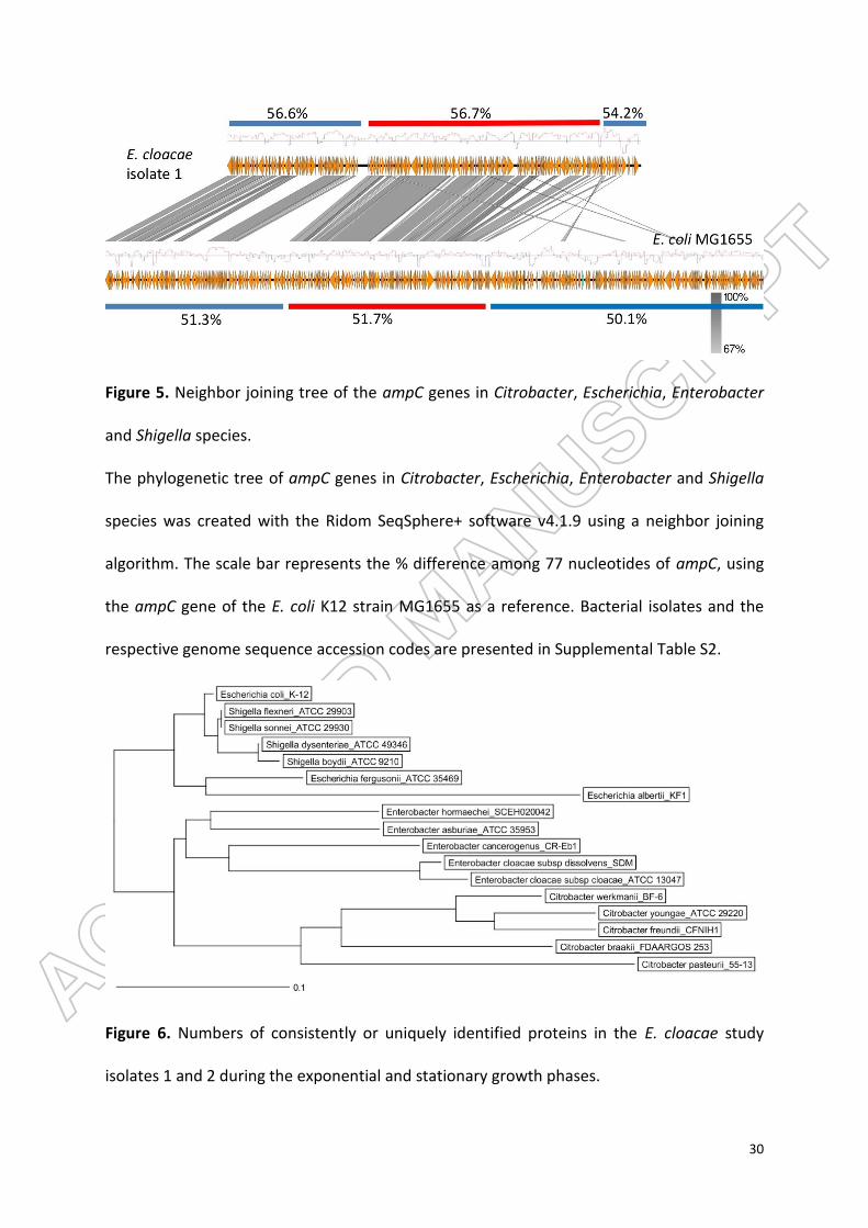
Diversity of ampC genes in MIR17-GI-like MGIs
The ampC genes of MIR17-GI-like MGIs are integrated at the same location in all Citrobacter,
Escherichia, Enterobacter, and Shigella genomes, next to sugE-blc genes. Nevertheless, the
sequences of these ampC genes are quite diverse. As shown in the phylogenetic tree in
Figure 5, the ampC genes of Citrobacter and Enterobacter species are relatively closely
related to each other, forming a distinct cluster from the ampC genes encountered in
Escherichia and Shigella species. Within E. coli and Shigella, the clustering of ampC genes
corresponds well with the different phylogenetic groups and species, although the ampC
genes of E. coli isolates belonging to phylogenetic group A are divided over four clusters
(Supplemental Figure S3). These findings are indicative of separate acquisitions of ampC by
the MIR17-GI-like MGIs over time.
Expression profile of MGI-encoded proteins from isolates 1 and 2
The frequent occurrence of MIR17-GI-like MGIs in Enterobacteriaceae raised the question to
what extent proteins encoded by such MGIs are expressed. To approximate protein
expression from MIR17-GI-like islands, cells of the E. cloacae study isolates 1 and 2 were
investigated using liquid chromatography and tandem mass spectrometry (LC-MS/MS). A
total number of 1300 different E. cloacae proteins was identified for both strains, including
857 proteins of isolate 1 and 1116 proteins of isolate 2 (Supplementary Table S1). The MS
analysis identified the largest numbers of different proteins in the stationary phase of
growth (Figure 6; Supplemental Figure S4). Further, label-free quantification of the proteins
identified by MS (i.e. spectral counting) showed similar patterns of representation of the

12
proteins detected at the highest levels among the two investigated isolates (Figure 7A). In
contrast, there was substantially more variation in the identified proteins from both isolates
that were detectable at relatively low levels. Importantly, for isolate 1, eleven of the 45
MIR17-GI-encoded proteins were identified, including the MIR17 carbapenemase that was
expressed at the highest levels in the stationary growth phase (Figure 7B, C). In fact, judged
by spectral counting, MIR17 is one of the 40 most abundant proteins that were identified in
isolate 1. Further, the MIR17-GI-encoded GroEL protein was the second most abundant
protein identified by MS. Nine proteins encoded by the MIR17-GI-like MGI of isolate 2 were
identified. Of note, the ACT-5 β-lactamase of isolate 2 remained undetected, which is
consistent with the susceptibility of this isolate to third-generation cephalosporins and
carbapenems.
Discussion
The present study highlights the identification of an ancient MGI-family integrating ampC-
like cephalosporinase genes in a wide range of Enterobacteriaceae. This MGI was first
identified in a carbapenem-resistant clinical isolate of E. cloacae, where it was shown to
carry the blaMIR-17 gene for an AmpC-like cephalosporinase. Accordingly, the respective MGI
was named MIR17-GI. Further inspection of MIR17-GI revealed a high abundance of
different resistance genes of which the MIR17 enzyme was found to be highly expressed.
Importantly, MIR17 displays carbapenemase activity that can be inhibited with cloxacillin,
suggesting that the carbapenem resistance of isolate 1 has to be attributed to this enzyme.
Interestingly, the MIR17-GI of study isolate 1 was found to be associated with a P4
prophage. This suggests that the transmission of MIR17-GI-like MGIs between

13
Enterobacteriaceae may have been phage-mediated. Consistent with this idea, all MIR17-GI-
like MGIs were found to be integrated into Phe-GAA tRNA genes that include a phage P4-
associated attachment site at the 5’ end of the MGI. Moreover, they are all flanked by a
conserved P4 integrase gene, with the Leu-CAA tRNA gene as attachment site at the 3’ end.
These features are typical for chromosomal P4-like gene clusters 24.
Resistance genes are frequently exchanged between microorganisms via MGIs and this
seems to be the case also for the MIR17-GI-like islands of which most, but not all, were
found to carry an ampC-like cephalosporinase gene. Such resistance genes that are carried
and integrated into the bacterial chromosome by phages are sometimes referred to as
‘morons’. In general, morons are composed of one or more genes that have no particular
function in the phage's lysogenic cycle, but that may provide benefits to the phage's host.
This coevolution of prophages and bacteria would be important for species to adapt to
various environmental niches 37. In the case of MIR17, the benefit for the E. cloacae host
would be the acquisition of resistance to cephalosporins or even carbapenems. In this
respect, it is important to note that such antibiotics are produced in nature by fungi and
Streptomycetes that may compete for the same ecological niches as E. cloacae. Morons are
frequently flanked by tRNAs and this was also found to be the case for blaMIR-17 . The idea
that blaMIR-17 is a moron is further supported by the observation that it is localized in the
vicinity of the bacterial high frequency lysogeny operon hflQXKC 38.
Lastly, we observed a diversity of genes encoding cephalosporinases among the identified
MIR17-GI-like mobile genomic islands. The integration of broad spectrum cephalosporinase
genes, as found in the E. cloacae study isolates is of major clinical importance. Particularly,
upon high-level expression, both the ACT- and the MIR-families of β-lactamases are capable

14
of hydrolyzing third-generation cephalosporins 11. Moreover, our study shows that the
carbapenem resistance of E. cloacae study isolate 1 can be attributed to the new allele
blaMIR-17 and the high-level production of the encoded MIR17 enzyme. The carbapenem
resistance of study isolate 1 may be further enhanced by a change in outer membrane
protein expression. In particular, the ompF gene of isolate 1, homologous to ompK35,
contains two point mutations compared to reference ompF gene of E. cloacae MGH 54
(aeebl-supercont1.1.C3), one of which results in a stop codon at position 200. Therefore, an
additional factor contributing to the carbapenem resistance of isolate 1 could be the
absence of an outer membrane porin, especially OmpF. The latter would be consistent with
previous studies on the role of porins in the resistance of Enterobacteriaceae to antibiotics
29,39,40.
In conclusion, we identified a novel family of MGIs harboring genes associated with the
resistance to cephalosporins, phage immunity, and accessory metabolic functions. These
MIR17-GI-like islands are present in various Enterobacteriaceae, including K. pneumoniae, E.
coli, and E. cloacae. Importantly, the MIR17-GI-like islands are associated with integrated
P4-like prophages, which implicate phages in the spread of high-level cephalosporin and
carbapenem resistance amongst Enterobacteriaceae. In view of the serious consequences of
high-level cephalosporin and carbapenem resistance for treatment of patients with
enterobacteriaceal infections, the discovery of an ancient mobile genomic island that has
facilitated the spread of ‘cephalosporinase and carbapenemase morons’ is of high clinical
relevance.
Materials and Methods

15
Bacterial isolates
The bacterial isolates used in this study and their antibiotic resistances are summarized in
Table 1. Species determination was performed by matrix-assisted laser
desorption/ionization-time-of-flight (MALDI-TOF) MS, using a Bruker Microflex (Bruker
Corporation, Billerica, USA).
Antibiotic susceptibility testing
Antibiotic susceptibility was routinely determined with the VITEK 2 system with an ASTn344
card (bioMérieux, Marcy l’Etoil, France). The VITEK 2 minimum inhibitory concentration
results were interpreted according to the advanced expert system following EUCAST
guidelines (www.eucast.org). The susceptibility to meropenem or imipenem was
subsequently verified on MHA using the AB Biodisk Etest according to manufacturer’s
guidelines (AB Biodisk, Mannheim, Germany). The presence of genes for carbapenemase,
ESBL and AmpC was verified with the Check-MDR CT103 XL microarray assay (Check-Points,
Wageningen, the Netherlands).
DNA sequence analyses
Nanopore sequencing. Bacteria were grown overnight at 37°C on Blood Agar (BA) plates.
Then single colonies were picked for overnight culture at 37°C on BA plates again. For DNA
extraction the DNeasy UltraClean Microbial Kit (Qiagen) was used with minor modifications.
A 10 µl-loopful of bacteria was directly transferred into a tube with microbeads and
microbeads solution. The incubation period was prolonged to 20 min, instead of 5 min. The
quality and quantity of isolated DNA was determined using a Qubit® 2.0 fluorometer
(ThermoFisher Scientific), an Agilent Tapestation 2200 (Agilent) and a NanoDrop
(ThermoFisher Scientific). Libraries were prepared without shearing to maximise sequencing

16
read length. The library was prepared using the 2D ligation sequencing kit (SQK-LSK208). The
protocol for 2D ligation sequencing kit was followed as described by the manufacturer. The
final library was loaded onto an FLO-MIN106 R9.4 flow cell. The run was performed on a
MinION device using the NC_48Hr_Sequencing_Run_FLO-MIN107_SQKLSK208 protocol with
976 available pores (464, 314, 161 and 37 pores per group). The run proceeded for the full
48 hours. Base calling was performed after the run, using Albacore v1.2.2 (Nanopore) with
the r94_250bps_2d.cfg workflow. Lastly, the quality of the data was analysed with Poretools
v0.6.0 41 and the fast5 files were transformed into a fastq file.
Illumina sequencing. Total DNA extraction for whole-genome sequencing was performed
directly from colonies of the respective isolates using the Ultraclean Microbial DNA Isolation
Kit (MO BIO Laboratories, Carlsbad, CA, US) according to the manufacturer’s protocol. DNA
concentrations were determined using a Qubit® 2.0 fluorometer and the dsDNA HS and/or
BR assay kit (Life technologies, Carlsbad, CA, US). Subsequently, DNA libraries were
prepared using the Nextera XT v3 kit (Illumina, San Diego, CA, US) according to the
manufacturer’s instructions. Sequence analysis was performed with an Illumina Miseq
System generating paired-end reads of 300 bp as described previously 42. De novo assembly
of paired-end reads was performed using CLC Genomics Workbench v8.5.3 (QIAGEN, Hilden,
Germany) after quality trimming (Qs ≥ 20) establishing a word size of 30. This resulted in
159 and 66 contigs (≥500 bp) with an average coverage of 72-fold. The acquired
antimicrobial resistance genes and multi-locus sequence types (MLST) were identified by
uploading the assembled genome sequences onto the Resfinder server v2.1. 43 and the
MLST 1.7 server 44, respectively. The raw WGS datasets generated in the current study are
available in the European Nucleotide Archive (ENA) repository under Bioproject PRJEB22119
(http://www.ebi.ac.uk/ena/).

17
Hybrid assembly. The hybrid assembly of the Illumina short reads and the MinION long
reads of isolate 339389L was performed using SPAdes version: 3.10.1
(http://bioinf.spbau.ru/en). The resulting assembly was submitted to NCBI under accession
number CP026536. Sequence similarity analyses between MIR17 and MGIs from Escherichia
coli were performed using the rapid annotation using subsystem technology (RAST) server
4.0 45.
Phylogenetic analyses
Phylogenetic trees based on single nucleotide polymorphisms (SNPs) were built using the
SNP comparison tool in Ridom SeqSphere v4.1.9 (Münster, Germany) with default settings
46. Properties of the enterobacteriaceal genomes used for phylogenetic analyses are
summarized in Supplemental Table S2. The relatedness of MIR17-GI-like MGIs was analyzed
using a local ad hoc cgMLST scheme based on 124 genes of the respective MGI from E. coli
K12 strain MG1655 (Supplemental Table S3). For visualization of the MGI comparisons,
BLAST+ 2.6.0 (NCBI, Bethesda, USA) and DNA plotter v 1.11 (Welcome Sanger Institute,
Cambrigde, UK) were used with default settings.
Identification of orthologous proteins
To identify orthologous proteins, reciprocal best hits (RBHs) were calculated. Galaxy, a
python script 47, was used to perform reciprocal protein BLAST searches (NCBI BLAST+ v.
2.3.0 48. Default parameters (minimum percentage identity: 70%; minimum High Scoring Pair
(HSP) coverage: 50%) were used and all redundancies were removed. RBHs were calculated
by blasting: (i) the two whole genome sequences; (ii) the MGI from isolate 1 and the whole
genome of isolate 2; (iii) the MGI from isolate 2 and the whole genome of isolate 1; and (iv)
the two MGIs.
18
Proteome analyses
E. cloacae isolates 1 and 2 were cultured in triplicate in brain heart infusion broth (BHI;
Oxoid, Basingstoke, UK) at 37 oC with vigorous shaking at 250 rpm. For sample preparation,
cells were collected in the mid-exponential and stationary growth phases by centrifugation
and disrupted by bead-beating with glass beads (~0.1 mm diameter) in a Precellys 24
homogenisator (Bertin Technologies, France) as described previously(49,50). Glass beads
and cell debris were removed by centrifugation (21,000 × g, 10 min, 4 °C). The cell extract
protein fraction was prepared and analyzed by LC-MS/MS using an Orbitrap Velos Pro mass
spectrometer (ThermoFisher, Waltham, MA USA) as described previously(49) . Briefly,
proteins were concentrated with Strataclean beads, subsequently reduced and alkylated,
digested with trypsin and then purified through StageTip purification. Desalted peptides
were loaded on an EASY-nLC™ II nano-flow LC system (ThermoFisher) with 10 µl buffer A
(0.1% (v/v) acetic acid) and a constant flow rate of 0.5 µL/min. Afterwards, the peptides
were separated by reversed phase chromatography with a 155 min non-linear gradient from
1 to 50 % buffer B (0.1 % (v/v) acetic acid in acetonitrile) with a constant flow rate of 0.3
µl/min and injected online into the mass spectrometer. The 20 most abundant precursor
ions were selected for collision-induced dissociation (CID) fragmentation after a survey scan
in the Orbitrap with a resolution of 60,000 and activated lockmass correction. MS/MS scans
were recorded in the dual pressure linear ion trap after fragmentation was performed for 10
msec with a normalized collision energy of 35.
Data analysis was performed according to Bonn et al. 49,51. In brief, database searching was
done with Sorcerer-SEQUEST 4 (Sage-N Research, Milpitas, USA). After data extraction from
raw files, the *.dta files were searched with Sequest against a target-decoy database with a
set of common laboratory contaminants. A non-redundant database for peptide/protein

19
searches was created from the genome sequences of isolate 1 (339389L) and isolate 2
(141024K), the genome sequence of the E. cloacae type strain ATCC 13047 as downloaded
from Uniprot (http://www.uniprot.org; 23rd of October 2015), plus five additional genome
sequences from unrelated clinical E. cloacae isolates. The used database includes protein
sequences that differ in at least 1 amino acid, and it contains 30486 proteins in total. Only
strict tryptic peptides with up to two missed cleavages were used for the database search.
Fixed modifications were not considered. Oxidation of methionine and
carbamidomethylation of cysteine were considered as variable modifications. Mass
tolerance for precursor ions was set to 10 ppm, and for fragment ions to 0.5 Da. Validation
of the MS/MS-based peptide and protein identification was performed with Scaffold
v.4.4.1.1 (Proteome Software, Portland, USA). Peptide identifications were only accepted if
they exceeded the following specific database search engine thresholds: the SEQUEST
identifications required at least deltaCn scores of > 0.1 and XCorr scores of > 2.2, 3.3 and 3.7
for doubly, triply and all higher charged peptides, respectively. Protein identifications
(Supplemental Table S1) were accepted if at least 2 identified peptides were detected with
above mentioned filter criteria in 2 out of 3 biological replicates. This resulted in a false-
positive discovery rate (FDR) below 0.2% on protein level as was verified by a search against
a concatenated target-pseudoreversed decoy database. All MS data have been deposited to
the ProteomeXchange Consortium via the PRIDE partner repository with the dataset
identifier PXD007113 52.
Ethics statement
The bacterial isolates used for the present analyses were collected in the course of routine
diagnostics and infection prevention control. Oral consent for the use of such clinical

20
samples for research purposes is routinely obtained upon patient admission to the UMCG,
in accordance with the guidelines of the Medical Ethics Committee of the University Medical
Center Groningen. All experiments were performed in accordance with the guidelines of the
Declaration of Helsinki and the institutional regulations, and all samples were anonymized.
Funding
This work was supported by the Graduate School for Medical Sciences of the University of
Groningen (to S.N. and T.S.), the People Programme (Marie Skłodowska-Curie Actions) of
the European Union’s Horizon 2020 Programme under REA grant agreement (no. 642836 to
S.G., D.B and J.M.v.D.), the Deutsche Forschungsgemeinschaft (SFB/TRR 34 framework to
D.B., F.B. and A.O.), the German Federal Ministry of Education and Research (Septomics to
F.B.), and the Fundamental Research Funds for the Central Universities (2016FZA7008 to
KZ). The funders had no role in study design, data collection and analysis, decision to
publish, or preparation of the manuscript.
Disclosure of Potential Conflicts of Interest
The authors declare that they have no financial and non-financial competing interests in
relation to the documented research.
Acknowledgements
We thank Giorgio Gabarrini and Eleni Tsompanidou for helpful discussions, and Monika
Chlebowicz and Natacha Couto for support with data submission.
21
References
(1) Darmon E, Leach DRF. Bacterial Genome Instability. Microbiol Mol Biol Rev 2014; 78: 1-
39.
(2) Arber W. Genetic variation: molecular mechanisms and impact on microbial evolution.
FEMS Microbiol Rev 2000; 24: 1-7.
(3) Bobay LM, Ochman H. The Evolution of Bacterial Genome Architecture. Front Genet
2017; 8: 10.3389.2017.
(4) Hacker J, Hochhut B, Middendorf B, Schneider G, Buchrieser C, Gottschalk G, et al.
Pathogenomics of mobile genetic elements of toxigenic bacteria. Int J Med Microbiol 2004;
293: 453-461.
(5) Hall JPJ, Brockhurst MA, Harrison E. Sampling the mobile gene pool: innovation via
horizontal gene transfer in bacteria. Philos Trans R Soc Lond B Biol Sci 2017; 372: pii:
20160424.
(6) Penades JR, Chen J, Quiles-Puchalt N, Carpena N, Novick RP. Bacteriophage-mediated
spread of bacterial virulence genes. Curr Opin Microbiol 2015; 23: 171-178.
(7) Bellanger X, Payot S, Leblond-Bourget N, Guedon G. Conjugative and mobilizable
genomic islands in bacteria: evolution and diversity. FEMS Microbiol Rev 2014; 38: 720-760.
(8) Canchaya C, Proux C, Fournous G, Bruttin A, Brussow H. Prophage genomics. Microbiol
Mol Biol Rev 2003; 67: 238-276.
(9) Mezzatesta ML, Gona F, Stefani S. Enterobacter cloacae complex: clinical impact and
emerging antibiotic resistance. Future Microbiol 2012; 7: 887-902.
(10) Fata F, Chittivelu S, Tessler S, Kupfer Y. Gas gangrene of the arm due to Enterobacter
cloacae in a neutropenic patient. South Med J 1996; 89: 1095-1096.
22
(11) Papanicolaou GA, Medeiros AA, Jacoby GA. Novel plasmid-mediated beta-lactamase
(MIR-1) conferring resistance to oxyimino- and alpha-methoxy beta-lactams in clinical
isolates of Klebsiella pneumoniae. Antimicrob Agents Chemother 1990; 34: 2200-2209.
(12) Lee EH, Nicolas MH, Kitzis MD, Pialoux G, Collatz E, Gutmann L. Association of two
resistance mechanisms in a clinical isolate of Enterobacter cloacae with high-level resistance
to imipenem. Antimicrob Agents Chemother 1991; 35: 1093-1098.
(13) Perez-Perez FJ, Hanson ND. Detection of plasmid-mediated AmpC beta-lactamase genes
in clinical isolates by using multiplex PCR. J Clin Microbiol 2002; 40: 2153-2162.
(14) Philippon A, Arlet G, Jacoby GA. Plasmid-Determined AmpC-Type beta-Lactamases.
Antimicrob Agents Chemother 2002; 46: 1-11.
(15) Chavda KD, Chen L, Fouts DE, Sutton G, Brinkac L, Jenkins SG, et al. Comprehensive
Genome Analysis of Carbapenemase-Producing Enterobacter spp.: New Insights into
Phylogeny, Population Structure, and Resistance Mechanisms. MBio 2016; 7: e02093-16.
(16) Conceicao T, Faria N, Pimentel M, Soveral G, Duarte A, Lito LM, et al. New chromosomal
AmpC beta-lactamase in Enterobacter cloacae. Antimicrob Agents Chemother 2004; 48:
1437.
(17) Jacoby GA. AmpC beta-Lactamases. Clin Microbiol Rev 2009; 22: 161-182.
(18) Mammeri H, Poirel L, Fortineau N, Nordmann P. Naturally Occurring Extended-
Spectrum Cephalosporinases in Escherichia coli. Antimicrob Agents Chemother 2006; 50:
2573-2576.
(19) Lawrence JG, Ochman H. Molecular archaeology of the Escherichia coli genome. Proc
Natl Acad Sci U S A 1998; 95: 9413-9417.

23
(20) Pham HN, Ohkusu K, Mishima N, Noda M, Monir Shah M, Sun X, et al. Phylogeny and
species identification of the family Enterobacteriaceae based on dnaJ sequences. Diagn
Microbiol Infect Dis 2007; 58: 153-161.
(21) Clermont O, Gordon D, Denamur E. Guide to the various phylogenetic classification
schemes for Escherichia coli and the correspondence among schemes. Microbiology 2015;
161: 980-988.
(22) Paradis S, Boissinot M, Paquette N, Belanger SD, Martel EA, Boudreau DK, et al.
Phylogeny of the Enterobacteriaceae based on genes encoding elongation factor Tu and F-
ATPase beta-subunit. Int J Syst Evol Microbiol 2005; 55: 2013-2025.
(23) Hata H, Natori T, Mizuno T, Kanazawa I, Eldesouky I, Hayashi M, et al. Phylogenetics of
family Enterobacteriaceae and proposal to reclassify Escherichia hermannii and Salmonella
subterranea as Atlantibacter hermannii and Atlantibacter subterranea gen. nov., comb. nov.
Microbiol Immunol 2016; 60: 303-311.
(24) Pierson LS,3rd, Kahn ML. Integration of satellite bacteriophage P4 in Escherichia coli.
DNA sequences of the phage and host regions involved in site-specific recombination. J Mol
Biol 1987; 196: 487-496.
(25) Tanaka Y, Tsumoto K, Nakanishi T, Yasutake Y, Sakai N, Yao M, et al. Structural
implications for heavy metal-induced reversible assembly and aggregation of a protein: the
case of Pyrococcus horikoshii CutA. FEBS Lett 2004; 556: 167-174.
(26) Chung YJ, Saier MH,Jr. Overexpression of the Escherichia coli sugE gene confers
resistance to a narrow range of quaternary ammonium compounds. J Bacteriol 2002; 184:
2543-2545.
(27) Cui C, Adler J. Effect of mutation of potassium-efflux system, KefA, on mechanosensitive
channels in the cytoplasmic membrane of Escherichia coli. J Membr Biol 1996; 150: 143-152.

24
(28) Bishop RE. The bacterial lipocalins. Biochim Biophys Acta 2000; 1482: 73-83.
(29) Liu YF, Yan JJ, Lei HY, Teng CH, Wang MC, Tseng CC, et al. Loss of outer membrane
protein C in Escherichia coli contributes to both antibiotic resistance and escaping antibody-
dependent bactericidal activity. Infect Immun 2012; 80: 1815-1822.
(30) Gupta P, Aggarwal N, Batra P, Mishra S, Chaudhuri TK. Co-expression of chaperonin
GroEL/GroES enhances in vivo folding of yeast mitochondrial aconitase and alters the
growth characteristics of Escherichia coli. Int J Biochem Cell Biol 2006; 38: 1975-1985.
(31) Cole ST, Guest JR. Genetic and physical characterization of lambda transducing phages
(lambda frdA) containing the fumarate reductase gene of Escherichia coli K12. Mol Gen
Genet 1980; 178: 409-418.
(32) Bishop RE, Leskiw BK, Hodges RS, Kay CM, Weiner JH. The entericidin locus of
Escherichia coli and its implications for programmed bacterial cell death. J Mol Biol 1998;
280: 583-596.
(33) Cheng X, Wang W, Molineux IJ. F exclusion of bacteriophage T7 occurs at the cell
membrane. Virology 2004; 326: 340-352.
(34) Coudron PE, Hanson ND, Climo MW. Occurrence of extended-spectrum and AmpC beta-
lactamases in bloodstream isolates of Klebsiella pneumoniae: isolates harbor plasmid-
mediated FOX-5 and ACT-1 AmpC beta-lactamases. J Clin Microbiol 2003; 41: 772-777.
(35) Susin MF, Baldini RL, Gueiros-Filho F, Gomes SL. GroES/GroEL and DnaK/DnaJ have
distinct roles in stress responses and during cell cycle progression in Caulobacter crescentus.
J Bacteriol 2006; 188: 8044-8053.
(36) Lawrence JG, Ochman H. Amelioration of bacterial genomes: rates of change and
exchange. J Mol Evol 1997; 44: 383-397.

25
(37) Cumby N, Davidson AR, Maxwell KL. The moron comes of age. Bacteriophage 2012; 2:
225-228.
(38) Kihara A, Akiyama Y, Ito K. Host regulation of lysogenic decision in bacteriophage
lambda: transmembrane modulation of FtsH (HflB), the cII degrading protease, by HflKC
(HflA). Proc Natl Acad Sci U S A 1997; 94: 5544-5549.
(39) Bajaj H, Scorciapino MA, Moynie L, Page MG, Naismith JH, Ceccarelli M, et al. Molecular
Basis of Filtering Carbapenems by Porins from beta-Lactam-resistant Clinical Strains of
Escherichia coli. J Biol Chem 2016; 291: 2837-2847.
(40) Majewski P, Wieczorek P, Ojdana D, Sienko A, Kowalczuk O, Sacha P, et al. Altered
Outer Membrane Transcriptome Balance with AmpC Overexpression in Carbapenem-
Resistant Enterobacter cloacae. Front Microbiol 2016; 7: 2054.
(41) Loman NJ, Quinlan AR. Poretools: a toolkit for analyzing nanopore sequence data.
Bioinformatics 2014; 30: 3399-3401.
(42) Zhou K, Ferdous M, de Boer RF, Kooistra-Smid AM, Grundmann H, Friedrich AW, et al.
The mosaic genome structure and phylogeny of Shiga toxin-producing Escherichia coli
O104:H4 is driven by short-term adaptation. Clin Microbiol Infect 2015; 21: 468.e7-468.18.
(43) Zankari E, Hasman H, Cosentino S, Vestergaard M, Rasmussen S, Lund O, et al.
Identification of acquired antimicrobial resistance genes. J Antimicrob Chemother 2012; 67:
2640-2644.
(44) Larsen MV, Cosentino S, Rasmussen S, Friis C, Hasman H, Marvig RL, et al. Multilocus
sequence typing of total-genome-sequenced bacteria. J Clin Microbiol 2012; 50: 1355-1361.
(45) Aziz RK, Bartels D, Best AA, DeJongh M, Disz T, Edwards RA, et al. The RAST Server:
rapid annotations using subsystems technology. BMC Genomics 2008; 9: 75.

26
(46) Leopold SR, Goering RV, Witten A, Harmsen D, Mellmann A. Bacterial whole-genome
sequencing revisited: portable, scalable, and standardized analysis for typing and detection
of virulence and antibiotic resistance genes. J Clin Microbiol 2014; 52: 2365-2370.
(47) Cock PJ, Chilton JM, Gruning B, Johnson JE, Soranzo N. NCBI BLAST+ integrated into
Galaxy. Gigascience 2015; 4: 39.
(48) Camacho C, Coulouris G, Avagyan V, Ma N, Papadopoulos J, Bealer K, et al. BLAST+:
architecture and applications. BMC Bioinformatics 2009; 10: 421.
(49) Bonn F, Bartel J, Buttner K, Hecker M, Otto A, Becher D. Picking vanished proteins from
the void: how to collect and ship/share extremely dilute proteins in a reproducible and
highly efficient manner. Anal Chem 2014; 86: 7421-7427.
(50) Maass S, Sievers S, Zuhlke D, Kuzinski J, Sappa PK, Muntel J, et al. Efficient, global-scale
quantification of absolute protein amounts by integration of targeted mass spectrometry
and two-dimensional gel-based proteomics. Anal Chem 2011; 83: 2677-2684.
(51) Bonn F, Pane-Farre J, Schluter R, Schaffer M, Fuchs S, Bernhardt J, et al. Global analysis
of the impact of linezolid onto virulence factor production in S. aureus USA300. Int J Med
Microbiol 2016; 306: 131-140.
(52) Vizcaino JA, Csordas A, del-Toro N, Dianes JA, Griss J, Lavidas I, et al. 2016 update of the
PRIDE database and its related tools. Nucleic Acids Res 2016; 44(D1): D447-56.

27
Figure legends
Figure 1. Imipenem Etest for E. cloacae study isolate 1 on Muller Hinton agar with or
without cloxacillin.
Figure 2. Similarity among MIR17-GI-like MGIs and schematic representation of the P4-like
prophage at the 3’ end of MIR17-GI.

28
(A) MIR17-GI-like MGIs identified in the four study isolates and E. coli K12 MG1655. The
alignment of MIR17-GI-like MGIs was performed with Easyfig
(http://mjsull.github.io/Easyfig/). The positions of relevant genes are indicated. (B) Genetic
map of the early operon of the P4-like prophage associated with the 3’ end of MIR17-GI,
following the tRNA-Leu-CAA gene. The relative positions of the genes for the phage
integrase, an ATP-ase of unknown function (ATP-ase), the phage polarity suppression
protein (psu), the phage capsid and scaffold protein (sid), the Ash family secondary
immunity repressor (cI); the DNA replication protein (repA), and the DNA primase are
indicated. Numbers correspond to the following gene annotations: 1, late gene regulator; 2,
phage DNA binding protein; 3, phage immunity repressor protein; 4, immunity derepression
protein; 5, phage protein of unknown function; 6, hypothetical protein for heme transfer
during cytochrome c biogenesis.

29
Figure 3: Phylogenetic tree of Citrobacter, Enterobacter, Escherichia, Klebsiella, Salmonella,
and Shigella species and conservation of MIR17-GI-like MGIs.
The phylogenetic tree of was created with the Ridom SeqSphere+ software v4.1.9 using a
neighbor joining algorithm. It is based on a SNP analysis of 39430 targets of the MGI from
the reference genome of the E. coli K12 strain MG1655. The scale bar under the tree
represents the phylogenetic distance (in %). The red bars indicate the conservation of the
MIR17-GI using the respective MGI of E. coli K12 strain MG1655 as the reference. For the
MGI comparisons, an 80% DNA similarity cut-off was used in DNA plotter. The scale bar
under the MGI alignment indicates the sequence position of the reference MGI. Bacterial
isolates and the respective genome sequence accession codes are presented in
Supplemental Table S2.
Figure 4. GC content of the MIR17-GI from E. cloacae and the related MGI from E. coli and
their flanking regions.
Red bars indicate the position of the respective MGI and blue bars the different flanking
regions. The overall GC% per region is indicated and the red-blue diagrams mark variations
in the respective GC profiles.
30
Figure 5. Neighbor joining tree of the ampC genes in Citrobacter, Escherichia, Enterobacter
and Shigella species.
The phylogenetic tree of ampC genes in Citrobacter, Escherichia, Enterobacter and Shigella
species was created with the Ridom SeqSphere+ software v4.1.9 using a neighbor joining
algorithm. The scale bar represents the % difference among 77 nucleotides of ampC, using
the ampC gene of the E. coli K12 strain MG1655 as a reference. Bacterial isolates and the
respective genome sequence accession codes are presented in Supplemental Table S2.
Figure 6. Numbers of consistently or uniquely identified proteins in the E. cloacae study
isolates 1 and 2 during the exponential and stationary growth phases.

31
The diagrams were created using the Venn diagram web tool of the VIB and the University
of Gent in Belgium (http://bioinformatics.psb.ugent.be/webtools/Venn/).
Figure 7. Proteomic profiles of the investigated E. cloacae study isolates 1 and 2 in the
exponential and stationary growth phases.
(A) Bar diagram depicting the relative amounts of 500/1300 identified E. cloacae proteins in
the exponential (E) and stationary (S) growth phases based on normalized spectral counts.
Relative amounts of identified proteins encoded by the MIR17-GI (B), and their respective
functions (C).

32
Supplemental Figure S1. Phylogenetic tree of E. coli, Salmonella and Shigella, and
conservation of the MIR17-GI mobile genomic island.
The phylogenetic tree of E. coli isolates, and Salmonella and Shigella species was created
with the Ridom SeqSphere+ software v4.1.9 using a neighbor joining algorithm. It is built on
a cgMLST analysis of 3839 targets from the reference genome of the E. coli K12 strain
MG1655. The scale bar under the tree represents the phylogenetic distance (in %). The red
bars indicate the conservation of the MIR17-GI using the respective MGI of E. coli K12 strain
MG1655 as the reference. For the MGI comparisons, an 80% DNA similarity cut-off was used
in DNA plotter. The scale bar under the MGI alignment indicates the sequence position of
the reference MGI. The color codes mark publicly available sequences of E. coli isolates, and
Salmonella and Shigella species that belong to particular phylogenetic groups or clades. The

33
MIR17-GI was represented in all 100 investigated genomes, most of which were derived
from a study by Clermont et al. 21. Of note, the ampC gene was absent from the MGI in all
sequenced Salmonella genomes. Bacterial isolates and the respective genome sequence
accession codes are presented in Supplemental Table S2.
Supplemental Figure S2. Genetic map of the 5’ end of the MIR17-GI mobile genomic island
of study isolate 1 up to the high frequency lysogeny operon.
MIR17-GI is integrated in the Phe-GAA tRNA gene at the 5’end, which has a P4 prophage-
associated attachment site in the 3’-5’direction. The positions of genes for AmpR, AmpD and
AmpE orthologues and other identified genes are indicated.
Supplemental Figure S3. Neighbor joining tree showing the diversity of ampC genes on
MIR17-GI-like MGIs in E. coli and Shigella.
The neighbor joining tree is based on genome sequences used to construct the phylogenetic
tree in Supplemental Figure S1. The scale bar represents the % difference among 197
nucleotides of ampC, using the ampC gene of the E. coli K12 strain MG1655 as a reference.
Bacterial isolates and the respective genome sequence accession codes are presented in
Supplemental Table S2.
Supplemental Figure S4. Numbers of expressed proteins in E. cloacae isolates 1 and 2.
The Venn diagram shows overlapping and uniquely expressed proteins of the E. cloacae
isolates 1 and 2 in the exponential (Exp) and stationary (St) growth phases. The diagram was
created using the Venn diagram web tool of the VIB and the University of Gent in Belgium
(http://bioinformatics.psb.ugent.be/webtools/Venn/).

34
Supplemental Table S1. E. cloacae proteins detected by Mass Spectroscopy.
The Table shows the identified proteins from E. cloacae isolates 1 and 2, and the
corresponding gene names. For each isolate, independent triplicate samples from
exponentially growing and stationary phase cells were analyzed by LC-MS/MS. The
Normalized spectral abundance factors are shown for each identified protein.
Supplemental Table S2. Bacterial isolates and the respective genome sequence accession
codes used for phylogenetic analyses.
Supplemental Table S3. Genes and the respective functions encoded by the MIR17-GI-like
MGI of E. coli K12 strain MG1655 used for phylogenetic analyses.

35
Table 1
Antibiotic Isolate 1 Patient 1 E. cloacae ST-232
Isolate 2 Patient 1 E. cloacae ST-97
Isolate 3 Patient 1 K. pneumoniae ST-20
Isolate 4 Patient 2 E. coli ST-131
MIC (mg/L) MIC (mg/L) MIC (mg/L) MIC (mg/L)
Amoxicillin+clavulanic acid
>32 >32 >32 >32
Cefuroxime >64 32 >64 >64
Cefotaxime >64 <1.0 16 >64
Ceftazidime >64 <1.0 1 >64
Cefoxitin >64 >64 >64 32
Cefepime 2 <1.0 2 16
Meropenem 8 <0.25# 0.032 4
Imipenem 8 <0.25# 0.5 0.75
ESBL* negative negative negative positive
Antibiotic susceptibility of the four bacterial isolates described in this study. All four isolates carried a MIR17-GI-related MGI. In three of the four isolates, a cephalosporinase gene (ampC) was integrated in this island. #Carbapenem-resistant colony variants grew into the carbapenem inhibition zone. *ESBL production was tested by E-tests and indicated by a > 8-fold reduction in MIC in the presence of clavulanic acid compared to cefepime, ceftazidim or cefotaxim alone.


![RESEARCH ARTICLE Open Access Differential regulation of ......Acquisition of genomic islands (GIs) plays a key role in bacterial evolution [1,2]. In silico analyses revealed that numerous](https://static.fdocuments.us/doc/165x107/6086f63332ee3d344e597c6a/research-article-open-access-differential-regulation-of-acquisition-of-genomic.jpg)