AN AN NUAL PUBLICATION VOLUME 4 / JUNE 1975 · AN AN NUAL PUBLICATION . VOLUME 4 / JUNE 1975.1...
37
AN AN NUAL PUBLICATION VOLUME 4 / JUNE 1975 .1 Bu LL ETI N P UBLISHED sr HACETTEPE .Y IVERSI7T PRESS
Transcript of AN AN NUAL PUBLICATION VOLUME 4 / JUNE 1975 · AN AN NUAL PUBLICATION . VOLUME 4 / JUNE 1975.1...
AN AN NUAL PUBLICATION VOLUME 4 / JUNE 1975
.1 B u LLETIN PUBLISHED sr HACETTEPE .YI VERSI 7T PRESS

HACETTEPE BUllETIN Of
NATURAL SCIENCES 110 ENGINEERIG AN ANNUAL PUBUCATION VOLUME 4 I JUNE 1975
EDITOR I ALAETTtN KUTSAL
EDITORIAL BOARD (HACETTEPE BULLETIN OF NATURAL SCIENCES AND ENGINEERING)
BAYSAL BATMAN I AHMET NOYAN I ALAATTtN KUTSAL (CHAlRMANOF EDITORIAL BOARD) I OKYAY ALPAUT I ACAR I~IN
MANAGING EDITOR &0 ART DIRECTOR I VURAL TORKER
ASSISTANT TO MANAGING EDITOR I S'OHEYLA KIYICI
PUBLISHED BT HACETTEPE UNIVERSITY PRESS
I

-.. .;.
SUBSCRIPTION RATES
TURKEY Annual subscription (including postage)
Single issue (not including postage)
7.50 TL
12.50 TL.
FOREIGN Annual subscription (including postage)
Single issue (not including postage)
s 1.25
s 17.5
Inquiries concerning articles. reprints or subscriptions should be forwarded to:
HACETIEPE ONlvERSiTESi BASIM VE YAYIM MERKEZi. ANKARA TURKEY
Printed by
Hacettepe University Press
Printing Division

VOLUME 4 I JUNE 1975
HACETTEPE BULLETIN OF
NATURAL SCIENCES ENGINEERINGAND
CONTENTS 1 Glucose 6-Phosphate Dehydrogenase from Rat Liver (II)
The Evidencefor A Specific Function ofLysine Residue. Substrate Specificity and Isoelectric Point ofthe Enzyme
ENGIN M. GOZUKARA
12 The Isolation of L-Asparaginase and L-Serine Dehydratase from Bacteria
NAZIF KOLANKAYA I ATILLA ATALAY I SEVDA GOKDENlz
19 The Most General Form ofthe CP Violating Interaction in the
(3,3) EB (3,3) Scheme NAMIK KEMAL PAK
25 Lower'Bounds on the Pion-Pion Physical Partial-Wave Amplitudes TARIK QELIK
31 Magnetic Field Effect on Exciton- Trap Triplet Annihilation in A Fluorene Single Crystal Doped with Pryene
ONDER PEKCAN
44 Volume Magnetic Susceptibility of Ethylenediamine Measured by N MR Method
DEMIR INAN
50 An Infrared and Raman Spectroscopic Study ofSome T -Picoline Type Werner Complexes and Their Aniline Clathrates
SEvlM AKYOZ
65 Estimation in a Heteroscedastic Linear Regression Model with Autocorrelated Disturbances M. ERDAL GURKAN I FETIH YILDIRIM
72 A Conjugate Family for Samplesfrom A Uniform Distribution SOLEYMAN GUNAY
77 Total Syntheses of Some 7-Methyl Substituted Estrogens
ENls OSKAY
I -
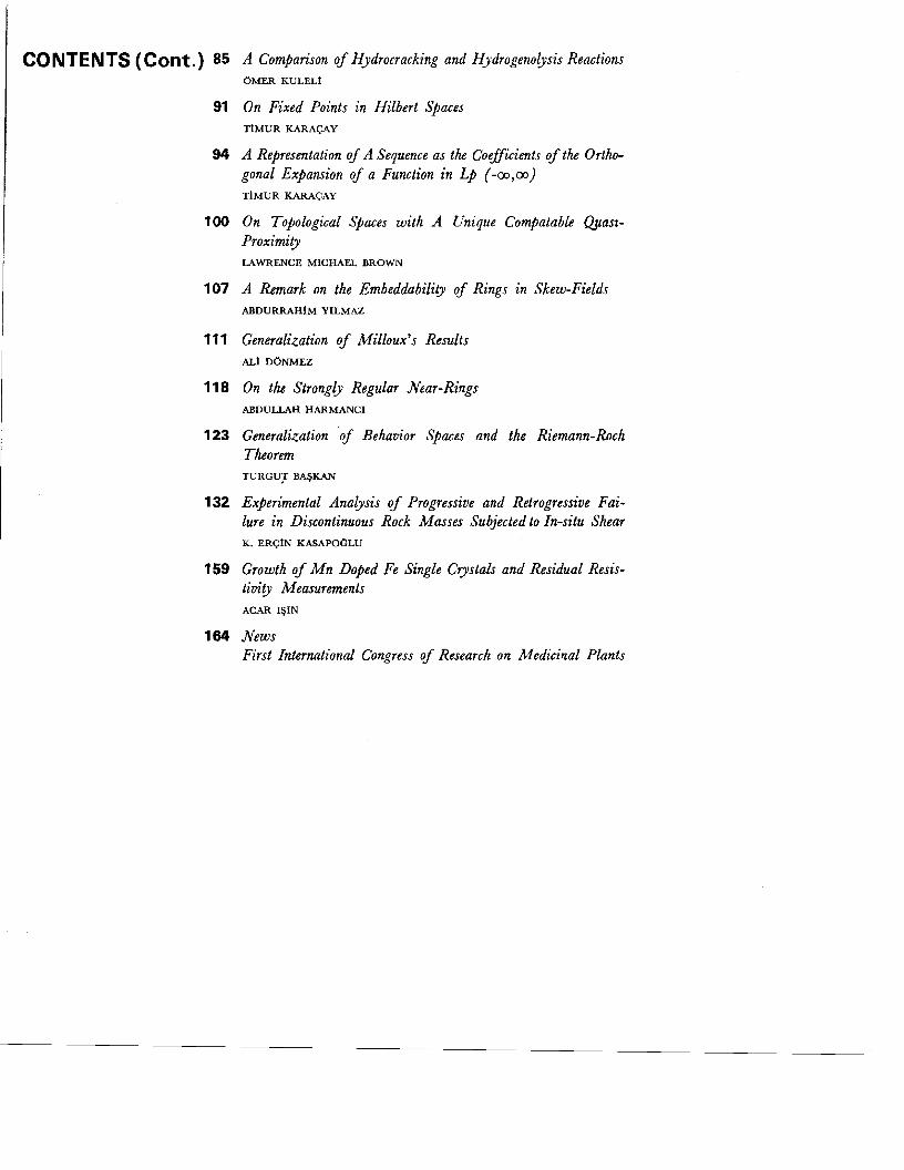
CONTENTS (Cont.) 85
91
94
100
107
111
118
123
132
159
164
A Comparison of Hydrocracking and Hydrogenolysis Reactions 6MER KULELt
On Fixed Points in Hilbert Spaces TIMUR KARACAY
A Representation of A Sequence as the Coefficients of the Orthogonal Expansion of a Function in Lp (-00,00) TIMUR KARA<;:AY
On Topological Spaces with A Unique Compatable QUaslProximity LAWRENCE MICHAEL BROWN
A Remark on the Embeddability of Rings in Skew-Fields ABDURRAHIM YILMAZ
Generalization of Milloux's Results ALt D6NMEZ
On the Strongly Regular Near-Rings ABDULLAH HARMANCI
Generalization of Behavior Spaces and the Riemann-Roch Theorem TURGU:r BA~KAN
Experimental Analysis of Progressive and Retrogressive Failure in Discontinuous Rock Masses Subjected to In-situ Shear K. ERQIN KASAPOCLU
Growth of Mn Doped Fe Single Crystals and Residual Resistivity Measurements ACAR I~IN
News First International Congress of Research on Medicinal Plants

HACETTEPE BULLETIN OF
NATURAL SCIENCES AND ENGINEERING VOLUME 4 I JUNE 1975
Glucose-6-Phosphate Dehydrogenase from Rat Liver (II) The Evidence for a Specific Function of Lysine Residue. Substrate Specificity and IsoelectricPoint of the Enzyme.
Engin M. Goziikara*
Introduction
Glucose-6-phosphate dehydrogenase is widely distributed in nature throughout both the animal and plant kingdoms. The enzyme first discovered in 1931 by Warburg and Chrisitian, and named as the "Zwischenferment". The discovery of this enzyme caused the coenzyme, nicotinamide adenin dinucleotide phosphate to be known.!?
This enzyme is the first regulatory enzyme in pentose phosphate pathway, which is known as a second way in glucose utilization. The fatty acids, cholesterol, reduced glutation and steroid hormones are required NADPH+H +, for thier synthesis, which is generated by the activity of this enzyme.t'v 13 If the enzyme deficient, these four biosynthesis can not take place in the cells. In this case erythrocytes are not able to survive and may be hemolysed.
It is rather important to know the mechanism of the catalytic activity of this enzyme, and which amino acids are involved in the active center of the enzyme. Probable amino acids which link with coenzyme NADP+ were discussed in the previous paper. 6
The aim of the presenet paper, is to discuss the amino acid to which substrate glucose-f-phosphate binds in the active center and also to discuss some physical features of the enzyme.
* Department of Molecular Biology, Hacettepe University, Beytepc Campus. Ankara-Turkey.

2 HACE'ITEPE BULLETIN OF NATURAL SCIENCES AND ENGINEERtNG
Pyridoxal 5'-phosphate has been utilized to detecet the E -amino group of lysine at the active site of various enzymes, including glucose6-P dehydrogenase from Leuconostoc mesenteroides,'? and Candida utilis:" Glutamate dehydrogenase.!? and tryptophanase? from Escherichia coli and ribonuclease from bovine panceras!' have been used for further studies of pyridoxal 5'-phosphate (PL 5'-P) binding to the E -amino group of the lysine residue. These enzymes were first treated with PL 5'-P, then reduced with sodium borohydrid (NaBH 4) and these derivatives were digested with trypsin and chymotrypsin. From these mixtures small peptides, containing 5'-phosphopyridoxyl residue were isolated and the amino acid sequences were determined. PL 5'·P were found to be bounded to the E -amino group of the lysine residue in all theree experiments. 9, II, 17
The isoelectric point and substrate specificity of glucose-6-P dehydrogenase are also discussed in this paper.
Materials and Methods
Glucose-6-phosphate dehydrogenase (D-glucose-6-phosphate: NADP+ oxidoreductase, EC. 1.1.1.49) from rat liver was used in all experiments in purified form. Fairly pure enzyme perparation was a gift by Dr Holten.
One unit of glucose-6-phosphate dehydrogenase activity is the amount required to form 1 micromole of NADPH + H + in one minute in the assay employed.f The initial reaction velocity was expresssed as micromoles ofNADPH + H + formed per minute. Glucose-6-phosphate dehydrogenase activity was measured at 340 mu wavelength by the initial rate of reduction of NADP+ at 30° C.
All solutions were prepared and used frehly and all assays were done in duplictate. In kinetic studies a Gilford model 240 spectrophometer was used and for routine assays, a Beckman model DB-G. The other details and experimental conditions were described in the previous paper. 6
Electrofocussing Experiment
The experiment was designed according to the manual book ofLKB-Laboratories. A 110 ml capacity electrofocussing column was used and glycerol was applied as a fillling material instead of sucrose. Glycerol was prepared in twenty four different concentrations, between 2.5 to 60 % and the amount was 4.6 ml of each. Ampholyne was mixed with glycerol as a carrier substance (Ampholyne, LKB-Producter AB, Stockholm) The best working pH range, was 4-6. Sulfuric acid was first added as a

- -
3 GLUCOSE-6-PHOSPHATE DEHYDROGENASE FROM RAT LIVER (II)
anodal buffer and then glycerol-ampholyne mixture was applied to the column very carefully in decreasing amounts starting by 60 %. 2.5 % mixture was added finally and the triethanolamine was applied as a cathodal buffer top of the column. 74 units of dialysed pur enzyme were applied to the middle of the column. Two electrodes were connected with a power supply and the experiment was continued for fifty hours with a 900 volts electric current. The column content was removed in 1 ml quantities from the column by a fraction coll~ctor. Each sample was tested for pH and enzyme activity. The column tube was enclosed in a glass water jacket through which water at 40 C. was circulated. This was necessary for preventing overheating by high voltage.
Chemicals
Glucose-6-phosphate, NADP+, and 2-deoxy-D-glucose-6-phospate were obtained from Boehringer Mannheim Corporation New York. Galactose6-phosphate and pyridoxa15'-phosphate from Sigma Chemical Company St. Louis, Missoury, ampholyne LKB-Laboratory Stockholm and the other chemicals used were analytical reagent grade from Mallincrodt Chemical Company St. Louis Missouri, U.S.A.
. Results
Inhibition of Glucose-6-Phosphate Dehydrogenase by Pyridoxal 5'-Phosphate
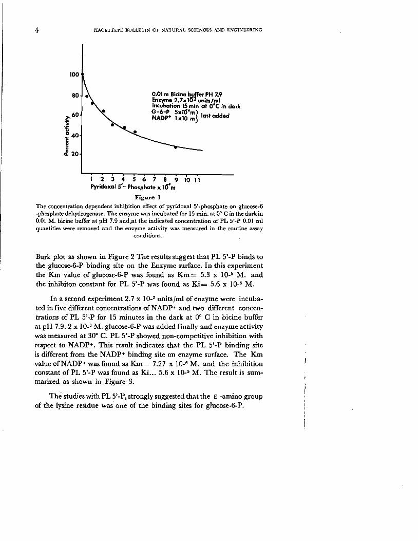
Pyridoxal 5' -phosphate was used by several investigators to determine the E -amino froup of the lysine residue in the active center of different enzymes. Pyridoxal 5'-phosphate (PL 5'-P) was found to inhibit glucose6-phosphate dehydrogenase (G-6-PDH) from rat liver. 2.7 x 10⁻² units/ ml of enzyme were incubated for 15 minutes at 00 C in the dark with different concentrations of PL 5' -P, and the enzyme activity was measure at 300 C by the addition of glucose-6-phosphate and NADP+ at the same time. The enzyme was incubated with PL 5,'-P in dark, in order to prevent the decomposition of PL 5'-P by light. The concentration dependence of pyridoxal 5'-phosphate inhibition in bicine buffer at pH 7.9 was illustrated in Figure 1. 3.5 x 10_4 M. PL 5'-P produces 50 % inhibition under the experimental conditions.
Six different concentrations of glucose-6-P, and two different concentrations ofPL 5'-P were incubated for 15 minutes at 00 C in the dark and 1 x 10_3 M. NADP+ was added last and enzyme activity was measured. The inhibition of PL 5'-P was found to be competitive with glucose-6-P. The result of this experiment was applied to Line-Weaver
4 HACETIEPE BULLETIN OF NATURAL SCIENCES AND ENGINEERING
100
last added
0.01 m Bicine buffer PH 7.9 Enzyme 2.7xl~2 units/ml incubation 15min at ooe in dark G-6-P 5Xl04 mjNAOP+ 1xl0 m
1 2 3 4 5 6 7 8 9 10 11 Pyridoxal 5'- Phosphate x 104 m
Figure 1
The concentration dependent inhibition effect of pyridoxal 5'-phosphate on glucose-f -phosphate dehydrogenase. The enzyme was incubated for IS min. at 0° C in the dark in 0.01 M. bicine buffer at pH 7.9 andat the indicated concentration of PL 5'-P 0.01 ml quantities were removed and the enzyme activity was measured in the routine assay
conditions.
Burk plot as shown in Figure 2 The results suggest that PL 5'-P binds to the glucose-6-P binding site on the Enzyme surface. In this experiment the K.m value of glucose-6-P was found as Km ee 5.3 x 10.5 M. and the inhibiton constant for PL 5'-P was found as Ki e- 5.6 x 10.5 M.
In a second experiment 2.7 x 10.2 units Iml of enzyme were incubated in five different concentrations ofNADP+ and two different concentrations of PL 5'·P for 15 minutes in the dark at 0° C in bicine butTer at pH 7.9. 2 x 10.3 M. glucose-6-P was added finally and enzyme activity was measured at 30° C. PL 5'-P showed non-competitive inhibition with respect to NADP+. This result indicates that the PL 5'-P binding site is different from the NADP+ binding site on enzyme surface. The Km value ofNADP+ was found as Km-e 7.27 x 10⁻⁶ M. and the inhibition constant of PL 5'-P was found as Ki... 5.6 x 10_5 M. The result is summarized as shown in Figure 3.
The studies with PL 5'-P, strongly suggested that the E -amino group of the lysine residue was' one of the binding sites for glucose-ti-P.

5 GLUCOSE-6-PHOSPHATE DEHYDROGENASE FROM RAT LIVER (II)
_.1_ V;
48
45
40
35
30
25
20
15
G-6-P only10 ......... PI.5"-P .sxll!:m
0.01 M Sic;ne bufl., PH 7.9 0.01 ml Enzyme 1.BS.16'm incubation 15min at cf C in dark
0---<> Pl5-P hlO m NADP lx1d'.lIm in 01experiments
1,0 1,5 2.0 _1_ x Uj'm(G-6-Pl
Figure 2 A double riciprocal plot showing competitive inhibition by pyridoxal 5'-phosphate with glucose-6-P in glucose-6-P dehydrogenase. The enzyme was incubated for 15 min. at 0° C. in the dark, in 0.01 M. bicine buffer at pH 7.9 Glucose-6-P and pyridoxal 5'-P concentrations were as indicated. I x 10-3M. NADP+ was added last and enzyme
activity was measured.
1 Vi
25
NADP+2xl05m
PL-5-P NADP + 5x10
5 PL-5-P
NADP+ only
25 50 75 100 1 125 -3
...,...~--:-x10 m (NADP)
Figure 3 A double reciprocal plot showing non-compatitive inhibition by pyridoxal 5'-phosphate with to NADP+. The enzyme was incubated for 15 min. at 0° C. in the dark, in 0.01 M. bicine buffer at pH 7.9. Pyridoxal 5'-P and NADP+ concentrations were as indicated. I x 10-3 M. glucose-6-P was added last and enzyme activity was mesured.
20
15
m
Jlkm_1_=7.27xIO" 137500

6 HACETIEPE BULLETIN OF NATURAL SCIENCES AND ENGINEERING
Substrate Specificity of Glucose-6-Phosphate Dehydrogenase Glucose-6-phosphate dehydrogenase from rat liver uses glucose-f-P as a natural subtrate. The enzyme was found to utilize 2-deoxy-D-glucose-6-P and galactose-6-P in addition to glucose-6-P. The relative rates of oxidation of these two compounds were rather low as compaired with that of glucose-6-P. The result is summarized in Table 1.
TABLE I
The Relative Oxidation Rates by Glucose-6-Phosphate Dehydrogenase for Glucose-6-Phosphate, 2-Deoxy-D-Glucose-6-Phosphate and Galactose-6- Phos
phate.
Substrate Concentration Per cent Activity
Glucose-6-P 100 2-deoxy-D-glucose-6-P 7.5 Galactose-6-P 8
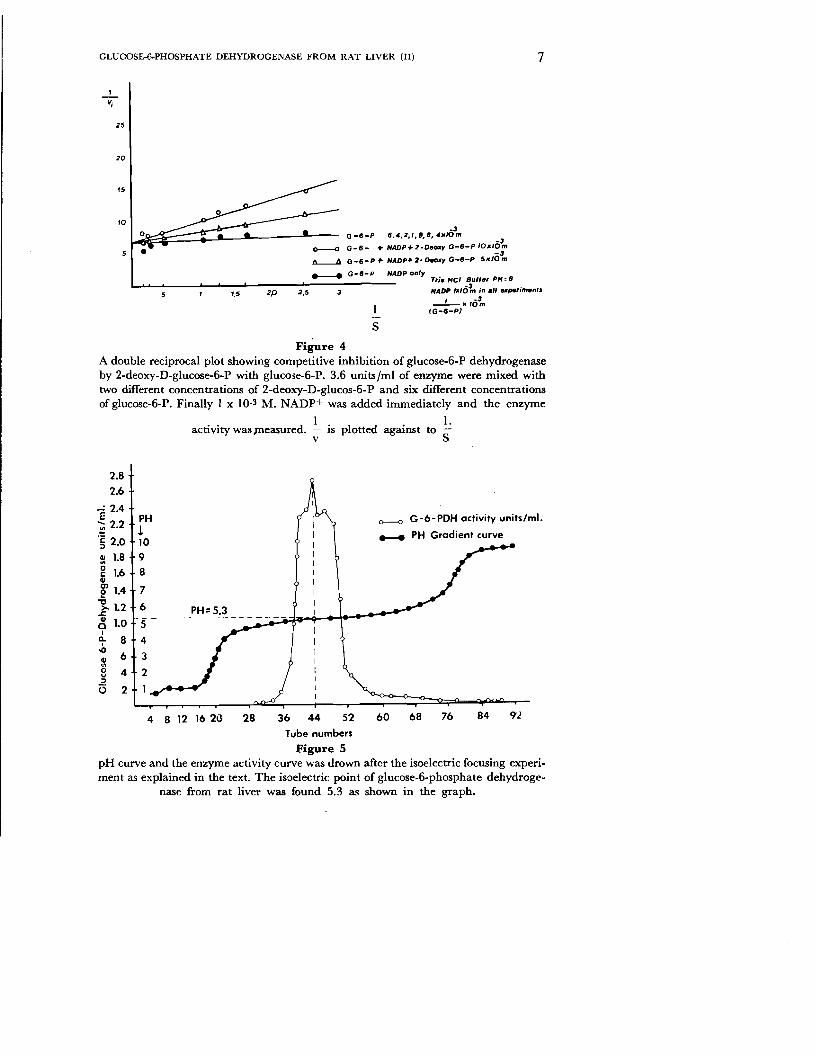
Seven different concentrations of glucose-6-P, two different concentrations of 2-deoxy-D-glucose-6-phosphate and 3.6 x 10_2 units Iml of enzyme were mixed and 1 x 10_3 M. NADP+ was added immediately and the enzyme activity was measured. The experiment result is presented in a Line-Weaver-Burk plot in fig. 4. The kinetic behaviour of 2-deoxy-D-glucose-6-P was competitive against to glucose-6-P. The Km value was found as Km= 6.7 x 10⁻⁵ M. and the inhibition constant Ki value was found as Ki= 5 x 10_4 M.
Isoelectric Focussing Experiment
The isoelectric focussing experiment was carried out as described in the material and methods section. Ampholyne is a substance which acts as an acid in a strongly acidic medium and as an alkali in a strongly basic medium. During a period of 50 hours under 900 volts a pH gradient is established in the column. The pH is 1 at the bottom of the column and 10 at the top. The enzyme in the column will move up to a definite zone where pH value is equal to its electric charge. In the zone where the pH is equal to the isoelectric point of the enzyme no further migration occurs. At the end of experimental period 1 ml mixture was removed from the column by a fraction collector. pH value and enzyme activity was determined for each tube and result is represented in fig. 5. The maximal activity was found as 2.8 units Iml of enzyme in the tube number 44.
The pH curve intersects the maximal activity peak point at a pH value of 5.3 which is called the isoelectric point (pI) of the enzyme. Two small peaks appeared on both sides of the optimal peak the pH value of which could not be determined because they were too close to the optimal point.
GLUCOSE-E.PHOSPHATE DEHYDROGENASE FROM RAT LIVER (II) 7
1
Vj
25
20
15
'0 -3
Q-6-P 6 .... 2,1,8,6, .. 'IIOm -3
G-6- + NADP",Z-Oeoxy G-6-P IO,,'Om
"'---.J:j G-II-P f" NAOPf- Z· O_y G-II-P 5.JO~ ....--..... G-5-P NADP only
Tria Hel Bu".' PH: 8
2,5 NADP IX/6~ in .11 upetimenrs1,5 2P _'-lC'O~I IG-II-pi
S
Figure 4 A double reciprocal plot showing competitive inhibition of glucose-6-P dehydrogenase by 2-deoxy-D-glucose-6-P with glucose-ti-P. 3.6 unitsjrnl of enzyme were mixed with two different concentrations of 2-deoxy-D-glucos-6-P and six different concentrations of glucose-6-P. Finally I x 10-3 M. NADP+ was added immediately and the enzyme
I . . I.activity was measured, IS plotted against to
v S
2.8
2.6
!1 2.4
~ 2.2
'§ 2.0
:l: 1.8 g 1.6 CIIe1.4
"'tl ~ 1.2 ~ 1.0 a.I
8 ~ 6
CII.,8 4 :>
(5 2
0--0 G-6-PDH activity units/mi.PH ! ..... PH Gradient curve 10 9
8
7
6 PH: 5.3 ::.:i~~......-\...............--- 5" - - - -- - - - -- - - - - -~--
4
3
2
I
4 8 12 1620 28 36 44 52 60 68 76 84
Tube numbers
Figure 5 pH curve and the enzyme activity curve was drown after the isoelectric focusing experiment as explained in the text. The isoelectric point of glucose-6-phosphate dehydroge
nase from rat liver was found 5.3 as shown in the graph.

8 HACETTEPE BULLETIN OF NATURAL SCIENCES AND ENGINEERING
Discussion
Pyridoxal 5'-phosphate was used to label the active lysine residues in several enzymes.x 9, II, 12,17 Pyridoxal 5'.P showed competitive inhibition with glucose-6-P and non-competitive inhibition with NADP+ as shown in fig. 3 and 4. Glucose-6-phosphate dehydrogenase isolated from Candida utilis by Domischke and Domagk was traeted with pyridoxal 5'-phosphate (PL 5'-P) and also showed competitive inhibition with glucose-ti-P". The same result was also obtained by Olive, Geroch and Levy with glucose-6-P dehydrogenase isolated from Leuconostoc mesenteroides by using PL 5'-P in a kinetic method.P Olive, Geroch and Levy suggested that the ionization of the phosphate group of glucose-6-P which has a pK' of 6.1 and should exist in the anionic form in order to bind to the protonated S -amino group of the lysine residue. The second probablitiy is that the aldehyde group of the PL 5'-P forms a Schiff's base with the E -amino group of a specific lysine residue of the enzyme protein.
To determine the pyridoxyllysine complex in different enzymes some detailed studies were done by several authors. Glutamate decarboxylase isolated from Escherichia coli by Strausbach and Fischer;" tryptophanase, by Kagamiyama, Morino and Snell? and ribonuclease from bovine pancreas by Means and Fenney!' were incubated with PL 5'-P and then reduced with sodium borohydride (NaBH4) . These complexes were digested with trypsin and chymotrypsin and small peptides containing Nf-pyridoxyllysine residue were isolated. The amino acid sequence of the small peptides were determined by these authors. PL 5'-P was found to be covalently bound to an s-amino group of the lysyl residue. The amino acid sequences are indicated as follows.
Glutamate decarboxylase Ser-Ile-Ser-Ala-Gly-His-Lys-Phe• I
PL 5'-P
Tryptophanase Ser-Ala-Lys-Lys-Asp-Ala
I PL 5'-P
In conclusion pyridoxal 5'-P and glucose-6-P were bound to the same site on glucose-6-P dehydrogenase and showed competitive inhibition. The evidence strongly suggests that the E -amino group of the lysine residue is one of the probable binding sites for glucose-6-P in the structure of glucose-6-P dehydrogenase form rat liver.
Glucose-6-P dehydrogenase was found to utilize 2-deoxy-D-glucose6·P and galactose-6-P as a substrate. The oxidation rates were found as low as 7.5 and 8 % as compared with glucose-6-P.

9 GLUCOSE-6-PHOSPHATE DEHYDROGENASE FROM RAT LIVER (II)
The enzyme isolated from human erythrocyte by Yoshida was found to utilize these two compounds as a substrate. 18 Scott and Tatum observedthat the enzyme isolated from Neurospora crassa also utilized these two substances. Therefore, in this respect there are similarities between the human erythrocyte, rat liver and Neurospora carssa Enzymes. From these experiment it is concluded that both substrates can not play an important role in physiological conditions.
Recently isoelectric focussing techniques have been developed for separation ofisoenzymes and proteins. These techniques are also avaliable for determination of the isoelectric point ofproteins. In our experiment the isoelectric point (pI) of glucose-6-P dehydrogenase was found to be 5.3 as showen in Figure 5, but the two points at both sides of this value remained undetermined. For the enzyme isolated from Neurospora crassa, three bands ofprotein in the pH rang~ of 4.8 to 5.2 were consistently observed by Scott.'! He estimated a difference in isoelectric point of less than 0.2 pH unit. The resolving power of isoelectric focussing is demonstrated by an experiment of Albers and Scanul. The authors used a narrow pH gradient for human serum lipoproteins and they separated fraction B from C with a pI difference of only 0.09 pH units. Probably using a narrow pH gradient in our experiment it is possible to separate the close points from the main peak. .
Acknowledgetnents
I wish to express my thanks to Dr. Holten in the Department of Biochemistry, University of California at Riverside for his encouragement during my stay at his laboratory as a post-doctoral Fellow. My biochemical studies for about theree yers were made possible by a scholarship from The Deparment of State Agency for International Development U.S.A. for which I am indebted.
Summary
Glucose-6-phosphate dehydrogenase from rat liver was used in purified form in all experiments.
The binding site of glucose-6-P to the active center of the enzyme was tested with pyridoxal 5'-phosphate by using a kinetic method PyridoxaI5'-phosphate showed a competitive inhibition with glucose-6-P in the experimental conditions. This result indicates that glucose-6-P and Phyridoxal 5'-phosphate (PL 5'-P) binds to the same site on the enzyme surface. The E -amino group of the lysine residue was found to be one of the probable binding sites of the substrate glucose-6-P.
10 HACETrEPE BULLETIN OF NATURAL SCIENCES AND ENGINEERING
Glucose-6-P dehydrogenase from rat liver was found to utilize 2-deoxy-D-glucose-6-P and galactose-6-P in addition to glucose-6-P. The relative oxidation rates of two compounds were very low (7.5 and 8 %) as compared with its natural substrate and ware thus thought not to be important in physiological conditions. 2-deoxy-D-glucose-6-P was run as a second substrate and showed a competitive inhibition with glucose-6-P.
In order to determine the isoelectric point of glucose-6-P dehydrogenase from rat liver an isoelectric focussing experiment was done in a glycerol-ampholyne mixture at a high voltage for 50 hours. The isoelectric point of the enzyme was found 5.3 in our experimental conditions.
Manuscript Received in January. 1975
REFERENCES
1. Albers, J. J., Scanu, A. M., 1971. Isoelectric fractionation and characterization of polypeptides from human serum very density lipoproteins. Biochim. Biophys. Acta, 236: 29, 1971.
2. Bottomley, R. H., Pitot, H. C., Potte;, V. Rl, and Morris, H. P.: 1963 Metabolic adaptation in rat hepatomas V-reciprocal relationship between threonine dehydratase and glucose-6-phosphate dehydrogenase. Cancer Research. 23: 300, 1963.
3. Chung, A. E. and Langdon, R. G.: Human erythrocyte glucose-6-phosphate dehydrogenase. I. Isolation and properties of the enzyme. ]. Biol. Chem., 238: 2309, 1963.
4. Dixon, M. and Webb, E.: Enzymes. Longman, 316-335, 1967.
5. Domschke, V. W., Domagk, G. F.: Die hemmbarkeit der glucose-6-Phosphatdehydrogenase und aderer enzyme des zuckerstoff-wechseles durch pyridoxal 5'-phosphat. Hoppe-Seyler's Z. Physiol. Chem, Bd. 350. S. 1111. 1969.
6. Gozukara, E. M.: Glucose-6-phosphate dehydrogenase from rat liver I. The role of sulfhydryl froups and the evidences for a specific function of tyrosine and histidine residues in the enzyme structure. Bullet. Hacet, Natur. Scien, and Eng. 1974 (in press).
7. Hori, S. H., Kamada, T., and Matsui, S. I.: Electrophoretic separation of the magnesium dependent glucose-6-phosphate dehydrogenase in rats. ]. Histochem. and Cytochem., 15: 419, 1967.
8. Instruction Manuel IKB-Produkter AB, Stokcholm-Bromma I, Sweden
9. Kagamiyama, H., Morino, &. and Snell, E. E.: The Chemical Structure of tryp
tophanase from Escherchia coli. I. Isolation and structure of a pyridoxyl decapepti de from borohydride-reduced holotryptophanase.]. Bioi. Chern., 245: 2819, 1970.
10. Keller, D. F.: G-6-PD Deficiency. CRC Press., 1971.
11. Means, G. E., and Feeney, R. E.: Affinity labelling of pancreatic ribonuclease. ]. BioI. Chern., 246: 5532, 1971.
12. Olive, C., Geroch, M. E., and Levy, H. R.: Glucose-6-Phosphate Dehydrogenase from Leuconostoc mesenteroides. Kinetic Studies. ]. Biol. Chern. 246: 2047, 1971.

11 GLUCOSE-6-PHOSPHATE DEHYDROGENASE FROM RAT LIVER (II)
13. Pontremoli, S., and Grazi, E.: Hexose-Monophosphate oxidation. Comprehensive Biochemistry. Ed. by Florkin and Statz, 17: 163, 1969.
14. Scott, W. A., and Tatum, E. L.: Glucose-6-Phosphate and Neurospora morphology Proc. N. Acien., 66: SIS, 1970.
15. Scott, W. A.: Physical Properties of glucose-6-phosphate dehydrogenase from Neurospora crassa, ]. Bioi. Chern., 246: 6353, 1971.
16. Scott, W. A., and Tatum, E. L.: Purufication and Partial characterisation of glucose-6-phosphate dehydrogenase from Neurospora crassa, ]. Bioi. Chern. 246: 6347, 1971.
17. Straussbach, P. H., Fischer, E. H.: Structure of binding site of pyridoxal 5'-phosphate to Escherchia coli Glutamate decarboxylase. Biochemistry, 9: 223, 1970.
18. Yoshida, A.: Glucose-6-phosphate dehydrogenase of human erythrocytes. I. Purification and characterisation of normal (B+) enzyme. ]. Bioi. Chern. 241: 4966, 1966.
Hacetlepe Bulletin of Natural Sciences and Engineering
1975 I Volume 4 I pp. 12 - 18
The Isolation of L-Asparaginase and L-Serine Dehydratase from Bacteria* Nazif Kolankaya** I Atilla Atalay** I Sevda GokdenizU
Introduction
Kidd (1953) first observed that certain transplanted lymphomas of mice and rats were strongly suppressed by treatment with guinea pig serum, and Broome (1961) provided evidence that L-asparaginase in the serum is the antitumoral factor. The finding by Mashburn and Wriston (1964) that L-asparaginase derived from Escherichia coli has antitumor activity similar to that of guinea pig serum opened up the possibility of large scale production of the enzyme for ultimate clinical trial. Thus, a large amount of purified enzyme has been supplied and employed in the clinical tests for acute leukemia and other malignant neoplasms in man by Hill, Roberts, Loeb, Khan, Maclellan, Hill (1967), Hill, Loeb, Maclellan, Khan, Roberts, Schields, Hill (1969) and Oetgen, Old, Boyse, Campbell, Philips, Clarkson, Tallal, Leeeper, Schwartz, Kim (1967). Khan and Hill (1969) showed that the administration of such an enzyme protein for a long duration produces the corresponding antibody in the living bodies, and the antibody causes and anaphylactic schok or neutralization of the drug effect. Therefore, the discovery of a new L-asparaginase immunologically different from that of E.coli has been greatly desired. Thus, in addition to E.coli some reports have been published on the production of tumor inhibitory L-asparaginase from Aspergillus terreus by DeAngeli, Pochiari, Tonolo, Zurita, Ciaranfi, Perin (1970), from Erwinia aroideae by Peterson, Ciegler(1969), from E.carotovora by Wade, Elsworth, Herbert, Keppie, Sargeant (1969), from Mycobacterium tuberculosis by Jayaram, Ramakrishnan, Vaidyanathan (1968), from Serratia marcescens by Heinemann, Howard, Palocz (1970), and
* This study was supported by the Scientific and Technical Research Council of Turkey. Grand TAG-Z89.
** Inst.of Biology. Faculty of Sicience, University of Hacettepe, Ankara, Turkey.

13 HACETTEPE BULLETIN OF NATURAL SCIENCES AND ENGINEERING
from Proteus vulgaris by Tosa, Sano, Yamato, Nakamura, Ando, Chibata (1971),. At the other hand, in recent years Regan, Vodopick, Takeda, Lee, Faulcon (1969) showed that leukocytes from chronic granulocytic leukemia apparently require serine for growth. It has been suggested that L-serine dehydratase might be useful in leukemia therapy.
In the present paper twenty nine strains of bacteria and five strains of fungi were tested for L-asparaginase and L-serine dehydratase activity, and partially purified L-asparaginase from P.vulgaris was measured for the effect of synthesis of DNA, RNA and protein on acute leukemia cells.
Materials and Methods
Bacterial and Fungal Strains: The bacteria and fungi marked with QMB were obtained from Quatermaster Research and Development Center U.S.Army, Natick, Mass. Culture Collection. E.coli N 300, E. coli B,E.coli Bland E.coli W were obtained from Michrobiology department, Harvard University. E.Coli K 12687 and E.coli K 12AB 735 were obtained from Microbiology department, California University. E.coli C 60015 was obtained from Microbiology Department, Sussex University. P.vulgaris A-232 and other strains of bacteria without code number were obtained from Institute of Hifzrsihha, Ankara,
Media and Culture Method ; To screen for L-asparaginase producing bacteria, a medium developed by Bilimoria (1969), containing 0.1 % pepton (Difco), 0.6 % beef extract(Difco), 0.33% KH2P04 and 0.1 % L-asparagine was used. Aspergillus cultures were grown on a medium of Deangeli, Pochiari, Tanola, Zurita, Ciaranfi and Perin (1970), containing 5 % pepton(Difco), 1 % beef extract(Difco) and 5 % mannitol. Media used for bacteria were adjusted to pH 7.0 and for fungi to pH 4.5 befor sterilization at 121°C of presure for 20 min. The media were distributed in 50 ml amounts to 250 ml shaking flasks, sterilized, and inoculated with a loopful of bacteria. Culture was carried out for 24 hours at 37°C on a reciprocating shaker (New Brunsvick Scientific Co) at 175 rev Imin. Five days old slant cultures of Aspergillus were suspended with 10 ml ofdistilled water and 1 ml of this suspention was used for inoculation. The strains of Aspergillus were grown at 30°C for 48 hours on a reciprocating shaker at 175 rev [tnu». Production was collected by centrifugation and the cell paste was then frozen and kept at-20°C.
L-Asparaginase Assay: L-Asparaginase activities were measured by the procedure described by Peterson and Ciegler (1969) with some modifications Frozen cells were suspended with distilled water. A 0.1 ml sample of cell suspention, 0.9 ml of 0.1 M sodium borate buffer (pH 8.5),

14 THE ISOLATION OF L-ASPARAGINASE AND L-SERINE DEHYDRATASE
and I ml of 0.04 M L-asparagine solution were combined and incubated for 10 min at 37 C. The reaction was stoped by the addition of 0.5 ml of IS % (W IV) trichloracetic acid. After centrifugation, a 0.5 ml portion of supernatant fluid was diluted to 4 ml with distilled water and treated with 1.0 ml of Nessler's reagent. The colour reaction was allowed to proceed for IS min. The yellow colour was read at 450 nm in a BauschLomb spectrophotometer. The OD was then compared to a standard curve prepared from solution of ammonium chloride as the ammonia source. One international unit (IU) of L-asparaginase is that amount of enzyme which liberates I micromole of ammonia in I min at 37 C. The remainder of the cell suspention was dried and the dry weight determined.
L-Serin Dehydratase Assay: L-Serine dehydratase activities were measured by the method of Nelson, Peterson, Ciegler (1973). The only difference of this method from that of L-asparaginase assay is the buffer used in the assay as 0.0 I M.
Partial Purification of L-asparaginase: All of the steps described below were carried out at 4 C. Frozen cells were thawed and suspended with 100 ml of 0.05 M phosphate buffer (pH 7.0) per 10 g cell paste, cooled in ice and sonicated in a Artek Model ISO Sonicator for IS min. Cell free extract was collected' by centrifugation (30 min at 30000xg) and treated with 0.05 volumes of 1.0 M MnCI 2 to partially remove nucleic acid. After the MnC12was added, the suspention was stirred for I hr, allowed to stand for IS min, and then centrifuged to remove precipitate. Solid (NH4)2S04 was added to the supernatant fluid to a concentration of 25 % saturation. After stirring for 30 min, the precipitate was collected by centrifugation at 17.000 rpm for 20 min. The (NH4) 2
S04 concentration was raised to 40 % and then 80 % saturation. The precipitates of each obtained was centrifuged as before. The (NH4)2 S04 precipitated fractions were thendialized against 0.05 M phosphate (pH 7.0) and the dializate was kept at -20°C.
Anti Leukemic Test: The anti leukemic action of L-asparaginase was tested against granulocytic leukemia cells. Leukemic blood was obtained from the Children Hospital, Hacettepe University. A 0.25 ml of heparinized blood was incubated in 5 ml of Dulbecco's modified Eagle medium with 10 per cent dialyzed (against 30 volumes of normal saline, with three changes at 4°C for 48 hours) calf serum at 37°C. A I IU of L-asparaginase was added to the assay tube. Deoxyribonuclei acid (DNA) and Ribonucleic acid (RNA) synthesis was measured by incorporation of tritiated thymidine (I uc Iml) and tritiated uridine (I uc Iml) into a fraction insoluble in trichloracetic acid by using filter disc method of

15 HACI':T1'EPE BULLETIN OF NATURAL SCIENCES AND ENGINEERING
Bollum (1968). Protein synthesis was measured by the procedure described by Mans and Novelli (1961), using tritiated methionine (1 ucjml), Protein Determination: Protein was determined by the method of Lowry, Rosebrough, Farr and Randall (1951).
Results
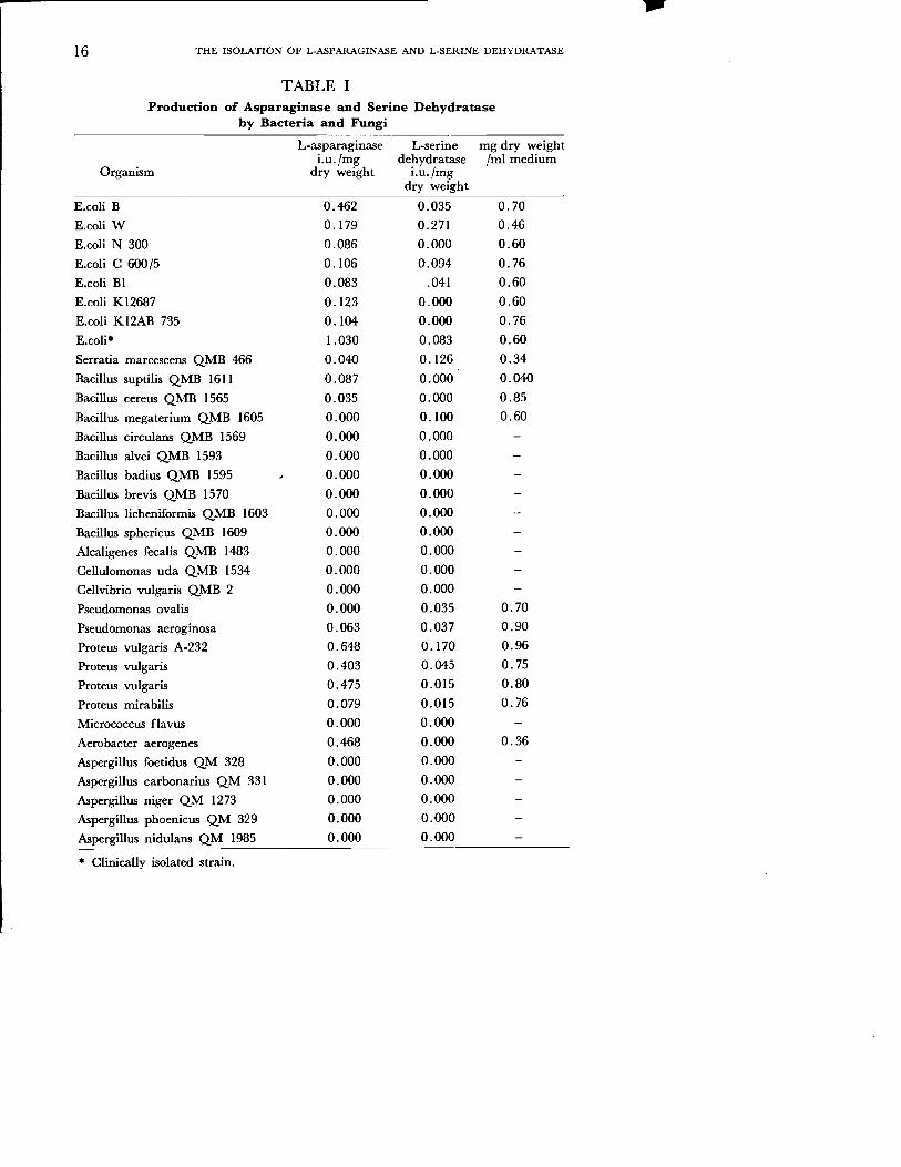
Twenty nine strains of bacteria and five strains of fungi were screened for L-asparaginase and L-serine dehydratase production. The results of these are summarized in Table 1. Proteus vulgaris A-232 was found to contain high levels of these enzymes. Of the other bacteria tested E. coli W, Serratia marcescens QMB 466 were potent sources of L-serin dehydratase. Clinically isolated strain of E.coli was found to synthesize remarkable quantities of L-asparaginase. Proteus vulgaris was selected for further studies because of high productivity on the production of both enzymes.
During the (NH4) 2S04 purification steps the highest L-asparaginase activity was found at 80 % saturation and after sonication L-serin dehydratase activity was lost. In addition, when relevant substrates were present in the growth medium, both of the enzymes induced (Table II). Partially purified L-asparaginase which was obtained from P.vulgaris A-232 inhibited DNA, RNA and protein synthesis of acute leukemia cells by 10 %, 3 %, 9 % respectively (Table III).
Conclusion
It has been shown that P.vulgaris A-232 produced both of the enzymes in high levels, this may give an advantage to produce the enzymes from one organism. Nelson, Peterson and Ciegler(1973) reported high yield of L-serine dehydratase from P.vulgaris NRRI B-123, but didn't measure L-asparaginase activity. In addition, an other strain of P.vu1garis was found to have high L-asparaginase activity. by Tosa, Sano, Yamato, Nakamura, Ando and Chibata (1968).
When the appropriate substrates added to the growth medium, the production of enzymes are increased. This suggested that the enzymes are inducible or substrates act as a nitrogen source.
Loosing the activity of L-serin dehydratase by sonication, might be from degradation of molecule during this procedure.
The inhibition of DNA, RNA and protein synthesis of acute leukemia cells by L-asparaginase from P.vulgaris A-232, shows that it has an anti tumoral activity, and possibly willi increase this property with further purification.
\
16 THE ISOLATION OF L-ASPARAGINASE AND L-SERINE DEHYDRATASE
TABLE I Production of Asparaginase and Serine Dehydratase
by Bacteria and Fungi
L-asparaginase L-serine mg dry weight i.u.jmg dehydratase Iml medium
Organism dry weight i.u.jrng dry weight
E.coli B 0.462 0.035 0.70
E.coli W 0.179 0.271 0.46
E.coli N 300 0.086 0.000 0.60
E.coli C 600/5 0.106 0-.094 0.76
E.coli BI 0.083 .041 0.60
E.coli KI2687 0.123 0.000 0.60
E.coli KI2AB 735 0.104 0.000 0.76
E.coli* 1.030 0.083 0.60
Serratia marcescens QMB 466 0.040 0.126 0.34
Bacillus suptilis QMB 1611 0.087 0.000 0.040
Bacillus cereus QMB 1565 0.035 0.000 0.85
Bacillus megaterium QMB 1605 0.000 0.100 0.60
Bacillus circulans QMB 1569 0.000 0.000
Bacillus alvei QMB 1593 0.000 0.000
Bacillus badius QMB 1595 0.000 0.000
Bacillus brevis QMB 1570 0.000 0.000
Bacillus licheniformis QMB 1603 0.000 0.000
Bacillus spheric us QMB 1609 0.000 0.000
Alcaligenes fecalis QMB 1483 0.000 0.000
Cellulomonas uda QMB 1534 0.000 0.000
Cellvibrio vulgaris QMB 2 0.000 0.000
Pseudomonas ovalis 0.000 0.035 0.70
Pseudomonas aeroginosa 0.063 0.037 0.90
Proteus vulgaris A-232 0.648 0.170 0.96
Proteus vulgaris 0.403 0.045 0.75
Proteus vulgaris 0.475 0.015 0.80
Proteus mirabilis 0.079 0.015 0.76
Micrococcus flavus 0.000 0.000
Aerobacter aerogenes 0.468 0.000 0.36
Aspergillus foetidus QM 328 0.000 0.000
Aspergillus carbonarius QM 331 0.000 0.000
Aspergillus niger QM 1273 0.000 0.000
Aspergillus phoenicus QM 329 0.000 0.000
Aspergillus nidulans QM 1985 0.000 0.000
* Clinically isolated strain.
----------------
17 HACETfEPE BULLETIN OF NATURAL SCIENCES AND ENGINEERING
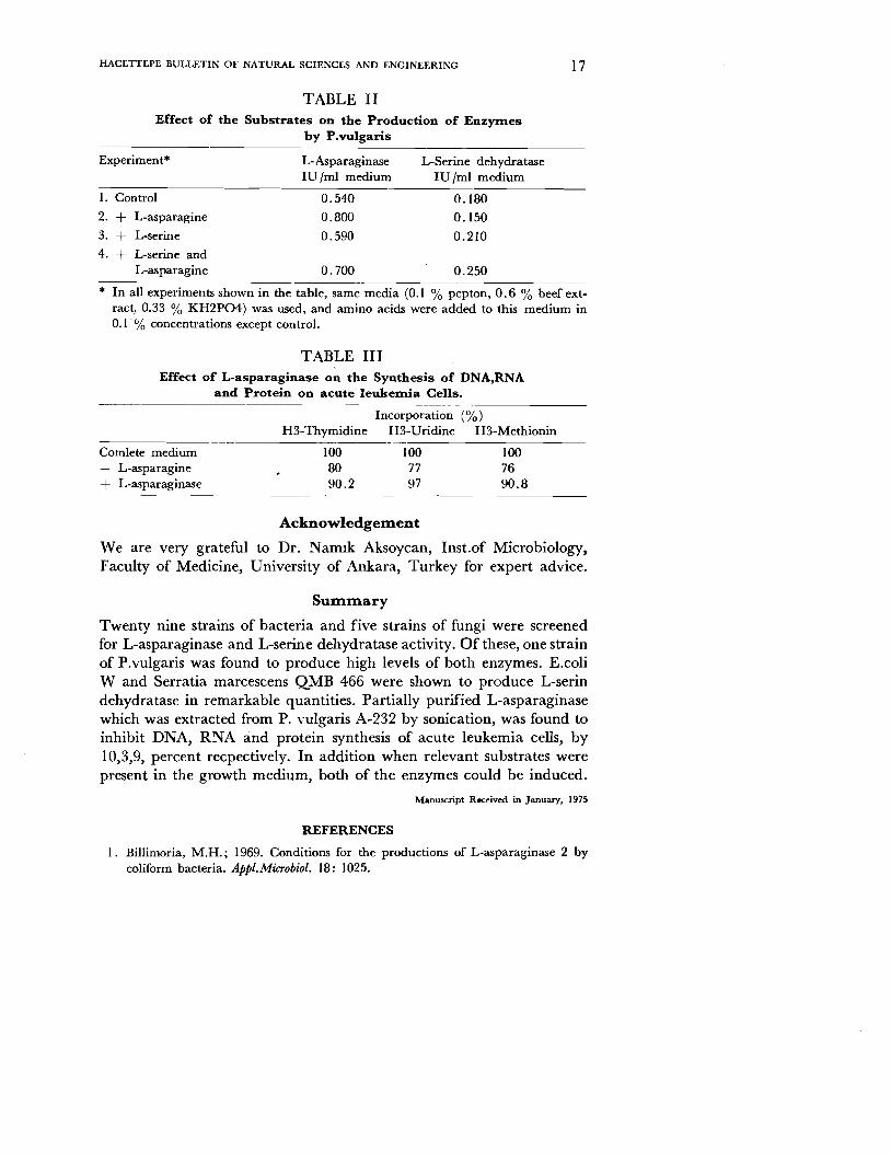
TABLE II Effect of the Substrates on the Production of Enzymes
by P.vulgaris
Experiment* L-Asparaginase L-Serine dehydratase IV Iml medium IV Iml medium
1. Control 0.540 0.180
2. + L-asparagine 0.800 0.150
3. + L-serine 0.590 0.210
4. + L-serine and L-asparagine 0.700 0.250
* In all experiments shown in the table, same media (0.1 % pepton, 0.6 % beef extract, 0.33 % KH2P04) was used, and amino acids were added to this medium in 0.1 % concentrations except control.
TABLE III Effect of L-asparaginase on the Synthesis of DNA,RNA
and Protein on acute Ieukerrria Cells.
Incorporation (%) H3-Thymidine H3-Vridine H3-Methionin
Cornlete medium 100 100 100 L-asparagine 80 77 76
+ L-asparaginase 90.2 97 90.8
Ackno~ledgenaent
We are very grateful to Dr. Narrnk Aksoycan, Inst.of Microbiology, Faculty of Medicine, University of Ankara, Turkey for expert advice.
Sunanaary
Twenty nine strains of bacteria and five strains of fungi were screened for L-asparaginase and L-serine dehydratase activity. Of these, one strain of P.vulgaris was found to produce high levels of both enzymes. E.coli Wand Serratia marcescens QMB 466 were shown to produce L-serin dehydratase in remarkable quantities. Partially purified L-asparaginase which was extracted from P. vulgaris A-232 by sonication, was found to inhibit DNA, RNA and protein synthesis of acute leukemia cells, by 10,3,9, percent recpectively. In addition when relevant substrates were present in the growth medium, both of the enzymes could be induced.
Manuscript Received in January, 1975
REFERENCES
I. Billimoria, M.H.; 1969. Conditions for the productions of L-asparaginase 2 by coliform bacteria. Appl.MiCTObiol. 18: 1025.

18 THE ISOLATION OF L-ASPARAGINASE AND L-SERINE DEHYDRATASE
2. Bollum, F.J., 1966. Filter paper disk techniques for assaying radioactive macromolecules, in Procedures in nucleic acid research. Ed. Cantoni and Davies, Harper and Row Publish. New York and London.
3. Broome, J.D., 1961. Evidence that the L-asparaginase activity of guinea pig serum is responsible for its antilymphoma effects. Nature (London) 191: 1114
4. Deangeli, L.C.; Pochiari, F.; Tanola, A.; Zurita, V.E.; Ciaranfi, E.; and Perin, A. 1970. Effect of L-asparaginase from Aspergillus terreus on ascites sarcoma in the rat. Nature (London) 225: 549
5. Heinemann, B.; Howard, A.J.; and Palocz, H.J., 1970. Influence of dissolvedoxygen levels on production of Lasparaginase and prodigiosin by Serratia marcescens. Appl.Miaobiol. 19:800
6. Hill, J.M.; Roberts, J.; Loeb, E.; Khan, A.; Maclellan, A.; and Hill, R.W., 1967. L-Asparaginase therapy for leukemia and other malignant neoplasms, J.Amer. Med.Ass. 202 :882.
7. Hill,J.M.; Loeb, E.; Maclellan, A; Khan, A.; Roberts,J.; Schields, W.F.; and Hill, N.O., 1969. Response to highly prufied L-asparaginase during therapy of acute leukemia. Cancer Res. 29:1574.
8. Jayaram, H.N.; Ramakrishnan, T.; and Viadyanathan, C.S., 1968. L-asparaginase from Mycobacterium tuberculosis strain H37Rv and H37RA. Arch.Biochem. Biophys. 126:165
9. Khan, A.; and Hill, J.M., 1969. Neutralizing precipitin in the serum of a patient treated with. L-asparaginase. J.Lab.Clin.Med. 73 :846
10. Kidd,J.G., 1953. Regression o{ transplanted lymphomas induced in vivo by means of normal guinea pig serum. LCourse of transplanted cancers of various kinds in mice and rats given guinea pig serum. J.Exp.Med. 98:565
11. Lowry, O.H.; Rosebrough, H.J.; Farr, A.J.; and Randall, R.J., 1951. Protein measurement with the folin phenol reagent. J. Bioi. Chern. 193 :265
12. Mans, R.].; and Novelli, G.D., 1961. Measurement of the incorporation of radioactive amino acids into protein by a filter paper disk method. Arch.Biochem.Biophys. 94:48
13. Mushbum, L.T.; and Wriston,J.C., 1964. Tumor inhibitory effects ofL-asparaginase from E.coli. Arch. Biochem. Biophys. 105:450
14. Nelson, G.E.N.; Peterson, R.E.; and Ciegler, A., 1973. Serin dehydratase from bacteria. J. Appl. Bacteriol, 36 :245
15. Oettgen, H.F.; Old, L.J.; Boyse, E.A.; Campbell, H.A.; Philips, F.S.; Clarkson, B.D.; Tallal, L.; Leeper, R.D.; Schwartz, M.K.; and Kim,J.H., 1967. Inhibition of leukemias in man by L-asparaginase. Cancer Res. 27 :2619
16. Peterson, R.E.; and Ciegler, A., 1969. L-Asparaginase production by Erwinia aroideae, Appi. Microbiol. 18:64 '
17. Regan,J.D.; Vodopick, H.; Takeda, S.; Lee, H.; and Faulcon, F.M., 1969. Serin requirement in leukemia and normal blood cells. Science 163: 1452
18. Tosa, T.; Sano, R.; Yamato, K.; Nakamura, M.; Ando, K.; and Chibata, 1., 1971. L-Asparaginase from Proteus vulgaris. Appl. Microbial, 22 :387
19. Wade, J .E.; Elsworth, R.; Herbert, D.; Keppie, J.; and Sargeant, K., 1968. A new L-asparaginase with anti tumor activity. Lancet 2 :776

HtJUUlp' Bu/JI/;" 01 NQJIlT,J Sd_1S ImIi E"gilU.ro.Z
1975 I Volume 4 I pp. 77 - 84
Total Syntheses of Some 7-Methyl Substituted' Estrogens Enis Oskay
Introduction
Within the last ten years many of the steroidal hormones have been totally synthesized.t-P A French firm, Roussel, has even started to manufacture most of the steroidal hormones, used in chemical therapy, by total synthesis.I ,2 So far nothing has been reported about the total syntheses of 7-methyl substituted steroids. While studying the effect of the 7-methyl group on a certain biological activityl4-16 several 7-methyl substituted steroids have been prepared by partial synthesis'Y'" with rather poor yields. For this purpose first ofall a double bond must be introduced to the steroid molecule at S-C. The resulting unsaturated molecule should then be oxidized to a 7-keto-~5 steroid. Finally the unsaturated ketone, thus obtained, when reacted with methyl magnesium iodide leads to a 7-oxo methylene steroid which could be hydrogenated with palladium oxide in glacial acetic acid-acetic anhydride mixture. For the oxidation of the A5 steroids to the 7-keto-A5 steroids t-butyl chromate or alternatively chromic oxide in pyridine is used. The first method suffers from a low yield as well as the extreme care necessary in working with large amounts of t-butyl chromate. The second method results in an even lower yield. One other method for the preparation of 7-keto-~5 steroids involves a three step sequence reported by Lenhard and Bernstein.P This method involves allylic bromination (C-7) of ~5 with N-bromo succinimide followed by reaction of the bromo conipound with neutral alumina to produce the 7-0H steroid which can then be oxidized with chromium trioxide in pyridine to 7-keto-~5 steroid.
• Faculty of Chemistry, Hacettepe University, Ankara-Turkey.

78 HACE1TEPE BULLETIN OF NATURAL SCIENCES AND ENGINEERING
In the work of Torgov". 6 for the total synthesis of d,I-8, l-l-bisdehydroestrone methyl ether (IV) (see Scheme I) triton B catalyzed condensation of crude 1,2,3,4-tetrahydro-6-methoxy-I-vinyl-I-naphthol (II) with 2-methylcyclopentane-I,3-dione afforded 2-(6-methoxy-I,2,3,4tetrahydronaphthylidene ethyl)-2-methylcyclopentane-l, 3-dione (III) which on cyclization with p-toluenesulphonic acid in refluxing benzene, afforded d,I-8, l-l-bisdehydroestrone methyl ether (IV).
SCHEME I
. CH~~H2
o CH2: CH-Mg B~ . I
TritonTHF CH30 b BoJ t~( II )Not puri f i ed
run 75 % (IV) 80 %
Method
In the present work (see Scheme II) subsititution of the tetralone (I) by a CH3 at C-3 could, according to the Scheme I, yield methyl substituted derivatives of II, III and IV (XIII, XIV, XV) and XX, XXI which could be converted to some other additional 7-methyl substituted estrogens. Since 3-methyl-6-methoxy tetralone (XII) is not a known compound 7-methyl estrogens described in this paper were totally synthesized from o-methoxybenzyl bromide (see Scheme II).

SCHEME II
Na-CCH3ICOOC2HSI2~ ~ fOOC2H5 --I~.. 0 ~OOH CH30QBr C~O~CH3 HOH C~O~CH3
IV) I VII67~. COOC2Hs I VIIJ99~. COOH
~ ~H30H. LiA1H, ~ ,OOH
C~O~CHl CH30~CH3 IIXI90~.H IVIII196~.H
1U19 2.C0 2
CC)0 0 C
I ,-,:::. Polypha. - acid
C~O.o CH3
(XIl78% H
IXXI I 85~. IXXI48 ~. (XIV) 50~.
J" !
CHIO0£>I Q CHI
{XXII J 80 % IXVllO%
j~,.,.''": OH
-C -CH " Na- C-CH .. llq. NH]
C",O CH30
IXXIII 190% IXVIII 50~. I XVII 60 Y. "'''''7CH30
", j..... OH OH
Coc-c,c-clHs
~HlO CHlO
(XVIlIl 1Z~. IXIXJ90%

80 HACETIEPE BULLETIN OF NATURAL SCIENCES ANO ENGINEERING
Results
Several 7-methyl substituted estrogenic steroids have been totally synthesized from o-methoxybenzyl bromide.
Experim.ental
Methyl-o-m.ethoxybenzyl-diethyhnalonate (VI). To a freshly prepared solution of sodium ethoxide, prepared from sodium (20 gr., 0.87 atom gram) in super dry ethanol (500 cc.), methyl-diethyl-rnalonate (151 gr., 0.8 mole gram) was added with stirring.. This was followed by the addition of o-methoxybenzyl bromide (174 gr., 0.86 mole gram). The reaction mixture was refluxed (2 hours). Most of the solvent was distilled off and the residue was poured onto crushed ice. The mixture was extracted with ether (3X). The extracts were washed (water) and dried (MgSO.). The solvent was removed and the residue distilled under reduced pressure to give methyl-o-methoxybenzyl-dieth
no 280 ylmalonate (VI, 170 gr. 67 %), b.p 138-142%.3 mm.; 1.4902; (Found: C, 65.20 %; H, 7.60 %. C\6H2~05 requires: C, 65.30 %; H, 7.54%).
Methyl-o-m.ethoxybenzyl-m.alonic acid (VII). To a refluxed solution of potassium hydroxide (280 gr.,.5 mole gram) in water (290 cc.) methylo-methoxybenzyl-diethylmalonate (VI, 295 gr., I mole gram) was added
..:.(3.0 minutes). The mixture was refluxed for an additional 2 hours period,
. cooled and acidified with cone. hydrcchloric acid. The precipitate was collected, washed (water) and dried (MgSO.). Yield: 232 gr. (almost quantitative);m.p 146-147°; (Found: C, 60.45 %; H, 5.90 %. C\2H\:.os requires: C, 60.50 %; H,5.93 %) .
. a~Methyl-a-o-lDethoxybenzylacetic acid (VIn). Methyl-o-methoxybenzyl-malonic acid (VII,77.6 gr., 0.4 mole gram) was heated at 200° for 2 hours and finally distilled to yield a-methyl-a-o-methoxybenzylacetic acid (VIII, 61 gr., 96 %); b.p 140-144° /1 mm.;no230 1.5218; (Found:
. C, :67.90 %.; H,.7-25%; C llH 1403r6,quires: C, 68.03 %; H, 7.27 %).
2,;Methyl-3-o:'m.ethoxyphenylpropan';I-01 (IX). A solution of amethyl-e-o-rnethoxybenzylacetic acid (VIII, 255 gr., 1.30 mole gram) in sodium dried ether (250 cc.) was added (60 minutis) to a susppension of lithium aluminium hydride (70 gr., 1.84 mole gram) in dry ether (500 cc.). The reaction mixture was refluxed for I hour and then cooled (0°). Excess of the hydride was carefully decomposed with water (150 cc.). This was followed by the addition of 20 % sulphuric acid (500 cc.). The mixture was extracted with ether. Ethereal layers were washed (water) and dried (MgSO.). Evaporation of the solvent

....
TOTAL SYNTHESES OF SOME 7-METHYL SUBSTITUTED ESTROGENS 81
and distillation of the residue yielded 2-methyl.3-o-methoxpyhenYlpro-', pan-l-ol (IX, 188 gr., sn %), b.p 116°fl mm.; nD240 1.5225; (Found: C, 73.35 %; H,8.90 %; CllH\602 requires: C, 73.39 %; H, 8.95 %);
I-Bromo-2-methyl-3-o-methoxyphenylpropane (X). Phosporous tri.. bromide (180 gr., 0.66 mole gram) was, added (45 minutes) to a solution of 2-methyl-3-o-methoxyphenylpropan-l-ol (IX, 180 gr., 1 mole gram) in sodium dried benzene (300 cc.) at 0°,The reaction mixture was then stirred (30 minutes) at room temperature and.Finally refluxed (4 hours). The mixture was cooled and poured onto crushed- ' ice. Benzene layer was separated, washed first with aqueous bicarbonate solution, then with water and dried (MgS04). The solvent was removed and the residue distilled to yield l-bromo-2-methyl-3-o-methoxyphenylpropane (X, 180 gr., 74 %), b.p 104° /004 mrn.; nD24°1.5405; (Found;" C, 54.20 %; H, 6.35 %; Cl1H\50Br requires: C, 54.34 %; H,6.25 %).
~-Methyl-y-o-methoxyphenylbutyric acid (XI). A Grignard reagent was prepared' from magnesium (12.2 gr., 0.5 atom gram) and l-bromo-2-methyl-3-o-methoxypheriylpropane (X, 122 gr., 0.5 ~ole gram) using a total 260 cc: of dry tetrahydro furane. This reagent was poured onto an excess of solid carbon dioxide and left to stand overnight. The mixture was the~ diluted with water (300 cc.), acidified' (cone. hydrochloric acid) and extracted with ether (3X). Ethereal layers were washed with 30 % aqueous sodium hydroxide solution' and the aqueous layers were acidified (conc.. hydrochloric acid). Acidic layers' were extracted with ether (3X). Ethereal layers were dried (MgS04) and the solvent removed. The residue was distilled to yield 13-methyl-y-o-. methoxyphenylbutyric acid (XI, 81 gr., 78 %),b.p .152° 10,5 rnm.jnDu o 1.5198; (Found: C, 69,15 %; H, 7.80 %; ClzH1603 requires: C, 69.22 %; H, 7.75 %).
3-Methyl-6~methoxy-a-tetralone' (XU). A. mixture of polyposphoric acid (125 cc.) and 13-methyl-y-o-methoxyphenylbutyric acid (XI,42 gr., 0.2 mole gram) was heated (100°) with vigorous shakin~ (45 minutes). The mixture was poured onto crushed ice, scratched and warmed until the red coloured gum turned to a colourless precipitate. The precipitate was collected, washed (water) and crystallised (petrol ether 40-60°). to' yield 3-methyl.6-methoxy-a-tetralone (XII,. 26-gfl, .' 70 %), m.p 71_72°; (Found: C, 75.70 %; H,_ 7.?O·,%; ,C\2HI402; requires: C, 75.76 %; H, 7,43 %). 2-(3-Methyl-6-methoxy-l, 2,3 - 4 -tetrahydronaphthylideDe-ethyl)~ .: 2-methylcyc1opentane-l, 3-dione .(XIV) and 2-(3-methyl-6-methoxy- 1,2, 3, 4- tetrahydronaphthylideneethyl) -2- ethyfcyclopentane-v r.

82 HACETIEPE BULLETIN OF NATURAL SCIENCES AND ENGINEERING
1,3-dione (XX). A Grignard reagent was prepared from magnesium (24.4gr., 1 mole gram) and vinyl bromide (108 gr., 1 mole gram) using a total amount of 400 cc. of dry tetrahydro furane as solvent. This reagent was reacted with 57 gr. (0.3 mole gram) of 3-methyl-6-methoxy-a-tetralone dissolved in 100 cc. of tetrahydro furane and 400 cc. of ether. The crude vinyl carbinol thus obtained was dissolved in 500 cc. of xylene and 110 cc. of tertiary butyl alcohol, and 33 gr (0.3 mole gram) of 2-methylcyc1opentane -1,3-dione or 37.8 gr. (0.3 mole gram) 2-ethylcyc1opentane-l,3-dione was added. Mter stirring (15 minutes) at room temperature 10 cc. of 40 % solution of triton B in methanol was added and the mixture stirred and heated at the reflux temperature for 2 hours. After cooling ether (1000 cc.) was added and the mixture stirred with cooling for 4-5 minutes. The mixture was filtered and the filtrate was washed (3X) with 5 % potassium hydroxide solution and with water (3X). The filtrate was then dried (MgSO.) and concentrated in vacuo. The residues were crystallised (petrol ether 40-60°) to yield: a- 2-(3-methyl-6-methoxy-l ,2,3,4-tetrahydronaphthylideneethyl)-2-methylcyc1opentane-l,3-dione (XIV, 48 gr., 50 %); (Found: C, 76.85 %; H, 7.70 %; C2oH2.03requires: C, 76.90 %; H,7.75 %), b- 2-(3-methyl-6-methoxy-l, 2, 3, 4-tetrahydronaphthylideneethyl)2-ethylcyc1opentane-l,3-dione (XX, 55 gr., 48 %), m.p 81°; (Found: C, 77.60 %; H, 8.00 %; C:llH2603requires: C, 77.27 %;H, 8.03 %).
d,1-8,14-Bisdehydro-7~-methyloestronemethylether (XV) and d,1-8,14-bisdehydro-7~, 18-dimethyloestronemethylether (XXI). A solution of XIV (20 gr., 0,07 mole gram) or of XX (50 gr., 0.16 mole gram) in methanol (500 cc.) containing 2N HCl (10 cc.) was heated (100°) for 2 hours. The reaction mixtures were diltuted with water (2000 cc.) and extracted (ether, 3X). The ethereal layers were washed (water) and dried (MgSO.). Organic solvents were removed and the residues crystallised to yield: a- d, 1-8,l4-bisdehydro-7~ -methyloestronemethylether (XV, 17 gr., 90 %), m.p 151-152°; (Found: C, 81.50 %; H, 7.60 %; C2oH2202 requires: C, 81.60 %; H, 7.54 %), b- d,1-8,14bisdehydro -7~, l8-dimethyloestronemethylether (XXI, 40 gr., 85%), m.p 105°; (Found: C, 81.65 %; H, 7.90 %; C2\H2.02 requires: C, 81.78 %; H, 7.85 %).
d,1-8-Dehydro-7~-met~yloestronemethylether(XVI) and d,l-8dehydro-7 ~-18-dimethyloestronemethylether(XXII). Hydrogenations of XV (15 gr., 0.05 mole gram) and of XXI (15 gr., 0.05 mole gram) inanalar benzene (150 cc.) in the presence of prereduced pallladium on calcium carbonate catalyst ~5 %,5 gr.) yielded by spontaneous crystallisation (methanol) from the filtrated, concentrated

TOTAL SYNTHESES OF SOME 7·METHYL SUBSTITUTED ESTROGENS 83
reaction products: a- d.l -8-dehydro-7~- methyloestronemethylether (XVI, 12 gr., 80 %), m.p 95-97°; (Found: C, 81.00 %; H, 8.20 %; C2oH2402 requires: C, 81.05 %; H, 8.17 %), b- d,l -8-dehydro-7~,
l8-dimethyloestronemethylether (XXII, 12 gr., 80 %),_ m.p 100°; (Found: C, 81.20 %; H, 10.30 %; C21H2602_ requires: C, 81.26 %; H, 10.30 %),.
d, 1-3-Methoxy-7 ~-methylestra-I,3,5(10), 8-tetraen-1713-0 I (XIX) and d,I-3-methoxy-7 ~-18-dimethylestra-I,3,5(10), 8-tetraen-1713o I (XXIII). Reduction of compounds XVI (2.9 gr., 0.01 mole gram) and XXII (3.1 gr., 0.01 mole gram) by sodium borohydride (1.5 gr., 0.04 mole gram) in methanol yielded: a- XIX (2.6 gr.,90 %), m.p 131.5°; (Found: C, 45 %; H, 8.80 %; C2oH2602 requires: C, 80.50 %; H,8. 78 %), b- XXIII (2.7 gr., 90 %), m.p 129°; (Found: C, 80.50 %; H, 9.00 %; C21H2802 requires: C, 80.73 %; H, 9.04 %).
d, 1-17 a-Ethynyl-3-methoxy-7~-methylestra-l,3,5 (10), 8-tetraen-17 13-01 (XVII) and d,I-17a-(1,3-hexadiynyl)-3-m.ethoxy-7~,18dimethylestra-I,3,5 (10), 8-tetraen-17 13-0 1 (XVIII). Compounds XVII and XVIII were prepared from XVI and appropriate sodium acetylides in liquid ammonia. Crystallisation (methanol) of the crude reaction products yielded: ;1- XVII (a gum, 50 %); (Found: C, 81.85, %; H, 8.20 %; C22H2602 requires: C, 81.95 %; H, 8.13 %) bXVIII (12 %), m.p 120-122°; (Found: C, 83,15 %; H, 8.20 %; C26H3002 requires: C, 83.40 %; H, 8.09 %).
Summary
Introduction of a 7-CH3group to the steroid skeleton is a tedious method involving several steps and suffering from low yields. Although within the last ten years many of the steroidal compounds have been totally synthesizedvP so for nothing has been reported about the total syntheses of 7-methyl substituted steroids. In the present work total syntheses of some 7-methyl steroids, starting with o-methoxybenzyl bromide, are described.
Manuscript Received in January, 1975
REFERENCES
I. Velluz, L., Recent Advances in the Total Syntheses of Steroids, Angew. Chem. Intern. Edit., 4 (1965) 181.
2. Velluz, L., et, al., Progres en Synthese totale steroide, Compt, Rend., 257 (1963) 3086.

84 HACETrEPE BULLETIN OF NATURAL SCIENCES AND ENGINEERING
3. Smith, H., et, aI., Totally synthetic (+ -) 13-hydroxy and methoxy gona-l,3,5 (10) - trien-l7-ones and related compounds, Experientia, 19 (1963) 394.
4. Smith, H., et. al., Totally Synthetic Steroid Hormones, Part I. Oestrone and related Oestrapolyenes, ]. Chern. Soc., (1963) 5072.
5. Torgov, I. V., et, al., Syntheses of derivatives of Oestrone and 19.Norsteroids from 6-methoxytetralone and 6-hydroxytetralone, Tetrahydron, 18 (1962) 1355.
6. Torgov, I. V.. et, aI., New Syntheses of oestrone, d,I-8-iso-oestrone and d,I-19 -Nortestosterone, Tetrahydron Letters, 20 (1963) 1553.
7. Asako, T., et. al., An improved Synthesis of Oestrogens, Proc, Chern. Soc., (1963) 139.
8. Asako, T., et, al., Syntheses of racemic and optically active 13 ~ethylgonanes,
Chern. Pharm. sai; 13 (1965) 1285.
9. Windholtz, T. B., et. al., Total Synthesis of 19-Norsteroids, ]. Org. Chern., 18 (1963) 1902.
10. Horeau, A., et, al., Determination des configurations par "Dedoublement Partial" III Alcools Steroides, Tetrahydron, 20 (1964) 2431.
11. Zurcher, R, F., Protonenresonanzspektroskopie und Steroidstruktur, Helo, Chim. ActQ, 46 (1963) 2054. .
12. Gibbian, H., et. al., Totalsynthese von naturlichem Ostradiolmethylather, Tetrahydron Letters, 21 (1966) 2321.
13. Oskay, E., Total synthetic estrogens and progestogens, Chim. Act. Turc., 2 (1974) 181.
14. Petrow, V., et. al., Brit. 1.081. 494.
15. Beyler, R. E., et, al., The synthesis of7a-and 7f3-methylhydrocortisones,]. A. C.S., 82 (1960) 170.
16. Bernstein, and Lenhard, H., The synthesis of 7-ketodesoxycorticosterone, ]. A. C. S., 78 (1956) 989.

Hacell'''' Bull,tin or NalllTal Sci..", tuUi Engineering
1975 / Volume 4 / pp. 85 - 90
A Comparison of Hydrocracking and Hydrogenolysis Reactions Omer Kuleli*
Introduction
Catalytic cracking of petroleum to obtain lower boiling hydrocarbon fractions is today the most important catalytic process. Reforming of the middle and heavy fractions of cracking products is a close second to this process. Hydrocracking is a special form of catalytic cracking, It is performed in the presence ofhydrogen and has proved to be very flexible to meet the fluctuating market demands for gasoline. By applying different operating conditions it is possible to convert 75 % of petroleum to gasoline or 60 % to middle distillates (light fuel oil, diesel, jet fuel) in the same reactor. Light gasoline, a major product of hydrocracking, has a high octane number due to its high isoparaffin content.
The hydrocracking is carried out at a hydrogen pressure of about 35-200 Atm and at 300-450 °C on bifunctional catalysts, such as Pt / Zeolite. Low space velocities (LHSV) of about one is used.
New restrictions on the lead-content of gasoline force the refiners to find alternative ways to produce gasolines of high octane numbers. The two strong alternatives are:
1. H ydrocracking,
2. Alkylation.
Since the erection of the first hydrocracking plant in 1959 the world hydrocracking capacity has been increased rapidly (1968: 25 Miot, 1970: 50 Miot) (1). The trend to no-lead gasoline indicates that many new hydrocracking plants will soon be operating all over the world.
" Assistant Professor, Department of Chemical Engineering, Hacettepe University, Ankara-Turkey.

86
... HACETTEPE BULLETIN OF NATURAL SCIENCES AND ENGINEERING
General
A general equation for the heteregeneous catalytic cracking reaction in the presence of hydrogen is
C - X + H2 -+ C - H + H - X (I)
where the atom X is C, N, S, 0 or a halogen. Hydrocracking requires a dual function (bifunctional) catalyst, having
a) Acid centers for cracking and isomerisation, and
b) Metal or metal oxides for hydrogenation /dehydrogenation. A side reaction of hydrocracking is hydrogenolysis and it is represented also by the equation (I). It requires monofunctional catalysts. These are usually hydrogen activating metals, such as Ni, Co, Ru, Pt, Ir, Rh, etc. Hydrogenolysis has drawn special attention in the search of alternative ways for the production ofSNG (Substitute Natural Gas). Indeed Hydromethanisation (2) and the SNG from naphtha - of British Gas Council are industrial applications ofhydrogenolysis. The addition ofmethanators to the existing Lurgi Pressure Gasifiers seems to be the most promising solution to satisfy the growing demand for SNG (3) The new and yet not commercially operated methanators require a thorough study of hydrogenolysis reactions.
This study intends to explain the differences between these two related reactions and to which extent hydrogenolysis plays a role in commercial hydrocracking processes.
Experimental
The experiments have been carried out in a flow system, made of stainless steel. High hydrogen pressures (up to 50 Atm) and low hydrocarbon space velocities (0,3-0,5 h-1) have been used. Hydrogen was dried and purified in a Deoxo unit. After being saturated with the hydrocarbon the mixture was preheated to reaction temperature before entering the reactor. Reaction temperature was controlled within ± 1°C. About one or two mililiters of catalyst were used for the conversion.
The reaction products were analysed by gas chromatography. Details of temperature programmed GC-analysis were explained elsewhere (4). For the complete analysis of mixtures, two capillary columns (PPG and Squalane) and a packed column (Reoplex/Ah0 3) were used.
Bifunctional hydrocracking catalyst (catalyst B), 0,5 % Pt /Ca / V-Zeolite, was a commercial one and obtained from The Union Carbide Int. Co. Monofunctional 5 % Pt/AI203 catalyst (catalyst M); used in

87 A COMPARISON OF HYDROCRACKING AND HYDROGENOLYSIS REACTIONS
this work, was prepeared as explained below: 0,440 g ofPtOz. xH20 was mixed with 6,688 g Al203 in water. This slurry was stirred continuously, water being slowly evaporated. More water was added and this procedure was repeated 5-6 times till Pt02 • xH20 was uniformly adsorbed on Al203• This way of preparation ensures an acid-free catalyst which is of vital importance in hydrogenolysis reactions. A reliable hydrogenolysis catalyst must not have acidic properties, otherwise it may initiate hydrocracking reactions of ionic nature.
Results and Discussions
Hydrocracking and hydrogenolysis of methylcyclopentane (MCP) and cyclohexane (CH) were carried out under the same reaction conditions. The aim was to find out the fraction of hydrogenolytic reactions taking part on bifunctional catalysts. A short comparison of the results is given in Table 1.
The bifunctional catalyst, Pt /Ca /Y-Zeolite (B), as compared to monofunctional Pt/Alz03 (M) was much more active. The combination of hydrogenation /dehydrogenation component (Pt) and the acid component (Zeolite) gives excellent rates of conversions. Hydrocarbon reactions on such bifunctional catalysts can be explained through carbenium-ion mechanism.
at P-p>SItlon
ivvCracking1~-Scission
The conversions on M are about three magnitudes of order smaller than those of B. This large difference indicates that the hydrogenolytic reactions are not significant during commercial hydrocracking operations
The careful study of the reaction products reveal the fact that these two reactions proceed quite differently from each other.

88 HACElTEPE BULLETIN OF NATURAL SCIENCES AND ENGINEERING
TABLE I
Comparison Between Monofunctional and Bifunctional Catalysis of Naphthene Conversion
Catalyst M: 5 % Pt/AI203 Monofunctional Catalyst B: 0,5 % Pt/Ca/Y-Zeolite Bifunctional T: 275°, H 2:HC(*): 20 Mol/Mol, P: 40 Atm, LHSV: 0,3 h"t
Feed Methylcyclopentane Cyclohexane Catalyst B M . B M Conversion (Wt %) 27,12 0,07 74,03 0,02
Analysis of products (Wt %)
Ring opening
Isomerization
Paraffins < C6
Naphthenes < C6
Aufbau (> C6) (**)
droducts of ring opening
n-Hexane
2 Me Pentane
3 Me Pentane
2,2 Di Me Butane
2,3 Di Me Butane
25,4
66,3
3,9
0,2
4,2
.38,3
32,9
21,8
4,2
2,8
30,0
57,9
12,1
3,7
60,4
35,9
6,2
92,3 68,4
0,6 31,6
400 ppm
0,9
11,2
31,8
21,7
3,1
2,2
100
Paraffins < C6
C t 1,3 30,6 2,2 100
C2 0,4. 2,2
C3 6,3 4,S C4 52,0 48,7
Cs 40,0 69,3 42,1
( *) Hz: HC: Ratio of hydrogen to hydrocarbon (**) Aufbau products are hydrocarbons which contain more carbon atoms than the
feed hydrocarbons.
1. The main reaction in both cases is isomerization, isomerization of CH being faster than that of MCP.
o
89 A COMPARISON OF~HYOROCRACKINGA.1IJO HYOROGENOLYSIS REACTIONS
Thermodynamic equilibrium is. reached with catalyst B.
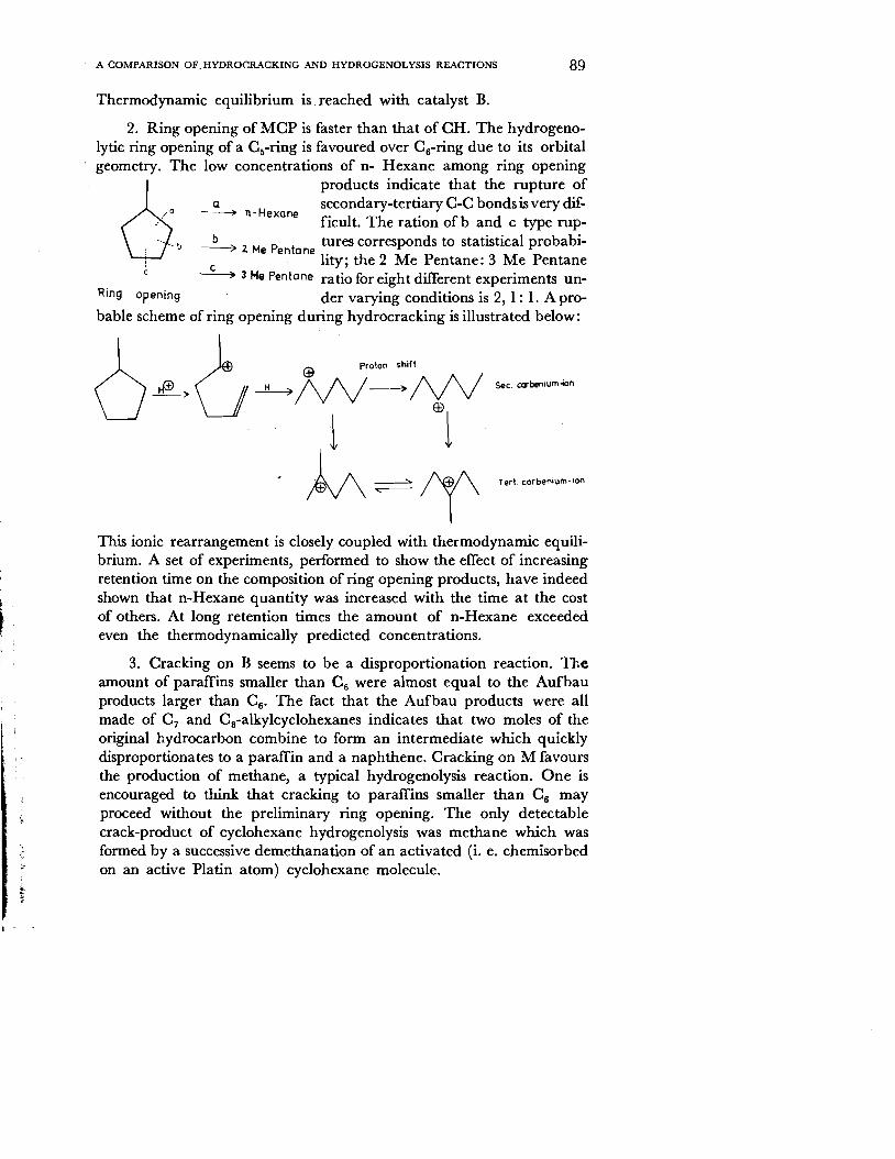
2. Ring opening of MCP is faster than that of CH. The hydrogenolytic ring opening of a C~-ring is favoured over Cs-ring due to its orbital geometry. The low concentrations of n- Hexane among ring opening
6 products indicate that the rupture of
a secondary-tertiary C-C bonds is very dif00 ~ n-Hexane
0'·0 ficult. The ration of band c type rup<; <b ~,., P t tures corresponds to statistic.al probabi
; .. ,.,e en one • : c . hty; the 2 Me Pentane: 3 Me Pentane c ------? 3 Me Pentane ratio for eight different experiments un-
Ring opening der varying conditions is 2, 1: 1. A probable scheme of ring opening during hydrocracking is illustrated below:
;.. :...
r
sec. cabenium-ion
This ionic rearrangement is closely coupled with thermodynamic equilibrium. A set of experiments, performed to show the effect of increasing retention time on the composition of ring opening products, have indeed shown that n-Hexane quantity was increased with the time at the cost of others. At long retention times the amount of n-Hexane exceeded even the thermodynamically predicted concentrations.
3. Cracking on B seems to be a disproportionation reaction. The amount of paraffins smaller than Cs were almost equal to the Aufbau products larger than CS' The fact that the Aufbau products were all made of C 7 and Cs-alkykyclohexanes indicates that two moles of the original hydrocarbon combine to form an intermediate which quickly disproportionates to a paraffin and a naphthene. Cracking on M favours the production of methane, a typical hydrogenolysis reaction. One is encouraged to think that cracking to paraffins smaller than Cs may proceed without the preliminary ring opening. The only detectable crack-product of cyclohexane hydrogenolysis was methane which was formed by a successive demethanation of an activated (i. e. chemisorbed on an active Platin atom) cyclohexane molecule.

90 HACETTEPE BULLETIN OF NATURAL SCIENCES AND ENGINEERING
Conclusion
The role of hydrogenolysis reactions taking part on the hydrogenation I dehydrogenation component of a bifunctional hydrocracking catalyst is of no practical importance. The existence of such reactions can at best be suggested by the presence of small amounts of methane and ethane. As is well known a carbenium ion mechanism operating through ~-scissions,
produces no hydrocarbon smaller than C 3• The reaction velocities of hydrocracking on such powerful catalysts are several magnitudes of order larger than those of hydrogenolysis.
Manuscript Received in January, 1975
REFERENCES i. Scott,]. W., Paterson, N. ]., Proc.7th World Petroleum Cogress, 4, 97 (1967).
2. Kikuchi, E., Morita, &., Yamamoto, K., Bull. ofthe Japan Petrol. Inst., 11,34 (1969).
3. Moeller, F. W., Roberts, H., Britz, B., Hydrocarbon Proc., 53 (4), 69 (1974).
4. Kuleli, 0., Dissertation, Universitaet Karlsruhe, s, 34 (1974).
5. Eberth, H., Diplomarbeit, Universitaet Karlsruhe (1971).