Amyloid Neuropathy - Alexander Lauder DS edits … Quiz: Question What type of amyloid is the most...
68
Amyloid Neuropathy Alexander Lauder Boston University School of Medicine, Third Year July 29, 2010
Transcript of Amyloid Neuropathy - Alexander Lauder DS edits … Quiz: Question What type of amyloid is the most...
Amyloid Neuropathy Alexander LauderBoston University School of Medicine, Third YearJuly 29, 2010
Pop Quiz: Question
� What type of amyloid is the most common cause
of amyloid neuropathy?
� ATTR
� AA� AA
� AL
� Aβ
� Aβ2-microglobulin

Pop Quiz: Answer
� AL

Outline� Properties of Amyloid
� Pathogenesis of Amyloidosis
� Classification & Organ System Involvement
� Amyloid Neuropathy
� Epidemiology
� Etiology
� Pathogenesis
� Clinical Findings
� Diagnostic Evaluation
� Treatment
� Prognosis
� Summary
Amyloid, what is it?
A pathologic protinaceous substance,
deposited in the extracellular space of various
tissues and organs.
Properties of Properties of
Amyloid
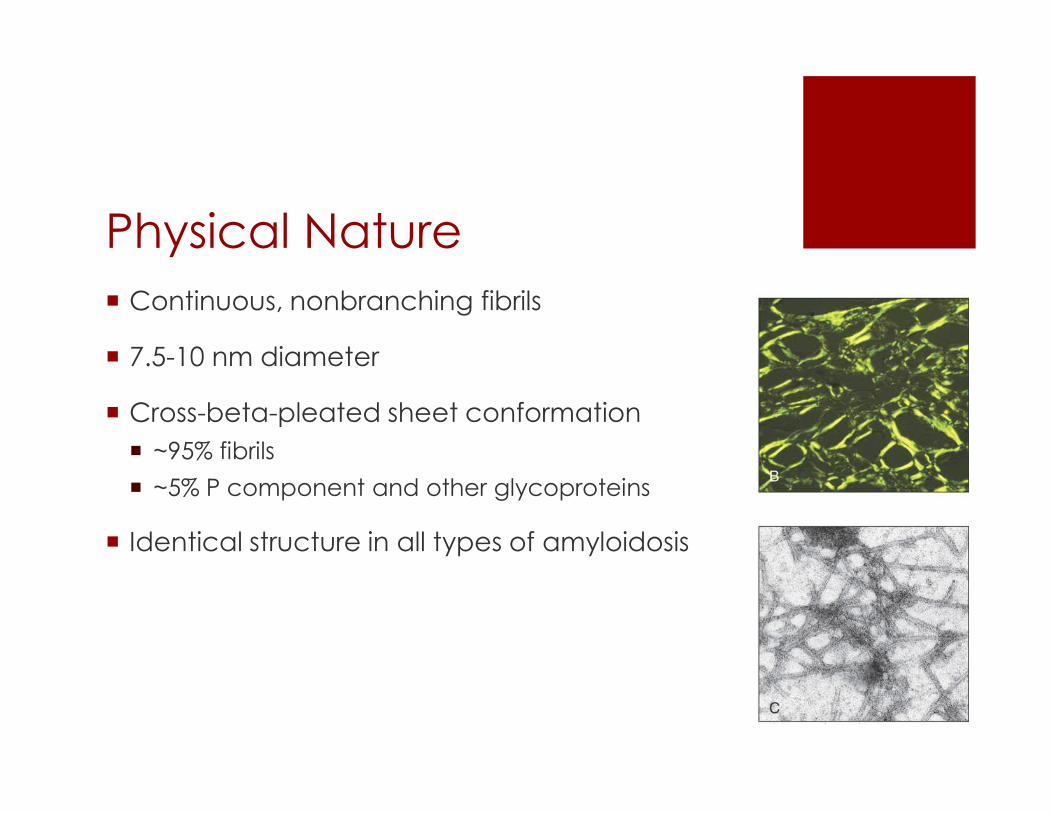
Physical Nature
� Continuous, nonbranching fibrils
� 7.5-10 nm diameter
� Cross-beta-pleated sheet conformation� Cross-beta-pleated sheet conformation
� ~95% fibrils
� ~5% P component and other glycoproteins
� Identical structure in all types of amyloidosis
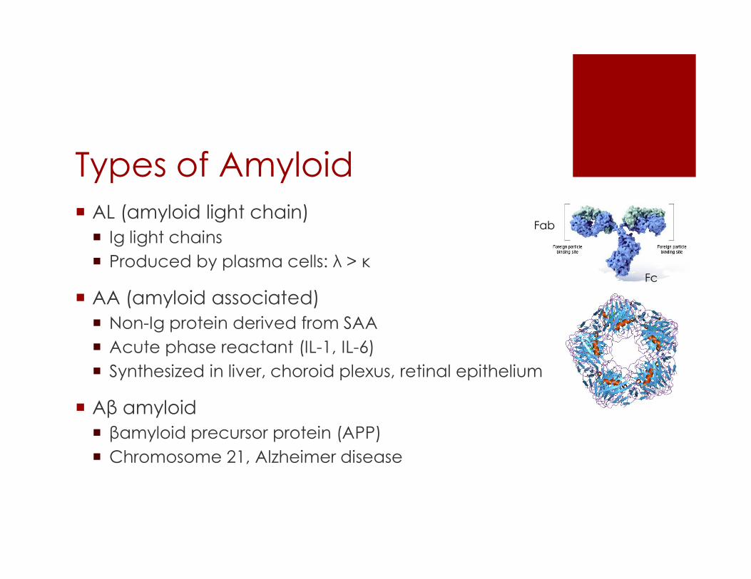
Types of Amyloid
� AL (amyloid light chain)
� Ig light chains
� Produced by plasma cells: λ > κ
� AA (amyloid associated)
Fab
Fc
� AA (amyloid associated)
� Non-Ig protein derived from SAA
� Acute phase reactant (IL-1, IL-6)
� Synthesized in liver, choroid plexus, retinal epithelium
� Aβ amyloid
� βamyloid precursor protein (APP)
� Chromosome 21, Alzheimer disease
Types of Amyloid, cont.� Transthyretin (TTR)
� Serum protein� Transports thyroxine and retinol� Mutant forms cause familial amyloid polyneuropathy or cardiomyopathyWild type form can lead to SSA, senile systemic (age-� Wild type form can lead to SSA, senile systemic (age-related) amyloidosis
� β2-microglobulin� Component of MHC class I� Long term hemodialysis
� Prion Proteins� “local amyloidosis”� Misfolded proteins aggregate in extracellular space
Pathogenesis
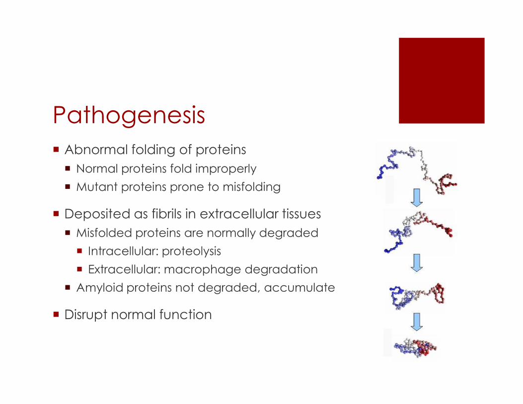
Pathogenesis
� Abnormal folding of proteins
� Normal proteins fold improperly
� Mutant proteins prone to misfolding
� Deposited as fibrils in extracellular tissues
� Misfolded proteins are normally degraded
� Intracellular: proteolysis
� Extracellular: macrophage degradation
� Amyloid proteins not degraded, accumulate
� Disrupt normal function
ClassificationSystemic
Localized
Hereditary

Systemic� AL: 1o Amyloidosis, associated with clonal B lymphoplasmacytic
diseases. All of these are AL, due to immunoglobulin light chains.
� AL: plasma cell dyscrasia up to 30% with diagnostic features of MM
� MM: high grade plasma cell dyscrasia with lytic bone lesions, � MM: high grade plasma cell dyscrasia with lytic bone lesions, hypercalcemia, anemia
� WM: Waldenstrom’s macroglobulinemia (lymphoplasmacyticlymphoma) with IgM AL
� Other B lymphomas
� AA: 2o Amyloidosis, associated with chronic inflammation, infection.
� Rheumatoid Arthritis
� Ankylosing Spondylitis
� Inflammatory Bowel Disease
� Hereditary Periodic Fever Syndromes like FMF

Localized
� Confined to a single organ
� Alzheimer’s Disease
� Prion Disease� Prion Disease

Hereditary
� TTR
� Familial Amyloid Polyneuropathy
� ApoAI� ApoAI
� Gelsolin
� Lysozyme
� Fibrinogen

Affected Organ Systems� Kidney: proteinuria, renal failure
� Spleen: splenomegaly
� Liver: hepatomegaly, elevated alkaline phosphatase� Liver: hepatomegaly, elevated alkaline phosphatase
� Heart: concentric ventricular hypertrophy, diastolic dysfunction, conduction system damage
� Endocrine: adrenal, thyroid, pituitary, pancreas
� GI: dysmotility, malabsorption, diarrhea or constipation
� Nerve: carpal tunnel syndrome, peripheral and/or autonomic neuropathy
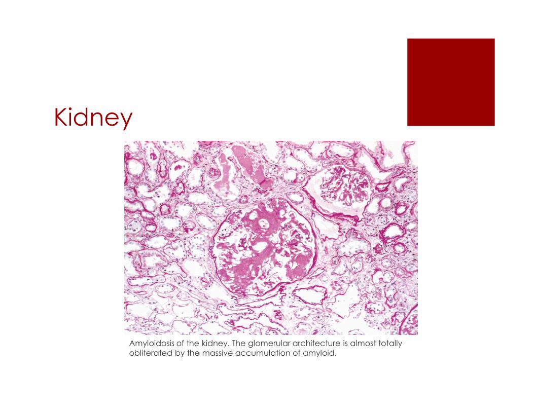
Kidney
Amyloidosis of the kidney. The glomerular architecture is almost totally
obliterated by the massive accumulation of amyloid.
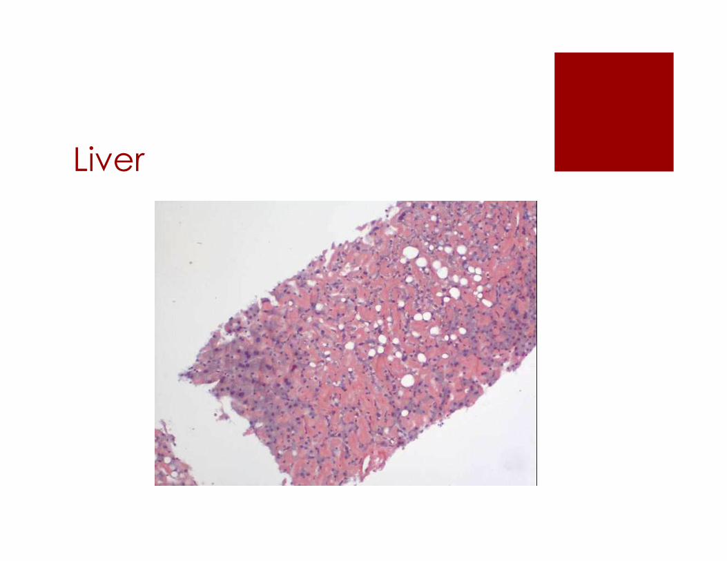
Liver
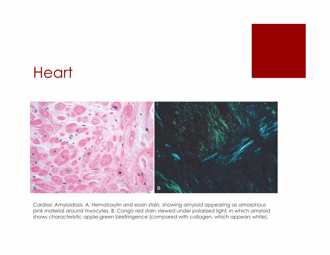
Heart
Cardiac Amyloidosis. A, Hematoxylin and eosin stain, showing amyloid appearing as amorphous
pink material around myocytes. B, Congo red stain viewed under polarized light, in which amyloid
shows characteristic apple-green birefringence (compared with collagen, which appears white).
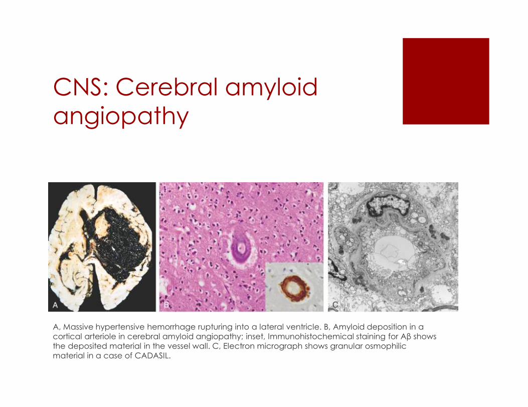
CNS: Cerebral amyloid
angiopathy
A, Massive hypertensive hemorrhage rupturing into a lateral ventricle. B, Amyloid deposition in a
cortical arteriole in cerebral amyloid angiopathy; inset, Immunohistochemical staining for Aβ shows
the deposited material in the vessel wall. C, Electron micrograph shows granular osmophilic
material in a case of CADASIL.
Amyloid
Neuropathy

History
� First described in families in Oporto, Portugal in
1952
� Andrade C. “A peculiar form of peripheral
neuropathy; familiar atypical generalized amyloidosis neuropathy; familiar atypical generalized amyloidosis
with special involvement of the peripheral nerves.”
Brain 75 (3): 408–27
Amyloidoses and
Neuropathy
� Associated with:
� AL Amyloidosis (30%)
� ATTR Amyloidosis: FAP familial amyloidotic
polyneuropathypolyneuropathy
� AF Amyloidosis: Other hereditary forms
� ApoAI
� Gelsolin: benign cranial and sensory
polyneuropathy
� Aβ2-microglobulin: carpal tunnel syndrome
� Not associated with:
� AA Amyloidosis
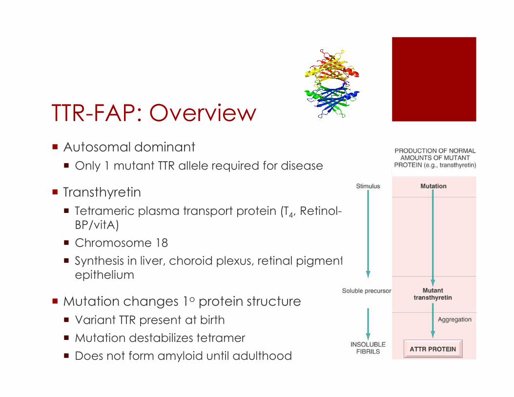
TTR-FAP: Overview
� Autosomal dominant
� Only 1 mutant TTR allele required for disease
� Transthyretin� Transthyretin
� Tetrameric plasma transport protein (T4, Retinol-
BP/vitA)
� Chromosome 18
� Synthesis in liver, choroid plexus, retinal pigment
epithelium
� Mutation changes 1o protein structure
� Variant TTR present at birth
� Mutation destabilizes tetramer
� Does not form amyloid until adulthood

TTR-FAP: >100 mutations
TTR-FAP: System involvement
� PNS: most common, peripheral neuropathy
� ANS: orthostatic hypotension, alteration in GI
motility
� Heart: restrictive cardiomyopathy
� Blood vessels:
� CNS leptomeningeal amyloidosis
� Cerebral infarcts & hemorrhage
� Renal involvement not common

TTR-FAP: Pathogenesis
Deposit around perforating arterioles
Amyloiddisplaces nerve
fiber
Compression-induced
demyelinationand nerve fiber
loss with intraneuralamyloid
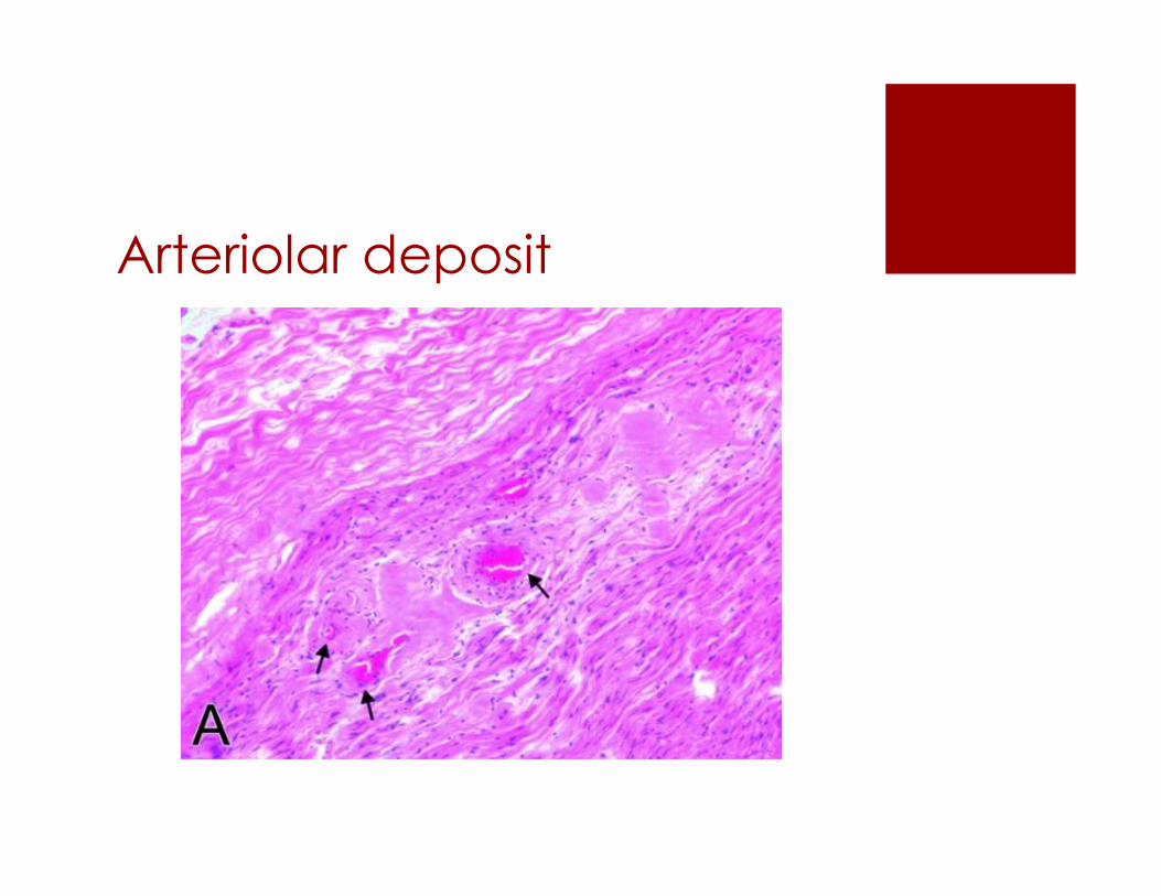
Arteriolar deposit
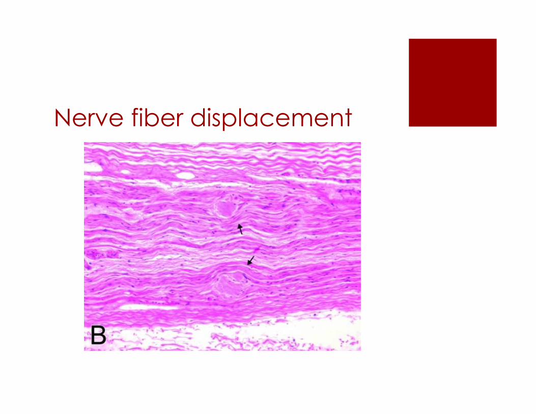
Nerve fiber displacement

Demyelination with deposits
Cross Section of Sciatic N. bundles

TTR-FAP:
Location of Deposition
� Peripheral nerves
� Dorsal root ganglia
� Leptomeninges around SC and brain� Leptomeninges around SC and brain
� NO CNS parenchymal involvement
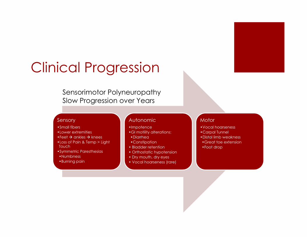
Clinical Progression
Sensory Autonomic Motor
Sensorimotor Polyneuropathy
Slow Progression over Years
Sensory
•Small fibers
•Lower extremities
•Feet � ankles � knees
•Loss of Pain & Temp > Light Touch
•Symmetric Paresthesias
•Numbness
•Burning pain
Autonomic
•Impotence
•GI motility alterations:
•Diarrhea
•Constipation
• Bladder retention
• Orthostatic hypotension
• Dry mouth, dry eyes
• Vocal hoarseness (rare)
Motor
•Vocal hoarseness
•Carpal Tunnel
•Distal limb weakness
•Great toe extension
•Foot drop
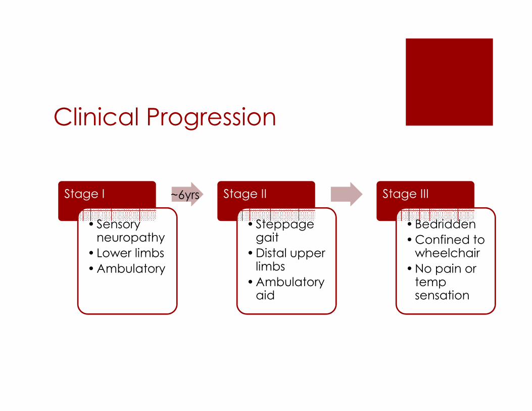
Clinical Progression
Stage I Stage II Stage III~6yrs
•Sensory neuropathy
•Lower limbs
•Ambulatory
•Steppagegait
•Distal upper limbs
•Ambulatory aid
•Bedridden
•Confined to wheelchair
•No pain or temp sensation
TTR-FAP: Other exam findings
� Neuroarthropathies (Charcot joints)
� Cranial neuropathies:
� Progressive involvement of CN� Progressive involvement of CN
� V: facial sensation
� VIII: impaired hearing
� XII: tongue movement
� Sparing of oculomotor nerve
� Carpal tunnel syndrome
� Blindness from vitreous opacities
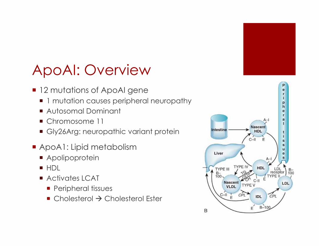
ApoAI: Overview
� 12 mutations of ApoAI gene
� 1 mutation causes peripheral neuropathy
� Autosomal Dominant
� Chromosome 11� Chromosome 11
� Gly26Arg: neuropathic variant protein
� ApoA1: Lipid metabolism
� Apolipoprotein
� HDL
� Activates LCAT
� Peripheral tissues
� Cholesterol � Cholesterol Ester
ApoAI: System involvement
� Renal amyloid deposition, main feature
� Liver
� Spleen� Spleen
� Heart
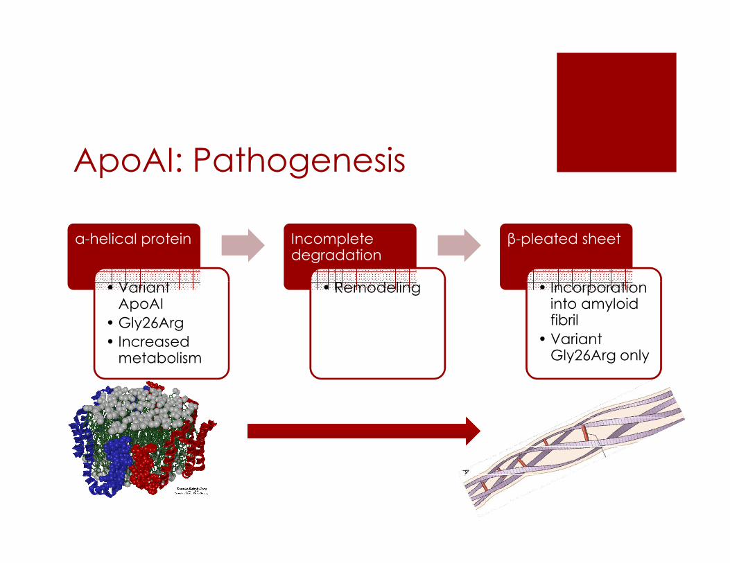
ApoAI: Pathogenesis
α-helical protein
• Variant
Incomplete degradation
• Remodeling
β-pleated sheet
• Incorporation • Variant ApoAI
• Gly26Arg
• Increased metabolism
• Remodeling • Incorporation into amyloidfibril
• Variant Gly26Arg only
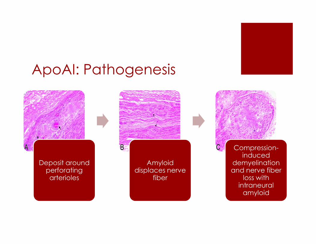
ApoAI: Pathogenesis
Deposit around perforating arterioles
Amyloiddisplaces nerve
fiber
Compression-induced
demyelinationand nerve fiber
loss with intraneuralamyloid

ApoAI:
Location of Deposition
� Peripheral nerves
� Dorsal root ganglia
� Leptomeninges around SC and brain� Leptomeninges around SC and brain
� NO CNS parenchymal involvement
� Similar to TTR-FAP
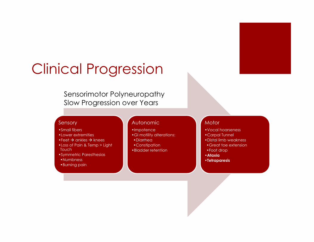
Clinical Progression
Sensory Autonomic Motor
Sensorimotor Polyneuropathy
Slow Progression over Years
Sensory
•Small fibers
•Lower extremities
•Feet � ankles � knees
•Loss of Pain & Temp > Light Touch
•Symmetric Paresthesias
•Numbness
•Burning pain
Autonomic
•Impotence
•GI motility alterations:
•Diarrhea
•Constipation
•Bladder retention
Motor
•Vocal hoarseness
•Carpal Tunnel
•Distal limb weakness
•Great toe extension
•Foot drop
•Ataxia
•Tetraparesis

ApoA1: Onset/Prognosis
� Adult onset: 30-40s
� Slowly progressive

Gelsolin: Overview� Plasma gelsolin
� Actin modulating protein
� Mutations result in abnormal proteolysis
� Asp187Asn
� Asp187Tyr
� Systems Involved:
� Nerve
� Vascular
� Renal
� Onset: ~40 years of age
� Involvement of CN VII leads to characteristic drooping of
facial muscles, wrinkling, ptosis
� Ptosis can be corrected surgically
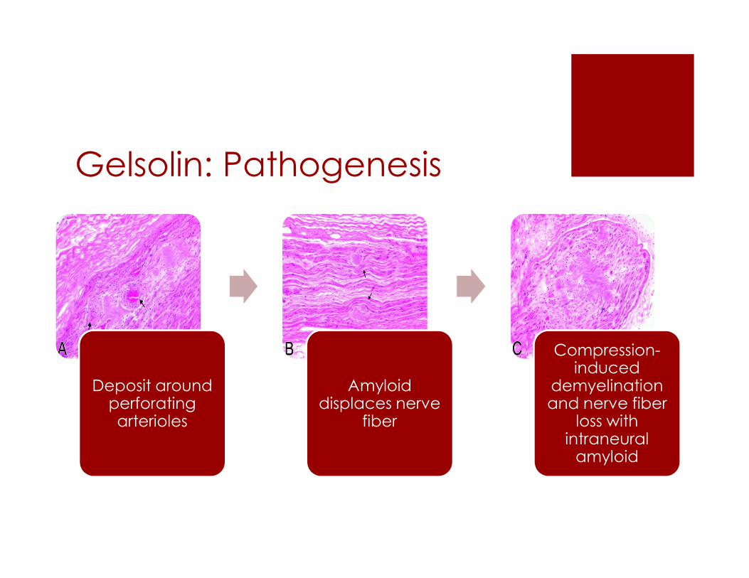
Gelsolin: Pathogenesis
Deposit around perforating arterioles
Amyloiddisplaces nerve
fiber
Compression-induced
demyelinationand nerve fiber
loss with intraneuralamyloid
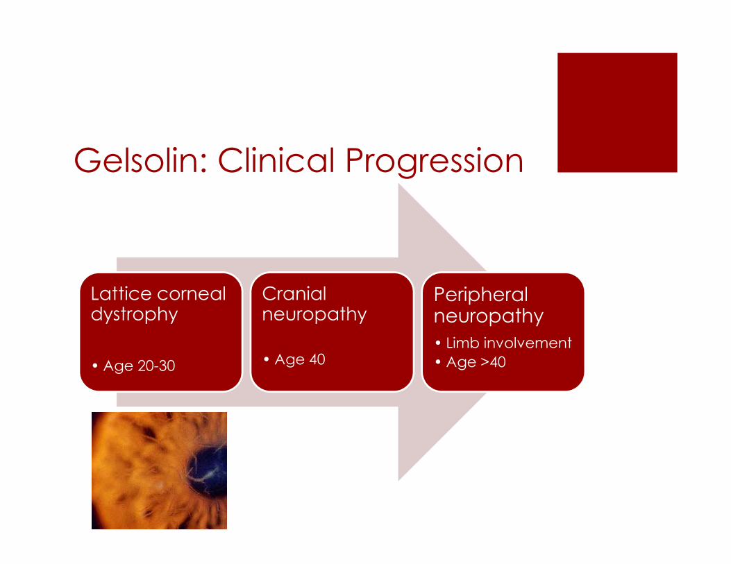
Gelsolin: Clinical Progression
Lattice corneal Cranial Peripheral Lattice corneal dystrophy
• Age 20-30
Cranial neuropathy
• Age 40
Peripheral neuropathy
• Limb involvement
• Age >40
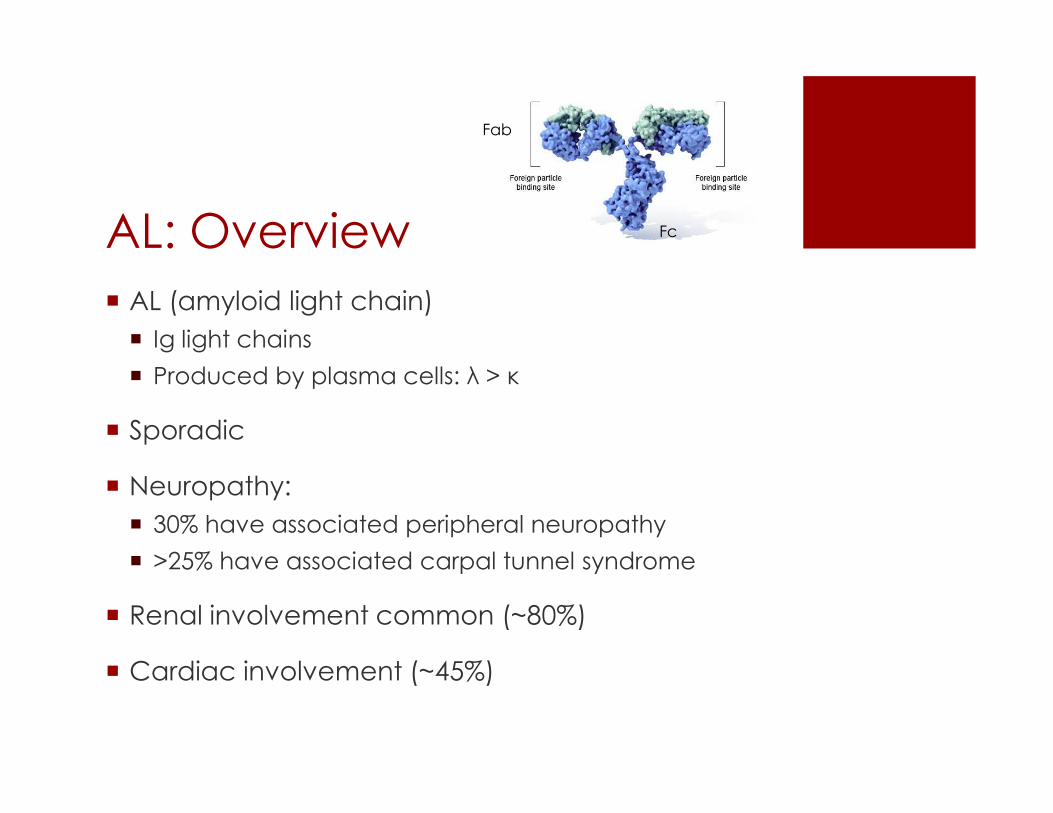
AL: Overview
� AL (amyloid light chain)
� Ig light chains
� Produced by plasma cells: λ > κ
Fab
Fc
� Sporadic
� Neuropathy:
� 30% have associated peripheral neuropathy
� >25% have associated carpal tunnel syndrome
� Renal involvement common (~80%)
� Cardiac involvement (~45%)

AL: Pathogenesis
Deposit around perforating arterioles
Amyloiddisplaces nerve
fiber
Compression-induced
demyelinationand nerve fiber
loss with intraneuralamyloid
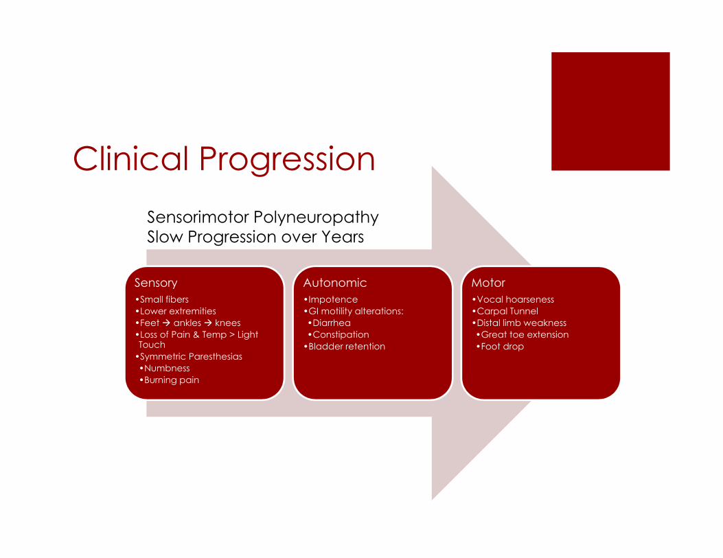
Clinical Progression
Sensory Autonomic Motor
Sensorimotor Polyneuropathy
Slow Progression over Years
Sensory
•Small fibers
•Lower extremities
•Feet � ankles � knees
•Loss of Pain & Temp > Light Touch
•Symmetric Paresthesias
•Numbness
•Burning pain
Autonomic
•Impotence
•GI motility alterations:
•Diarrhea
•Constipation
•Bladder retention
Motor
•Vocal hoarseness
•Carpal Tunnel
•Distal limb weakness
•Great toe extension
•Foot drop
Diagnostic Diagnostic
Evaluation
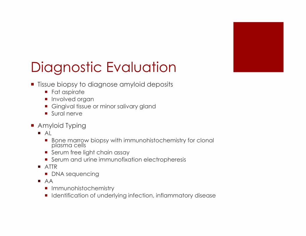
Diagnostic Evaluation� Tissue biopsy to diagnose amyloid deposits
� Fat aspirate
� Involved organ
� Gingival tissue or minor salivary gland
� Sural nerve
� Amyloid Typing� AL
� Bone marrow biopsy with immunohistochemistry for clonalplasma cells
� Serum free light chain assay
� Serum and urine immunofixation electropheresis
� ATTR
� DNA sequencing
� AA
� Immunohistochemistry
� Identification of underlying infection, inflammatory disease
Tissue biopsy
� Light microscopy (H&E): amorphous, eosinophilic,
hyaline, extracellular substance
� Deposits induce pressure atrophy
� Peripheral nerve Wallerian Degeneration
� Distal degeneration of axon & myelin sheath
� Proximal axonal degeneration to next node
� Cell body swells, nucleus is peripheralized
� Congo red stain: apple-green birefringence
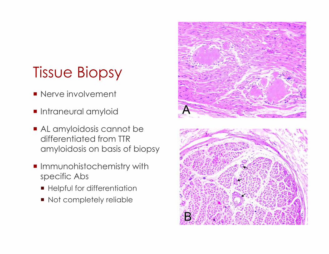
Tissue Biopsy
� Nerve involvement
� Intraneural amyloid
� AL amyloidosis cannot be � AL amyloidosis cannot be
differentiated from TTR
amyloidosis on basis of biopsy
� Immunohistochemistry with
specific Abs
� Helpful for differentiation
� Not completely reliable
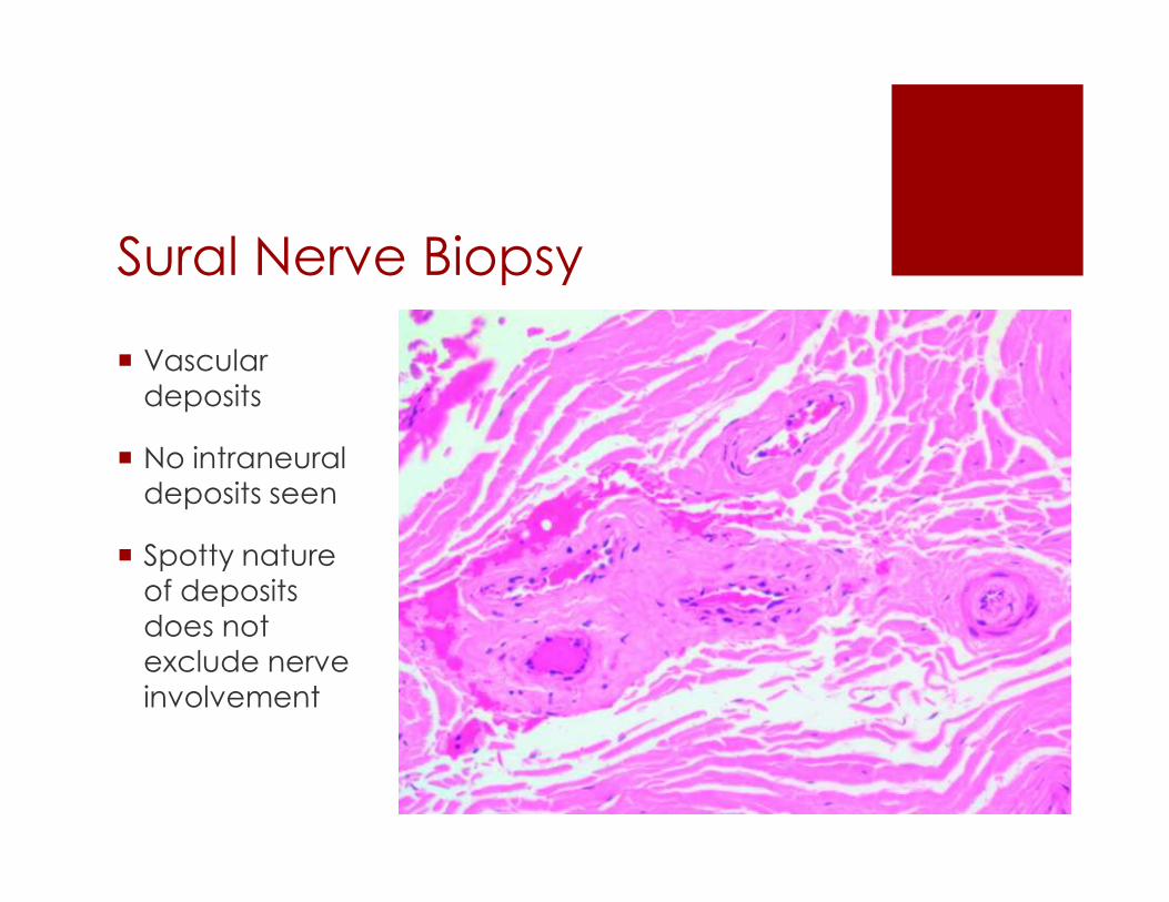
Sural Nerve Biopsy
� Vascular
deposits
� No intraneural � No intraneural
deposits seen
� Spotty nature
of deposits
does not
exclude nerve
involvement
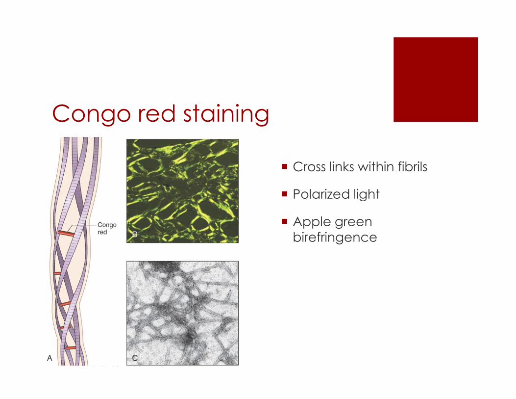
Congo red staining
� Cross links within fibrils
� Polarized light� Polarized light
� Apple green
birefringence

Commercial DNA Testing
� Available for TTR
� Full TTR DNA testing if mutation unknown
� Specific TTR sequence testing if mutation known
� Not available for:
� ApoAI
� Gelsolin
Treatment
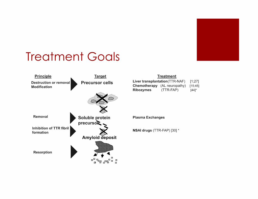
Treatment Goals

Treatment
� Nonspecific
� Specific
Nonspecific Treatment
� Treat painful neuropathic symptoms
� Agents:
� Gabapentin� Gabapentin
� Amitryptyline
� Pregabalin
� Duloxetine
� Tricyclic antidepressants, may exacerbate orthostasis
� Opioid analgesics
� Response to drug may change as disease
progresses
Specific Treatment: TTR-FAP
� Orthotopic liver transplantation
� Remove mutant TTR, synthesized in liver
� Val30Met mutation best prognosis: 80% 5 year survival
� Other mutations: 55-60% 5 year survival� Other mutations: 55-60% 5 year survival
� Some evidence of efficacy for ApoAI
� Vitrectomy for corneal amyloid deposits
� Small molecules to stabilize the normal TTR tetramer
� Diflunisal (NSAID)
� Tafamidis
Specific Treatment: Gelsolin
� Lattice corneal dystrophy
� Method: Corneal transplantation
� Cutis laxis & blepharochalasis (resultant from
facial palsy)
� Method: Plastic surgery
� Note: Gelsolin is essential protein of actin function
� Rx aimed to eliminate production will likely not be
tolerated
Specific Treatment: AL
� Anti-plasma cell chemotherapy
� Oral melphalan chemotherapy (relatively
ineffective)
� High dose IV melphalan and autologous stem cell � High dose IV melphalan and autologous stem cell
transplantation
� New agents
� Bortezomib, proteasome inhibitor
� Lenalidomide, immunomodulator
� Others

Summary
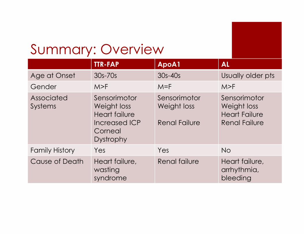
Summary: OverviewTTR-FAP ApoA1 AL
Age at Onset 30s-70s 30s-40s Usually older pts
Gender M>F M=F M>F
Associated
Systems
Sensorimotor
Weight loss
Heart failure
Sensorimotor
Weight loss
Sensorimotor
Weight loss
Heart FailureHeart failure
Increased ICP
Corneal
Dystrophy
Renal Failure
Heart Failure
Renal Failure
Family History Yes Yes No
Cause of Death Heart failure,
wasting
syndrome
Renal failure Heart failure,
arrhythmia,
bleeding
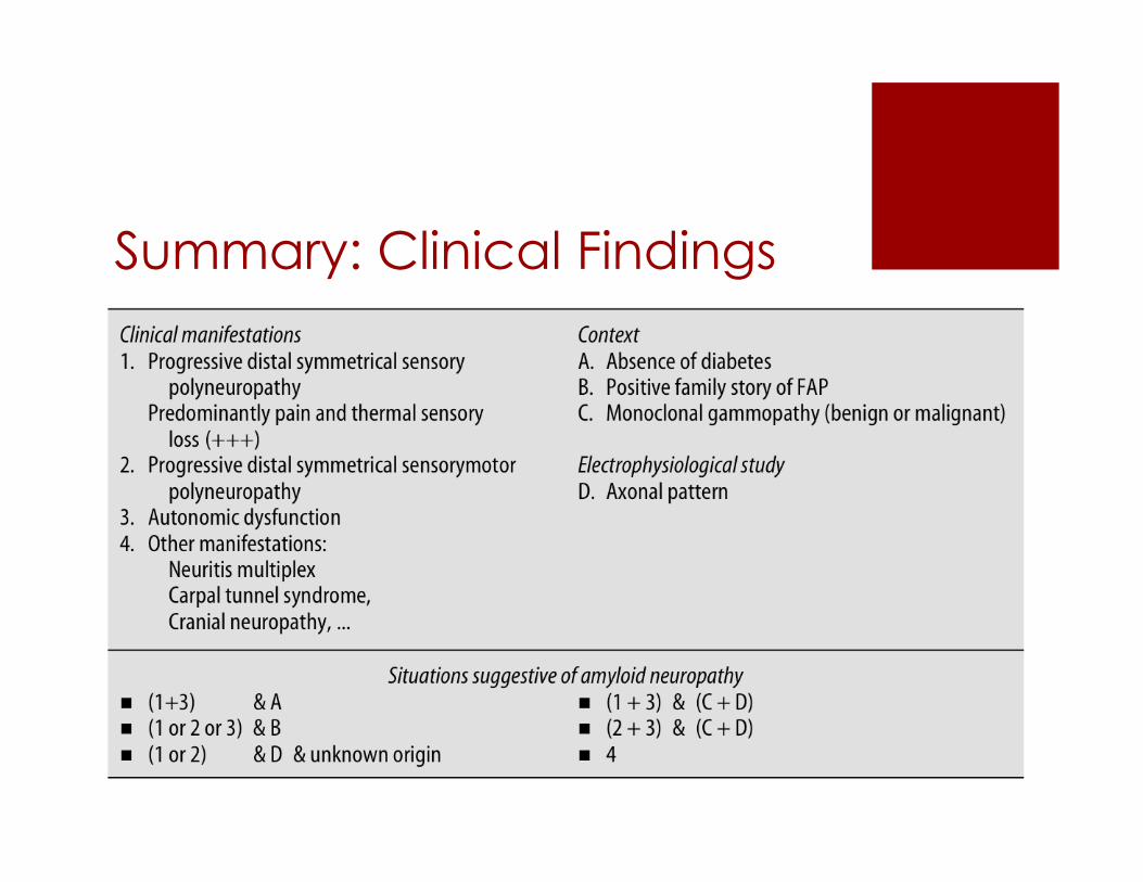
Summary: Clinical Findings
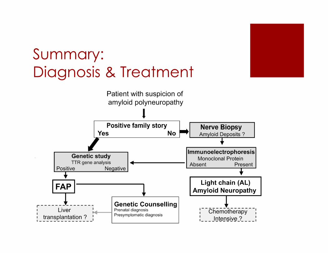
Summary:
Diagnosis & Treatment
Take Home Points
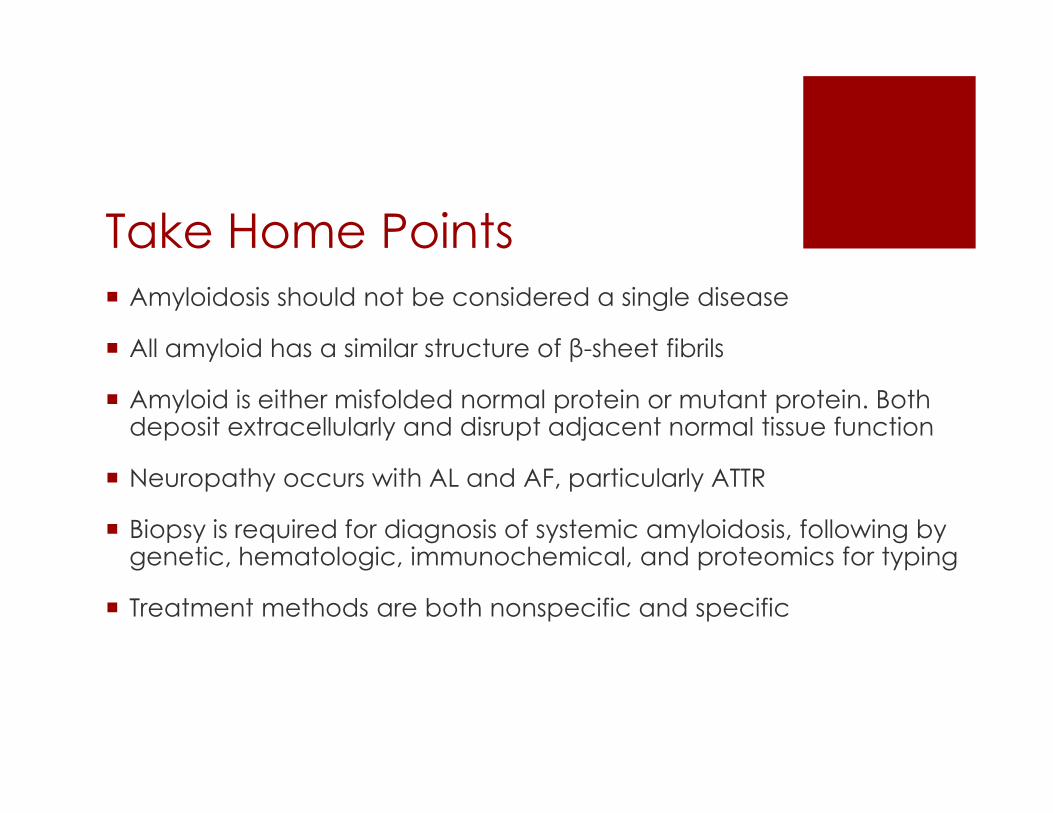
� Amyloidosis should not be considered a single disease
� All amyloid has a similar structure of β-sheet fibrils
� Amyloid is either misfolded normal protein or mutant protein. Both � Amyloid is either misfolded normal protein or mutant protein. Both deposit extracellularly and disrupt adjacent normal tissue function
� Neuropathy occurs with AL and AF, particularly ATTR
� Biopsy is required for diagnosis of systemic amyloidosis, following by genetic, hematologic, immunochemical, and proteomics for typing
� Treatment methods are both nonspecific and specific

Thank you
References & Images� Benson M, Kincaid J. The molecular biology and clinical features of
amyloid neuropathy. Muscle & Nerve (2007) 36: 411-423.
� Adams, D. Hereditary and acquired amyloid neuropathies. J Neurol(2001) 248: 647-657.
� Kumar V, Abbas A, Fausto N. Robbins and Cotran Pathologic Basis of Disease. Eighth Edition. Saunders Elselvier, 2010.Disease. Eighth Edition. Saunders Elselvier, 2010.
� Yazaki M et al. Rapidly progressive amyloid polyneuropathyassociated with a novel variant of transthyretin Ser 25. Muscle Nerve (2002) 25: 244-250.
� Gillmore J et al. Organ transplantation in hereditary apolipoprotein AI amyloidosis. Am J Transplant (2006) 6: 2342-2347.
� Benson M. The hereditary amyloidoses. Best Pract Res Clin Rheumatol(2003) 17: 909-927.
� Images used with permission of Elsevier Publishing.
� Goljan E. Rapid Review Pathology. Second Edition. Mosby Elsevier, 2007.
� Kumar, V. Robins & Cotran Pathologic Basis of Disease. Eighth Edition. Saunders Elselvier, 2010.