
Amyloid beta protein may initiate a cascade leading to AD pathology.
17
Amyloid beta protein may initiate a cascade leading to AD pathology.
-
date post
21-Dec-2015 -
Category
Documents
-
view
215 -
download
0
Transcript of Amyloid beta protein may initiate a cascade leading to AD pathology.

Amyloid beta protein may
initiate a
cascade leading to AD
pathology.

APP
secretases
A
nucleus
aggregation, fibrils and amyloid plaques
other substratese.g.notch
Clearance: Microglia and blood vessels
Enzymes break down Aneurotoxicity

CSF A equilibrium depends on:
Production
Uptake
Removal
Breakdown
Aggregation oligomers, fibrils
Deposition: A42 first
Plaques

Ais the initiator and main culprit in amyloid deposition
Down’s syndrome study by C. Lemere
A42 is the initial amyloid species deposited in brain A42 exceeds A40 in amyloid deposits Toxicity and amyloid fibril
formation: A42 > 40 Selectively in presenilin
mutations in most APP mutations High plasma A42 is linked
to a LOAD locus on chr 10
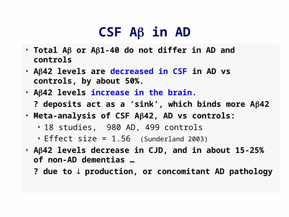
CSF A in AD• Total A or A1-40 do not differ in AD and controls• A42 levels are decreased in CSF in AD vs controls,
by about 50%.• A42 levels increase in the brain.
? deposits act as a ‘sink’, which binds more A42• Meta-analysis of CSF A42, AD vs controls:
• 18 studies, 980 AD, 499 controls• Effect size = 1.56 (Sunderland 2003)
• A42 levels decrease in CJD, and in about 15-25% of non-AD dementias …
? due to production, or concomitant AD pathology

CSF A and brain A deposition
APP tg mouse: brain vs CSF A De Mattos, 2002
R2 = 0.63
R2 = 0.57
Human: postmortem CSF A42vs neuritic plaque count
Strozyk, 2003
CSF
A42
neuritic plaques

CSF A42 meta-analysis (Sunderland, JAMA 2003)

CSF A-beta42 and APO-E

CSF A42 in very mild AD/MCI
N % with CSF A42 in AD range
MMSE > 23/30
Galasko et al 1998 24 64 %
Hulstaert et al, 1999 23 70 %
Riemenschneider et al, 2000 25 72 %
Andreasen et al, 2000 20 75 %
MCI with progression
Maruyama et al, 2001 19 45 %
Riemenschneider et al, 2002 18 85 %
Andreasen et al, 2003 44 77 %

CSF biomarkers in MCI and early ADRiemenschneider et al, 2002

A was immunoprecipitated from 2 ml of CSF from an AD patient, and visualized on a bicine gel that resolves A38, 40 and 42
Peptide
CSF Stds.
Measuring A subtypes

A species in CSF
Wiltfang et al, J Neurochem 2002
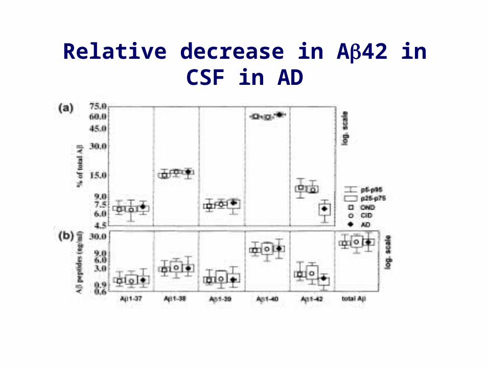
Relative decrease in A42 in CSF in AD
CSF A as an index of drug treatment?
• Half-life of A in CSF is about 30 minutes• CSF and plasma A are not correlated in humans• May be easier to show effects in controls than in
AD, because levels are not already decreased.
• Limited published data ….- -secretase inhibitors: CSF and plasma A40 and 42 in APP tg mice - Some NSAIDs may selectively decrease A42 in tg mice and increase A38 - Rivastigmine x 1 year had no effect on CSF A42

R=.90, p<.001
CSF A42 remains stable in AD over 12 months

Summary
• CSF A42 is decreased in AD, in 70-85% of patients, but less consistently so in MCI.
• A40 levels are not altered.
• Diagnostic potential of CSF A42 is limited, but may improve if it is part of a panel of biomarkers.
• CSF and possibly plasma A may be used to monitor certain types of anti-amyloid therapy, e.g. for proof of principle, or dose finding
• Several forms of A can be measured in CSF; data on A subtypes and on oligomers will be of interest.

Plasma A in inherited and sporadic AD
Scheuner 1996
in PS and APP mutations and DS, not sporadic AD
Mayeux 1999
risk of developing AD for highest quartile of plasma A42


![Colloid-amyloid Bodies in PUVA-treated Human Psoriatic ...Amyloid of primary cutaneous amyloidoses such as lichen amyloidosus [5, 17], macular amyloidosis [6] and amyloid dep- osition](https://static.fdocuments.us/doc/165x107/5e62f6a65098527daa05e73b/colloid-amyloid-bodies-in-puva-treated-human-psoriatic-amyloid-of-primary-cutaneous.jpg)