Amphiphilic Aminoglycosides as Medicinal Agents
37
International Journal of Molecular Sciences Review Amphiphilic Aminoglycosides as Medicinal Agents Clément Dezanet 1 , Julie Kempf 1 , Marie-Paule Mingeot-Leclercq 2, * and Jean-Luc Décout 1, * 1 Molecular Pharmacochemistry Department, University Grenoble Alpes, CNRS, 470 Rue de la Chimie, F-38000 Grenoble, France; [email protected] (C.D.); [email protected] (J.K.) 2 Cellular and Molecular Pharmacology Unit, Louvain Drug Research Institute, Catholic University of Louvain, Avenue E. Mounier 73, UCL B1.73.05, 1200 Brussels, Belgium * Correspondence: [email protected] (M.-P.M.-L.); [email protected] (J.-L.D.) Received: 6 September 2020; Accepted: 2 October 2020; Published: 8 October 2020 Abstract: The conjugation of hydrophobic group(s) to the polycationic hydrophilic core of the antibiotic drugs aminoglycosides (AGs), targeting ribosomal RNA, has led to the development of amphiphilic aminoglycosides (AAGs). These drugs exhibit numerous biological effects, including good antibacterial effects against susceptible and multidrug-resistant bacteria due to the targeting of bacterial membranes. In the first part of this review, we summarize our work in identifying and developing broad-spectrum antibacterial AAGs that constitute a new class of antibiotic agents acting on bacterial membranes. The target-shift strongly improves antibiotic activity against bacterial strains that are resistant to the parent AG drugs and to antibiotic drugs of other classes, and renders the emergence of resistant Pseudomonas aeruginosa strains highly difficult. Structure–activity and structure–eukaryotic cytotoxicity relationships, specificity and barriers that need to be crossed in their development as antibacterial agents are delineated, with a focus on their targets in membranes, lipopolysaccharides (LPS) and cardiolipin (CL), and the corresponding mode of action against Gram-negative bacteria. At the end of the first part, we summarize the other recent advances in the field of antibacterial AAGs, mainly published since 2016, with an emphasis on the emerging AAGs which are made of an AG core conjugated to an adjuvant or an antibiotic drug of another class (antibiotic hybrids). In the second part, we briefly illustrate other biological and biochemical effects of AAGs, i.e., their antifungal activity, their use as delivery vehicles of nucleic acids, of short peptide (polyamide) nucleic acids (PNAs) and of drugs, as well as their ability to cleave DNA at abasic sites and to inhibit the functioning of connexin hemichannels. Finally, we discuss some aspects of structure–activity relationships in order to explain and improve the target selectivity of AAGs. Keywords: aminoglycosides; amphiphilic; antibacterial; antibiotic; cardiolipin; delivery vehicles; lipopolysaccharides; membranes 1. Introduction Aminoglycosides (AGs), for example, neomycin B (NEO) 1, paromomycin (PARO) 2, kanamycins A and B (KANA, KANB) 3, 4 and tobramycin 5 (Figure 1), constitute a large family of highly potent broad-spectrum antibiotic drugs. These naturally occurring hydrophilic pseudo-oligosaccharides, which are polycationic species at physiological pH, target ribosomal RNA and disrupt protein synthesis [1]. Unfortunately, the widespread clinical use of AGs has strongly reduced their clinical efficacy through the selection of resistant bacteria [2]. In the search for new antibacterial agents targeting bacterial membranes, our team and others have synthesized and identified antibacterial amphiphilic aminoglycosides (antibacterial AAGs), which constitute a new class of polycationic antibiotic agents [3–8]. AAGs can be defined as substances made of a hydrophilic AG core Int. J. Mol. Sci. 2020, 21, 7411; doi:10.3390/ijms21197411 www.mdpi.com/journal/ijms
Transcript of Amphiphilic Aminoglycosides as Medicinal Agents

International Journal of
Molecular Sciences
Review
Amphiphilic Aminoglycosides as Medicinal Agents
Clément Dezanet 1, Julie Kempf 1, Marie-Paule Mingeot-Leclercq 2,* and Jean-Luc Décout 1,*1 Molecular Pharmacochemistry Department, University Grenoble Alpes, CNRS, 470 Rue de la Chimie,
F-38000 Grenoble, France; [email protected] (C.D.); [email protected] (J.K.)2 Cellular and Molecular Pharmacology Unit, Louvain Drug Research Institute,
Catholic University of Louvain, Avenue E. Mounier 73, UCL B1.73.05, 1200 Brussels, Belgium* Correspondence: [email protected] (M.-P.M.-L.);
[email protected] (J.-L.D.)
Received: 6 September 2020; Accepted: 2 October 2020; Published: 8 October 2020�����������������
Abstract: The conjugation of hydrophobic group(s) to the polycationic hydrophilic core of theantibiotic drugs aminoglycosides (AGs), targeting ribosomal RNA, has led to the development ofamphiphilic aminoglycosides (AAGs). These drugs exhibit numerous biological effects, includinggood antibacterial effects against susceptible and multidrug-resistant bacteria due to the targetingof bacterial membranes. In the first part of this review, we summarize our work in identifyingand developing broad-spectrum antibacterial AAGs that constitute a new class of antibiotic agentsacting on bacterial membranes. The target-shift strongly improves antibiotic activity against bacterialstrains that are resistant to the parent AG drugs and to antibiotic drugs of other classes, and rendersthe emergence of resistant Pseudomonas aeruginosa strains highly difficult. Structure–activity andstructure–eukaryotic cytotoxicity relationships, specificity and barriers that need to be crossed intheir development as antibacterial agents are delineated, with a focus on their targets in membranes,lipopolysaccharides (LPS) and cardiolipin (CL), and the corresponding mode of action againstGram-negative bacteria. At the end of the first part, we summarize the other recent advances inthe field of antibacterial AAGs, mainly published since 2016, with an emphasis on the emergingAAGs which are made of an AG core conjugated to an adjuvant or an antibiotic drug of anotherclass (antibiotic hybrids). In the second part, we briefly illustrate other biological and biochemicaleffects of AAGs, i.e., their antifungal activity, their use as delivery vehicles of nucleic acids, of shortpeptide (polyamide) nucleic acids (PNAs) and of drugs, as well as their ability to cleave DNA atabasic sites and to inhibit the functioning of connexin hemichannels. Finally, we discuss some aspectsof structure–activity relationships in order to explain and improve the target selectivity of AAGs.
Keywords: aminoglycosides; amphiphilic; antibacterial; antibiotic; cardiolipin; delivery vehicles;lipopolysaccharides; membranes
1. Introduction
Aminoglycosides (AGs), for example, neomycin B (NEO) 1, paromomycin (PARO) 2, kanamycinsA and B (KANA, KANB) 3, 4 and tobramycin 5 (Figure 1), constitute a large family of highly potentbroad-spectrum antibiotic drugs. These naturally occurring hydrophilic pseudo-oligosaccharides,which are polycationic species at physiological pH, target ribosomal RNA and disrupt proteinsynthesis [1]. Unfortunately, the widespread clinical use of AGs has strongly reduced their clinicalefficacy through the selection of resistant bacteria [2]. In the search for new antibacterial agentstargeting bacterial membranes, our team and others have synthesized and identified antibacterialamphiphilic aminoglycosides (antibacterial AAGs), which constitute a new class of polycationicantibiotic agents [3–8]. AAGs can be defined as substances made of a hydrophilic AG core
Int. J. Mol. Sci. 2020, 21, 7411; doi:10.3390/ijms21197411 www.mdpi.com/journal/ijms

Int. J. Mol. Sci. 2020, 21, 7411 2 of 37
on which lipophilic/hydrophobic group(s) are grafted, to produce, at physiological pH, cationicamphiphilic species.
Int. J. Mol. Sci. 2020, 21, x FOR PEER REVIEW 2 of 36
core on which lipophilic/hydrophobic group(s) are grafted, to produce, at physiological pH, cationic amphiphilic species.
O
O
HO
NH2
R1
HOO
NH2
H2N
O
3: Kanamycin A: R1= R2= OH4: Kanamycin B: R1= OH, R2= NH2, R3= H5: Tobramycin: R1= NH2, R2= R3= H
HOH2N
OHOH
R2
OH
O
O
HO
R
HONH2
OOH
NH2
H2N
O
O
HO
OH
1: Neomycin B: R= NH22: Paromomycin: R= OH
6: Neamine: R1= NH2, R2= OH7: Paromamine: R1= OH, R2= OH8: Nebramine: R1= NH2, R
2= H
Ring II
Ring III
1
23
5 6
1'2'3'
4' 5'
O
NH2HO
H2N
6'Ring I: Deoxystreptamine
(2DOS)
Ring II
O
O
HO
R1
R2
NH2
HOOH
NH2
H2N
O
O
R4O
R1
R3O
NHTrHO
OR2
NHTr
TrHN
9: R1= NHTr, R2= R3= R4= H10: R1= NHTr, R2= R3= PMB, R4= H11: R1= NHTr, R2= R3= R4= PMB12: R1= NHTr, R2= PMB, R3= R4= H13: R1= OTr, R2= R3= R4= H
4'
3'
6'
65
6
Ring II
Figure 1. Structures of natural antibiotic aminoglycosides 1–5, of some corresponding constitutive derivatives 6–8 and of synthetic intermediates used to prepare amphiphilic aminoglycosides (AAGs) 9–13 (Tr = trityl group = triphenylmethyl, PMB = para-methoxyphenyl group).
Originally, AAGs were developed in the search for new AGs that are less susceptible to resistance. In this approach, Hanessian, Westhof and coworkers reported the first example of in vivo antibacterial lipophilic ether-modified derivatives of PARO targeting ribosomal RNA with a novel mode of binding, which are able to inhibit AG-deactivating enzymes [9,10].
In the never-ceasing fight against pathogenic multidrug-resistant bacteria, the identification of new targets, and of the corresponding drugs, is imperative [11], and an interest in membrane-targeting drug is emerging [12]. The expected major advantages of such drugs are (i) their activity against bacterial persisters and antibiotic-tolerant bacterial populations [13], characterized by a low metabolic activity [14,15]; (ii) their low tendency for the development of bacterial resistance [16]; and (iii) the fact that they do not need to cross the bacterial outer membrane (OM) of Gram-negative bacteria.
Indeed, this mode of action should limit the emergence of resistance through drug modifications by intracellular resistance-causing enzymes and efflux pumps expressed by multidrug-resistant (MDR) bacteria. Moreover, interaction with key membrane components, present in multiple copies in the bacterial membranes, can cause many antibacterial destructuring effects. The bacteria that are resistant to these effects have to perform biochemical modifications of multiple membrane components [17]. The corresponding metabolic modifications have a high energetic cost for the selected resistant bacteria, resulting in slow growth, rapid reversibility of the emerging resistance and high sensitivity to antibiotic drugs of other classes.
In such an approach, the antibacterial potential of cationic amphiphilic compounds that target bacterial membranes is attractive from a drug development perspective. The repurposing of the old antibacterial cationic cyclopeptide polymyxin E, colistin (COL, Figure 2), acting on the bacterial outer membrane (OM), as a last-line antibiotic to treat multidrug-resistant (MDR) Gram-negative bacterial infections, especially those caused by antibiotic-resistant Pseudomonas aeruginosa strains, illustrate
Figure 1. Structures of natural antibiotic aminoglycosides 1–5, of some corresponding constitutivederivatives 6–8 and of synthetic intermediates used to prepare amphiphilic aminoglycosides (AAGs)9–13 (Tr = trityl group = triphenylmethyl, PMB = para-methoxyphenyl group).
Originally, AAGs were developed in the search for new AGs that are less susceptible to resistance.In this approach, Hanessian, Westhof and coworkers reported the first example of in vivo antibacteriallipophilic ether-modified derivatives of PARO targeting ribosomal RNA with a novel mode of binding,which are able to inhibit AG-deactivating enzymes [9,10].
In the never-ceasing fight against pathogenic multidrug-resistant bacteria, the identification ofnew targets, and of the corresponding drugs, is imperative [11], and an interest in membrane-targetingdrug is emerging [12]. The expected major advantages of such drugs are (i) their activity againstbacterial persisters and antibiotic-tolerant bacterial populations [13], characterized by a low metabolicactivity [14,15]; (ii) their low tendency for the development of bacterial resistance [16]; and (iii) the factthat they do not need to cross the bacterial outer membrane (OM) of Gram-negative bacteria.
Indeed, this mode of action should limit the emergence of resistance through drug modificationsby intracellular resistance-causing enzymes and efflux pumps expressed by multidrug-resistant (MDR)bacteria. Moreover, interaction with key membrane components, present in multiple copies in thebacterial membranes, can cause many antibacterial destructuring effects. The bacteria that are resistantto these effects have to perform biochemical modifications of multiple membrane components [17].The corresponding metabolic modifications have a high energetic cost for the selected resistant bacteria,resulting in slow growth, rapid reversibility of the emerging resistance and high sensitivity to antibioticdrugs of other classes.
In such an approach, the antibacterial potential of cationic amphiphilic compounds that targetbacterial membranes is attractive from a drug development perspective. The repurposing of the oldantibacterial cationic cyclopeptide polymyxin E, colistin (COL, Figure 2), acting on the bacterial outermembrane (OM), as a last-line antibiotic to treat multidrug-resistant (MDR) Gram-negative bacterial

Int. J. Mol. Sci. 2020, 21, 7411 3 of 37
infections, especially those caused by antibiotic-resistant Pseudomonas aeruginosa strains, illustrate wellthe interesting nature of cationic amphiphilic antibiotic drugs [18]. Despite several studies on COLrepurposing, many issues related to emerging bacterial resistances, toxicity and pharmacokinetics stillneed to be elucidated [18,19].
Int. J. Mol. Sci. 2020, 21, x FOR PEER REVIEW 3 of 36
well the interesting nature of cationic amphiphilic antibiotic drugs [18]. Despite several studies on COL repurposing, many issues related to emerging bacterial resistances, toxicity and pharmacokinetics still need to be elucidated [18,19].
NH HNO
NH
ONH HN O
O
H3N
OHN
HOO
NH3
HN O
NH3
HN
ONHO
OH
HN
O
H3N
HNO
H3N
MeMe
Me
Me
Figure 2. Structure of polymyxin E (COL), showing the five amine functions protonated at physiological pH.
In the search for membrane-targeting antibiotics, we have focused our work on Gram-negative bacteria, such as P. aeruginosa, an opportunistic pathogen that causes a wide range of severe opportunistic infections in patients with serious underlying medical conditions, such as those with burns, surgical wounds or people with cystic fibrosis [20].
The interaction of cationic species with bacterial membrane components such as anionic phospholipids can produce membrane disruption and depolarization. The cell envelope of Gram-negative bacteria contains two membranes, the inner membrane (IM) and the OM, separated by the periplasm. The OM of Gram-negative bacteria has a unique architecture that acts as a potent permeability barrier against antibiotics. The OM is composed of lipopolysaccharide (LPS), phospholipids, outer membrane β-barrel proteins (OMPs) and lipoproteins. These components are synthesized in the cytoplasm or in the IM, and are then selectively transported to the OM by specific transport machines. Recent reviews on the transport and assembly systems of OM components have been published with the aim of developing inhibitors targeting these systems [21–24].
In the search for new antibacterial agents, we identified broad-spectrum antibacterial AAGs carrying two or three lipophilic groups and, for the first time, we revealed their effects on bacterial membranes [5,25]. There was a corresponding strong increase in the AG lipophilicity results in the bacterial target shift from rRNA to membranes and a significant improvement in activity against bacterial strains resistant to the parent AG drugs and to antibiotic drugs of other classes (penicillins, fluoroquinolones, macrolides, etc.). The identification of antibacterial AAGs acting on the bacterial membranes against AG-resistant bacteria and MDR bacteria offered a promising direction for the development of novel antibiotics. Most of the antibacterial AAGs identified were mainly active against susceptible and MDR Gram-positive bacteria and a few appeared to be active against susceptible and resistant Gram-positive and Gram-negative bacteria.
In the first part of this review article, we summarize the results that we obtained in the development of broad-spectrum antibacterial amphiphilic neamine (NEA) 6, paromamine (PARA) 7 and 6-amino-6-deoxy-1-methylglucosamine (1-methyl neosamine, corresponding to ring II in NEO) derivatives (Figure 1), including some recent unpublished results, with a focus on the most active NEA derivatives. Structure–activity and structure–eukaryotic cytotoxicity relationships, as well as specificities and particularities in their mode of action and barriers that need to be crossed in their development as medicinal agents, are delineated. Since the integrity of the biophysical properties of bacterial membranes are required for maintaining their permeability functions, as well as the right environment for proteins embedded within, we explored the effects of AAGs on two major lipids, LPS and cardiolipin (CL), that are involved in one of the most critical biophysical characteristics of
Figure 2. Structure of polymyxin E (COL), showing the five amine functions protonated atphysiological pH.
In the search for membrane-targeting antibiotics, we have focused our work on Gram-negativebacteria, such as P. aeruginosa, an opportunistic pathogen that causes a wide range of severe opportunisticinfections in patients with serious underlying medical conditions, such as those with burns, surgicalwounds or people with cystic fibrosis [20].
The interaction of cationic species with bacterial membrane components such as anionicphospholipids can produce membrane disruption and depolarization. The cell envelope ofGram-negative bacteria contains two membranes, the inner membrane (IM) and the OM, separatedby the periplasm. The OM of Gram-negative bacteria has a unique architecture that acts as apotent permeability barrier against antibiotics. The OM is composed of lipopolysaccharide (LPS),phospholipids, outer membrane β-barrel proteins (OMPs) and lipoproteins. These components aresynthesized in the cytoplasm or in the IM, and are then selectively transported to the OM by specifictransport machines. Recent reviews on the transport and assembly systems of OM components havebeen published with the aim of developing inhibitors targeting these systems [21–24].
In the search for new antibacterial agents, we identified broad-spectrum antibacterial AAGscarrying two or three lipophilic groups and, for the first time, we revealed their effects on bacterialmembranes [5,25]. There was a corresponding strong increase in the AG lipophilicity results in thebacterial target shift from rRNA to membranes and a significant improvement in activity againstbacterial strains resistant to the parent AG drugs and to antibiotic drugs of other classes (penicillins,fluoroquinolones, macrolides, etc.). The identification of antibacterial AAGs acting on the bacterialmembranes against AG-resistant bacteria and MDR bacteria offered a promising direction for thedevelopment of novel antibiotics. Most of the antibacterial AAGs identified were mainly active againstsusceptible and MDR Gram-positive bacteria and a few appeared to be active against susceptible andresistant Gram-positive and Gram-negative bacteria.
In the first part of this review article, we summarize the results that we obtained in thedevelopment of broad-spectrum antibacterial amphiphilic neamine (NEA) 6, paromamine (PARA) 7and 6-amino-6-deoxy-1-methylglucosamine (1-methyl neosamine, corresponding to ring II in NEO)derivatives (Figure 1), including some recent unpublished results, with a focus on the most activeNEA derivatives. Structure–activity and structure–eukaryotic cytotoxicity relationships, as well asspecificities and particularities in their mode of action and barriers that need to be crossed in theirdevelopment as medicinal agents, are delineated. Since the integrity of the biophysical properties ofbacterial membranes are required for maintaining their permeability functions, as well as the right

Int. J. Mol. Sci. 2020, 21, 7411 4 of 37
environment for proteins embedded within, we explored the effects of AAGs on two major lipids,LPS and cardiolipin (CL), that are involved in one of the most critical biophysical characteristics ofthe OMs of Gram-negative bacteria, their asymmetry. LPS is located at the outer leaflet of the OMand CL is located mostly within the IM, and also within the OM [26]. Some selectivity results fromthe binding of AAGs to these lipids in bacterial membranes in comparison to mammalian ones, sincebacterial membranes (i) are more negatively charged than eukaryotic membranes; (ii) contain a higherproportion of negative intrinsic curvature lipids, where proteins involved in the formation of thedivision plane are located [27,28]; and (iii) exhibit a dilational modulus of elasticity that is much lowerthan the one found in mammalian membranes [29].
At the end of the first part of this review article, other advances in the field of antibacterial AAGs,published since 2016 after the appearance of several review articles [7,8,30–32], are reviewed with anemphasis on AAGs made of an AG core conjugated to an adjuvant or an antibiotic drug of anotherclass (antibiotic hybrids), as recently developed by Schweizer, Zhanel and coworkers.
Numerous other biological and biochemical effects of AAGs have been reported, and, in the secondpart of this article, we illustrate briefly some of these effects, i.e., their antifungal activity; their use asdelivery vehicles of nucleic acids, of short peptide nucleic acids and of drugs; their ability to cleave DNAat abasic sites and their ability to inhibit the functioning of connexin hemichannels. Structure–activityrelationships are finally discussed, in order to explain and improve their target selectivity.
2. Antibacterial Amphiphilic Aminoglycosides (Antibacterial AAGs)
The chemical strategies used for the preparation of new AGs and AAGs and their biologicalactivities were recently reviewed [1]. The recent progress in AG conjugation for RNA targetingwas also summarized in 2020 [33]. AAGs were synthesized by modification of the AG’s primaryamine or hydroxyl functions. Lipophilic groups were conjugated to the AG’s core of NEO 1 (NEO),NEA 6, PARO 2, PARA 7, KANA 3 and KANB 4, TOB 5 and nebramine 8 (NEB) (Figure 1) usingseveral strategies [3–8,30–38]. The amine functions of the selected AG were converted to alkyl- oraryl-amide(s) or to carbamates, leading to a decrease in the number of positive charges present atphysiological pH. In another approach, the hydroxyl groups were converted to ether and thioethergroups. Such modification(s) of the amine or hydroxyl function(s) of the AG cores should produceAAGs that are not a substrate of AG-deactivating enzymes (AG nucleotidyltransferases (ANTs), AGphosphotransferases (APHs) and AG acetyltransferases (AACs) [2], and therefore produces AAGswhich are active against bacteria expressing resistance-causing enzyme(s).
The AAGs identified to be most active against Gram-positive and Gram-negative bacteria aremainly dialkyl and/or trialkyl derivatives of NEA 6 (Figure 1), [5,34,35], of 1-methyl neosamine [36]and of NEB 8, which were identified more recently and are briefly described [37,38]. In this partof the review, we summarize the main results obtained in the development of broad-spectrumantibacterial amphiphilic NEA 6, PARA 7 and 1-methyl neosamine derivatives. Some physicochemicalproperties of amphiphilic NEA derivatives and a method developed for their dosage are also reported(unpublished results).
2.1. Broad-Spectrum Antibacterial AAGs: Antibacterial NEA, PARA and6-Amino-6-Deoxy-1-Methylglucosamine Derivatives, Structure–Activity and Structure–CytotoxicityRelationships, Modes of Action
2.1.1. Synthesis of NEA and PARA Derivatives
Our first works in the field of AGs were developed in the search for anti-HIV agents targetingviral RNA [39]. The AG core of NEA 6, carrying four amine and four hydroxyl functions, has beenselected for modification in order to reduce the number of cationic groups present in the synthesizedAAGs and, as a consequence, to limit the unspecific binding of the resulting species to biologicalpolyanionic components.

Int. J. Mol. Sci. 2020, 21, 7411 5 of 37
Methods of protection and deprotection of amine and hydroxyl functions of NEA 6,obtained by methanolysis of NEO 1, were developed. From N-tetratritylated NEA 9,the 3′,6-O-di-para-methoxybenzyl (diPMB) and 3′,4′,6-O-triPMB derivatives 10 and 11 (Figure 1) wereprepared for selective O-alkylation and deprotection under acidic conditions to give 4′-,5-O-monoalkyland 4′,5-O-dialkyl derivatives [39,40]. The O-alkylation of N-tetratritylated NEA 9 in DMF using sodiumhydride (NaH) as a base allowed the preparation, in one step, of 3′,6-O-dialkyl and 3′,4′,6-O-trialkylNEA derivatives [5]. Using phase transfer conditions, it was possible to prepare, in high yields,the 6-PMB tetratrityl NEA derivative 12, which was alkylated to selectively produce 3′,4′-O-dialkylderivatives [36,41]. Methanolysis of PARO 2 gave PARA 7, which was tetratritylated, leading to13, which was used to prepare 3′,6-O-dialkyl and 3′,4′,6-O-trialkyl PARA derivatives [34]. Majorincreases in the selectivity and/or reactivity of AAGs in O-mono- and O,O′-di-alkylation were observedunder phase-transfer conditions in the presence of tetrabutylammonium fluoride (TBAF) or iodide(TBAI) [41] in comparison to alkylation in DMF solution using NaH [5]. The use of TBAI under phasetransfer conditions allowed the rapid and efficient preparation of 3′,6-dialkyl and 3′,6-diarylalkyl NEAderivatives, avoiding the O-trialkylation observed with NaH in DMF [5,33,34].
2.1.2. Structure–Activity Relationships
First Identified Broad-Spectrum Antibacterial Amphiphilic NEA Derivatives
In the search for new antibacterial agents, first, we introduced onto the hydroxyl functionsof NEA lipophilic benzyl, 2-naphthylmethylene (2NM) (Figure 3), 2-pyridinylmethylene and2-quinolinylmethylene groups from the corresponding halides [5]. The resulting 3′,6-di-; 3′,4′-di-and 3′,4′,6-triarylalkyl NEA derivatives were isolated as tetratrifluoroacetate salts, after a finaldeprotection step in TFA, and were evaluated for their antibacterial activity against susceptible andresistant Gram-positive Staphylococcus aureus (for instance, methicillin-resistant S. aureus (MRSA)and vancomycin-resistant S. aureus (VRSA)) and Gram-negative bacteria (Acinetobacter baumannii,Escherichia coli, Klebsiella pneumonia, P. aeruginosa, etc.).Int. J. Mol. Sci. 2020, 21, x FOR PEER REVIEW 6 of 36
O
O
R4O
R1
R3O
NH2HO
OR2
NH2
H2N
2NM
1NM
14: R1= NH2, R2= R3= 2NM, R4= H15: R1= NH2, R2= H, R3= R4= 2NM16: R1= NH2, R2= R3= R4= 2NM17: R1= NH2, R2= R3= 1NM, R4= H18: R1= NH2, R2= R3= R4= 1NM19: R1= OH, R2= R3= 2NM, R4= H20 R1= OH, R2= R3= R4= 2NM21: R1= OH, R2= R3= 1NM, R4= H22: R1= OH, R2= R3= R4= 1NM23: R1= NH2, R2= R3= R4= n-hexyl
4'
3'
6'
65
4 CF3CO2H
Figure 3. Structures of the first identified broad-spectrum antibacterial amphiphilic neamine (NEA) (6) and paromamine (PARA) (7) derivatives [5,34].
Comparison of the Antibacterial Activities of NEA and PARA Derivatives
In the PARA core, the 6′-amino group of NEA is replaced by a hydroxyl group and, as a consequence, the corresponding AAGs bear, at physiological pH, one less positive charge in comparison to their NEA homologs [34]. The comparison of the antibacterial activities of the previously prepared 2NM derivatives (14 and 16) with those of 1-naphthylmethylene (1NM, 17 and 18) NEA derivatives and of the corresponding PARA derivatives 19–22 (Figure 3), revealed the better good broad-spectrum activity of the 3′,4′,6-triNM NEA derivatives, with the MIC mainly decreased by four times. Therefore, the NEA core was selected in the search for more active compounds than 14–18 [34]. The presence of an amino group at position 6′ that is protonated at physiological pH of the aminoglycoside increases the antibacterial effect, without being essential to the antibacterial effect.
In order to compare the antibacterial activity of 3′,6-dialkyl and 3′,4′,6-trialkyl NEA derivatives and the effect of the alkyl chain lipophilicity on their activity, NEA derivatives carrying two or three linear alkyl groups, butyl (Bu), hexyl (Hx), nonyl (Nn) and octadecyl (Ocd) groups, as well as arylalkyl groups, benzyl and 2-naphthylalkyl with alkyl, methyl, n-propyl, n-butyl and n-hexyl (2NM, 2NP, 2NB and 2NH, respectively), were synthesized and evaluated [34]. The 3′- and 6-mono-Ocd derivatives were also prepared and found to be inactive against Gram-positive and Gram-negative bacteria. The 3′,4′,6-tri2NM (16), -triHx (23) (Figure 3) and the 3′,6-diNn (24), -di2NP (26) and -di2NB (27) NEA derivatives (Figure 4) showed good activity against susceptible and resistant Gram-positive and Gram-negative bacteria, with the 3′,6-dinonyl derivative 24 being the most active, and the di2NP 26 and di2NB 27 also being the most active against Gram-positive bacteria. The main result of this study was the delineation of windows of critical lipophilicity values for the active 3′,6-dialkyl and 3′,4′,6-trialkylNEA derivatives from the plot of 1/MIC versus clogP (calculated octanol/water partition coefficient; see the paragraph below on the fine-tuning of the structure–activity relationships). NEA AAGs 24, 26 and 27 were selected for further study as the most active and the less cytotoxic derivatives at 10 µM to murine J774 macrophages, in comparison to the trihexyl derivative 23 that has shown slightly lower broad-spectrum activity [34]. These three AAGs were shown to be more active against COL-resistant P. aeruginosa (PA272, PA307, PA343 and PA2938; MIC = 2–8 µg/mL) than the 3,’4′,6-tri2NM NEA derivative 16 (MIC = 4–32 µg/mL) [42].
Figure 3. Structures of the first identified broad-spectrum antibacterial amphiphilic neamine (NEA) (6)and paromamine (PARA) (7) derivatives [5,34].
The 3′,6-di-(14); 3′,4′-di-(15) and 3′,4′,6-tri-(16) 2-naphthylmethylene (2NM) derivatives (Figure 3)showed good activity against susceptible and resistant S. aureus strains expressing resistance pumps(NorA or MsrA), against AG-inactivating enzymes and against MRSA and VRSA strains, which are

Int. J. Mol. Sci. 2020, 21, 7411 6 of 37
resistant to methicillin and vancomycin, respectively (minimum inhibitory concentrations (MICs)4–16 µg/mL), whereas the 3′-, 4′-, 5′-, 6-mono-2NM derivatives were inactive (MICs > 128 µg/mL).The tri2NM derivative 16 showed better antibacterial activity against the selected Gram-positive strains(MICs 2–4 µg/mL). It also exhibited good activity (MICs 4–16 µg/mL) against sensitive and resistantGram-negative bacteria, both on Enterobacteriaceae and non-Enterobacteriacea, including P. aeruginosa,expressing efflux pumps or AG-deactivating enzymes, or rRNA methylases [2] (Citrobacter amalonaticusarm 06AB0010, E. coli 06AB003arm, Enterobacter aerogenes 06AB008 arm). Derivatives 14 and 16 revealedweak and aspecific binding to a model bacterial 16S rRNA as compared to NEO 1. Derivative 16 alsoshowed a low ability to decrease 3H leucine incorporation into proteins in P. aeruginosa, suggesting that16 acts against Gram-negative bacteria with a mechanism different from inhibition of protein synthesis,probably by membrane destabilization [5]. For the first time, AAGs (14 and 16) were shown to targetthe membranes of P. aeruginosa with induction of depolarization [25].
Comparison of the Antibacterial Activities of NEA and PARA Derivatives
In the PARA core, the 6′-amino group of NEA is replaced by a hydroxyl group and, as a consequence,the corresponding AAGs bear, at physiological pH, one less positive charge in comparison to theirNEA homologs [34]. The comparison of the antibacterial activities of the previously prepared 2NMderivatives (14 and 16) with those of 1-naphthylmethylene (1NM, 17 and 18) NEA derivatives andof the corresponding PARA derivatives 19–22 (Figure 3), revealed the better good broad-spectrumactivity of the 3′,4′,6-triNM NEA derivatives, with the MIC mainly decreased by four times. Therefore,the NEA core was selected in the search for more active compounds than 14–18 [34]. The presence ofan amino group at position 6′ that is protonated at physiological pH of the aminoglycoside increasesthe antibacterial effect, without being essential to the antibacterial effect.
In order to compare the antibacterial activity of 3′,6-dialkyl and 3′,4′,6-trialkyl NEA derivativesand the effect of the alkyl chain lipophilicity on their activity, NEA derivatives carrying two or threelinear alkyl groups, butyl (Bu), hexyl (Hx), nonyl (Nn) and octadecyl (Ocd) groups, as well as arylalkylgroups, benzyl and 2-naphthylalkyl with alkyl, methyl, n-propyl, n-butyl and n-hexyl (2NM, 2NP, 2NBand 2NH, respectively), were synthesized and evaluated [34]. The 3′- and 6-mono-Ocd derivativeswere also prepared and found to be inactive against Gram-positive and Gram-negative bacteria.The 3′,4′,6-tri2NM (16), -triHx (23) (Figure 3) and the 3′,6-diNn (24), -di2NP (26) and -di2NB (27)NEA derivatives (Figure 4) showed good activity against susceptible and resistant Gram-positiveand Gram-negative bacteria, with the 3′,6-dinonyl derivative 24 being the most active, and the di2NP26 and di2NB 27 also being the most active against Gram-positive bacteria. The main result ofthis study was the delineation of windows of critical lipophilicity values for the active 3′,6-dialkyland 3′,4′,6-trialkylNEA derivatives from the plot of 1/MIC versus clogP (calculated octanol/waterpartition coefficient; see the paragraph below on the fine-tuning of the structure–activity relationships).NEA AAGs 24, 26 and 27 were selected for further study as the most active and the less cytotoxicderivatives at 10 µM to murine J774 macrophages, in comparison to the trihexyl derivative 23 that hasshown slightly lower broad-spectrum activity [34]. These three AAGs were shown to be more activeagainst COL-resistant P. aeruginosa (PA272, PA307, PA343 and PA2938; MIC = 2–8 µg/mL) than the3,’4′,6-tri2NM NEA derivative 16 (MIC = 4–32 µg/mL) [42].

Int. J. Mol. Sci. 2020, 21, 7411 7 of 37
Int. J. Mol. Sci. 2020, 21, x FOR PEER REVIEW 7 of 36
O
O
HO
NH2
O
NH2HO
O
NH2
H2N
O
O
HO
NH2
O
NH2HO
O
NH2
H2N
4 CF3CO2H
3'
6
28
4 CF3CO2H
O
O
O
NH2
O
NH2HO
OH
NH2
H2N
4'
3'
4 CF3CO2H
24 25
O
O
O
NH2
O
NH2 HO
OH
NH2
H2N
4 CF3CO2H
n
26: n= 327: n= 4
n
Figure 4. Structures of the identified broad-spectrum antibacterial amphiphilic dialkyl (24 and 25) and dialkylnaphthyl (26–28) NEA derivatives [34].
AG antibiotics are effective against biofilms and recently approaches and possible mechanisms for the application of AGs to treat biofilm-associated infections were briefly reviewed [43].
The 3′,6-dinonyl NEA derivative 24 was shown to be bactericidal against P. aeruginosa at its MIC and to inhibit the P. aeruginosa biofilm formation at a two-fold MIC [42].
Comparison of the Antibacterial Activities of 3′,6- and 3′,4′-Dialkyl NEA Derivatives
In order to investigate the role of the attachment position of the second alkyl chain in antibacterial activities and cytotoxicities, the 3′,4′-diNn and -di2NP analogues 25 and 28 of the corresponding 3′,6-dialkyl derivatives 24 and 26 (Figure 4), were synthesized from 6-(para-methoxy)benzyl-N-tetratrityl NEA 12, obtained selectively under phase transfer conditions of alkylation [36,41]. Against susceptible and resistant Gram-positive bacteria, the 3′,6-di2NP and 3′,4′-di2NP derivatives 26 and 28 showed similar good activity. Against Gram-negative bacteria, the activity of 28 was better. The 3′,6-dinonyl derivative 24 appeared to be more active than its 3′,4-dinonyl isomer 25 and was found to be the most active synthesized NEA derivative against susceptible and resistant Gram-positive and Gram-negative bacteria [36].
6-Amino-6-Deoxy-1-Methylglucosamine (1-Methyl Neosamine) Derivatives, Analogues of 3′,4′-Dialkyl NEA Derivatives
In order to evaluate the importance, in the antibacterial activity of 3′,4′-dinonyl derivative 25, of the integrity of the 2-deoxystreptamine ring I (Figure 5), amphiphilic 3,4-dialkyl derivatives 29–36 of 6-amino-6-deoxyglucosamine, named neosamine, corresponding to NEA ring II, were synthesized from the 1-allyl intermediate derivative 37, prepared from N-acetylglucosamine (in 8 or 9 steps) (Figure 5) [36]. The most hydrophilic derivatives 30–36 mainly showed similar good activity against susceptible and resistant Gram-positive and Gram-negative bacteria (MIC = 1–2 and 4–8 µg/mL, respectively), slightly better than the corresponding activity of the 3,4′-dinonyl NEA derivative 25. The azido derivative 29 revealed an activity against Gram-positive bacteria similar to 30–36 but was
Figure 4. Structures of the identified broad-spectrum antibacterial amphiphilic dialkyl (24 and 25) anddialkylnaphthyl (26–28) NEA derivatives [34].
AG antibiotics are effective against biofilms and recently approaches and possible mechanisms forthe application of AGs to treat biofilm-associated infections were briefly reviewed [43].
The 3′,6-dinonyl NEA derivative 24 was shown to be bactericidal against P. aeruginosa at its MICand to inhibit the P. aeruginosa biofilm formation at a two-fold MIC [42].
Comparison of the Antibacterial Activities of 3′,6- and 3′,4′-Dialkyl NEA Derivatives
In order to investigate the role of the attachment position of the second alkyl chain in antibacterialactivities and cytotoxicities, the 3′,4′-diNn and -di2NP analogues 25 and 28 of the corresponding3′,6-dialkyl derivatives 24 and 26 (Figure 4), were synthesized from 6-(para-methoxy)benzyl-N-tetratritylNEA 12, obtained selectively under phase transfer conditions of alkylation [36,41]. Against susceptibleand resistant Gram-positive bacteria, the 3′,6-di2NP and 3′,4′-di2NP derivatives 26 and 28 showedsimilar good activity. Against Gram-negative bacteria, the activity of 28 was better. The 3′,6-dinonylderivative 24 appeared to be more active than its 3′,4-dinonyl isomer 25 and was found to be the mostactive synthesized NEA derivative against susceptible and resistant Gram-positive and Gram-negativebacteria [36].
6-Amino-6-Deoxy-1-Methylglucosamine (1-Methyl Neosamine) Derivatives, Analogues of3′,4′-Dialkyl NEA Derivatives
In order to evaluate the importance, in the antibacterial activity of 3′,4′-dinonyl derivative 25, ofthe integrity of the 2-deoxystreptamine ring I (Figure 5), amphiphilic 3,4-dialkyl derivatives 29–36 of6-amino-6-deoxyglucosamine, named neosamine, corresponding to NEA ring II, were synthesizedfrom the 1-allyl intermediate derivative 37, prepared from N-acetylglucosamine (in 8 or 9 steps)(Figure 5) [36]. The most hydrophilic derivatives 30–36 mainly showed similar good activity againstsusceptible and resistant Gram-positive and Gram-negative bacteria (MIC = 1–2 and 4–8 µg/mL,respectively), slightly better than the corresponding activity of the 3,4′-dinonyl NEA derivative 25. The

Int. J. Mol. Sci. 2020, 21, 7411 8 of 37
azido derivative 29 revealed an activity against Gram-positive bacteria similar to 30–36 but was mainlyinactive against Gram-negative bacteria (MIC = 32–>128 µg/mL). The major difference observed incomparison to the 3′,6- and 3′,4′-dinonyl NEA derivatives, 24 and 25, was a better activity against theresistant Acinetobacter lwoffi strain Al88-483 of 30–32 and 34–36 (MIC = 4–8 µg/mL), the only selectedbacteria against which 24 revealed an MIC value higher than 2–4 µg/mL (32 µg/mL). These resultsshowed that the integrity of ring I is not necessary to have good broad-spectrum antibacterial activity.They suggest that, at least, one amine function, protonated at physiological pH, should be present inthe flexible chain replacing ring I, since the azido derivative 29 is mainly inactive and the hydroxylderivative 32 is less active than 31 and 33–36 against susceptible and resistant Gram-negative bacteria,especially against P. aeruginosa strains (MICs = 64 and 4–8 µg/mL, respectively).
Int. J. Mol. Sci. 2020, 21, x FOR PEER REVIEW 8 of 36
mainly inactive against Gram-negative bacteria (MIC = 32–>128 µg/mL). The major difference observed in comparison to the 3′,6- and 3′,4′-dinonyl NEA derivatives, 24 and 25, was a better activity against the resistant Acinetobacter lwoffi strain Al88-483 of 30–32 and 34–36 (MIC = 4–8 µg/mL), the only selected bacteria against which 24 revealed an MIC value higher than 2–4 µg/mL (32 µg/mL). These results showed that the integrity of ring I is not necessary to have good broad-spectrum antibacterial activity. They suggest that, at least, one amine function, protonated at physiological pH, should be present in the flexible chain replacing ring I, since the azido derivative 29 is mainly inactive and the hydroxyl derivative 32 is less active than 31 and 33–36 against susceptible and resistant Gram-negative bacteria, especially against P. aeruginosa strains (MICs = 64 and 4–8 µg/mL, respectively).
Figure 5. Structures of the broad-spectrum antibacterial 3′,4′-dinonyl NEA derivative and of the corresponding antibacterial analogues synthesized in the 1-methyl neosamine series [36].
Fine-Tuning of the Structure–Activity Relationships (SAR)
In the previous studies, the 3′,6-dinonyl NEA derivative 24 has been found to be the most active AAG against susceptible and resistant Gram-negative bacteria with the exception of resistant A. lwoffi (Al88–483). In the search for the best antibacterial amphiphilic NEA derivatives to develop further, different 3′,6- and 3′,4′-dialkyl NEAs having lipophilicity values close to that of 24 were synthesized and studied [35].
3′,6-homodialkyl NEA derivatives carrying n-heptyl (Hp, C7); n-octyl (Oc, C8); n-decyl (De, C10) and n-undecyl (Ud, C11) groups, 38–41, respectively, were prepared (Figure 6). Two 3′,6-
Figure 5. Structures of the broad-spectrum antibacterial 3′,4′-dinonyl NEA derivative and of thecorresponding antibacterial analogues synthesized in the 1-methyl neosamine series [36].
Fine-Tuning of the Structure–Activity Relationships (SAR)
In the previous studies, the 3′,6-dinonyl NEA derivative 24 has been found to be the most activeAAG against susceptible and resistant Gram-negative bacteria with the exception of resistant A. lwoffi(Al88–483). In the search for the best antibacterial amphiphilic NEA derivatives to develop further,different 3′,6- and 3′,4′-dialkyl NEAs having lipophilicity values close to that of 24 were synthesizedand studied [35].

Int. J. Mol. Sci. 2020, 21, 7411 9 of 37
3′,6-homodialkyl NEA derivatives carrying n-heptyl (Hp, C7); n-octyl (Oc, C8); n-decyl (De, C10)and n-undecyl (Ud, C11) groups, 38–41, respectively, were prepared (Figure 6). Two 3′,6-heterodialkylderivatives, 3′-n-heptyl-6′n-undecyl (Ud, C11) 42 and 3′-n-undecyl -6′-n-heptyl (Ud, C11) 43, and twodialkyl derivatives bearing the same branched chain, 3′,6-di(3,7-(dimethyl)octyl (diDiMOc) NEA 46and its 3′,4′-isomer 47, were also synthesized (Figure 6). Two 3′,6-heterodialkyl NEA derivatives, 42and 43, having lipophilicities close to that of 24 (3′- or 6-heptyl and 6- or 3′-undecyl) were synthesizedin order to evaluate the effects of the presence of dissymmetric alkyl chains on the antibacterial effectsand the eukaryotic cytotoxicity. Two fluorescent 3′,6-dialkyl NEA derivatives, 44 and 45, carrying onen-heptyl group and one (1-pyrenyl)butyl group, and also having lipophilicities close to those of 24,were also prepared as probes for mechanistic studies (Figure 6).
Int. J. Mol. Sci. 2020, 21, x FOR PEER REVIEW 9 of 36
heterodialkyl derivatives, 3′-n-heptyl-6′n-undecyl (Ud, C11) 42 and 3′-n-undecyl -6′-n-heptyl (Ud, C11) 43, and two dialkyl derivatives bearing the same branched chain, 3′,6-di(3,7-(dimethyl)octyl (diDiMOc) NEA 46 and its 3′,4′-isomer 47, were also synthesized (Figure 6). Two 3′,6-heterodialkyl NEA derivatives, 42 and 43, having lipophilicities close to that of 24 (3′- or 6-heptyl and 6- or 3′-undecyl) were synthesized in order to evaluate the effects of the presence of dissymmetric alkyl chains on the antibacterial effects and the eukaryotic cytotoxicity. Two fluorescent 3′,6-dialkyl NEA derivatives, 44 and 45, carrying one n-heptyl group and one (1-pyrenyl)butyl group, and also having lipophilicities close to those of 24, were also prepared as probes for mechanistic studies (Figure 6).
Figure 6. Structures of antibacterial homodialkyl (38–41, 46, 47) and heterodialkyl (42–45) NEA derivatives synthesized [35].
Two windows of lipophilicities have been related to the antibacterial activities of the 3′,6-dialkyl and -dialkylaryl derivatives and to the 3′,4′,6-trialkyl and –trialkylaryl derivatives, respectively, through the plot of 1/MIC versus clogP (calculated octanol/water partition coefficient) [34]. The fine-tuning of the delineated critical window of lipophilicity related to the antibacterial activity of the 3′,6-dialkyl and -dialkylaryl derivatives was possible with the new 3′,6-dialkyl derivatives synthesized [35]. It revealed differences between the series of dinaphthylalkyl NEAs and the series of homodialkyl NEAs (Table 1, Figure 7) [35]. Against P. aeruginosa, the dinaphthylpropyl derivative 26 is slightly less active than the alkyl derivatives 24 and 39, despite close lipophilicities. Against Gram-positive bacteria, the activity is similar (MICs 1–2 µg/mL). The dinaphthylalkyl derivative 26 is less cytotoxic (Table 2) than the dialkyl derivatives 24 and 39 at 30 µM. Fridman and coworkers reported that an increased degree of unsaturation in the lipophilic chain of antifungal AAGs derived from TOB 5 significantly reduced the immediate toxicity to mammalian cells in comparison to saturated derivatives [44].
Figure 6. Structures of antibacterial homodialkyl (38–41, 46, 47) and heterodialkyl (42–45) NEAderivatives synthesized [35].
Two windows of lipophilicities have been related to the antibacterial activities of the 3′,6-dialkyland -dialkylaryl derivatives and to the 3′,4′,6-trialkyl and –trialkylaryl derivatives, respectively, throughthe plot of 1/MIC versus clogP (calculated octanol/water partition coefficient) [34]. The fine-tuning ofthe delineated critical window of lipophilicity related to the antibacterial activity of the 3′,6-dialkyl and-dialkylaryl derivatives was possible with the new 3′,6-dialkyl derivatives synthesized [35]. It revealeddifferences between the series of dinaphthylalkyl NEAs and the series of homodialkyl NEAs (Table 1,Figure 7) [35]. Against P. aeruginosa, the dinaphthylpropyl derivative 26 is slightly less active than thealkyl derivatives 24 and 39, despite close lipophilicities. Against Gram-positive bacteria, the activityis similar (MICs 1–2 µg/mL). The dinaphthylalkyl derivative 26 is less cytotoxic (Table 2) than the

Int. J. Mol. Sci. 2020, 21, 7411 10 of 37
dialkyl derivatives 24 and 39 at 30 µM. Fridman and coworkers reported that an increased degree ofunsaturation in the lipophilic chain of antifungal AAGs derived from TOB 5 significantly reduced theimmediate toxicity to mammalian cells in comparison to saturated derivatives [44].
Table 1. Minimum inhibitory concentrations (MICs) of the NEA derivatives 24, 26, 39 and of NEO 1 andNEA 6, representative aminoglycosides (AGs) against susceptible and resistant Staphylococcus aureusand Pseudomonas aeruginosa strains [35]. MRSA: methicillin-resistant S. aureus.
AGsLipophilicity
Expressedas clogP
MIC µg/mL
S. aureus P. aeruginosa
ATCC25923
SA-1PumpNorA
ATCC33592
HA-MRSA
ATCC27853 Psa. FO3 a PA22 b PA406 c
NEO 1 −29.9 1–2 0.5–1 >128 64 128 32–64 2–4
NEA 6 −19.4 16–32 8 >128 >128 >128 >128 64
3′,6-diNn 24 −11.9 1 1 2–4 2–4 4–8 4 2–4
3′,6-di2NP 26 −11.4 2 2 2 8-16 16 16 2–4
3′,6-diOc 39 −12.7 1 1 2 2 8 8 2a: Psa.F03 AAC6′-IIA; b: surexp MexXY; c: PAO509.5 ∆triABC.
Int. J. Mol. Sci. 2020, 21, x FOR PEER REVIEW 10 of 36
Table 1. Minimum inhibitory concentrations (MICs) of the NEA derivatives 24, 26, 39 and of NEO 1 and NEA 6, representative aminoglycosides (AGs) against susceptible and resistant Staphylococcus aureus and Pseudomonas aeruginosa strains [35]. MRSA: methicillin-resistant S. aureus.
AGs Lipophilicity Expressed as
clogP
MIC µg/mL S. aureus P. aeruginosa
ATCC 25923
SA-1 Pump NorA
ATCC 33592 HA-
MRSA
ATCC 27853
Psa. FO3 a PA22 b PA406 c
NEO 1 −29.9 1–2 0.5–1 >128 64 128 32–64 2–4 NEA 6 −19.4 16–32 8 >128 >128 >128 >128 64
3′,6-diNn 24 −11.9 1 1 2–4 2–4 4–8 4 2–4 3’,6-di2NP 26 −11.4 2 2 2 8-16 16 16 2–4 3´,6-diOc 39 −12.7 1 1 2 2 8 8 2
a: Psa.F03 AAC6´-IIA; b: surexp MexXY; c: PAO509.5 ∆triABC.
Table 2. Viability (%) of murine J774 macrophages determined using the MTT assay in the presence of 10 and 30 µM of the NEA derivatives 24, 26 and 39 in comparison to NEO 1 and NEA 6, representative AGs; the numbers of independent experiments are mentioned after the viability values in brackets [35].
AAG Lipophilicity Expressed as clogP
Viability % 10 µM 30 µM
NEO 1 −29.9 87.3 (10) 69.8 (2) NEA 6 −19.4 94.8 (9) 84.4 (2)
3′,6-diNn 24 −11.9 86.7 (9) 67.4 (3) 3′,6-di2NP 26 −11.4 91.1 (13) 89.5 (2) 3´,6-diOc 39 −12.7 91.3 (4) 65.1 (3)
Figure 7. Values of 1/(MIC (mL/µg) as a function of clogP values for 3′,6-dinaphthylalkyl NEAs (di2NM 14, di2NP 26, di2NB 27 and di2-naphthylhexyl) and 3′,6-dialkyl NEAs (diC4, diC6, diC7 38,
Figure 7. Values of 1/(MIC (mL/µg) as a function of clogP values for 3′,6-dinaphthylalkyl NEAs (di2NM14, di2NP 26, di2NB 27 and di2-naphthylhexyl) and 3′,6-dialkyl NEAs (diC4, diC6, diC7 38, diC8 39,diC9 24, diC10 40, diC11 41 and diC18). (A) Against MRSA; (B) against susceptible P. aeruginosa ATCC27853. Naphthylalkyl derivatives: red squares; alkyl derivatives: green triangles [35].

Int. J. Mol. Sci. 2020, 21, 7411 11 of 37
Table 2. Viability (%) of murine J774 macrophages determined using the MTT assay in the presence of10 and 30 µM of the NEA derivatives 24, 26 and 39 in comparison to NEO 1 and NEA 6, representativeAGs; the numbers of independent experiments are mentioned after the viability values in brackets [35].
AAGLipophilicity
Expressed as clogPViability %
10 µM 30 µM
NEO 1 −29.9 87.3 (10) 69.8 (2)
NEA 6 −19.4 94.8 (9) 84.4 (2)
3′,6-diNn 24 −11.9 86.7 (9) 67.4 (3)
3′,6-di2NP 26 −11.4 91.1 (13) 89.5 (2)
3′,6-diOc 39 −12.7 91.3 (4) 65.1 (3)
In [35], four new broad-spectrum antibacterial derivatives of similar lipophilicities were identified,two 3′,6-dialkyl derivatives carrying two linear octyl and decyl chains 39 and 40, and, 3′,6- and3′,4′-dialkyl derivatives bearing the same branched 3,7-(dimethyl)octyl (DiMOc) chains 46 and 47.In comparison to 24 and 25, this study revealed that the grafting of branched alkyl chains of similarlipophilicities (NEAs 46 and 47) preserves or increases the antibacterial activity and increases theeukaryotic cytotoxicity. 3′,6-heterodialkyl NEAs 42–43, of lipophilicities close to those of 24 and25, showed good broad-spectrum antibacterial activity, and it was shown that the dissymmetry oftheir chains increases the cytotoxicity. The fluorescent pyrenyl derivatives 44 and 45, having goodantibacterial activity, are under study for related mechanistic studies (unpublished results).
The previously identified 3′,6-dinonyl NEA 24 and the 3′,6-dioctyl NEA 39, identified in this study,appeared to be the most active amphiphilic NEAs against susceptible and resistant Gram-positive andGram-negative bacteria (for example, Table 1) and the least cytotoxic broad spectrum antibacterial3′,6-dialkyl derivatives (without aryl groups) to mammalian cells (Table 2). These derivatives andthe 3′,6-diarylalkyl NEA derivative 26, which showed a better activity against Gram-positive thanagainst Gram-negative bacteria and is the less active cytotoxic derivative at 30 µM (Tables 1 and 2),merit further interest in an antibacterial development.
Emergence of Resistance to Amphiphilic NEA Derivatives: MIC Changes against P. aeruginosa upon aLong Exposures to AAGs
We studied, in comparison to ciprofloxacin (CIP), the MIC changes against susceptible P. aeruginosaATCC 27,853 upon several days’ exposure to a half-MIC of 3′,6-di2NP NEA 26 and its 3′,4′-isomer28 [36], and to the 3′,6-diNn NEA derivative 24 [35].
Exposure of P. aeruginosa to subinhibitory concentrations of CIP or di2NP NEAs caused a decreasein susceptibility that appeared later for the di2NP derivatives. The exposure of susceptible P. aeruginosato a half-MIC of 24 at different times, over more than one month, also demonstrated the expectedhigh difficulty of resistance emergence to AAGs, with a much weaker and slower increase of MIC incomparison to CIP. Under these conditions, the MIC increased slightly from 1 to 4 µg/mL at day 15, 30and 38 as compared to the MIC values of CIP, which increased faster, from 0.5 to 16 µg/mL at days 6, 21and 24 [35].
Solubility of AAGs at High Concentration in Aqueous Solutions and Dosage of 24 for Studies In Vivo(Unpublished Results)
The synthesized AAGs were isolated and evaluated mainly as tetratrifluoroacetates of high Mw(24: 1031 g/mol; 26: 1115 g/mol) isolated after lyophilization as white foams. They are hygroscopicand highly soluble in water, at more than 50 mM for 24 and 26, for example. The solubility Sdecreases strongly in the presence of glucose (S < 5 mM), of 0.9% aqueous NaCl (S < 3 mM) and of1× phosphate-buffered saline (PBS) aqueous solution (S < 1 mM). This behavior corresponds to a
Int. J. Mol. Sci. 2020, 21, 7411 12 of 37
difficulty in the in vivo antibacterial evaluation of 24 and 26, especially through intravenous injectionsat high concentration.
The dinonyl derivative 24 does not incorporate a chromophore absorbing in the near andmedium UV useful for dosage. Therefore, a spectrofluorimetric method of dosage was optimizedby derivatization of 24 with fluorescamine from a method described to quantify the concentration ofAGs such as NEO 1 [45,46], KAN and TOB 5 [46,47]. In the reported method, acetone has been usedas a solvent of fluorescamine. It was replaced by less volatile acetonitrile, resulting in an increasedfluorescence intensity that was found to be linearly correlated to the concentration of the dinonylderivative 24, in the range of 1 to 30 µM.
2.1.3. Targets and Modes of Action against Gram-Negative Bacteria
In the search for new antibacterials and for the fine-tuning of the most promising AAGs,the understanding of the molecular mechanisms involved in the antibacterial activity is critical.The OM of Gram-negative bacteria has a unique architecture that acts as a potent permeability barrieragainst antibiotics. Since the biophysical characteristics of bacterial membranes, and especially OMasymmetry, are required to maintain their permeability functions as well as the right environment forproteins embedded within, we focused our studies on the effect of AAGs on the two lipids involved inmembrane asymmetry, LPS and cardiolipin.
Anionic LPS
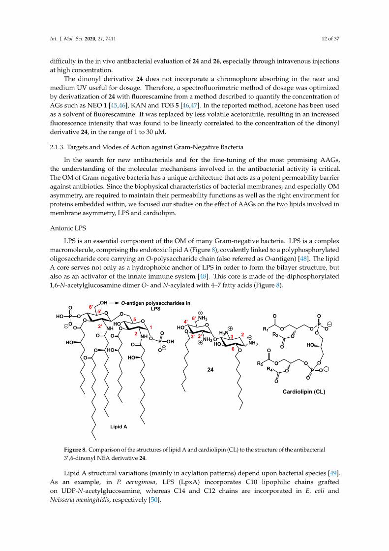
LPS is an essential component of the OM of many Gram-negative bacteria. LPS is a complexmacromolecule, comprising the endotoxic lipid A (Figure 8), covalently linked to a polyphosphorylatedoligosaccharide core carrying an O-polysaccharide chain (also referred as O-antigen) [48]. The lipidA core serves not only as a hydrophobic anchor of LPS in order to form the bilayer structure, butalso as an activator of the innate immune system [48]. This core is made of the diphosphorylated1,6-N-acetylglucosamine dimer O- and N-acylated with 4–7 fatty acids (Figure 8).
Int. J. Mol. Sci. 2020, 21, x FOR PEER REVIEW 12 of 36
2.1.3. Targets and Modes of Action against Gram-Negative Bacteria
In the search for new antibacterials and for the fine-tuning of the most promising AAGs, the understanding of the molecular mechanisms involved in the antibacterial activity is critical. The OM of Gram-negative bacteria has a unique architecture that acts as a potent permeability barrier against antibiotics. Since the biophysical characteristics of bacterial membranes, and especially OM asymmetry, are required to maintain their permeability functions as well as the right environment for proteins embedded within, we focused our studies on the effect of AAGs on the two lipids involved in membrane asymmetry, LPS and cardiolipin.
Anionic LPS
LPS is an essential component of the OM of many Gram-negative bacteria. LPS is a complex macromolecule, comprising the endotoxic lipid A (Figure 8), covalently linked to a polyphosphorylated oligosaccharide core carrying an O-polysaccharide chain (also referred as O-antigen) [48]. The lipid A core serves not only as a hydrophobic anchor of LPS in order to form the bilayer structure, but also as an activator of the innate immune system [48]. This core is made of the diphosphorylated 1,6-N-acetylglucosamine dimer O- and N-acylated with 4–7 fatty acids (Figure 8). Lipid A structural variations (mainly in acylation patterns) depend upon bacterial species [49]. As an example, in P. aeruginosa, LPS (LpxA) incorporates C10 lipophilic chains grafted on UDP-N-acetylglucosamine, whereas C14 and C12 chains are incorporated in E. coli and Neisseria meningitidis, respectively [50].
OP
O
O
OO
O
O
R1
HOO
R2
P OO
OOO
O
R3
O
R4O
OHOO
NH
OO
OH
NHO
PO
HOO
O
O PO
OOHO
HO
O
O
O
HO
O
O
HO
12
52'
5'
O
O
HO
NH3
ONH3
HOO
NH3
H3N3'
6
24
6'
2'
4'
23
Lipid A
Cardiolipin (CL)
O-antigen polysaccharides in LPS6'
Figure 8. Comparison of the structures of lipid A and cardiolipin (CL) to the structure of the antibacterial 3′,6-dinonyl NEA derivative 24.
In the continuous search for membrane-targeting antibiotics, compounds that interfere in LPS biogenesis or regulatory pathways offer opportunities for development as antibiotics that may be useful against pathogenic bacteria [22,23,51,52].
The electrostatic attraction between the amphiphilic NEA derivatives and the negatively charged lipid or LPS headgroups (i.e., core oligosaccharide and Lipid A phosphates in LPS) is likely the dominant driving force of the interaction [53,54]. However, other parameters governing the interactions between LPS and amphiphilic NEA derivatives, including (i) the area per lipid, (ii) the ordering and the hydration behavior, (iii) the spatial conformation and (iv) the molecular shape of the lipid A moiety, also play a critical role and are discussed hereunder.
Figure 8. Comparison of the structures of lipid A and cardiolipin (CL) to the structure of the antibacterial3′,6-dinonyl NEA derivative 24.
Lipid A structural variations (mainly in acylation patterns) depend upon bacterial species [49].As an example, in P. aeruginosa, LPS (LpxA) incorporates C10 lipophilic chains graftedon UDP-N-acetylglucosamine, whereas C14 and C12 chains are incorporated in E. coli andNeisseria meningitidis, respectively [50].

Int. J. Mol. Sci. 2020, 21, 7411 13 of 37
In the continuous search for membrane-targeting antibiotics, compounds that interfere in LPSbiogenesis or regulatory pathways offer opportunities for development as antibiotics that may beuseful against pathogenic bacteria [22,23,51,52].
The electrostatic attraction between the amphiphilic NEA derivatives and the negatively chargedlipid or LPS headgroups (i.e., core oligosaccharide and Lipid A phosphates in LPS) is likely the dominantdriving force of the interaction [53,54]. However, other parameters governing the interactions betweenLPS and amphiphilic NEA derivatives, including (i) the area per lipid, (ii) the ordering and thehydration behavior, (iii) the spatial conformation and (iv) the molecular shape of the lipid A moiety,also play a critical role and are discussed hereunder.
First, the area per lipid of the lipid A molecule is critical for characterizing the LPS/lipid Apacking. The acyl chains in lipid A occupy a smaller volume than in most of the phospholipids andcould be associated with a better packing [55]. Especially, in the P. aeruginosa membrane, the tailvolume of the penta-acyl lipid A is smaller as compared to that found in other bacteria having hexa- orhepta-acyl chains in their membranes, such as Salmonella minnesota [55–57].
Amphiphilic NEA derivatives were inserted into LPS monolayer, as indicated after spreadingthem in the subphase and measuring the compression isotherms of rough mutant. A shift to molecularareas higher than those for the pure buffer subphase was observed [42]. This results in a higher packing.Addition of 3′,6-dinonyl NEA 24 in the presence of Ca2+ gave rise to an effect on the molecular areathat was weaker than the effect obtained in the absence of Ca2+, suggesting that CaCl2 reduced theinsertion of the 3′,6-dinonyl NEA derivative [42].
As mentioned, and demonstrated by displacement assays, it is not only electrostatic interactionsthat are involved in the binding of amphiphilic NEA derivatives to LPS—the length and branching ofacyl chains in LPS are also critical. Different 3′,6-dialkyl NEA derivatives were found to bind to LPS witha clear ranking, depending upon the length of the linear alkyl chain (3′,6-diheptyl NEA 38 < 3′,6-dioctylNEA 39 < 3′,6-dinonyl NEA 24 < 3′,6-didecyl NEA 40 < 3′,6-undedecyl NEA 41). The presence of abranched alkyl chain also plays a critical role (3′,6-dinonyl NEA 24 < 3′,6-di(dimethyloctyl) NEA 46(Swain et al., unpublished results).
Second, closely interconnected with LPS/lipid A packing, the order and lipid phase might alsobe key clues for the interaction between LPS/lipid A and amphiphilic aminoglycoside antibiotics.The order parameter of lipid A from P. aeruginosa can be determined using Fourier-transform infrared(FT-IR) spectroscopy [58]. The antibacterial amphiphilic NEA derivatives make the LPS film statemore liquid-like, as suggested by the decrease in the excitation generalized polarization (GPex) of afluorescent laurdan probe (6-dodecanoyl-2-dimethylamine-naphthalene), which is sensitive to lipidhydration, inserted into LPS micelles [59,60]. This indicates an increasing liquid-like state in the lipid Aregion of LPS. The effect was dependent upon the presence of aryl groups (3′,6-di2NP 26 < 3′,6-diNB27 < 3′,4′,6-tri2NM 16). The effect induced by 3′,6-dinonyl NEA 24 was not significantly different fromthat observed with 3′,6-diNP 26, of similar lipophilicity. In contrast, COL, NEA and the 3′,6-di2NMderivative 14 had no influence on the packing of lipid A in micelles, as indicated by unchanged GPexvalues compared to the value for the control. The appearance of fluidic material extends previousdata related to the formation of a supramolecular network between multi-cationic and multi-anionicsubstances [61,62]. The liquid-like state in the lipid A region of LPS was likely more predominant thanobserved with glycerophospholipid. Divalent calcium ions could form salt bridges due to the closeproximity of lipid A phosphate groups [63]. This binding is tight enough that the charge density inthe solvent can approach zero [55]. All kinks in the water density lines, which indicate the enhancedwater penetration, are located around z = 8 nm in the vicinity of the peaks of both lipid A phosphatesand calcium ions [55]. The presence of divalent cations explains why the water molecules are able topenetrate deeper into the lipid A leaflet than the phospholipid leaflet.
The conformation of lipid A could be the third critical parameter for designing new potentialantibiotics. The spatial conformation of lipid A is an indicator of lipid A bioactivity [58,64,65].The backbone inclination angle of asymmetric penta-acyl lipid A influences the position and orientation

Int. J. Mol. Sci. 2020, 21, 7411 14 of 37
of the phosphate groups in LPS. Other inclination angles also provide information to further determinethe lipid A conformation, such as the angles between the membrane surface and the vectors from4′-carbon to 4′-phosphorus, 1-carbon to 1-phosphorus and 4′-carbon to 1-phosphorus atoms [55].This might help us to design new positions for substitution or new branching.
Lastly, the molecular shape of LPS/lipid A could play a critical role in its interaction withamphiphilic NEA derivatives. The penta-acyl LPS has a multilamellar structure due to its cylindricalmolecular shape. To be biologically appropriate (for interaction with Toll-like receptors, for example),this structure has to be converted into a physiologically active conformation [58]. The transitionfrom a unilamellar into a cubic inverted structure suggests that a subsequent intercalation into thehydrophobic moiety takes place, resulting in a change of the molecular shape of the LPS/lipid Amolecules with a more conical conformation [66].
Assuming the volume of the hydrophobic part increases from the linear dialkyl (3′,6-dinonylNEA 24) to the dinaphthylalkyl (3′,6-di2NM NEA 14, 3′,6-di2NP NEA 26, 3′,6-di2NB NEA 27) andthe bulky 3′,4′,6-tri2NM NEA 16 moieties, the maximal binding would be inversely proportionalto the volume of the hydrophobic moiety. The 3′,6-dinonyl NEA 24 shows a molecular shape of aninverted cone, with a large hydrophilic part and a small hydrophobic one. This could allow a closeinteraction between this derivative and the lipid A moiety of the LPS unit, characterized by a conicalcomplementary shape (with a small hydrophilic part and a large hydrophobic part). Very interestingly,LPS acylation can be modified through the activity of the palmitoyl lipid A transferase PagP, leading tonew modulation of LPS activity [67].
In summary, the small area per lipid and large order parameter of lipid A indicate a leaflet withlow fluidity and highly organized molecules. Calcium ions form salt bridges with phosphates in lipidA and stabilize the corresponding structure. The overall hydrated lipid A leaflet displays solvationdifferences due to the tilted lipid A backbone. This inclination causes the 1-phosphate in lipid A toproject toward the outer aqueous environment and the 4′-phosphate to become buried between the acylchains of lipid A [55]. Studies have highlighted the formation of a fluidic cross-linked supramolecularnetwork between LPS and amphiphilic NEA derivatives, in parallel with the length and the logPvalues of the corresponding derivatives. Both hydrophobic and electrostatic interactions are required.
The molecular shape of the 3′,6-dialkyl NEA derivatives induced by the nature of the graftedhydrophobic moieties (naphthylalkyl instead of alkyl) and the flexibility of the hydrophobic moiety arecritical for their fluidifying effect and their ability to displace cations bridging LPS. These parameterscould be exploited for the development of new amphiphilic NEA derivatives [42].
How these characteristics in the interactions between LPS and amphiphilic NEA derivatives resultin antibacterial activity is still unclear. Activity was associated with the self-promoted insertion ofthe molecule within the lipid A region of the OM outer leaflet in P. aeruginosa, leading to an increasein OM permeability. The fluorescent probe 1-N-phenylnapthylamine (NPN) was used to probe thepermeability of the OM, since NPN is a small molecule that cannot effectively cross the OM. It is weaklyfluorescent in aqueous solution but fluoresces strongly when it binds to phospholipids [68].
An ascending time dependence of the Ca2+ effect in NPN uptake experiments was observedfrom 3′,6-di2NM NEA 14 to 3′,6-di2NP NEA 26 and 3′,6-diNB NEA 27 [42]. For linear dialkylNEA derivatives, no clear effect corresponding to the increase of side chain lengths was observed.Regarding the effect of branched alkyl groups present in the 3′,6-di(dimethyloctyl) derivative 46,the dose required to obtain 50% of the maximal effect is lower for the branched derivative as comparedto the unbranched analogues, and even the maximal effect was equal for both compounds (Swain et al.,unpublished results).
Cardiolipin (CL)
CL (Figure 8) is a tetra-acylated diphosphatidylglycerol derivative found in plants and animals,but also in bacteria. This structurally unusual phospholipid carries two negative charges due to itsdimeric structure, consisting of two phosphatidyl residues connected by a glycerol bridge and four

Int. J. Mol. Sci. 2020, 21, 7411 15 of 37
associated fatty acyl chains (made of various alkyl chains R1, R2, R3 and R4) [69]. Due to variability infatty acyl chains (length, degree of unsaturation, etc.), CL is characterized by numerous molecularspecies, leading to a high potential for adaptability or responses to stress. CL is known for its sensitivityto stressors, such as the addition of organic solvents or high salt content or the presence of quaternaryammonium compounds [70,71]. Changes in the molecular species suggest CL restructuring. Indeed,CL is a critical phospholipid in maintaining the function and morphology of bacteria. CL was one ofthe main components of the IM, along with phosphatidylethanolamine (PE) and phosphatidylglycerol(PG) [72,73]. Very interestingly, CL is also located in the OM of most Gram-negative bacteria [26,74].
Like LPS, CL is characterized by its own membrane parameters, including (i) molecular area,(ii) degree of unsaturation and length of acyl chains and (iii) the ability to induce negative curvatureand form microdomains, parameters which are critical in the biophysical properties of membranes inwhich CL is inserted.
With the aim of characterizing the effect of the interactions between amphiphilic NEAderivatives with CL on the molecular area, we determined the compression isotherms correspondingto the lipid monolayers for CL spread on a subphase containing 3′,6-dinonyl NEA 24 (ascompared to those of 1-palmitoyl-2-oleoyl-sn-glycero-3-phosphatidylethanolamine (POPE) and1-palmitoyl-2-oleoyl-sn-glycero-3-phospho-(1′-rac-glycerol) (POPG)). The isotherms were shiftedto higher molecular areas than the pure buffer subphase, although to a much lower extent for POPE,compared with POPG and CL. At 30 mNewtons/m, a value close to the estimated surface pressureof biological membranes in vivo, the mean molecular area for POPE was not significantly modifiedby the presence of 3′,6-dinonyl NEA 24 in the subphase. With POPG and CL, the mean moleculararea increased by 1.8 and 1.6, respectively. This discrepancy indicates a much higher adsorption of 24into POPG or CL than into POPE. The shift of the mean molecular area was not significantly differentbetween POPG and CL.
Furthermore, order and interdigitation could be affected by AAGs. Bacterial CL structure ischaracterized by a high degree of symmetry and unsaturation [71,75,76], as well as by long fatty acylchains. Both the degree of unsaturation and the length of acyl chains can modulate order and crosstalkbetween leaflets [77–79]. CL might be a reservoir for unsaturated fatty acid chains, as suggested by thecontent of unsaturated fatty acid in cls/cls2-mutant cells, which is approximatively 40% lower than inwild-type cells [71]. The relative concentrations of C16 and C18 for the barotolerant Pseudomonas sp.BT1 were approximately 60% and 40%, respectively [72]. PE and CL have a phase transition well abovethat of PG when the fatty acid content is identical for both lipids. In general, lipids with long fatty acylchains undergo phase transition at higher temperature than lipids with short chains [80]. Long chainsexplain why CL stimulates changes in the physical properties of the membrane and why it decreasesthe lateral interaction within the monolayer leaflet, which favors the creation of membrane folds [81].
Another striking characteristic of CL is the comparatively small cross-section of its headgrouprelative to the cross-section of its four large tail groups. This discrepancy results in a molecularshape with a large intrinsic negative curvature [82–85] and a non-bilayer anionic phospholipid alsodescribed previously for PE [86] that facilitates the insertion of membrane proteins. The cross-sectionalsize difference further explains the location of CL-enriched regions at the pole and/or the divisionseptum [87], which can in turn be related to the polar localization of many proteins, including thoseinvolved in cell division and osmosensing [88]. The intrinsic negative curvature of CL can result in theformation of microdomains (clusters).
The presence and the role of these microdomains can be affected by membrane-acting antibiotics.Using membrane models mimicking P. aeruginosa plasma membrane composition (POPE:POPG:CL),we demonstrated the ability of 3′,6-dinonyl NEA 24 to induce in a CL dependent manner, to increasemembrane permeability through reduced hydration and decreased ability of the membrane to mixand fuse, as shown by monitoring calcein release, GPex of laurdan and fluorescence dequenchingof octadecyl rhodamine B, respectively [89]. After incubation with 3′,6-dinonyl NEA 24, CL wasco-localized with PE in small clusters, sections with increased membrane curvature, and fusion points

Int. J. Mol. Sci. 2020, 21, 7411 16 of 37
in the joined giant unilamellar vesicles (GUV) population. Moreover, a clear overall lipid reorganizationwas observed [89].
CL-interacting proteins and functions regulated by CL are affected by the amphiphilic AG, as wedemonstrated an inhibition of the respiratory chain and changes in bacterial shape. The latter effectwas characterized by the loss of the bacterial rod shape through a decrease in length and an increasein curvature. It resulted from the effect on MreB, a CL-dependent cytoskeleton protein, as well as adirect effect of 3′,6-dinonyl NEA 24 on CL [90]. In E. coli, CL is known to enhance the activity of theglycosyltransferase MurG involved in peptidoglycan biosynthesis [91]. Some correlation (phosphatidicacid > CL > PG) with the activity of dynamin-related Protein 1 (Drp1) has been reported [92]. In addition,CL content plays a critical role since low levels of CL in the membrane of Pseudomonas putida allowthe tetradecyltrimethylammonium cation (TTAB) to cross the membrane, reach its site of action andkill the cell, as bacteria cannot counteract the fluidizing effect of the detergent [71]. On the contrary,the addition of CL to the culture medium delayed the growth of P. aeruginosa, favored asymmetricalgrowth and enhanced the efficiency of 3′,6-dinonyl NEA 24 [93].
These results shed light on how targeting CL microdomains may be of great interest for developingnew antibacterial therapies. Some recent evidence also highlights the role of CL for outer membranevesicle (OMV) formation. These vesicles could be important as inter-kingdom players. Experimentalevidence that bacterial OMVs, by sequestering of cationic peptides, may protect pathogenic yeastagainst the combined action of antifungal drugs has been reported [94].
2.1.4. Targets and Modes of Action against Gram-Positive Bacteria
We also investigated the mechanism of action against Gram-positive bacteria, S. aureus andBacillus subtilis [95]. Time-killing experiments were performed to demonstrate the bactericidal effectinduced by 3′,6-dinonyl NEA 24 against S. aureus MSSA and MRSA (methicillin susceptible andresistant S. aureus, respectively). The displacement of the BODIPY™-TR cadaverine probe bound tolipoteichoic acids (LTA) showed that 24 interacts with the bacterial surface components. The abilityof 24 to enhance membrane depolarization and induce membrane permeability was highlighted,using fluorescent probes, DiSC3C(5) and propidium iodide, respectively. These effects were observedfor both MSSA and MRSA, as well as for B. subtilis. The disruption of membrane integrity of thebacterial cell wall was revealed by electronic microscopy, and changes in the localization of lipids fromthe enriched-septum region and the impairment of the formation of the septum were observed byfluorescence microscopy. This study revealed that 24 interferes with multiple targets, suggesting a lowability of Gram-positive bacteria to acquire resistance to this antibacterial agent.
2.2. Recent Reports on AAGs in the Field of Antibacterial Agents
Here, the main reports in the field of antibacterial AAGs, published after the appearance of severalreview articles since 2016 [7,8,30–32], are summarized with an emphasis on AAGs made of an AGcore conjugated to an adjuvant or an antibiotic drug of another class (antibiotic hybrids), as recentlydeveloped by Schweizer, Zhanel and coworkers.
The grafting of a metal binding site on antimicrobial peptides (AMPs) improves theirantibacterial efficiency [96–98]. In the search for antibacterial AAGs bearing a metal bindingmotif, 3′,4′,6-tri2NM NEA (16) [22] derivatives functionalized at position 5 through a short spacerby a Zn(II) or Cu(II) chelating group, tris(2-pyridylmethyl)amine (TPA), di(picolyl)amine (DPA)and tetraazacyclotetradecane (Cyclam) were synthesized [99]. NEA-cyclam and Zn(NEA-TPA)derivatives were found to be the most efficient compounds active against clinical MDR strain isolateEnterobacter aerogenes EA289, with MICs in the range of 16–4 and 4 µM, respectively, whereas usualantibiotics such as β-lactams and phenicols were inactive and ciprofloxacin was weakly active.NEA-Cyclam and Zn(NEA-TPA) were shown to target and permeabilize the OM of EA289. All NEAconjugates were able to block the efflux of 1,2′-dinaphthylamine in EA289 by acting on the effluxtransporter located in the inner membrane [99].

Int. J. Mol. Sci. 2020, 21, 7411 17 of 37
A series of pyrene-NEO (PYR-NEO) conjugates were synthesized and their binding affinity toA-site RNA targets, resistance to AG-deactivating enzymes and antibacterial activity against a widevariety of bacterial strains of clinical relevance were studied [100]. As observed previously with NEAconjugates [35], the conjugation significantly alters the affinities of NEO for bacterial A-site targets.PYR-NEO conjugates exhibited broad-spectrum activity towards Gram-positive bacteria, includingimproved activity against NEO-resistant methicillin-resistant S. aureus (MRSA) strains. The conjugationsignificantly increased the resistance of NEO to AG-deactivating enzyme modification.
New antibacterial properties of amphiphilic AG conjugates were also recently highlighted.Antimicrobial hybrids that are AAGs have emerged in a promising strategy to combat bacterialresistance as drugs alone and as adjuvants in combination with existing antibiotics. The concepts,advances and challenges of antibiotic hybrids, including AAGs linked to an established antibioticagent, were reviewed recently [101,102]. Such AAG hybrids can promote the influx of antibioticsthrough the OM of Gram-negative bacteria via the self-promoted uptake mechanism by displacementof the divalent cations (Ca2+ or Mg2+) that stabilize LPS [103].
Lysine-NEO conjugates have been synthesized and exhibited mainly decreased antibacterialactivities [104]. Amphiphilic tobramycin (TOB) conjugates to lysine and anthracenyl groups 48(Figure 9) appeared to be able to sensitize MDR Gram-negative bacteria to antibiotic drugs [105,106].
Int. J. Mol. Sci. 2020, 21, x FOR PEER REVIEW 17 of 36
through the OM of Gram-negative bacteria via the self-promoted uptake mechanism by displacement of the divalent cations (Ca2+ or Mg2+) that stabilize LPS [103].
Lysine-NEO conjugates have been synthesized and exhibited mainly decreased antibacterial activities [104]. Amphiphilic tobramycin (TOB) conjugates to lysine and anthracenyl groups 48 (Figure 9) appeared to be able to sensitize MDR Gram-negative bacteria to antibiotic drugs [105,106].
O
O
HO
NH2
NH2
OO
NH2
H2N
OHO
H2NOH
OHN
O
ClNH2
NH2
7 HCl
O
O
HO
NH2
NH2
OO
NH2
H2N
OHO
H2NOH
OHN
H2N
OHN
ONH
7 HCl
O
O
HO
NH2
NH2
OO
NH2
H2N
OHO
H2NOH
OHN
N
7 HCl
O
O
HO
NH2
NH2
OO
NH2
H2N
OHO
H2NOH
OHN
F
O
O
O 6 HCl
5n
48: n= 3, 7, 11
12
51: TOB-DBP
49: TOB-NMPn= 2, 4, 6, 8, 10 12
n
12
50: TOB-PAR
Figure 9. Structures of the tobramycin (TOB) conjugates to lysine 48 [105,106], and to the efflux pump inhibitors (EPIs), 1-(1′-naphthylmethyl)piperazine (NMP) (49), paroxetine (PAR) (50) and dibasic naphthyl peptide (DBP) (51) [107,108].
Combination studies indicate that these amphiphilic TOB-lysine conjugates 48 sensitize Gram-negative bacteria to antibiotics. The most potent derivative (n = 11, Figure 9) synergizes rifampicin and minocycline against MDR and extensively drug resistant (XDR) P. aeruginosa isolates and enhances efficacy of both antibiotics in the Galleria mellonella larvae in vivo infection model [105]. Mode of action studies indicate that the amphiphilic TOB-lysine adjuvants enhance OM cell penetration and affect the proton motive force, which energizes efflux pumps. The TOB-lysine conjugate 48 (n = 11) (Figure 9) also potentiates the antibacterial efficacy of eight clinically used antibiotics against wild-type, multidrug-resistant and extensively drug-resistant P. aeruginosa isolates from Canadian hospitals [106]. Antibiotics that are synergistic with 48 (n = 11) included moxifloxacin (MOX), ciprofloxacin (CIP), erythromycin, chloramphenicol, trimethoprim, novobiocin, linezolid, and fosfomycin. Novobiocin showed the highest synergy.
TOB conjugates to the efflux pump inhibitors (EPIs) 1-(1′-naphthylmethyl)piperazine (NMP) (49), paroxetine (PAR) (50), and dibasic naphthyl peptide (DBP) (51) (Figure 9) also overcome resistance against MDR P. aeruginosa isolates by enhancing bacterial OM penetration and reducing efflux [107]. These AAGs enhance the synergy and efficacy of EPIs in combination with tetracycline antibiotics against MDR Gram-negative bacteria, including P. aeruginosa. In addition to potentiating tetracycline antibiotics, TOB-EPI conjugates can also suppress resistance development to the tetracycline antibiotic minocycline, thereby providing a strategy to develop more effective adjuvants
Figure 9. Structures of the tobramycin (TOB) conjugates to lysine 48 [105,106], and to the efflux pumpinhibitors (EPIs), 1-(1′-naphthylmethyl)piperazine (NMP) (49), paroxetine (PAR) (50) and dibasicnaphthyl peptide (DBP) (51) [107,108].
Combination studies indicate that these amphiphilic TOB-lysine conjugates 48 sensitizeGram-negative bacteria to antibiotics. The most potent derivative (n = 11, Figure 9) synergizesrifampicin and minocycline against MDR and extensively drug resistant (XDR) P. aeruginosa isolatesand enhances efficacy of both antibiotics in the Galleria mellonella larvae in vivo infection model [105].

Int. J. Mol. Sci. 2020, 21, 7411 18 of 37
Mode of action studies indicate that the amphiphilic TOB-lysine adjuvants enhance OM cell penetrationand affect the proton motive force, which energizes efflux pumps. The TOB-lysine conjugate 48 (n = 11)(Figure 9) also potentiates the antibacterial efficacy of eight clinically used antibiotics against wild-type,multidrug-resistant and extensively drug-resistant P. aeruginosa isolates from Canadian hospitals [106].Antibiotics that are synergistic with 48 (n = 11) included moxifloxacin (MOX), ciprofloxacin (CIP),erythromycin, chloramphenicol, trimethoprim, novobiocin, linezolid, and fosfomycin. Novobiocinshowed the highest synergy.
TOB conjugates to the efflux pump inhibitors (EPIs) 1-(1′-naphthylmethyl)piperazine (NMP) (49),paroxetine (PAR) (50), and dibasic naphthyl peptide (DBP) (51) (Figure 9) also overcome resistanceagainst MDR P. aeruginosa isolates by enhancing bacterial OM penetration and reducing efflux [107].These AAGs enhance the synergy and efficacy of EPIs in combination with tetracycline antibioticsagainst MDR Gram-negative bacteria, including P. aeruginosa. In addition to potentiating tetracyclineantibiotics, TOB-EPI conjugates can also suppress resistance development to the tetracycline antibioticminocycline, thereby providing a strategy to develop more effective adjuvants to rescue tetracyclineantibiotics from resistance in MDR Gram-negative bacteria. A strong synergy with MOX, ciprofloxacin(CIP), rifampicin and fosfomycin was also observed against MDR/XDR P. aeruginosa [108].
AAG hybrids incorporating a fluoroquinolone were synthesized through conjugation of theMOX and CIP to the TOB core via an aliphatic alkyl linker. Among the TOB-MOX AAGs 52–54(Figure 10) synthesized, the hybrid 52 was shown to enhance OM permeability and reduce efflux bydissipating the proton motive force of the cytoplasmic membrane in P. aeruginosa [109]. It protectsGalleria mellonella larvae from the lethal effects of MDR P. aeruginosa. Attempts to select for resistanceover a period of 25 days resulted in a two-fold increase in the MIC for the hybrid, whereas MOX orTOB resulted in a 16- and 512-fold increase in MIC. TOB-ciprofloxacin hybrid (TOB-CIP) adjuvants, inwhich CIP is attached at the TOB 5- (hybrids 55, Figure 10), 2”- or 6”-position, were shown to rescue theactivity of fluoroquinolone antibiotics against MDR and XDR P. aeruginosa isolates in vitro and enhancefluoroquinolone efficacy in the Galleria mellonella in vivo infection model [110]. Structure–activitystudies revealed that the presence of both TOB and CIP, which are separated by a C12 tether, is criticalfor the function of the adjuvant. Mechanistic studies indicate that the antibacterial modes of CIP areretained, whereas the role of TOB is limited to destabilization of the OM in the hybrid.
In vitro screening of six anticancer drugs for their potential use in antimicrobial therapysuggested the feasibility of repurposing the anticancer drug mitomycin C against MDR Gram-negativebacteria [111]. In combination with the TOB-CIP hybrid 55 (n = 12, Figure 10), the antibacterialactivity of mitomycin C was enhanced against MDR clinical isolates of P. aeruginosa, A. baumannii,E. coli, K. pneumoniae and Enterobacter cloacae. Synergy was inherent to TOB-CIP and neither TOB norCIP individually synergized with mitomycin C. The TOB-based hybrid adjuvants able to potentiatemultiple classes of legacy antibiotics against various MDR Gram-negative bacteria were modified byreplacing the TOB domain with the smaller pseudo-disaccharide NEB through selective cleavage of theα-D-glucopyranosyl linkage of TOB 8, which is an NEA (7) analogue lacking the 3′-hydroxyl function(Figures 2 and 10) [112]. The hybrid AAGs NEB-moxifloxacin (NEB-MOX) 56, NEB-ciprofloxacin(NEB-CIP) 57 and NEB-1-(1′-naphthylmethyl)piperazine (NEB-NMP) 58 were synthesized (Figure 10).Potent synergism was found for combinations of NEB-based hybrid adjuvants with multiple classes ofantibiotics, including fluoroquinolones (MOX and CIP), tetracyclines (minocycline) and rifamycins(rifampicin) against both wild-type and MDR P. aeruginosa clinical isolates. The combination of theoptimized NEB-CIP hybrid 56 (Figure 10) and rifampicin protects G. mellonella larvae from the lethaleffects of extensively drug-resistant (XDR) P. aeruginosa. Mechanistic evaluation of NEB-based hybridadjuvants revealed that the hybrids affect the OM and IM of wild-type P. aeruginosa PAO1.

Int. J. Mol. Sci. 2020, 21, 7411 19 of 37
Int. J. Mol. Sci. 2020, 21, x FOR PEER REVIEW 18 of 36
to rescue tetracycline antibiotics from resistance in MDR Gram-negative bacteria. A strong synergy with MOX, ciprofloxacin (CIP), rifampicin and fosfomycin was also observed against MDR/XDR P. aeruginosa [108].
AAG hybrids incorporating a fluoroquinolone were synthesized through conjugation of the MOX and CIP to the TOB core via an aliphatic alkyl linker. Among the TOB-MOX AAGs 52–54 (Figure 10) synthesized, the hybrid 52 was shown to enhance OM permeability and reduce efflux by dissipating the proton motive force of the cytoplasmic membrane in P. aeruginosa [109]. It protects Galleria mellonella larvae from the lethal effects of MDR P. aeruginosa. Attempts to select for resistance over a period of 25 days resulted in a two-fold increase in the MIC for the hybrid, whereas MOX or TOB resulted in a 16- and 512-fold increase in MIC. TOB-ciprofloxacin hybrid (TOB-CIP) adjuvants, in which CIP is attached at the TOB 5- (hybrids 55, Figure 10), 2′’- or 6′’-position, were shown to rescue the activity of fluoroquinolone antibiotics against MDR and XDR P. aeruginosa isolates in vitro and enhance fluoroquinolone efficacy in the Galleria mellonella in vivo infection model [110]. Structure–activity studies revealed that the presence of both TOB and CIP, which are separated by a C12 tether, is critical for the function of the adjuvant. Mechanistic studies indicate that the antibacterial modes of CIP are retained, whereas the role of TOB is limited to destabilization of the OM in the hybrid.
O
O
HO
NH2
NH2
OO
NH2
H2N
OHO
H2NOH
OHN
NN
FOO
HO 6 HCl
O
O
HO
NH2
NH2
OOH
NH2
H2N
N
NN
FO
HO2C
5 HCl
X
O
O
HO
NH2
NH2
OOH
NH2
H2N
5 HCl
O
NH2O
HO
NH2
OOH
NH2
H2N
NN
6 HCl
O
O
HO
OR2O
H2NOH
OR3
NH2
NH2
R1OO
NH2
H2N
5 HCl
N
OMe
N
F
H
N
O
HO2C H
5
55: TOB-CIP (n= 4, 6, 8, 10, 12)
2"
6" 55
5
57: NEB-CIP
56: NEB-MOX
n
58: NEB-NMPn= 2, 4, 6, 8, 10 12
TOB-MOX: 52: R1= X, R2= R3= H; 53: R1= H, R2= X, R3= H; 54: R1= R2= H, R3= X
12
X=
12
n
Figure 10. Structures of the TOB (52–54) and nebramine (NEB) (56) conjugates to the fluoroquinolones moxifloxacin (MOX) and ciprofloxacin (CIP) [109,111], respectively 55 and 57, and, of the NEB conjugates to the efflux pump inhibitor 1-(1′-naphthylmethyl)piperazine (NEB-NMP) 58 [112].
In vitro screening of six anticancer drugs for their potential use in antimicrobial therapy suggested the feasibility of repurposing the anticancer drug mitomycin C against MDR Gram-
Figure 10. Structures of the TOB (52–54) and nebramine (NEB) (56) conjugates to the fluoroquinolonesmoxifloxacin (MOX) and ciprofloxacin (CIP) [109,111], respectively 55 and 57, and, of the NEB conjugatesto the efflux pump inhibitor 1-(1′-naphthylmethyl)piperazine (NEB-NMP) 58 [112].
TOB-polymyxin B-3 hybrids showed potent activity against carbapenem-resistant as well asMDR or extensively drug-resistant (XDR) P. aeruginosa clinical isolates [113]. The most potent hybridincorporating a C12 spacer was able to synergize with currently used antibiotics against wild-type andMDR/XDR P. aeruginosa and Acinetobacter baumannii.
More recently, TOB-rifampicin conjugates were also shown to break the intrinsic resistance inP. aeruginosa, due to chromosomally encoded low OM permeability and constitutively over-expressedefflux pumps, and sensitize MDR and XDR P. aeruginosa to doxycycline and chloramphenicol in vitroand in vivo [114]. Tetracyclines and chloramphenicol are model compounds for bacteriostatic effects,but when combined with TOB-rifampicin adjuvants, their effects became bactericidal at sub MIC levels.Potentiation of tetracyclines was found to be correlated with SAR of this class of drugs and consistentwith OM permeabilization and efflux pump inhibition.
Conjugation of TOB and cyclam to give 59 (Figure 11) also abrogates the ribosomal effects ofTOB but confers a potent adjuvant property that restores the full antibiotic activity of aztreonam andmeropenem against carbapenem-resistant P. aeruginosa [115]. Therapeutic levels of susceptibility wereattained in several MDR clinical isolates, and time-kill assays revealed a synergistic dose-dependentpharmacodynamic relationship. A triple combination of the adjuvant with ceftazidime/avibactam(approved), aztreonam/avibactam (phase III) and meropenem/avibactam enhances the efficaciesof β-lactam/beta-lactamase inhibitors against recalcitrant strains, suggesting rapid access of thecombination to their periplasmic targets. The adjuvants, and their combinations, were non-hemolyticand non-cytotoxic, and preliminary in vivo evaluation of 59 (n = 8) in G. mellonella suggested therapeuticpotential for the double and triple combinations. The TOB–cyclam conjugate 59 (n = 8) mitigates the

Int. J. Mol. Sci. 2020, 21, 7411 20 of 37
effect of OprD/OprF porin loss in P. aeruginosa and potentiates β-lactam/β-lactamase inhibitors againstcarbapenem-resistant clinical isolates, highlighting the complexity of resistance to β-lactam antibiotics.
Int. J. Mol. Sci. 2020, 21, x FOR PEER REVIEW 19 of 36
negative bacteria [111]. In combination with the TOB-CIP hybrid 55 (n = 12, Figure 10), the antibacterial activity of mitomycin C was enhanced against MDR clinical isolates of P. aeruginosa, A. baumannii, E. coli, K. pneumoniae and Enterobacter cloacae. Synergy was inherent to TOB-CIP and neither TOB nor CIP individually synergized with mitomycin C. The TOB-based hybrid adjuvants able to potentiate multiple classes of legacy antibiotics against various MDR Gram-negative bacteria were modified by replacing the TOB domain with the smaller pseudo-disaccharide NEB through selective cleavage of the α-D-glucopyranosyl linkage of TOB 8, which is an NEA (7) analogue lacking the 3′-hydroxyl function (Figures 2 and 10) [112]. The hybrid AAGs NEB-moxifloxacin (NEB-MOX) 56, NEB-ciprofloxacin (NEB-CIP) 57 and NEB-1-(1′-naphthylmethyl)piperazine (NEB-NMP) 58 were synthesized (Figure 10). Potent synergism was found for combinations of NEB-based hybrid adjuvants with multiple classes of antibiotics, including fluoroquinolones (MOX and CIP), tetracyclines (minocycline) and rifamycins (rifampicin) against both wild-type and MDR P. aeruginosa clinical isolates. The combination of the optimized NEB-CIP hybrid 56 (Figure 10) and rifampicin protects G. mellonella larvae from the lethal effects of extensively drug-resistant (XDR) P. aeruginosa. Mechanistic evaluation of NEB-based hybrid adjuvants revealed that the hybrids affect the OM and IM of wild-type P. aeruginosa PAO1.
TOB-polymyxin B-3 hybrids showed potent activity against carbapenem-resistant as well as MDR or extensively drug-resistant (XDR) P. aeruginosa clinical isolates [113]. The most potent hybrid incorporating a C12 spacer was able to synergize with currently used antibiotics against wild-type and MDR/XDR P. aeruginosa and Acinetobacter baumannii.
More recently, TOB-rifampicin conjugates were also shown to break the intrinsic resistance in P. aeruginosa, due to chromosomally encoded low OM permeability and constitutively over-expressed efflux pumps, and sensitize MDR and XDR P. aeruginosa to doxycycline and chloramphenicol in vitro and in vivo [114]. Tetracyclines and chloramphenicol are model compounds for bacteriostatic effects, but when combined with TOB-rifampicin adjuvants, their effects became bactericidal at sub MIC levels. Potentiation of tetracyclines was found to be correlated with SAR of this class of drugs and consistent with OM permeabilization and efflux pump inhibition.
Conjugation of TOB and cyclam to give 59 (Figure 11) also abrogates the ribosomal effects of TOB but confers a potent adjuvant property that restores the full antibiotic activity of aztreonam and meropenem against carbapenem-resistant P. aeruginosa [115]. Therapeutic levels of susceptibility were attained in several MDR clinical isolates, and time-kill assays revealed a synergistic dose-dependent pharmacodynamic relationship. A triple combination of the adjuvant with ceftazidime/avibactam (approved), aztreonam/avibactam (phase III) and meropenem/avibactam enhances the efficacies of β-lactam/beta-lactamase inhibitors against recalcitrant strains, suggesting rapid access of the combination to their periplasmic targets. The adjuvants, and their combinations, were non-hemolytic and non-cytotoxic, and preliminary in vivo evaluation of 59 (n = 8) in G. mellonella suggested therapeutic potential for the double and triple combinations. The TOB–cyclam conjugate 59 (n = 8) mitigates the effect of OprD/OprF porin loss in P. aeruginosa and potentiates β-lactam/β-lactamase inhibitors against carbapenem-resistant clinical isolates, highlighting the complexity of resistance to β-lactam antibiotics.
O
O
HO
NH2
NH2
OO
NH2
H2N
OHO
H2NOH
OH
O
O
HO
NH2
NH2
OOH
NH2
H2N
5
59: n= 4 (9 TFA), 8 (9 TFA), 12 (9 HCl)
8 5
60: NEB-CIP (8 HCl)
n
NH
NH
HN
N
NH
NH
HN
N
Figure 11. Structures of the TOB-cyclam and NEB-cyclam hybrids 59 [115] and 60 [116].
The NEB-cyclam conjugate 60 (Figure 11) was shown to potentiate β-lactam antibiotics, as wellas other antibiotic drugs, against P. aeruginosa in vitro [116]. This adjuvant is able to synergize withβ-lactams aztreonam and ceftazidime against MDR and extremely drug-resistant clinical isolatesthrough a hypothesized mechanism of OM permeabilization.
AGs were also modified for obtaining antibacterial amphiphilic nanoparticles. The brainneurotransmitter dopamine has been polymerized under aerobic conditions to produce polydopamine(PDA). Recently, self-polymerized PDA nanoparticles were tethered to AGs (gentamicin, KANA andNEO) [117]. These nanoconjugates were evaluated for their antimicrobial potency against variousbacterial strains, including resistant ones, and their cytocompatibility. Of the three nanoconjugates(PDA-gentamicin, PDA-KANA and PDA-NEO), the PDA-KANA (PDA-K) nanoconjugate exhibitedthe highest activity against potent pathogens, the least toxicity to human embryonic kidney (HEK 293)cells and intense toxic effects to human glioblastoma (U87) cells.
2.3. AAG Positioning as Potential Antibacterial Drug Candidates, Toxicity
Dialkyl and trialkyl NEA derivatives have broad-spectrum antibacterial activity againstsusceptible and resistant Gram-positive and Gram-negative bacteria and their activity is relatedto favorable lipophilicity windows. Fridman and coworkers identified one amphiphilic 4′,5,6-triheptylNEB derivative 80 (Figure 12) that is active against susceptible and resistant Gram-positive andGram-negative bacteria (MRSA, E. coli, K. Pneumonia, P. aeruginosa strains, etc.) [37]. The correspondingtrihexyl and trioctyl derivatives were found to be active against Gram-positive bacteria and inactive orweakly active against Gram-negative bacteria. As a consequence, the antibacterial Gram-positive andGram-negative activity of amphiphilic NEB derivatives corresponds to a lipophilicity window that ismuch narrower than the windows delineated in the NEA series for trialkyl and dialkyl derivatives.This result is probably related to the higher lipophilicity of the NEB derivatives in comparison to thecorresponding 3′,4′,6-trialkyl NEA derivatives [34,35].
A chemical way to increase the antimicrobial activity of the anti-Gram-positive andanti-Gram-negative NEB, TOB and PARO AAGs was reported in 2015 [38]. AAGs acting against a panelof Gram-positive and Gram-negative bacteria were obtained by di-N-methylation of all amine functionsof the corresponding AAGs; for example, the 4′,5-dinonyl NEB derivative 81 was methylated to givethe tetra-N-dimethyl derivative 82. However, strong hemolytic effects of the N-dimethyl NEB, TOB andPARO derivatives were observed and were related to the strong increase in lipophilicity in comparisonto the parent non-N-dimethylated AAGs (an increase in the calculated logP by about a three ordersof magnitude). Cetrimonium and gramicidin D, which are both in antibacterial topical clinical use,appeared to be significantly more hemolytic than all studied antibacterial AAGs. These results suggest

Int. J. Mol. Sci. 2020, 21, 7411 21 of 37
a possible means of development of potent and broad-spectrum antibacterial membrane-disruptingAAGs for the treatment of persistent topical infections [38].
Int. J. Mol. Sci. 2020, 21, x FOR PEER REVIEW 20 of 36
Figure 11. Structures of the TOB-cyclam and NEB-cyclam hybrids 59 [115] and 60 [116].
The NEB-cyclam conjugate 60 (Figure 11) was shown to potentiate β-lactam antibiotics, as well as other antibiotic drugs, against P. aeruginosa in vitro [116]. This adjuvant is able to synergize with β-lactams aztreonam and ceftazidime against MDR and extremely drug-resistant clinical isolates through a hypothesized mechanism of OM permeabilization.
AGs were also modified for obtaining antibacterial amphiphilic nanoparticles. The brain neurotransmitter dopamine has been polymerized under aerobic conditions to produce polydopamine (PDA). Recently, self-polymerized PDA nanoparticles were tethered to AGs (gentamicin, KANA and NEO) [117]. These nanoconjugates were evaluated for their antimicrobial potency against various bacterial strains, including resistant ones, and their cytocompatibility. Of the three nanoconjugates (PDA-gentamicin, PDA-KANA and PDA-NEO), the PDA-KANA (PDA-K) nanoconjugate exhibited the highest activity against potent pathogens, the least toxicity to human embryonic kidney (HEK 293) cells and intense toxic effects to human glioblastoma (U87) cells.
2.3. AAG Positioning as Potential Antibacterial Drug Candidates, Toxicity
Dialkyl and trialkyl NEA derivatives have broad-spectrum antibacterial activity against susceptible and resistant Gram-positive and Gram-negative bacteria and their activity is related to favorable lipophilicity windows. Fridman and coworkers identified one amphiphilic 4′,5,6-triheptyl NEB derivative 80 (Figure 12) that is active against susceptible and resistant Gram-positive and Gram-negative bacteria (MRSA, E. coli, K. Pneumonia, P. aeruginosa strains, etc.) [37]. The corresponding trihexyl and trioctyl derivatives were found to be active against Gram-positive bacteria and inactive or weakly active against Gram-negative bacteria. As a consequence, the antibacterial Gram-positive and Gram-negative activity of amphiphilic NEB derivatives corresponds to a lipophilicity window that is much narrower than the windows delineated in the NEA series for trialkyl and dialkyl derivatives. This result is probably related to the higher lipophilicity of the NEB derivatives in comparison to the corresponding 3′,4′,6-trialkyl NEA derivatives [34,35].
O
O
O
NR2
NR2
OOH
NR2
R2NO
O
O
NH2
NH2
OO
NH2
H2N
80 81: R= H 82: R= Me
5 6
4'
Figure 12. Structures of the synthesized broad-spectrum antibacterial 4′,5,6-tri- and 4′,5-di-alkylated NEB derivatives [37,38].
A chemical way to increase the antimicrobial activity of the anti-Gram-positive and anti-Gram-negative NEB, TOB and PARO AAGs was reported in 2015 [38]. AAGs acting against a panel of Gram-positive and Gram-negative bacteria were obtained by di-N-methylation of all amine functions of the corresponding AAGs; for example, the 4′,5-dinonyl NEB derivative 81 was methylated to give the tetra-N-dimethyl derivative 82. However, strong hemolytic effects of the N-dimethyl NEB, TOB and PARO derivatives were observed and were related to the strong increase in lipophilicity in comparison to the parent non-N-dimethylated AAGs (an increase in the calculated logP by about a three orders of magnitude). Cetrimonium and gramicidin D, which are both in antibacterial topical clinical use, appeared to be significantly more hemolytic than all studied antibacterial AAGs. These
Figure 12. Structures of the synthesized broad-spectrum antibacterial 4′,5,6-tri- and 4′,5-di-alkylatedNEB derivatives [37,38].
In order to evaluate the immediate toxicity of antibacterial AAGs, we performed a toxicity studyin SKH1 male mice with AAGs 24 and 26 delivered in aqueous solution at concentrations higherthan 5 µM, intravenously, intraperitoneally and subcutaneously in 100 µL injections twice a day.This study revealed immediate toxic effects (tail necrosis or swelling in the area of the injections andhigh mean body weight decrease; unpublished results obtained in collaboration with the preclinicalContract Research Organization (CRO), VOXCAN, Marcy l’Étoile, France). Such effects could berelated, at least in part, to cellular membrane effects and to a low solubility in saline and bufferedaqueous solutions. These results show that antibacterial AAGs have to be formulated to be deliveredin vivo at high concentrations, for example in liposomes like the anionic and amphiphilic antifungaldrug amphothericin B, which self-aggregates and is highly toxic [118]. Such formulations should allowthe reduction of the concentration-dependent local toxic effects and the release of the AAGs at anefficient concentration.
The antibacterial potential of AAGs in antibiotherapy as adjuvants to antibiotic drugs alsosuggests their potential use at lower concentrations, rather than alone as antibiotic drugs. Recently,other adjuvants that are membrane-affecting small molecules were shown to be able to suppress COLresistance by abolishing or reducing the extent of lipid A modification induced by the mcr-1 geneand pmrAB system in E. coli [119,120]. AAGs 24, 26 and 27 are active against clinical COL-resistantP. aeruginosa strains (MICs = 28 µg/mL) and they cooperatively bind to LPS, increasing the OMpermeability [42]. The much weaker and slower increase of MIC, in comparison to the fluoroquinoloneCIP, of the 3′,6-dinonyl (24) and 3′,6-di2NP (26) NEA derivatives observed against P. aeruginosaillustrates the difficulty of resistance emergence to AAGs. These results suggest that 24 and/or 26 couldbe used at low concentrations as adjuvants, to reduce resistance to COL.
Another application of antibacterial AAGs could be found in their development as materialsuseful for antibacterial and anti-biofilm coating of surfaces, for example of catheters. Since trialkylNEA derivatives are broad-spectrum antibacterial agents, their covalent attachment by one of the threealkyl groups to insoluble crosslinked polymers could produce interesting antibacterial materials.
3. Other Biological Activities of AAGs
Since the antibacterial activity of AAGs is related to narrow windows of lipophilicity and theirmolecular shape, it should be possible to discriminate different targets in adjusting, for example,the AAG lipophilicity and/or the corresponding hydrophilic/hydrophobic balance for different biologicaland medicinal applications.

Int. J. Mol. Sci. 2020, 21, 7411 22 of 37
3.1. Recent Advances in the Field of Antifungal AAGs
Recent years saw the expansion of AAGs in the search for new antifungal agents and probes forrelated mechanistic studies. The corresponding advances were reviewed in 2020 [121]. AAGs wereshown to disrupt fungal cell membranes. As an example, the first antifungal non-antibacterial AAGsidentified were KAN derivatives. Their development, through the delineation of structure–activityrelationships, led to the novel antifungal agent K20 (Figure 13), which is capable of inhibitingmany fungal species such as Fusarium graminearum, the causal agent in wheat Fusarium head blight(FHB) [122–128]. Plasma membrane permeabilization is probably the principal antifungal mechanismof action of AAGs, leading to the suggestion that they mechanistically and structurally comprise anovel class of antifungal agents [121,124,127].Int. J. Mol. Sci. 2020, 21, x FOR PEER REVIEW 22 of 36
O
O
HO
NH2
OH
HOO
NH2
H2N
OHO
H2NOH
O
HO
K20 S
OO6"
Figure 13. Structure of the amphiphilic aminoglycoside K20, capable of inhibiting many fungal species such as Fusarium graminearum, the causal agent wheat Fusarium head blight (FHB) [125–128].
Numerous antifungal AAG derivatives of KANA and KANB, NEB, trehalose, TOB and NEO were identified as antifungal agents or fluorogenic derivatives used as probes for related mechanistic studies. Recently, in a collaboration with D. Aldebert and M. Cornet (TIMC-IMAG/University Grenoble Alpes), we also identified some interesting antifungal antibacterial amphiphilic NEA and neosamine derivatives (unpublished results).
3.2. AAG Vehicles for Nucleic Acids, Effects on DNA or RNA
3.2.1. Gene and siRNA delivery
AAGs have also received interest in relation to different medicinal applications, especially for intracellular delivery of active agents, for example, nucleic acids or analogues, non-covalently or covalently bound to AAGs or AGs. The synthesis and development of gene delivery vehicles was recently reviewed [129]. Here, we mainly illustrate the precursor works of J.M. Lehn, P. Lehn and coworkers.
Amphiphilic TOB, KANA, PARO, NEO and NEA derivatives were demonstrated to be efficient intracellular delivery vehicles for biologically relevant molecules such as genes [130–134] and small interfering RNA (siRNA) [135]. The transfection potential of KANA and of its tri-guanidinylated derivative, conjugated to cholesterol through the formation of a carbamate group, was explored [130–132]. The KANA conjugate 61 (Figure 14) was highly efficient for gene transfection into a variety of mammalian cell lines when used either alone or as a liposomal formulation with the neutral phospholipid dioleoylphosphatidylethanolamine (DOPE). The polyguanidinylated derivative mediated in vitro gene transfection. In addition, the colloidally stable KANA-cholesterol conjugate/DOPE lipoplexes were found to be efficient for in vivo gene transfection into mouse airways. AAGs consisting of cholesteryl or dioleyl moieties linked via various spacers to PARO or NEO headgroups are also efficient for gene transfection both in vitro and into mouse airways in vivo, and the physico-chemical properties of their DNA lipocomplexes were investigated in order to delineate structure–activity relationships [130].
3 CF3COOH
O
O
HO
NH
OH
HOO
NH2
H2N
OHO
H2NOH
HO
OH
O
O
61 Figure 14. Structure of one of the kanamycin (KANA)-cholesterol conjugates, 61, developed for gene transfection [130].
Figure 13. Structure of the amphiphilic aminoglycoside K20, capable of inhibiting many fungal speciessuch as Fusarium graminearum, the causal agent wheat Fusarium head blight (FHB) [125–128].
Numerous antifungal AAG derivatives of KANA and KANB, NEB, trehalose, TOB and NEOwere identified as antifungal agents or fluorogenic derivatives used as probes for related mechanisticstudies. Recently, in a collaboration with D. Aldebert and M. Cornet (TIMC-IMAG/University GrenobleAlpes), we also identified some interesting antifungal antibacterial amphiphilic NEA and neosaminederivatives (unpublished results).
3.2. AAG Vehicles for Nucleic Acids, Effects on DNA or RNA
3.2.1. Gene and siRNA Delivery
AAGs have also received interest in relation to different medicinal applications, especially forintracellular delivery of active agents, for example, nucleic acids or analogues, non-covalently orcovalently bound to AAGs or AGs. The synthesis and development of gene delivery vehicles wasrecently reviewed [129]. Here, we mainly illustrate the precursor works of J.M. Lehn, P. Lehnand coworkers.
Amphiphilic TOB, KANA, PARO, NEO and NEA derivatives were demonstrated to beefficient intracellular delivery vehicles for biologically relevant molecules such as genes [130–134]and small interfering RNA (siRNA) [135]. The transfection potential of KANA and of itstri-guanidinylated derivative, conjugated to cholesterol through the formation of a carbamategroup, was explored [130–132]. The KANA conjugate 61 (Figure 14) was highly efficient for genetransfection into a variety of mammalian cell lines when used either alone or as a liposomal formulationwith the neutral phospholipid dioleoylphosphatidylethanolamine (DOPE). The polyguanidinylatedderivative mediated in vitro gene transfection. In addition, the colloidally stable KANA-cholesterolconjugate/DOPE lipoplexes were found to be efficient for in vivo gene transfection into mouse airways.AAGs consisting of cholesteryl or dioleyl moieties linked via various spacers to PARO or NEOheadgroups are also efficient for gene transfection both in vitro and into mouse airways in vivo, and thephysico-chemical properties of their DNA lipocomplexes were investigated in order to delineatestructure–activity relationships [130].

Int. J. Mol. Sci. 2020, 21, 7411 23 of 37
Int. J. Mol. Sci. 2020, 21, x FOR PEER REVIEW 22 of 36
O
O
HO
NH2
OH
HOO
NH2
H2N
OHO
H2NOH
O
HO
K20 S
OO6"
Figure 13. Structure of the amphiphilic aminoglycoside K20, capable of inhibiting many fungal species such as Fusarium graminearum, the causal agent wheat Fusarium head blight (FHB) [125–128].
Numerous antifungal AAG derivatives of KANA and KANB, NEB, trehalose, TOB and NEO were identified as antifungal agents or fluorogenic derivatives used as probes for related mechanistic studies. Recently, in a collaboration with D. Aldebert and M. Cornet (TIMC-IMAG/University Grenoble Alpes), we also identified some interesting antifungal antibacterial amphiphilic NEA and neosamine derivatives (unpublished results).
3.2. AAG Vehicles for Nucleic Acids, Effects on DNA or RNA
3.2.1. Gene and siRNA delivery
AAGs have also received interest in relation to different medicinal applications, especially for intracellular delivery of active agents, for example, nucleic acids or analogues, non-covalently or covalently bound to AAGs or AGs. The synthesis and development of gene delivery vehicles was recently reviewed [129]. Here, we mainly illustrate the precursor works of J.M. Lehn, P. Lehn and coworkers.
Amphiphilic TOB, KANA, PARO, NEO and NEA derivatives were demonstrated to be efficient intracellular delivery vehicles for biologically relevant molecules such as genes [130–134] and small interfering RNA (siRNA) [135]. The transfection potential of KANA and of its tri-guanidinylated derivative, conjugated to cholesterol through the formation of a carbamate group, was explored [130–132]. The KANA conjugate 61 (Figure 14) was highly efficient for gene transfection into a variety of mammalian cell lines when used either alone or as a liposomal formulation with the neutral phospholipid dioleoylphosphatidylethanolamine (DOPE). The polyguanidinylated derivative mediated in vitro gene transfection. In addition, the colloidally stable KANA-cholesterol conjugate/DOPE lipoplexes were found to be efficient for in vivo gene transfection into mouse airways. AAGs consisting of cholesteryl or dioleyl moieties linked via various spacers to PARO or NEO headgroups are also efficient for gene transfection both in vitro and into mouse airways in vivo, and the physico-chemical properties of their DNA lipocomplexes were investigated in order to delineate structure–activity relationships [130].
3 CF3COOH
O
O
HO
NH
OH
HOO
NH2
H2N
OHO
H2NOH
HO
OH
O
O
61 Figure 14. Structure of one of the kanamycin (KANA)-cholesterol conjugates, 61, developed for gene transfection [130]. Figure 14. Structure of one of the kanamycin (KANA)-cholesterol conjugates, 61, developed for genetransfection [130].
NEA derivatives 62–65, bearing long dialkyl chains, one or two NEA headgroups and four toten protonatable amine functions, were also prepared for gene transfection through the selectivealkylation of the 4′- or 5-hydroxyl function in ring I and ring II of NEA, respectively (Figure 14) [134].The transfection activity of the twelve derivatives synthesized was investigated in vitro in genetransfection experiments, using several mammalian cell lines. The most efficient derivative, 63(Figure 15), carries two octadecyl chains linked to a diaminoethyl spacer attached at the 4′-position ofthe NEA core. Both the presence of that spacer and such an attachment position, i.e., at an extremity ofthe NEA core, appear to play a role in the increase of the gene transfection efficiency.
Int. J. Mol. Sci. 2020, 21, x FOR PEER REVIEW 23 of 36
NEA derivatives 62–65, bearing long dialkyl chains, one or two NEA headgroups and four to ten protonatable amine functions, were also prepared for gene transfection through the selective alkylation of the 4′- or 5-hydroxyl function in ring I and ring II of NEA, respectively (Figure 14) [134]. The transfection activity of the twelve derivatives synthesized was investigated in vitro in gene transfection experiments, using several mammalian cell lines. The most efficient derivative, 63 (Figure 15), carries two octadecyl chains linked to a diaminoethyl spacer attached at the 4′-position of the NEA core. Both the presence of that spacer and such an attachment position, i.e., at an extremity of the NEA core, appear to play a role in the increase of the gene transfection efficiency.
O
O
O
NH2
HONH2
OHOH
NH2
H2N
HN
N
O
O
O
NH2
HONH2
OHOH
NH2
H2N
N
O
O
HO
NH2
HONH2
OOH
NH2
H2N
O
O
HO
NH2
HONH2
OOH
NH2
H2N
HN
N
N
6 CF3CO2H
5 CF3CO2H
5 CF3CO2H
6 CF3CO2H
4'
4'
5
5
13b
62
63
64
65 Figure 15. Structures of the most efficient NEA-based vectors for gene transfection [134].
Chemically synthesized small interfering RNAs (siRNAs) can be used to induce specific and reversible gene expression silencing in mammalian cells. Cationic lipids commonly used for DNA transfection have also been used for siRNA transfection. To deliver siRNA into cells, two dioleyl chains were grafted through a succinyl spacer to the TOB, KANA, PARO and NEO cores to give AAGs 66, 67, 68 and 69, respectively (Figure 16) [135]. The most active AAG/siRNA complexes for gene silencing were the PARO and NEO derivatives 68 and 69, which were characterized as small particles exhibiting lamellar microdomains, corresponding to siRNA sandwiched between the lipid bilayers.
Figure 15. Structures of the most efficient NEA-based vectors for gene transfection [134].
Chemically synthesized small interfering RNAs (siRNAs) can be used to induce specific andreversible gene expression silencing in mammalian cells. Cationic lipids commonly used for DNA

Int. J. Mol. Sci. 2020, 21, 7411 24 of 37
transfection have also been used for siRNA transfection. To deliver siRNA into cells, two dioleyl chainswere grafted through a succinyl spacer to the TOB, KANA, PARO and NEO cores to give AAGs 66, 67,68 and 69, respectively (Figure 16) [135]. The most active AAG/siRNA complexes for gene silencingwere the PARO and NEO derivatives 68 and 69, which were characterized as small particles exhibitinglamellar microdomains, corresponding to siRNA sandwiched between the lipid bilayers.Int. J. Mol. Sci. 2020, 21, x FOR PEER REVIEW 24 of 36
O
O
HO
OH
HONH2
OOH
NH2
H2N
O
O
OHO
NH2HO
OHNH
H2N
OO
N
O
O
HO
NH2
HONH2
OOH
NH2
H2N
O
O
OHO
NH2HO
OHH2N
SO
O
N
HN
O
O
HO
OH
HOO
HN
H2N
OHOH2N
OHOH
HO
R3
HN
OO
N
O
O
HO
NH2
HOO
NH2
H2N
OHOH2N
OHOH
OO
NHN
4 CF3COOH
4 CF3COOH
6 CF3COOH
3 CF3COOH
66
67
68
69 Figure 16. Structures of the AAGs developed for small interfering RNA (siRNA) delivery made of the TOB, KANA, PARO and NEO cores, respectively, linked to two dioleyl chains by a succinyl spacer [135].
More recently, in the search for gene delivery vehicles with inherent antibacterial properties, NEO AAGs 1, PARO 2 and NEA 6 were conjugated to the tetramino-tetrahexyloxycalix [4]arene scaffold [136]. The three synthesized conjugates exhibited greater DNA binding ability than the gold standard transfectant 25 kDa bPEI, as well as a striking DNA packing ability. An antibacterial effect of the NEO and PARO AAGs and their DNA lipoplexes was observed against E. coli.
3.2.2. Peptide (Polyamide) Nucleic Acid (PNA)-AG Conjugates to Target RNA
In the antisense approach developed to target, for example, messager RNA sequences by hybridization and to inhibit the corresponding translation, conjugates of AGs (NEO, NEA, PARA, ribostamycin and methyl neobiosamine) to oligo-2′-deoxyribonucleotides (ODNs) were synthesized in order to allow the ODN cellular uptake [137–142]. However, in the resulting conjugates, the strong binding of AGs to the ODN through intramolecular charge–charge interaction between the protonated AG core and the phosphodiester backbone can disturb the selective binding to the RNA target.
Peptide nucleic acid oligomers (PNAs) are a class of antisense DNA analogues devoid of charges under physiological conditions, first synthesized by Buchardt, Nielsen and coworkers in 1991 [143]. Their design, based upon a repeating N-(2-aminoethyl)glycine polyamide backbone to which nucleobases are attached through a methylene carbonyl linkage to the α-amino group, allows the
Figure 16. Structures of the AAGs developed for small interfering RNA (siRNA) delivery made of theTOB, KANA, PARO and NEO cores, respectively, linked to two dioleyl chains by a succinyl spacer [135].
More recently, in the search for gene delivery vehicles with inherent antibacterial properties,NEO AAGs 1, PARO 2 and NEA 6 were conjugated to the tetramino-tetrahexyloxycalix [4]arenescaffold [136]. The three synthesized conjugates exhibited greater DNA binding ability than the goldstandard transfectant 25 kDa bPEI, as well as a striking DNA packing ability. An antibacterial effect ofthe NEO and PARO AAGs and their DNA lipoplexes was observed against E. coli.
3.2.2. Peptide (Polyamide) Nucleic Acid (PNA)-AG Conjugates to Target RNA
In the antisense approach developed to target, for example, messager RNA sequences byhybridization and to inhibit the corresponding translation, conjugates of AGs (NEO, NEA, PARA,ribostamycin and methyl neobiosamine) to oligo-2′-deoxyribonucleotides (ODNs) were synthesized inorder to allow the ODN cellular uptake [137–142]. However, in the resulting conjugates, the strong

Int. J. Mol. Sci. 2020, 21, 7411 25 of 37
binding of AGs to the ODN through intramolecular charge–charge interaction between the protonatedAG core and the phosphodiester backbone can disturb the selective binding to the RNA target.
Peptide nucleic acid oligomers (PNAs) are a class of antisense DNA analogues devoid ofcharges under physiological conditions, first synthesized by Buchardt, Nielsen and coworkers in1991 [143]. Their design, based upon a repeating N-(2-aminoethyl)glycine polyamide backbone towhich nucleobases are attached through a methylene carbonyl linkage to the α-amino group, allows theperfect H-bonding to the RNA and DNA complementary sequences. PNAs are chemically stable andshow superior resistance to nucleases and proteases than nucleic acids. However, their low solubilityin water and their poor cellular uptake hampered their development in the antisense approach. PNAscan be used in an antibacterial approach, consisting in specifically targeting any single pathogen.For example, potent antibacterial antisense peptide–peptide nucleic acid conjugates were developedagainst P. aeruginosa [144]. A recent review highlighted the potential of gene-specific oligonucleotidesand PNAs as antibacterial agents in order to broaden the range of potential targets to any gene with aknown sequence in any bacterium [145].
AGs are attractive cationic vehicles to conjugate to PNAs. The resulting AAG conjugates shouldbe more soluble in water than the PNAs, and the conjugation could allow an efficient cellularuptake of the PNAs. Therefore, chemical methods for the conjugation of PNAs to AGs have beendeveloped [142,146–150]. We conjugated the NEA core with a 16-mer PNA targeting HIV-1 TAR RNA(anti-TAR PNA) [146]. In the resulting conjugate 73 (Figure 16), the presence of the NEA core not onlystrongly increased the solubility in water and enhanced the cellular uptake of PNA, it also conferred aunique metal ion-independent, target-specific RNA cleavage property to the conjugate [146,148].
We also conjugated the 6-amino-6-deoxy-1-methylglucosamine (1-methyl neosamine) corecorresponding to ring II of NEA, to a 16-mer PNA targeting HIV-1 TAR RNA [151]. The resultingconjugate 74 (Figure 17) was stable under acidic conditions and displayed very high target specificityin vitro and strongly inhibited Tat-mediated transactivation of HIV-1 LTR transcription in a cellculture system. The fluorescent derivative 75 was efficiently taken up by the human cells and welldistributed in both the cytosol and nucleus without endosomal entrapment because co-treatment withendosome-disrupting agent had no effect on its cellular distribution.
Int. J. Mol. Sci. 2020, 21, x FOR PEER REVIEW 25 of 36
perfect H-bonding to the RNA and DNA complementary sequences. PNAs are chemically stable and show superior resistance to nucleases and proteases than nucleic acids. However, their low solubility in water and their poor cellular uptake hampered their development in the antisense approach. PNAs can be used in an antibacterial approach, consisting in specifically targeting any single pathogen. For example, potent antibacterial antisense peptide–peptide nucleic acid conjugates were developed against P. aeruginosa [144]. A recent review highlighted the potential of gene-specific oligonucleotides and PNAs as antibacterial agents in order to broaden the range of potential targets to any gene with a known sequence in any bacterium [145].
AGs are attractive cationic vehicles to conjugate to PNAs. The resulting AAG conjugates should be more soluble in water than the PNAs, and the conjugation could allow an efficient cellular uptake of the PNAs. Therefore, chemical methods for the conjugation of PNAs to AGs have been developed [142,146–150]. We conjugated the NEA core with a 16-mer PNA targeting HIV-1 TAR RNA (anti-TAR PNA) [146]. In the resulting conjugate 73 (Figure 16), the presence of the NEA core not only strongly increased the solubility in water and enhanced the cellular uptake of PNA, it also conferred a unique metal ion-independent, target-specific RNA cleavage property to the conjugate [146,148].
We also conjugated the 6-amino-6-deoxy-1-methylglucosamine (1-methyl neosamine) core corresponding to ring II of NEA, to a 16-mer PNA targeting HIV-1 TAR RNA [151]. The resulting conjugate 74 (Figure 17) was stable under acidic conditions and displayed very high target specificity in vitro and strongly inhibited Tat-mediated transactivation of HIV-1 LTR transcription in a cell culture system. The fluorescent derivative 75 was efficiently taken up by the human cells and well distributed in both the cytosol and nucleus without endosomal entrapment because co-treatment with endosome-disrupting agent had no effect on its cellular distribution.
A strategy consisting of recognizing and selectively targeting RNA duplex sequences (ds) with chemically modified dsRNA-binding PNAs (dbPNAs), incorporating thiopseudoisocytosine (L) and guanidine-modified 5-methyl cytosine (Q) residues at physiologically relevant conditions, was developed to facilitate the sequence-specific recognition of Watson−Crick G-C and C-G pairs [152]. A short 10-mer dbPNA targeting a highly conserved panhandle duplex structure of influenza A virion, not accessible to traditional antisense DNA or RNA with a similar length, was conjugated to the NEA core to enhance the cellular uptake [153]. It was shown to inhibit innate immune receptor RIG-I binding to panhandle structure and thus RIG-I ATPase activity. These findings provide the foundation for developing novel dbPNAs for the detection of influenza viral RNAs and therapeutics with optimal antiviral and immunomodulatory activities. An antisense PNA was also conjugated to the NEA core to enter cells and was used to target and probe the tau pre-mRNA exon 10 5′-splice hairpin structure through strand invasion [154].
Anti-TAR PNA-aminoglucosamine conjugates74: X= TCCCAGGCTCAGATCT75: X= TCCCAGGCTCAGATCTLys(Fluorescein)
O
O
HO
NH2
HONH2 O
OHNH2
H2N
HNO
O NH
TCCCAGGCTCAGATCT CONH2
73: Anti-TAR PNA-NEA conjugate
O
OMe
O
NH2
HONH2
HNX CONH2
O5
4
Figure 17. Structures of the anti-HIV (anti-TAR RNA) PNA conjugates to NEA [145,149] and to 6-amino-6-deoxy-1-methylglucosamine (1-methyl neosamine) [151].
Figure 17. Structures of the anti-HIV (anti-TAR RNA) PNA conjugates to NEA [145,149] and to6-amino-6-deoxy-1-methylglucosamine (1-methyl neosamine) [151].
A strategy consisting of recognizing and selectively targeting RNA duplex sequences (ds)with chemically modified dsRNA-binding PNAs (dbPNAs), incorporating thiopseudoisocytosine(L) and guanidine-modified 5-methyl cytosine (Q) residues at physiologically relevant conditions,was developed to facilitate the sequence-specific recognition of Watson−Crick G-C and C-G pairs [152].A short 10-mer dbPNA targeting a highly conserved panhandle duplex structure of influenza A virion,not accessible to traditional antisense DNA or RNA with a similar length, was conjugated to theNEA core to enhance the cellular uptake [153]. It was shown to inhibit innate immune receptor RIG-Ibinding to panhandle structure and thus RIG-I ATPase activity. These findings provide the foundation

Int. J. Mol. Sci. 2020, 21, 7411 26 of 37
for developing novel dbPNAs for the detection of influenza viral RNAs and therapeutics with optimalantiviral and immunomodulatory activities. An antisense PNA was also conjugated to the NEA coreto enter cells and was used to target and probe the tau pre-mRNA exon 10 5′-splice hairpin structurethrough strand invasion [154].
3.2.3. Some Particular Effects of AAGs on DNA and RNA
The study of 6”-substituted variants of TOB, derivatives with C-12 or C-14 linear alkyl substituents,revealed a potent inhibition of reverse transcription in vitro, which was related to nucleic acid bindingwith high affinity and the formation of high-molecular weight complexes [155]. Stable complexformation was observed with DNA or RNA in single- or double-stranded form, suggesting that theformation of micelles and/or vesicles with surface-bound nucleic acids may be a useful tool to localizenucleic acids to surfaces or deliver nucleic acids to cells or organelles.
Abasic sites are probably the most common lesions in DNA, resulting from the hydrolytic cleavageof glycosidic bonds that can occur spontaneously and through DNA alkylation, especially at N-7of purine [156]. The Nobel Prize in Chemistry for 2015 was awarded to T. Lindahl, P. Modrich andA. Sancar for mapping at a molecular level how cells repair damaged DNA in order to preservegenetic information. Compounds able to specifically bind and react at abasic sites have attracted muchattention for therapeutic and diagnostic purposes. We studied the efficient cleavage activity of AGantibiotic drugs and of some AAGs at abasic sites, introduced either by depurination in a plasmidicDNA or site-specifically in a synthetic oligonucleotide [157]. NEO was found to be the most efficientcleaving AG drug, followed by the AAG 76 (Figure 18) resulting from the conjugation of the NEA coreto the nucleic base adenine (EC50 = 0.09 and 0.15 µM, respectively, and EC50(NEA) = 0.9 µM).
Int. J. Mol. Sci. 2020, 21, x FOR PEER REVIEW 26 of 36
3.2.3. Some Particular Effects of AAGs on DNA and RNA
The study of 6”-substituted variants of TOB, derivatives with C-12 or C-14 linear alkyl substituents, revealed a potent inhibition of reverse transcription in vitro, which was related to nucleic acid binding with high affinity and the formation of high-molecular weight complexes [155]. Stable complex formation was observed with DNA or RNA in single- or double-stranded form, suggesting that the formation of micelles and/or vesicles with surface-bound nucleic acids may be a useful tool to localize nucleic acids to surfaces or deliver nucleic acids to cells or organelles.
Abasic sites are probably the most common lesions in DNA, resulting from the hydrolytic cleavage of glycosidic bonds that can occur spontaneously and through DNA alkylation, especially at N-7 of purine [156]. The Nobel Prize in Chemistry for 2015 was awarded to T. Lindahl, P. Modrich and A. Sancar for mapping at a molecular level how cells repair damaged DNA in order to preserve genetic information. Compounds able to specifically bind and react at abasic sites have attracted much attention for therapeutic and diagnostic purposes. We studied the efficient cleavage activity of AG antibiotic drugs and of some AAGs at abasic sites, introduced either by depurination in a plasmidic DNA or site-specifically in a synthetic oligonucleotide [157]. NEO was found to be the most efficient cleaving AG drug, followed by the AAG 76 (Figure 18) resulting from the conjugation of the NEA core to the nucleic base adenine (EC50 = 0.09 and 0.15 µM, respectively, and EC50(NEA) = 0.9 µM).
OH
O
O
HO
NH2
HONH2
OOH
NH2
H2N
O
O
HO
OHO
NH2HO
NH
O
N NMe
Me
HNN
S
HN
77
O
O
O
NH2
HONH2
OH
OH
NH2
H2N
4 CF3CO2H4'
HN
N
N
N
NH
6
76
Figure 18. Structure of the most efficient DNA-cleaving AAG identified, 76, at abasic sites [157], and of the amphiphilic azobenzene-NEO conjugate 77 forming nanostructures [158].
3.3. AAGs as Intracellular Delivery Vectors of Drugs
The cationic amphiphilic stimuli-responsive azobenzene-NEO conjugate 77 (Figure 18) was synthesized and its self-assembly in aqueous solutions into nanostructures, in which eosin and aspirin were successfully encapsulated, was demonstrated [158]. The ability of the resulting nanostructures to act as drug carriers, in a UV- and visible light-mediated release pattern or through azoreductase-mediated cleavage of the azo moiety, was also investigated. Stimuli responsiveness of nanostructures and their on/off-switch-like behavior appeared to be of interest, in relation to their use as controlled drug delivery systems and in other biomedical applications, such as colon-specific delivery and gene delivery. Amphiphilic PARO-derived nanoparticles were also prepared for drug delivery from mPEG-PAE (methoxy-terminated poly(ethylene glycol)-poly(aminoether)) nanoparticles [159]. They were able to carry significant amounts of the anticancer drug doxorubicin,
Figure 18. Structure of the most efficient DNA-cleaving AAG identified, 76, at abasic sites [157], and ofthe amphiphilic azobenzene-NEO conjugate 77 forming nanostructures [158].
3.3. AAGs as Intracellular Delivery Vectors of Drugs
The cationic amphiphilic stimuli-responsive azobenzene-NEO conjugate 77 (Figure 18) wassynthesized and its self-assembly in aqueous solutions into nanostructures, in which eosin andaspirin were successfully encapsulated, was demonstrated [158]. The ability of the resultingnanostructures to act as drug carriers, in a UV- and visible light-mediated release pattern or throughazoreductase-mediated cleavage of the azo moiety, was also investigated. Stimuli responsiveness ofnanostructures and their on/off-switch-like behavior appeared to be of interest, in relation to their use ascontrolled drug delivery systems and in other biomedical applications, such as colon-specific delivery

Int. J. Mol. Sci. 2020, 21, 7411 27 of 37
and gene delivery. Amphiphilic PARO-derived nanoparticles were also prepared for drug deliveryfrom mPEG-PAE (methoxy-terminated poly(ethylene glycol)-poly(aminoether)) nanoparticles [159].They were able to carry significant amounts of the anticancer drug doxorubicin, and cell-based studiesindicated that the nanoparticles loaded with doxorubicin were able to induce a significant loss in theviability of cancer cells.
3.4. A New Target of AAGs
Connexin hemichannels (HCs) from adjacent cells form gap junctional channels to mediatecell-to-cell communication. Abnormal opening of “free” undocked HCs can produce cell damage andparticipate in the mechanism of disorders such as cardiac infarction, stroke, deafness, skin diseases andcataracts. Therefore, inhibitors of connexin HCs have great pharmacological potential. AGs have beenrecently identified as connexin HC inhibitors, but their antibiotic effect is an issue for the treatmentof disorders where infections do not play a role [160]. Several AAGs without antibiotic effect weresynthesized and tested for their inhibition against connexin HCs, using a newly developed cell-basedbacterial growth complementation assay. Several leads, 79 (Figure 19), that are non-bactericidaland non-toxic or moderately toxic to mammalian HeLa cells, with superior potency than the parentcompound, KANA, were identified.
Int. J. Mol. Sci. 2020, 21, x FOR PEER REVIEW 27 of 36
and cell-based studies indicated that the nanoparticles loaded with doxorubicin were able to induce a significant loss in the viability of cancer cells.
3.4. A New Target of AAGs
Connexin hemichannels (HCs) from adjacent cells form gap junctional channels to mediate cell-to-cell communication. Abnormal opening of “free” undocked HCs can produce cell damage and participate in the mechanism of disorders such as cardiac infarction, stroke, deafness, skin diseases and cataracts. Therefore, inhibitors of connexin HCs have great pharmacological potential. AGs have been recently identified as connexin HC inhibitors, but their antibiotic effect is an issue for the treatment of disorders where infections do not play a role [160]. Several AAGs without antibiotic effect were synthesized and tested for their inhibition against connexin HCs, using a newly developed cell-based bacterial growth complementation assay. Several leads, 79 (Figure 19), that are non-bactericidal and non-toxic or moderately toxic to mammalian HeLa cells, with superior potency than the parent compound, KANA, were identified.
O
O
HO
NH2
OH
HOO
NH2
H2N
OHO
H2NOR
OR
HO
4"
6"
R=
X
X= Me, Cl
79
Figure 19. Structures of AAGs that are non-bactericidal and non-toxic or moderately toxic to mammalian HeLa cells, which are connexin hemichannel (HC) inhibitors [160].
4. Discussion and Conclusion
AAGs have been found to have many potential applications of biological and medicinal interest. Several interesting key targets were identified for antibacterial AAGs—ribosomal RNA (a target of AAGs carrying small lipophilic groups) [9,10], as well as LPS [25,42,89] and cardiolipin [89,90,93] in membranes of Gram-negative bacteria and lipoteichoic acids in membranes of Gram-positive bacteria [95]. The presence of these different bacterial targets allows for broad-spectrum antibacterial activity and limits the emergence of resistance to antibiotic AAGs. It could be a limitation in the bacterial/mammalian cell selectivity leading to toxic effects, since cardiolipin is present in mammalian cell membranes. The selectivity of AAGs for bacterial membranes should be improved through mechanistic studies and the careful design of the active species, for example, in order to selectively target LPS or cardiolipin clusters, especially LPS, which is a unique component of bacterial membranes.
We showed that the broad-spectrum antibacterial activity of AAGs is dependent on their lipophilicity, corresponding to narrow windows of clogP (or clogD) [34,35]. Several types of AAGs may be defined, for example, from the composition of their lipophilic part. A first type corresponds to antibacterial and/or antifungal AAGs carrying one or more lipophilic groups (alkyl, arylalkyl) including only carbon and hydrogen atoms, like broad-spectrum antibacterial NEA and PARA derivatives [5,34–36]. Such AAGs target bacterial and/or fungal membranes. The identification of non-antibacterial antifungal AAGs targeting fungal membranes demonstrates that the main targets of AAGs can be discriminated [121–127]. This selectivity could be found in the difference between monoalkylated and di- or tri-alkylated derivatives of similar lipophilicities.
A second type of AAGs corresponds to AG conjugates in which the lipophilic part incorporates heteroatoms (mainly O and N). In a promising strategy to combat bacterial resistance, antibacterial AAGs have also emerged as adjuvants in combination with existing antibiotics. AG conjugates to an efflux pump inhibitor or to an antibiotic drug of another class (antibiotic hybrids) can promote, as
Figure 19. Structures of AAGs that are non-bactericidal and non-toxic or moderately toxic to mammalianHeLa cells, which are connexin hemichannel (HC) inhibitors [160].
4. Discussion and Conclusions
AAGs have been found to have many potential applications of biological and medicinal interest.Several interesting key targets were identified for antibacterial AAGs—ribosomal RNA (a target ofAAGs carrying small lipophilic groups) [9,10], as well as LPS [25,42,89] and cardiolipin [89,90,93]in membranes of Gram-negative bacteria and lipoteichoic acids in membranes of Gram-positivebacteria [95]. The presence of these different bacterial targets allows for broad-spectrum antibacterialactivity and limits the emergence of resistance to antibiotic AAGs. It could be a limitation in thebacterial/mammalian cell selectivity leading to toxic effects, since cardiolipin is present in mammaliancell membranes. The selectivity of AAGs for bacterial membranes should be improved throughmechanistic studies and the careful design of the active species, for example, in order to selectivelytarget LPS or cardiolipin clusters, especially LPS, which is a unique component of bacterial membranes.
We showed that the broad-spectrum antibacterial activity of AAGs is dependent on theirlipophilicity, corresponding to narrow windows of clogP (or clogD) [34,35]. Several types of AAGsmay be defined, for example, from the composition of their lipophilic part. A first type correspondsto antibacterial and/or antifungal AAGs carrying one or more lipophilic groups (alkyl, arylalkyl)including only carbon and hydrogen atoms, like broad-spectrum antibacterial NEA and PARAderivatives [5,34–36]. Such AAGs target bacterial and/or fungal membranes. The identification ofnon-antibacterial antifungal AAGs targeting fungal membranes demonstrates that the main targetsof AAGs can be discriminated [121–127]. This selectivity could be found in the difference betweenmonoalkylated and di- or tri-alkylated derivatives of similar lipophilicities.

Int. J. Mol. Sci. 2020, 21, 7411 28 of 37
A second type of AAGs corresponds to AG conjugates in which the lipophilic part incorporatesheteroatoms (mainly O and N). In a promising strategy to combat bacterial resistance, antibacterial AAGshave also emerged as adjuvants in combination with existing antibiotics. AG conjugates to an effluxpump inhibitor or to an antibiotic drug of another class (antibiotic hybrids) can promote, as adjuvants,the uptake of antibiotics through the OM of Gram-negative bacteria via the self-promoted uptakemechanism by displacement of the divalent cations (Ca2+ or Mg2+), which stabilize LPS [101–116].The high affinity of strongly lipophilic AAGs of the first and second types for nucleic acids was used todevelop efficient non-antibacterial vehicles for nucleic acid uptake in mammalian cells [129–135]. AAGshave also been employed for drug delivery [158,159] and their cationic characters should be exploitedto deliver anionic drugs. More recently, weakly lipophilic monoalkyl AAGs were demonstrated to benon-antibacterial inhibitors of connexin hemichannels [160].
Altogether, these results suggest that it should be possible to discriminate different anionictargets through a fine adjustment of the lipophilicity, of the number of positive charges presentat physiological conditions, of the hydrophilicity/hydrophobicity balance and the correspondingmolecular shape of AAGs. In our opinion, the number of charges should be limited in order toincrease the selectivity, as well as the lipophilicity, which increased the cytotoxicity in the NEA series.The detailed understanding of the mechanisms of the antibacterial and antifungal effects, as well as theinternalization in mammalian cells, of AAGs having different lipophilicities, and of their lipoplexes,should contribute strongly to a structural design that can improve their activity and their selectivity.There is no doubt that they will be used as efficient tools in many future biological, pharmacologicaland medicinal studies.
Funding: This research received no external funding.
Acknowledgments: Many thanks to all who contributed to the works reported here and to the financial agenciesthat supported them, the Belgian Funds for Scientific Research (Fonds National de la Recherche Scientifique(grants no. 3.4.588.10, 3.4.578.12, T.1003.14, J.0205.16, and T.175.20), French National Research Agency (ANR;“Physique et Chimie du Vivant” 1998 and ANR-07-PCVI-0030), Indo−French Centre for the Promotion ofAdvanced Research (IFCPAR, project no. 3405-1), Région Rhône-Alpes (ARC 1, grant no. 12-00887201), LABEXARCANE (ANR-11-LABX-003 and CBH-EUR-GS (ANR-17-EURE-0003)), SATT Linksium Grenoble Alpes, (projectANTIGRAM), «Fondation pour la Recherche Médicale» (FRM, post-doctoral grant for CD and JK, DBF20161136768).
Conflicts of Interest: The authors declare no conflict of interest.
Abbreviations
AAG Amphiphilic aminoglycosideAG AminoglycosideBu n-butylCIP CiprofloxacinCL CardiolipinclogP Calculated log(octanol/water partition coefficient)COL ColistinDiMOc 3,7-(dimethyl)octylDBP Dibasic naphthyl peptideDe DecylEPI Efflux pump inhibitorHx n-hexylHp n-heptylIM Inner membraneKANA Kanamycin A 3KANB Kanamycin B 4LPS LipopolysaccharidesMDR Multidrug-resistantMIC Minimum inhibitory concentration

Int. J. Mol. Sci. 2020, 21, 7411 29 of 37
MOX MoxifloxacinMRSA Methicillin resistant S. aureus2NB 2-naphthylbutylNEA Neamine 6NEB Nebramine 8NEO Neomycin B 12NH 2-naphthylhexyl2NM 2-naphthylmethylNMP 1-(1′-naphthylmethyl)piperazineNn n-nonyl2NP 2-naphthylpropylOc n-octylOcd n-octadecyl (C18)OM Outer membranePAR ParoxetinePARA Paromamine 7PARO Paromomycin 2PDA PolydopaminePE PhosphatidylethanolaminePG PhosphatidylglycerolPMB para-methoxybenzylPOPE 1-palmitoyl-2-oleoyl-sn-glycero-3-phosphatidylethanolaminePOPG 1-palmitoyl-2-oleoyl-sn-glycero-3-phospho-(1′-rac-glycerol)PNA Peptide (polyamide) nucleic acidSAR Structure-activity relationshipsTOB Tobramycin 5Tr TritylUd n-undecyl (C11)XDR Extensively drug-resistant
References
1. Chandrika, N.T.; Garneau-Tsodikova, S. Comprehensive review of chemical strategies for the preparation ofnew aminoglycosides and their biological activities. Chem. Soc. Rev. 2018, 47, 1189–1249.
2. Garneau-Tsodikova, S.; Labby, K.J. Mechanisms of resistance to aminoglycoside antibiotics: Overview andperspectives. MedChemComm 2016, 7, 11–27.
3. Zhang, J.; Chiang, F.I.; Takemoto, J.Y.; Bensaci, M.; Litke, A.; Czyryca, P.G.; Chang, C.W.T. Surprisingalteration of antibacterial activity of 5′’-modified neomycin against resistant bacteria. J. Med. Chem. 2008, 51,7563–7573. [PubMed]
4. Bera, S.; Zhanel, G.G.; Schweizer, F. Design, synthesis and antibacterial activities of neomycin-lipid conjugates:Polycationic lipids with potent gram-positive activity. J. Med. Chem. 2008, 51, 6160–6164.
5. Baussanne, I.; Bussière, A.; Halder, S.; Ganem-Elbaz, C.; Ouberai, M.; Riou, M.; Paris, J.M.; Ennifar, E.;Mingeot-Leclercq, M.P.; Décout, J.L.; et al. Synthesis and antimicrobial evaluation of amphiphilic neaminederivatives. J. Med. Chem. 2010, 53, 119–127.
6. Herzog, I.M.; Green, K.D.; Berkov-Zrihen, Y.; Feldman, M.; Vidavski, R.R.; Eldar-Boock, A.; Satchi-Fainaro, R.;Eldar, A.; Garneau-Tsodikova, S.; Fridman, M.; et al. 6”-Thioether tobramycin analogues: Towards selectivetargeting of bacterial membranes. Angew. Chem. Int. Ed. Engl. 2012, 51, 1–6.
7. Gorityala, B.K.; Guchhait, G.; Schweizer, F. Carbohydrates in drug design and discovery. In Chapter AmphiphilicAminoglycoside Antimicrobials in Antibacterial Discovery; Jimenez-Barbero, J., Canada, F.J., Martin-Santamaria, S.,Eds.; Royal Society of Chemistry: London, UK, 2015; pp. 255–285.
8. Mingeot-Leclercq, M.P.; Décout, J.L. Bacterial lipid membranes as promising targets to fight antimicrobialresistance, molecular foundations and illustration through the renewal of aminoglycoside antibiotics andemergence of amphiphilic aminoglycosides. MedChemComm 2016, 7, 586–611.

Int. J. Mol. Sci. 2020, 21, 7411 30 of 37
9. François, B.; Szychowski, J.; Adhikari, S.S.; Pachamuthu, K.; Swayze, E.E.; Griffey, R.H.; Migawa, M.T.;Westhof, E.; Hanessian, S. Antibacterial aminoglycosides with a modified mode of binding to theribosomal-RNA decoding site. Angew. Chem. Int. Ed. 2004, 43, 6735–6738.
10. Szychowski, J.; Kondo, J.; Zahr, O.; Auclair, K.; Westhof, E.; Hanessian, S.; Keillor, J.W. Inhibition ofaminoglycoside-deactivating enzymes APH(3′)-IIIa and AAC(6′)-II by amphiphilic paromomycin O2”-etheranalogues. ChemMedChem 2011, 6, 1961–1966.
11. Baral, B.; Mozafari, M.R. Strategic moves of “superbugs” against available chemical scaffolds: Signaling,regulation, and challenges. ACS Pharmacol. Transl. Sci. 2020, 3, 373–400.
12. MacNair, C.R.; Tsai, C.N.; Brown, E.D. Creative targeting of the gram-negative outer membrane in antibioticdiscovery. Ann. N. Y. Acad. Sci. 2020, 1459, 69–85. [PubMed]
13. Schmidt, N.W.; Deshayes, S.; Hawker, S.; Blacker, A.; Kasko, A.M.; Wong, G.C.L. Engineering persister-specificantibiotics with synergistic antimicrobial functions. ACS Nano 2014, 8, 8786–8793. [PubMed]
14. Allison, K.R.; Brynildsen, M.P.; Collins, J.J. Heterogeneous bacterial persisters and engineering approaches toeliminate them. Curr. Opin. Microbiol. 2011, 14, 593–598. [PubMed]
15. Hurdle, J.G.; O’Neill, A.J.; Chopra, I.; Lee, R.E. Targeting bacterial membrane function: An underexploitedmechanism for treating persistent infections. Nat. Rev. Microbiol. 2011, 9, 62–75.
16. Ganewatta, M.S.; Tang, C. Controlling macromolecular structures towards effective antimicrobial polymers.Polymers 2015, 63, A1–A29.
17. Steinbuch, K.B.; Fridman, M. Mechanisms of resistance to membrane-disrupting antibiotics in gram-positiveand gram-negative bacteria. MedChemComm 2016, 7, 86–102.
18. Yin, J.; Meng, Q.; Cheng, D.; Fu, J.; Luo, Q.; Liu, Y.; Yu, Z. Mechanisms of bactericidal action and resistance ofpolymyxins for Gram-positive bacteria. Appl. Microbiol. Biotechnol. 2020, 104, 3771–3780.
19. Moubareck, C.A. Polymyxins and bacterial membranes: A review of antibacterial activity and mechanismsof resistance. Membranes 2020, 10, 30.
20. Gellatly, S.L.; Hancock, R.E. Pseudomonas aeruginosa: New insights into pathogenesis and host defenses.Pathog. Dis. 2013, 67, 159–173.
21. Airoldi, C.; Sommaruga, S.; Merlo, S.; Sperandeo, P.; Cipolla, L.; Polissi, A.; Nicotra, F. Targeting bacterialmembranes: Identification of Pseudomonas aeruginosa D-arabinose-5P isomerase and NMR characterisationof its substrate recognition and binding properties. Chembiochem 2011, 12, 719–727.
22. Putker, F.; Bos, M.P.; Tommassen, J. Transport of lipopolysaccharide to the Gram-negative bacterial cellsurface. FEMS Microbiol. Rev. 2015, 39, 985–1002. [PubMed]
23. Sperandeo, P.; Martorana, A.M.; Polissi, A. Lipopolysaccharide biogenesis and transport at the outermembrane of Gram-negative bacteria. Biochim. Biophys. Acta Mol. Cell. Biol. Lipids 2017, 1862, 1451–1460.[PubMed]
24. Choi, U.; Lee, C.R. Antimicrobial agents that inhibit the outer membrane assembly machines of Gram-negativebacteria. J. Microbiol. Biotechnol. 2019, 29, 1–10. [PubMed]
25. Ouberai, M.; El Garch, F.; Bussière, A.; Riou, M.; Alsteens, D.; Lins, L.; Baussanne, I.; Dufrêne, Y.F.; Brasseur, R.;Décout, J.L.; et al. The Pseudomonas aeruginosa membranes: A target for a new amphiphilic aminoglycosidederivative? Biochem. Biophys. Acta Biomembr. 2011, 1808, 1716–1727.
26. Dong, H.; Zhang, Z.; Tang, X.; Huang, S.; Li, H.; Peng, B.; Dong, C. Structural insights into cardiolipin transferfrom the inner membrane to the outer membrane by PbgA in gram-negative bacteria. Sci. Rep. 2016, 6, 10.
27. Bramkamp, M.; van Baarle, S. Division site selection in rod-shaped bacteria. Curr. Opin. Microbiol. 2009, 12,683–688.
28. Renner, L.D.; Weibel, D.B. MinD and MinE interact with anionic phospholipids and regulate division planeformation in Escherichia Coli. J. Biol. Chem. 2012, 287, 38835–38844.
29. Evans, E.A.; Waugh, R. Osmotic correction to elastic area compressibility measurements on red cell membrane.Biophys. J. 1977, 20, 307–313.
30. Fosso, Y.; Li, Y.; Garneau-Tsodikova, S. New trends in the use of aminoglycosides. MedChemComm 2014, 5,1075–1091.
31. Chandrika, N.T.; Garneau-Tsodikova, S.A. Review of patents (2011–2015) towards combating resistance toand toxicity of aminoglycosides. MedChemComm 2016, 7, 50–68.
32. Bera, S.; Mondal, D.; Palit, S.; Schweizer, F. Structural modifications of the neomycin class of aminoglycosides.MedChemComm 2016, 7, 1499–1534. [CrossRef]

Int. J. Mol. Sci. 2020, 21, 7411 31 of 37
33. Aradi, K.; di Giorgio, A.; Duca, M. Recent progresses in aminoglycoside conjugation for RNA targeting:Antimicrobials and beyond. Chem. Eur. J. 2020, 26. [CrossRef] [PubMed]
34. Zimmermann, L.; Bussière, A.; Ouberai, M.; Baussanne, I.; Jolivalt, C.; Mingeot-Leclercq, M.P.; Décout, J.L.Tuning the antibacterial activity of amphiphilic neamine derivatives and comparison to paromaminehomologues. J. Med. Chem. 2013, 56, 7691–7705. [CrossRef] [PubMed]
35. Zimmermann, L.; Kempf, J.; Briée, F.; Swain, J.; Mingeot-Leclercq, M.P.; Décout, J.L. Broad-spectrumantibacterial amphiphilic aminoglycosides: A new focus on the structure of the lipophilic groups extends theseries of active dialkyl NEAs. Eur. J. Med. Chem. 2018, 157, 1512–1525. [CrossRef]
36. Zimmermann, L.; Das, I.; Désiré, J.; Sautrey, G.; Barros, R.S.V.; el Khoury, M.; Mingeot-Leclercq, M.P.;Décout, J.L. New broad-spectrum antibacterial amphiphilic aminoglycosides active against resistant bacteria:From neamine derivatives to smaller neosamine analogues. J. Med. Chem. 2016, 59, 9350–9369. [CrossRef]
37. Berkov-Zrihen, Y.; Herzog, I.M.; Benhamou, R.I.; Feldman, M.; Steinbuch, K.B.; Shaul, P.; Lerer, S.; Eldar, A.;Fridman, M. Tobramycin and Nebramine as pseudo-oligosaccharide scaffolds for the development ofantimicrobial cationic amphiphiles. Chem. Eur. J. 2015, 21, 4340–4349. [CrossRef]
38. Benhamou, R.I.; Shaul, P.; Herzog, I.M.; Fridman, M. Di-N-methylation of anti-Gram-positiveaminoglycoside-derived membrane disruptors improves antimicrobial potency and broadens spectrum toGram-negative bacteria. Angew. Chem. Int. Ed. Engl. 2015, 54, 13617–13621. [CrossRef]
39. Riguet, E.; Désiré, J.; Bailly, C.; Décout, J.L. A route for preparing new NEA derivatives targeting HIV-1 TARRNA. Tetrahedron 2004, 60, 8053–8064. [CrossRef]
40. Riguet, E.; Désiré, J.; Boden, O.; Ludwig, V.; Göbel, M.; Bailly, C.; Décout, J.L. NEA dimers targeting theHIV-1 TAR RNA. Bioorg. Med. Chem. Lett. 2005, 15, 4651–4655. [CrossRef]
41. Jackowski, O.; Bussière, A.; Vanhaverbeke, C.; Baussanne, I.; Peyrin, E.; Mingeot-Leclercq, M.P.; Décout, J.L.Major increases of the reactivity and selectivity in aminoglycoside O-alkylation due to the presence offluoride ions. Tetrahedron 2012, 68, 737–746. [CrossRef]
42. Sautrey, G.; Zimmermann, L.; Deleu, M.; Delbar, A.; Souza, M.L.; Jeannot, K.; Van, B.; Buyck, J.M.; Décout, J.L.;Mingeot-Leclercq, M.P. New amphiphilic NEA derivatives active against resistant Pseudomonas aeruginosa andtheir interactions with lipopolysaccharides. Antimicrob. Agents Chemother. 2014, 58, 4420–4430. [CrossRef][PubMed]
43. Khan, F.; Nguyen Pham, D.T.; Kim, Y.M. Alternative strategies for the application of aminoglycosideantibiotics against the biofilm-forming human pathogenic bacteria. Appl. Microbiol. Biotechn. 2020, 104,1955–1976. [CrossRef] [PubMed]
44. Steinbuch, K.B.; Benhamou, R.I.; Levin, L.; Stein, R.; Fridman, M. Increased degree of unsaturation in thelipid of antifungal cationic amphiphiles facilitates selective fungal cell disruption. ACS Infect. Dis. 2018, 4,825–836. [CrossRef] [PubMed]
45. Kusnlr, J.; Barna, K. Fluorimetric determination of some basic antibiotics at very low concentrations.Z. Anal. Chem. 1974, 271, 288. [CrossRef]
46. El-Shabrawy, Y. Fluorimetric determination of aminoglycoside antibiotics in pharmaceutical preparationsand biological fluids. Spectrosc. Lett. 2002, 35, 99–109. [CrossRef]
47. Tekkeli, S.E.K.; Önal, A.; Sagirli, A.O. Spectrofluorimetric determination of tobramycin in human serum andpharmaceutical preparations by derivatization with fluorescamine. Luminescence 2012, 29, 87–91. [CrossRef]
48. Raetz, C.R.; Whitfield, C. Lipopolysaccharide Endotoxins. Annu. Rev. Biochem. 2002, 71, 635–700. [CrossRef]49. Raetz, C.R.H.; Reynolds, C.M.; Trent, M.S.; Bishop Russell, E. Lipid A modification systems in gram-negative
bacteria. Annu. Rev. Biochem. 2007, 76, 295–329. [CrossRef]50. Wang, X.; Quinn, P.J. Lipopolysaccharide: Biosynthetic pathway and structure modification. Prog. Lipid Res.
2010, 49, 97–107. [CrossRef]51. Bohl, T.E.; Aihara, H. Current progress in the structural and biochemical characterization of proteins involved
in the assembly of lipopolysaccharide. Int. J. Microbiol. 2018, 2018, 5319146. [CrossRef]52. Qiao, S.; Luo, Q.; Zhao, Y.; Zhang, X.C.; Huang, Y. Structural basis for lipopolysaccharide insertion in the
bacterial outer membrane. Nature 2014, 511, 108–111. [CrossRef] [PubMed]53. Lam, N.H.; Ha, B.Y. Surface-lattice model describes electrostatic interactions of ions and polycations with
bacterial lipopolysaccharides: Ion valence and polycation’s excluded area. Langmuir 2014, 30, 13631–13640.[CrossRef] [PubMed]

Int. J. Mol. Sci. 2020, 21, 7411 32 of 37
54. Neville, F.; Hodges, C.S.; Liu, C.; Konovalov, O.; Gidalevitz, D. In situ characterization of lipid A interactionwith antimicrobial peptides using surface X-ray scattering. Biochim. Biophys. Act. 2006, 1758, 232–240.[CrossRef]
55. Li, A.; Schertzer, J.W.; Yong, X. Molecular dynamics modeling of pseudomonas aeruginosa outer membranes.Phys. Chem. Chem. Phys. 2018, 20, 23635–23648. [CrossRef] [PubMed]
56. Brandenburg, K.; Funari, S.S.; Koch, M.H.; Seydel, U. Investigation into the acyl chain packing of endotoxinsand phospholipids under near physiological conditions by WAXS and FTIR spectroscopy. J. Struct. Biol.1999, 128, 175–186. [CrossRef] [PubMed]
57. Dias, R.P.; Lin, L.; Soares, T.A.; Alexov, E. Modeling the electrostatic potential of asymmetriclipopolysaccharide membranes: The MEMPOT algorithm Implemented in DelPhi. J. Comput. Chem.2014, 35, 1418–1429. [CrossRef] [PubMed]
58. Brandenburg, K.; Andra, J.; Muller, M.; Koch, M.H.; Garidel, P. physicochemical properties of bacterialglycopolymers in relation to bioactivity. Carbohydr. Res. 2003, 338, 2477–2489. [CrossRef] [PubMed]
59. Parasassi, T.; Gratton, E.; Yu, W.M.; Wilson, P.; Levi, M. Two-photon fluorescence microscopy of Laurdangeneralized polarization domains in model and natural membranes. Biophys. J. 1997, 72, 2413–2429.[CrossRef]
60. Kathi Scheinpflug, K.; Krylova, O.; Strahl, H. Measurement of cell membrane fluidity by laurdan GP:Fluorescence spectroscopy and microscopy. Methods Mol. Biol. 2017, 1520, 159–174.
61. Pristovsek, P.; Kidric, J. Solution structure of polymyxins B and E and effect of binding to lipopolysaccharide:An NMR and molecular modeling study. J. Med. Chem. 1999, 42, 4604–4613. [CrossRef]
62. Velkov, T.; Thompson, P.E.; Nation, R.L.; Li, J. Structure-activity relationships of polymyxin antibiotics.J. Med. Chem. 2010, 53, 1898–1916. [CrossRef] [PubMed]
63. Beveridge, T.J. Structures of gram-negative cell walls and their derived membrane vesicles. J. Bacteriol. 1999,181, 4725–4733. [CrossRef] [PubMed]
64. Seydel, U.; Oikawa, M.; Fukase, K.; Kusumoto, S.; Brandenburg, K. Intrinsic conformation of lipid A isresponsible for agonistic and antagonistic activity. Eur. J. Biochem. 2000, 267, 3032–3039. [CrossRef] [PubMed]
65. Mashburn-Warren, L.; Howe, J.; Garidel, P.; Richter, W.; Steiniger, F.; Roessle, M.; Brandenburg, K.; Whiteley, M.Interaction of quorum signals with outer membrane lipids: Insights into prokaryotic membrane vesicleformation. Mol. Microbiol. 2008, 69, 491–502. [CrossRef]
66. Howe, J.; Hammer, M.; Alexander, C.; Rossle, M.; Fournier, K.; Mach, J.P.; Waelli, T.; Gorczynski, R.M.;Ulmer, A.J.; Zahringer, U.; et al. Biophysical characterization of the interaction of endotoxins withhemoglobins. Med. Chem. 2007, 3, 13–20. [CrossRef] [PubMed]
67. Dalebroux, Z.D.; Matamouros, S.; Whittington, D.; Bishop, R.E.; Miller, S.I. PhoPQ Regulates acidicglycerophospholipid content of the Salmonella typhimurium outer membrane. Proc. Natl. Acad. Sci. USA 2014,111, 1963–1968. [CrossRef] [PubMed]
68. Helander, I.M.; Mattila-Sandholm, T. Fluorometric assessment of Gram-negative bacterial permeabilization.J. Appl. Microbiol. 2000, 88, 213–219. [CrossRef]
69. Olofsson, G.; Sparr, E. Ionization constants pKa of cardiolipin. PLoS ONE 2013, 8, e73040. [CrossRef]70. Boeris, P.S.; Domenech, C.E.; Lucchesi, G.I. Modification of phospholipid composition in pseudomonas
putida a ATCC 12633 induced by contact with tetradecyltrimethylammonium. J. Appl. Microbiol. 2007, 103,1048–1054. [CrossRef]
71. Lopez, G.A.; Heredia, R.M.; Boeris, P.S.; Lucchesi, G.I. Content of cardiolipin of the membrane and sensitivityto cationic surfactants in Pseudomonas putida. J. Appl. Microbiol. 2016, 121, 1004–1014. [CrossRef]
72. Kaneko, H.; Takami, H.; Inoue, A.; Horikoshi, K. Effects of hydrostatic pressure and temperature on growthand lipid composition of the inner membrane of Barotolerant Pseudomonas Sp. BT1 isolated from the deep-sea.Biosci. Biotechnol. Biochem. 2000, 64, 72–79. [CrossRef] [PubMed]
73. Broniatowski, M.; Mastalerz, P.; Flasinski, M. Studies of the interactions of ursane-type bioactive terpeneswith the model of escherichia coli inner membrane-langmuir monolayer approach. Biochim. Biophys. Acta2015, 1848, 469–476. [CrossRef] [PubMed]
74. Dalebroux, Z.D.; Edrozo, M.B.; Pfuetzner, R.A.; Ressl, S.; Kulasekara, B.R.; Blanc, M.P.; Miller, S.I. Delivery ofcardiolipins to the Salmonella outer membrane is necessary for survival within host tissues and virulence.Cell. Host Microbe 2015, 17, 441–451. [CrossRef] [PubMed]

Int. J. Mol. Sci. 2020, 21, 7411 33 of 37
75. Ghorbal, S.K.; Chatti, A.; Sethom, M.M.; Maalej, L.; Mihoub, M.; Kefacha, S.; Feki, M.; Landoulsi, A.;Hassen, A. Changes in membrane fatty acid composition of Pseudomonas aeruginosa in response to UV-Cradiations. Curr. Microbiol. 2013, 67, 112–117. [CrossRef] [PubMed]
76. Pizzuto, M.; Lonez, C.; Baroja-Mazo, A.; Martinez-Banaclocha, H.; Tourlomousis, P.; Gangloff, M.; Pelegrin, P.;Ruysschaert, J.M.; Gay, N.J.; Bryant, C.E. Saturation of acyl chains converts cardiolipin from an antagonist toan activator of toll-like receptor-4. Cell. Mol. Life Sci. 2019, 76, 3679–3680. [CrossRef]
77. Garrett, T.A.; O’Neill, A.C.; Hopson, M.L. Quantification of cardiolipin molecular species in escherichiacoli lipid extracts using liquid chromatography/electrospray ionization mass spectrometry. Rapid. Commun.Mass Spectrom. 2012, 26, 2267–2274. [CrossRef]
78. Oemer, G.; Lackner, K.; Muigg, K.; Krumschnabel, G.; Watschinger, K.; Sailer, S.; Lindner, H.; Gnaiger, E.;Wortmann, S.B.; Werner, E.R.; et al. Molecular structural diversity of mitochondrial cardiolipins. Proc. Natl.Acad. Sci. USA 2018, 115, 4158–4163. [CrossRef]
79. Prossnigg, F.; Hickel, A.; Pabst, G.; Lohner, K. Packing behaviour of two predominant anionic phospholipidsof bacterial cytoplasmic membranes. Biophys. Chem. 2010, 150, 129–135. [CrossRef]
80. Pluschke, G.; Overath, P. Function of phospholipids in Escherichia coli. Influence of changes in polar headgroup composition on the lipid phase transition and characterization of a mutant containing only saturatedphospholipid acyl chains. J. Biol. Chem. 1981, 256, 3207–3212.
81. Nichols-Smith, S.; Teh, S.Y.; Kuhl, T.L. Thermodynamic and mechanical properties of model mitochondrialmembranes. Biochim. Biophys. Acta 2004, 1663, 82–88. [CrossRef]
82. Dowhan, W. Molecular basis for membrane phospholipid diversity: Why are there so many lipids?Annu. Rev. Biochem. 1997, 66, 199–232. [CrossRef] [PubMed]
83. Kozlovsky, Y.; Chernomordik, L.V.; Kozlov, M.M. Lipid intermediates in membrane fusion: Formation,structure, and decay of hemifusion diaphragm. Biophys. J. 2002, 83, 2634–2651. [CrossRef]
84. Epand, R.F.; Martinou, J.C.; Fornallaz-Mulhauser, M.; Hughes, D.W.; Epand, R.M. The apoptotic proteintBid promotes leakage by altering membrane curvature. J. Biol. Chem. 2002, 277, 32632–32639. [CrossRef][PubMed]
85. Beales, P.A.; Bergstrom, C.L.; Geerts, N.; Groves, J.T.; Vanderlick, T.K. Single vesicle observations of thecardiolipin-cytochrome C interaction: Induction of membrane morphology changes. Langmuir 2011, 27,6107–6115. [CrossRef]
86. Rogasevskaia, T.P.; Coorssen, J.R. A new approach to the molecular analysis of docking, priming, and regulatedmembrane fusion. J. Chem. Biol. 2011, 4, 117–136. [CrossRef]
87. Epand, R.M.; Epand, R.F. Lipid domains in bacterial membranes and the action of antimicrobial agents.Biochim. Biophys. Acta 2009, 1788, 289–294. [CrossRef]
88. Romantsov, T.; Gonzalez, K.; Sahtout, N.; Culham, D.E.; Coumoundouros, C.; Garner, J.; Kerr, C.H.; Chang, L.;Turner, R.J.; Wood, J.M. Cardiolipin synthase A colocalizes with cardiolipin and osmosensing transporterProP at the poles of Escherichia coli cells. Mol. Microbiol. 2018, 107, 623–638. [CrossRef]
89. Sautrey, G.; el Koury, M.; dos Santos, A.G.; Zimmermann, L.; Deleu, M.; Lins, L.; Décout, J.L.;Mingeot-Leclercq, M.P. Negatively charged lipids as a potential target for new amphiphilic aminoglycosideantibiotics: A biophysical study. J. Biol. Chem. 2016, 291, 13864–13874. [CrossRef]
90. El Khoury, M.; Swain, J.; Sautrey, G.; Zimmermann, L.; van der Smissen, P.; Décout, J.L.; Mingeot-Leclercq, M.P.Targeting bacterial cardiolipin enriched microdomains: An antimicrobial strategy used by amphiphilicaminoglycoside antibiotics. Sci. Rep. 2017, 7, 12. [CrossRef]
91. Van den Brink-van der Laan, E.; Boots, J.W.P.; Spelbrink, R.E.J.; Kool, G.M.; Breukink, E.; Killian, J.A.;de Kruijff, B. Membrane interaction of the glycosyltransferase MurG: A special role for cardiolipin. J. Bacteriol.2003, 185, 3773–3779. [CrossRef]
92. Ugarte-Uribe, B.; Müller, H.M.; Otsuki, M.; Nickel, W.; García-Sáez, A.J. Dynamin-related protein 1 (Drp1)promotes structural intermediates of membrane division. J. Biol. Chem. 2014, 289, 30645–30656. [CrossRef][PubMed]
93. Swain, J.; El Khoury, M.; Kempf, J.; Briée, F.; van der Smissen, P.; Décout, J.L.; Mingeot-Leclercq, M.P. Effectof cardiolipin on the antimicrobial activity of new amphiphilic aminoglycoside derivative on P. aeruginosa.PLoS ONE 2018, 13, e0201752. [CrossRef] [PubMed]

Int. J. Mol. Sci. 2020, 21, 7411 34 of 37
94. Roszkowiak, J.; Jajor, P.; Guła, G.; Gubernator, J.; Zak, A.; Drulis-Kawa, Z.; Augustyniak, D. Interspeciesouter membrane vesicles (OMVs) modulate the sensitivity of pathogenic bacteria and pathogenic yeasts tocationic peptides and serum complement. Int. J. Mol. Sci. 2019, 20, 5577. [CrossRef] [PubMed]
95. Swain, J.; El Khoury, M.; Flament, A.; Dezanet, C.; Briée, F.; van der Smissen, P.; Decout, J.L.;Mingeot-Leclercq, M.P. Antimicrobial activity of amphiphilic neamine derivatives: Understanding themechanism of action on gram-positive bacteria. Biochem. Biophys. Acta Biomembr. 2019, 1861, 10. [CrossRef][PubMed]
96. Libardo, M.D.; Cervantes, J.L.; Salazar, J.C.; Angeles-Boza, A.M. Improved bioactivity of antimicrobialpeptides by addition of amino-terminal copper and nickel (ATCUN) binding motifs. ChemMedChem. 2014, 9,1892–1901. [CrossRef]
97. Libardo, M.D.J.; Nagella, S.; Lugo, A.; Pierce, S.; Angeles-Boza, A.M. Copper binding tripeptide motif increasespotency of the antimicrobial peptide anoplin via reactive oxygen species generation. Biochem. Biophys. Res.Commun. 2015, 456, 446–451. [CrossRef]
98. Joyner, J.C.; Hodnick, W.F.; Cowan, T.D.; Boyd, R.; Cowan, J.A. Antimicrobial metallopeptides with broadnuclease and ribonuclease activity. Chem. Commun. 2013, 49, 2118–2120. [CrossRef]
99. Allam, A.; Maigre, L.; Alves de Sousa, R.; Dumont, E.; Vergalli, J.; Pages, J.M.; Artaud, I. New amphiphilicneamine conjugates bearing a metal binding motif active against MDR E. aerogenes gram-negative bacteria.Eur. J. Med. Chem. 2017, 12, 748–756. [CrossRef]
100. Story, S.; Skriba, M.J.; Maiti, K.; Ranjan, N.; Degtyareva, N.N.; Green, K.D.; Khodaverdian, V.; Oyelere, A.K.;Garneau-Tsodikova, S.; Arya, D.P.; et al. Synthesis, antimicrobial activity, attenuation of aminoglycosideresistance in MRSA, and ribosomal A-site binding of pyrene-neomycin conjugates. Eur. J. Med. Chem. 2019,163, 381–393. [CrossRef]
101. Domalaon, R.; Idowu, T.; Zhanel, G.G.; Schweizer, F. Antibiotic hybrids: The next generation of agents andadjuvants against gram-negative pathogens? Clin. Microb. Rev. 2018, 31, 45. [CrossRef]
102. Gupta, V.; Datta, P. Next-generation strategy for treating drug resistant bacteria: Antibiotic hybrids. Indian J.Med. Res. 2019, 7, 97–106.
103. Schweizer, F. Enhancing uptake of antibiotics into gram-negative bacteria using nonribosome-targetingaminoglycoside-based adjuvants. Future Med. Chem. 2019, 11, 1519–1522. [CrossRef] [PubMed]
104. Bera, S.; Zhanel, G.; Schweizer, F. Synthesis and antibacterial activity of amphiphilic lysine-ligated neomycinB conjugates. Carbohydr. Res. 2011, 346, 560–568.
105. Lyu, Y.; Yang, X.; Goswami, S.; Gorityala, B.K.; Idowu, T.; Domalaon, R.; Zhanel, G.G.; Shan, A.; Schweizer, F.Amphiphilic tobramycin-lysine conjugates sensitize multidrug resistant gram-negative bacteria to rifampicinand minocycline. J. Med. Chem. 2017, 60, 3684–3702. [CrossRef] [PubMed]
106. Lyu, Y.; Domalaon, R.; Yang, X.; Schweizer, F. Amphiphilic lysine conjugated to tobramycin synergizeslegacy antibiotics against wild-type and multidrug-resistant Pseudomonas aeruginosa. Peptide Sci. 2019, 111, 7.[CrossRef]
107. Yang, X.; Goswami, S.; Gorityala, B.K.; Domalaon, R.; Lyu, Y.; Kumar, A.; Zhanel, G.G.; Schweizer, F.A Tobramycin vector enhances synergy and efficacy of efflux pump inhibitors against multidrug-resistantGram-negative bacteria. J. Med. Chem. 2017, 60, 3913–3932. [CrossRef]
108. Yang, X.; Domalaon, R.; Lyu, Y.; Zhanel, G.G.; Schweizer, F. Tobramycin-linked efflux pumpinhibitor conjugates synergize fluoroquinolones, rifampicin and fosfomycin against multidrug-resistantPseudomonas aeruginosa. J. Clin. Med. 2018, 7, 158. [CrossRef] [PubMed]
109. Gorityala, B.K.; Guchhait, G.; Goswami, S.; Fernando, D.M.; Kumar, A.; Zhanel, G.G.; Schweizer, F. Hybridantibiotic overcomes resistance in P. aeruginosa by enhancing outer membrane penetration and reducingefflux. J. Med. Chem. 2016, 59, 8441–8455.
110. Gorityala, B.K.; Guchhait, G.; Fernando, D.M.; Deo, S.; McKenna, S.A.; Zhanel, G.G.; Kumar, A.; Schweizer, F.Adjuvants based on hybrid antibiotics overcome resistance in Pseudomonas aeruginosa and enhancefluoroquinolone efficacy. Angew. Chem. Int. Ed. Engl. 2016, 55, 555–559. [CrossRef]
111. Domalaon, R.; Ammeter, D.; Brizuela, M.; Gorityala, B.K.; Zhanel, G.G.; Schweizer, F. Repurposedantimicrobial combination therapy: Tobramycin-Ciprofloxacin hybrid augments activity of the anticancerdrug Mitomycin C against Multidrug-Resistant Gram-Negative bacteria. Front. Microbiol. 2019, 10, 9.[CrossRef]

Int. J. Mol. Sci. 2020, 21, 7411 35 of 37
112. Yang, X.; Ammeter, D.; Idowu, T.; Domalaon, R.; Brizuela, M.; Okunnu, O.; Bi, L.; Guerrero, Y.A.; Zhanel, G.G.;Kumar, A.; et al. Amphiphilic nebramine-based hybrids rescue legacy antibiotics from intrinsic resistance inmultidrug-resistant gram-negative bacilli. Eur. J. Med. Chem. 2019, 175, 187–200. [CrossRef] [PubMed]
113. Domalaon, R.; Yang, X.; Lyu, Y.; Zhanel, G.G.; Schweizer, F. Polymyxin B-3-tobramycin hybrids withPseudomonas aeruginosa-selective antibacterial activity and strong potentiation of rifampicin, minocycline,and vancomycin. ACS Infect. Dis. 2017, 3, 941–954. [CrossRef] [PubMed]
114. Idowu, T.; Arthur, G.; Zhanel, G.G.; Schweizer, F. Heterodimeric rifampicin-tobramycin conjugatesbreak intrinsic resistance of Pseudomonas aeruginosa to doxycycline and chloramphenicol in vitro andin a Galleria mellonella in vivo model. Eur. J. Med. Chem. 2019, 174, 16–32. [CrossRef] [PubMed]
115. Idowu, T.; Ammeter, D.; Arthur, G.; Zhanel, G.G.; Schweizer, F. Potentiation of β-lactam antibioticsand β -lactam/β-lactamase inhibitor combinations against MDR and XDR Pseudomonas aeruginosa usingnon-ribosomal Tobramycin-Cyclam conjugates. J. Antimicrob. Chemother. 2019, 74, 2640–2648. [CrossRef][PubMed]
116. Ammeter, D.; Idowu, T.; Zhanel, G.G.; Schweizer, F. Development of a nebramine-cyclam conjugate asan antibacterial adjuvant to potentiate beta-lactam antibiotics against multidrug-resistant P. aeruginosa.J. Antibiot. 2019, 72, 816–826. [CrossRef]
117. Singh, I.; Priyam, A.; Jha, D.; Dhawan, G.; Gautam, H.K.; Kumar, P. Polydopamine-aminoglycosidenanoconjugates: Synthesis, characterization, antimicrobial evaluation and cytocompatibility. Mater. Sci.Eng. C 2020, 107, 11. [CrossRef]
118. Torrado, J.J.; Espada, R.; Ballesteros, M.P.; Torrado-Santiago, S. Amphotericin B formulations and drugtargeting. J. Pharm. Sci. 2008, 97, 2405–2425.
119. Barker, W.T.; Martin, S.E.; Chandler, C.E.; Nguyen, T.V.; Harris, T.L.; Goodell, C.; Melander, R.J.; Doi, Y.;Ernst, R.K.; Melander, C. Small molecule adjuvants that suppress both chromosomal and mcr-1 encodedcolistin-resistance and amplify colistin efficacy in polymyxin-susceptible bacteria. Bioorg. Med. Chem. 2017,25, 5749–5753. [CrossRef]
120. Kathayat, D.; Antony, L.; Deblais, L.; Helmy, Y.A.S.; Caria, J.; Rajashekara, G. Small molecule adjuvantspotentiate Colistin activity and attenuate resistance development in Escherichia coli by affecting pmrABsystem. Infect. Drug Resist. 2020, 13, 2205–2222. [CrossRef]
121. Howard, K.C.; Dennis, E.K.; Watt, D.S.; Garneau-Tsodikova, S. A comprehensive overview of the medicinalchemistry of antifungal drugs including: Perspectives and promise. Chem. Soc. Rev. 2020, 49, 2426–2480.[CrossRef]
122. Chang, C.W.T.; Takemoto, J.Y. Antifungal amphiphilic aminoglycosides. Med. Chem. Commun. 2014, 5,1048–1057. [CrossRef] [PubMed]
123. Chang, C.W.T.; Fosso, M.; Kawasaki, Y.; Shrestha, S.; Bensaci, M.F.; Wang, J.; Evans, C.K.; Takemoto, J.Y.Divergent synthesis of three classes of antifungal amphiphilic kanamycin derivatives. J. Antibiot. 2010, 63,667–672. [CrossRef] [PubMed]
124. Takemoto, J.Y.; Wegulo, S.N.; Yuen, G.Y.; Stevens, J.A.; Jochum, C.C.; Chang, C.W.T.; Kawasaki, Y.; Miller, G.W.Suppression of wheat Fusarium head blight by novel amphiphilic aminoglycoside fungicide K20. Fungal Biol.2017, 122, 465–470. [CrossRef] [PubMed]
125. Subedi, Y.P.; AlFindee, M.N.; Takemoto, J.Y.; Chang, C.W.T. Antifungal amphiphilic kanamycins: New lifefor an old drug. MedChemComm 2018, 9, 909–919. [CrossRef]
126. Subedi, Y.P.; Roberts, P.; Grilley, M.; Takemoto, J.Y.; Chang, C.T. Development of fungal selective amphiphilickanamycin: Cost-effective synthesis and use of fluorescent analogs for mode of action investigation.ACS Infect. Dis. 2019, 5, 473–483. [CrossRef]
127. Alfindee, M.N.; Subedi, Y.P.; Grilley, M.M.; Takemoto, J.Y.; Chang, C.W.T. Antifungal activities of4”,6”-disubstituted amphiphilic kanamycins. Molecules 2019, 24, 1882. [CrossRef]
128. Subedi, Y.P.; Pandey, U.; Alfindee, M.N.; Montgomery, H.; Roberts, P.; Wight, J.; Nichols, G.; Grilley, M.;Takemoto, J.Y.; Chang, C.W.T.; et al. Scalable and cost-effective tosylation-mediated synthesis of antifungaland fungal diagnostic 6”-Modified amphiphilic kanamycins. Eur. J. Med. Chem. 2019, 182, 8. [CrossRef]
129. Bellucci, M.C.; Volonterio, A. Aminoglycosides: From antibiotics to building blocks for the synthesis anddevelopment of gene delivery vehicles. Antibiotics 2020, 9, 504. [CrossRef]

Int. J. Mol. Sci. 2020, 21, 7411 36 of 37
130. Belmont, P.; Aissaoui, A.; Hauchecorne, M.; Oudrhiri, N.; Petit, L.; Vigneron, J.P.; Lehn, J.M.; Lehn, P.Aminoglycoside-derived cationic lipids as efficient vectors for gene transfection in vitro and in vivo.J. Gene Med. 2002, 4, 517–526. [CrossRef]
131. Sainlos, M.; Belmont, P.; Vigneron, J.P.; Lehn, P.; Lehn, J.M. Aminoglycoside-derived cationic lipids for genetransfection: Synthesis of kanamycin A derivatives. Eur. J. Org. Chem. 2003, 15, 2764–2774. [CrossRef]
132. Sainlos, M.; Hauchecorne, M.; Oudrhiri, N.; Zertal-Zidani, S.; Aissaoui, A.; Vigneron, J.P.; Lehn, J.M.; Lehn, P.Kanamycin A-derived cationic lipids as vectors for gene transfection. ChemBioChem 2005, 6, 1023–1033.[CrossRef] [PubMed]
133. Mevel, M.; Sainlos, M.; Chatin, B.; Oudrhiri, N.; Hauchecorne, M.; Lambert, O.; Vigneron, J.P.; Lehn, P.;Pitard, B.; Lehn, J.M. Paromomycin and neomycin B derived cationic lipids: Synthesis and transfectionstudies. J. Control. Release 2012, 158, 461–469. [CrossRef] [PubMed]
134. Le Gall, T.; Baussanne, I.; Halder, S.; Carmoy, N.; Montier, T.; Lehn, P.; Décout, J.L. Synthesis and transfectionproperties of a series of lipidic neamine derivatives. Bioconjugate Chem. 2009, 20, 2032–2046. [CrossRef][PubMed]
135. Desigaux, L.; Sainlos, M.; Lambert, O.; Chèvre, R.; Letrou-Bonneval, E.; Vigneron, J.P.; Lehn, P.; Lehn, J.M.;Pitard, B. Self-assembled lamellar complexes of siRNA with lipidic aminoglycoside derivatives promoteefficient siRNA delivery and interference. Proc. Natl. Acad. Sci. USA 2007, 104, 16534–16539. [CrossRef][PubMed]
136. Bono, N.; Pennetta, C.; Sganappa, A.; Giupponi, E.; Sansone, F.; Volonterio, A.; Candiani, G. Design andsynthesis of biologically active cationic amphiphiles built on the calix [4]arene scaffold. Int. J. Pharm. 2018,549, 436–445. [CrossRef] [PubMed]
137. Irudayasamy, C.; Xi, H.; Arya, D.P. Sequence specific targeting of RNA with an oligonucleotide-neomycinconjugate. Bioconjugate Chem. 2007, 18, 160–169.
138. Ketomäki, K.; Virta, P. Synthesis of aminoglycoside conjugates of 2′-O-methyl oligoribonucleotides.Bioconjugate Chem. 2008, 19, 766–777. [CrossRef]
139. Kiviniemi, A.; Virta, P.; Lönnberg, H. Utilization of intrachain 4′-C-azidomethylthymidine for preparation ofoligodeoxyribonucleotide conjugates by click chemistry in solution and on a solid support. Bioconjugate Chem.2008, 19, 1726–1734. [CrossRef]
140. Kiviniemi, A.; Virta, P.; Lönnberg, H. Solid supported synthesis and click conjugation of 4′-C-alkynefunctionalized oligodeoxyribonucleotides. Bioconjugate Chem. 2010, 21, 1890–1901. [CrossRef]
141. Kiviniemi, A.; Virta, P. Synthesis of aminoglycoside-3′- conjugates of 2′-O-methyl oligoribonucleotides andtheir invasion to a 19F labeled HIV-1 TAR model. Bioconjugate Chem. 2011, 22, 1559–1566. [CrossRef]
142. Mei, H.; Xing, L.; Cai, L.; Hong-Wei, J.; Zhao, P.; Yang, Z.J.; Zhang, L.R.; Zhang, L.H. Studies on the synthesisof neamine−dinucleosides and neamine−PNA conjugates and their interaction with RNA. Bioorg. Med.Chem. Lett. 2008, 18, 5355–5358. [CrossRef] [PubMed]
143. Nielsen, P.E.; Egholm, M.; Berg, R.H.; Buchardt, O. Sequence selective recognition of DNA by stranddisplacement with a thymine-substituted polyamide. Science 1991, 254, 1497–1500. [CrossRef] [PubMed]
144. Ghosal, A.; Nielsen, P. Potent antibacterial antisense peptide–peptide nucleic acid conjugates againstPseudomonas aeruginosa. Nucleic Acid Ther. 2012, 22, 323–334. [CrossRef] [PubMed]
145. Wojciechowska, M.; Rownicki, M.; Mieczkowski, A.; Miszkiewicz, J.; Trylska, J. Antibacterial peptide nucleicacids-facts and perspectives. Molecules 2020, 25, 559. [CrossRef] [PubMed]
146. Riguet, E.; Tripathi, S.; Chaubey, B.; Désiré, J.; Pandey, V.N.; Décout, J.L. A peptide nucleic acid−neamineconjugate that targets and cleaves HIV-1 TAR RNA inhibits viral replication. J. Med. Chem. 2004, 47,4806–4809. [CrossRef]
147. Charles, I.; Arya, D.P. Synthesis of neomycin−DNA/peptide nucleic acid conjugates. J. Carbohydr. Chem.2005, 24, 145–160. [CrossRef]
148. Soonsil, H.; Hyun, L.K.; Jaehoon, Y. A strategy for the design of selective RNA binding agents. Preparationand RRE RNA binding affinities of a neomycin−peptide nucleic acid heteroconjugate library. Bioorg. Med.Chem. Lett. 2006, 16, 4757–4759.
149. Chaubey, B.; Tripathi, S.; Désiré, J.; Baussanne, I.; Décout, J.L.; Pandey, V.N. Mechanism of RNA cleavagecatalyzed by sequence specific polyamide nucleic acid−neamine conjugate. Oligonucleotides 2007, 17, 302–313.[CrossRef]

Int. J. Mol. Sci. 2020, 21, 7411 37 of 37
150. Alguacil, J.; Defaus, S.; Claudio, A.; Trapote, A.; Masides, M.; Robles, J. A straigtforward preparation ofaminoglycoside−dinucleotide and−diPNA conjugates via Click ligation assisted by microwaves. Eur. J.Org. Chem. 2010, 20, 3102–3109. [CrossRef]
151. Das, I.; Désiré, J.; Manvar, D.; Baussanne, I.; Décout, J.L. A peptide nucleic acid-aminosugar conjugatetargeting transactivation response element of HIV-1 RNA genome shows a high bioavailability in humancells and strongly inhibits Tat-mediated transactivation of HIV-1 transcription. J. Med. Chem. 2012, 55,6021–6032. [CrossRef]
152. Devi, G.; Yuan, Z.; Lu, Y.; Zhao, Y.; Chen, G. Incorporation of thio-pseudoisocytosine into triplex-formingpeptide nucleic acids for enhanced recognition of RNA duplexes. Nucleic Acids Res. 2014, 42, 4008–4018.[CrossRef] [PubMed]
153. Kesy, J.; Patil, K.; Subaschandrabose, R.; Shu, Z.; Yong, H.Y.; Zimmermann, L.; Ong, A.A.L.; Toh, D.F.K.;Krishna, M.S.; Yang, L.; et al. A Short chemically-modified dsRNA-binding PNA (dbPNA) inhibits influenzaviral replication by targeting viral RNA panhandle structure. Bioconjugate Chem. 2019, 30, 931–943. [CrossRef][PubMed]
154. Ong, A.A.L.; Tan, J.; Bhadra, M.; Dezanet, C.; Patil, K.M.; Chong, M.S.; Kierzek, R.; Decout, J.L.; Roca, X.;Chen, G.; et al. RNA secondary structure-based design of antisense peptide nucleic acids for modulatingdisease-associated aberrant tau pre-mRNA alternative splicing. Molecules 2019, 24, 3020. [CrossRef] [PubMed]
155. Ying, L.; Zhu, H.; Fosso, M.Y.; Garneau-Tsodikova, S.; Fredrick, K. Modified aminoglycosides bind nucleicacids in high-molecular-weight complexes. Antibiotics 2020, 9, 93. [CrossRef] [PubMed]
156. Greenberg, M.M. The formamidopyrimidines: Purine lesions formed in competition with 8-oxopurines fromoxidative stress. Accounts Chem. Res. 2012, 45, 588–597. [CrossRef]
157. Perigolo, M.; Constant, J.F.; Peuchmaur, M.; Pitta, I.; Décout, J.L. Antibiotic drugs aminoglycosides cleaveDNA at abasic sites: Shedding new light on their toxicity? Chem. Res. Tox. 2013, 26, 1710–1719. [CrossRef]
158. Deka, S.R.; Yadav, S.; Mahato, M.; Sharma, A.K. Azobenzene-aminoglycoside: Self-assembled smartamphiphilic nanostructures for drug delivery. Colloid Surface B 2015, 135, 150–157. [CrossRef]
159. Miryala, B.; Godeshala, S.; Grandhi, T.S.P.; Christensen, M.D.; Tian, Y.; Rege, K. Aminoglycoside-derivedamphiphilic nanoparticles for molecular delivery. Colloid Surface B 2016, 146, 924–937. [CrossRef]
160. AlFindee, M.N.; Subedi, Y.P.; Fiori, M.C.; Krishnan, S.; Kjellgren, A.; Altenberg, G.A.; Chang, C.W.T.Inhibition of connexin hemichannels by new amphiphilic aminoglycosides without antibiotic activity.ACS Med. Chem. Lett. 2018, 9, 697–701. [CrossRef]
© 2020 by the authors. Licensee MDPI, Basel, Switzerland. This article is an open accessarticle distributed under the terms and conditions of the Creative Commons Attribution(CC BY) license (http://creativecommons.org/licenses/by/4.0/).