Amperometric Detection and Quantification of 8-Hydroxy-2′-deoxyguanosine (8-OHdG) Using Dendrimer...
7
Full Paper Amperometric Detection and Quantification of 8-Hydroxy-2’-deoxyguanosine (8-OHdG) Using Dendrimer Modified Electrodes Alejandro Gutie ´rrez, a Soraya Osegueda, a Silvia Gutie ´rrez-Granados, a Alejandro Alatorre, a Ma. Guadalupe Garcȷa, a * Luis A. Godȷnez b a Instituto de Investigaciones Cientȷficas, Universidad de Guanajuato, Cerro de la Venada S/N, Col. Pueblito de Rocha, C.P. 36050, Guanajuato, Mexico *e-mail: [email protected] b Centro de Investigacio ´n y Desarrollo Tecnolo ´gico en Electroquȷmica, CIDETEQ. Parque Tecnolo ´ gico Quere ´taro, Sanfandila, Pedro Escobedo, Qro, Me ´xico. C. P. 76703, Mexico Received: May 27, 2008 Accepted: August 5, 2008 Abstract 8-Hydroxy-2’-deoxyguanosine (8-OHdG) detection by high performance liquid chromatography (HPLC) with amperometric detection was studied using a Au electrode modified with different dendrimer based thin films. Gold electrode is thiol-modified, forming self-assembled monolayers on which different generation PAMAM dendrimers with terminal functional groups COOH and NH 2 have been attached using peptidic bonds. Results obtained in synthetic samples show low limits of detection and quantification for 8-OHdG (1.2 10 9 and 3.7 10 9 M respectively), with matrix interference elimination, thus avoiding sample pretreatment. Best results are obtained with electrodes modified with aliphatic amino thiols and 3.5 and 4.5 generation carboxylated dendrimers (Au/AET/DG 3.5 and Au/AET/DG 4.5 ), demonstrating that these materials constitute a good alternative for 8-OHdG determination in biological fluids. Keywords: Modified electrodes, PAMAM dendrimers, 8-Hydroxy-2’-deoxyguanosine, Sensor, Thiols DOI: 10.1002/elan.200804324 1. Introduction Oxidative damage to biomolecules such as DNA occurs as a result of the generation of the oxidants such as superoxide anions, hydrogen peroxide and hydroxyl radicals. 8-Hy- droxy-2’-deoxyguanosine (8-OHdG) is for instance a prod- uct of DNA hydroxylation and is considered a biomarker of damage caused by oxidative stress [1, 2]. DNA adduct formation plays an important role in chemical carcino- genesis [3], as well as diabetes [4] and many other degenerative diseases [5]. Moreover, a correlation has been established between an increase in oxidative stress and human daily habits [6 – 9]. Average concentration in healthy humans is 23.0 ng/mL, a value which increases when oxidative stress rises [2]. Classic detection techniques for 8-OHdG involves so- phisticated and laborious methods, such as the use of genotoxic marking agents [10, 11], and immunochemical methods [12 – 15]. Analytical techniques include capillary electrophoresis with spectrophotometric detection (CE- UV), but it provides low concentration sensitivity, reduced limits of detection [16, 17] and liquid chromatography with mass detection (HPLC MS-MS) [18, 19] with a limit of detection of 1 ng/mL obtained. An attractive option, in order to simplicify the pretreat- ment of the sample is the use of electrochemical techniques that can take advantage of the fact that a large number of DNA adducts are electrochemically active [20]. 8-OHdG determination by high-performance liquid chromatography with electrochemical detection (HPLC-ECD) combines sensitivity with selectivity. A 8-OHdG electrochemical study using the HPLC-ECD technique with glassy carbon electrodes demonstrated a correlation between 8-OHdG levels and oxidative stress in different matrices [21 – 23]. However, an important drawback is the passivation of the electrodes with biological samples, requiring a pretreat- ment, resulting in excessive handling of the sample and, therefore, substantial analyte loss. Thus, need arises to modify electrode surface to prevent passivation while providing good selectivity and, at the same time, eliminate treatment and matrix interference effects in biological samples. In surface electrode design, self-assembled monolayers constitute the most popular approach to the prepare of thin film modified electrodes. Self-assembled monolayers (SAMs) are organic arrangements formed by spontaneous adsorption on a surface [24], and other molecule adsorption [25 – 38]. Intermolecular noncovalent interactions and chemical affinity between the functional groups of adsor- 2294 Electroanalysis 20, 2008, No. 21, 2294 – 2300 # 2008 WILEY-VCH Verlag GmbH&Co. KGaA, Weinheim
-
Upload
alejandro-gutierrez -
Category
Documents
-
view
217 -
download
5
Transcript of Amperometric Detection and Quantification of 8-Hydroxy-2′-deoxyguanosine (8-OHdG) Using Dendrimer...
Full Paper
Amperometric Detection and Quantification of8-Hydroxy-2’-deoxyguanosine (8-OHdG) Using DendrimerModified ElectrodesAlejandro Gutierrez,a Soraya Osegueda,a Silvia Gutierrez-Granados,a Alejandro Alatorre,a Ma. Guadalupe Garc�a,a*Luis A. God�nezb
a Instituto de Investigaciones Cient�ficas, Universidad de Guanajuato, Cerro de la Venada S/N, Col. Pueblito de Rocha, C. P. 36050,Guanajuato, Mexico
*e-mail: [email protected] Centro de Investigacion y Desarrollo Tecnologico en Electroqu�mica, CIDETEQ. Parque Tecnologico Queretaro, Sanfandila,Pedro Escobedo, Qro, Mexico. C. P. 76703, Mexico
Received: May 27, 2008Accepted: August 5, 2008
Abstract8-Hydroxy-2’-deoxyguanosine (8-OHdG) detection by high performance liquid chromatography (HPLC) withamperometric detection was studied using a Au electrode modified with different dendrimer based thin films. Goldelectrode is thiol-modified, forming self-assembled monolayers on which different generation PAMAM dendrimerswith terminal functional groups �COOH and �NH2 have been attached using peptidic bonds. Results obtained insynthetic samples show low limits of detection and quantification for 8-OHdG (1.2� 10�9 and 3.7� 10�9 Mrespectively), with matrix interference elimination, thus avoiding sample pretreatment. Best results are obtained withelectrodes modified with aliphatic amino thiols and 3.5 and 4.5 generation carboxylated dendrimers (Au/AET/DG3.5
and Au/AET/DG4.5), demonstrating that these materials constitute a good alternative for 8-OHdG determination inbiological fluids.
Keywords: Modified electrodes, PAMAM dendrimers, 8-Hydroxy-2’-deoxyguanosine, Sensor, Thiols
DOI: 10.1002/elan.200804324
1. Introduction
Oxidative damage to biomolecules such asDNAoccurs as aresult of the generation of the oxidants such as superoxideanions, hydrogen peroxide and hydroxyl radicals. 8-Hy-droxy-2’-deoxyguanosine (8-OHdG) is for instance a prod-uct of DNA hydroxylation and is considered a biomarker ofdamage caused by oxidative stress [1, 2]. DNA adductformation plays an important role in chemical carcino-genesis [3], as well as diabetes [4] and many otherdegenerative diseases [5]. Moreover, a correlation hasbeen established between an increase in oxidative stressand human daily habits [6 – 9]. Average concentration inhealthy humans is 23.0 ng/mL, a valuewhich increases whenoxidative stress rises [2].Classic detection techniques for 8-OHdG involves so-
phisticated and laborious methods, such as the use ofgenotoxic marking agents [10, 11], and immunochemicalmethods [12 – 15]. Analytical techniques include capillaryelectrophoresis with spectrophotometric detection (CE-UV), but it provides low concentration sensitivity, reducedlimits of detection [16, 17] and liquid chromatography withmass detection (HPLC MS-MS) [18, 19] with a limit ofdetection of 1 ng/mL obtained.
An attractive option, in order to simplicify the pretreat-ment of the sample is the use of electrochemical techniquesthat can take advantage of the fact that a large number ofDNA adducts are electrochemically active [20]. 8-OHdGdetermination by high-performance liquid chromatographywith electrochemical detection (HPLC-ECD) combinessensitivity with selectivity. A 8-OHdG electrochemicalstudy using the HPLC-ECD technique with glassy carbonelectrodes demonstrated a correlation between 8-OHdGlevels and oxidative stress in different matrices [21 – 23].However, an important drawback is the passivation of theelectrodes with biological samples, requiring a pretreat-ment, resulting in excessive handling of the sample and,therefore, substantial analyte loss. Thus, need arises tomodify electrode surface to prevent passivation whileproviding good selectivity and, at the same time, eliminatetreatment and matrix interference effects in biologicalsamples.In surface electrode design, self-assembled monolayers
constitute the most popular approach to the prepare of thinfilm modified electrodes. Self-assembled monolayers(SAMs) are organic arrangements formed by spontaneousadsorption on a surface [24], and other molecule adsorption[25 – 38]. Intermolecular noncovalent interactions andchemical affinity between the functional groups of adsor-
2294
Electroanalysis 20, 2008, No. 21, 2294 – 2300 E 2008 WILEY-VCH Verlag GmbH&Co. KGaA, Weinheim

bate and support, control the formation and features of theorganic coating, such as its packing, orientation and surfacephysicochemical properties [39, 40]. Due to their reprodu-cibility and stability, thesemonomolecular layers can also betemplates onto which other organic compounds, such asdendrimers [40 – 42] may be used to form highly orderstructures.Since, dendrimers are characterized by endoreceptor
cavities and reactive terminal functional groups such as�COOH and �NH2 with exoreceptor properties [41], theresulting mixed coatings are anticipated to significantlypromote the interfacial concentration of certain speciessuch as 8-OHdG [43]. God�nez et al. [42, 44, 45] reportedelectrostatic adsorption and covalent of Poly (amidoamine)(PAMAM) dendrimers on thiol-modified gold surfaces forthe development of dopamineKs sensors, demonstrating thatthese materials constitute a good alternative for electro-chemical sensor development.Breet et al. [46, 47] reported electrochemical oxidation of
8-oxoguanine and electrochemical DNA sensor for detec-tion of DNA damage, electroanalytical determinations ofthis analyte were carried out and the detection limit was 8�10�7 M. Electroanalytical methods augurs well for thedetermination of 8-oxoguanine in biological fluids, such asserum or urine.In this context, the goal of this study is to develop an
electrochemical sensor for 8-hydroxy-2’-deoxyguanosinedetection and quantification by HPLC-ECD in syntheticand urine samples, using Au electrodes modified with thiol-based self-assembled monolayers further modified withdendrimers. In this way, the Au electrode is modified withthiolmonolayerswith two terminal functional groups,�NH2
and�COOH, to which different generations of dendrimerscontaining terminal functional groups �NH2 and �COOHsubsequently bond through the formation of amide bonds[41]. Six different systems are studied. These will beprepared and characterized, and their catalytic effectstudied as amperometric sensors of 8-OHdG throughHPLC-ECD in biological fluids.
2. Experimental
2.1. Reagents
A gold electrode is modified with monolayers of either oftwo types of thiols: 2-aminoethanothiol hydrochloride(AET/N) (SIGMA) or 3-mercaptopropionic acid (MPA/C) (ALDRICH, 99%). Each of the SAM prepared issubsequently attached to a dendrimer by amide bonds usinga coupling agent, 1-ethyl-3-[3-dimethylaminopropyl] carbo-diimide hydrochloride (EDC). Dendrimers used are gen-eration 3.5, 4.0, 4.5 and 5.0, all from Aldrich. A DG3.5
solution with terminal carboxyl groups and a DG4 solutionwith terminal amino groups are prepared at 10% wt inmethanol. DG4.5 with terminal carboxyl groups, DG5 withterminal amino groups, both are prepared at 5%wt solutionin methyl alcohol.
2.1.1. Au Electrode Cleaning
The Au disk electrode (0.2827 cm2) is sequentially polishedwith 1, 0.3 and 0.05 mm particle size alumina slurries on amicrocloth. Then the electrode was immersed in Milli-Qwater and subjected to ultrasound stirring for 5 min. Afterwashing, the electrode was subjected to an electrochemicaltreatment, carrying out a potential sweep between�200 mV and þ1500 mV in a 50 mM H2SO4 solution witha scan rate of 50 mV/s. A cyclic voltammogram character-istic for Au polycrystalline electrode clean was obtained[25].
2.1.2. The Modification of Au Electrodes
The clean Au electrode was immersed in a 2 mM thiolsolution for 4 hours, to form the covalent attachment of thetiols compounds to the Au electrode surface. Subsequently,the electrode was placed in contact with a 20 mMdendrimersolution and a 5 mM EDC solution for during 12 hours, atroom temperature to promote the amide bond formationbetween acid and amino groups. Amide bond formationswere confirmed by recording [Ru(NH3)6]Cl3 couple redox[45].The following six systems have been studied (see Fig. 1):1. Au/AET/N terminal group�NH2
2. Au/MPA/C terminal group�COOH3. Au/AET/N/DG3.5
4. Au/AET/N/ DG4.5
5. Au/MPA/C/DG4
6. Au/MPA/C/DG5
2.1.3. The Chromatographic Separation of 8-OHdG
Chromatographic analysis with electrochemical detectionwas carried out using aBASEpsilon amperometric detector(Bioanalytical Systems, Inc. (BAS), West Lafayette, IN). A
Fig. 1. Schematic representation of the methodology used toprepare gold electrodes modified with thiols and dendrimers.
2295Detection of 8-Hydroxy-2’-Deoxyguanosine
Electroanalysis 20, 2008, No. 21, 2294 – 2300 www.electroanalysis.wiley-vch.de E 2008 WILEY-VCH Verlag GmbH&Co. KGaA, Weinheim

Hypersil BDS C-18 column (250� 4.6 mm, 5 mm) was usedas an analytical column. To perform the isocratic elution at0.8 mL/min flow rate, a mixture of MeOH/ 1� 10�3 M aphosphate buffer solution pH 6.60/MeCN 7/90/3% (v/v)(I¼ 0.1) was used as mobile phase. The thin-layer electro-chemical cell employed a stainless-steel counter electrodeand a 0.2827 cm2 BAS MF1006 gold working electrode towhich various potentials were set using a BAS RE-6 Ag/AgCl (3 M KCl) reference electrode. Mobile phase andurine sample dilutions were prepared with 18 MW MilliQwater and 8-OHdG standard obtained from Sigma. Allexperiments were carried out at room temperature.
3. Results and Discussion
3.1. Experiments on Au
The obtained of the chromatogram of a 0.6 mM 8-OHdGsolution using anAu electrode at a potential ofþ900 mVvs.Ag/AgCl [21]. There is a signal at retention time of 6.7 min,with a current density of 0.6 mA cm�2 (data not shown).The results represent a mean �SD (standard deviation).
Calculated from four independent experiments.In addition Figure 2A, shows the experiment on the
influence of potential oxidation in 8-OHdG with a currentdensity for this purpose, a series of chromatograms weretaken applying a constant potential between þ300 andþ1300 mV.The graph shows that the current increases uponpotential increase, exhibiting a maximum current density atþ900 mV for the case of the unmodified Au electrode.Applying this potential, the calibration curve is constructed(Fig. 2B) for the detection of 8-OHdG. This curve indicatesa sensitivity detection of 7709 AM�1 cm�2 , from which wecan calculate a limit of detection (LOD) of 25� 10�9 M anda limit of quantification (LOQ) of 82.5� 10�9 M. Thesevalues can be improved by modifying the electrode surfaceto lower these detection limits to ng/L and eliminate samplepretreatment. The Au electrode is therefore modified firstwith different types of thiols, and then, dendrimers areattached on top of the self-assembled monolayer.
3.2. Experiments on Thiol-Modified Au
TheAuelectrodesweremodifiedwith thiols to obtain eitherAu/AET/N or Au/MPA/C. It should be noted that theexperiment was carried out at pH 6.60, at which thefunctional groups involved are ionized thus,�NH2 bears apositive charge�COOH a negative charge and the analyte(8-OHdG) a positive charge. Using the Au/AET/N and Au/MPA/C systems, other chromatogram series were obtainedat different potential values (hydrodynamic voltammo-grams) (Fig. 3); the amperometric current response is shownas a function of the potential applied for these two systems inFigure 3. As can be observed, the Au/MPA/C (curve a) andtheAu/AET/N (curve b) electrodes show a constant currentdensity in low potentials of oxidation (between þ300 and
þ700 mV) . The maximum current density at a potentialoxidation of þ1100 mV was presented, although the samecurrent magnitudes are not exhibited for both electrodes inthis potential. These differences in response should berelated by the interfacial concentration of the analyte, andits positive charge at pH 6.60 (pKaKs between 6 and 8).Once the potential (þ1100 mV, Au/AET/N orþ1000 mV, Au/MPA/C) had been selected, the calibrationcurves were obtained (Fig. 4), and these responses for eachof the modified electrodes were compared with the electro-chemical response of 8-OHdG.These results can be interpreted on the basis of the 8-
OHdG charge (positive charge) [48]; i.e., the Au/MPA/Celectrode is negatively charged and an increase in themagnitudeof current densitywas observed, unlikeAu/AET/N electrodes, which are positively charged and do not easilyattract the analyte, thus reducing sensitivity. These resultsare consistent with the calibration curves shown in Figure 4,where the Au/MPA/C electrode shows the highest slope,which means greater sensitivity when compared to the Au/AET/N response.
Fig. 2. A) Hydrodynamic voltammograms of oxidized 8-OHdG(0.6 mM) at Au. B) Calibration plot of the J vs. the concentrationof 8-OHdG. The results represent a mean �RSD. Calculated fromfour independent experiments. MeOH/ 1� 10�3 M a phosphatebuffer solution pH 6.60/MeCN 7/90/3% (v/v) (I¼ 0.1) as themobile phase.
2296 A. Gutierrez et al.
Electroanalysis 20, 2008, No. 21, 2294 – 2300 www.electroanalysis.wiley-vch.de E 2008 WILEY-VCH Verlag GmbH&Co. KGaA, Weinheim

3.3. Au/Thiol/Dendrimer Experiments
TheAu/AET/N electrodes bear amino terminal groups thatcan be conveniently used as templates for�COOH termi-nated dendrimer adsorption (dendrimer DG3.5 and DG4.5),to obtain the Au/AET/N/DG3.5 and Au/AET/N/DG4.5
electrodes. In a symmetric fashion, the Au/MPA/C electro-des with peripheric acid groups can be threaded todendrimers with amino groups to obtain the Au/MPA/C/DG4 and Au/MPA/C/DG5 electrodes.Figure 5 shows hydrodynamic voltammograms (HPLC
chromatograms taken at various potentials) obtained for the4 modified electrodes and Au electrode, which show that,the maximum oxidation current density of 8-OHdG variesfor the 5 systems under study.As can be seen, the modified electrodes with peripheral
acid groups such as Au/AET/N/DG3.5 (curve b) and Au/
AET/N/DG4.5 (curve c) have their highest density current atan oxidation potential of þ1100 mV. Although the twodendrimers are of different generations (3.5 and 4.5), theyexhibit almost identical behavior upon variation in workingpotential. For the modified electrode with generation 4dendrimers with peripheral carboxyls Au/MPA/C/DG4
(curve d), current density was maintained constant in theþ500 mV toþ1300 mV range, where themaximum currentdensity not change throughout the interval. For the Au/MPA/C/DG5 (curve e) electrode on the other hand, thecurrent density is constant up to þ800 mV, obtaining themaximum current density at þ1000 mV. Figure 5 clearlyshows two types of responses that are heavily dependent onthe charge of the electrodeKs peripheral groups with respectto the charge of the analyte. The Aumodified electrode wascompared with the Au electrode and showed a highersuperficial negative charge. The positively charged elec-trode compared to the Au electrode without chargedemonstrated a similar result. In potentials where thehighest current density is exhibited for the 4 modifiedelectrode systems, the calibration curves were obtained.In thisway, Figure 6 shows the calibration curves obtained
for the Au/AET/N/DG3.5, Au/AET/N/DG4.5, Au/MPA/C/DG4 and Au/MPA/C/DG5 systems, calculated at a potentialofþ1100 mV. As can be clearly seen, the calibration curvesshow a greater electrochemical response for the negativelycharged dendrimers, exhibiting a higher slope.Moreover, dendrimer structure and/or generation play(s)
an important role in the analyte electrochemical response.In this regard, Figure 6 shows the calibration curves of 8-OHdG for four dendrimer generations (3.5, 4, 4.5, and 5),where highest sensitivity (highest slope) is obtained withelectrodes Au/AET/N/DG3.5 (curve b) and Au/AET/N/DG4.5 (curve c), where the dendrimer surfaces (carboxylgroups) are negatively charged at the pH conditions under
Fig. 3. Hydrodynamics voltammograms of oxidized 8-OHdG(0.6 mM) at a) MPA/C, b) AET/N. MeOH/ 1� 10�3 M a phosphatebuffer solution pH 6.60/MeCN 7/90/3% (v/v) (I¼ 0.1 ) as themobile phase.
Fig. 4. Plot of the J vs. concentration of 8-OHdG. The electro-chemical data were obtained using two electrodes: a) MPA/C, atþ1000 mV; b) AET/N, at þ1100 mV.
Fig. 5. Hydrodynamic voltammograms of oxidized 8-OHdG(0.6 mM) at a) Au, b) Au/AET/N/DG3.5, c) Au/AET/N/DG4.5, d)Au/MPA/C/DG4, and e) Au/MPA/C/DG5. MeOH/ 1� 10�3 M aphosphate buffer solution pH 6.60/MeCN 7/90/3% (v/v) (I¼ 0.1 )as mobile phase.
2297Detection of 8-Hydroxy-2’-Deoxyguanosine
Electroanalysis 20, 2008, No. 21, 2294 – 2300 www.electroanalysis.wiley-vch.de E 2008 WILEY-VCH Verlag GmbH&Co. KGaA, Weinheim

study (pH 6.60). Therefore, the best response with modifiedelectrodes is obtained with the mixed amino thiol (AET)and the carboxylated dendrimer (DG3.5 andDG4.5) thin film.As dendrimer generation, i.e., size and dendron density, it
is interesting to note that when using a PAMAMgeneration5 dendrimer, the analyte is no longer detected. This isprobably because, at pH 6.60, when the monolayer ispositively charged, the analyte is repelled (decreasingelectrochemical response and thus current), as opposed tothe case in which the monolayer or multilayer is negativelycharged, and the analyte is attracted to the electrode surfaceby electrostatic forces (increasing the electrochemicalresponse). Table 1 shows the analytical parameters obtainedfrom the calibration curves of all the systems studied.As can be seen, the oxidation potential of the analyte
changes as the Au electrode is chemically modified fromþ900 to þ1100 mV, because analyte interphasial concen-tration, thermodynamics and kinetics of electron transfermust be changing on the different electrode surfacesexplored. In agreement with slope values, the sensitivity of
the Au/AET/N/DG3.5 and Au/AET/N/DG4.5 electrodesincreases 3 times when compared to the clean Au electrodeand by almost 4 times relative to the thiol-modified electro-des, with amaximum sensitivity of 17650 AM�1 cm�2. This isalso consistent with values of detection and quantificationlimits, where, with the same electrode systems, 1.2 and 1.4(� 10�9) M are obtained [16, 17, 19]. It is interesting to notealso that these limiting values, are close to those inwhich thisanalyte is found in actual urine samples.As previouslymentioned, two of themain problems in the
determination of 8-OHdG in biological samples by electro-chemical analytical methods correspond to sample lossduring the pretreatment process and passivation of theworking electrode surface (vitreous carbon or clean Au).Therefore, the electrochemical detection experiments werecarried out using the modified electrodes and non-pre-treated urine samples.Figure 7 shows the chromatograms using a cleanAu and a
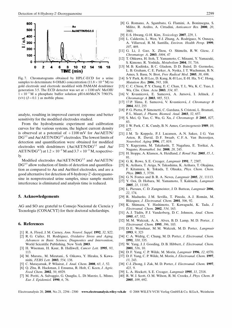
Au/AET/N/DG3.5 electrode for a sample of diluted urine(1 :3) enriched with 11.8� 10�6 M of 8-OHdG. A peak canbeobserved at an elution timeof 6.9 min for both electrodes,but for the Au/AET/N/DG3.5 modified electrode the signalrelated to 8-OHdG exhibits higher current and the chro-matogram shows decreased matrix interference. A gradientis used in the chromatographic separation, reducing analysistime. The results indicate that the dendritic coatings preventpassivation of the electrode, and eliminate the need forsample pretreatment, thus allowing the analysis of trace-level 8-OHdG in a complexmatrix such as urine. Therefore,the most important contribution of this study is related tothe potential application of these modified electrodes as anew family of amperometric sensors for real samples.The amperometric study was undertaken over 20 hour
period. During the period, the electrodes were stable andlater the electrodes became unspecific for the analyte.System with more stable dendritic materials will be
studied in the future by this group of investigator.
4. Conclusions
The experiments carried out clearly show electrostaticdiscrimination by monolayers or multilayer towards the
Fig. 6. Plot of the J vs. the concentration of 8-OHdG. Theelectrochemical data were obtained using various electrodes: a)Au, at þ900 mV; b) Au/AET/N/DG3.5 and c) Au/AET/N/DG4.5 atþ1100 mV; d) Au/AMP/C/DG4 and e) Au/AMP/C/DG5 atþ1000 mV.
Table 1. Electroanalytical data obtained from studied experiments for 8-OHdG at HPLC-ECD.
Sistema Emax
(mV) [a]Jmax
(mA cm�2) [a]Sensitivity(A M�1 cm�2)(P< 0.05) [b]
R [b] DL(1� 10�9 M) [b]
QL(1� 10�9 M) [b]
Au 900 3.9� 103 7709� 200 0.999 2.5 82.5Au/AET/N 1100 3.4� 103 4588� 50 0.999 71.5 217.0Au/MPA/C 1000 5.0� 103 5196� 60 0.999 14.5 44.0Au/AET/N/DG3.5 1100 10.3� 103 17650� 10 0.999 1.2 3.7Au/AET/N/DG4.5 1100 11.0� 103 17638� 14 0.999 1.4 4.0Au/MPA/C/DG4 1000 3.0� 103 6727� 143 0.997 7.5 22.6Au/MPA/C/DG5 1000 4.0� 103 5236� 278 0.993 8.0 23.0
[a] from Figure 1A, 2, 4;[b] calculated from data of Figure 1B, 3, 5.
2298 A. Gutierrez et al.
Electroanalysis 20, 2008, No. 21, 2294 – 2300 www.electroanalysis.wiley-vch.de E 2008 WILEY-VCH Verlag GmbH&Co. KGaA, Weinheim
analyte, resulting in improved current response and bettersensitivity for the modified electrodes studied.From the hydrodynamic experiment and calibration
curves for the various systems, the highest current densityis observed at a potential of þ1100 mV for Au/AET/N/DG3.5 andAu/AET/N/DG4.5 electrodes. The lowest limits ofdetection and quantification were obtained for modifiedelectrodes with dendrimers (Au/AET/N/DG3.5 and Au/AET/N/DG4.5) at 1.2� 10�9 M and 3.7� 10�9 M, respective-ly.Modified electrodes Au/AET/N/DG3.5 and Au/AET/N/
DG4.5 allow reduction of limits of detection and quantifica-tion as compared to Au and Au/thiol electrodes, and are agood alternative for detection of 8-hydroxy-2’-deoxyguano-sine in nonpretreated urine samples, since sample matrixinterference is eliminated and analysis time is reduced.
5. Acknowledgements
AG and SO are grateful to Consejo Nacional de Ciencia yTecnolog�a (CONACYT) for their doctoral scholarships.
6. References
[1] R. A. Floyd, J. M. Carney, Ann. Neurol. Suppl. 1992, 32, S22.[2] R. G. Cutler, H. Rodriguez, Oxidative Stress and Aging.
Advances in Basic Science, Diagnostics and Intervention,World Scientific Publishing, New York 2003.
[3] H. Wiseman, H. Kaur, B. Halliwell, Cancer Lett. 1995, 93,113.
[4] M. Murata., M. Mizutani., S. Oikawa, Y. Hiraku, S. Kawa-nishi, FEBS Lett. 2003, 554, 138.
[5] C. Matayatsuk, P. Wilairat, J. Anal. Chem. 2008, 63, 1, 52.[6] Q. Zhu, R. Hackman, J. Ensunsa, R. Holt, C. Keen, J. Agric.
Food Chem. 2002, 50, 6929.[7] M. Periti, A. Salvaggio, G. Quaglia, L. Di Marzio, L. Misno,
Eur. J. Epidemiol. 1990, 6, 76.
[8] G. Romano, A. Sgambato, G. Flamini, A. Boninsegna, S.Milito, R. Ardito, A. Cittadini, Anticancer Res. 2000, 20,3801.
[9] E-S. Hwang, G-H. Kim, Toxicology 2007, 229, 1.[10] L. Calderon, L. Wen, Y-J. Zhang, A. Rodr�guez, N. Osnaya,
A. Villarreal, R. M. Santilla, Environ. Health Persp. 1999,107, 469.
[11] G. Li, J. Gao, X. Zhou, O. Shimelis, R. W. Giese, J.Chromatogr. A. 2003, 1004, 47
[12] T. Ohkuwa, H. Itoh, T. Yamamoto, C. Minami, Y. Yamazaki,S. Kimono, R. Yoshida, Metabolism 2004, 53, 716.
[13] M. B. Kadiiska, B. C. Gladem, D. D. Baird, D. Germolec,L. B. Graham, C. E. Parker, A. Nyska, J. T. Wachsman, B. N.Ames, S. Basu, N. Brot, Free Radical Med. 2005, 38, 698.
[14] S-Y Park, K-H Lee, D. Kang, K-H Lee, E-H. Ha, Y-C. Hong,Mutation Res. 2006, 593, 108.
[15] C. C. Chiou, P. Y. Chang, E. C. Chan, T. L. Wu, K. C. Tsao, J.Wu, Clin. Chim. Acta 2003, 334, 87.
[16] V. Kvasnicova, E. Samcova, A. Jursova, I. Jel�nek, J.Chromatogr A 2003, 985, 513.
[17] 17.P. Tuma, E. Samcova, V. Kvasnicova, J. Chromatogr B2004, 813, 255
[18] P. G. Pietta, P. Simonetti, C. Gardana, S. Cristoni, L. Bramati,P. L. Mauri, J. Pharm. Biomed. Anal. 2003, 32, 657.
[19] S. Mei, Q. Yao, C. Wu, G. Xu, J. Chromatogr. B 2005, 827,83.
[20] J. W. Park, C. K. Cundy, B. N. Ames, Carcinogenesis 1989, 10,827.
[21] J. M. Te Koppele, P. J. Lucassen, A. N. Sakee, J. G. VanAsten, R. David, D. F. Swaab, C. F. A. Van Bezooijen,Neurobiol. Aging 1996, 17, 819.
[22] Y. Kageyama, M. Takahashi, T. Nagafusa, E. Torikai, A.Nagano, Reumathol. Int. 2008, 28, 245.
[23] H. Stoppe, A. Klassen, A. Heidland, J. Renal Nut. 2005, 15, 1,81.
[24] G. K. Rowe, S. E. Creager, Langmuir 1991, 7, 2307.[25] K. Arihara, T. Ariga, N. Takashima, K. Arihara, T. Okajima,
F. Kitamura, K. Tokuda, T. Ohsaka, Phys. Chem. Chem.Phys. 2003, 5, 3758.
[26] G. N. Fontes and B. R. A. Neves, Langmuir 2005, 21, 11113.[27] Y. Ooi, D. Hobara, M. Yamamoto, T. Kakiuchi, Langmuir
2005, 21, 11185.[28] L. Picraux, C. D. Zangmeister, J. D. Batteas, Langmuir 2006,
22, 174.[29] R. Madueno, J. M. Sevilla, T. Pineda, A. J. Roman, M.
Blazquez, J. Electroanal. Chem. 2001, 506, 92.[30] K. Shimazu, Y. Hashimoto, T. Kawaguchi, K. Tada, J.
Electroanal. Chem. 2002, 534, 163.[31] A. J. TTdçs, P. J. Vanderberg, D. C. Johnson, Anal. Chem.
1995, 67, 552.[32] M. M. Walczak, A. A. Alves, B. D. Lamp, M. D. Porter, J.
Electroanal. Chem. 1995, 396, 103.[33] D. E. Weisshaar, M. M. Walczak, M. D. Porter, Langmuir
1993, 9, 323[34] C. A. Widrig, C. Chung, M. D. Porter, J. Electroanal. Chem.
1991, 310, 335.[35] W. Yang, J. J. Gooding, D. B. Hibbert, J. Electroanal. Chem.
2001, 516, 10.[36] D. F. Yang, C. P. Wilde, M. Morin, Langmuir 1996, 12, 6570.[37] D. F. Yang, C. P. Wilde, M. Morin, J. Electroanal. Chem. 1997,
13, 243.[38] C-J. Zhong, J. Zak, M. D. Porter, J. Electroanal. Chem. 1997,
421, 9.[39] L. A. Hockett, S. E. Creager, Langmuir 1995, 11, 2318.[40] R. W. J. Scott, O. M. Wilson, R. M. Crooks, J. Phys. Chem. B
2005, 109, 692.
Fig. 7. Chromatograms obtained by HPLC-ECD for a urinesamples to determinate 8-OHdG concentration (11.8� 10�6 M) togold electrode and electrode modified with PAMAM dendrimergeneration 3.5. The ECD detector was set at þ1100 mV. MeOH/1� 10�3 M a phosphate buffer solution pH 6.60/MeCN 7/90/3%(v/v) (I¼ 0.1 ) as mobile phase.
2299Detection of 8-Hydroxy-2’-Deoxyguanosine
Electroanalysis 20, 2008, No. 21, 2294 – 2300 www.electroanalysis.wiley-vch.de E 2008 WILEY-VCH Verlag GmbH&Co. KGaA, Weinheim

[41] L. Crespo, G. Sanclimens, M. Pons, E. Giralt, M. Royo, F.Albericio, Chem. Rev. 2005, 105, 1663.
[42] J. Ledesma-Garc�a J. Manr�quez, S. Gutierrez-Granados,L. A. God�nez, Electroanalysis 2003, 15, 659.
[43] G. R. Newkome, C. N. Moorefield, F. Vçgtle, DendriticMolecules: Concepts, Syntheses and Perspectives, VCH,Weinheim, Germany 1996.
[44] E. Bustos, J. Manr�quez, L. Echegoyen, L. A. God�nez,Chem. Commun. 2005, 1613.
[45] E. B. Bustos, Ma. G. G. Jimenez, B. R. Diaz-Sanchez, E.Juaristi, T. W. Chapman, L. A. God�nez, Talanta 2007, 72,1586
[46] A. M. O. Brett, J. A. P. Piedade, S. H. P. Serrano, Electro-analysis 2000, 12, 969.
[47] V. C. Diculescu, A. M. C. Paquim, A. M. O. Brett, Sensors2005, 5, 377.
[48] K. Vuorensola, H. Siren, J. Chromatogr. A 2000, 895, 317.
2300 A. Gutierrez et al.
Electroanalysis 20, 2008, No. 21, 2294 – 2300 www.electroanalysis.wiley-vch.de E 2008 WILEY-VCH Verlag GmbH&Co. KGaA, Weinheim