AMP-deaminase in elasmobranch fish: A comparative...
30
Please note that this is an author-produced PDF of an article accepted for publication following peer review. The definitive publisher-authenticated version is available on the publisher Web site 1 Comparative Biochemistry and Physiology Part B: Biochemistry and Molecular Biology August 2005; 141(4) : 472-479 http://dx.doi.org/10.1016/j.cbpc.2005.05.009 © 2005 Elsevier Inc. All rights reserved. Archimer http://www.ifremer.fr/docelec/ Archive Institutionnelle de l’Ifremer AMP-deaminase in elasmobranch fish: A comparative histochemical and enzymatic study Marie T. Thébault a,* , Lahoucine Izem b , Jean Paul Leroy c , Eric Gobin c , Gregory Charrier a and Jean Paul Raffin d a LEMAR, UMR CNRS 6539, Université de Bretagne Occidentale, Institut Universitaire Européen de la Mer, Place Copernic, Technopole Brest-Iroise, 29 280, Plouzané, France b Station de Biologie Marine du Museum National d'Histoire Naturelle et du Collège de France, Concarneau, France c Laboratoire d'Anatomie-pathologie, CHU Morvan, 29609, Brest-Cedex, France d Laboratoire de Microbiologie des Milieux Extrêmes, UMR 6197, IFREMER, B.P. 70, 29280 Plouzané Cedex, France *: [email protected] Tel.: +33 2 98 49 86 12; fax: +33 2 98 49 86 45. Abstract: AMP-deaminase activity was measured in white muscle from a wide range of fish, including one cyclostome, 13 chondrosteans, and one teleost to elucidate the pattern of the AMP-deaminase activity in white muscle of fish. Compared to a mammalian (rat) muscle extract, low enzyme activities are found in the cyclostome and two elasmobranchs from two families (Scyliorhinidae, Hexanchidae). In contrast, higher AMP-deaminase activities, similar to mammals, are expressed in Squalidae, all families of skates, Chimaeridae and in the teleostean fish. We then compared AMP-deaminase activities in red and white muscles from two representative elasmobranch fish, the dogfish (Scyliorhinus canicula) and the thornback ray (Raja clavata). The fibre type composition and distribution of the locomotory musculature were determined in these two elasmobranchs to establish a relationship between the morphology, the type of fibres of the locomotion-implicated muscles and the AMP-deaminase activity. Experimental data are discussed with respect to the layout of fibres in the myotome. In both species, three fibre types were identified. In the two fish myotomes, most of the axial muscles are white fibres while red fibres constitute a thin sheet. Some differences were observed between the two species in the distribution of intermediate fibres: in dogfish, these are located between the red and white fibres; in thornback ray, some are dispersed within the white fibre region, while others form an intermediary layer like in dogfish. These results suggest that in the course of evolution, an amplification of the AMP-deaminase activity in muscle was coupled with increase of complexity of the muscular structure. Keywords: AMP-deaminase; Elasmobranch; Evolution; Fish; Histology; Muscle; Red fibres; White fibres
Transcript of AMP-deaminase in elasmobranch fish: A comparative...
Ple
ase
note
that
this
is a
n au
thor
-pro
duce
d P
DF
of a
n ar
ticle
acc
epte
d fo
r pub
licat
ion
follo
win
g pe
er re
view
. The
def
initi
ve p
ublis
her-a
uthe
ntic
ated
ver
sion
is a
vaila
ble
on th
e pu
blis
her W
eb s
ite
1
Comparative Biochemistry and Physiology Part B: Biochemistry and Molecular Biology August 2005; 141(4) : 472-479 http://dx.doi.org/10.1016/j.cbpc.2005.05.009 © 2005 Elsevier Inc. All rights reserved.
Archimer http://www.ifremer.fr/docelec/Archive Institutionnelle de l’Ifremer
AMP-deaminase in elasmobranch fish: A comparative histochemical and
enzymatic study
Marie T. Thébaulta,*, Lahoucine Izemb, Jean Paul Leroyc, Eric Gobinc,
Gregory Charriera and Jean Paul Raffind
a LEMAR, UMR CNRS 6539, Université de Bretagne Occidentale, Institut Universitaire Européen de la Mer, Place Copernic, Technopole Brest-Iroise, 29 280, Plouzané, France b Station de Biologie Marine du Museum National d'Histoire Naturelle et du Collège de France, Concarneau, France c Laboratoire d'Anatomie-pathologie, CHU Morvan, 29609, Brest-Cedex, France d Laboratoire de Microbiologie des Milieux Extrêmes, UMR 6197, IFREMER, B.P. 70, 29280 Plouzané Cedex, France *: [email protected] Tel.: +33 2 98 49 86 12; fax: +33 2 98 49 86 45.
Abstract: AMP-deaminase activity was measured in white muscle from a wide range of fish, including one cyclostome, 13 chondrosteans, and one teleost to elucidate the pattern of the AMP-deaminase activity in white muscle of fish. Compared to a mammalian (rat) muscle extract, low enzyme activities are found in the cyclostome and two elasmobranchs from two families (Scyliorhinidae, Hexanchidae). In contrast, higher AMP-deaminase activities, similar to mammals, are expressed in Squalidae, all families of skates, Chimaeridae and in the teleostean fish. We then compared AMP-deaminase activities in red and white muscles from two representative elasmobranch fish, the dogfish (Scyliorhinus canicula) and the thornback ray (Raja clavata). The fibre type composition and distribution of the locomotory musculature were determined in these two elasmobranchs to establish a relationship between the morphology, the type of fibres of the locomotion-implicated muscles and the AMP-deaminase activity. Experimental data are discussed with respect to the layout of fibres in the myotome. In both species, three fibre types were identified. In the two fish myotomes, most of the axial muscles are white fibres while red fibres constitute a thin sheet. Some differences were observed between the two species in the distribution of intermediate fibres: in dogfish, these are located between the red and white fibres; in thornback ray, some are dispersed within the white fibre region, while others form an intermediary layer like in dogfish. These results suggest that in the course of evolution, an amplification of the AMP-deaminase activity in muscle was coupled with increase of complexity of the muscular structure.
Keywords: AMP-deaminase; Elasmobranch; Evolution; Fish; Histology; Muscle; Red fibres; White fibres
Revie
w C
opy
3
INTRODUCTION
The locomotory musculature of mammals, amphibians and birds contains a
complex distribution of several fibre types classified into three types (I, IIA and IIB),
which differ in mean size and in staining properties (Burke et al. 1971, Lannergreen and
Hoh 1984, Rowlerson and Spurway 1985, Chayen et al. 1987). On the basis of some
enzyme activities, type I fibers, also called slow twitch fibres, have a slow contraction
velocity, are very resistant to fatigue and have a high capacity to generate ATP by
oxidative metabolic processes. Type IIA fibres, also called fast oxidative fibres, have a fast
contraction velocity and a very high capacity for generating ATP mainly by oxidative
metabolic processes, and are resistant to fatigue. Type IIB fibres, also called fast glycolytic
fibres, contain relatively few mitochondria, fatigue easily, and generate ATP mainly by
anaerobic metabolic processes. Fish differ considerably in the organization of muscular
system and metabolic processes between the three types. Indeed, the body musculature is
myomeric, and different fibre types, rather than being intermixed as in mammals, occupy
distinct areas (Johnston 1981). The red ones (slow fibres), in more taxonomically
primitive groups of fish, form a thin superficial layer under the skin, whereas the white
ones (fast fibres) are involved in the constitution of the trunk muscles (Bone 1966).
Intermediate fibres, displaying different diameters and histochemical properties, form an
intermediary layer.
In mammals, there are multiple tissue-specific AMP-deaminase isoenzymes which
can be distinguished by immunological, kinetic and chromatographic criteria (Ogasawara
et al. 1972, 1975, 1978) and the concentration of the enzyme found in skeletal muscle is
3 of 31
Friday , May 13, 2005
Elsevier

Revie
w C
opy
4
considerably higher than in other tissues (Lowenstein 1972). AMP-deaminase (EC 3.5.4.6)
concentration is high in the white fibres (Purzycka 1962). Red muscle also showed high
AMP-deaminase activity, but it was about a half of the activity observed in white muscle
(Raggi et al. 1969, Fishbein et al. 1979, Ogasawara et al. 1983). Like higher vertebrates,
the teleost fish display high AMP-deaminase activities in their white fibres (Kaletha et al.
1991, Raffin et al. 1993). In contrast, a previous report described a low AMP-deaminase
activity in white fibres of the dogfish Scyliorhinus canicula (Raffin and Leray 1980).
AMP-deaminase activity in red muscles was earlier studied in carp (Van Waarde and
Kesbeke, 1981). To our knowledge, the activity of the enzyme in elasmobranch fish red
muscle has not been investigated up to now.
Amplification of the AMP-deaminase activity in the white muscle of vertebrates,
and especially its significance, has been widely debated. Red and white muscles are known
to function in steady-state and burst movements, respectively, and to be different in their
metabolic patterns. White muscle is, indeed, known to have a limited oxidative capacity,
and inside its ATP generation is highly dependent upon anaerobic metabolism. So, its high
content in AMP-deaminase is probably related to the stabilizing function of this enzyme in
the regulation of adenine nucleotides metabolism during intense energy demand; it would
be done through the stabilization of the adenylate energy charge, the ATP-to-ADP ratio
and phosphorylation potential (Lowenstein 1972, Chapman and Atkinson 1973, Veech et
al. 1979, Meyer and Terjung 1980, Van Waarde 1988).
The present study was aimed at (1) precising the pattern of AMP-deaminase
activity in white muscle of fish, by measuring the activity of enzyme in a wide range of
fish, including one cyclostom, 13 chondrosteans, and one teleost, (2) establishing a
4 of 31
Friday , May 13, 2005
Elsevier

Revie
w C
opy
5
relationship between the morphology, the types of fibres of the locomotion-implicated
muscles, and the AMP-deaminase activity, by studying the histological picture of the
white muscle on two representative species of benthic elasmobranch, the dogfish
Scyliorhinus canicula and the rayfish Raja clavata. Results are discussed in relation with
the AMP-deaminase activity displayed by the white fibres as well as the way they are laid
out in the myotome.
5 of 31
Friday , May 13, 2005
Elsevier

Revie
w C
opy
6
MATERIALS AND METHODS
Animals
Cyclostoms were obtained from a local fishery in Gironde (France). Rainbow trout
were obtained from a local hatchery near Concarneau (France). Rats (Wistar) were
purchased from the Centre d’Elevage Janvier (Le Genest St-Isle, France). Chondrostean
fish were caught by trawling in the gulf of Gascogne during 1 month IFREMER campaign
(EVOE 882), or obtained from from local sellers (Brest, France). Muscle samples were
taken immediately, frozen in liquid nitrogen, and kept at –80°C until AMP-deaminase
assay.
Specimens of dogfish (Scyliorhinus canicula) and rayfish (Raja clavata) weighing
around 0.5 and 1.3 kg, respectively, were caught in the Bay of Concarneau and kept in
running-seawater tanks at 15°C for at least 7 days prior to experiments. Once a week, they
were on a diet of squid and fish flesh. Our investigations were conducted only on fish
obviously in good condition.
Histology
The metabolic characteristics of muscle fibers can be displayed through the use of
histochemical staining techniques (Dubowitz et al., 1985).
The fish were anesthetized with tricaine methane sulfate (MS 222), killed by
decapitation and descaled. Tissue blocks, 3-4-cm in length, containing red, intermediate
and white muscles were dissected on a 4-cm layer from the median part of the trunk
(squales) and the fin (skates). The samples were oriented so that the muscle fibres were cut
6 of 31
Friday , May 13, 2005
Elsevier
Revie
w C
opy
7
at right angles to their long axis.
For study of fibres-direction and -type, the pieces oriented longitudinally or
transversally were fixed in Bouin’s fluid for 24-48 h at 4°C, then dehydrated through
ascending ethanol series and embedded in paraffin wax. Sections were then colored with
hematein-eosin.
For histochemical study, the tissue was rapidly frozen in isopentane cooled to its
freezing point with liquid nitrogen (-160°C ), and then sectioned in a cryostat at -25°C.
Longitudinal and transversal sections (5 µm) were mounted on glass slides, air-dried at
room temperature and stained for glycogen with the periodic-acid schiff reagent (PAS)
(Pearse, 1968), and for lipids using Sudan Black (Chiffelle and Putt, 1951).
Ten-µm-thick transverse sections were cut and mounted on glass slides and then
stained to evidence the following enzymes:
- NADH Tetrazolium Reductase (NADH-TR, EC 1.6.99.3): this enzyme is used for
evaluation of the oxidative capacity of fibre types (Nachlas et al., 1958). The incubation
medium contained 10 ml 0.2 M Tris/HCl buffer (pH 7.4), 10 ml of 2.4 mM nitro-blue
tetrazolium and 0.27 mM NADH. Incubation at 37°C took 20-30 min.
- succinic dehydrogenase (SDH, EC 1.3.99.1): this mitochondrial enzyme is used
to separate oxidative from anaerobic fibres (Nachlas et al., 1957). The incubation medium
was made of 10 ml of 0.2 M potassium phosphate buffer (pH 7.4), 10 ml of 2.4 mM nitro-
blue tetrazolium, and 100 mM Na-succinate. The incubation lasted 2 h at 37°C.
- Menadione-linked α-glycerophosphate deshydrogenase (Mα-GPDH, EC
1.1.99.5): used as a measure of aerobic metabolic capacities (Wattenberg and Leong,
1960). This enzyme is an intramitochondrial flavoprotein. The enzyme activity can be
7 of 31
Friday , May 13, 2005
Elsevier

Revie
w C
opy
8
increased by vitamin K3 (menadione), which acts as a hydrogen acceptor. The incubation
medium consisted of 10 ml 0.2 M Tris/HCl buffer (pH 7.4), 10 ml of 2.4 mM nitro-blue
tetrazolium, α-glycerophosphate (3 mg.ml-1), and 0.01% menadione. Samples were
incubated at 37°C for 20-30 min.
- Phosphorylase (EC 2.4.1.1): The incubation medium consisted of 5 ml of 0.2 M
Na acetate buffer (pH 5.6-6.0), 2 ml of absolute ethanol, 5 ml of bidistilled water with
25 mg of glucose-1-phosphate, 1.2 M adenosine-5'-monophosphate (AMP) and 5 mg of
glycogen. Samples were incubated for 1 h at 37°C and then stained with Lugol (10%)
(Swanson, 1948).
- AMP-deaminase: as described in Fishbein et al. (1980): The incubation medium
contained 0.7 ml 3 M KCl, 1.2 M AMP, 2.4 mM nitro-blue tetrazolium, and 0.1 mM
dithiothreitol, at pH 6.1. The incubation lasted for 1 h at 20°C. Positive controls for
staining consisted of tissue sections from human skeletal muscle. Substrate-free controls
were also prepared for all the reactions, except for the PAS stain where the oxidation by
periodic acid was omitted. In each case, negative result was obtained, and thus confirmed
the reaction specificity.
Assay of AMP-deaminase activity
Pieces from axial muscle and from fin muscle were rapidly dissected from squales
and skates, respectively.
A 10% (w/v) suspension was made in 0.089 M potassium phosphate (pH 6.5), 0.18 M
KCl, 0.1 mM dithiothreitol. The tissue was homogeneized in an Ultra-Turrax
homogeneizer for 2 x 20 s, with intermediate cooling in ice. Then the extract was diluted
8 of 31
Friday , May 13, 2005
Elsevier

Revie
w C
opy
9
in the same volume of homogeneization buffer, gently stirred for 1 h at room temperature
and used for determination of enzyme activity without prior centrifugation.
The chromatographic method previously reported by Raffin and Thébault (1991)
was used to determine AMP-deaminase activity through assessment of IMP produced from
AMP. A solution of cacodylate buffer (50 mM, pH 6.5) was made as previously described
(Raffin et al. 1993b). The AMP concentration was 10 mM and the incubation temperature
was 26°C for all the fish and 37°C for the rat. One unit of AMP-deaminase activity
corresponds to the amount of enzyme that deaminates 1 µmole of substrate min-1 under
our experimental conditions; activity is expressed as U g-1 wet weight).
9 of 31
Friday , May 13, 2005
Elsevier

Revie
w C
opy
10
RESULTS
AMP-deaminase activity in different fish species
Steady activity difference between fish species could be measured in the white
muscle (Table 1). The level of activity in the cyclostom Petromyzon marinus and the
teleost Oncorhynchus mykiss white muscles was similar to that of the mammals. Among
the chondrosteans, those belonging to the families of Chimaeridae, Rajidae, Squalidae,
Torpinidae, and Triakidae displayed an AMP-deaminase activity similar to that of the
mammals in their white muscles, whereas the activity was significantly lower in
Hexanchidae and Scyliorhinidae.
In order tofind out if the AMP-deaminase followed the same pattern in red muscle,
the activity of the enzyme from red muscle was compared to that of white muscle, in two
representative elasmobranch species, Raja clavata and Scyliorhinus canicula (Table 2). In
the dogfish, the AMP-deaminase activity was sixfold lower in the red muscle than in the
white muscle. Also, in the rayfish, the activity tended to be lower in the red muscle, but the
difference was not significant.
Histological studies and AMP-deaminase assays in rayfish and dogfish
Histological studies and enzyme assays were performed in two elasmobranch
species, Raja clavata and Scyliorhinus canicula.
Muscle fibres orientation and type
In S. canicula, the fibres-containing compartments are about 2.5-mm-long and
10 of 31
Friday , May 13, 2005
Elsevier

Revie
w C
opy
11
parallelly aligned from head to tail. The muscle fibres are longitudinally directed. On the
contrary in R. clavata, the muscle fibers are at right angle to the vertebrate chord and
radiate towards the fin. In transversal sections, size and histo-enzymology reactions
allowed us to classify the muscle fibres into three groups, white (W), red (R) and
intermediate (I).
Figure 1 clearly shows the three types of fibres identified in the dogfish lateral
muscle as well as portions of the myotomal musculature at the anterior end of the body.
Just underneath the skin, a 1-mm-thin cortical zone of about 15 layers contains the
NADH-TR-positive red fibres running almost parallel to the long axis of the body; these
small-size fibres (67 µm in diameter) contain a dark colored cytoplasm and peripheral
nuclei.
Near the vertebral axis, a larger zone containing almost all the cones (95%) is
composed of large NADH-TR-negative fibres (155 µm in diameter) tilted towards the long
axis; some partially internalized nuclei are visible. Their cytoplasm was weakly stained.
Adjacent to the white muscle, a thin zone of fibres (117-µm in diameter), with
partially internalized nuclei, was moderately stained after standard and NADH-TR staining
reactions.
Table 3 illustrates the data obtained by histo-enzymology on the localization of
glycogen, lipids, NADH-TR, menadione-linked α-glycerophosphate dehydrogenase and
phosphorylase. None of the fibres, whatever their type, were stained with Sudan Black.
The red and intermediary fibres were fairly stained with PAS, whereas the white ones
displayed a milder coloration. NADH-TR activity was found essentially in the red muscle
fibres. The intermediate fibres were moderately stained, whereas the white ones showed
very mild coloration.
11 of 31
Friday , May 13, 2005
Elsevier

Revie
w C
opy
12
Figure 2 illustrates the characteristics of the rayfish fin muscle. The red-fibre zone
under the skin is thicker than in the dogfish muscle (2 -2.5 mm) and larger underneath the
animal. The mean diameter of the cells is 50 µm. The white-fibre zone accounts for 95%
of the cones with cells of 180-µm in mean diameter. The intermediate fibres consist of
115-µm-mean-diameter cells. Some of them display a mosaic distribution within the white
fibres, while others form an intermediary layer like in dogfish. The histochemical
characteristics of muscle fibres were similar to those of the dogfish (Table 3).
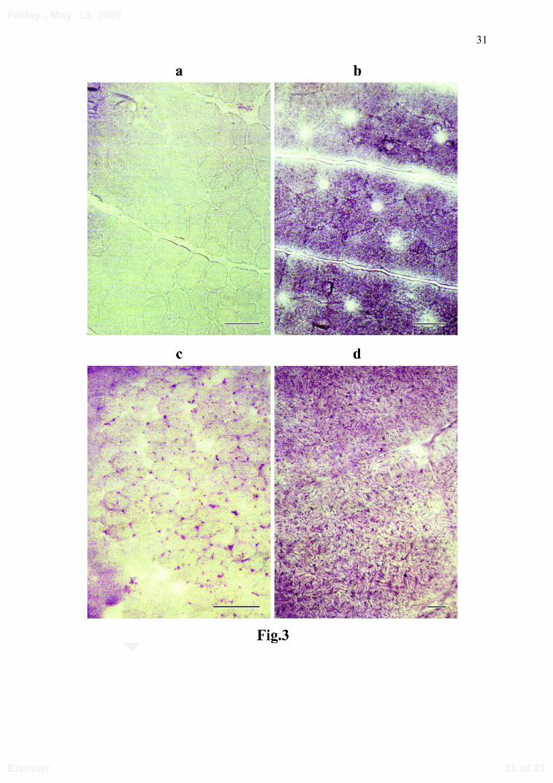
Stain for AMP-deaminase
The AMP-deaminase staining of the dogfish and rayfish myotomes produced
similar results with no staining of the red fibres (Figures 3 a and c). As for the white fibres,
in dogfish they exhibited a blue reticular staining on a diffusely pink-colored cytoplasm.
On the other hand, in rayfish they were heavily stained with a high degree of granularity
(Figures 3 b and d).
12 of 31
Friday , May 13, 2005
Elsevier

Revie
w C
opy
13
DISCUSSION
The concentration of AMP-deaminase found in skeletal muscle of mammals is
considerably higher than in other mammalian tissues (Ogasawara et al. 1983), and than in
invertebrate white muscle (Raffin and Thébault 1987). Our results show that a significant
increase of the AMP-deaminase activity took place in the elasmobranch group. Then, the
enzyme activation was a several-step mechanism which occured during the appearance of
vertebrates. Amplification of AMP-deaminase activity could be related to the extensive
events that occurred, simultaneous to the gene duplication, at the very beginning of
vertebrate evolution (Ohno et al. 1969).
Molecular studies have demonstrated that mammalian AMP-deaminases are made
manifest through the expression of a multigene family. Previous molecular studies have
reported two genes, AMPD1 and AMPD2, that produce isoform M and L transcripts,
respectively (Morisaki et al. 1990). AMPD1 is expressed at high levels in adult skeletal
muscle, while the AMPD2 transcript is detected primarily in adult nonskeletal muscle
tissues and embryos (Sabina et al. 1989, Morisaki et al. 1990). A third AMP-deaminase
gene has been identified in human, AMPD3, encoding transcripts that are specific for the
E isoforms in erythrocytes (Mahnke-Zizelman and Sabina 1992). Detailed studies on
AMP-deaminase in white muscle from Schyliorhinus canicula and Raja clavata showed
that the difference in the enzyme activity found between the two species was correlated
with the appearance, in Rajidae, of an enzymatic form very close to the AMPD1 gene
product of higher vertebrates (Raffin et al. 1993 a). Thus, in this animal group, the AMP-
deaminase activity difference of 3-4 fold could indicate a genetic modification of the
AMP-deaminase molecule.
13 of 31
Friday , May 13, 2005
Elsevier

Revie
w C
opy
14
Very little information has been available on AMP-deaminase isozymes from red
muscle. Our study shows that, in Scyliorhinus canicula, the AMP-deaminase activity was
much lower in red fibres than in white fibres, as in mammals (Ogasawara et al. 1978). In
Raja clavata, a non significant difference between the activities of red and white fibres
could result from the difficulty to separate the different types of fibres in the fins of skates,
as shown by high values of the standard deviation. In mammals, two different isozymes
exist in red muscle; one is identical to the isozyme in heart (isoform L), and the other of
the isozyme in white muscle (isoform M) (Raggi et al. 1979, Ogasawara et al. 1983). To
our knowledge, nothing is known about the isozyme pattern in fish red and white muscles.
Rayfish and dogfish are benthic fish that live at depths from 20 to 400 m. But, both
species differ in the way they swim. The dogfish swimming is based on undulatory side-
movements of the trunk and tail resulting from contractions of axial muscles. Active
swimming to escape a predator is achieved through vigorous contractions of the axial
muscle fibres. The rayfish moves at low speed by undulatory vertical movements of the
large pectoral fins; its caudal muscles contract noticeably, but only during vigorous bursts
of activity.
Histochemistry has been acknowledged as a valuable tool in investigations into
muscle tissues in lower vertebrates (Johnston et al. 1975, Mellgren and Mathisen 1966).
The fin muscle fibres of the rayfish are differenciated in three distinct types: red,
intermediate and white, like in dogfish and other elasmobranch species (Bone 1966, Bone
and Chubb 1978, Kryvi and Totland 1978, Johnston 1981); the intermediate and white
fibres show great similarities in their respective morphology. However, the way they are
arranged in the rayfish differs from that of the dogfish: the red-fibre zone is thicker,
particularly in the superior part of the fin. Near the vertebrate chord, the muscular mass
14 of 31
Friday , May 13, 2005
Elsevier

Revie
w C
opy
15
consists mainly of white fibres; on the other hand, even though the intermediate fibres are
mostly found between the red and white ones where they form a strip, some of them,
which account for 5% of the total fibres, are dispersed between the white fibres. This
layout is similar to one found in some teleost fish (Johnston et al. 1977, Chayen et al.
1987).
In fish, the recruitment of the different types of fibres with respect to the swimming
speed has been thoroughly investigated, and is known to depend on the motor system
innervation (Johnston 1981). In dogfish, each white fibre is innervated by two separate
axons which fuse to form a single end-plate (Bone 1964, 1972). In such innervation
systems, only the red fibres are implicated in slow swimming, whereas the white ones
become active for fast and vigorous movements (Bone 1966, Johnston 1981). In multiple
innervated systems, several studies have highlighted that, at sustainable swimming speeds
in some species, the white and red fibres are both recruited (Hudson 1973, Davison and
Goldspink 1977, Johnston et al. 1977, Bone et al. 1978, Johnston and Moon 1980)
whereas, in other species, they are recruited in more primitive pattern (Freadman 1979). In
the white fibres of Torpedo, the more superficial ones have a basket-like end plate, like
those of the dogfish (Bone 1964). However, to our knowledge, neither the innervation
system of rayfish fin muscles, nor its fibre recruitment as a function of speed has been
investigated. A peculiarity of the rayfish myotome is the dispersion of intermediate fibres
among the white ones. According to some authors the intermediate fibres would be
associated with the fish ability to swim continuously for a long time (Bokdawala 1967,
Patterson et al. 1975), but this assumption is still questioned.
Though our histo-enzymology-based study failed to evidence differences between
rayfish and dogfish in the activities of the mitochondrial marker enzymes (SDH, NADH-
15 of 31
Friday , May 13, 2005
Elsevier
Revie
w C
opy
16
TR), phosphorylase and Mα-GPD enzymes, a marked difference was observed for AMP-
deaminase in white fibres. These experimental data, together with the AMP-deaminase
assays in the white and red fibres of both species, corroborated the information ona higher
activity in the rayfish fin muscles. As observed in mammals, their white fibres contained
more AMP-deaminase than the red ones. Further, molecular studies will have to be carried
out to determine if this is related to, either a modification of the enzyme structure, or a
higher expression rate of the corresponding gene. Indeed, the high AMP-deaminase
activity in rayfish white fibres could be correlated to the increase in muscle complexity.
Fibre typing in AMP-deaminase staining is more based on difference in color
rather than in intensity. Because of the lack of inherent relation between the color
differentiation and the enzyme reaction, the intensity of the pink color and the degree of
blue granularity may depend on the amount of cytoplasmic enzyme and that of the enzyme
bound to mitochondria and sarcoplasmic reticulum in the intermyofibrillar network,
respectively (Fishbein et al. 1980). In mammals, type-II fibres show a diffuse pink
staining, whereas type-I fibers display a granular blue staining. The slight stain obtained
for the dogfish white fibres may result from their low content in enzyme as confirmed by
the low activity found in solution assays.
Prior studies have demonstrated that, in mammalian muscle, AMPD1 binds to the
S-2 fragment of myosin, and that complex formation makes the reaction less susceptible to
inhibition by GTP (Ashby and Frieden, 1977, 1978; Ashby, Frieden and Bishoff, 1979).
Binding to the myofibril varies with the state of muscle contraction in vivo and is required
for activation of the enzyme in myocytes (Hisatome et al., 1998). As the experiments of
Lushchak and Storey (1994) and Lushchak et al. (1998) on teleost muscle show clearly
that enzyme binding to the particulate fraction of muscle increases in exercising skeletal
16 of 31
Friday , May 13, 2005
Elsevier

Revie
w C
opy
17
muscle and in muscle of fish exposed to hypoxia, it is very likely that the control of AMP-
deaminase by the combined effects of allosteric modifiers and enzyme interactions with
cellular structural elements has a more general character. However, the association of
AMP-deaminase to particular fraction of muscle in elasmobranch fish displaying a low
AMP-deaminase activity has been investigated. So a possible regulation by binding to
cellular structure elements cannot be proposed in these fish.
In conclusion, this study shows that transition from ancestral to evolved AMP-
deaminase forms has occurred in at least two taxa: the one that evolved to rajiform
elasmobranch fish and the other to the land vertebrates. An important point is that the
enzyme activation and appearance of a molecular form similar to the mammal AMP-
deaminase occurred within the phylogenic evolution of the major fish groups. Finally, our
comparative study of two elasmobranches suggests that enhancement of AMP-deaminase
activity goes along with a higher complexity of the muscular structure as evidenced by the
rudimentary mosaïc arrangement between red and white fibres.
ACKNOWLEDGEMENTS
We thank Dr. Joël Querellou for access to HPLC equipement at Ifremer.
17 of 31
Friday , May 13, 2005
Elsevier

Revie
w C
opy
18
REFERENCES
Ashby, B., Frieden, C. 1977. Interaction of AMP-aminohydrolase with myosin and its
subfragments. J. Biol. Chem. 252, 1869-1872.
Ashby, B., Frieden, C., Bishoff, R. 1979. Immunofluorescent and histochemical
localization of AMP-deaminase in skeletal muscle. J. Cell Biology 81, 361-373.
Ashby, B., Frieden, C. 1978. Adenylate deaminase. Kinetic and binding studies on the
rabbit muscle enzyme. J. Biol. Chem. 253, 8728-8735.
Bokdawala, F.D. 1967. A histochemical study of fat in the red and white muscle fibres
of fin skeletal muscle. J. Anim. Morphol. Physiol. 14, 231-241.
Bone, Q. 1964. Patterns of muscular innervation in the lower chordates. Int. Rev.
Neurobiol. 6, 99-147.
Bone, Q. 1966. On the function of the two types of myotomal muscle fibres in
elasmobranch fish. J. Mar. Biol. Ass. UK. 46, 321-349.
Bone, Q. 1972. The dogfish neuromuscular junction: Dual innervation of vertebrate
striated muscle fibres? J. Cell. Sci. 10, 657-665.
Bone, Q., Chubb, A.D. 1978. The histochemical demonstration of myofibrillar ATPase
in elasmobranch muscle. Histochem. 10, 489-494.
Bone, Q., Kicnuik, J., Jones, D.R. 1978. On the role of the different fibre types in fish
myotomes at intermediate swimming speeds. Fish. Bull. USA. 76, 691-699.
Broberg, S., Sahlin, K. 1989. Adenine nucleotide degradation in human skeletal muscle
during prolonged exercise. J. Appl. Physiol. 67, 116-122.
Burke, R.E., Levine, D.N., Zajac, F.E., Tsairis, P., Engel, W.K. 1971. Mammalian
18 of 31
Friday , May 13, 2005
Elsevier
Revie
w C
opy
19
motor units: physiological-histochemical correlation in three types in cat
gastrocnemius. Science 174, 709-712.
Chapman, A.G., Atkinson, D.E. 1973. Stabilization of adenylate energy charge by the
AMP-deaminase reaction. J. Biol. Chem. 248, 8309-8312.
Chayen, N., Freundlich, A., Squire, J.M. 1987. Comparative histochemistry of a flatfish
fin muscle and of other vertebrate muscles used for ultrastructural studies. J
.Muscle Res. Cell. Motil. 4, 358-371.
Chiffelle, T.L., Putt, F.A. 1951. Propylene and ethylene glycol as solvents for Sudan IV
and Sudan Black B. Stain Technol. 26, 51-56.
Davison, W,. Goldspink ,G. 1977. The effect of prolonged exercise on the lateral
musculature of the brown trout (Salmo trutta). J. Exp. Biol. 70, 1-12.
Dubowitz, V., Sewry, C.A., Fitzsimons, R.B. 1985. Histological and histochemical
stains and reactions. In: Saunders, W.B. (Ed.), Muscle biopsy, a practical
approach, 2nd edn, Baillière Tindall, London, pp. 19-40.
Fishbein, W.N., Griffin, J.L., Nagarajan, K. 1979. Immunologic uniqueness of muscle
adenylate deaminase and genetic transmission of the deficiency state. Clin. Res.
27, 274A.
Fishbein, W.N., Griffin, J.L., Armbrustmacher, V.W. 1980. Stain for skeletal muscle
adenylate deaminase. Arch. Pathol. Lab. Med. 104, 462-46.
Freadman, M.A. 1979. Role of partitioning of swimming musculature of striped bass,
Morone saxatilis Walbaum and Bluefish, Pomatomus saltatrix L. J. Fish Biol. 15,
417-423.
Hisatome, I., Morisaki, T., Kamma, H., Sugama, T., Morisaki, H., Ohtahara, A.,
19 of 31
Friday , May 13, 2005
Elsevier

Revie
w C
opy
20
Holmes, E.W. 1998. Control of AMP deaminase 1 binding to myosin heavy
chain. Am. J. Physiol. 275, C870-C881.
Hudson, R.C.L. 1973. On the function of the white muscles in teleosts at intermediate
swimming speeds. J. Exp. Biol. 58, 509-522.
Johnston, I.A., Ward, P.S., Goldspink, G. 1975. Studies on the swimming musculature
of the rainbow trout. I. Fibre types. J. Fish Biol. 7, 451-458.
Johnston, I.A., Davison, W., Goldspink, G. 1977. Energy metabolism of carp
swimming muscle. J. Comp. Physiol. 114 B, 203-216.
Johnston, I.A., Moon, T.W. 1980. Endurance exercise training in the fast and low
muscles of a teleost fish (Pollachius virens). J. Comp. Physiol. 135 B, 147-156.
Johnston, I.A. 1981. Structure and function of fish muscles. Symp. Zool. Soc. Lond. 48,
71-113.
Kaletha, K., Thébault, M.T., Raffin, J.P. 1991. Comparative studies on heart and
skeletal muscle AMP-deaminase from rainbow trout (Salmo gairdneri). Comp.
Biochem. Physiol. 99 B, 751-754.
Krivy, H., Totland, G.K. 1978. Fibre types in locomotory muscles of the cartilagenous
fish Chimaera monstrosa. J. Fish Biol. 12, 257-265.
Lannergreen, J., Hoh, J.F.K. 1984. Myosin isoenzymes in single muscle fibres of
Xenopus laevis: analysis of five different functional types. Proc. Roy. Soc. Lond.
Ser. 222 B, 401-408.
Lowenstein, J.M. 1972. Ammonia production in muscle and other tissues. The purine
nucleotide cycle. Physiol. Rev. 52, 382-414.
Lushchak, V.I., Storey, K.B. 1994. Influence of exercise on the distribution of enzymes
20 of 31
Friday , May 13, 2005
Elsevier

Revie
w C
opy
21
in trout white muscle and kinetic properties of AMP-deaminase from free and
bound fractions. Fish Physiol. Biochem. 13, 407-418.
Lushchak, V.I., Smirnova, Y.D., Storey, K.B. 1998. AMP-deaminase from sea-scorpion
white muscle: properties and redistribution under hypoxia. Comp. Biochem.
Physiol., 119 B, 611-618.
Mahnke-Zizelman, D.K., Sabina, R.L. 1992. Cloning of human AMP-deaminase
isoform EcDNAs. Evidence for a third AMPD gene exhibiting alternatively
spliced 5’-exons. J. Biol. Chem. 267, 20866-20877.
Mellgren, S.I., Mathisen, J.S. 1966. Oxidative enzymes, glycogen and lipid in striated
muscle. A histochemical study in the atlantic hagfish [Myxine glutinosa (L)]. Z.
Zellforsh Mikrosk. Anat. 71, 169-188.
Meyer, R.A., Terjung, R.L. 1980. AMP deamination and IMP reamination in working
skeletal muscle. Am. J. Physiol. 239, C32-C38, 1980.
Morisaki, T., Sabina, R., Holmes, E.W. 1990. Adenylate deaminase. A multigene family in
humans and rats. J. Biol. Chem. 265, 11482-11486.
Nachlas, N.M., Tsou, K.G., DeCheng, C.S., Seligman, A.M. 1957. Cytochemical
demonstration of succinic dehydrogenase by the use of a new p-nitrophenyl
substituted ditetrazole. J. Histochem. Cytochem. 5, 420-436.
Nachlas, N.M., Walker, D.G., Seligman, A.M. 1958. A histochemical method for the
demonstration of diphosphopyridine nucleotide diaphorase. J. Biophys. Biochem.
Cytol. 4, 29-38.
Ogasawara, N., Yoshino, M., Kawamura, Y. 1972. Multiple forms of AMP-deaminase
in rat brain. Biochim. Biophys. Acta 258, 680-684.
21 of 31
Friday , May 13, 2005
Elsevier

Revie
w C
opy
22
Ogasawara, N., Goto, H., Watanabe, T. 1975. Isozymes of rat AMP-deaminase.
Biochim. Biophys. Acta 403, 530-537.
Ogasawara, N., Goto, H., Yamada, Y., Watanabe, T. 1978. Distribution of AMP-
deaminase isozymes in rat tissues. Eur. J. Biochem. 87, 297-304.
Ogasawara, N., Goto, H., Yamada, Y. 1983. AMP-deaminase isozymes in rabbit red
and white muscles and heart. Comp. Biochem. Physiol. 76 B, 471-473.
Ohno, S., Muramoto, J., Stenius, C., Christian, L., Kittrell, W.A., Atkin, N.B. 1969.
Microchromosomes in Holocephalian, Chondrostean and Holostean fishes.
Chromosoma (Berl.) 26, 35-40.
Patterson, S., Johnston, I.A., Goldspink, G. 1975. A histochemical study of the lateral
muscles of five teleosts species. J. Fish Biol. 7, 159-166.
Pearse, A.G.E. 1968. Histochemistry. Theoretical and Applied, 3rd edn Vol.1.. J.& A.
Churchill Ltd., London, 759 pp.
Purzycka, J. 1962. AMP and adenosine aminohydrolases in rat tissues. Acta Biochim.
Pol. 9, 83-93.
Raffin, J.P., Leray, C. 1980. Comparative study on AMP-deaminase in gill, muscle and
blood of fish. Comp. Biochem. Physiol. 67 B, 533-540.
Raffin, J.P., Thébault, M.T. 1987. Purification and partial characterization of an AMP-
deaminase from the marine invertebrate Palaemon serratus. Comp. Biochem.
Physiol. 88 B, 1071-1076.
Raffin, J.P., Thébault, M.T. 1991. A specific AMP-deaminase assay and its application
to tissues homogenates. Comp. Biochem. Physiol. 99 B, 125-127.
Raffin, J.P., Thébault, M.T., Izem, L. 1993a. The genetic amplification of muscular
22 of 31
Friday , May 13, 2005
Elsevier
Revie
w C
opy
23
AMP-deaminase: Towards the identification of evolutionary steps. Trends
Comp. Biochem. Physiol. 1 B, 171-181.
Raffin, J.P., Izem, L., Thébault, M.T. 1993b. Amplification of myoadenylate deaminase
during evolution. 2. Purification and properties of the enzyme from two
elasmobranch fishes, Scyliorhinus canicula and Raja clavata. Comp. Biochem.
Physiol. 106 B, 999-1007.
Raggi, A., Ronca-Testoni, S., Ronca, G. 1969. Muscle AMP aminohydrolase: II.
Distribution of AMP aminohydrolase, myokinase and creatine kinase activities in
skeletal muscle. Biochim. Biophys. Acta 178, 619-622.
Raggi, A., Bergamini, C., Ronca, G. 1979. Isozymes of AMP-deaminase in red and
white muscle. FEBS Lett. 56, 19-23.
Rowlerson, A., Spurway, Y.N. 1985. How many fibre types in amphibian limb
muscles? A comparison of Rana and Xenopus. J. Physiol. 358, 78P.
Sabina, R., Ogasawara, N., Holmes, E.W. 1989. Expression of three stage-specific
transcripts of AMP-deaminase during myogenesis. Mol. Cell. Biol. 9, 2244-
2246.
Swanson, M.A. 1948. Studies on the structure of polysaccharides. IV. Relation of the
iodine colour to the structure. J. Biol. Chem. 172, 825-837.
Van Waarde, A., Kesbeke, F. 1981. Regulatory properties of AMP-deaminase from
lateral red muscle and dorsal white muscle of goldfish, Carassius auratus (L).
Comp. Biochem. Physiol. 69B, 413-423.
Van Waarde, A. 1988. Operation of the purine nucleotide cycle in animal tissues. Biol
.Rev. 63, 259-298.
23 of 31
Friday , May 13, 2005
Elsevier

Revie
w C
opy
24
Veech, R.L., Lawson, J.W.R., Cornell, N.W., Krebs, H.A. 1979. Cytosolic
phosphorylation potential. .J Biol. Chem. 254, 6358-6547.
Wattenberg, L.W., Leong, J.L. 1960. Effects of coenzyme Q10 and menadione on succinic
dehydrogenase activity as measured by tetrazolium salt reduction. J. Histochem.
Cytochem. 8, 296-303.
24 of 31
Friday , May 13, 2005
Elsevier
Revie
w C
opy
25
CAPTIONS TO FIGURES
Fig. 1: Transverse section of axial muscle of the dogfish. a: hematein-eosin stain ; b: NADH-TR stain.
White fibres (W) ; Red fibres (R) ; Intermediate fibres (P)
Bar represents 100 µm.
Fig. 2: Transverse sections of axial muscle of the rayfish. a: hematein-eosin stain ; b: NADH-TR stain.
White fibres (W) ; Red fibres (R) ; Intermediate fibres (P).
Bar represents 100 µm.
Fig. 3: AMP-deaminase histochemistry.
AMP-deaminase activity was assayed in transverse sections of dogfish and rayfish axial muscles.
a: dogfish red fibres ; b: dogfish white fibres ; c: rayfish red fibres ; d: rayfish white fibres.
Bar represents 100 µm.
25 of 31
Friday , May 13, 2005
Elsevier

Review Copy26
Table 1. Determination of AMP-deaminase activity in white muscle of different fish species, compared with mammals
Species Type of muscle Activity (U/g FW)
Cyclostoms Petromyzon marinus (6)
Chimaeridae Chimaera monstrosa (4)
Hexanchidae Hexanchus griseus (1)
Scyliorhinidae Scyliorhinus canicula (6)
Galeus melastomus (6)
Triakidae Galeorhinus galeus (5)
Squalidae Etmopterus spinax (6)
Squalus acanthias (6)
Torpenidae Torpedo marmorata (5)
Rajidae Raja circularis (6)
Raja clavata (6)
Raja fullonica (4)
Raja montagui (6)
Raja naevus (6)
Salmonidae Oncorhynchus mykiss (5)
Mammal Rat (Wistar) (6)
White muscle (median part of the trunk)
White muscle (median part of the trunk)
White muscle (median part of the trunk)
White muscle (median part of the trunk)
White muscle (median part of the trunk)
White muscle (median part of the trunk)
White muscle (fin)
White muscle (fin)
White muscle (fin)
White muscle (fin)
White muscle (fin)
White muscle (fin)
White muscle (fin)
White muscle (fin)
White muscle (median part of the trunk)
Fast lateral gastrocnemius
44.1 ± 14.0
202 ± 162
40.6
19.3 ± 3.24
72.6 ± 29.9
116 ± 58.9
108 ± 31.2
177 ± 69.8
163 ± 32.7
259 ±134
247 ± 103
288 ± 54.9
323 ± 88.6
173 ± 78.6
139 ± 10.4
153 ± 46.6
n: number of specimen
AMP-deaminase activity is expressed as Units/g of wet weight. One enzyme activity unit was defined as the amount of enzyme
that catalyzes the formation of 1 µmol of IMP per min at 10 mM substrate concentration and 26°C.
26 of 31
Friday , May 13, 2005
Elsevier

Review Copy27
Table 2. AMP-deaminase activity in dogfish and rayfish myotome extracts.
Enzyme origin Activity in red fibres
(U/g FW)
Activity in white fibres
(U/g FW)
Scyliorhinus canicula 3.19 ± 1.89 19.30 ± 3.24*
Raja clavata 162 ± 14.5° 247 ± 103°
One enzyme activity unit was defined as the amount of enzyme that catalyzes the formation of 1 µmol of IMP
per min at 10 mM substrate concentration and 25°C.
Data are mean ± SD from 6 measurements.
Significant differences between red and white fibres: * (p≤ 0.001)
Significant differences between dogfish and rayfish:° (p≤ 0.001)
27 of 31
Friday , May 13, 2005
Elsevier

Review Copy28
Table 3. Summary of the histo-enzymologic study of myotomal muscles from the dogfish (Scyliorhinus canicula) and the rayfish
(Raja clavata), compared to human.
Tissue NADH-TR SDH Μ α−GPD Phyla PAS Sudan Black AMPdase
Red fibres
Intermediatefibres
White fibres
S
+++
++
+
R
+++
++
+
H
+++
++
+
S
+++
+
+
R
+ ++
+
++
H
+++
+
S
-/+
++
++
R
+/-
++
++
H
+
++
+++
S
+
++
++°
R
++
H
+
++
+++
S
++
++
+
R
+++
++
+
H
+
++
+++
S
-
-
-
R
-
-
-
H
++
+/-
+/-
S
-
+
+
R
-
+++
+++
H
+
++
++
S: Scyliorhinus canicula; R: Raja clavata; H: Human
NADH-TR: NADH Tetrazolium Reductase; SDH: Succinic Dehydrogenase; M α−GPD: Menadione-linked α- glycerophosphate dehydrogenase;
Phyla: Phosphorylase; PAS: periodic acid schiff reagent; AMPdase: AMP-deaminase.
+++: heavily stained; ++: fairly stained; +: very lightly stained; -: no staining.
28 of 31
Friday , May 13, 2005
Elsevier

Revie
w C
opy
29
29 of 31
Friday , May 13, 2005
Elsevier

Revie
w C
opy
30
30 of 31
Friday , May 13, 2005
Elsevier
Revie
w C
opy
31
31 of 31
Friday , May 13, 2005
Elsevier