
Amersfoort - Clinical Microbiology Reviews - American Society for
36
CLINICAL MICROBIOLOGY REVIEWS, July 2003, p. 379–414 Vol. 16, No. 3 0893-8512/03/$08.000 DOI: 10.1128/CMR.16.3.379–414.2003 Copyright © 2003, American Society for Microbiology. All Rights Reserved. Receptors, Mediators, and Mechanisms Involved in Bacterial Sepsis and Septic Shock Edwin S. Van Amersfoort,† Theo J. C. Van Berkel, and Johan Kuiper* Division of Biopharmaceutics, Leiden/Amsterdam Center of Drug Research, Leiden University, Leiden, The Netherlands INTRODUCTION .......................................................................................................................................................380 SEPSIS AND SEPTIC SHOCK ................................................................................................................................380 BACTERIAL CELL WALL ARCHITECTURE.......................................................................................................381 Lipopolysaccharide .................................................................................................................................................381 Lipoteichoic and Teichoic Acids ...........................................................................................................................383 Peptidoglycan...........................................................................................................................................................383 Purification and Aggregate Structure ..................................................................................................................383 HOST RESPONSE TO CELL WALL CONSTITUENTS .....................................................................................384 Cellular Defense ......................................................................................................................................................384 Humoral Defense ....................................................................................................................................................385 The Liver ..................................................................................................................................................................385 Liver cell types ....................................................................................................................................................386 Experiments with Radiolabeled Lipopolysaccharide In Vivo ...........................................................................386 Macrophages and the Response to Lipopolysaccharide and Lipoteichoic Acid ............................................386 Detoxification of Lipopolysaccharide ...................................................................................................................387 LIPOPOLYSACCHARIDE AND LIPOTEICHOIC ACID RECEPTORS...........................................................387 Lipopolysaccharide-Binding Protein ....................................................................................................................388 CD14 .........................................................................................................................................................................388 sCD14 .......................................................................................................................................................................389 Toll-Like Receptors.................................................................................................................................................390 2 -Integrins..............................................................................................................................................................392 Selectins ...................................................................................................................................................................393 Scavenger Receptors ...............................................................................................................................................393 Moesin ......................................................................................................................................................................394 Heptose-Specific Lipopolysaccharide Receptor ..................................................................................................394 LIPOPOLYSACCHARIDE- AND LIPOTEICHOIC ACID-BINDING PROTEINS ..........................................394 Neutrophilic Lipopolysaccharide-Binding Molecules ........................................................................................394 Bactericidal/permeability-increasing protein ..................................................................................................394 CAP18 ...................................................................................................................................................................394 CAP37 ...................................................................................................................................................................395 Lactoferrin ...........................................................................................................................................................395 Lysozyme ..............................................................................................................................................................395 Other Lipopolysaccharide-Binding Proteins .......................................................................................................395 Development of Bactericidal and Lipopolysaccharide-Neutralizing Peptides ................................................395 LIPOPROTEINS.........................................................................................................................................................395 Metabolism ..............................................................................................................................................................395 Apolipoprotein E .....................................................................................................................................................396 Anti-Inflammatory Role .........................................................................................................................................396 Lipid Metabolism and Infection ...........................................................................................................................398 CLINICAL AND EXPERIMENTAL SEPSIS THERAPIES .................................................................................399 Immunosuppression and Neutralization of Proinflammatory Cytokines .......................................................400 CONCLUDING REMARKS ......................................................................................................................................400 REFERENCES ............................................................................................................................................................401 * Corresponding author. Mailing address: Division of Biopharma- ceutics, Gorlaeus Laboratories, Leiden/Amsterdam Center for Drug Research, P.O. Box 9502, 2300 RA Leiden, The Netherlands. Phone: 31-71-5274378. Fax: 31-71-5276032. E-mail: [email protected] .nl. † Present address: Regulatory Affairs, Organon NV, 5340 BH Oss, The Netherlands. 379 Downloaded from https://journals.asm.org/journal/cmr on 09 December 2021 by 47.58.174.122.
Transcript of Amersfoort - Clinical Microbiology Reviews - American Society for

CLINICAL MICROBIOLOGY REVIEWS, July 2003, p. 379–414 Vol. 16, No. 30893-8512/03/$08.00�0 DOI: 10.1128/CMR.16.3.379–414.2003Copyright © 2003, American Society for Microbiology. All Rights Reserved.
Receptors, Mediators, and Mechanisms Involved inBacterial Sepsis and Septic Shock
Edwin S. Van Amersfoort,† Theo J. C. Van Berkel, and Johan Kuiper*Division of Biopharmaceutics, Leiden/Amsterdam Center of Drug Research,
Leiden University, Leiden, The Netherlands
INTRODUCTION .......................................................................................................................................................380SEPSIS AND SEPTIC SHOCK ................................................................................................................................380BACTERIAL CELL WALL ARCHITECTURE.......................................................................................................381
Lipopolysaccharide .................................................................................................................................................381Lipoteichoic and Teichoic Acids ...........................................................................................................................383Peptidoglycan...........................................................................................................................................................383Purification and Aggregate Structure ..................................................................................................................383
HOST RESPONSE TO CELL WALL CONSTITUENTS .....................................................................................384Cellular Defense......................................................................................................................................................384Humoral Defense ....................................................................................................................................................385The Liver..................................................................................................................................................................385
Liver cell types ....................................................................................................................................................386Experiments with Radiolabeled Lipopolysaccharide In Vivo ...........................................................................386Macrophages and the Response to Lipopolysaccharide and Lipoteichoic Acid ............................................386Detoxification of Lipopolysaccharide ...................................................................................................................387
LIPOPOLYSACCHARIDE AND LIPOTEICHOIC ACID RECEPTORS...........................................................387Lipopolysaccharide-Binding Protein ....................................................................................................................388CD14 .........................................................................................................................................................................388sCD14 .......................................................................................................................................................................389Toll-Like Receptors.................................................................................................................................................390�2-Integrins..............................................................................................................................................................392Selectins ...................................................................................................................................................................393Scavenger Receptors...............................................................................................................................................393Moesin ......................................................................................................................................................................394Heptose-Specific Lipopolysaccharide Receptor ..................................................................................................394
LIPOPOLYSACCHARIDE- AND LIPOTEICHOIC ACID-BINDING PROTEINS ..........................................394Neutrophilic Lipopolysaccharide-Binding Molecules ........................................................................................394
Bactericidal/permeability-increasing protein ..................................................................................................394CAP18...................................................................................................................................................................394CAP37...................................................................................................................................................................395Lactoferrin ...........................................................................................................................................................395Lysozyme ..............................................................................................................................................................395
Other Lipopolysaccharide-Binding Proteins.......................................................................................................395Development of Bactericidal and Lipopolysaccharide-Neutralizing Peptides................................................395
LIPOPROTEINS.........................................................................................................................................................395Metabolism ..............................................................................................................................................................395Apolipoprotein E.....................................................................................................................................................396Anti-Inflammatory Role .........................................................................................................................................396Lipid Metabolism and Infection ...........................................................................................................................398
CLINICAL AND EXPERIMENTAL SEPSIS THERAPIES .................................................................................399Immunosuppression and Neutralization of Proinflammatory Cytokines .......................................................400
CONCLUDING REMARKS......................................................................................................................................400REFERENCES ............................................................................................................................................................401
* Corresponding author. Mailing address: Division of Biopharma-ceutics, Gorlaeus Laboratories, Leiden/Amsterdam Center for DrugResearch, P.O. Box 9502, 2300 RA Leiden, The Netherlands. Phone:31-71-5274378. Fax: 31-71-5276032. E-mail: [email protected].
† Present address: Regulatory Affairs, Organon NV, 5340 BH Oss,The Netherlands.
379
Dow
nloa
ded
from
http
s://j
ourn
als.
asm
.org
/jour
nal/c
mr
on 0
9 D
ecem
ber
2021
by
47.5
8.17
4.12
2.

INTRODUCTION
Throughout the ages, mankind has suffered from diseasescaused by microorganisms. These microorganisms oftencaused severe disease and significantly reduced life expecta-tion. The era of modern microbiology started with the obser-vations by Antonie van Leeuwenhoek at the end of the seven-teenth century. Later, Klebs indicated the presence of bacteriain lesions, whereas Koch established that “each infectious dis-ease stems from a specific microbe” and made a pure cultureof Bacillus anthracis. During these years the first exotoxins,heat-sensitive substances secreted actively by many bacteriaand causing illness, were isolated (453). In 1892, Pfeiffer’sdiscovery of a heat-stabile toxin synthesized by Vibrio choleraeinitiated lipopolysaccharide (LPS) research.
Under normal circumstances, many bacteria live in coexist-ence with humans. The skin, digestive tract, upper respiratorytract, external urogenital organs, and conjunctiva all containcommensal bacteria that do not cause disease. In particular,the intestinal tract contains billions of bacteria such as Esche-richia coli that contribute to the function of the intestine.Similarly, bacteria such as Lactobacillus acidophilus are in-volved in maintaining an acidic climate in the vagina whilebacteria such as Staphylococcus epidermidis on the skin aid inthe defense against invading microorganisms through produc-tion of several bactericidal substances. The presence of bacte-ria on or in these organs is not a threat to the body because thenasal and oral cavities, respiratory and digestive tracts, andurogenital organs are connected to the “external environment”and are thus separated from the normally sterile “internalenvironment.”
Pathogenic as well as commensal microorganisms evoke animmune response if they, or their constituents, pass the barrierbetween the external and internal environment. After recog-nition of the bacteria or their products, the body launches anattack, kills the bacteria, and repairs putative damage. Thissequence of events is highly regulated, enabling the body tocombat infection by a tailor-made attack that is fierce enoughto eradicate the bacteria but not so fierce as to cause unnec-essary damage to the body.
As some of the first living organisms on Earth, bacteriaevolved and have been endowed with an enormous capacity toadapt to changes in environment. Bacteria are the result ofmillions of years of evolution and are—despite their simplicitycompared to multicellular organisms—highly refined.
The scope of the review is to discuss the different compo-nents of the various bacteria that are involved in the process ofsepsis and/or septic shock. The interactions of the variousbacterial components with receptors and other proteins arediscussed in detail. The consequences of binding of the bacte-rial components to these receptors and other proteins for theprocess of sepsis and septic shock is discussed in terms ofcellular activation and production of pro- and anti-inflamma-tory proteins. Finally, some newer therapies for the treatmentof sepsis are reviewed.
SEPSIS AND SEPTIC SHOCK
Until the beginning of the 20th century, reports describinginfections other than those due to Salmonella enterica serovar
Typhi (typhoid fever) and Yersinia pestis (plague) were rare.Sepsis and septic shock, caused by gram-negative and gram-positive bacteria, fungi, viruses, and parasites, have becomeincreasingly important over the past decades (168). In theUnited States, the septicemia rates more than doubled be-tween 1979 and 1987 causing up to 250,000 deaths annually(403, 413). In three distinct studies, the proportion of infec-tions due to gram-negative bacteria varied between 30 and80% and that of infections due to gram-positive bacteria variedbetween 6 and 24% of the total number of cases of sepsis, withthe remainder being accounted for by other pathogenic organ-isms (168). However, the contribution of gram-positive bacte-ria to sepsis has increased, and in the early 1990s it accountedfor more than 50% of all cases of septicemia (27, 161), withStaphylococcus aureus and S. epidermidis being responsible formore than half of the cases of sepsis due to gram-positivebacteria (27, 161). The increasing septicemia rates are proba-bly caused by the increasing use of catheters and other invasiveequipment, by chemotherapy, and by immunosuppression inpatients with organ transplants or inflammatory diseases. Fur-thermore, improvements in medical care have resulted inlonger life spans for the elderly and patients with metabolic,neoplastic, or immunodeficiency disorders. These groups re-main at increased risk for infection (42, 44).
Due to differences in interpretation of the clinical condition“septic shock,” reported mortality rates in patients with septicshock vary from 20 to 80% (42). The mortality is related toboth the severity of sepsis and the underlying disease that isnearly always present (42, 43, 413). In many cases of sepsis, thepresence of microorganisms (bacteremia) or LPS in the blood(endotoxemia) cannot be established, which has promptedmodification of the definitions of sepsis and septic shock (42,43, 561). The definitions are as follows: bacteremia, positiveblood cultures; sepsis, clinical evidence of infection, tachypnea(�20 breaths/min), tachycardia (�90 beats/min), hyperther-mia, or hypothermia; sepsis syndrome, sepsis plus hypoxemiaor elevated plasma lactate levels or oliguria; and septic shock,sepsis syndrome plus hypotension (despite adequate volumeresuscitation).
The clinical phenomena preceding the development of sep-sis and septic shock are highly complex. Paradoxically, as men-tioned above, persons with a weakened immune system aremost likely to develop sepsis, but the detrimental processesthat may ultimately lead to the death of the patient are mostlycaused by an exaggerated, systemic response to an infection.The widespread activation of cells responsive to bacteria orbacterial components results in the release of an array of in-flammatory mediators, such as cytokines, chemokines, prosta-glandins and lipid mediators, and reactive oxygen species.These compounds induce vasodilatation and upregulation ofadhesion molecules, resulting in extravasation of neutrophilsand monocytes; activation of leukocytes, lymphocytes, and en-dothelial cells; and myocardial suppression (218, 251, 413,570). Besides stimulation of coagulation by cytokines, bacterialcomponents may directly interact with the coagulation system.The resulting disseminated intravascular coagulation causeshypoperfusion and hypoxia. Together with the damage causedby the intra- and extravascular phagocytic cells, these condi-tions lead to organ failure (338, 551). This may initiate theoften lethal stage of sepsis, in which multiple-organ failure,
380 VAN AMERSFOORT ET AL. CLIN. MICROBIOL. REV.
Dow
nloa
ded
from
http
s://j
ourn
als.
asm
.org
/jour
nal/c
mr
on 0
9 D
ecem
ber
2021
by
47.5
8.17
4.12
2.

mostly involving the lungs (acute respiratory distress syn-drome), liver, and kidneys, develops (42, 413, 585). In addition,the hypoperfusion caused by disseminated intravascular coag-ulation may impair the gut mucosal barrier and result in trans-location of bacteria to the mesenteric lymph nodes and, underconditions of ongoing stress, to several organs and the circu-lation. The released bacteria will “feed” the multiple-organfailure and significantly worsen the prognosis (608).
There are marked differences in the responses to gram-positive and gram-negative bacteria. Whereas gram-negativebacteria all contain LPS as their major pathogenic determi-nant, gram-positive bacteria contain a number of immunogeniccell wall components besides the highly deleterious exotoxins(403, 495). The immunological response to gram-negative bac-teria mainly involves leukocytes and the production of cyto-kines such as tumor necrosis factor alpha (TNF-�), interleu-kin-1 (IL-1), and IL-6. The release of exotoxins, many of whichare superantigens, by gram-positive bacteria activates T cells,resulting in a different cellular response and different cytokineprofile, with relatively low levels of TNF-�, IL-1, and IL-6 andincreased levels of IL-8 (44, 403, 495).
BACTERIAL CELL WALL ARCHITECTURE
LPS and lipoteichoic acid (LTA) are the main buildingblocks of the outer leaflets of bacterial cell wall membranesand as such contribute to and are essential for stability andgrowth. Often they are not directly exposed to the externalenvironment because many naturally occurring gram-positiveand gram-negative bacteria are fitted with a thick polysaccha-ride capsule (455). In Fig. 1, schematic representations of thegram-positive and gram-negative cell walls are shown.
Lipopolysaccharide
LPS is a major constituent of the outer membrane of gram-negative bacteria and is the only lipid constituent of the outerleaflet; a single E. coli cell contains approximately 3.5 � 106
LPS molecules (454). Other componentsof the bacterial outermembrane are glycerolphospholipids in the inner leaflet andinner membrane and proteins (e.g., pore proteins such asOmpA in E. coli), some of which are firmly associated with theLPS molecules (328). LPS is an essential compound of the cellwall and is a prerequisite for bacterial viability. The LPS mol-ecules is not toxic when it is incorporated into the bacterialouter membrane, but after release from the bacterial wall, itstoxic moiety, lipid A, is exposed to immune cells, thus evokingan inflammatory response. LPS and other cell wall constituentsare released from the bacterial cells when they multiply butalso when bacteria die or lyse (209,209,454). Various endoge-nous factors like complement and bactericidal proteins cancause disintegration of bacteria, resulting in the release of LPS(82). In addition, some antibiotics are known to cause therelease of LPS from bacteria (71).
The LPS molecule consists of four different parts (Fig. 2)(328,443,454). The first and most essential part is lipid A, thecovalently linked lipid component of LPS. Six or more fattyacid residues are linked to two phosphorylated glucosaminesugars. Four of these fatty acids carry a hydroxyl group on thethird carbon, whereas the other two are not hydroxylated. Allbacterial species carry unique LPS, and some of the variationsreside in the lipid A moiety: (i) acylation pattern, which iscommonly asymmetric (4 � 2), or a symmetric (3 � 3) config-uration (e.g., in Neisseria meningitidis); (ii) length of the fattyacid residues; typically three or four different fatty acids arepresent, with a length between 10 and 16 C atoms (average, 14C atoms); (iii) the presence of 4-amino-deoxy-L-arabinoseand/or phosphoethanolamine linked to the phospho groups onthe glucosamine sugars; and (iv) The number of fatty acids(most common bacteria contain six fatty acid residues). Exper-iments with synthetic lipid A have shown that this part of theLPS molecule represents the toxic moiety (274). A number ofsynthetic derivatives of lipid A (dephosphorylated or deacy-lated) have been tested in vivo and in vitro, and the potency ofthese molecules was 10- to 1,000-fold reduced with respect to
FIG. 1. Cell wall structure of bacteria. All types of bacteria contain a cell membrane surrounded by a PGN-containing layer. LTA and LAMare inserted into the cell membrane of gram-positive bacteria. LPS forms the outer layer of the outer membrane of gram-negative bacteria. Themycobacteria also contain a carbohydrate shell, but not all bacteria contain a capsule.
VOL. 16, 2003 BACTERIAL SEPSIS 381
Dow
nloa
ded
from
http
s://j
ourn
als.
asm
.org
/jour
nal/c
mr
on 0
9 D
ecem
ber
2021
by
47.5
8.17
4.12
2.

the original lipid A molecule (99,325,378). In addition, thelipid a Precursor, lipid IVA, dose dependently inhibits theeffects of lipid A, as shown by reduced TNF-� and prostaglan-din E2 (PGE2) production in vitro (169). Another lipid Aprecursor, lipid X, has limited lipid A antagonist activity (169).This illustrates that lipid A-induced cell activation requires astricter structure than lipid A binding to the receptor per se.
The second part of the LPS molecule is the inner core, whichconsists of two or more 2-keto-3-deoxyoctonic acid (KDO)sugars linked to the lipid A glucosamine and two or threeheptose (L-glycero-D-manno-heptose) sugars linked to theKDO. Both sugars are unique to bacteria. The smallest LPSmolecule produced by gram-negative bacteria under naturalconditions is Re-LPS (lipid A with one or two KDO sugars),but longer LPS molecules are more common. The Rd1- andRd2-LPS serotypes contain a complete inner core and an innercore lacking two heptose sugars, respectively.
The outer core, the third part of the LPS molecule, consistsof common sugars and is more variable than the inner core. Itis normally three sugars long with one or more covalentlybound sugars as side chains. LPS serotypes consisting of lipidA and the complete inner and outer core are denoted Ra-LPS,whereas the Rb- and Rc-LPS serotypes only contain a part ofthe outer core.
The fourth moiety of the LPS molecule is the O antigen.This part of the LPS molecule is attached to the terminal sugarof the outer core, extends from the bacterial surface, and ishighly immunogenic. It is composed of units of common sug-ars, but there is a huge interspecies and interstrain variation inthe composition and length. In a single LPS preparation, thelength of the O antigen may vary from 0 to as many as 40repeating units, but it generally consists of 20 to 40 repeatingunits. Each unit is composed of three sugars with a single sugarconnected to the first and third sugar of the unit. LPS mole-
FIG. 2. Structure of lipid A (443) (A) and whole LPS (B). The composition and length of several LPS serotypes are indicated.
382 VAN AMERSFOORT ET AL. CLIN. MICROBIOL. REV.
Dow
nloa
ded
from
http
s://j
ourn
als.
asm
.org
/jour
nal/c
mr
on 0
9 D
ecem
ber
2021
by
47.5
8.17
4.12
2.

cules with O antigen are denoted S-LPS. Colonies from bac-teria with O-antigen-containing LPS have a smooth (S) ap-pearance on the plate, while bacteria that express anO-antigen-lacking LPS have a rough (R) appearance.
Lipoteichoic and Teichoic Acids
LTA resembles LPS in certain respects and can therefore beconsidered the gram-positive counterpart of LPS (Fig. 3). Itcontains a diacylglycerol lipid moiety instead of a phospholip-id-like structure as well as highly charged glycerophosphaterepeating units, in contrast to the oligosaccharide-repeatingunits in LPS. Like LPS, LTA is essential for bacterial growth(131). It may be involved in the regulation of the Ca2� andMg2� ion concentration in the cell wall and in the regulation ofthe activity of autolytic enzymes, and it may function as acarrier in cell wall teichoic acid synthesis (132, 460). The ar-chitecture of the gram-positive cell wall is markedly differentfrom that of gram-negative bacteria, since it contains only asingle cell membrane in which LTA molecules are inserted.The outside of the gram-positive cell wall is covered with athick layer consisting of peptidoglycan (PGN) and teichoic acid(Fig. 1) (95).
The gram-positive bacterial cell membrane contains, in ad-dition to LTA, other lipid constituents such as diglucosyldia-cylglycerol, phosphatidylglycerol, diacylglycerol, and lysylphos-phatidylglycerol (131, 132). In Staphylococcus aureus LTA, twoacyl chains (the first generally unbranched and 16, 18, or 20 Catoms in length and the second shorter and often branched)are linked to the 1 and 2 positions of the glucosylglycerolmoiety (Fig. 3) (133). Comparison of the basic structure ofLTAs from various species has revealed that all LTAs containa single unbranched polyglycerophosphate chain phosphodi-ester-linked to the nonreducing hexapyranosyl residue of thediacylglycerol moiety (133). Marked interspecies differenceswere observed in the length of the acyl chains and in length andthe carbohydrate composition of the glycerophosphate tail(130, 212).
A long tail of repeating 1,3-linked glycerophosphate units isconnected to the glucoside moiety (131–133). The number ofrepeating units varies widely, depending on the species, strain,and growth conditions, but generally ranges between 4 and 30for S. aureus (130, 465). D-Alanine may be incorporated at the2 position of the glycerophosphate tail, but the extent of ala-nine substitution depends on factors, such as species, strain,growth conditions, and growth stage (130, 131, 272). Variousconditions may lead to the release of LTA from the cell wall,including the presence of certain antibiotics (352, 552).
In S. aureus, about 50% of the total mass of the cell wallconsists of teichoic acid (165). Teichoic acid is composed oflong chains of ribitol phosphate units that are partially re-placed by ester-linked D-alanine. Teichoic acid is linked to themuramic acid of the cell wall PGNs via phosphodiester bonds(165).
Peptidoglycan
A major component of the cell wall of gram-positive bacteriais PGN, which is—although to a much lesser extent—alsofound in gram-negative bacteria. The glycan strands of the cellwall consist of repeating disaccharide N-acetylmuramic acid-(�1–4)-N-acetylglucosamine (MurNAc-GlcNAc) units (165,384). The glycan strands may vary in length between 5 and 30subunits, depending on the bacterial species. In most cases, theD-lactyl moiety of each MurNAc is amide linked to the shortpeptide component of PGN. The tetrapeptides, consisting ofL-alanine, D-glutamine, L-lysine, and D-alanine, are cross-linked with other peptides that are attached to neighboringglycan strands, thereby generating a three-dimensional molec-ular network that surrounds the cell and provides the desiredexoskeletal function (165, 384).
Purification and Aggregate Structure
Due to their lipophilic and hydrophilic moieties, LPS andLTA form aggregates in solution. Purified LTA forms simple,spherical micelles with a diameter of approximately 22 nm andconsisting of around 150 LTA molecules (131). Due to itsconical shape, LTA does not have the capacity to form mem-branes and therefore needs to be inserted in membranesformed by other lipids like those present in the bacterial cellmembrane (131). In contrast, LPS aggregates may form severaltypes of micellar structures due to a larger number of acylresidues in lipid A than in LTA. This results in an increasedcross-sectional area of the lipophilic and hydrophilic moietiesand a more cylindrical shape. For every LPS serotype, thetemperature and ionic strength define its micellar structurethat shifts from lamellar to cubic and/or hexagonal (51, 52).The capacity of different LPS serotypes to exert their endotoxicactivity (e.g., cell activation) appears to be related to theirmicellar structure and fluidity (52, 329, 472). The data fromSchromm and coworkers provide an explanation for the factthat several LPS serotypes, such as that from Rhodobactersphaeroides, are poor activators or, rather, are LPS antagonists:these LPS serotypes are in the lamellar state under physiolog-
FIG. 3. Structure of LTA from S. aureus (131). Ala, alanine.
VOL. 16, 2003 BACTERIAL SEPSIS 383
Dow
nloa
ded
from
http
s://j
ourn
als.
asm
.org
/jour
nal/c
mr
on 0
9 D
ecem
ber
2021
by
47.5
8.17
4.12
2.

ical conditions which has been shown to correlate with pooragonistic activity (329, 472).
LPS and LTA can be purified using a number of isolationprocedures employing phenol and—depending on the LPSserotype—chloroform and petroleum ether, yielding LPS andLTA preparations that may be contaminated with minoramounts of protein, phospholipids, divalent anions, and DNA(66, 152, 367). In particular, the protein contaminants mayaffect the physiological behavior of LPS, including cellularbinding, endotoxic potency, and lipoprotein binding (339, 340,435, 504, 508). However, bacterial DNA is also able to activatemacrophages (483). Similarly, the presence of a minor nonpro-tein constituent of LTA preparations with stimulatory capacityhas been described (283, 284). Most of the contaminants inLPS and LTA preparations can be removed only by reextrac-tion, reversed-phase chromatography, or electrodialysis (151,340, 488). Under physiological conditions, the LPS or LTA andtheir contaminants may behave as inseparable complexes im-peding the analysis of the contribution of these contaminantsto physiological processes (504).
Due to their numerous peptide cross-links, PGNs are iso-lated from rough cell wall preparations as insoluble fragmentsthat can be broken down to soluble PGN by trypsin-mediatedhydrolysis of the peptide bonds (334). Different levels of bio-activity have been described for isolated PGNs, which are up to1,000 times less active than LPS. However, using the PGNhydrolase of Streptococcus pneumoniae, soluble PGN was ob-tained with bioactivity in the same range as that of LPS (334).
HOST RESPONSE TO CELL WALL CONSTITUENTS
As soon as a bacterium enters the body, it is confronted withtwo lines of defense: a humoral line and a cellular line. Thehumoral factors comprise complement, antibodies, and acute-phase proteins. In the cellular line of defense, in particular themononuclear cells (monocytes and macrophages) and the neu-trophils are of great significance since these cells may recog-nize bacterial cell wall constituents directly or indirectly aftercomplement and antibody bind to the bacterium and its con-stituents.
Under physiological conditions, the immune cells are con-tinuously exposed to low levels of LPS derived from gastroin-testinal bacteria that enter the body via the portal vein. ThisLPS is taken up by macrophages and may be essential tomaintain a basal level of attentiveness of the immune system.At the end of the 19th century, LPS was mainly regarded as an“endotoxin,” although Coley showed that heat-killed Serratiamarcescens caused necrosis and hemorrhage of various tumorsas well as causing fever (588). About 40 years later, Shearidentified LPS as the agent responsible for the necrosis andhemorrhage. Buchner discovered that the immunological de-fense system could be nonspecifically activated against infec-tion by injection with bacterial extracts (565, 588). It is nowthought that continuous challenges with small amounts of bac-terial constituents may be necessary to keep the immune sys-tem alert to infections. Indeed, low levels of LPS are present inhealthy individuals without causing disease (137, 512, 565).
Cellular Defense
As described in more detail below, LPS and other bacterial(surface) components are recognized by complement and an-tibodies, leading to opsonisation and lysis of the bacterium.Phagocytes (monocytes, macrophages, and polymorphonuclearleukocytes [PMN]) are able to recognize opsonized bacterialcomponents by complement receptors and Fc receptors (whichbind immunoglobulin G [IgG] antibodies) (140). Furthermore,they express receptors that recognize bacterial components. Inthe host response to bacteria, the mononuclear phagocytes(monocytes and macrophages) are of major importance (365).This was further illustrated in a mouse strain unresponsive toLPS: C3H/HeJ mice. When bone marrow from LPS-responsiveC3H/HeN mice was injected into irradiated C3H/HeJ recipi-ents, they became responsive to LPS (363). In addition, C3H/HeJ mice could be rendered sensitive to LPS after injection ofmacrophages from C3H/HeN mice (147). Recognition of LPSor other bacterial components by these cells initiates a cascadeof release of inflammatory mediators, vascular and physiolog-ical changes, and recruitment of immune cells. An LPS-acti-vated macrophage becomes metabolically active and producesintracellular stores of oxygen free radicals and other microbi-cidal agents (lysozyme, cationic proteins, acid hydrolases, andlactoferrin) and secretes inflammatory mediators (214, 353,456). One of the key mediators is TNF-� (39). After exposureto LPS, TNF-� is one of the first cytokines released by mac-rophages. TNF-� mRNA is constitutively transcribed inKupffer cells, allowing rapid release of TNF-� after an inflam-matory challenge (175). IL-1 and IL-6 are not constitutivelyexpressed, but the mRNAs of these cytokines, as well as that ofTNF-�, are immediately transcribed after a challenge, andmaximum mRNA levels have been found 40 min post-chal-lenge in mouse liver macrophages (175, 331).
The release of TNF-�, IL-1, IL-6, IL-8, IL-12, platelet-acti-vating factor (PAF), chemokines, and eicosanoids has pro-found effects on the surrounding tissue (179, 252, 330, 438). Inconcert with the complement pathway-derived anaphylatoxinsC3a and C5a, several of these inflammatory mediators attractPMN from the circulation and activate them. The extravasa-tion of PMN is enabled by vasodilatation and upregulation ofadhesion molecules on endothelial cells, PMN, and macro-phages (242, 258, 556). The PMN react to these stimuli byintravascular aggregation, adherence to the endothelium, dia-pedesis, and the production of inflammatory mediators likeTNF-�, leukotriene B4, and PAF (370, 550). The (activated)PMN express CD14, CD11/CD18, and several complementand Fc receptors and are thus able to recognize and phago-cytose LPS, bacterial fragments, and whole bacteria. As spe-cialized phagocytes, PMN produce an impressive series ofmicrobicidal agents, such as lysozyme, bactericidal/permeability-increasing protein (BPI), enzymes, and oxygen free radicals(62, 457). These agents are used mainly for lysosomal killing ofmicroorganisms. However, adherence of the PMN to endothe-lial cells and the presence of high concentrations of stimuli mayalso result in the release of microbicidal agents; much of theendothelial damage observed in sepsis is caused by theseagents (42). Endothelial cells respond to LPS (via solubleCD14) and to the circulating cytokines by the release of IL-1,IL-6, eicosanoids, the vasoactive agents endothelium derived
384 VAN AMERSFOORT ET AL. CLIN. MICROBIOL. REV.
Dow
nloa
ded
from
http
s://j
ourn
als.
asm
.org
/jour
nal/c
mr
on 0
9 D
ecem
ber
2021
by
47.5
8.17
4.12
2.

relaxation factor and endothelin-1, chemokines, and colony-stimulating factors (CSF) (332). The inflammatory mediatorssecreted by the different cell populations attract and activate Band T lymphocytes. In turn, the latter release mediators such asIL-2, gamma interferon (IFN-�), and granulocyte-macrophage(GM)-CSF (42). IL-2 and GM-CSF are involved in prolifera-tion and activation of PMN and mononuclear cells, whereasIFN-� enhances the effects of LPS on mononuclear cells (4, 42,206, 241, 610). The actions of the activated immune cells com-bined with the effects of the inflammatory mediators causesymptoms such as fever, endothelial damage, capillary leakage,peripheral vascular dilatation, coagulation disorders, micro-thrombi, and myocardial depression. These phenomena mayfinally result in multiple organ dysfunction, shock, and death(42).
Compared to LPS, relatively little is known about the actionsof LTA in vivo and in vitro. In contrast to gram-negativebacteria, in which LPS is the major biologically active moiety,in gram-positive bacteria LTA, PGNs, and exotoxins are highlyrelevant with respect to the immunological response (403).LTA and PGNs are able to induce the release of nitric oxide(NO), IL-1, IL-6, and TNF-� by monocytes and macrophagesand to activate the oxidative burst in vitro (40, 100, 259, 263,518, 574). Furthermore, the effects of LTA and PGNs may besynergistic (90). Like LPS, the bacterial species largely deter-mines the potency of the biological actions of LTA (40, 259,261). In vivo both LTA and PGNs cause the release of NO,TNF-�, and IFN-� and induce circulatory failure (89, 90, 261),which indicates that gram-positive bacterial components suchas LTA and PGNs induce similar effects to LPS both in vitroand in vivo.
In vivo challenges with viable and killed bacteria revealmarked differences between gram-positive and gram-negativebacteria in the kinetics of bacterium-induced TNF-� release,and similar differences were observed in vitro (73, 491). Incontrast, LPS and LTA exhibit similar kinetics of TNF-� re-lease in vivo (90). Despite the differences between bacteria andLPS, it has recently been shown that S. enterica serovar Typhi-murium and its LPS induce similar changes in macrophage-gene expression in vitro, confirming the early observations thatLPS mimics whole gram-negative bacteria in many respects(462). The pathogenesis of gram-positive bacteria depends to alarge extent on the production of powerful exotoxins. Gram-positive bacterial sepsis differs from gram-negative bacterialsepsis in that the gram-positive bacteria often arise from skin,wounds, soft tissue structures, and catheter sites rather thanenteric or genitourinary sources. Additionally, gram-positiveorganisms require a highly orchestrated host response, withintracellular killing by neutrophils and macrophages (403).This is often not the case for gram-negative pathogens, whichmay be readily killed in the extracellular space by antibody andcomplement (424, 495). Exotoxins may act as bacterial super-antigens, which are potent T-cell-stimulatory protein mole-cules, produced by for instance S. aureus and S. pyogenes.These superantigens are able to induce toxic shock syndromeand can sometimes cause multiple organ failure (25, 44). Thesuperantigenic activity of the bacterial exotoxins can be attrib-uted to their ability to cross-link major histocompatibility com-plex class II molecules on antigen-presenting cells outside thepeptide groove with T-cell receptors to form a trimolecular
complex (312). Each superantigen is known to interact with aspecific V(beta) element of the T-cell receptor. This trimolecu-lar interaction leads to uncontrolled release of a number ofproinflammatory cytokines, especially IFN-� and TNF-�, thekey cytokines causing toxic shock syndrome (58). Besides thehighly deleterious exotoxins, gram-positive bacteria contain anumber of immunogenic cell wall components, such as LTAand PGNs (403, 495).
Humoral Defense
Bacteria activate both complement pathways: E. coli poly-saccharide surface components (O antigen, capsule, and LPS)trigger the alternative pathway by binding to complement fac-tor 3 (C3) (246, 436, 521). Lipid A binds C1q and activates theclassical pathway (609). The classical complement pathway isalso activated in the presence of specific antibodies (IgG andIgM) against gram-negative bacterial constituents. In all threecases, C3b is deposited on the molecule or cell surface, whichpromotes phagocytosis by macrophages and neutrophils andleads to insertion of C5–C9 (membrane attack complex) intothe cell surface, in many cases leading to lysis of the bacterium(83, 140). However, long O-antigen chains in gram-negativebacteria or the thick PGN layer of gram-positive bacteria mayprotect the bacteria from complement-mediated lysis (180).Similar to LPS. LTA activates the classical pathway by inter-acting with C1 and C1q (324). In addition, erythrocyte bound-LTA activates the alternative complement pathway resulting inlysis of the erythrocytes (229, 578).
With the cleavage of C3 and C5, the chemoattractive andvasoactive agents C3a and C5a are released. They cause in-creased vascular permeability, upregulate adhesion moleculeexpression on endothelial cells and neutrophils, and attractand activate these phagocytes. Furthermore, they activate ba-sophilic granulocytes and mast cells: these cells release a vari-ety of vasoactive compounds (such as histamine), facilitatingthe invasion of phagocytes (116, 223, 280, 370, 434, 457, 521,550).
During infection, liver parenchymal cells are stimulated byTNF-�, IL-1, and IL-6 to produce acute-phase proteins. Theseproteins comprise C-reactive protein, serum amyloid A, li-popolysaccharide-binding protein (LBP), serum amyloid P, he-mopexin, haptoglobin, complement C3 and C9, �1-acid glyco-protein, �2-macroglobulin, and some proteinase inhibitors(129, 476, 498). The expression is differentially upregulatedfrom severalfold (C3 and C9) to even 1,000-fold (C-reactiveprotein) (129). Some of the acute-phase proteins, like LBPmodulate the immune response reactions by activation ofphagocytes and antigen-presenting cells, but basically theacute-phase response is considered to alleviate the damagecaused during infection (129, 280, 444). Albumin is a so-callednegative acute-phase protein since its production is down reg-ulated during inflammation (129).
The Liver
The liver is the largest solid organ in the body, constituting2 to 5% of the body weight in adults. Via the portal vein, theliver is provided with nutrients from the gastrointestinal tract.A major function of the liver is the uptake of these nutrients
VOL. 16, 2003 BACTERIAL SEPSIS 385
Dow
nloa
ded
from
http
s://j
ourn
als.
asm
.org
/jour
nal/c
mr
on 0
9 D
ecem
ber
2021
by
47.5
8.17
4.12
2.

and their subsequent storage, metabolic conversion, and dis-tribution to blood and bile. Of interest for this review, the liveris considered to be of major importance in the body’s defensemechanism against bacteria and foreign macromolecules de-rived from bacteria and microorganisms (92, 270).
Liver cell types. The liver consists of five kinds of cells: liverparenchymal cells (hepatocytes), endothelial cells, fat-storingcells, pit cells, and Kupffer cells. The liver parenchymal cellsrepresent 60% of the liver cells (423). Parenchymal liver cellsare metabolically highly active and contain huge numbers oflysosomes, peroxisomes, Golgi complexes, and mitochondria(92). Important and specific metabolic pathways are the ureacycle, regulation of lipid metabolism, production of bile acid inrelation to bile secretion, and hormonally regulated glycogen-olysis and gluconeogenesis (423). Furthermore, liver parenchy-mal cells are major producers of plasma proteins (e.g., albu-min) and, as mentioned above, acute-phase proteins (129).Sinusoidal endothelial cells account for approximately 19% ofall liver cells. In contrast to vascular endothelial cells, thesecells have no basement membrane and possess slender pro-cesses containing fenestrae, allowing direct contact betweenthe plasma and the cells behind the endothelial barrier. Liverendothelial cells express several receptors that allow the en-docytosis of (foreign) ligands, and during a bacterial infection,they produce several cytokines and eicosanoids (82, 278, 452).Fat-storing cells are characterized by the presence of vitaminA-rich fat droplets in the cytoplasm. Some specific functionsare uptake and storage of retinoids, as well as synthesis andsecretion of extracellular matrix proteins (162). Pit cells arelocated in the sinusoids and exert natural killer (NK) activity(92). Kupffer cells are the liver macrophages; they are stellateand are situated in the sinusoids, where they are attached toendothelial cells (and parenchymal cells) by their pseudopodia.They constitute 80 to 90% of the fixed tissue macrophages(reticuloendothelial system) and account for approximately15% of the liver cells. Kupffer cells remove all kinds of old,unnecessary, and damaged material from the circulation (im-mune complexes, erythrocytes, tumor cells, cellular debris, andapoptotic cells) (485, 530, 571). In addition, they remove for-eign materials from the blood with high efficacy (92, 278). Inrelation to the defense against bacteria and bacterial compo-nents, Kupffer cells are highly relevant (270, 530), playing amajor role in both clearance and detoxification of LPS fromthe circulation (especially the portal vein) and the productionof inflammatory mediators in response to LPS (85, 139, 482,530, 571).
Experiments with Radiolabeled Lipopolysaccharide In Vivo
Klein et al. injected radiolabeled, live E. coli into the femoralvein of rats (270). At 5 min after injection, 80% of the bacteriahad already been taken up by liver cells and the rest of thebacteria could be found in the lungs, spleen, and blood. Uptakewas followed by degradation, which was almost complete after24 h (270). However, the clearance of bacteria is species andstrain specific, with generally higher residual levels for virulentstrains than for avirulent strains (41). Mathison and Ulevitchinjected rabbits intravenously (i.v.) with 250 �g of either E. coliO111:B4 S-LPS or S. enterica serovar Minnesota Re595 Re-LPS and observed a rapid serum decay (t1/2 � 30 min) and high
clearance capacity of the liver (346). In an electron microscopystudy by Van Bossuyt et al., at 5 min after the injection ofradioactive S. enterica serovar Abortus-equi S-LPS into theportal vein, maximal association with Kupffer cells was ob-served (542). The association gradually decreased over 3 days,but increasing association with liver parenchymal cells wasdetected several hours after injection of the LPS and wasparalleled by excretion of radioactivity into the bile. Mathisonet al. obtained similar results (346). Freudenberg et al. ob-served that injected S. enterica serovar abortus-equi S-LPSbound to Kupffer cells and granulocytes (145, 149). Also inthese experiments, LPS was redistributed from Kupffer cells toliver parenchymal cells. Although in general the decay of LPSin serum was rapid, with t1/2 varying between 15 min and 3 hdepending on the route of administration, dose, and animalused, bioactive LPS could be recovered from plasma for a longtime (346, 432, 466). Using radioiodinated Re595-LPS, wefound rapid decay in serum (t1/2 � 5 min) and a high liveruptake predominantly due to uptake by the liver Kupffer andendothelial cells (75%), showing that the liver binding anddecay in serum may vary with the LPS serotype and prepara-tion used (146, 557).
Macrophages and the Response to Lipopolysaccharideand Lipoteichoic Acid
Macrophages play a pivotal role in the cellular response toLPS. The reticuloendothelial system consists of specializedtissue macrophages responsible for the primary response tomicroorganisms in most tissues. As described above, an im-mune reaction is aimed at eradicating the invading microor-ganism (lysis, phagocytosis) and preventing the spread of mi-croorganisms and their toxic components or products(coagulation) to the rest of the body. It has been shown thatmacrophages are able to remove endotoxin and bacteria fromthe lymph and blood circulation and respond to the binding ofLPS by the production of inflammatory mediators. The cells ofthe reticuloendothelial system have acquired tissue specificcharacteristics, which result in differences in their response toLPS. On challenge with LPS, LTA, or other bacterial compo-nents, macrophages release a series of inflammatory mediatorssuch as TNF-�, IL-1, IL-6, eicosanoids, PAF, NO, and reactiveoxygen. Not only free LPS, LTA, and PGN but also live andkilled bacteria can elicit the release of TNF-� (21, 73, 178, 262,400, 491). The lipid mediators—eicosanoids and PAF—re-leased by the macrophages and liver sinusoidal endothelialcells have important functions as well (197, 230, 254, 279).Besides the vasoactive functions of these agents (368, 369),PGE1 and PGE2 inhibit the transcription of TNF-� mRNA inmacrophages, resulting in a long-term inhibition of TNF-�release (175, 317, 389, 458). The last group of products re-leased in response to LPS are the reactive oxygen species.Activation of macrophages and infiltrating PMN by bacterialcomponents and by TNF-� and other inflammatory mediatorsinduces the intracellular production of O2
, H2O2, and otherpotent microbicidal products (32, 361). Although these com-pounds are responsible for killing phagocytosed microorgan-isms, they are released at high concentrations of activators andcause extensive tissue damage. Nitric oxide (NO) is a micro-bicidal product which is produced by macrophages, endothelial
386 VAN AMERSFOORT ET AL. CLIN. MICROBIOL. REV.
Dow
nloa
ded
from
http
s://j
ourn
als.
asm
.org
/jour
nal/c
mr
on 0
9 D
ecem
ber
2021
by
47.5
8.17
4.12
2.
cells, and hepatocytes. Once secreted, it is rapidly converted tonitrate and nitrite and has a wide range of physiological effects(74, 298, 406, 493). Besides its (beneficial) microbicidal andtumoricidal effects, NO causes vasodilation, endothelial dam-age, damage to hepatocytes, inhibition of acute-phase proteinproduction, and increased leukocyte adhesion in liver andlungs (85, 213, 237, 575).
Gadolinium chloride (GdCl3) causes a transient depletion ofthe large, ED2-positive Kupffer cell fraction in rats (81, 192).Administration of GdCl3 reduces death and hepatic damage inrats treated with a lethal dose of LPS but does not preventTNF-� production (231). Bautista et al. described a similartechnique with liposome-encapsulated dichloromethylene bi-phosphonate (Cl2MBP); this reagent eliminated 90% of thelargest Kupffer cell fraction and 50% of the smaller Kupffercells (33). In addition, macrophages in the spleen are depletedafter injection of these liposomes whereas circulating mono-cytes are spared (558). After i.v. injection of LPS into Cl2MBP-liposome treated rats, serum TNF-� levels were significantlyreduced (33). Similar decreases in TNF-�, IL-1, and IL-6 pro-duction in liver slices from Cl2MBP-liposome treated micewere observed (331).
An in vitro study with splenic macrophages and Kupffer cellshas shown that splenic macrophages produce significantlymore LPS-induced TNF-� than do Kupffer cells but that thelatter phagocytose more latex beads in vitro and in vivo (486).In addition, Lichtman et al. showed that there are major dif-ferences in the activation pathway between peritoneal macro-phages and Kupffer cells (316). Whereas the response of peri-toneal macrophages to LPS was dependent on CD14 (see thesection on LPS and LTA receptors, below), a mainly CD14-independent activation pathway was utilized in Kupffer cells.The route of LPS entry into the body may also alter the im-mune response. Asari et al. have shown that the peak TNFlevels and the kinetics of TNF release after intraperitoneal(i.p.) versus i.v. injection differ, confirming that the macro-phages from relevant organs respond differently (13).
Detoxification of Lipopolysaccharide
There are several lines of evidence that LPS is processedafter uptake by macrophages and PMN. Several investigatorsobserved the displacement of LPS from Kupffer cells to hepa-tocytes after incubation times varying from several hours todays, indicating preferential binding of native LPS to Kupffercells and preferential binding of Kupffer cell-released LPS toliver parenchymal cells (139, 148, 543). Indeed, the observationthat LPS, both modified and unmodified, binds to liver paren-chymal cells may indicate that these cells are involved in theclearance of LPS from the circulation (94, 412). This is con-firmed by the observations that LPS or LPS metabolites areexcreted in bile and feces (138, 542). One of the intracellulardegradation pathways may be the removal of fatty acids byacyloxyacyl hydrolase. This enzyme is present in the lysosomesof PMN and macrophages (253, 327, 373). Deacylated LPSprobably has decreased biological activity (148, 188, 426, 450,543) and actually antagonises the actions of native LPS (268,316). A second method of processing may be digestion of theO antigen. LPS released by Kupffer cells showed a decreasedsugar/lipid ratio compared to native LPS (138, 139). Hampton
and Raetz described the dephosphorylation of LPS after bind-ing to the scavenger receptor (186). Like deacylated LPS, de-phosphorylated LPS appears to have a decreased biologicalactivity (99). Poelstra et al. proposed that alkaline phosphataseis involved in detoxification of LPS (425). Treatment of LPSwith alkaline phosphatase results in dephosphorylation invitro, whereas blocking of alkaline phosphatase in vivo causesan enhanced sensitivity to E. coli in mice (425). Treon et al.isolated LPS that was released by Kupffer cells (531a). TheKupffer cell-released LPS exhibited a higher binding to liverparenchymal cells and a markedly reduced induction of TNF-�production by peritoneal macrophages. Furthermore, bindingto the liver hepatocytes could not be inhibited by excessamounts of LPS, indicating that the LPS structure had beenchanged significantly (139). However, the nature of thechanges and the receptors responsible for the uptake of themodified LPS were not identified.
LIPOPOLYSACCHARIDE AND LIPOTEICHOICACID RECEPTORS
Over the past 20 years, one of the major aims in LPS re-search has been the elucidation of the sequence of eventsbetween the binding of LPS to a cell and the response of thecell. One of the first LPS receptors to be characterized was theCD11b/CD18 or CR3 receptor (593). Binding of LPS-coatederythrocytes to PMN is mediated through this receptor. How-ever, it turned out that the cells were not sufficiently activatedthrough the CD11b/CD18 receptor, and the quest for identi-fication of the cell-activating LPS receptor was continued. In1990, CD14 (previously known only as a monocyte-specificantigen) was identified as the receptor involved in cellularactivation (596). However, because CD14 lacks a transmem-brane signaling domain, the involvement of an accessory re-ceptor was proposed. Quite recently, the Toll-like receptors(TLR) were identified as the putative signaling receptor forLPS, LTA, and a variety of other microbial constituents (428).Although the precise nature of the CD14-TLR interactions hasnot been clarified, the events occurring after binding to theTLR are now being unraveled. In this section the variousreceptors involved in the uptake of, and in some cases activa-tion by, LPS and LTA are described in further detail. Theserum proteins LBP and soluble CD14 function as accessoryreceptors and are therefore also described in this section.Other LPS- or LTA-binding serum constituents are describedin the next section. LPS and LTA receptors are listed in Table1, along with some of their ligands.
TABLE 1. LPS and LTA receptors and some of their ligands
Receptor Ligand(s)
CD14 and TLR...........LPS, LTA, PGN, other microbial constituents,apoptotic cells
�2-Integrins..................C3bi, C3b, ICAM-1, LPSSR-A.............................Oxidized LDL, apoptotic cells, LPS, LTAMARCO ......................BacteriaL-selectin......................GlyCAM-1, CD34, MAdCAM-1, Sgp200, LPS,
LTAP-selectin......................PSGL-1 (Sialyl Lewisx moiety), LPSHeptose receptor ........LPS
VOL. 16, 2003 BACTERIAL SEPSIS 387
Dow
nloa
ded
from
http
s://j
ourn
als.
asm
.org
/jour
nal/c
mr
on 0
9 D
ecem
ber
2021
by
47.5
8.17
4.12
2.
Lipopolysaccharide-Binding Protein
LBP was first isolated from rabbit acute-phase serum byTobias et al. (526). They observed differences in the binding ofLPS to high-density lipoprotein HDL in normal and acute-phase serum and discovered that LPS in acute-phase serumwas mainly complexed with a protein. The LBP was recoveredfrom serum as a 58- and 60.5-kDa protein, the difference inmolecular mass reflecting different degrees of glycosylation(444, 526).
LBP is an acute-phase protein (473, 476) and is induced byIL-6 and IL-1 (176, 444, 572). Besides the liver, the lungs,kidneys, and heart are also involved in the production of LBP(506). The constitutive levels of LBP in serum are low (1 to 15�g/ml) but increase greatly during infection (155, 289, 474, 476,528). In humans during the acute phase of trauma or sepsis,LBP levels are at a maximum on days 2 to 3 (476). Most of theLBP in serum is associated with lipoproteins, and LBP inserum is mainly associated with low-density lipoprotein (IDL),very-low-density lipoprotein (VLDL), or HDL (414, 569, 600).
LBP binds to smooth and rough LPS, lipid A, and lipid IVA.The affinity of LBP for lipid A is high, with the Kd varying from1 to 58 nM (158, 527). The binding site for lipid A is situatedin the N-terminal part between amino acids (aa) 91 and 108,with positively charged arginine and lysine residues within thisregion fulfilling an essential role (290, 519). The C-terminalpart of the LBP molecule, however, mediates the transfer ofLPS to CD14 (187, 522). The binding of LBP to killed bacteriais markedly higher than the binding to living bacteria (307).
LBP catalyses the transfer of LPS to CD14, thus enhancingthe LPS-induced activation of monocytes, macrophages, andPMN by 100- to 1,000-fold (475). The CD14-mediated activa-tion of peritoneal macrophages by heat-killed Staphylococcusaureus bacteria, LTA, cell wall PGN, or mycobacterial lipopro-teins is not enhanced by LBP (345, 357, 478, 524). In thepresence of LBP, LPS induced an enhanced intracellular kill-ing and secretion of TNF-� and NO by murine macrophages(68), increased adherence of human PMN to endothelial cells(590), LBP and CD14 release by HepG2 human hepatomacells (383), and release of tissue factor by THP-1 cells in vitro(500), whereas the addition of anti-LBP or anti-CD14 antibod-ies abrogated the effect of LBP (154, 500, 590). Application ofanti-LBP antibodies together with LPS protected D-galac-tosamine-sensitized mice from death (155, 156, 307a).
Besides its proinflammatory role, LBP may also have anti-inflammatory actions, such as the LBP-mediated catalysis ofLPS and LTA transfer to HDL and other lipoproteins (see thesection on lipoproteins below). (177, 526, 600, 602). Recently,LBP was shown to be involved in the neutralization of LTA byHDL, extending the anti-inflammatory role of LBP to gram-positive organisms (177). Interestingly, as mentioned above,LBP is not essential for binding of gram-positive cell wallcomponents to CD14 while promoting the neutralization ofLTA by lipoproteins, suggesting a solely anti-inflammatoryfunction in the response to gram-positive organisms. The in-jection of LBP into D-galactosamine-sensitised mice decreasedLPS-induced TNF and IL-6 release and significantly reducedmortality, and LBP was also found to be protective during aninfection with live E. coli (289). Interestingly, Jack et al. ob-served that LBP knockout mice were less susceptible to a
challenge with LPS but more susceptible to live S. entericaserovar Typhimurium (240), which is in line with the putativeprotective effect of LBP during bacterial infection. Wurfel etal. observed the absence of a response to LPS in whole bloodfrom LBP knockout mice (601).
LBP binds certain phospholipids, which relates to its struc-tural homology to other lipid-binding proteins like phospho-lipid transfer protein (PLTP), which is able to transfer LPS toHDL (182, 414, 612). By analysis of sequence homologies, itwas found that LBP belongs to a family of lipid-binding pro-teins also containing BPI, PLTP, and cholesteryl ester transferprotein (3, 224, 266, 287).
CD14
LPS binding to and activation of mononuclear cells fromCD18-deficient patients indicated the presence of additionalreceptors on macrophages and PMN (592, 597). The additionof anti-CD14 antibodies prevented binding of the LPS-coatederythrocytes to macrophages and decreased LPS-inducedTNF-� release (596). Transfection of CD14-negative CHOand 70Z/3 cells with CD14 conferred responsiveness to LPSand positively identified CD14 as an LPS receptor (170, 302).Kirkland et al. determined the binding affinity of the LPS-LBPcomplex to CD14-transfected CHO cells and THP-1 cells andfound Kd values of 2.7 � 108 to 4.8 � 108 M (265). Themechanism of LPS binding to CD14 is shown in Fig. 4.
Mice transgenic for human CD14 are three times more sen-sitive to LPS than are wild-type mice (128). In contrast, CD14-deficient mice are highly resistant to a challenge with LPS(200). However, the CD14-deficient mice were less sensitive toa challenge with live gram-negative bacteria, due to the accel-erated clearance of bacteria by PMN (200, 201). The clearanceof live S. aureus and the TNF-� levels were even higher inCD14-deficient than in wild-type mice after a challenge withlive S. aureus, indicating that CD14 is not of great importancein the in vivo defense against these gram-positive bacteria(202). Anti-CD14 antibodies applied to LPS-treated rabbitsand cynomolgus monkeys decreased hypotension, neutropenia,TNF-� release, and organ damage, indicating that blocking ofCD14 in severe endotoxemia could protect against the detri-mental effects (308, 468).
Because CD14 is a glycosylphosphatidylinositol-linked re-ceptor that lacks a transmembrane domain, it probably needsan accessory molecule for signal transduction (499). This hy-pothesis was confirmed using different anti-CD14 antibodiesthat either blocked LPS binding to CD14, or did not block LPSbinding while preventing LPS-induced cell activation (163,303). The accessory receptor has been identified as being amember of the TLR family.
Binding of LPS to a cell does not result in an immediateresponse. A time lapse of 15 to 30 min between LPS bindingand LPS-induced responses such as cytokine release and ad-hesion was observed, which suggests that a time-consumingprocess such as internalization is necessary to enable signaling(93, 315). Indeed, several, but not all, studies have revealedthat blocking internalization or endosome fusion also blocksLPS-induced signaling (93, 315, 427, 431). Although the pre-cise mechanisms are not completely understood, it has beenshown that monomeric LPS is transported into the cell to the
388 VAN AMERSFOORT ET AL. CLIN. MICROBIOL. REV.
Dow
nloa
ded
from
http
s://j
ourn
als.
asm
.org
/jour
nal/c
mr
on 0
9 D
ecem
ber
2021
by
47.5
8.17
4.12
2.

Golgi complex, thus activating the cell. In contrast, particulate(bacterium) or aggregated (micelles) LPS is transportedthrough a CD14-dependent pathway to the lysosomes withoutactivating the cell (461).
Although LPS was the first CD14 ligand discovered, manyother microbial ligands for CD14 were later identified. Theseare also ligands for the TLRs, and these are discussed below.Molecular cloning of the CD14 gene revealed a 1.4-kb tran-script encoding a 356-aa protein (126). CD14 is glycosylphos-phatidylinositol linked and has a high leucine content (17.7%human CD14, 15.5% murine CD14) (484). A repeatingleucine-rich, 24-residue motif (LxxLxLx) can be recognized(127, 484). The LPS-binding site and the sites involved in theinteraction of CD14 with the putative accessory receptors havebeen identified in the N-terminal part of CD14 (250, 613). Twoputative LPS-binding sites were mentioned: aa 39 to 44 (501)and aa 57 to 64 (248, 355). Stelter et al. also measured the LPS-or E. coli-induced activation and found that the substitutionmutant with substitution at aa 39 to 44 was not capable ofinducing activation (501). Two other regions essential forsCD14-mediated signaling of endothelial and smooth musclecells were also identified: aa 9 to 13 and aa 91 to 101 (249, 502).
CD14 is expressed by cells of the myeloid lineage (mono-cytes, macrophages, and PMN), B cells, liver parenchymalcells, gingival fibroblasts, and microglial cells (9, 322, 383, 421,507, 616). Differential expression of CD14 is observed: perito-neal and pleural macrophages have a high level of constitutiveCD14 expression, whereas (murine) Kupffer cells, alveolar
macrophages, monocytes, and PMN have a low level of con-stitutive CD14 expression (9, 341, 350, 617). CD14 is absentfrom early progenitor (myeloid) cells, but CD14 expressionincreases with maturation (164). In vivo challenge of mice withLPS results in up regulation of CD14 on Kupffer cells but alsoin the heart, lungs, spleen, and kidneys (119, 350). HumanPMN express low levels of CD14, but the expression can beupregulated by TNF-�, granulocyte (G) CSF, GM-CSF andfMLP (formyl-Met-Leu-Phe) within 20 min, indicating that theCD14 originates from intracellular stores (205, 595).
sCD14
In 1986, the excretion of the MY-4 antigen (CD14) bymonocytes was observed (36). The glycoprotein was laterfound to be the soluble form of CD14: sCD14 (98, 285). Inaddition to macrophages, liver parenchymal cells are involvedin the release of sCD14 (322, 383, 505). The release of sCD14by mononuclear cells and PMN is dose dependently induced byLPS and TNF-�, whereas IFN-� and IL-4 inhibit the release ofsCD14 (291, 477). In the steady state, the concentration ofsCD14 is 2 to 6 �g/ml in human serum (528). In septic shockpatients, sCD14 levels are increased, the levels have beenfound to correlate with mortality (292). Active soluble CD14was also found in human milk at 10-fold-higher concentrationsthan in serum. The milk sCD14 may play a role in the bacterialcolonization of the gut (286).
Endothelial and epithelial cells do not express membrane
FIG. 4. Binding of bacterial ligands to CD14 and sCD14. The involvement of LBP, (s)CD14, and TLR2 and TLR4 in the activation ofCD14-expressing cells (e.g., macrophages) and of cells that do not express CD14 (e.g., endothelial cells) is shown. LPS (left) and PGN (right)represent TLR4- and TLR2-specific ligands, respectively.
VOL. 16, 2003 BACTERIAL SEPSIS 389
Dow
nloa
ded
from
http
s://j
ourn
als.
asm
.org
/jour
nal/c
mr
on 0
9 D
ecem
ber
2021
by
47.5
8.17
4.12
2.

CD14 and become up to 10,000-fold more sensitive to LPS inthe presence of serum (Fig. 4). The LPS sensitivity can beblocked by anti-CD14 or by immunodepletion of sCD14 fromthe serum (150, 566). Presentation of sCD14-LPS to endothe-lial or epithelial cells results in up regulation of adhesion mol-ecules, excretion of IL-6 and IL-8, and endothelial damage(150, 433, 566, 616) The role of LBP in the transfer of LPS tosCD14 is unclear (433).
In addition to its proinflammatory actions, sCD14 exertsanti-inflammatory actions. First, it accelerates the LBP-medi-ated transfer of LPS to HDL, but it has been found to beinessential in this process (599, 612). Second, injection ofsCD14 into LPS-challenged mice is protective against LPS-induced death (204), although in a similar experiment byStelter et al., sCD14 provided less protection, with significantdecreases in mortality but not in cytokine release and organdamage (503). In both experiments, the amount of sCD14injected was approximately 25-fold greater than the endoge-nous sCD14 levels. In vitro experiments revealed that moder-ate concentrations of sCD14 enhanced the LPS-induced acti-vation of monocytes, macrophages, and PMN (183, 533)whereas inhibition of LPS-induced activation was observed athigher sCD14/LBP ratios (203, 477, 533).
Toll-Like Receptors
Due to the absence of a transmembrane signaling domain inCD14 and the necessity for a signaling receptor for sCD14, thepresence of an additional molecule involved in LPS bindingand signaling was expected. This putative signaling receptorwas found after the cloning of the defective gene in the LPS-unresponsive C3H/HeJ and C57BL/10ScCr mice (428, 430,441). This molecule turned out to be Toll-like receptor 4(TLR4), named after the homologous Toll protein in Drosoph-ila melanogaster (359, 439). The putative signaling pathwaycomponents in mammals and Drosophila are CD14 3 TLR43 MyD88 3 IRAK 3 TRAF6 3 IB 3 NF-B for inflam-matory mediators and Spatzle 3 Toll 3 Tube 3 Pelle 3dTRAF3 Cactus3 Dorsal for antimicrobial peptides, whereIRAK is IL-1R-associated kinase and TRAF6 is TNF receptor-associated factor 6 (49, 439). To date, TLR1 to TLR10 havebeen identified and are all expected to be involved in immuneresponses (63, 376, 400a). The TLRs, the IL-1 receptor, theIL-18 receptor, and a number of mammalian and nonmamma-lian proteins exhibit a striking similarity with respect to theToll/IL-1 receptor domain (TIR); hence, this family of recep-tors is called the TIR superfamily (400a). Three major groupscan be determined: the immunoglobulin domain subgroup,containing the IL-1RI and the IL-18R; the leucine-rich-repeatsubgroup, containing the TLRs; and the adaptor subgroup,which includes the MyD88 protein essential for TLR2 andTLR4 mediated signaling (400a).
So far, the specificities of TLR2, TLR3, TLR4, TLR5, andTLR9 (partially) have been shown to be involved in recogni-tion of microbial components. A substantial amount of datasuggests that TLR4 is involved mainly in the recognition ofLPS from gram-negative bacteria. TLR2 recognizes gram-pos-itive cell wall constituents such as PGN and LTA but alsorecognizes microbial lipoproteins and lipopeptides and yeasts.In addition, TLR3 recognizes viral double-stranded RNA
dsRNA, TLR5 recognizes bacterial flagellin, and TLR9 recog-nizes bacterial CpG DNA (references are given in Table 2). Ofthe remaining TLRs identified, TLR1 may function as an ac-cessory receptor for TLR2 in the recognition of Neisseria men-ingitidis cell wall components, whereas other investigators ob-served the heterodimerization of the signaling domain ofTLR2 with either TLR1 or TLR6, enabling the recognition ofzymosan, group B streptococcal soluble factor, or gram-posi-tive lipopeptides and lipoproteins (211, 407, 516, 603). In theresponse to S. aureus modulin, however, TLR1 inhibited andTLR6 enhanced the TLR2-mediated response, indicating amodulatory role for these proteins (184). This is confirmed bythe findings of Spitzer et al., who observed the inhibition ofTLR4-mediated responses by TLR1 in endothelial cells (494).A similar role could be envisioned for some other TLRs forwhich no microbial specificity has been determined.
Some contradictory results with regard to receptor specific-ities were obtained. The LTA-induced activation throughTLR4 instead of TLR2 was also reported (349, 513), as well asthe LPS-induced activation through TLR2 instead of TLR4(267, 606). The latter has been clarified, because repurified
TABLE 2. Microbial ligands of the TLRs
Receptor Origin Microbial ligands
TLR2 Gram-positive bacteria Peptidoglycan (478, 611) andLTA (478)
Gram-negative bacteria LPS proteins (216, 301), LPSfrom Leptospira interrogans(587) and Porphyromonasgingivalis (22)
Mycobacteria Lipoarabinomannan (356), cellwall (538) and lipoproteins/lipopeptides (53)
Borrelia burgdorferi Lipoproteins/lipopeptides (215,320)
Treponema spp. Glycolipids (405, 471) andlipoproteins/lipopeptides(320)
Mycoplasma spp. Lipoproteins (320) andlipopeptides (515)
S. aureus Phenol-soluble modulin fromS. aureus (184)
S. pneumoniae Cell wall (611)Group B streptococci Soluble factor (GBS-F) (211)Neisseria meningitidis Porins (344)Yeast (zymosan) Complete cells (537)Human protein Heat shock protein 70 (14,
541)
TLR3 Virus dsRNA (6)
TLR4 Gram-negative bacteria LPS (221, 428, 441)Gram-positive bacteria LTA (514)Mycobacteria Heat-sensitive compound
(357)Chlamydia pneumoniae Heat shock protein 60 (55)Respiratory syncytial virus F protein (282)Treponema brennaborense Glycolipids (471)Human protein Heat shock protein 60 (396)Human protein Heat shock protein 70 (14,
541)
TLR5 Bacteria with flagella Flagellin (199)
TLR9 Bacteria CpG DNA (210)
390 VAN AMERSFOORT ET AL. CLIN. MICROBIOL. REV.
Dow
nloa
ded
from
http
s://j
ourn
als.
asm
.org
/jour
nal/c
mr
on 0
9 D
ecem
ber
2021
by
47.5
8.17
4.12
2.

LPS, totally devoid of associated endotoxin protein, activatedcells through TLR4 but lacked TLR2 binding (216). However,LPS from Leptospira interrogans (587) and Porphyromonas gin-givalis (22) have been found to act exclusively through TLR2.LPS from the LTA-induced activation through TLR2 was ob-served in overexpression experiments (478). In contrast, thelack of a response to LTA was observed in TLR4-deficientmice, whereas TLR2-deficient mice were responsive to LTA(513). Unidentified cofactors in the overexpression experi-ments may hamper the accurate characterization of ligandspecificity, which is not the case for the knockout models (5).
The activation of cells by microbial components is depen-dent on CD14 (myeloid cells) or sCD14 (endothelial and ep-ithelial cells). It has now been shown that the microbial com-ponents interact primarily with CD14 and subsequently withthe TLRs. In fact, by using photoactivated cross-linking, it wasshown that LPS becomes cross-linked to TLR4 and MD-2 onlyif the latter are coexpressed with CD14 (79). The extracellularprotein MD-2 is closely associated with TLR4 and is essentialfor LPS binding to this receptor (487).
The role of sCD14 in TLR-mediated signaling has not beenspecifically addressed. In several of the TLR-overexpressionstudies in vitro, however, serum was found to enhance activa-tion by the microbial components and the activation could be(partially) inhibited by the addition of anti-CD14 antibodies(118, 215, 320). This reveals that sCD14 in serum can replacesurface-bound CD14 and that CD14-mediated signaling is notsignificantly different in myeloid cells expressing membraneCD14 and other cell types that are dependent on sCD14.However, in deletion and substitution studies with membrane,CD14 and sCD14, the same binding site for LPS, but differentsites for mCD14 and sCD14-mediated signaling were found(502, 563). Whether these differences actually reflect the in-volvement of distinct accessory receptors (e.g., TLRs) remainsto be determined.
Human TLR4 is an 841-aa protein with a molecular mass of92 kDa, whereas TLR2 is an 85-kDa protein (359, 614). Astructural similarity between CD14 and the TLRs is the pres-ence of a leucine rich-repeat (LRR) in the extracellular do-main. CD14 and TLR4 contain 10 and 21 of these LRR moi-eties, respectively, which suggests that both receptors maycontain a similar binding site for LPS (127, 359). In contrast toTLR4, the binding site in CD14 is only partially situated in theLRR region (248, 250). An acyclic LPS agonist exhibitedTLR4-mediated binding which was independent of LBP and(s)CD14, although sCD14 strongly enhanced the cellular re-sponse (318). In addition, the proximity of LPS-CD14-TLR4prior to signaling has been illustrated (244). Poltorak et al.showed that C3H/HeJ macrophages (expressing a nonfunc-tional TLR4) transfected with either human or murine TLR4responded differently to LA-14-PP (deficient in secondary acylchains): whereas the cells transfected with murine TLR4 pro-duced TNF-�, the cells transfected with human TLR4 were notactivated (429). The authors proposed that LPS physically in-teracts with TLR4, enabling this molecule to discriminate be-tween lipid A and the partially deacylated LA-14-PP. Similarly,human THP-1 (CD14�) cells transfected with hamster TLR4responded to Rhodobacter sphaeroides LPS whereas the samecells transfected with human TLR4 did not (319).
Several approaches using knockout mice or cell lines trans-
fected with mutated putative signaling proteins have pin-pointed important participants in the signaling pathway. De-fective proteins such as MyD88 (255, 513, 515, 614), IRAK(607, 614), TRAF (607, 614), and NIK (NF-B-inducing ki-nase) (607), resulted in blocked or muted responses to gram-positive and/or gram-negative cell wall constituents. In addi-tion, similar experiments with knockout mice have identifiedMD-2 as essential accessory molecules in TLR4-mediated sig-naling and intracellular trafficking (379, 487), whereas MD-1 isinstrumental in the LPS-induced B-cell proliferation and anti-body production through RP105 (380, 394). A substantial partof the TLR2 and TLR4 signaling pathways coincides, but thereis now increasing evidence that alternative pathways also exist.This is exemplified by the observation that Kupffer cells fromMyD88-deficient mice release IL-18 but do not produce IL-1�and IL-12 while Kupffer cells from TLR4-deficient mice do notproduce any of these cytokines after stimulation with LPS(481) (the signaling pathways of TLR2 and TLR4 are shown inFig. 5).
The expression patterns of the TLRs vary widely, butwhereas TLR1 transcripts are present in almost all myeloidand lymphoid cells, the TLR3 mRNA is present at substantiallevels only in NK cells (Table 3) (219). TLR2 and TLR4 exhibitcomparable expression patterns and in steady state are repre-sented mainly on PMN, monocytes, macrophages, and den-dritic cells (376, 377). However, both receptors are also present
FIG. 5. TLR signaling pathways. The shared signaling pathway forTLR2 and TLR4 is depicted. IRAK, IL-1R-associated kinase; TRAF6,tumor necrosis factor receptor-associated factor 6; TAK1, transform-ing growth factor �-activated kinase; TAB1, TAK1-binding protein;NIK, NF-B-inducing kinase; MKK, mitogen-activated protein kinasekinase; JNK, c-Jun N-terminal kinase; IKK, IB kinase; AP-1, activa-tor protein 1.
VOL. 16, 2003 BACTERIAL SEPSIS 391
Dow
nloa
ded
from
http
s://j
ourn
als.
asm
.org
/jour
nal/c
mr
on 0
9 D
ecem
ber
2021
by
47.5
8.17
4.12
2.

on various other cell types including epithelial and endothelialcells (59, 118, 614); TLR2 is also present on hepatocytes (323).In comparison to CD14, the number of TLR4 molecules onmonocytes is small: CD14 is expressed at ca.115,000 moleculesper monocyte whereas TLR4 is present at ca.1,300 moleculesper monocyte, which has led some investigators to propose thatTLR4 expression may be a limiting factor in the response toLPS (9, 564). The expression levels of TLR2 and TLR4 aremodulated by LPS and other microbial components. Duringinfection, TLR2 and TLR4 are expressed on cells otherwiseexpressing very low levels of these receptors. The treatment ofrats with LPS, IL-1, or TNF results in a marked upregulationof TLR2 mRNA on macrophages, monocytes, liver parenchy-mal cells, and a variety of cells in other tissues (323, 348, 349,564). TLR4 mRNA levels on liver parenchymal cells, kidneys,and spleen was unchanged after an LPS challenge, whereas thelevels were increased in the heart and lungs and decreased inthe brain (349). Although LPS induces an increase in TLR4mRNA in a number of cell types, surface expression of TLR4is decreased on murine macrophages (348, 390) and humanmonocytes (47).
Recently, the expression of a splice variant of TLR4 in micewas observed, resulting in the presence of a soluble TLR4 inserum (239). The soluble TLR4 inhibited TNF-� release andNF-B activation in macrophages, possibly representing anovel component involved in immunomodulation. Althoughthe TLR4 is a link in a highly sensitive signaling pathway, theabsence of this receptor corresponds to a decreased responseto LPS, as expected, but also in an increased sensitivity to livegram-negative bacteria (440). However, Haziot et al. haveshown that CD14- and TLR4-deficient mice and TLR2-defi-cient hamsters exhibit a highly efficient LPS- or E. coli-inducedPMN infiltration into the peritoneum, resulting in increasedclearance of bacteria (201). They propose a novel CD14-,TLR4-, TLR2-, complement-, and coagulation factor-indepen-dent pathway for the PMN response.
In addition to a direct role in innate immunity, several in-vestigators have revealed a modulating role of the TLR path-way in the development of adaptive immune responses.MyD88-deficient mice were defective in the activation of an-tigen-specific Th1 immune responses (469), whereas the ab-sence of TLR4 affected Th2 responses (77).
These data, taken together, indicate an important role for
TLRs in discrimination between a variety microbial compo-nents. Additional research must reveal the precise interactionsbetween (s)CD14 and the TLRs, as well as the functions of theremaining TLRs in the innate immune response.
�2-Integrins
The CD18 antigens, or �2-integrins, comprise a family ofthree closely related cell surface glycoproteins with a varyingCD11 �-chain and an identical CD18 �-chain: (i) �1�2-inte-grin, LFA-1 or CD11a/CD18; (ii) �2�2-integrin, CR3 (comple-ment receptor), MAC-1, or CD11b/CD18; and (iii) �3�2-inte-grin, CR4, p150,95, or CD11c/CD18. LFA-1 is expressed on allleukocytes; CR3 is expressed on monocytes, macrophages,PMN, and lymphocytes; while CR4 is expressed abundantly onmonocytes and macrophages (591). LFA-1 recognizes the ad-hesion molecules ICAM-1 and ICAM-2, CR3 recognizes sur-face-bound C3bi and surface-bound fibrinogen, and CR4 bindssurface-bound fibrinogen as well (591, 598).
Many strains of E. coli are recognized by macrophages with-out the intervention of antibodies and complement. CD18 onthe surface may be responsible for recognition of the bacteria:(i) Spreading of macrophages on a surface coated with anti-CD18 antibodies prevents the binding of E. coli (593); (ii)spreading of macrophages on a surface coated with Re-LPS orlipid IVA prevents the binding of bacteria and C3bi-coatederythrocytes (593); (iii) LPS-coated erythrocytes (E-LPS) bindto macrophages in the absence of LBP, and the binding isproportional to the amount of LPS bound to the erythrocytes(593); and (iv) monocytes from CD18-deficient patients areunable to bind E-lipid IVA, E-LPS, and unopsonized bacteria(594). Furthermore, all three members of the CD18 family arecapable of binding LPS. The part of the LPS recognized byCD18 resides in the lipid A region because Re-LPS and thelipid A precursor lipid IVA are recognized. Since the acylmoieties of the LPS are embedded in the outer cell wall of thebacteria, only the phosphorylated glucosamines of lipid IVA
are exposed; therefore, this is probably the moiety of the LPSmolecule that is recognized by CD18 (593, 594). This hypoth-esis is strengthened by the observation that Leishmania mexi-cana lipophosphoglycan, rich in phosphosugars, binds to thesame site as LPS on CD18 (517). In addition, Blastomycesdermatidis yeasts, an acylpolygalactosyl from Klebsiella pneu-moniae, and group A and B streptococci bind to CD11b/CD18,while S. aureus and Listeria monocytogenes bind CD11c/CD18(76, 217, 358, 388). The binding site on the CD18 antigens forLPS is distinct from the binding site for endogenous ligands(594).
In several studies it was shown that the activation of mono-nuclear cells by several microbial antigens, including LPS, ispartially inhibited by antibodies to CD18, CD11b, or CD11c,which may indicate that the �2-integrins are involved in cellularactivation (76, 358). CD11/CD18 and CD14 may have a com-mon signaling pathway (170, 232, 234). There are indicationsthat CD11/CD18 also utilizes the TLR4 pathway for LPS-induced activation, albeit with substantially lower efficiency, asshown by the differences in activation kinetics (233, 420). Dueto the lower efficiency of CD11/CD18-TLR4 than of CD14-TLR4, the physiological relevance of the former is probablylimited. Whether CD11/CD18 contributes to gram-positive
TABLE 3. Constitutive TLR mRNA expression patternsin human leukocytesa
Receptor
Expression of receptor inb:
T-lympho-cytes
B-lympho-cytes
NKcells Monocytes Dendritic
cells
TLR1 � � � � �TLR2 � � TLR3 � � TLR4 � TLR5 � � � TLR6 � � � � �TLR9 �
a Data from reference 219.b All levels within one cell type are related to the highest level of TLR
expression for that particular cell type. Of the listed cell types, T cells have thelowest expression levels and monocytes have the highest.
392 VAN AMERSFOORT ET AL. CLIN. MICROBIOL. REV.
Dow
nloa
ded
from
http
s://j
ourn
als.
asm
.org
/jour
nal/c
mr
on 0
9 D
ecem
ber
2021
by
47.5
8.17
4.12
2.
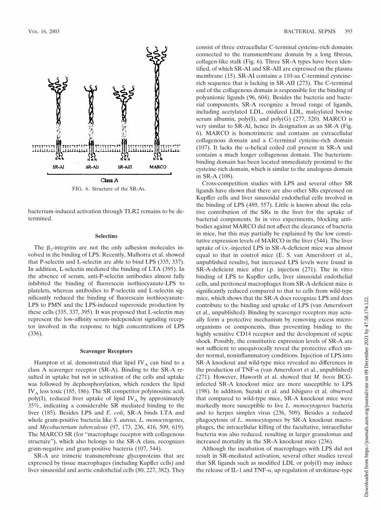
bacterium-induced activation through TLR2 remains to be de-termined.
Selectins
The �2-integrins are not the only adhesion molecules in-volved in the binding of LPS. Recently, Malhotra et al. showedthat P-selectin and L-selectin are able to bind LPS (335, 337).In addition, L-selectin mediated the binding of LTA (395). Inthe absence of serum, anti-P-selectin antibodies almost fullyinhibited the binding of fluorescein isothiocyanate-LPS toplatelets, whereas antibodies to P-selectin and L-selectin sig-nificantly reduced the binding of fluorescain isothiocyanate-LPS to PMN and the LPS-induced superoxide production bythese cells (335, 337, 395). It was proposed that L-selectin mayrepresent the low-affinity serum-independent signaling recep-tor involved in the response to high concentrations of LPS(336).
Scavenger Receptors
Hampton et al. demonstrated that lipid IVA can bind to aclass A scavenger receptor (SR-A). Binding to the SR-A re-sulted in uptake but not in activation of the cells and uptakewas followed by dephosphorylation, which renders the lipidIVA less toxic (185, 186). The SR competitor polyinosinic acid,poly(I), reduced liver uptake of lipid IVA by approximately35%, indicating a considerable SR mediated binding to theliver (185). Besides LPS and E. coli, SR-A binds LTA andwhole gram-positive bacteria like S. aureus, L. monocytogenes,and Mycobacterium tuberculosis (97, 173, 236, 416, 509, 619).The MARCO SR (for “macrophage receptor with collagenousstructure”), which also belongs to the SR-A class, recognizesgram-negative and gram-positive bacteria (107, 544).
SR-A are trimeric transmembrane glycoproteins that areexpressed by tissue macrophages (including Kupffer cells) andliver sinusoidal and aortic endothelial cells (80, 227, 382). They
consist of three extracellular C-terminal cysteine-rich domainsconnected to the transmembrane domain by a long fibrous,collagen-like stalk (Fig. 6). Three SR-A types have been iden-tified, of which SR-AI and SR-AII are expressed on the plasmamembrane (15). SR-AI contains a 110-aa C-terminal cysteine-rich sequence that is lacking in SR-AII (273). The C-terminalend of the collagenous domain is responsible for the binding ofpolyanionic ligands (96, 604). Besides the bacteria and bacte-rial components, SR-A recognize a broad range of ligands,including acetylated LDL, oxidized LDL, maleylated bovineserum albumin, poly(I), and poly(G) (277, 520). MARCO isvery similar to SR-Al, hence its designation as an SR-A (Fig.6). MARCO is homotrimeric and contains an extracellularcollagenous domain and a C-terminal cysteine-rich domain(107). It lacks the �-helical coiled coil present in SR-A andcontains a much longer collagenous domain. The bacterium-binding domain has been located immediately proximal to thecysteine-rich domain, which is similar to the analogous domainin SR-A (108).
Cross-competition studies with LPS and several other SRligands have shown that there are also other SRs expressed onKupffer cells and liver sinusoidal endothelial cells involved inthe binding of LPS (489, 557). Little is known about the rela-tive contribution of the SRs in the liver for the uptake ofbacterial components. In in vivo experiments, blocking anti-bodies against MARCO did not affect the clearance of bacteriain mice, but this may partially be explained by the low consti-tutive expression levels of MARCO in the liver (544). The liveruptake of i.v.-injected LPS in SR-A-deficient mice was almostequal to that in control mice (E. S. van Amersfoort et al.,unpublished results), but increased LPS levels were found inSR-A-deficient mice after i.p. injection (271). The in vitrobinding of LPS to Kupffer cells, liver sinusoidal endothelialcells, and peritoneal macrophages from SR-A-deficient mice issignificantly reduced compared to that to cells from wild-typemice, which shows that the SR-A does recognize LPS and doescontribute to the binding and uptake of LPS (van Amersfoortet al., unpublished). Binding by scavenger receptors may actu-ally form a protective mechanism by removing excess micro-organisms or components, thus preventing binding to thehighly sensitive CD14 receptor and the development of septicshock. Possibly, the constitutive expression levels of SR-A arenot sufficient to unequivocally reveal the protective effect un-der normal, noninflammatory conditions. Injection of LPS intoSR-A knockout and wild-type mice revealed no differences inthe production of TNF-� (van Amersfoort et al., unpublished)(271). However, Haworth et al. showed that M. bovis BCG-infected SR-A knockout mice are more susceptible to LPS(198). In addition, Suzuki et al. and Ishiguro et al. observedthat compared to wild-type mice, SR-A knockout mice weremarkedly more susceptible to live L. monocytogenes bacteriaand to herpes simplex virus (236, 509). Besides a reducedphagocytosis of L. monocytogenes by SR-A knockout macro-phages, the intracellular killing of the facultative, intracellularbacteria was also reduced, resulting in larger granulomas andincreased mortality in the SR-A knockout mice (236).
Although the incubation of macrophages with LPS did notresult in SR-mediated activation, several other studies revealthat SR ligands such as modified LDL or poly(I) may inducethe release of IL-1 and TNF-�, up regulation of urokinase-type
FIG. 6. Structure of the SR-As.
VOL. 16, 2003 BACTERIAL SEPSIS 393
Dow
nloa
ded
from
http
s://j
ourn
als.
asm
.org
/jour
nal/c
mr
on 0
9 D
ecem
ber
2021
by
47.5
8.17
4.12
2.

plasminogen activator on macrophages, and up regulation ofadhesion molecules on endothelial cells (185, 222, 247, 410,411). In addition, Kobayashi et al. observed reduced IL-1 pro-duction in SR-A knockout mice after an LPS challenge andproposed that binding of LPS to SR-A induces the release ofIL-1 (271). However, no additional (in vitro) data to supportthis hypothesis were provided.
Based on available data, SR-A can be considered anti-in-flammatory due to the uptake of LPS or other bacterial com-pounds, circumventing the CD14-TLR signaling pathway. LPSaffect the expression of the SR-A. On human macrophages, theexpression of SR-A decreases after stimulation with LPS (553).Similarly, the SR-A mRNA is transiently down regulated afteran in vivo challenge with LPS in mice (461). However, treat-ment of mice with inflammatory stimuli like BCG or heat-killed Corynebacterium parvum results in increased expressionon liver Kupffer cells (181, 198, 523). Also, the expression ofMARCO can be upregulated by inflammatory stimuli; in mice,treatments with BCG, C. parvum, K. pneumoniae, and LPS allresulted in a marked upregulation of MARCO expression inthe liver (181, 544, 545). Moreover, a similar pattern of highMARCO expression was observed in the livers of two new-borns who died of sepsis (108).
Moesin
A 78-kDa LPS-binding protein was shown to be identical tomoesin (membrane-organizing extension spike protein) (529).An anti-moesin antibody completely inhibited the LPS-in-duced TNF release by monocytes, but LPS binding was notblocked (529). Due to the complete inhibition of LPS-medi-ated signaling by the anti-moesin antibody, whereas an anti-CD14 antibody only partially blocked responses, it is temptingto speculate that moesin may represent an element in theCD14-TLR4 signaling pathway. The fact that the anti-moesinantibody does not inhibit S. aureus-mediated inhibition pre-cludes the binding of the antibody to TLR2 but not to TLR4.
Heptose-Specific Lipopolysaccharide Receptor
A receptor on liver parenchymal cells was identified thatspecifically recognized heptose residues in the inner core ofLPS (412). This receptor may be involved in the transfer ofLPS from the circulation to bile as proposed by Van Bossuyt etal. (542).
LIPOPOLYSACCHARIDE- AND LIPOTEICHOICACID-BINDING PROTEINS
Neutrophilic Lipopolysaccharide-Binding Molecules
Bactericidal/permeability-increasing protein. In 1978, Weisset al. reported the isolation of a potent bactericidal protein,BPI, from human PMN (583). The cationic, 55-kDa proteinresided in the azurophilic granules of PMN and, to a lesserextent, in monocytes and macrophages. It binds highly specif-ically to gram-negative bacteria and kills bacteria by increasingthe permeability of the outer membrane and by activating cellwall-degrading outer membrane enzymes (110, 583). No bac-tericidal activity of BPI against gram-positive bacteria was ob-served (583). The initial effect of the decreased integrity of the
outer membrane of gram-negative bacteria is growth arrest(�1 min) followed by changes in the cytoplasmic membrane,resulting in impaired energy metabolism, irreversible growtharrest, and, finally, death (109, 583). The bactericidal activitiesof other granule constituents such as the p15s and defensinsare synergistic with that of BPI (311, 577), but the serumprotein LBP may also enhance the bactericidal activity of BPI(220). PMN may also excrete BPI, and excreted BPI has iden-tical bactericidal effects to intracellular BPI (109). Binding ofBPI to gram-negative bacteria is mediated by LPS. The initialinteraction probably involves negatively charged groups in theinner core and lipid A and additional interactions with thehydrophobic moieties of lipid A, resulting in a high-affinitybinding (Kd � 2 to 5 nM) (159, 160, 401). In vitro experimentswith BPI have shown that the sensitivity of various gram-negative species to the bactericidal effect of BPI varies consid-erably. Strains expressing rough LPS are more sensitive thanstrains expressing smooth LPS. Limited proteolysis of BPIyields a C-terminal 30-kDa fragment and an N-terminal 25-kDa fragment (401). The latter contains the LPS-binding andbactericidal capacity (401, 402). The 25-kDa fragment, a re-combinant 23-kDa N-terminal fragment (rBPI23), and holo-BPI have comparable affinities for free LPS, irrespective of theserotype (401).
Binding of BPI to LPS neutralizes the biological activity ofLPS in vitro. BPI attenuates LPS-mediated endothelial dam-age and IL-6 production as well as LPS-mediated NO, TNF-�,IL-1, IL-6, and IL-8 production by macrophages and wholeblood (11, 67, 91, 362, 584). In vivo, BPI or rBPI23 reducedLPS- or bacterium-induced TNF-� release, liver damage, NOproduction, and mortality and protected against cardiovasculardepression (7, 117, 275, 321). Combining rBPI with antibioticsenhances its protective effect (7). BPI causes a significant re-duction in serum LPS, TNF, IL-6, IL-8, and IL-10 levels and inseveral other parameters in healthy volunteers (567). The re-sults of a clinical study of children with meningococcal sepsiswere promising: of 26 patients only 1 died (4%), which wasfavorable compared to the predicted mortalities, which variedbetween 15 and 49% based on various parameters such as LPSlevels, serum IL-6 levels, etc. (167). However, in a larger pla-cebo-controlled study, the mortality in the rBPI21-treated pa-tients was not significantly reduced, although there was a trendtoward improved outcome in the primary outcome variables(309). The feasibility of the use of BPI for the treatment ofsepsis and septic shock in humans may be restricted due to thelimited half-life of approximately 10 min in vivo (28, 29).
CAP18. Like BPI, CAP18 was isolated from PMN, but the18-kDa protein bears no structural and sequence similarity toBPI and other LPS-binding proteins (297). CAP18 binds LPSbut is also bactericidal to bacteria such as E. coli and S. aureus(381). Defensins, lactoferrin, and lysozyme synergistically en-hance the bactericidal activity of CAP18 (23, 381). The pro-duction of CAP18 by human lung epithelial cells and the re-lease of CAP18 onto the airway surface of the cells indicatethat it may also have a function in mucosal defense (23).Intratracheal instillation of CAP18-adenovirus reduced the re-sponse to Pseudomonas aerigunosa, whereas i.v.-injected virusprotected against LPS and E. coli (24). The LPS-binding siteresides in the C-terminal 36-aa part of the molecule, and pep-
394 VAN AMERSFOORT ET AL. CLIN. MICROBIOL. REV.
Dow
nloa
ded
from
http
s://j
ourn
als.
asm
.org
/jour
nal/c
mr
on 0
9 D
ecem
ber
2021
by
47.5
8.17
4.12
2.
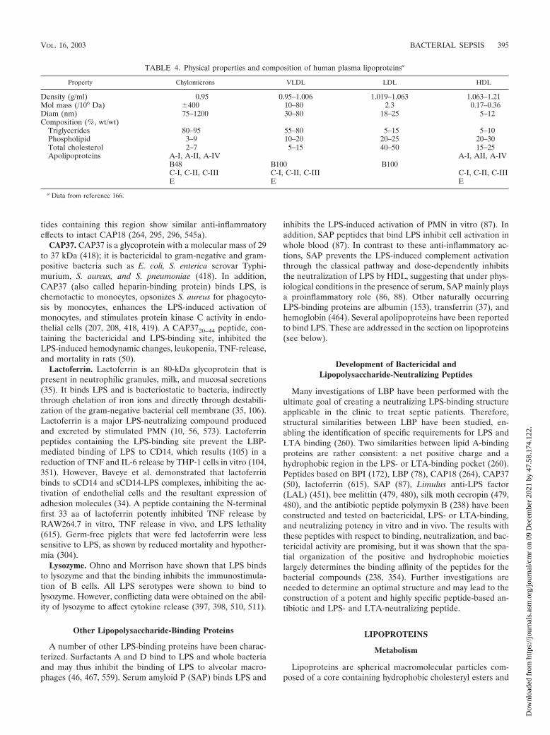
tides containing this region show similar anti-inflammatoryeffects to intact CAP18 (264, 295, 296, 545a).
CAP37. CAP37 is a glycoprotein with a molecular mass of 29to 37 kDa (418); it is bactericidal to gram-negative and gram-positive bacteria such as E. coli, S. enterica serovar Typhi-murium, S. aureus, and S. pneumoniae (418). In addition,CAP37 (also called heparin-binding protein) binds LPS, ischemotactic to monocytes, opsonizes S. aureus for phagocyto-sis by monocytes, enhances the LPS-induced activation ofmonocytes, and stimulates protein kinase C activity in endo-thelial cells (207, 208, 418, 419). A CAP3720–44 peptide, con-taining the bactericidal and LPS-binding site, inhibited theLPS-induced hemodynamic changes, leukopenia, TNF-release,and mortality in rats (50).
Lactoferrin. Lactoferrin is an 80-kDa glycoprotein that ispresent in neutrophilic granules, milk, and mucosal secretions(35). It binds LPS and is bacteriostatic to bacteria, indirectlythrough chelation of iron ions and directly through destabili-zation of the gram-negative bacterial cell membrane (35, 106).Lactoferrin is a major LPS-neutralizing compound producedand excreted by stimulated PMN (10, 56, 573). Lactoferrinpeptides containing the LPS-binding site prevent the LBP-mediated binding of LPS to CD14, which results (105) in areduction of TNF and IL-6 release by THP-1 cells in vitro (104,351). However, Baveye et al. demonstrated that lactoferrinbinds to sCD14 and sCD14-LPS complexes, inhibiting the ac-tivation of endothelial cells and the resultant expression ofadhesion molecules (34). A peptide containing the N-terminalfirst 33 aa of lactoferrin potently inhibited TNF release byRAW264.7 in vitro, TNF release in vivo, and LPS lethality(615). Germ-free piglets that were fed lactoferrin were lesssensitive to LPS, as shown by reduced mortality and hypother-mia (304).
Lysozyme. Ohno and Morrison have shown that LPS bindsto lysozyme and that the binding inhibits the immunostimula-tion of B cells. All LPS serotypes were shown to bind tolysozyme. However, conflicting data were obtained on the abil-ity of lysozyme to affect cytokine release (397, 398, 510, 511).
Other Lipopolysaccharide-Binding Proteins
A number of other LPS-binding proteins have been charac-terized. Surfactants A and D bind to LPS and whole bacteriaand may thus inhibit the binding of LPS to alveolar macro-phages (46, 467, 559). Serum amyloid P (SAP) binds LPS and
inhibits the LPS-induced activation of PMN in vitro (87). Inaddition, SAP peptides that bind LPS inhibit cell activation inwhole blood (87). In contrast to these anti-inflammatory ac-tions, SAP prevents the LPS-induced complement activationthrough the classical pathway and dose-dependently inhibitsthe neutralization of LPS by HDL, suggesting that under phys-iological conditions in the presence of serum, SAP mainly playsa proinflammatory role (86, 88). Other naturally occurringLPS-binding proteins are albumin (153), transferrin (37), andhemoglobin (464). Several apolipoproteins have been reportedto bind LPS. These are addressed in the section on lipoproteins(see below).
Development of Bactericidal andLipopolysaccharide-Neutralizing Peptides
Many investigations of LBP have been performed with theultimate goal of creating a neutralizing LPS-binding structureapplicable in the clinic to treat septic patients. Therefore,structural similarities between LBP have been studied, en-abling the identification of specific requirements for LPS andLTA binding (260). Two similarities between lipid A-bindingproteins are rather consistent: a net positive charge and ahydrophobic region in the LPS- or LTA-binding pocket (260).Peptides based on BPI (172), LBP (78), CAP18 (264), CAP37(50), lactoferrin (615), SAP (87), Limulus anti-LPS factor(LAL) (451), bee melittin (479, 480), silk moth cecropin (479,480), and the antibiotic peptide polymyxin B (238) have beenconstructed and tested on bactericidal, LPS- or LTA-binding,and neutralizing potency in vitro and in vivo. The results withthese peptides with respect to binding, neutralization, and bac-tericidal activity are promising, but it was shown that the spa-tial organization of the positive and hydrophobic moietieslargely determines the binding affinity of the peptides for thebacterial compounds (238, 354). Further investigations areneeded to determine an optimal structure and may lead to theconstruction of a potent and highly specific peptide-based an-tibiotic and LPS- and LTA-neutralizing peptide.
LIPOPROTEINS
Metabolism
Lipoproteins are spherical macromolecular particles com-posed of a core containing hydrophobic cholesteryl esters and
TABLE 4. Physical properties and composition of human plasma lipoproteinsa
Property Chylomicrons VLDL LDL HDL
Density (g/ml) 0.95 0.95–1.006 1.019–1.063 1.063–1.21Mol mass (/106 Da) �400 10–80 2.3 0.17–0.36Diam (nm) 75–1200 30–80 18–25 5–12Composition (%, wt/wt)
Triglycerides 80–95 55–80 5–15 5–10Phospholipid 3–9 10–20 20–25 20–30Total cholesterol 2–7 5–15 40–50 15–25Apolipoproteins A-I, A-II, A-IV A-I, AII, A-IV
B48 B100 B100C-I, C-II, C-III C-I, C-II, C-III C-I, C-II, C-IIIE E E
a Data from reference 166.
VOL. 16, 2003 BACTERIAL SEPSIS 395
Dow
nloa
ded
from
http
s://j
ourn
als.
asm
.org
/jour
nal/c
mr
on 0
9 D
ecem
ber
2021
by
47.5
8.17
4.12
2.

triglycerides surrounded by a shell of phospholipids, unesteri-fied free cholesterol, and one or more proteins called apoli-poproteins. The cholesterol and apolipoproteins stabilize theparticles, but the apolipoproteins are also involved in lipidrecruitment, modulation of enzyme activity, and modulation ofreceptor-mediated binding and endocytosis. In human blood,four major lipoprotein classes can be distinguished accordingto their density: chylomicrons, VLDL, LDL, and HDL. Theselipoprotein classes differ with respect to size, electrophoreticmobility, and lipid and apolipoprotein composition (Table 4)(166). Two major metabolic routes can be distinguished: anexogenous pathway is involved in the transport and metabo-lism of intestinally derived dietary lipids, and an endogenouspathway is involved in the transport and metabolism of pro-cessed lipids. In short, dietary lipids that are absorbed in theintestines are packaged into chylomicrons, large triglyceride-rich particles, and transported from the lymph to the bloodcirculation (333). On entering the circulation, lipoproteinlipase (LPL) processes the chylomicrons, delivering free fattyacids to peripheral tissues (fat and muscle tissue), whereas thecholesterol-rich core remnants are taken up mainly via ApoE-specific recognition sites on liver parenchymal cells (333). Theliver secretes ApoB100-containing VLDL that in the circula-tion becomes enriched in ApoC and ApoE. These particles arealso rich in triglycerides and serve as a source of free fatty acidsfor extrahepatic tissues. LPL and hepatic lipase (HL) processthese particles, resulting in a loss of triglycerides, phospholip-ids, ApoC, and ApoE. The remnants (also called intermediate-density lipoproteins [IDL]) are further processed to becomeLDL with ApoB100 as its sole apolipoprotein. These choles-terol-rich particles are taken up in the liver and peripheraltissues through the LDL receptor. To maintain cholesterolhomeostasis, excess cholesterol in extrahepatic tissues must bereturned to the liver, which is the only organ capable of dis-posing of cholesterol via the bile. This process is known asreverse cholesterol transport and involves the last lipoproteinparticle, HDL. The enzyme lecithin:cholesterol acyltransferasepromotes the esterification of cholesterol in HDL, creating aconcentration gradient and thus facilitating the efflux of freecholesterol from extrahepatic tissues to HDL. In exchange fortriglycerides, cholesteryl ester transfer protein-mediated trans-fer of cholesteryl esters to LDL and VLDL occurs. Subse-quently, these particles are taken up by the liver. In addition,direct transfer of cholesteryl esters from HDL to the liveroccurs through recognition by SR-BI. In contrast to the recep-tor-mediated uptake of VLDL and LDL, SR-BI promotes onlythe selective transfer of cholesteryl ester without causing theuptake of the HDL particle (2).
Apolipoprotein E
ApoE was identified in 1973 as a component of triglyceride-rich VLDL and was initially referred to as arginine-rich proteindue to its relatively high arginine content compared to otherapolipoproteins known at that time (490, 540). The ApoEmolecule is synthesized as a 317-aa prepeptide after cotrans-lational cleavage yielding a 299-aa protein with a molecularmass of 34.2 kDa. ApoE is synthesized in a wide variety oftissues, including the liver, central nervous system, kidneys,adrenal glands, testes, and ovaries, but not the intestines (111,
496). However, the highest levels of ApoE mRNA are found inthe liver parenchymal cells and the second highest are found inthe astrocytes in the brain. Furthermore, ApoE is also pro-duced by macrophages from the liver, lungs, and spleen (12,26). In the absence of lipids, ApoE self-associates to formtetramers over a wide concentration range (580). In contrast,self-association does not occur on lipid surfaces. ApoE con-tains two domains that are joined by a protease-susceptiblehinge region. Thrombin digestion of ApoE yields two frag-ments, of which the 10-kDa C-terminal fragment harbors thelipid-binding domain whereas the LDL receptor (LDLr)-bind-ing domain is situated in the 22-kDa N-terminal fragment (235,581). The N-terminal domain contains four antiparallel heli-ces, of which helix 4 contains the LDLr-binding domain. Thishelix is unusually rich in basic amino acids (Arg, Lys, and His).ApoE contains two heparin-binding sites, of which one is lo-cated within the LDLr-binding site (582). Similarly, ApoE in-teracts with heparan sulfate proteoglycans (HSPG). HSPG areabundantly present on many different cell types, including vas-cular endothelial cells and liver parenchymal cells within thespace of Disse (605). In the liver, HSPG may be involved inboth the secretion of ApoE and the binding of lipoprotein-bound ApoE (243).
Several physiological and pathological functions for apoEhave been proposed. These include the role of ApoE in lipidmetabolism (as described above), intracellular lipid redistribu-tion (281, 448), atherogenesis (75, 463, 549), neurobiology(nerve regeneration, association of the ApoE4 serotype withneuropathologic lesions in Alzheimer’s disease) (342, 580),and immunomodulation (inhibition of proliferation of periph-eral blood mononuclear cells and lymphocyte activation; inhi-bition of TNF secretion by glial cells) (20, 228, 299, 364, 399,417).
Anti-Inflammatory Role
Ulevitch et al. were the first to observe that if LPS is mixedwith serum or plasma, a decrease in buoyant density results.Preincubation of LPS with plasma decreased the ability of LPSto induce neutropenia and a pyrogenic response in rabbits.Further investigations showed that the LPS was bound to HDLand that a plasma protein aided in the binding (525, 534, 536).In rabbits, the uptake of LPS by the adrenal glands was in-creased after LPS binding to HDL, which indicates that bind-ing of LPS to HDL results in a decreased recognition by LPSreceptors (347, 372). Since then, in vivo and in vitro experi-ments have shown that LPS and LTA bind to and are neutral-ized by lipid emulsions (177, 445), chylomicrons (194, 195,447), VLDL (102, 194, 459, 464, 576), LDL (102, 177, 366, 459,464, 554, 576), Lp(a) (385), HDL (102, 144, 177, 374, 459, 464,535, 576), ApoA-I (112, 135, 539), ApoB (112, 464), and ApoE(449, 555).
In vivo experiments have shown that the injection of LPS-HDL complexes may affect the decay of LPS in serum (144,375) and inhibits the LPS-induced release of cytokines com-pared to LPS alone (194, 536). In addition, in several experi-ments the infusion of recombinant HDL (rHDL), VLDL, orlymph-derived chylomicrons and lipid emulsion also resultedin inhibition of LPS-induced physiological changes (120, 195,226, 310, 445). Pajkrt et al. infused rHDL into humans, who
396 VAN AMERSFOORT ET AL. CLIN. MICROBIOL. REV.
Dow
nloa
ded
from
http
s://j
ourn
als.
asm
.org
/jour
nal/c
mr
on 0
9 D
ecem
ber
2021
by
47.5
8.17
4.12
2.

were subsequently challenged with a low dose of LPS andobserved a significant reduction in the release of proinflam-matory cytokines and a partial reduction in the activationand/or release of components involved in coagulation (408,409). The in vitro data mainly parallel those found in vivo.These data confirm that binding of LPS to lipoproteins de-creases its ability to activate macrophages and endothelial cells(30, 57, 60, 196, 306, 366, 415, 554, 579) (Table 5). Recently,Grunfeld et al. have shown a similar inhibitory effect of li-poprotein binding on LTA-induced activation of macrophages(177). The inhibition of LPS-induced activation is dependentlargely on the concentration of lipoproteins: in the presence oflow concentrations of lipoproteins, cells become activated be-fore neutralization by lipoproteins occurs (386). It has beenshown that a 5-min exposure to LPS is sufficient to maximallybind and stimulate monocytes in whole blood, which impliesthat in the circulation neutralization by (infused) lipoproteinsis a rapid process (157). However, Kitchens et al. illustratedthat lipoproteins can remove LPS bound to the cell surface.This transfer is dependent on the presence of sCD14 andfurther enhances the protective effect of lipoproteins (269).
Mice with high HDL levels due to overexpression of humanApoA-I levels and rats rendered hyperlipidemic by estradioltreatment are less sensitive to LPS compared to the respectivecontrols (120, 310). Hypercholesterolemic rabbits exhibitedhigher TNF- levels and mortality than did the normolipidemiccontrols (54). LDL receptor knockout mice, with cholesterollevels twice those in C57BL/6 mice, are less susceptible to LPSthan are wild-type mice, as shown by decreased mortality andreduced release of TNF, IL-1, and IL-6 (387). In contrast, theseverely hypercholesterolemic ApoE knockout mice weremore sensitive to LPS (84). It is likely that the lack of ApoE isthe cause of this increased sensitivity.
The binding of LPS to HDL is mediated by LBP and sCD14,although only the first has been shown to be essential (600,602). Wright et al. showed that most LBP is associated withApoA-I-containing particles, which may explain the preferen-tial binding of LPS to HDL (414, 600). PLTP may also aid in
the transfer of LPS to HDL (182). This was confirmed byobservations that both LBP and PLTP could extract LPS fromgram-negative bacterial cell membranes and mediate the trans-fer to lipoproteins (mainly HDL) (562). However, experimentsby Vreugdenhil et al. indicated that LBP is associated mainlywith ApoB-containing lipoproteins (e.g., LDL) (569). Theyhave shown that the association of LBP with ApoB is strongerthan with ApoA-I, that LBP in serum is present mainly onLDL and VLDL, and that LDL- and VLDL-associated LBPenhanced LPS binding to these lipoproteins. Using a solid-phase binding assay, they also observed that in the absence ofLBP, LPS preferentially bound to LDL and VLDL. The dis-crepancies in the results of Wright et al. (600) and Vreugdenhilet al. (569) may be explained by the differences in the methodsused. Whereas the first group used affinity chromatographywith either anti-ApoA-I or anti-LBP antibodies to coprecipateApoA-I and LBP, Vreugdenhil et al. employed agarose gelelectrophoresis in combination with immunoblotting to detectLBP-containing lipoproteins. In our in vivo and in vitro exper-iments, we have observed LPS and LTA binding to all lipopro-teins in the serum, which may indicate that LBP transfers LPSand LTA to all lipoproteins (van Amersfoort et al., unpub-lished). However, although we observed binding to all lipopro-tein classes, based on lipid or protein contents LTA seemed toexhibit preferential binding to HDL (Van Amersfoort et al.,unpublished). Other investigators have also observed eitherpreferential binding of LPS to HDL (375, 464, 535), to LDLand/or VLDL (102, 554), or to all lipoproteins (102, 459, 554,562, 576) depending on the lipid and/or protein distribution inthe distinct lipoprotein fractions. A correlation between thecholesterol contents of the lipoprotein fractions and LPS as-sociation was proposed by Van Lenten et al. (554), but withrHDL the neutralizing potency correlates with the phospho-lipid contents of the particle (72). Using the synthetic LPS-likeantagonist E5531, it was found that association with the dif-ferent lipoproteins did correlate with protein (HDL), choles-terol (VLDL, LDL), and triglyceride (VLDL) contents, whichmay imply that factors other than the composition of the li-
TABLE 5. Inhibitory effects of lipoproteins on LPS and LTA toxicity
LPS and LTA property
Effect of:
Reference(s)HDL rHDL LDL VLDL or
chylomicronsEmul-sion
Total lipo-protein ApoE
In vivoInhibition of cytokine release 2 2 2 2 60, 72, 120, 195, 225, 226, 437,
555a
Decreased pyrogenicity 2 2 534, 535Decreased mortality after LPS challenge 2 2 2 2 2 2 2 120, 194, 195, 310, 445, 555Decreased mortality and TNF release after CLPb 2 446Increased excretion of 125I-LPS into bile 2 447Decreased Kupffer cell binding 2 449
In vitroInhibition of macrophage or monocyte activation
(cytokine release)2 2 2 2 2 2 30, 60, 61, 135, 136, 177, 196,
306, 415, 545, 554, 579Inhibition of PMN priming 2 568Inhibition of VCAM-1 expression by HUVECc 2 57Inhibition of LAL activation 2 2 2 103,112,193
a E. S. van Amersfoort et al., unpublished results.b CLP, cecal ligation puncture; this experimental procedure results in polymicrobial peritonitis and sepsis.c HUVEC, human umbilical vein epithelial cells.
VOL. 16, 2003 BACTERIAL SEPSIS 397
Dow
nloa
ded
from
http
s://j
ourn
als.
asm
.org
/jour
nal/c
mr
on 0
9 D
ecem
ber
2021
by
47.5
8.17
4.12
2.

poproteins are also involved (459, 576). The association withthe lipoproteins in sera from mouse strains with various li-poprotein profiles correlates with the cholesterol contents ofLDL and VLDL but not of HDL (van Amersfoort et al.,unpublished). The increased binding of LTA to HDL that wehave observed may be caused by the involvement of LBP in thetransfer of LTA to HDL, as proposed by Grunfeld et al. (177).The proposed LPS and LTA neutralization pathways are de-picted in Fig. 7.
The lipoprotein distribution of LPS may depend on the typeof LPS used, the origin of the serum (animal species andlipoprotein status), or, when using plasma, the type of antico-agulant used (e.g., citrate, EDTA, or heparin) (101, 102, 554).Furthermore, as mentioned above, the use of a density gradi-ent, agarose gel electrophoresis, or column chromatographymay introduce additional variations.
So far, only the binding of LPS to lipoproteins has beendiscussed, but binding to apolipoproteins has also been re-ported. Emancipator et al. and Usynin et al. have shown thatboth ApoB and ApoA-I are able to bind and neutralize LPS(112, 539). Our group has shown that LPS binds ApoE andcauses a redistribution in vivo, reducing the uptake by Kupffercells and promoting the binding to liver parenchymal cells(449). In addition, we have shown that ApoE binds LTA,resulting in a similar redistribution from Kupffer cells to liverparenchymal cells and in a strongly decreased TNF release invivo (Van Amersfoort et al., unpublished).
Lipid Metabolism and Infection
Injection of LPS or bacteria leads to a marked hyperlipid-emic response, caused primarily by an increase in the level ofVLDL triglycerides (470). Remarkably, the underlying mech-anisms differ with the dose of LPS administered to the animal.With low doses, an increase in hepatic triglyceride productionoccurs, whereas at high doses of LPS (�500 �g/kg of body
weight), a decrease in the clearance of triglyceride-rich li-poproteins occurs (125). Experiments with IL-1 receptor an-tagonist (IL-1ra) and TNF antibodies showed that these phe-nomena could be only partially mimicked by administration ofIL-1 or TNF-� (125). Subsequently, it was shown that �1-adrenergic receptors were involved in the low-dose-LPS-in-duced increase in triglyceride production whereas the �2-ad-renergic receptors were involved in the high-dose-LPS-inducedinhibition of LPL activity, confirming a partial cytokine-inde-pendent pathway (391). LPS and the proinflammatory cyto-kines induced by LPS affect a wide range of enzymes andreceptors involved in lipid metabolism, which include LPL,hepatic lipase, PLTP, LRP, and LDL receptor (Table 6).
However, the extent and kinetics of the effects differedwidely among these species and in some cases were even con-tradictory. This is not surprising since there are substantialspecies-specific differences in lipid metabolism, resulting inmarked differences in lipoprotein profiles. For instance, inmice almost all serum cholesterol resides in HDL, whereas inhumans a significant portion of the serum cholesterol can berecovered from the VLDL and LDL. However, a TNF-inducedincrease in triglyceride levels was also observed in humans,which was paralleled by an increased rather than decreasedlipolysis (497). The increased LPS- and LTA-induced triglyc-eride levels, along with the evidence that LPS and LTA areneutralized by lipoproteins, led to the hypothesis that thesemetabolic changes may be protective during bacterial infection(189, 293, 313). To test this hypothesis, rats were treated withestradiol or 4-aminopyrolo-(3,4-D)-pyrimide to induce hypo-lipidemia, after which they were challenged with LPS. TheTNF-� levels and mortality were significantly increased in thehypolipidemic animals (120). However, these animals could berescued by the injection of lipoproteins prior to the challengewith LPS, confirming that hypolipidemic animals are moresensitive to LPS. Fraunberger et al. collected serum from sep-
FIG. 7. LPS and LTA transfer to lipoproteins. Black arrows indicate established pathways. In contrast to LPS (left), LTA may also associatewith lipoproteins in the absence of LBP (extra arrow in right panel).
398 VAN AMERSFOORT ET AL. CLIN. MICROBIOL. REV.
Dow
nloa
ded
from
http
s://j
ourn
als.
asm
.org
/jour
nal/c
mr
on 0
9 D
ecem
ber
2021
by
47.5
8.17
4.12
2.

tic patients and measured lipoprotein and apolipoprotein con-tents. In the septic patients, there was a marked increase intriglyceride levels, whereas cholesterol, ApoB and ApoA-I lev-els were reduced. They also observed an inverse relationshipbetween serum cholesterol and TNF-� levels. A similar inverserelationship could be established between mortality and serumcholesterol levels (141–143). However, they did not show dataon the serum triglyceride levels in this group of patients, whichprecludes a final conclusion about whether cholesterol or tri-glyceride levels may be protective in septic shock.
We have shown that serum ApoE levels are increased inmice which were injected with LPS, LTA, or live bacteria (555;van Amersfoort et al., unpublished). Along with the findingthat ApoE is able to bind LPS and LTA, this also indicates thatApoE may be protective against LPS and LTA (449; vanAmersfoort et al., unpublished). Fraunberger et al. observedan increase in HDL- and LDL-associated ApoE levels in hu-man serum, whereas an increase in the HDL-associated ApoElevel in LPS-treated African green monkeys was observed byAuerbach and Parks (19, 141). In contrast, decreased amountsof ApoE were detected in VLDL and LDL from septic rats(293). Administration of LPS to Syrian hamsters caused areduction in liver ApoE mRNA levels but also a decreasedrelease of ApoE and ApoB by the perfused rat liver (122, 532).In LPS-treated Syrian hamsters, the levels of ApoE mRNAwere also reduced extrahepatically (191). LPS also caused a de-crease in the release of ApoE by macrophages (Table 6) (294).
CLINICAL AND EXPERIMENTAL SEPSIS THERAPIES
Due to the complexity of immunologic defense and sepsis,the development of pharmacologic interventions is difficult (1,218). One approach would be to prevent infection in patientsat high risk. However, timely treatment to prevent sepsis or
septic shock is often not possible, or the prevention of infectionis simply insufficient. The standard treatment of bacterial sep-sis often consists of administration of fluid and vasopressors torestore blood pressure and organ blood flow, oxygenation, andadministration of antibiotics (65, 442, 589). Although eradica-tion of the microbial organism and treatment of the patho-physiological changes are essential, alternative approaches ac-tively suppressing the deleterious effects of inflammation whileretaining the antimicrobial defense are needed. In this section,some approaches to the treatment of sepsis and septic shockare briefly described.
The first alternative approach to treatment of sepsis wasthrough antibody preparations to LPS. Three anti-lipid A IgMpreparations were used, HA-1A and E5 and a polyclonal an-tiserum against the LPS core of E. coli J5. The human anti-E. coli J5 (Rc-LPS) antiserum was first used to treat a smallgroup of patients with septic shock. This cross-reactive anti-serum barely significantly improved the overall prognosis inseptic shock, but it was not possible to determine whether theantibodies or other compounds (e.g., inflammatory mediators)from the donor conferred protection (618). E5 decreased mor-tality in a first trial but provided no protection in successivetrials (8, 174). HA-1A (Centoxin), was protective in one trialbut had no effect in a second and tended to increase mortalityin a subgroup of patients in a third trial (31, 69, 548). Very littleis known about the mechanism of protection of these antibod-ies. Although it was expected that the HA-1A antibody recog-nized lipid A specifically, it was later discovered that it boundrather nonspecifically to hydrophobic substances (31). Otherattempts to treat patients with an anti-LPS antibody have alsofailed (218). As listed in Table 7 a substantial number ofpatients have been treated with the various preparations, butno overall benefit was observed.
TABLE 6. Effects of inflammation and inflammatory mediators on lipid metabolism
Lipid undergoingmetabolism
Effect ofa:Reference(s)
LPS LTA TNF-� IL-1 IFN-� GM-CSF
Serum or plasmaTG 1§ 1 1 1 19, 121, 124, 276, 391,
392, 497TC 21 1 7 121, 122, 124LCAT 2 19PLTP 2 245CETP 2 2 2 190
TissueLPL 2 2 256, 257, 392
LiverHL 2 7 2 123LDL receptor 2 314
Monocytes/macrophagesLRP receptor 2 2 288
ApoESerum ApoE 1 19, 314, 555a
Liver ApoE 2 122, 532b
Macrophage ApoE 2 (2) 2 360, 586, 620
a1, increase; 2, decrease; 7, no change; 21, increased as well as decreased levels were observed.b E. S. van Amersfoort et al., unpublished results.
VOL. 16, 2003 BACTERIAL SEPSIS 399
Dow
nloa
ded
from
http
s://j
ourn
als.
asm
.org
/jour
nal/c
mr
on 0
9 D
ecem
ber
2021
by
47.5
8.17
4.12
2.

The recombinant 23-kDa N-terminal part of BPI, which is apowerful LPS-neutralizing agent, has also been used to treatseptic patients. Although the initial results of a small trial inchildren with meningococcal sepsis were promising, a largertrial showed no real benefit (167, 309). In addition to thedisappointing results of these trials, the use of therapeuticseffective only against gram-negative organisms may not bedesirable due to the delay caused by the time-consuming iden-tification of the pathogenic organism.
Both in vitro studies with whole blood from septic patientsand in vivo studies of healthy volunteers challenged with LPSindicate that treatment with recombinant HDL could be effec-tive in combating septic shock. However, no data are yet avail-able from clinical trials with recombinant HDL (171, 408, 409).
Immunosuppression and Neutralization ofProinflammatory Cytokines
Although general immunosuppression seems to be an obvi-ous choice in the treatment of sepsis, the overall mortalityincreased (70). Similarly, nonsteroidal anti-inflammatory drugsthat suppress the COX enzymes were also proven ineffective.Since TNF-� is a key mediator in the pathogenesis of sepsis, arecombinant humanized murine anti-TNF antibody prepara-tion (Remicade or infliximab) and a TNF-R:Fc fusion protein(Enbrel, or etanercept) have been tested for the treatment ofsepsis (64, 134). In clinical trials these preparations provedineffective. Paradoxically, these TNF antagonists are very ef-fective against Crohn’s disease (inflammatory bowel disease)and rheumatoid arthritis, while they are contraindicated forinfection and sepsis (113–115). Another important cytokine ininflammation and sepsis is IL-1. A recombinant IL-1 receptorantagonist has been tested in two large phase III trials but wasnot effective (404, 531). In healthy human volunteers, LPStolerance could be induced by the infusion of monophosphoryllipid A, but no clinical trials with septic patients have beenperformed (18). Interestingly, cross-tolerance to gram-positiveinfection could be induced in mice, indicating that this therapymight be effective in gram-negative and gram-positive sepsis(16).
The use of corticosteroids has been a subject of debate formany years. Corticosteroids are known to regulate a number of
immune responses. They inhibit a number of inflammatoryprocesses, such as leukocyte infiltration and cytokine produc-tion, and have a positive effect on cardiac output by a blockadeof NO synthesis (48, 371). Based on these effects, patients havebeen treated in two large clinical trials using corticoids in theearly phase of sepsis (326). The results of these trials indicatedeventually that there were no beneficial effects of corticoids inthe early phase of sepsis. More recently, it was observed thatpatients with a so-called adrenocortical insufficiency may havea poor prognosis (45, 70, 305, 492). Based on this hypothesis,trials were started in which patients were treated with a lowerdose of corticosteroids (300 mg of hydrocortisone per day) fora number of days at a later stage in sepsis. The outcome of thislower dose of steroids seems to be more promising than thehigher dose of steroids in the early phase of sepsis (45, 70, 305,492).
The latest success in the treatment of sepsis in adults was thePROWESS study (Human Activated PROtein C WorldwideEvaluation in Severe Sepsis) (38). In this large double-blindstudy, the effect of activated protein C was studied. After thetreatment of 1,520 patients, the study was abrogated because ofthe significant survival advantage in the actively treated group(24.7% mortality versus 30.8% for placebo [P � 0.005]). Themode of action of activated protein C is most probably basedon its ability to inhibit both thrombosis (lower levels of D-dimer) and inflammation (as seen in a reduced level of IL-6),whereas fibrinolysis is promoted. However, more studies areneeded before activated protein C is widely introduced as atherapeutic in the treatment. Although the blocking or modu-lation of a number of other targets, including complement andcoagulation factors, neutrophil adherence, and NO release, arepromising in animals, it remains to be determined whetherthese therapeutic approaches will be effective in humans (1,218, 547).
CONCLUDING REMARKS
Despite the extensive array of protective mechanisms em-ployed by bacteria, in mammals an innate, sensitive system todetect and eradicate bacteria has evolved. The immune systemis a double-edged sword: besides its ability to effectively detectbacteria and mount an immune response to eradicate them, itmay also cause serious damage to the body, even leading todeath. The rise and fall of an immune response is regulated bya complex, extensive system of pro- and anti-inflammatoryreceptors, cytokines, and other serum factors affecting the bac-teria directly or modulating the cellular response to the bac-teria. It is difficult to unequivocally attribute a fully pro- oranti-inflammatory role to inflammatory mediators and recep-tors. The experiments with TNF knockout mice and TNF re-ceptor knockout mice, for instance, have shown that these miceexhibit a drastically reduced sensitivity to LPS but succumb tosmall amounts of live bacteria or yeast (343, 422).
In human patients with sepsis, several putative therapieshave aimed at the neutralization of the proinflammatory cyto-kines TNF and IL-1. These therapies have proven ineffectiveor even counterproductive in some cases, leading to unchangedor increased mortality in the drug-treated patient groups (64,134). This shows that treatment of sepsis or septic shock maynot simply be a matter of administrating anti-inflammatory
TABLE 7. Randomized controlled trials of immunotherapy insepsis and septic shocka
Type of trial No. oftrials
Total no. ofpatients
Mortality (%) inpatients receiving:
Placebo Therapy
Anti-endotoxin 4 2,010 35 35Anti-IL-1R 3 1,898 35 31Anti-bradykinin 2 755 36 39Anti-PAF 2 870 50 45Anti-TNF 8 4,132 41 40Soluble TNF-R 2 688 38 40NSAIDS 3 514 40 37Steroids 9 1,267 35 39Activated protein C 1 1,690 31 25
All studies 33 13,824 38 37
a Adapted from reference 17.
400 VAN AMERSFOORT ET AL. CLIN. MICROBIOL. REV.
Dow
nloa
ded
from
http
s://j
ourn
als.
asm
.org
/jour
nal/c
mr
on 0
9 D
ecem
ber
2021
by
47.5
8.17
4.12
2.

agents or neutralizing their proinflammatory counterparts.Such therapeutic interventions may cause a shifted proinflam-matory and anti-inflammatory balance, thus compromising theendogenous regulatory processes controlling the inflammatorycascade. Therapy of sepsis and septic shock will therefore haveto simultaneously aim at maintaining homeostasis (part of themechanism of action of the newly approved recombinant hu-man activated protein C) and at eradication of the bacteria.Instead of combating the detrimental immune response duringsepsis, an attempt should be made to restore the balance be-tween the pro- and anti-inflammatory responses.
REFERENCES
1. Abraham, E. 1999. Why immunomodulatory therapies have not worked insepsis. Intensive Care Med. 25:556–566.
2. Acton, S., A. Rigotti, K. T. Landschulz, S. Xu, H. H. Hobbs, and M. Krieger.1996. Identification of scavenger receptor SR-BI as a high density lipopro-tein receptor. Science 271:518–520.
3. Agellon, L. B., E. M. Quinet, T. G. Gillette, D. T. Drayna, M. L. Brown, andA. R. Tall. 1990. Organization of the human cholesteryl ester transferprotein gene. Biochemistry 29:1372–1376.
4. Akagawa, K. S., and T. Tokunaga. 1985. Lack of binding of bacteriallipopolysaccharide to mouse lung macrophages and restoration of bindingby gamma-interferon. J. Exp. Med. 162:1444–1459.
5. Akira, S. 2000. Toll-like receptors: lessons from knockout mice. Biochem.Soc. Trans. 28:551–556.
6. Alexopoulou, L., A. C. Holt, R. Medzhitov, and R. A. Flavell. 2001. Recog-nition of double-stranded RNA and activation of NF-B by Toll-like re-ceptor 3. Nature 413:732–738.
7. Ammons, W. S., F. R. Kohn, and A. H. C. Kung. 1994. Protective effects ofan N-terminal fragment of bactericidal/permeability-increasing protein inrodent models of Gram-negative sepsis: role of bactericidal properties.J. Infect. Dis. 170:1473–1482.
8. Angus, D. C., M. C. Birmingham, R. A. Balk, P. J. Scannon, D. Collins, J. A.Kruse, D. R. Graham, H. V. Dedhia, S. Homann, N. Maclntyre, and E5Study Investigators. 2000. E5 murine monoclonal antiendotoxin antibodyin gram-negative sepsis: a randomized controlled trial. JAMA 283:1723–1730.
9. Antal-Szalmas, P., J. A. G. Van Strijp, A. J. L. Weersink, J. Verhoef, andK. P. M. Van Kessel. 1997. Quantitation of surface CD14 on human mono-cytes and neutrophils. J. Leukoc. Biol. 61:721–728.
10. Appelmelk, B. J., Y. Q. An, M. Geerts, B. G. Thijs, H. A. De Boer, D. M.MacLaren, J. De Graaff, and J. H. Nuijens. 1994. Lactoferrin is a lipidA-binding protein. Infect. Immun. 62:2628–2632.
11. Arditi, M., J. Zhou, S. H. Huang, P. M. Luckett, M. N. Marra, and K. S.Kim. 1994. Bactericidal/permeability-increasing protein protects vascularendothelial cells from lipopolysaccharide-induced activation and injury.Infect. Immun. 62:3930–3936.
12. Armbrust, T., and G. Ramadori. 1996. Functional characterization of twodifferent Kupffer cell populations of normal rat liver. J. Hepatol. 25:518–528.
13. Asari, Y., M. Majima, K. Sugimoto, M. Katori, and T. Ohwada. 1996.Release site of TNF� after intravenous and intraperitoneal injection of LPSfrom Escherichia coli in rats. Shock 5:208–212.
14. Asea, A., M. Rehli, E. Kabingu, J. A. Boch, O. Bare, P. E. Auron, M. A.Stevenson, and S. K. Calderwood. 2002. Novel signal transduction pathwayutilized by extracellular HSP70. Role of Toll-like receptor (TLR) 2 andTLR4. J. Biol. Chem. 277:15028–15034.
15. Ashkenas, J., M. Penman, E. Vasile, S. Acton, M. Freeman, and M. Krieger.1993. Structures and high and low affinity ligand binding properties ofmurine type I and type II macrophage scavenger receptors. J. Lipid Res.34:983–1000.
16. Astiz, M. E., A. Galera, D. C. Saha, C. Carpati, and E. C. Rackow. 1994.Monophosphoryl lipid A protects against gram-positive sepsis and tumornecrosis factor. Shock 2:271–274.
17. Astiz, M. E., and E. C. Rackow. 1998. Septic shock. Lancet 351:1501–1505.18. Astiz, M. E., E. C. Rackow, J. G. Still, S. T. Howell, A. Cato, K. B. Von
Eschen, J. T. Ulrich, J. A. Rudbach, G. McMahon, and R. Vargas. 1995.Pretreatment of normal humans with monophosphoryl lipid A inducestolerance to endotoxin: a prospective, double-blind, randomized, controlledtrial. Crit. Care Med. 23:9–17.
19. Auerbach, B. J., and J. S. Parks. 1989. Lipoprotein abnormalities associatedwith lipopolysaccharide-induced lecithin: cholesterol acyltransferase andlipase deficiency. J. Biol. Chem. 264:10264–10270.
20. Avila, E. M., G. Holdsworth, N. Sasaki, R. L. Jackson, and J. A. Harmony.1982. Apoprotein E suppresses phytohemagglutinin-activated phospholipidturnover in peripheral blood mononuclear cells. J. Biol. Chem. 257:5900–5909.
21. Aybay, C., and T. Imir. 1998. Comparison of the effects of Salmonellaminnesota Re595 lipopolysaccharide, lipid A and monophosphoryl lipid Aon nitric oxide, TNF-�, and IL-6 induction from RAW 264.7 macrophages.FEMS Immunol. Med. Microbiol. 22:263–273.
22. Bainbridge, B. W., and R. P. Darveau. 2001. Porphyromonas gingivalis lipo-polysaccharide: an unusual pattern recognition receptor ligand for theinnate host defense system. Acta Odontol. Scand. 59:131–138.
23. Bals, R., X. Wang, M. Zasloff, and J. M. Wilson. 1998. The peptide anti-biotic LL-37/hCAP-18 is expressed in epithelia of the human lung where ithas broad antimicrobial activity at the airway surface. Proc. Natl. Acad. Sci.USA 95:9541–9546.
24. Bals, R., D. J. Weiner, A. D. Moscioni, R. L. Meegalla, and J. M. Wilson.1999. Augmentation of innate host defense by expression of a cathelicidinantimicrobial peptide. Infect. Immun. 67:6084–6089.
25. Bannan, J., K. Visvanathan, and J. B. Zabriskie. 1999. Structure andfunction of streptococcal and staphylococcal superantigens in septic shock.Infect. Dis. Clin. North Am. 13:387–396, ix.
26. Basu, S. K., M. S. Brown, Y. K. Ho, R. J. Havel, and J. L. Goldstein. 1981.Mouse macrophages synthesize and secrete a protein resembling apoli-poprotein E. Proc. Natl. Acad. Sci. USA 78:7545–7549.
27. Bates, D. W., K. E. Pruess, and T. H. Lee. 1995. How bad are bacteremiaand sepsis? Outcomes in a cohort with suspected bacteremia. Arch. Intern.Med. 155:593–598.
28. Bauer, R. J., K. Der, N. Ottah-Ihejeto, J. Barrientos, and A. H. Kung. 1997.The role of liver and kidney on the pharmacokinetics of a recombinantamino terminal fragment of bactericidal/permeability-increasing protein inrats. Pharm. Res. 14:224–229.
29. Bauer, R. J., M. L. White, N. Wedel, B. J. Nelson, N. Friedmann, A. Cohen,W. N. M. Hustinx, and A. H. C. Kung. 1996. A phase I safety and phar-macokinetic study of a recombinant amino terminal fragment of bacteri-cidal/permeability-increasing protein in healthy male volunteers. Shock5:91–96.
30. Baumberger, C., R. J. Ulevitch, and J. M. Dayer. 1991. Modulation ofendotoxic activity of lipopolysaccharide by high-density lipoprotein. Patho-biology 59:378–383.
31. Baumgartner, J. D. 1990. Monoclonal anti-endotoxin antibodies for thetreatment of Gram-negative bacteremia and septic shock. Eur. J. Clin.Microbiol. Infect. Dis. 9:711–716.
32. Bautista, A. P., A. Schuler, Z. Spolarics, and J. J. Spitzer. 1991. Tumornecrosis factor-� stimulates superoxide anion generation by perfused ratliver and Kupffer cells. Am. J. Physiol. 261:G891–G895.
33. Bautista, A. P., N. Skrepnik, M. R. Niesman, and G. J. Bagby. 1994.Elimination of macrophages by liposome-encapsulated dichloromethylenediphosphonate suppresses the endotoxin-induced priming of Kupffer cells.J. Leukoc. Biol. 55:321–327.
34. Baveye, S., E. Elass, D. G. Fernig, C. Blanquart, J. Mazurier, and D.Legrand. 2000. Human lactoferrin interacts with soluble CD14 and inhibitsexpression of endothelial adhesion molecules, E-selectin and ICAM-1, in-duced by the CD14-lipopolysaccharide complex. Infect. Immun. 68:6519–6525.
35. Baveye, S., E. Elass, J. Mazurier, G. Spik, and D. Legrand. 1999. Lacto-ferrin: a multifunctional glycoprotein involved in the modulation of theinflammatory process. Clin. Chem. Lab. Med. 37:281–286.
36. Bazil, V., V. Horejsi, M. Baudys, H. Kristofova, J. L. Strominger, W.Kostka, and I. Hilgert. 1986. Biochemical characterization of a soluble formof the 53-kDa monocyte surface antigen. Eur. J. Immunol. 16:1583–1589.
37. Berger, D., and H. G. Beger. 1987. Evidence for endotoxin binding capacityof human Gc-globulin and transferrin. Clin. Chim. Acta 163:289–299.
38. Bernard, G. R., J. L. Vincent, P. F. Laterre, S. P. LaRosa, J. F. Dhainaut,A. Lopez-Rodriguez, J. S. Steingrub, G. E. Garber, J. D. Helterbrand, E. W.Ely, and C. J. Fisher, Jr. 2001. Efficacy and safety of recombinant humanactivated protein C for severe sepsis. N. Engl. J. Med. 344:699–709.
39. Beutler, B., I. W. Milsark, and A. C. Cerami. 1985. Passive immunizationagainst cachectin/tumor necrosis factor protects mice from lethal effect ofendotoxin. Science 229:869–871.
40. Bhakdi, S., T. Klonisch, P. Nuber, and W. Fischer. 1991. Stimulation ofmonokine production by lipoteichoic acids. Infect. Immun. 59:4614–4620.
41. Biozzi, G., J. G. Howard, B. N. Halpern, C. Stiffel, and D. Mouton. 1960.The kinetics of blood clearance of isotopically labelled Salmonella enteritidisby the reticulo-endothelial system in mice. Immunology 3:74–89.
42. Bone, R. C. 1991. The pathogenesis of sepsis. Ann. Intern. Med. 115:457–469.
43. Bone, R. C. 1993. Why new definitions of sepsis are needed. Am. J. Med.95:348–350.
44. Bone, R. C. 1994. Gram-positive organisms and sepsis. Arch. Intern. Med.154:26–34.
45. Bone, R. C., C. J. Fisher, Jr., T. P. Clemmer, G. J. Slotman, C. A. Metz, andR. A. Balk. 1987. A controlled clinical trial of high-dose methylprednisolonein the treatment of severe sepsis and septic shock. N. Engl. J. Med. 317:653–658.
46. Borron, P., J. C. McIntosh, T. R. Korfhagen, J. A. Whitsett, J. Taylor, andJ. R. Wright. 2000. Surfactant-associated protein A inhibits LPS-induced
VOL. 16, 2003 BACTERIAL SEPSIS 401
Dow
nloa
ded
from
http
s://j
ourn
als.
asm
.org
/jour
nal/c
mr
on 0
9 D
ecem
ber
2021
by
47.5
8.17
4.12
2.

cytokine and nitric oxide production in vivo. Am. J. Physiol. Lung Cell Mol.Physiol. 278:L840–L847.
47. Bosisio, D., N. Polentarutti, M. Sironi, S. Bernasconi, K. Miyake, G. R.Webb, M. U. Martin, A. Mantovani, and M. Muzio. 2002. Stimulation oftoll-like receptor 4 expression in human mononuclear phagocytes by inter-feron-�: a molecular basis for priming and synergism with bacterial lipo-polysaccharide. Blood 99:3427–3431.
48. Boumpas, D. T., G. P. Chrousos, R. L. Wilder, T. R. Cupps, and J. E. Balow.1993. Glucocorticoid therapy for immune-mediated diseases: basic andclinical correlates. Ann. Intern. Med. 119:1198–1208.
49. Bowie, A., and L. A. O’Neill. 2000. The interleukin-1 receptor/Toll-likereceptor superfamily: signal generators for pro-inflammatory interleukinsand microbial products. J. Leukoc. Biol. 67:508–514.
50. Brackett, D. J., M. R. Lerner, M. A. Lacquement, R. He, and H. A. Pereira.1997. A synthetic lipopolysaccharide-binding peptide based on the neutro-phil-derived protein CAP37 prevents endotoxin-induced responses in con-scious rats. Infect. Immun. 65:2803–2811.
51. Brandenburg, K., M. H. J. Koch, and U. Seydel. 1992. Phase diagram ofdeep rough mutant lipopolysaccharide from Salmonella minnesota R595. J.Struct. Biol. 108:93–106.
52. Brandenburg, K., U. Seydel, A. B. Schromm, H. Loppnow, M. H. J. Koch,and E. T. Rietschel. 1996. Conformation of lipid A, the endotoxic center ofbacterial lipopolysaccharide. J. Endotoxin Res. 3:173–178.
53. Brightbill, H. D., D. H. Libraty, S. R. Krutzik, R. B. Yang, J. T. Belisle, J. R.Bleharski, M. Maitland, M. V. Norgard, S. E. Plevy, S. T. Smale, P. J.Brennan, B. R. Bloom, P. J. Godowski, and R.L. Modlin. 1999. Host de-fense mechanisms triggered by microbial lipoproteins through toll-like re-ceptors. Science 285:732–736.
54. Brito, B. E., E. L. Romano, and C. Grunfeld. 1995. Increased lipopolysac-charide-induced tumour necrosis factor levels and death in hypercholester-olaemic rabbits. Clin. Exp. Immunol. 101:357–361.
55. Bulut, Y., E. Faure, L. Thomas, H. Karahashi, K. S. Michelsen, O. Equils,S. G. Morrison, R. P. Morrison, and M. Arditi. 2002. Chlamydial heat shockprotein 60 activates macrophages and endothelial cells through Toll-likereceptor 4 and MD2 in a MyD88-dependent pathway. J. Immunol. 168:1435–1440.
56. Caccavo, D., A. Afeltra, S. Pece, G. Giuliani, M. Freudenberg, C. Galanos,and E. Jirillo. 1999. Lactoferrin-lipid A-lipopolysaccharide interaction: in-hibition by anti-human lactoferrin monoclonal antibody AGM 10.14. Infect.Immun. 67:4668–4672.
57. Calabresi, L., G. Franceschini, C. R. Sirtori, A. De Palma, M. Saresella, P.Ferrante, and D. Taramelli. 1997. Inhibition of VCAM-1 expression inendothelial cells by reconstituted high density lipoproteins. Biochem. Bio-phys. Res. Commun. 238:61–65.
58. Cameron, S. B., M. C. Nawijn, W. W. Kum, H. F. Savelkoul, and A. W.Chow. 2001. Regulation of helper T cell responses to staphylococcal super-antigens. Eur. Cytokine Netw. 12:210–222.
59. Cario, E., I. M. Rosenberg, S. L. Brandwein, P. L. Beck, H. C. Reinecker,and D. K. Podolsky. 2000. Lipopolysaccharide activates distinct signalingpathways in intestinal epithelial cell lines expressing toll-like receptors.J. Immunol. 164:966–972.
60. Casas, A. T., A. P. Hubsch, B. C. Rogers, and J. E. Doran. 1995. Recon-stituted high-density lipoprotein reduces LPS-stimulated TNF�. J. Surg.Res. 59:544–552.
61. Cavaillon, J. M., C. Fitting, N. Haeffner-Cavaillon, S. J. Kirsch, and H. S.Warren. 1990. Cytokine response by monocytes and macrophages to freeand lipoprotein-bound lipopolysaccharide. Infect. Immun. 58:2375–2382.
62. Chatham, W. W., and W. D. Blackburn, Jr. 1993. Fixation of C3 to IgGattenuates neutrophil HOCl generation and collagenase activation. J. Im-munol. 151:949–958.
63. Chuang, T. H., and R. J. Ulevitch. 2000. Cloning and characterization of asub-family of human toll-like receptors: hTLR7, hTLR8 and hTLR9. Eur.Cytokine Netw. 11:372–378.
64. Clark, M. A., L. D. Plank, A. B. Connolly, S. J. Streat, A. A. Hill, R. Gupta,D. N. Monk, A. Shenkin, and G. L. Hill. 1998. Effect of a chimeric antibodyto tumor necrosis factor-� on cytokine and physiologic responses in patientswith severe sepsis-a randomized, clinical trial. Crit. Care Med. 26:1650–1659.
65. Cohen, J., and M. P. Glauser. 1991. Septic shock: treatment. Lancet 338:736–739.
66. Coley, J., M. Duckworth, and J. Baddiley. 1975. Extraction and purificationof lipoteichoic acids from Gram-positive bacteria. Carbohydr. Res. 40:41–52.
67. Corradin, S. B., D. Heumann, P. Gallay, J. Smith, J. Mauel, and M. P.Glauser. 1994. Bactericidal/permeability-increasing protein inhibits induc-tion of macrophage nitric oxide production by lipopolysaccharide. J. Infect.Dis. 169:105–111.
68. Corradin, S. B., J. Mauel, P. Gallay, D. Heumann, R. J. Ulevitch, and P. S.Tobias. 1992. Enhancement of murine macrophage binding of and responseto bacterial lipopolysaccharide (LPS) by LPS-binding protein. J. Leukoc.Biol. 52:363–368.
69. Costongs, L. G. P., P. Speelman, J. J. Van Lieshout, S. J. H. Van Deventer,
M. J. Lubbers, and H. G. Schipper. 1993. Immunotherapie met de anti-endotoxine-antistof HA-1A (Centoxin) bij patienten met het sepsissynd-room: matige resultaten na geprotocolleerde selectie van patienten. Ned.Tijdschr. Geneeskd. 137:355–360.
70. Cronin, L., D. J. Cook, J. Carlet, D. K. Heyland, D. King, M. A. Lansang,and C. J. Fisher, Jr. 1995. Corticosteroid treatment for sepsis: a criticalappraisal and meta- analysis of the literature. Crit. Care Med. 23:1430–1439.
71. Crosby, H. A., J. F. Bion, C. W. Penn, and T. S. J. Elliott. 1994. Antibiotic-induced release of endotoxin from bacteria in vitro. J. Med. Microbiol.40:23–30.
72. Cue, J. I., J. T. DiPiro, L. J. Brunner, J. E. Doran, M. E. Blankenship, A. R.Mansberger, and M. L. Hawkins. 1994. Reconstituted high density lipopro-tein inhibits physiologic and tumor necrosis factor � responses to lipopoly-saccharide in rabbits. Arch. Surg. 129:193–197.
73. Cui, W., D. C. Morrison, and R. Silverstein. 2000. Differential tumor ne-crosis factor � expression and release from peritoneal mouse macrophagesin vitro in response to proliferating gram-positive versus gram-negativebacteria. Infect. Immun. 68:4422–4429.
74. Cunha, F. Q., J. Assreuy, D. W. Moss, D. Rees, L. M. C. Leal, S. Moncada,M. Carrier, C. A. O’Donnell, and F. Y. Liew. 1994. Differential induction ofnitric oxide synthase in various organs of the mouse during endotoxaemia:role of TNF-� and IL-1�. Immunology 81:211–215.
75. Curtiss, L. K., and W. A. Boisvert. 2000. Apolipoprotein E and atheroscle-rosis. Curr. Opin. Lipidol. 11:243–251.
76. Cuzzola, M., G. Mancuso, C. Beninati, C. Biondo, F. Genovese, F. Toma-sello, T. H. Flo, T. Espevik, and G. Teti. 2000. �2-Integrins are involved incytokine responses to whole Gram-positive bacteria. J. Immunol. 164:5871–5876.
77. Dabbagh, K., M. E. Dahl, P. Stepick-Biek, and D. B. Lewis. 2002. Toll-likereceptor 4 is required for optimal development of Th2 immune responses:role of dendritic cells. J. Immunol. 168:4524–4530.
78. Dankesreiter, S., A. Hoess, J. Schneider-Mergener, H. Wagner, and T.Miethke. 2000. Synthetic endotoxin-binding peptides block endotoxin-trig-gered TNF-alpha production by macrophages in vitro and in vivo andprevent endotoxin-mediated toxic shock. J. Immunol. 164:4804–4811.
79. da Silva, C. J., K. Soldau, U. Christen, P. S. Tobias, and R. J. Ulevitch.2001. Lipopolysaccharide is in close proximity to each of the proteins in itsmembrane receptor complex. Transfer from CD14 to TLR4 and MD-2.J. Biol. Chem. 276:21129–21135.
80. Daugherty, A., J. A. Cornicelli, K. Welch, S. M. Sendobry, and D. L. Rateri.1997. Scavenger receptors are present on rabbit aortic endothelial cells invivo. Arterioscler. Thromb. Vasc. Biol. 17:2369–2375.
81. Deaciuc, I. V., G. J. Bagby, M. R. Niesman, N. Skrepnik, and J. J. Spitzer.1994. Modulation of hepatic sinusoidal endothelial cell function by Kupffercells: an example of intercellular communication in the liver. Hepatology19:464–470.
82. De Bleser, P. J., F. Braet, P. Lovisetti, K. Vanderkerken, B. Smedsrod, E.Wisse, and A. Geerts. 1994. Cell biology of liver endothelial and Kupffercells. Gut 35:1509–1516.
83. De Boer, J. P., A. A. Creasey, A. Chang, D. Roem, A. J. M. Eerenberg, C. E.Hack, and F. B. Taylor, Jr. 1993. Activation of the complement system inbaboons challenged with live Escherichia coli: correlation with mortality andevidence for a biphasic activation pattern. Infect. Immun. 61:4293–4301.
84. de Bont, N., M. G. Netea, P. N. Demacker, I. Verschueren, B. J. Kullberg,K. W. van Dijk, J. W. van der Meer, and A. F. Stalenhoef. 1999. Apoli-poprotein E knock-out mice are highly susceptible to endotoxemia andKlebsiella pneumoniae infection. J. Lipid Res. 40:680–685.
85. Decker, K. 1990. Biologically active products of stimulated liver macro-phages (Kupffer cells). Eur. J. Biochem. 192:245–261.
86. de Haas, C. J., M. J. Poppelier, K. P. Van Kessel, and J. A. Van Strijp. 2000.Serum amyloid P component prevents high-density lipoprotein-mediatedneutralization of lipopolysaccharide. Infect. Immun. 68:4954–4960.
87. de Haas, C. J., M. E. Van Der Tol, K. P. Van Kessel, J. Verhoef, and J. A.Van Strijp. 1998. A synthetic lipopolysaccharide-binding peptide based onamino acids 27–39 of serum amyloid P component inhibits lipopolysaccha-ride-induced responses in human blood. J. Immunol. 161:3607–3615.
88. de Haas, C. J., E. M. van Leeuwen, T. van Bommel, J. Verhoef, K. P. VanKessel, and J. A. Van Strijp. 2000. Serum amyloid P component bound togram-negative bacteria prevents lipopolysaccharide-mediated classicalpathway complement activation. Infect. Immun. 68:1753–1759.
89. De Kimpe, S. J., M. L. Hunter, C. E. Bryant, C. Thiemermann, and J. R.Vane. 1995. Delayed circulatory failure due to the induction of nitric oxidesynthase by lipoteichoic acid from Staphylococcus aureus in anaesthetizedrats. Br. J. Pharmacol. 114:1317–1323.
90. De Kimpe, S. J., M. Kengatharan, C. Thiemermann, and J. R. Vane. 1995.The cell wall components peptidoglycan and lipoteichoic acid from Staph-ylococcus aureus act in synergy to cause shock and multiple organ failure.Proc. Natl. Acad. Sci. USA 92:10359–10363.
91. Dentener, M. A., E. J. U. Von Asmuth, G. J. M. Francot, M. N. Marra, andW. A. Buurman. 1993. Antagonistic effects of lipopolysaccharide bindingprotein and bactericidal/permeability-increasing protein on lipopolysaccha-
402 VAN AMERSFOORT ET AL. CLIN. MICROBIOL. REV.
Dow
nloa
ded
from
http
s://j
ourn
als.
asm
.org
/jour
nal/c
mr
on 0
9 D
ecem
ber
2021
by
47.5
8.17
4.12
2.

ride-induced cytokine release by mononuclear phagocytes: competition forbinding to lipopolysaccharide. J. Immunol. 151:4258–4265.
92. Desmet, V. J. 1994. Introduction. Organizational principles, p. 3–14. In I. M.Arias, J. L. Boyer, N. Fausto, W. B. Jakoby, D. A. Schachter, and D. A.Shafritz (eds.), The liver: biology and pathobiology. Raven Press, NewYork, N.Y.
93. Detmers, P. A., N. Thieblemont, T. Vasselon, R. Pironkova, D. S. Miller,and S. D. Wright. 1996. Potential role of membrane internalization andvesicle fusion in adhesion of neutrophils in response to lipopolysaccharideand TNF. J. Immunol. 157:5589–5596.
94. Dıaz-Laviada, I., J. Ainaga, M. T. Portoles, J. L. Carrascosa, A. M. Muncio,and R. Pagani. 1991. Binding studies and localization of Escherichia colilipopolysaccharide in cultured hepatocytes by an immunocolloidal-goldtechnique. Histochem. J. 23:221–228.
95. Dmitriev, B. A., S. Ehlers, and E. T. Rietschel. 1999. Layered mureinrevisited: a fundamentally new concept of bacterial cell wall structure,biogenesis and function. Med. Microbiol. Immunol. (Berlin) 187:173–181.
96. Doi, T., K. Higashino, Y. Kurihara, Y. Wada, T. Miyazaki, H. Nakamura,S. Uesugi, T. Imanishi, Y. Kawabe, H. Itakura, Y. Yazaki, A. Matsumoto,and T. Kodama. 1993. Charged collagen structure mediates the recognitionof negatively charged macromolecules by macrophage scavenger receptors.J. Biol. Chem. 268:2126–2133.
97. Dunne, D. W., D. Resnick, J. W. Greenberg, M. Krieger, and K. A. Joiner.1994. The type I macrophage scavenger receptor binds to Gram-positivebacteria and recognizes lipoteichoic acid. Proc. Natl. Acad. Sci. USA 91:1863–1867.
98. Durieux, J. J., N. Vita, O. Popescu, F. Guette, J. Calzada-Wack, R. Munker,R. E. Schmidt, J. Lupker, P. Ferrara, H. W. L. Ziegler-Heitbrock, andM. O. Labeta. 1994. The two soluble forms of the lipopolysaccharide re-ceptor, CD14: characterization and release by normal human monocytes.Eur. J. Immunol. 24:2006–2012.
99. Dziarski, R. 1991. Peptidoglycan and lipopolysaccharide bind to the samebinding site on lymphocytes. J. Biol. Chem. 266:4719–4725.
100. Dziarski, R., Y. P. Jin, and D. Gupta. 1996. Differential activation ofextracellular signal-regulated kinase (ERK) 1, ERK2, p38, and c-Jun NH2-terminal kinase mitogen-activated protein kinases by bacterial peptidogly-can. J. Infect. Dis. 174:777–785.
101. Eggesbo, J. B., I. Hjermann, A. T. Hostmark, and P. Kierulf. 1996. LPSinduced release of IL-1�, II-6, IL-8 and TNF-� in EDTA or heparinanticoagulated whole blood from persons with high or low levels of serumHDL. Cytokine 8:152–160.
102. Eggesbo, J. B., T. Lyberg, T. Aspelin, I. Hjermann, and P. Kierulf. 1996.Different binding of 125I-LPS to plasma proteins from persons with high orlow HDL. Scand. J. Clin. Lab. Investig. 56:533–543.
103. Eichbaum, E. B., H. W. Harris, J. P. Kane, and J. H. Rapp. 1991. Chylo-microns can inhibit endotoxin activity in vitro. J. Surg. Res. 51:413–416.
104. Elass-Rochard, E., D. Legrand, V. Salmon, A. Roseanu, M. Trif, P. S.Tobias, J. Mazurier, and G. Spik. 1998. Lactoferrin inhibits the endotoxininteraction with CD14 by competition with the lipopolysaccharide-bindingprotein. Infect. Immun. 66:486–491.
105. Elass-Rochard, E., A. Roseanu, D. Legrand, M. Trif, V. Salmon, C. Motas,J. Montreuil, and G. Spik. 1995. Lactoferrin-lipopolysaccharide interac-tion: involvement of the 28–34 loop region of human lactoferrin in thehigh-affinity binding to Escherichia coli O55B5 lipopolysaccharide. Bio-chem. J. 312:839–845.
106. Ellison, R. T., III, and T. J. Giehl. 1991. Killing of gram-negative bacteriaby lactoferrin and lysozyme. J. Clin. Investis. 88:1080–1091.
107. Elomaa, O., M. Kangas, C. Sahlberg, J. Tuukkanen, R. Sormunen, A.Liakka, I. Thesleff, G. Kraal, and K. Tryggvason. 1995. Cloning of a novelbacteria-binding receptor structurally related to scavenger receptors andexpressed in a subset of macrophages. Cell 80:603–609.
108. Elomaa, O., M. Sankala, T. Pikkarainen, U. Bergmann, A. Tuuttila, A.Raatikainen-Ahokas, H. Sariola, and K. Tryggvason. 1998. Structure of thehuman macrophage MARCO receptor and characterization of its bacteria-binding region. J. Biol. Chem. 273:4530–4538.
109. Elsbach, P., and J. Weiss. 1993. Bacterial/permeability increasing proteinand host defense against Gram-negative bacteria and endotoxin. Curr.Opin. Immunol. 5:103–107.
110. Elsbach, P., J. Weiss, R. C. Franson, S. Beckerdite-Quagliata, A. Schneider,and L. Harris. 1979. Separation and purification of a potent bactericidal/permeability-increasing protein and a closely associated phospholipase A2from rabbit polymorphonuclear leukocytes. J. Biol. Chem. 254:11000–11009.
111. Elshourbagy, N. A., W. S. Liao, R. W. Mahley, and J. M. Taylor. 1985.Apolipoprotein E mRNA is abundant in the brain and adrenals, as well asin the liver, and is present in other peripheral tissues of rats and marmosets.Proc. Natl. Acad. Sci. USA 82:203–207.
112. Emancipator, K., G. Csako, and R. J. Elin. 1992. In vitro inactivation ofbacterial endotoxin by human lipoproteins and apolipoproteins. Infect.Immun. 60:596–601.
113. EMEA. 1999. Opinion of the committee for proprietary medicinal productson the granting of a marketing authorisation for etanercept. CPMP/2402/
1999-EN, EMEA/H/C/262, ANNEX 1. 18–11-1999. EMEA, London, En-gland.
114. EMEA. 1999. Opinion of the committee for proprietary medicinal productson the granting of a marketing authorisation for infliximab. CPMP/1478/99-EN; EMEA/H/C/240, ANNEX 1. 20–5-1999. EMEA, London, England.
115. EMEA. 2000. EMEA public statement on infliximab (Remicade): reports oftuberculosis infections. EMEA/CPMP/4445/00. 20–12-2000. EMEA, Lon-don, England.
116. Espevik, T., M. Otterlei, G. Skjåk-Bræk, L. Ryan, S. D. Wright, and A.Sundan. 1993. The involvement of CD14 in stimulation of cytokine pro-duction by uronic acid polymers. Eur. J. Immunol. 23:255–261.
117. Evans, T. J., A. Carpenter, D. Moyes, R. Martin, and J. Cohen. 1995.Protective effects of a recombinant amino-terminal fragment of humanbactericidal/permeability-increasing protein in an animal model of Gram-negative sepsis. J. Infect. Dis. 171:153–160.
118. Faure, E., O. Equils, P. A. Sieling, L. Thomas, F. X. Zhang, C. J. Kirsch-ning, N. Polentarutti, M. Muzio, and M. Arditi. 2000. Bacterial lipopoly-saccharide activates NF-B through toll-like receptor 4 (TLR-4) in culturedhuman dermal endothelial cells. Differential expression of TLR-4 andTLR-2 in endothelial cells. J. Biol. Chem. 275:11058–11063.
119. Fearns, C., V. Kravchenko, R. J. Ulevitch, and D. J. Loskutoff. 1995. MurineCD14 gene expression in vivo: extramyeloid synthesis and regulation bylipopolysaccharide. J. Exp. Med. 181:857–866.
120. Feingold, K. R., J. L. Funk, A. H. Moser, J. K. Shigenaga, J. H. Rapp, andC. Grunfeld. 1995. Role for circulating lipoproteins in protection fromendotoxin toxicity. Infect. Immun. 63:2041–2046.
121. Feingold, K. R., and C. Grunfeld. 1987. Tumor necrosis factor-� stimulateshepatic lipogenesis in the rat in vivo. J. Clin. Investis. 80:184–190.
122. Feingold, K. R., I. Hardardottir, R. Memon, E. J. Krul, A. H. Moser, J. M.Taylor, and C. Grunfeld. 1993. Effect of endotoxin on cholesterol biosyn-thesis and distribution in serum lipoproteins in Syrian hamsters. J. LipidRes. 34:2147–2158.
123. Feingold, K. R., R. A. Memon, A. H. Moser, J. K. Shigenaga, and C.Grunfeld. 1999. Endotoxin and interleukin-1 decrease hepatic lipasemRNA levels. Atherosclerosis 142:379–387.
124. Feingold, K. R., M. Soued, S. Adi, I. Staprans, R. Neese, J. Shigenaga, W.Doerrler, A. H. Moser, C. A. Dinarello, and C. Grunfeld. 1991. Effect ofinterleukin-1 on lipid metabilism in the rat: similarities to and differencesfrom tumor necrosis factor. Arterioscler. Thromb. 11:495–500.
125. Feingold, K. R., I. Staprans, R. A. Memon, A. H. Moser, J. K. Shigenaga,W. Doerrler, C. A. Dinarello, and C. Grunfeld. 1992. Endotoxin rapidlyinduces changes in lipid metabolism that produce hypertriglyceridemia: lowdoses stimulate hepatic triglyceride production while high doses inhibitclearance. J. Lipid Res. 33:1765–1776.
126. Ferrero, E., and S. M. Goyert. 1988. Nucleotide sequence of the geneencoding the monocyte differentiation antigen, CD14. Nucleic Acids Res.16:4173.
127. Ferrero, E., C. L. Hsieh, U. Francke, and S. M. Goyert. 1990. CD14 is amember of the family of the leucine-rich proteins and is encoded by a genesyntenic with multiple receptor genes. J. Immunol. 145:331–336.
128. Ferrero, E., D. Jiao, B. Z. Tsuberi, L. Tesio, G. Wei Rong, and A. Haziot.1993. Transgenic mice expressing human CD14 are hypersensitive to lipo-polysaccharide. Proc. Natl. Acad. Sci. USA 90:2380–2384.
129. Fey, G. H., G. M. Hocke, D. R. Wilson, J. A. Ripperger, T. S. C. Juan, M. Z.Cui, and G. J. Darlington. 1994. Cytokines and the acute phase response ofthe liver, p. 113–143. In I. M. Arias, J. L. Boyer, N. Fausto, W. B. Jakoby,D. A. Schachter, and D. A. Shafritz (ed.), The liver: biology and pathobi-ology. Raven Press, New York, N.Y.
130. Fischer, W. 1993. Molecular analysis of lipid macroamphiphiles by hydro-phobic interaction chromatography, exemplified with lipoteichoic acids.Anal. Biochem. 208:49–56.
131. Fischer, W. 1994. Lipoteichoic acid and lipids in the membrane of Staph-ylococcus aureus. Med. Microbiol. Immunol. (Berlin) 183:61–76.
132. Fischer, W., and H. U. Koch. 1985. Alanyl lipoteichoic acid of Staphylococ-cus aureus: functional and dynamic aspects. Biochem. Soc. Trans. 13:984–986.
133. Fischer, W., T. Mannsfeld, and G. Hagen. 1990. On the basic structure ofpoly(glycerophosphate) lipoteichoic acids. Biochem. Cell Biol. 68:33–43.
134. Fisher, C. J., J. M. Agosti, S. M. Opal, S. F. Lowry, R. A. Balk, J. C. Sadoff,E. Abraham, R. M. Schein, E. Benjamin, and The Soluble TNF ReceptorSepsis Study Group. 1996. Treatment of septic shock with the tumor ne-crosis factor receptor:Fc fusion protein. N. Engl. J. Med. 334:1697–1702.
135. Flegel, W. A., M. W. Baumstark, C. Weinstock, A. Berg, and H. Northoff.1993. Prevention of endotoxin-induced monokine release by human low-and high-density lipoproteins and by apolipoprotein A-I. Infect. Immun.61:5140–5146.
136. Flegel, W. A., A. Wolpl, D. Mannel, and H. Northoff. 1989. Inhibition ofendotoxin-induced activation of human monocytes by human lipoproteins.Infect. Immun. 57:2237–2245.
137. Fox, E. S., P. Thomas, and S. A. Broitman. 1987. Comparative studies ofendotoxin uptake by isolated rat Kupffer and peritoneal cells. Infect. Im-mun. 55:2962–2966.
VOL. 16, 2003 BACTERIAL SEPSIS 403
Dow
nloa
ded
from
http
s://j
ourn
als.
asm
.org
/jour
nal/c
mr
on 0
9 D
ecem
ber
2021
by
47.5
8.17
4.12
2.

138. Fox, E. S., P. Thomas, and S. A. Broitman. 1988. Uptake and modificationof 125I-lipopolysaccharide by isolated rat Kupffer cells. Hepatology 8:1550–1554.
139. Fox, E. S., P. Thomas, and S. A. Broitman. 1989. Clearance of gut-derivedendotoxins by the liver. Release and modification of 3H, 14C-lipopolysac-charide by isolated rat Kupffer cells. Gastroenterology 96:456–461.
140. Frank, M. M., and L. F. Fries. 1991. The role of complement in inflam-mation and phagocytosis. Immunol. Today 12:322–331.
141. Fraunberger, P., K. G. Parhofer, P. Cremer, A. Gerling, B. Siegele, A. K.Walli, and D. Seidel. 1997. Lipid and apolipoprotein composition of li-poproteins in hypocholesterolemia of sepsis. Arteriosclerosis 134:S349.
142. Fraunberger, P., G. Pilz, P. Cremer, K. Werdan, and A. K. Walli. 1998.Association of serum tumor necrosis factor levels with decrease of choles-terol during septic shock. Shock 10:359–363.
143. Fraunberger, P., S. Schaefer, K. Werdan, A. K. Walli, and D. Seidel. 1999.Reduction of circulating cholesterol and apolipoprotein levels during sep-sis. Clin. Chem. Lab. Med. 37:357–362.
144. Freudenberg, M. A., T. C. Bog-Hansen, U. Back, and C. Galanos. 1980.Interaction of lipopolysaccharides with plasma high-density lipoprotein inrats. Infect. Immun. 28:373–380.
145. Freudenberg, M. A., N. Freudenberg, and C. Galanos. 1982. Time course ofcellular distribution of endotoxin in liver, lungs and kidneys of rats. Br. J.Exp. Pathol. 63:56–65.
146. Freudenberg, M. A., and C. Galanos. 1990. Bacterial lipopolysaccharides:structure, metabolism and mechanisms of action. Int. Rev. Immunol. 6:207–221.
147. Freudenberg, M. A., D. Keppler, and C. Galanos. 1986. Requirement forlipopolysaccharide-responsive macrophages in galactosamine-induced sen-sitization to endotoxin. Infect. Immun. 51:891–895.
148. Freudenberg, M. A., B. Kleine, and C. Galanos. 1984. The fate of lipopoly-saccharide in rats: evidence for chemical alteration in the molecule. Rev.Infect. Dis. 6:483–487.
149. Freudenberg, N., M. A. Freudenberg, K. Bandara, and C. Galanos. 1985.Distribution and localization of endotoxin in the reticulo-endothelial sys-tem (RES) and in the main vessels of the rat during shock. Pathol. Res.Pract. 179:517–527.
150. Frey, E. A., D. S. Miller, T. Gullstein-Jahr, A. Sundan, V. Bazil, T. Espevik,B. B. Finlay, and S. D. Wright. 1992. Soluble CD14 participates in theresponse of cells to lipopolysaccharide. J. Exp. Med. 176:1665–1671.
151. Galanos, C., and O. Luderitz. 1975. Electrodialysis of lipopolysaccharidesand their conversion to uniform salt forms. Eur. J. Biochem. 54:603–610.
152. Galanos, C., O. Luderitz, and O. Westphal. 1969. A new method for theextraction of R lipopolysaccharides. Eur. J. Biochem. 9:245–249.
153. Galanos, C., E. T. Rietschel, O. Luderitz, O. Westphal, Y. B. Kim, andD. W. Watson. 1972. Biological activities of lipid A complexed with bovine-serum albumin. Eur. J. Biochem. 31:230–233.
154. Gallay, P., C. Barras, P. S. Tobias, T. Calandra, M. P. Glauser, and D.Heumann. 1994. Lipopolysaccharide (LPS)-binding protein in human se-rum determines the tumor necrosis factor response of monocytes to LPS.J. Infect. Dis. 170:1319–1322.
155. Gallay, P., D. Heumann, D. Le Roy, C. Barras, and M. P. Glauser. 1993.Lipopolysaccharide-binding protein as a major plasma protein responsiblefor endotoxemic shock. Proc. Natl. Acad. Sci. USA 90:9935–9938.
156. Gallay, P., D. Heumann, D. Le Roy, C. Barras, and M. P. Glauser. 1994.Mode of action of anti-lipopolysaccharide-binding protein antibodies forprevention of endotoxemic shock in mice. Proc. Natl. Acad. Sci. USA91:7922–7926.
157. Gallay, P., C. V. Jongeneel, C. Barras, M. Burnier, J. D. Baumgartner,M. P. Glauser, and D. Heumann. 1993. Short time exposure to lipopoly-saccharide is sufficient to activate human monocytes. J. Immunol. 150:5086–5093.
158. Gazzano-Santoro, H., K. Meszaros, C. Birr, S. F. Carroll, G. Theofan, A. H.Horwitz, E. Lim, S. Aberle, H. Kasler, and J. B. Parent. 1994. Competitionbetween rBP123, a recombinant fragment of bactericidal/permeability-in-creasing protein, and lipopolysaccharide (LPS)-binding protein for bindingto LPS and gram-negative bacteria. Infect. Immun. 62:1185–1191.
159. Gazzano-Santoro, H., J. B. Parent, P. J. Conlon, G. H. Kasler, C. M. Tsai,D. A. Lill-Elghanian, and R. I. Hollingsworth. 1995. Characterization of thestructural elements in lipid A required for binding of a recombinant frag-ment of bactericidal/permeability-increasing protein rBPI23. Infect. Im-mun. 63:2201–2205.
160. Gazzano-Santoro, H., J. B. Parent, L. Grinna, A. Horwitz, T. Parsons, G.Theofan, P. Elsbach, J. Weiss, and P. J. Conlon. 1992. High-affinity bindingof the bactericidal/permeability-increasing protein and a recombinant ami-no-terminal fragment to the lipid A region of lipopolysaccharide. Infect.Immun. 60:4754–4761.
161. Geerdes, H. F., D. Ziegler, H. Lode, M. Hund, A. Loehr, W. Fangmann, andJ. Wagner. 1992. Septicemia in 980 patients at a university hospital inBerlin: prospective studies during 4 selected years between 1979 and 1989.Clin. Infect. Dis. 15:991–1002.
162. Geerts, A., P. De Bleser, M. L. Hautekeete, T. Niki, and E. Wisse. 1994.Fat-storing (Ito) cell biology, p. 819–838. In I. M. Arias, J. L. Boyer, N.
Fausto, W. B. Jakoby, D. A. Schachter, and D. A. Shafritz (ed.), The liver:biology and pathobiology. Raven Press, New York, N.Y.
163. Gegner, J. A., R. J. Ulevitch, and P. S. Tobias. 1995. Lipopolysaccharide(LPS) signal transduction and clearance: dual roles for LPS binding proteinand membrane CD14. J. Biol. Chem. 270:5320–5325.
164. Gessani, S., U. Testa, B. Varano, P. Di Marzio, P. Borghi, L. Conti, T.Barberi, E. Tritarelli, R. Martucci, D. Seripa, C. Peschle, and F. Belardelli.1993. Enhanced production of LPS-induced cytokines during differentia-tion of human monocytes to macrophages. J. Immunol. 151:3758–3766.
165. Giesbrecht, P., T. Kersten, H. Maidhof, and J. Wecke. 1998. Staphylococcalcell wall: morphogenesis and fatal variations in the presence of penicillin.Microbiol. Mol. Biol. Rev. 62:1371–1414.
166. Ginsberg, H. N. 1998. Lipoprotein physiology. Endocrinol. Metab. Clin.North Am. 27:503–519.
167. Giroir, B. P., P. A. Quint, P. Barton, E. A. Kirsch, L. Kitchen, B. Goldstein,B. J. Nelson, N. I. Wedel, S. F. Carroll, and P. J. Scannon. 1997. Preliminaryevaluation of recombinant amino-terminal fragment of human bactericidal/permeability-increasing protein in children with severe meningococcal sep-sis. Lancet 350:1439–1443.
168. Glauser, M. P., G. Zanetti, J. D. Baumgartner, and J. Cohen. 1991. Septicshock: pathogenesis. Lancet 338:732–736.
169. Golenbock, D. T., R. Y. Hampton, N. Qureshi, K. Takayama, and C. R. H.Raetz. 1991. Lipid A-like molecules that antagonize the effects of endotox-ins on human monocytes. J. Biol. Chem. 266:19490–19498.
170. Golenbock, D. T., Y. Liu, F. H. Millham, M. W. Freeman, and R. A. Zoeller.1993. Surface expression of human CD14 in Chinese hamster ovary fibro-blasts imparts macrophage-like responsiveness to bacterial endotoxin.J. Biol. Chem. 268:22055–22059.
171. Gordon, B. R., T. S. Parker, D. M. Levine, S. D. Saal, J. C. Wang, B. J.Sloan, P. S. Barie, and A. L. Rubin. 1996. Low lipid concentrations incritical illness: implications for preventing and treating endotoxemia. Crit.Care Med. 24:584–589.
172. Gray, B. H., J. R. Haseman, and K. H. Mayo. 1995. BPI-derived syntheticpeptides: synergistic effects in tethered bactericidal and endotoxin neutral-izing peptides. Biochim. Biophys. Acta 1244:185–190.
173. Greenberg, J. W., W. Fischer, and K. A. Joiner. 1996. Influence of lipotei-choic acid structure on recognition by the macrophage scavenger receptor.Infect. Immun. 64:3318–3325.
174. Greenman, R. L., R. M. H. Schein, M. A. Martin, R. P. Wenzel, N. R.MacIntyre, G. Emmanuel, H. Chmel, R. B. Kohler, M. McCarthy, J. Plouffe,J. A. Russell, and the XOMA Sepsis Study Group. 1991. A controlledclinical trial of E5 murine monoclonal IgM antibody to endotoxin in thetreatment of gram-negative sepsis. JAMA 266:1097–1102.
175. Grewe, M., R. Gausling, K. Gyufko, R. Hoffmann, and K. Decker. 1994.Regulation of the mRNA expression for tumor necrosis factor-� in rat livermacrophages. J. Hepatol. 20:811–818.
176. Grube, B. J., C. G. Cochane, R. D. Ye, C. E. Green, M. E. McPhail, R. J.Ulevitch, and P. S. Tobias. 1994. Lipopolysaccharide binding protein ex-pression in primary human hepatocytes and HepG2 hepatoma cells. J. Biol.Chem. 269:8477–8482.
177. Grunfeld, C., M. Marshall, J. K. Shigenaga, A. H. Moser, P. Tobias, andK. R. Feingold. 1999. Lipoproteins inhibit macrophage activation by lipo-teichoic acid. J. Lipid Res. 40:245–252.
178. Gupta, D., Y. P. Jin, and R. Dziarski. 1995. Peptidoglycan induces tran-scription and secretion of TNF-� and activation of Lyn, extracellular signal-regulated kinase, Rsk signal transduction proteins in mouse macrophages.J. Immunol. 155:2620–2630.
179. Hack, C. E., L. A. Aarden, and L. G. Thijs. 1997. Role of cytokines in sepsis.Adv. Immunol. 66:101–195.
180. Haeney, M. R. 1998. The role of the complement cascade in sepsis. JAntimicrob. Chemother. 41(Suppl. A):41–46.
181. Hagiwara, S. I., M. Takeya, H. Suzuki, T. Kodama, L. J. van der Laan, G.Kraal, N. Kitamura, and K. Takahashi. 1999. Role of macrophage scav-enger receptors in hepatic granuloma formation in mice. Am. J. Pathol.154:705–720.
182. Hailman, E., J. Albers, G. Wolfbauer, A. Y. Tu, and S. D. Wright. 1996.Neutralization and transfer of lipopolysaccharide by phospholipid transferprotein. J. Biol. Chem. 271:12172–12178.
183. Hailman, E., T. Vasselon, M. Kelley, L. A. Busse, M. C. T. Hu, H. Lichen-stein, P. A. Detmers, and S. D. Wright. 1996. Stimulation of macrophagesand neutrophils by complexes of lipopolysaccharide and soluble CD14.J. Immunol. 156:4384–4390.
184. Hajjar, A. M., D. S. O’Mahony, A. Ozinsky, D. M. Underhill, A. Aderem,S. J. Klebanoff, and C. B. Wilson. 2001. Cutting edge: functional interac-tions between toll-like receptor (TLR) 2 and TLR1 or TLR6 in response tophenol-soluble modulin. J. Immunol. 166:15–19.
185. Hampton, R. Y., D. T. Golenbock, M. Penman, M. Krieger, and C. R. H.Raetz. 1991. Recognition and plasma clearance of endotoxin by scavengerreceptors. Nature 352:342–352.
186. Hampton, R. Y., and C. R. H. Raetz. 1991. Macrophage catabolism of lipidA is regulated by endotoxin stimulation. J. Biol. Chem. 266:19499–19509.
187. Han, J., J. C. Mathison, R. J. Ulevitch, and P. S. Tobias. 1994. Lipopoly-
404 VAN AMERSFOORT ET AL. CLIN. MICROBIOL. REV.
Dow
nloa
ded
from
http
s://j
ourn
als.
asm
.org
/jour
nal/c
mr
on 0
9 D
ecem
ber
2021
by
47.5
8.17
4.12
2.

saccharide (LPS) binding protein, truncated at Ile-197, binds LPS but doesnot transfer LPS to CD14. J. Biol. Chem. 269:8172–8175.
188. Hara-Kuge, S., F. Amano, M. Nishijima, and Y. Akamatsu. 1990. Isolationof a lipopolysaccharide (LPS)-resistant mutant, with defective LPS binding,of cultured macrophage-like cells. J. Biol. Chem. 265:6606–6610.
189. Hardardottir, I., C. Grunfeld, and K. R. Feingold. 1994. Effects of endo-toxin and cytokines on lipid metabolism. Curr. Opin. Lipidol. 5:207–215.
190. Hardardottir, I., A. H. Moser, J. Fuller, C. Fielding, K. Feingold, and C.Grunfeld. 1996. Endotoxin and cytokines decrease serum levels and extrahepatic protein and mRNA levels of cholesteryl ester transfer protein insyrian hamsters. J Clin. Investis. 97:2585–2592.
191. Hardardottir, I., J. Sipe, A. H. Moser, C. J. Fielding, K. R. Feingold, and C.Grunfeld. 1997. LPS and cytokines regulate extra hepatic mRNA levels ofapolipoproteins during the acute phase response in Syrian hamsters. Bio-chim. Biophys. Acta 1344:210–220.
192. Hardonk, M. J., F. W. J. Dijkhuis, C. E. Hulstaert, and J. Koudstaal. 1992.Heterogeneity of rat liver and spleen macrophages in gadolinium chloride-induced elimination and repopulation. J. Leukoc. Biol. 52:296–302.
193. Harris, H. W., E. B. Eichbaum, J. P. Kane, and J. H. Rapp. 1991. Detectionof endotoxin in triglyceride-rich lipoproteins in vitro. J. Lab. Clin. Med.118:186–193.
194. Harris, H. W., C. Grunfeld, K. R. Feingold, and J. H. Rapp. 1990. Humanvery low density lipoproteins and chylomicrons can protect against endo-toxin-induced death in mice. J. Clin. Investig. 86:696–702.
195. Harris, H. W., C. Grunfeld, K. R. Feingold, T. E. Read, J. P. Kane, A. L.Jones, E. B. Eichbaum, G. F. Bland, and J. H. Rapp. 1993. Chylomicronsalter the fate of endotoxin, decreasing tumor necrosis factor release andpreventing death. J. Clin. Investig. 91:1028–1034.
196. Harris, H. W., D. C. Rockey, and P. Chau. 1998. Chylomicrons alter thehepatic distribution and cellular response to endotoxin in rats. Hepatology27:1341–1348.
197. Hashimoto, N., T. Watanabe, Y. Shiratori, Y. Ikeda, K. K. Han, H. Yamada,H. Mitsui, H. Maekawa, Y. Okada, M. Kashiwagi, K. Kurokawa, H. Kato,and G. Toda. 1993. Production of prostanoids by cultured hepatic endo-thelial cells and their interaction to hepatic parenchymal cells, p. 36–39. InD. L. Knook and E. Wisse (ed.), Cells of the hepatic sinusoid. Kupffer CellFoundation, Leiden, The Netherlands.
198. Haworth, R., N. Platt, S. Keshav, D. Hughes, E. Darley, H. Suzuki, Y.Kurihara, T. Kodama, and S. Gordon. 1997. The macrophage scavengerreceptor type A is expressed by activated macrophages and protects thehost against lethal endotoxic shock. J. Exp. Med. 186:1431–1439.
199. Hayashi, F., K. D. Smith, A. Ozinsky, T. R. Hawn, E. C. Yi, D. R. Goodlett,J. K. Eng, S. Akira, D. M. Underhill, and A. Aderem. 2001. The innateimmune response to bacterial flagellin is mediated by Toll-like receptor 5.Nature 410:1099–1103.
200. Haziot, A., E. Ferrero, F. Kontgen, N. Hijiya, S. Yamamoto, J. Silver, C. L.Stewart, and S. M. Goyert. 1996. Resistance to endotoxic shock and re-duced dissemination of Gram-negative bacteria in CD14-deficient mice.Immunity 4:407–414.
201. Haziot, A., N. Hijiya, S. C. Gangloff, J. Silver, and S. M. Goyert. 2001.Induction of a novel mechanism of accelerated bacterial clearance by lipo-polysaccharide in CD14-deficient and toll-like receptor 4-deficient mice.J. Immunol. 166:1075–1078.
202. Haziot, A., N. Hijiya, K. Schultz, F. Zhang, S. C. Gangloff, and S. M.Goyert. 1999. CD14 plays no major role in shock induced by Staphylococcusaureus but down-regulates TNF-alpha production. J. Immunol. 162:4801–4805.
203. Haziot, A., G. W., Rong, V. Bazil, J. Silver, and S. M. Goyert. 1994. Re-combinant soluble CD14 inhibits LPS-induced tumor necrosis factor-� pro-duction by cells in whole blood. J. Immunol. 152:5868–5876.
204. Haziot, A., G. W. Rong, X. Y. Lin, J. Silver, and S. M. Goyert. 1995.Recombinant soluble CD14 prevents mortality in mice treated with endo-toxin (lipopolysaccharide). J. Immunol. 154:6529–6532.
205. Haziot, A., B. Z. Tsuberi, and S. M. Goyert. 1993. Neutrophil CD14:biochemical properties and role in the secretion of tumor necrosis factor-�in response to lipopolysaccharide. J. Immunol. 150:5556–5565.
206. Heinzel, F. P., R. M. Rerko, P. Ling, J. Hakimi, and D. S. Schoenhaut. 1994.Interleukin 12 is produced in vivo during endotoxemia and stimulatessynthesis of gamma interferon. Infect. Immun. 62:4244–4249.
207. Heinzelmann, M., M. A. Mercer-Jones, H. Flodgaard, and F. N. Miller.1998. Heparin-binding protein (CAP37) is internalized in monocytes andincreases LPS-induced monocyte activation. J. Immunol. 160:5530–5536.
208. Heinzelmann, M., A. Platz, H. Flodgaard, and F. N. Miller. 1998. Heparinbinding protein (CAP37) is an opsonin for Staphylococcus aureus and in-creases phagocytosis in monocytes. Inflammation 22:493–507.
209. Hellman, J., P. M. Loiselle, M. M. Tehan, J. E. Allaire, L. A. Boyle, J. T.Kurnick, D. M. Andrews, K. K. Sik, and H. S. Warren. 2000. Outer mem-brane protein A, peptidoglycan-associated lipoprotein, and murein lipopro-tein are released by Escherichia coli bacteria into serum. Infect. Immun.68:2566–2572.
210. Hemmi, H., O. Takeuchi, T. Kawai, T. Kaisho, S. Sato, H. Sanjo, M.
Matsumoto, K. Hoshino, H. Wagner, K. Takeda, and S. Akira. 2000. AToll-like receptor recognizes bacterial DNA. Nature 408:740–745.
211. Henneke, P., O. Takeuchi, J. A. Van Strijp, H. K. Guttormsen, J. A. Smith,A. B. Schromm, T. A. Espevik, S. Akira, V. Nizet, D. L. Kasper, and D. T.Golenbock. 2001. Novel engagement of CD14 and multiple toll-like recep-tors by group B streptococci. J. Immunol. 167:7069–7076.
212. Hether, N. W., and L. L. Jackson. 1983. Lipoteichoic acid from Listeriamonocytogenes. J. Bacteriol. 156:809–817.
213. Hickey, M. J., K. A. Sharkey, E. G. Sihota, P. H. Reinhardt, J. D. Mac-Micking, C. Nathan, and P. Kubes. 1997. Inducible nitric oxide synthase-deficient mice have enhanced leukocyte-endothelium interactions in endo-toxemia. FASEB J. 11:955–964.
214. Hiemstra, P. S., P. B. Eisenhauer, S. S. L. Harwig, M. T. Van Den Barse-laar, R. Van Furth, and R. I. Lehrer. 1993. Antimicrobial proteins ofmurine macrophages. Infect. Immun. 61:3038–3046.
215. Hirschfeld, M., C. J. Kirschning, R. Schwandner, H. Wesche, J. H. Weis,R. M. Wooten, and J. J. Weis. 1999. Cutting edge: inflammatory signaling byBorrelia burgdorferi lipoproteins is mediated by toll-like receptor 2. J. Im-munol. 163:2382–2386.
216. Hirschfeld, M., Y. Ma, J. H. Weis, S. N. Vogel, and J. J. Weis. 2000. Cuttingedge: repurification of lipopolysaccharide eliminates signaling through bothhuman and murine toll-like receptor 2. J. Immunol. 165:618–622.
217. Hmama, Z., A. Mey, G. Normier, H. Binz, and J. P. Revillard. 1994. CD14and CD11b mediate serum-independent binding to human monocytes of anacylpolygalactoside isolated from Klebsiella pneumoniae. Infect. Immun.62:1520–1527.
218. Horn, K. D. 1998. Evolving strategies in the treatment of sepsis and sys-temic inflammatory response syndrome (SIRS). Q. J. Med. 91:265–277.
219. Hornung, V., S. Rothenfusser, S. Britsch, A. Krug, B. Jahrsdorfer, T. Giese,S. Endres, and G. Hartmann. 2002. Quantitative expression of toll-likereceptor 1–10 mRNA in cellular subsets of human peripheral blood mono-nuclear cells and sensitivity to CpG oligodeoxynucleotides. J. Immunol.168:4531–4537.
220. Horwitz, A. H., R. E. Williams, and G. Nowakowski. 1995. Human lipo-polysaccharide-binding protein potentiates bactericidal activity of humanbactericidal/permeability-increasing protein. Infect. Immun. 63:522–527.
221. Hoshino, K., O. Takeuchi, T. Kawai, H. Sanjo, T. Ogawa, Y. Takeda, K.Takeda, and S. Akira. 1999. Cutting edge: toll-like receptor 4 (TLR4)-deficient mice are hyporesponsive to lipopolysaccharide: evidence forTLR4 as the Lps gene product. J. Immunol. 162:3749–3752.
222. Hsu, H. Y., D. P. Hajjar, K. M. Khan, and D. J. Falcone. 1998. Ligandbinding to macrophage scavenger receptor-A induces urokinase-type plas-minogen activator expression by a protein kinase-dependent signaling path-way. J. Biol. Chem. 273:1240–1246.
223. Hsueh, W., X. Sun, L. N. Rioja, and F. Gonzalez-Crussi. 1990. The role ofthe complement system in shock and tissue injury induced by tumournecrosis factor and endotoxin. Immunology 70:309–314.
224. Hubacek, J. A., C. Buchler, C. Aslanidis, and G. Schmitz. 1997. Thegenomic organization of the genes for human lipopolysaccharide bindingprotein (LBP) and bactericidal permeability increasing protein (BPI) ishighly conserved. Biochem. Biophys. Res. Commun. 236:427–430.
225. Hubsch, A. P., A. T. Casas, and J. E. Doran. 1995. Protective effects ofreconstituted high-density lipoprotein in rabbit gram-negative bacteremiamodels. J. Lab. Clin. Med. 126:548–558.
226. Hubsch, A. P., F. S. Powell, P. G. Lerch, and J. E. Doran. 1993. A recon-stituted, apolipoprotein A-I containing lipoprotein reduces tumor necrosisfactor release and attenuates shock in endotoxemic rabbits. Circ. Shock40:14–23.
227. Hughes, D. A., I. P. Fraser, and S. Gordon. 1995. Murine macrophagescavenger receptor: in vivo expression and function as a receptor for mac-rophage adhesion in lymphoid and non-lymphoid organs. Eur. J. Immunol.25:466–473.
228. Hui, D. Y., J. A. Harmony, T. L. Innerarity, and R. W. Mahley. 1980.Immunoregulatory plasma lipoproteins. Role of apoprotein E and apopro-tein B. J Biol. Chem. 255:11775–11781.
229. Hummell, D. S., A. J. Swift, A. Tomasz, and J. A. Winkelstein. 1985.Activation of the alternative complement pathway by pneumococcal lipo-teichoic acid. Infect. Immun. 47:384–387.
230. Hwang, D. 2001. Modulation of the expression of cyclooxygenase-2 by fattyacids mediated through toll-like receptor 4-derived signaling pathways.FASEB J. 15:2556–2564.
231. Iimuro, Y., M. Yamamoto, H. Kohno, J. Itakura, H. Fujii, and Y. Matsu-moto. 1994. Blockade of liver macrophages by gadolinium chloride reduceslethality in endotoxemic rats—analysis of mechanisms of lethality in endo-toxemia. J. Leukoc. Biol. 55:723–728.
232. Ingalls, R. R., M. A. Arnaout, and D. T. Golenbock. 1997. Outside-insignaling by lipopolysaccharide through a tailless integrin. J. Immunol.159:433–438.
233. Ingalls, R. R., and D. T. Golenbock. 1995. CD11c/CD18, a transmembranesignaling receptor for lipopolysaccharide. J. Exp. Med. 181:1473–1479.
234. Ingalls, R. R., B. G. Monks, R. Savedra, Jr., W. J. Christ, R. L. Delude, A. E.
VOL. 16, 2003 BACTERIAL SEPSIS 405
Dow
nloa
ded
from
http
s://j
ourn
als.
asm
.org
/jour
nal/c
mr
on 0
9 D
ecem
ber
2021
by
47.5
8.17
4.12
2.

Medvedev, T. Espevik, and D. T. Golenbock. 1998. CD11/CD18 and CD14share a common lipid A signaling pathway. J. Immunol. 161:5413–5420.
235. Innerarity, T. L., E. J. Friedlander, S. C. Rall, Jr., K. H. Weisgraber, andR. W. Mahley. 1983. The receptor-binding domain of human apolipopro-tein E. Binding of apolipoprotein E fragments. J. Biol. Chem. 258:12341–12347.
236. Ishiguro, T., M. Naito, T. Yamamoto, G. Hasegawa, F. Gejyo, M. Mit-suyama, H. Suzuki, and T. Kodama. 2001. Role of macrophage scavengerreceptors in response to Listeria monocytogenes infection in mice. Am. J.Pathol. 158:179–188.
237. Itoh, Y., T. Okanoue, F. Enjyo, Y. Nagao, T. Mori, S. Sakamoto, and K.Kashima. 1993. Regulation of hepatocyte acute phase proteins by Kupffercells: implications for the involvement of nitric oxide, p. 70–71. In D. L.Knook and E. Wisse (ed.), Cells of the hepatic sinusoid. Kupffer CellFoundation, Leiden, The Netherlands.
238. Iwagaki, A., M. Porro, and M. Pollack. 2000. Influence of synthetic anti-endotoxin peptides on lipopolysaccharide (LPS) recognition and LPS-in-duced proinflammatory cytokine responses by cells expressing membrane-bound CD14. Infect. Immun. 68:1655–1663.
239. Iwami, K., T. Matsuguchi, A. Masuda, T. Kikuchi, T. Musikacharoen, andY. Yoshikai. 2000. Cutting edge: naturally occurring soluble form of mousetoll-like receptor 4 inhibits lipopolysaccharide signaling. J Immunol. 165:6682–6686.
240. Jack, R. S., X. Fan, M. Bernheiden, G. Rune, M. Ehlers, A. Weber, G.Kirsch, R. Mentel, B. Furll, M. Freudenberg, G. Schmitz, F. Stelter, and C.Schutt. 1997. Lipopolysaccharide-binding protein is required to combat amurine gram-negative bacterial infection. Nature 389:742–745.
241. Jaeschke, H. 1996. Chemokines, neutrophils, and inflammatory liver injury.Shock 6:403–404.
242. Jaeschke, H., and C. W. Smith. 1997. Cell adhesion and migration. III.Leukocyte adhesion and transmigration in the liver vasculature. Am. J.Physiol. 273:G1169–G1173.
243. Ji, Z. S., S. Fazio, Y. L. Lee, and R. W. Mahley. 1994. Secretion-capture rolefor apolipoprotein E in remnant lipoprotein metabolism involving cell sur-face heparan sulfate proteoglycans. J Biol. Chem. 269:2764–2772.
244. Jiang, Q., S. Akashi, K. Miyake, and H. R. Petty. 2000. Lipopolysaccharideinduces physical proximity between CD14 and toll-like receptor 4 (TLR4)prior to nuclear translocation of NF-B. J. Immunol. 165:3541–3544.
245. Jiang, X. C., and C. Bruce. 1995. Regulation of murine plasma phospho-lipid transfer protein activity and mRNA levels by lipopolysaccharide andhigh cholesterol diet. J. Biol. Chem. 270:17133–17138.
246. Joiner, K. A., R. Goldman, M. Schmetz, M. Berger, C. H. Hammer, M. M.Frank, and L. Leive. 1984. A quantitative analysis of C3 binding to O-antigen capsule, lipopolysaccharide, and outer membrane protein of E. coliO111B4. J. Immunol. 132:369–375.
247. Jovinge, S., M. P. S. Ares, B. Kallin, and J. Nilsson. 1996. Human mono-cytes/macrophages release TNF-� in response to ox-LDL. Arterioscler.Thromb. Vasc. Biol. 16:1573–1579.
248. Juan, T. S. C., E. Hailman, M. J. Kelley, L. A. Busse, E. Davy, C. J. Empig,L. O. Narhi, S. D. Wright, and H. S. Lichenstein. 1995. Identification of alipopolysaccharide binding domain in CD14 between amino acids 57 and64. J. Biol. Chem. 270:5219–5224.
249. Juan, T. S. C., E. Hailman, M. J. Kelley, S. D. Wright, and H. S. Lichen-stein. 1995. Identification of a domain in soluble CD14 essential for lipo-polysaccharide (LPS) signaling but not LPS binding. J. Biol. Chem. 270:17237–17242.
250. Juan, T. S. C., M. J. Kelley, D. A. Johnson, L. A. Busse, E. Hailman, S. D.Wright, and H. S. Lichenstein. 1995. Soluble CD14 truncated at amino acid152 binds lipopolysaccharide (LPS) and enables cellular response to LPS.J. Biol. Chem. 270:1382–1387.
251. Karima, R., S. Matsumoto, H. Higashi, and K. Matsushima. 1999. Themolecular pathogenesis of endotoxic shock and organ failure. Mol. Med.Today 5:123–132.
252. Katori, M., and M. Majima. 2000. Cyclooxygenase-2: its rich diversity ofroles and possible application of its selective inhibitors. Inflamm. Res.49:367–392.
253. Katz, S. S., Y. Weinrauch, R. S. Munford, P. Elsbach, and J. Weiss. 1999.Deacylation of lipopolysaccharide in whole Escherichia coli during destruc-tion by cellular and extracellular components of a rabbit peritoneal inflam-matory exudate. J. Biol. Chem. 274:36579–36584.
254. Kawada, N., Y. Mizoguchi, K. Kobayashi, T. Monna, S. Morisawa, N. Ueda,Y. Omoto, Y. Takahashi, and S. Yamamoto. 1992. Possible induction offatty acid cyclo-oxygenase in lipopolysaccharide-stimulated rat Kupffercells. Gastroenterology 103:1026–1033.
255. Kawai, T., O. Adachi, T. Ogawa, K. Takeda, and S. Akira. 1999. Unrespon-siveness of MyD88-deficient mice to endotoxin. Immunity 11:115–122.
256. Kawakami, M., and A. Cerami. 1981. Studies of endotoxin-induced de-crease in lipoprotein lipase activity. J. Exp. Med. 154:631–639.
257. Kawakami, M., P. H. Pekala, M. D. Lane, and A. Cerami. 1982. Lipoproteinlipase suppression in 3T3-L1 cells by an endotoxin-induced mediator fromexudate cells. Proc. Natl. Acad. Sci. USA 79:912–916.
258. Kawamura, N., N. Imanishi, H. Koike, H. Nakahara, L. Phillips, and S.
Morooka. 1995. Lipoteichoic acid-induced neutrophil adhesion via E-selec-tin to human umbilical vein endothelial cells (HUVECs). Biochem. Bio-phys. Res. Commun. 217:1208–1215.
259. Keller, R., W. Fischer, R. Keist, and S. Bassetti. 1992. Macrophage re-sponse to bacteria: induction of marked secretory and cellular activities bylipoteichoic acids. Infect. Immun. 60:3664–3672.
260. Kellogg, T. A., C. A. Weiss III, J. W. Johnston, K. R. Wasiluk, and D. L.Dunn. 1999. Antiendotoxin agents share molecular homology within theirlipopolysaccharide binding domains. J. Surg. Res. 85:136–141.
261. Kengatharan, K. M., S. De Kimpe, C. Robson, S. J. Foster, and C. Thie-mermann. 1998. Mechanism of gram-positive shock: identification of pep-tidoglycan and lipoteichoic acid moieties essential in the induction of nitricoxide synthase, shock, and multiple organ failure. J. Exp. Med. 188:305–315.
262. Kengatharan, K. M., S. J. De Kimpe, and C. Thiemermann. 1996. Role ofnitric oxide in the circulatory failure and organ injury in a rodent model ofgram-positive shock. Br. J. Pharmacol. 119:1411–1421.
263. Kengatharan, M., S. J. De Kimpe, and C. Thiemermann. 1996. Analysis ofthe signal transduction in the induction of nitric oxide synthase by lipotei-choic acid in macrophages. Br. J. Pharmacol. 117:1163–1170.
264. Kirikae, T., M. Hirata, H. Yamasu, F. Kirikae, H. Tamura, F. Kayama, K.Nakatsuka, T. Yokochi, and M. Nakano. 1998. Protective effects of a human18-kilodalton cationic antimicrobial protein (CAP18)-derived peptideagainst murine endotoxemia. Infect. Immun. 66:1861–1868.
265. Kirkland, T. N., F. Finley, D. J. Leturcq, A. M. Moriarty, J. D. Lee, R. J.Ulevitch, and P. S. Tobias. 1993. Analysis of lipopolysaccharide binding byCD14. J. Biol. Chem. 268:24818–24823.
266. Kirschning, C. J., J. Au-Young, N. Lamping, D. Reuter, D. Pfeil, J. J.Seilhamer, and R. R. Schumann. 1997. Similar organization of the lipo-polysaccharide-binding protein (LBP) and phospholipid transfer protein(PLTP) genes suggests a common gene family of lipid-binding proteins.Genomics 46:416–425.
267. Kirschning, C. J., H. Wesche, A. T. Merrill, and M. Rothe. 1998. Humantoll-like receptor 2 confers responsiveness to bacterial lipopolysaccharide.J. Exp. Med. 188:2091–2097.
268. Kitchens, R. L., and R. S. Munford. 1995. Enzymatically deacylated lipo-polysaccharide (LPS) can antagonize LPS at multiple sites in the LPSrecognition pathway. J. Biol. Chem. 270:9904–9910.
269. Kitchens, R. L., G. Wolfbauer, J. J. Albers, and R. S. Munford. 1999.Plasma lipoproteins promote the release of bacterial lipopolysaccharidefrom the monocyte cell surface. J. Biol. Chem. 274:34116–34122.
270. Klein, A., M. Zhadkewich, J. Margolick, J. Winkelstein, and G. Bulkley.1994. Quantitative discrimination of hepatic reticuloendothelial clearanceand phagocytic killing. J. Leukoc. Biol. 55:248–252.
271. Kobayashi, Y., C. Miyaji, H. Watanabe, H. Umezu, G. Hasegawa, T. Abo,M. Arakawa, N. Kamata, H. Suzuki, T. Kodama, and M. Naito. 2000. Roleof macrophage scavenger receptor in endotoxin shock. J. Pathol. 192:263–272.
272. Koch, H. U., R. Doker, and W. Fischer. 1985. Maintenance of D-alanineester substitution of lipoteichoic acid by reesterification in Staphylococcusaureus. J. Bacteriol. 164:1211–1217.
273. Kodama, T., M. Freeman, L. Rohrer, J. Zabrecky, P. Matsudaira, and M.Krieger. 1990. Type I macrophage scavenger receptor contains �-helicaland collagen-like coiled coils. Nature 343:531–535.
274. Kotani, S., H. Takada, M. Tsujimoto, T. Ogawa, I. Takahashi, T. Ikeda, K.Otsuka, H. Shimauchi, N. Kasai, J. Mashimo, S. Nagao, A. Tanaka, S.Tanaka, K. Harada, K. Nagaki, H. Kitamura, T. Shiba, S. Kusumoto, M.Imoto, and H. Yoshimura. 1985. Synthetic lipid A with endotoxic andrelated biological activities comparable to those of a natural lipid A from anEscherichia coli Re-mutant. Infect. Immun. 49:225–237.
275. Koyama, S., T. Shibamoto, W. S. Ammons, and Y. Saeki. 1995. rBP123attenuates endotoxin-induced cardiovascular depression in awake rabbits.Shock 4:74–78.
276. Krauss, R. M., C. Grunfeld, W. T. Doerrier, and K. R. Feingold. 1990.Tumor necrosis factor acutely increases plasma levels of very low densitylipoproteins of normal size and composition. Endocrinology 127:1016–1021.
277. Krieger, M., and J. Herz. 1994. Structures and functions of multiligandlipoprotein receptors: macrophage scavenger receptors and LDL receptor-related protein (LRP). Annu. Rev. Biochem. 63:601–637.
278. Kuiper, J., A. Brouwer, D. L. Knook, and T. J. C. Van Berkel. 1994. Kupfferand sinusoidal endothelial cells, p. 791–818. In I. M. Arias, J. L. Boyer, N.Fausto, W. B. Jakoby, D. A. Schachter, and D. A. Shafritz (ed.), The liver:biology and pathobiology. Raven Press, New York, N.Y.
279. Kuiper, J., F. J. Zijlstra, J. A. A. M. Kamps, and T. J. C. Van Berkel. 1988.Identification of prostaglandin D2 as the major eicosanoid from liver en-dothelial and Kupffer cells. Biochim. Biophys. Acta 959:143–152.
280. Kuipers, B., T. Van Der Poll, M. Levi, S. J. H. Van Deventer, H. Ten Cate,Y. Imai, E. Hack, and J. W. Ten Cate. 1994. Platelet-activating factorantagonist TCV-309 attenuates the induction of the cytokine network inexperimental endotoxemia in chimpanzees. J. Immunol. 152:2438–2446.
281. Kuipers, F., M. C. Jong, Y. Lin, M. Eck, R. Havinga, V. Bloks, H. J.
406 VAN AMERSFOORT ET AL. CLIN. MICROBIOL. REV.
Dow
nloa
ded
from
http
s://j
ourn
als.
asm
.org
/jour
nal/c
mr
on 0
9 D
ecem
ber
2021
by
47.5
8.17
4.12
2.

Verkade, M. H. Hofker, H. Moshage, T. J. Berkel, R. J. Vonk, and L. M.Havekes. 1997. Impaired secretion of very low density lipoprotein-triglyc-erides by apolipoprotein E-deficient mouse hepatocytes. J. Clin. Investig.100:2915–2922.
282. Kurt-Jones, E. A., L. Popova, L. Kwinn, L. M. Haynes, L. P. Jones, R. A.Tripp, E. E. Walsh, M. W. Freeman, D. T. Golenbock, L. J. Anderson, andR. W. Finberg. 2000. Pattern recognition receptors TLR4 and CD14 me-diate response to respiratory syncytial virus. Nat. Immunol. 1:398–401.
283. Kusunoki, T., E. Hailman, T. S. C. Juan, H. S. Lichenstein, and S. D.Wright. 1995. Molecules from Staphylococcus aureus that bind CD14 andstimulate innate immune responses. J. Exp. Med. 182:1673–1682.
284. Kusunoki, T. and S. D. Wright. 1996. Chemical characteristics of Staphy-lococcus aureus molecules that have CD14-dependent cell-stimulatory ac-tivity. J. Immunol. 157:5112–5117.
285. Labeta, M. O., J. J. Durieux, N. Fernandez, F. Herrmann, and P. Ferrara.1993. Release from a human monocyte-like cell line of two different solubleforms of the lipopolysaccharide receptor, CD14. Eur. J. Immunol. 23:2144–2151.
286. Labeta, M. O., K. Vidal, J. E. Nores, M. Arias, N. Vita, B. P. Morgan, J. C.Guillemot, D. Loyaux, P. Ferrara, D. Schmid, M. Affolter, L. K. Bory-siewicz, A. Donnet-Hughes, and E. J. Schiffrin. 2000. Innate recognition ofbacteria in human milk is mediated by a milk-derived highly expressedpattern recognition receptor, soluble CD14. J. Exp. Med. 191:1807–1812.
287. Lagrost, L., C. Desrumaux, D. Masson, V. Deckert, and P. Gambert. 1998.Structure and function of the plasma phospholipid transfer protein. Curr.Opin. Lipidol. 9:203–209.
288. LaMarre, J., B. B. Wolf, E. L. Kittler, P. J. Quesenberry, and S. L. Gonias.1993. Regulation of macrophage alpha 2-macroglobulin receptor/low den-sity lipoprotein receptor-related protein by lipopolysaccharide and inter-feron-�. J. Clin. Investig. 91:1219–1224.
289. Lamping, N., R. Dettmer, N. W. Schroder, D. Pfeil, W. Hallatschek, R.Burger, and R. R. Schumann. 1998. LPS-binding protein protects micefrom septic shock caused by LPS or gram-negative bacteria. J. Clin. Inves-tig. 101:2065–2071.
290. Lamping, N., A. Hoess, B. Yu, T. C. Park, C. Kirschning, D. Pfeil, D.Reuter, S. D. Wright, F. Herrmann, and R. R. Schumann. 1996. Effects ofsite-directed mutagenesis of basic residues (arg 94, lys 95, lys 99) of lipo-polysaccharide (LPS)-binding protein on binding and transfer of LPS andsubsequent immune cell activation. J. Immunol. 96:4648–4656.
291. Landmann, R., A. E. Fisscher, and J. P. Obrecht. 1992. Interferon-� andinterleukin-4 down-regulate soluble CD14 release in human monocytes andmacrophages. J. Leukoc. Biol. 52:323–330.
292. Landmann, R., W. Zimmerli, S. Sansano, S. Link, A. Hahn, M. P. Glauser,and T. Calandra. 1995. Increased circulating soluble CD14 is associatedwith high mortality in Gram-negative shock. J. Infect. Dis. 171:639–644.
293. Lanza-Jacoby, S., H. Phetteplace, N. Sedkova, and G. Knee. 1998. Sequen-tial alterations in tissue lipoprotein lipase, triglyceride secretion rates, andserum tumor necrosis factor-� during Escherichia coli bacteremic sepsis inrelation to the development of hypertriglyceridemia. Shock 9:46–51.
294. Larkin, L., L. M. Khachigian, and W. Jessup. 2000. Regulation of apoli-poprotein E production in macrophages. Int. J. Mol. Med. 6:253–258.
295. Larrick, J. W., M. Hirata, R. F. Balint, J. Lee, J. Zhong, and S. C. Wright.1995. Human CAP18: a novel antimicrobial lipopolysaccharide-bindingprotein. Infect. Immun. 63:1291–1297.
296. Larrick, J. W., M. Hirata, H. Zheng, J. Zhong, D. Bolin, J. M. Cavaillon,H. S. Warren, and S. C. Wright. 1994. A novel granulocyte-derived peptidewith lipopolysaccharide-neutralizing activity. J. Immunol. 152:231–240.
297. Larrick, J. W., J. G. Morgan, I. Palings, M. Hirata, and M. H. Yen. 1991.Complementary DNA sequence of rabbit CAP18—a unique lipopolysac-charide binding protein. Biochem. Biophys. Res. Commun. 179:170–175.
298. Laskin, D. L., D. E. Heck, C. R. Gardner, L. S. Feder, and J. D. Laskin.1994. Distinct patterns of nitric oxide production in hepatic macrophagesand endothelial cells following acute exposure of rats to endotoxin. J. Leu-koc. Biol. 56:751–758.
299. Laskowitz, D. T., S. Goel, E. R. Bennett, and W. D. Matthew. 1997. Apo-lipoprotein E suppresses glial cell secretion of TNF-�. J. Neuroimmunol.76:70–74.
300. Reference deleted.301. Lee, H. K., J. Lee, and P. S. Tobias. 2002. Two lipoproteins extracted from
Escherichia coli K-12 LCD25 lipopolysaccharide are the major componentsresponsible for Toll-like receptor 2-mediated signaling. J. Immunol.168:4012–4017.
302. Lee, J. D., K. Kato, T. N. Kirkland, and R. J. Ulevitch. 1992. Transfectionof CD14 into 70Z/3 cells dramatically enhances the sensitivity to complexesto lipopolysaccharide (LPS) and LPS binding protein. J. Exp. Med. 175:1697–1705.
303. Lee, J. D., V. Kravchenko, T. N. Kirkland, J. Han, N. Mackman, A. M.Moriarty, D. J. Leturcq, P. S. Tobias, and R. J. Ulevitch. 1993. Glycosyl-phosphatidylinositol-anchored or integral membrane forms of CD14 medi-ate identical cellular responses to endotoxin. Proc. Natl. Acad. Sci. USA90:9930–9934.
304. Lee, W. J., J. L. Farmer, M. Hilty, and Y. B. Kim. 1998. The protective
effects of lactoferrin feeding against endotoxin lethal shock in germfreepiglets. Infect. Immun. 66:1421–1426.
305. Lefering, R. and E. A. Neugebauer. 1995. Steroid controversy in sepsis andseptic shock: a meta-analysis. Crit. Care Med. 23:1294–1303.
306. Lemaire, L. C., J. J. van Lanschot, T. Van Der Poll, W. A. Buurman, S. J.van Deventer, and D. J. Gouma. 1998. Lymph of patients with a systemicinflammatory response syndrome inhibits lipopolysaccharide-induced cyto-kine production. J. Infect. Dis. 178:883–886.
307. Lengacher, S., C. V. Jongeneel, D. Le Roy, J. D. Lee, V. Kravchenko, R. J.Ulevitch, M. P. Glauser, and D. Heumann. 1997. Reactivity of murine andhuman recombinant LPS-binding protein (LBP) with LPS and Gram neg-ative bacteria. J. Inflamm. 47:165–172.
307a.Le Roy, D., F. Di Padova, R. Tees, S. Lengacher, R. Landmann, M. P.Glauser, T. Calandra, and D. Heumann. 1999. Monoclonal antibodies tomurine lipopolysaccharide (LPS)-binding protein (LBP) protect mice fromlethal endotoxemia by blocking either the binding of LPS to LBP or thepresentation of LPS/LBP complexes to CD14. J. Immunol. 162:7454–7460.
308. Leturcq, D. J., A. M. Moriarty, G. Talbott, R. K. Winn, T. R. Martin, andR. J. Ulevitch. 1996. Antibodies against CD14 protect primates from en-dotoxin-induced shock. J. Clin. Investig. 98:1533–1538.
309. Levin, M., P. A. Quint, B. Goldstein, P. Barton, J. S. Bradley, S. D. Shemie,T. Yeh, S. S. Kim, D. P. Cafaro, P. J. Scannon, B. P. Giroir, and the rBPI21Meningococcal Sepsis Study Group. 2000. Recombinant bactericidal/per-meability-increasing protein (rBPI21) as adjunctive treatment for childrenwith severe meningococcal sepsis: a randomised trial. Lancet 356:961–967.
310. Levine, D. M., T. S. Parker, T. M. Donnelly, A. Walsh, and A. L. Rubin.1993. In vivo protection against endotoxin by plasma high density lipopro-tein. Proc. Natl. Acad. Sci. USA 90:12040–12044.
311. Levy, O., C. E. Ooi, P. Elsbach, M. E. Doerfler, R. I. Lehrer, and J. Weiss.1995. Antibacterial proteins of granulocytes differ in interaction with en-dotoxin. J. Immunol. 154:5403–5410.
312. Li, H., A. Llera, E. L. Malchiodi, and R. A. Mariuzza. 1999. The structuralbasis of T cell activation by superantigens. Annu. Rev. Immunol. 17:435–466.
313. Liao, W., and C. H. Floren. 1993. Endotoxin, cytokines, and hyperlipidemia.Scand. J. Gastroenterol. 28:97–103.
314. Liao, W., M. Rudling, and B. Angelin. 1999. Endotoxin suppresses mousehepatic low-density lipoprotein-receptor expression via a pathway indepen-dent of the toll-like receptor 4. Hepatology 30:1252–1256.
315. Lichtman, S. N., J. Wang, and J. J. Lemasters. 1998. Lipopolysaccharide-stimulated TNF-� release from cultured rat Kupffer cells: sequence ofintracellular signaling pathways. J. Leukoc. Biol. 64:368–372.
316. Lichtman, S. N., J. Wang, and J. J. Lemasters. 1998. LPS receptor CD14participates in release of TNF-� in RAW 264.7 and peritoneal cells but notin Kupffer cells. Am. J. Physiol. Gastrointest. Liver Physiol. 38:G39–G46.
317. Lichtman, S. N., J. Wang, J. H. Schwab, and J. J. Lemasters. 1993. Com-parison of peptidoglycan-polysaccharide and lipopolysaccharide stimula-tion of Kupffer cells to produce tumor necrosis factor and interleukin-1.Hepatology 19:1013–1022.
318. Lien, E., J. C. Chow, L. D. Hawkins, P. D. McGuinness, K. Miyake, T.Espevik, F. Gusovsky, and D. T. Golenbock. 2001. A novel synthetic acycliclipid A-like agonist activates cells via the lipopolysaccharide/Toll-like re-ceptor 4 signaling pathway. J. Biol. Chem. 276:1873–1880.
319. Lien, E., T. K. Means, H. Heine, A. Yoshimura, S. Kusumoto, K. Fukase,M. J. Fenton, M. Oikawa, N. Qureshi, B. Monks, R. W. Finberg, R. R.Ingalls, and D. T. Golenbock. 2000. Toll-like receptor 4 imparts ligand-specific recognition of bacterial lipopolysaccharide. J. Clin. Investig. 105:497–504.
320. Lien, E., T. J. Sellati, A. Yoshimura, T. H. Flo, G. Rawadi, R. W. Finberg,J. D. Carroll, T. Espevik, R. R. Ingalls, J. D. Radolf, and D. T. Golenbock.1999. Toll-like receptor 2 functions as a pattern recognition receptor fordiverse bacterial products. J. Biol. Chem. 274:33419–33425.
321. Lin, Y., F. R. Kohn, A. H. C. Kung, and W. S. Ammons. 1994. Protectiveeffect of a recombinant fragment of bactericidal/permeability increasingprotein against carbohydrate dyshomeostasis and tumor necrosis factor-�elevation in rat endotoxemia. Biochem. Pharmacol. 47:1553–1559.
322. Liu, S., L. S. Khemlani, R. A. Shapiro, M. L. Johnson, K. Liu, D. A. Geller,S. C. Watkins, S. M. Goyert, and T. R. Billiar. 1998. Expression of CD14 byhepatocytes: upregulation by cytokines during endotoxemia. Infect. Immun.66:5089–5098.
323. Liu, S., A. N. Salyapongse, D. A. Geller, Y. Vodovotz, and T. R. Billiar. 2000.Hepatocyte toll-like receptor 2 expression in vivo and in vitro: role ofcytokines in induction of rat TLR2 gene expression by lipopolysaccharide.Shock 14:361–365.
324. Loos, M., F. Clas, and W. Fischer. 1986. Interaction of purified lipoteichoicacid with the classical complement pathway. Infect. Immun. 53:595–599.
325. Loppnow, H., H. Brade, I. Durrbaum, C. A. Dinarello, S. Kusumoto, E. T.Rietschel, and H. D. Flad. 1989. IL-1 induction-capacity of defined lipo-polysaccharide partial structures. J. Immunol. 142:3229–3238.
326. Lucas, C. E., and A. M. Ledgerwood. 1984. The cardiopulmonary responseto massive doses of steroids in patients with septic shock. Arch. Surg.119:537–541.
VOL. 16, 2003 BACTERIAL SEPSIS 407
Dow
nloa
ded
from
http
s://j
ourn
als.
asm
.org
/jour
nal/c
mr
on 0
9 D
ecem
ber
2021
by
47.5
8.17
4.12
2.

327. Luchi, M., and R. S. Munford. 1993. Binding, internalization, and deacy-lation of bacterial lipopolysaccharide by human neutrophils. J. Immunol.151:959–969.
328. Lugtenberg, B., and L. Van Alphen. 1983. Molecular architecture andfunctioning of the outer membrane of Escherichia coli and other Gram-negative bacteria. Biochim. Biophys. Acta 737:51–115.
329. Luhm, J., A. B. Schromm, U. Seydel, K. Brandenburg, N. Wellinghausen, E.Riedel, R. R. Schumann, and L. Rink. 1998. Hypothermia enhances thebiological activity of lipopolysaccharide by altering its fluidity state. Eur.J. Biochem. 256:325–333.
330. Lukacs, N. W., C. Hogaboam, E. Campbell, and S. L. Kunkel. 1999. Che-mokines: function, regulation and alteration of inflammatory responses.Chem. Immunol. 72:102–120.
331. Luster, M. I., D. R. Germolec, T. Yoshida, F. Kayama, and M. Thompson.1994. Endotoxin-induced cytokine gene expression and excretion in theliver. Hepatology 19:480–488.
332. Mahalingam, S., and G. Karupiah. 1999. Chemokines and chemokine re-ceptors in infectious diseases. Immunol. Cell Biol. 77:469–475.
333. Mahley, R. W., and Z. S. Ji. 1999. Remnant lipoprotein metabolism: keypathways involving cell-surface heparan sulfate proteoglycans and apoli-poprotein E. J. Lipid Res. 40:1–16.
334. Majcherczyk, P. A., H. Langen, D. Heumann, M. Fountoulakis, M. P.Glauser, and P. Moreillon. 1999. Digestion of Streptococcus pneumoniaecell walls with its major peptidoglycan hydrolase releases branched stempeptides carrying proinflammatory activity. J. Biol. Chem. 274:12537–12543.
335. Malhotra, R., and M. I. Bird. 1997. L-selectin—a signalling receptor forlipopolysaccharide. Chem. Biol. 4:543–547.
336. Malhotra, R., R. Priest, and M. I. Bird. 1996. Role for L-selectin in lipo-polysaccharide-induced activation of neutrophils. Biochem. J. 320:589–593.
337. Malhotra, R., R. Priest, M. R. Foster, and M. I. Bird. 1998. P-selectin bindsto bacterial lipopolysaccharide. Eur. J. Immunol. 28:983–988.
338. Mammen, E. F. 1998. The haematological manifestations of sepsis. J. An-timicrob. Chemother. 41(Suppl. A):17–24.
339. Manthey, C. L., P. Y. Perera, B. E. Henricson, T. A. Hamilton, N. Qureshi,and S. N. Vogel. 1994. Endotoxin-induced early gene expression in C3H/HeJ (Lpsd) macrophages. J. Immunol. 153:2653–2663.
340. Manthey, C. L., and S. N. Vogel. 1994. Elimination of trace endotoxinprotein from rough chemotype LPS. J. Endotoxin Res. 1:84–91.
341. Marchant, A., J. Duchow, J. P. Delville, and M. Goldman. 1992. Lipopoly-saccharide induces up-regulation of CD14 molecule on monocytes in hu-man whole blood. Eur. J. Immunol. 22:1663–1665.
342. Marin, D. B., B. Breuer, M. L. Marin, J. Silverman, J. Schmeidler, D.Greenberg, S. Flynn, M. Mare, M. Lantz, L. Libow, R. Neufeld, L. Altstiel,K. L. Davis, and R. C. Mohs. 1998. The relationship between apolipopro-tein E, dementia, and vascular illness. Atherosclerosis 140:173–180.
343. Marino, M. W., A. Dunn, D. Grail, M. Inglese, Y. Noguchi, E. Richards, A.Jungbluth, H. Wada, M. Moore, B. Williamson, S. Basu, and L. J. Old.1997. Characterization of tumor necrosis factor-deficient mice. Proc. Natl.Acad. Sci. USA 94:8093–8098.
344. Massari, P., P. Henneke, Y. Ho, E. Latz, D. T. Golenbock, and L. M.Wetzler. 2002. Cutting edge: Immune stimulation by neisserial porins istoll-like receptor 2 and MyD88 dependent. J. Immunol. 168:1533–1537.
345. Mathison, J. C., P. S. Tobias, E. Wolfson, and R. J. Ulevitch. 1992. Plasmalipopolysaccharide (LPS)-binding protein; a key component in macrophagerecognition of Gram-negative LPS. J. Immunol. 149:200–206.
346. Mathison, J. C., and R. J. Ulevitch. 1979. The clearance, tissue distribution,and cellular localization of intravenously injected lipopolysaccharide inrabbits. J. Immunol. 123:2133–2143.
347. Mathison, J. C., and R. J. Ulevitch. 1985. Uptake and subcellular localiza-tion of bacterial lipopolysaccharide in the adrenal gland. Am. J. Pathol.120:79–86.
348. Matsuguchi, T., T. Musikacharoen, T. Ogawa, and Y. Yoshikai. 2000. Geneexpressions of toll-like receptor 2, but not toll-like receptor 4, is induced byLPS and inflammatory cytokines in mouse macrophages. J. Immunol. 165:5767–5772.
349. Matsumura, T., A. Ito, T. Takii, H. Hayashi, and K. Onozaki. 2000. Endo-toxin and cytokine regulation of toll-like receptor (TLR) 2 and TLR4 geneexpression in murine liver and hepatocytes. J. Interferon Cytokine Res.20:915–921.
350. Matsuura, K., T. Ishida, M. Setoguchi, Y. Higuchi, S. Akizuki, and S.Yamamoto. 1994. Upregulation of CD14 expression in Kupffer cells bylipopolysaccharide. J. Exp. Med. 179:1671–1676.
351. Mattsby-Baltzer, I., A. Roseanu, C. Motas, J. Elverfors, I. Engberg, andL. A. Hanson. 1996. Lactoferrin or a fragment thereof inhibits the endo-toxin-induced interleukin-6 response in human monocytic cells. Pediatr.Res. 40:257–262.
352. Mattsson, E., H. Van Dijk, J. Verhoef, R. Norrby, and J. Rollof. 1996.Supernatants from Staphylococcus epidermidis grown in the presence ofdifferent antibiotics induce differential release of tumor necrosis factoralpha from human monocytes. Infect. Immun. 64:4351–4355.
353. Mayer, A. M. S., and J. A. Spitzer. 1991. Continuous infusion of Escherichiacoli endotoxin in vivo primes superoxide anion release in rat polymorpho-
nuclear leukocytes and Kupffer cells in a time-dependent manner. Infect.Immun. 59:4590–4598.
354. Mayo, K. H., J. Haseman, E. Ilyina, and B. Gray. 1998. Designed �-sheet-forming peptide 33mers with potent human bactericidal/permeability in-creasing protein-like bactericidal and endotoxin neutralizing activities. Bio-chim. Biophys. Acta 1425:81–92.
355. McGinley, M. D., L. O. Narhi, M. J. Kelley, E. Davy, J. Robinson, M. F.Rohde, S. D. Wright, and H. S. Lichenstein. 1995. CD14: Physical proper-ties and identification of an exposed site that is protected by lipopolysac-charide. J. Biol. Chem. 270:5213–5218.
356. Means, T. K., E. Lien, A. Yoshimura, S. Wang, D. T. Golenbock, and M. J.Fenton. 1999. The CD14 ligands lipoarabinomannan and lipopolysaccha-ride differ in their requirement for Toll-like receptors. J. Immunol. 163:6748–6755.
357. Means, T. K., S. Wang, E. Lien, A. Yoshimura, D. T. Golenbock, and M. J.Fenton. 1999. Human toll-like receptors mediate cellular activation byMycobacterium tuberculosis. J. Immunol. 163:3920–3927.
358. Medvedev, A. E., T. Flo, R. R. Ingalls, D. T. Golenbock, G. Teti, S. N. Vogel,and T. Espevik. 1998. Involvement of CD14 and complement receptorsCR3 and CR4 in nuclear factor-B activation and TNF production inducedby lipopolysaccharide and group B streptococcal cell walls. J. Immunol.160:4535–4542.
359. Medzhitov, R., P. Preston-Hurlburt, and C. A. Janeway, Jr. 1997. A humanhomologue of the Drosophila Toll protein signals activation of adaptiveimmunity. Nature 388:394–397.
360. Menju, M., S. Tajima, and A. Yamamoto. 1989. Expression of the apoli-poprotein E gene in a human macrophage-like cell line, THP-1. J Biochem.(Tokyo) 106:505–510.
361. Meyer, J., F. Hinder, J. Stothert Jr, L. D. Traber, D. N. Herndon, J. T.Flynn, and D. L. Traber. 1994. Increased organ blood flow in chronicendotoxemia is reversed by nitric oxide synthase inhibition. J. Appl. Physiol.76:2785–2793.
362. Meszaros, K., J. B. Parent, H. Gazzano-Santoro, R. Little, A. Horwitz, T.Parsons, G. Theofan, L. Grinna, J. Weickmann, P. Elsbach, J. Weiss, andP. J. Conlon. 1993. A recombinant amino terminal fragment of bactericidal/permeability-increasing protein inhibits the induction of leukocytes re-sponses by LPS. J. Leukoc. Biol. 54:558–563.
363. Michalek, S. M., R. N. Moore, J. R. McGhee, D. L. Rosenstreich, and S. E.Mergenhagen. 1980. The primary role of lymphoreticular cells in the me-diation of host responses to bacterial endotoxim. J. Infect. Dis. 141:55–63.
364. Mistry, M. J., M. A. Clay, M. E. Kelly, M. A. Steiner, and J. A. Harmony.1995. Apolipoprotein E restricts interleukin-dependent T lymphocyte pro-liferation at the G1A/G1B boundary. Cell. Immunol. 160:14–23.
365. Moore, R. N., K. J. Goodrum, and L. J. Berry. 1976. Mediation of anendotoxic effect by macrophages. J. Reticuloendothel. Soc. 19:187–197.
366. Morel, D. W., P. E. DiCorleto, and G. M. Chisolm. 1986. Modulation ofendotoxin-induced endothelial cell toxicity by low density lipoprotein. Lab.Investig. 55:419–426.
367. Morrison, D. C., and L. Leive. 1975. Fractions of lipopolysaccharide fromEscherichia coli O111:B4 prepared by two extraction procedures. J. Biol.Chem. 250:2911–2919.
368. Mozes, T., F. J. Zijlstra, and J. P. C. Heiligers. 1992. Interactions betweenplatelet activating factor and eicosanoids during endotoxic shock in anaes-thetized pigs. Mediat. Inflamm. 1:183–190.
369. Mozes, T., F. J. Zijlstra, J. P. C. Heiligers, C. J. A. M. Tak, S. Ben Efraim,I. L. Bonta, and P. R. Saxena. 1991. Sequential release of tumour necrosisfactor, platelet activating factor and eicosanoids during endotoxin shock inanaesthetized pigs; protective effects of indomethacin. Br. J. Pharmacol.104:691–699.
370. Mulligan, M. S., C. W. Smith, D. C. Anderson, R. F. Todd III, M. Miyasaka,T. Tamatami, T. B. Issekutz, and P. A. Ward. 1993. Role of leukocyteadhesion molecules in complement-induced lung injury. J. Immunol. 150:2401–2406.
371. Munck, A., P. M. Guyre, and N. J. Holbrook. 1984. Physiological functionsof glucocorticoids in stress and their relation to pharmacological actions.Endocr. Rev. 5:25–44.
372. Munford, R. S., and J. M. Dietschy. 1985. Effects of specific antibodies,hormones, and lipoproteins on bacterial lipopolysaccharides injected intothe rat. J. Infect. Dis. 152:177–184.
373. Munford, R. S., and C. L. Hall. 1989. Purification of acyloxyacyl hydrolase,a leukocyte enzyme that removes secondary acyl chains from bacteriallipopolysaccharides. J. Biol. Chem. 264:15613–15619.
374. Munford, R. S., C. L. Hall, and J. M. Dietschy. 1981. Binding of Salmonellatyphimurium lipopolysaccharides to rat high-density lipoproteins. Infect.Immun. 34:835–843.
375. Munford, R. S., C. L. Hall, J. M. Lipton, and J. M. Dietschy. 1982. Bio-logical activity, lipoprotein-binding behavior, and in vivo disposition ofextracted and native forms of Salmonella typhimurium lipopolysaccharides.J. Clin. Investig. 70:877–888.
376. Muzio, M., D. Bosisio, N. Polentarutti, G. D’amico, A. Stoppacciaro, R.Mancinelli, C. van’t Veer, G. Penton-Rol, L. P. Ruco, P. Allavena, and A.Mantovani. 2000. Differential expression and regulation of toll-like recep-
408 VAN AMERSFOORT ET AL. CLIN. MICROBIOL. REV.
Dow
nloa
ded
from
http
s://j
ourn
als.
asm
.org
/jour
nal/c
mr
on 0
9 D
ecem
ber
2021
by
47.5
8.17
4.12
2.

tors (TLR) in human leukocytes: selective expression of TLR3 in dendriticcells. J. Immunol. 164:5998–6004.
377. Muzio, M., N. Polentarutti, D. Bosisio, P. P. Manoj Kumar, and A. Man-tovani. 2000. Toll-like receptor family and signalling pathway. Biochem.Soc. Trans. 28:563–566.
378. Muller-Loennies, S., U. Zahringer, U. Seydel, S. Kusumoto, A. J. Ulmer,and E. T. Rietschel. 1998. What we know and don’t know about the chem-ical and physical structure of lipopolysaccharide in relation to biologicalactivity. Prog. Clin. Biol. Res. 397:51–72.
379. Nagai, Y., S. Akashi, M. Nagafuku, M. Ogata, Y. Iwakura, S. Akira, T.Kitamura, A. Kosugi, M. Kimoto, and K. Miyake. 2002. Essential role ofMD-2 in LPS responsiveness and TLR4 distribution. Nat. Immunol. 3:667–672.
380. Nagai, Y., R. Shimazu, H. Ogata, S. Akashi, K. Sudo, H. Yamasaki, S.Hayashi, Y. Iwakura, M. Kimoto, and K. Miyake. 2002. Requirement forMD-1 in cell surface expression of RP105/CD180 and B- cell responsive-ness to lipopolysaccharide. Blood 99:1699–1705.
381. Nagaoka, I., Y. Tsutsumi-Ishii, S. Yomogida, and T. Yamashita. 1997.Isolation of cDNA encoding guinea pig neutrophil cationic antibacterialpolypeptide of 11 kDa (CAP11) and evaluation of CAP11 mRNA expres-sion during neutrophil maturation. J. Biol. Chem. 272:22742–22750.
382. Naito, M., H. Suzuki, T. Mori, A. Matsumoto, T. Kodama, and K. Taka-hashi. 1992. Coexpression of type I and type II human macrophage scav-enger receptors in macrophages of various organs and foam cells in ath-erosclerotic lesions. Am. J. Pathol. 141:591–599.
383. Nanbo, A., H. Nishimura, T. Muta, and S. Nagasawa. 1999. Lipopolysac-charide stimulates HepG2 human hepatoma cells in the presence of lipo-polysaccharide-binding protein via CD14. Eur. J. Biochem. 260:183–191.
384. Navarre, W. W., and O. Schneewind. 1999. Surface proteins of gram-positive bacteria and mechanisms of their targeting to the cell wall enve-lope. Microbiol. Mol. Biol. Rev. 63:174–229.
385. Netea, M. G., N. de Bont, P. N. Demacker, B. J. Kullberg, L. E. Jacobs, T. J.Verver-Jansen, A. F. Stalenhoef, and J. W. van der Meer. 1998. Lipopro-tein(a) inhibits lipopolysaccharide-induced tumor necrosis factor � produc-tion by human mononuclear cells. Infect. Immun. 66:2365–2367.
386. Netea, M. G., P. N. Demacker, B. J. Kullberg, L. E. Jacobs, T. J. Verver-Jansen, O. C. Boerman, A. F. Stalenhoef, and J. W. van der Meer. 1998.Bacterial lipopolysaccharide binds and stimulates cytokine-producing cellsbefore neutralization by endogenous lipoproteins can occur. Cytokine 10:766–772.
387. Netea, M. G., P. N. M. Demacker, B. J. Kullberg, O. C. Boerman, I.Verschueren, A. F. H. Stalenhoef, and J. W. M. Van Der Meer. 1996.Low-density lipoprotein receptor-deficient mice are protected against lethalendotoxemia and severe Gram-negative infections. J. Clin. Investig. 97:1366–1372.
388. Newman, S. L., S. Chaturverdi, and B. S. Klein. 1995. The WI- antigen ofBlastomyces dermatitidis yeasts mediates binding to human macrophageCD11b/CD18 (CR3) and CD14. J. Immunol. 154:753–761.
389. Niho, Y., H. Niiro, Y. Tanaka, H. Nakashima, and T. Otsuka. 1998. Role ofIL-10 in the crossregulation of prostaglandins and cytokines in monocytes.Acta Haematol. 99:165–170.
390. Nomura, F., S. Akashi, Y. Sakao, S. Sato, T. Kawai, M. Matsumoto, K.Nakanishi, M. Kimoto, K. Miyake, K. Takeda, and S. Akira. 2000. Cuttingedge: endotoxin tolerance in mouse peritoneal macrophages correlates withdown-regulation of surface Toll-like receptor 4 expression. J. Immunol.164:3476–3479.
391. Nonogaki, K., A. H. Moser, K. R. Feingold, and C. Grunfeld. 1994. Alpha-adrenergic receptors mediate the hypertriglyceridemia induced by endo-toxin, but not tumor necrosis factor, in rats. Endocrinology 135:2644–2650.
392. Nonogaki, K., A. H. Moser, X. M. Pan, I. Staprans, C. Grunfeld, and K. R.Feingold. 1995. Lipoteichoic acid stimulates lipolysis and hepatic triglycer-ide secretion in rats in vivo. J. Lipid Res. 36:1987–1995.
393. Reference deleted.394. Ogata, H., I. Su, K. Miyake, Y. Nagai, S. Akashi, I. Mecklenbrauker, K.
Rajewsky, M. Kimoto, and A. Tarakhovsky. 2000. The toll-like receptorprotein RP105 regulates lipopolysaccharide signaling in B cells. J. Exp.Med. 192:23–29.
395. Ogata, M., M. F. Fletcher, M. Kloczewiak, P. M. Loiselle, E. M. Zanzot,M. W. Vermeulen, and H. S. Warren. 1997. Effect of anticoagulants onbinding and neutralization of lipopolysaccharide by the peptide immuno-globulin conjugate CAP18106–138-immunoglobulin G in whole blood. Infect.Immun. 65:2160–2167.
396. Ohashi, K., V. Burkart, S. Flohe, and H. Kolb. 2000. Heat shock protein 60is a putative endogenous ligand of the toll-like receptor-4 complex. J. Im-munol. 164:558–561.
397. Ohno, N., and D. C. Morrison. 1989. Lipopolysaccharide interaction withlysozyme. J. Biol. Chem. 264:4434–4441.
398. Ohno, N., and D. C. Morrison. 1989. Lipopolysaccharide interactions withlysozyme differentially affect lipopolysaccharide immunostimulatory activ-ity. Eur. J. Biochem. 186:629–636.
399. Okano, Y., M. Macy, A. D. Cardin, and J. A. Harmony. 1985. Suppression
of lymphocyte activation by plasma lipoproteins: modulation by cell numberand type. Exp. Cell Biol. 53:199–212.
400. Olynyk, J. K., G. M. Matuschak, A. J. Lechner, R. S. Britton, T. L. Tredway,R. O’Neill, and B. R. Bacon. 1994. Differential production of TNF byKupffer cells after phagocytosis of E. coli and C. albicans. Am. J. Physiol.267:G213–G219.
400a.O’Neill, L. 2000. The Toll/interleukin-1 receptor domain: a molecularswitch for inflammation and host defence. Biochem. Soc. Trans. 28:557–563.
401. Ooi, C. E., J. Weiss, M. E. Doerfler, and P. Elsbach. 1991. Endotoxin-neutralizing properties of the 25 kD N-terminal fragment and a newlyisolated 30kD C-terminal fragment of the 55–60 kD bactericidal/permeabil-ity-increasing protein of human neutrophils. J. Exp. Med. 174:649–655.
402. Ooi, C. E., J. Weiss, P. Elsbach, B. Frangione, and B. Mannion. 1987. A25-kDa NH 2-terminal fragment carries all the antibacterial activities of thehuman neutrophil 60-kDa bactericidal/permeability-increasing protein.J. Biol. Chem. 262:14891–14894.
403. Opal, S. M., and J. Cohen. 1999. Clinical gram-positive sepsis: does itfundamentally differ from gram-negative bacterial sepsis? Crit. Care Med.27:1608–1616.
404. Opal, S. M., C. J. Fisher, Jr., J. F. Dhainaut, J. L. Vincent, R. Brase, S. F.Lowry, J. C. Sadoff, G. J. Slotman, H. Levy, R. A. Balk, M. P. Shelly, J. P.Pribble, J. F. LaBrecque, J. Lookabaugh, H. Donovan, H. Dubin, R. Baugh-man, J. Norman, E. DeMaria, K. Matzel, E. Abraham, M. Seneff, and TheInterleukin-1 Receptor Antagonist Sepsis Investigator Group. 1997. Con-firmatory interleukin-1 receptor antagonist trial in severe sepsis: a phaseIII, randomized, double-blind, placebo-controlled, multicenter trial. Crit.Care Med. 25:1115–1124.
405. Opitz, B., N. W. Schroder, I. Spreitzer, K. S. Michelsen, C. J. Kirschning,W. Hallatschek, U. Zahringer, T. Hartung, U. B. Gobel, and R. R. Schu-mann. 2001. Toll-like receptor-2 mediates Treponema glycolipid and lipo-teichoic acid-induced NF-B translocation. J. Biol. Chem. 276:22041–22047.
406. Ou, J., T. M. Carlos, S. C. Watkins, J. E. Saavedra, L. K. Keefer, Y. M. Kim,B. G. Harbrecht, and T. R. Billiar. 1997. Differential effects of nonselectivenitric oxide synthase (NOS) and selective inducible NOS inhibition onhepatic necrosis, apoptosis, ICAM-1 expression, and neutrophil accumula-tion during endotoxemia. Nitric Oxide 1:404–416.
407. Ozinsky, A., D. M. Underhill, J. D. Fontenot, A. M. Hajjar, K. D. Smith,C. B. Wilson, L. Schroeder, and A. Aderem. 2000. The repertoire for patternrecognition of pathogens by the innate immune system is defined by coop-eration between toll-like receptors. Proc. Natl. Acad. Sci. USA 97:13766–13771.
408. Pajkrt, D., J. E. Doran, F. Koster, P. G. Lerch, B. Arnet, T. Van Der Poll,J. W. Ten Cate, and S. J. H. Van Deventer. 1996. Antiinflammatory effectsof reconstituted high-density lipoprotein during human endotoxemia. J.Exp. Med. 184:1601–1608.
409. Pajkrt, D., P. G. Lerch, T. Van Der Poll, M. Levi, M. Illi, J. E. Doran, B.Arnet, A. Van Den Ende, J. W. Ten Cate, and S. J. H. Van Deventer. 1997.Differential effects of reconstituted high-density lipoprotein on coagulation,fibrinolysis, and platelet activation during human endotoxemia. Thromb.Haemostasis 77:303–307.
410. Palkama, T. 1991. Induction of interleukin-1 production by ligands bindingto the scavenger receptor in human monocytes and the THP-1 cell line.Immunology 74:432–438.
411. Palkama, T., M. L. Majuri, P. Mattila, M. Hurme, and R. Renkonen. 1993.Regulation of endothelial adhesion molecules by ligands binding to thescavenger receptor. Clin. Exp. Immunol. 92:353–360.
412. Parent, J. B. 1990. Membrane receptors on rat hepatocytes for the innercore region of bacterial lipopolysaccharide. J. Biol. Chem. 265:3455–3461.
413. Parillo, J. E. 1993. Pathogenetic mechanisms of septic shock. N. Engl.J. Med. 328:1471–1477.
414. Park, C. T., and S. D. Wright. 1996. Plasma lipopolysaccharide-bindingprotein is found associated with a particle containing apolipoprotein A-I,phospholipid, and factor H-related proteins. J. Biol. Chem. 271:18054–18060.
415. Parker, T. S., D. M. Levine, J. C. C. Chang, J. Laxer, C. C. Coffin, and A. L.Rubin. 1995. Reconstituted high-density lipoprotein neutralizes gram-neg-ative bacterial lipopolysaccharides in human whole blood. Infect. Immun.63:253–258.
416. Peiser, L., P. J. Gough, T. Kodama, and S. Gordon. 2000. Macrophage classA scavenger receptor-mediated phagocytosis of Escherichia coli: role of cellheterogeneity, microbial strain, and culture conditions in vitro. Infect. Im-mun. 68:1953–1963.
417. Pepe, M. G., and L. K. Curtiss. 1986. Apolipoprotein E is a biologicallyactive constituent of the normal immunoregulatory lipoprotein, LDL-In.J. Immunol. 136:3716–3723.
418. Pereira, H. A. 1995. CAP37, a neutrophil-derived multifunctional inflam-matory mediator. J. Leukoc. Biol. 57:805–812.
419. Pereira, H. A., P. Moore, and P. Grammas. 1996. CAP37, a neutrophilgranule-derived protein stimulates protein kinase C activity in endothelialcells. J. Leukoc. Biol. 60:415–422.
VOL. 16, 2003 BACTERIAL SEPSIS 409
Dow
nloa
ded
from
http
s://j
ourn
als.
asm
.org
/jour
nal/c
mr
on 0
9 D
ecem
ber
2021
by
47.5
8.17
4.12
2.

420. Perera, P. Y., T. N. Mayadas, O. Takeuchi, S. Akira, M. Zaks-Zilberman,S. M. Goyert, and S. N. Vogel. 2001. CD11b/CD18 acts in concert withCD14 and toll-like receptor (TLR) 4 to elicit full lipopolysaccharide andtaxol-inducible gene expression. J Immunol. 166:574–581.
421. Peterson, P. K., G. Gekker, S. Hu, W. S. Sheng, W. R. Anderson, R. J.Ulevitch, P. S. Tobias, K. V. Gustafson, T. W. Molitor, and C. C. Chao.1995. CD14 receptor-mediated uptake of nonopsonized Mycobacterium tu-berculosis by human microglia. Infect. Immun. 63:1598–1602.
422. Pfeffer, K., T. Matsuyama, T. M. Kundig, A. Wakeham, K. Kishihara, A.Shahinian, K. Wiegmann, P. S. Ohashi, M. Kronke, and T. W. Mak. 1993.Mice deficient for the 55 kd tumor necrosis factor receptor are resistant toendotoxic shock, yet succumb to L. monocytogenes infection. Cell 73:457–467.
423. Phillips, M. J., and P. Satir. 1988. The cells. The hepatocyte: organization,p. 9–10. In I. M. Arias, W. B. Jakoby, H. Popper, D. Schachter, and D. A.Shafritz (ed.), The liver: biology and pathobiology. Raven Press, New York,N.Y.
424. Pieters, J. 2001. Evasion of host cell defense mechanisms by pathogenicbacteria. Curr. Opin. Immunol. 13:37–44.
425. Poelstra, K., W. W. Bakker, P. A. Klok, J. A. Kamps, M. J. Hardonk, andD. K. Meijer. 1997. Dephosphorylation of endotoxin by alkaline phospha-tase in vivo. Am. J. Pathol. 151:1163–1169.
426. Pohlman, T. H., R. S. Munford, and J. M. Harlan. 1987. Deacylated lipo-polysaccharide inhibits neutrophil adherence to endothelium induced bylipopolysaccharide in vitro. J. Exp. Med. 165:1393–1402.
427. Pollack, M., C. A. Ohl, D. T. Golenbock, F. Di Padova, L. M. Wahl, N. L.Koles, G. Guelde, and B. G. Monks. 1997. Dual effects of LPS antibodies oncellular uptake of LPS and LPS-induced proinflammatory functions. J. Im-munol. 159:3519–3530.
428. Poltorak, A., X. He, I. Smirnova, M. Y. Liu, C. V. Huffel, X. Du, D. Birdwell,E. Alejos, M. Silva, C. Galanos, M. Freudenberg, P. Ricciardi-Castagnoli,B. Layton, and B. Beutler. 1998. Defective LPS signaling in C3H/HeJ andC57BL/10ScCr mice: mutations in Tlr4 gene. Science 282:2085–2088.
429. Poltorak, A., P. Ricciardi-Castagnoli, S. Citterio, and B. Beutler. 2000.Physical contact between lipopolysaccharide and toll-like receptor 4 re-vealed by genetic complementation. Proc. Natl. Acad. Sci. USA 97:2163–2167.
430. Poltorak, A., I. Smirnova, X. He, M. Y. Liu, C. Van Huffel, O. McNally, D.Birdwell, E. Alejos, M. Silva, X. Du, P. Thompson, E. K. Chan, J. Ledesma,B. Roe, S. Clifton, S. N. Vogel, and B. Beutler. 1998. Genetic and physicalmapping of the Lps locus: identification of the toll-4 receptor as a candidategene in the critical region. Blood Cells Mol. Dis. 24:340–355.
431. Poussin, C., M. Foti, J. L. Carpentier, and J. Pugin. 1998. CD14-dependentendotoxin internalization via a macropinocytic pathway. J. Biol. Chem.273:20285–20291.
432. Praaning-Van Dalen, D. P., A. Brouwer, and D. L. Knook. 1981. Clearancecapacity of rat liver Kupffer, endothelial, and parenchymal cells. Gastroen-terology 81:1036–1044.
433. Pugin, J., C. C. Schurer-Maly, D. J. Leturcq, A. M. Moriarty, R. J. Ulevitch,and P. S. Tobias. 1993. Lipopolysaccharide activation of human endothelialand epithelial cells is mediated by lipopolysaccharide-binding protein andsoluble CD14. Proc. Natl. Acad. Sci. USA 90:2744–2748.
434. Puschel, G. P., U. Hespeling, M. Oppermann, and P. Dieter. 1993. Increasein prostanoid formation in rat liver macrophages (Kupffer cells) by humananaphylatoxin C3a. Hepatology 18:1516–1521.
435. Quakyi, E. K., H. D. Hochstein, and C. M. Tsai. 1997. Modulation of thebiological activities of meningococcal endotoxins by association with outermembrane proteins is not inevitably linked to toxicity. Infect. Immun. 65:1972–1979.
436. Quezado, Z. M. N., W. D. Hoffman, J. A. Winkelstein, I. Yatsiv, C. A. Koev,L. C. Cork, R. J. Elin, P. Q. Eichacker, and C. Natanson. 1994. The thirdcomponent of complement protects against Escherichia coli endotoxin-induced shock and multiple organ failure. J. Exp. Med. 179:569–578.
437. Quezado, Z. M. N., C. Natanson, S. M. Banks, D. W. Alling, C. A. Koev,R. L. Danner, R. J. Elin, J. M. Hosseini, T. S. Parker, D. M. Levine, A. L.Rubin, and W. D. Hoffman. 1995. Therapeutic trial of reconstituted humanhigh-density lipoprotein in a canine model of Gram-negative septic shock.J. Pharmacol. Exp. Ther. 272:604–611.
438. Qureshi, N., J. P. Honovich, H. Hara, R. J. Cotter, and K. Takayama. 1988.Location of fatty acids in lipid A obtained from lipopolysaccharide ofRhodopseudomonas sphaeroides ATCC 17023. J. Biol. Chem. 263:5502–5504.
439. Qureshi, S. T., P. Gros, and D. Malo. 1999. Host resistance to infection:genetic control of lipopolysaccharide responsiveness by TOLL-like receptorgenes. Trends Genet. 15:291–294.
440. Qureshi, S. T., P. Gros, and D. Malo. 1999. The Lps locus: genetic regu-lation of host responses to bacterial lipopolysaccharide. Inflamm. Res.48:613–620.
441. Qureshi, S. T., L. Lariviere, G. Leveque, S. Clermont, K. J. Moore, P. Gros,and D. Malo. 1999. Endotoxin-tolerant mice have mutations in Toll-likereceptor 4 (TLR4). J. Exp. Med. 189:615–625.
442. Rackow, E. C., and M. E. Astiz. 1991. Pathophysiology and treatment ofseptic shock. JAMA 266:548–554.
443. Raetz, C. R. H. 1990. Biochemistry of endotoxins. Annu. Rev. Biochem.59:129–170.
444. Ramadori, G., K. H. Meyer zum Buschenfelde, P. S. Tobias, J. C. Mathison,and R. J. Ulevitch. 1990. Biosynthesis of lipopolysaccharide-binding proteinin rabbit hepatocytes. Pathobiology 58:89–94.
445. Read, T. E., C. Grunfeld, Z. Kumwenda, M. C. Calhoun, J. P. Kane, K. R.Feingold, and J. H. Rapp. 1995. Triglyceride-rich lipoproteins improvesurvival when given after endotoxin in rats. Surgery 117:62–67.
446. Read, T. E., C. Grunfeld, Z. L. Kumwenda, M. C. Calhoun, J. P. Kane, K. R.Feingold, and J. H. Rapp. 1995. Triglyceride-rich lipoproteins prevent sep-tic death in rats. J. Exp. Med. 182:267–272.
447. Read, T. E., H. W. Harris, C. Grunfeld, K. R. Feingold, M. C. Calhoun, J. P.Kane, and J. H. Rapp. 1993. Chylomicrons enhance endotoxin excretion inbile. Infect. Immun. 61:3496–3502.
448. Rensen, P. C., M. C. Jong, L. C. van Vark, H. van der Boom, W. L.Hendriks, T. J. van Berkel, E. A. Biessen, and L. M. Havekes. 2000. Apo-lipoprotein E is resistant to intracellular degradation in vitro and in vivo.Evidence for retroendocytosis. J. Biol. Chem. 275:8564–8571.
449. Rensen, P. C. N., M. Van Oosten, E. Van De Bilt, M. Van Eck, J. Kuiper,and T. Van Berkel. 1997. Human recombinant apolipoprotein E redirectslipopolysaccharide from Kupffer cells to liver parenchymal cells in rats invivo. J. Clin. Investig. 99:2438–2445.
450. Repo, H., R. Renkonen, I. M. Helander, and M. Leirisalo-Repo. 1994.Alkali-treated LPS of Yersinia enterocolitica does not induce expression ofE-selectin, ICAM-1 or VCAM-1 on endothelial cells but may mediateantibody- and complement-dependent cell injury. Scand. J. Immunol. 39:241–248.
451. Ried, C., C. Wahl, T. Miethke, G. Wellnhofer, C. Landgraf, J. Schneider-Mergener, and A. Hoess. 1996. High affinity endotoxin-binding and neu-tralizing peptides based on the crystal structure of recombinant Limulusanti-lipopolysaccharide factor. J. Biol. Chem. 271:28120–28127.
452. Rieder, H., K. H. Meyer zum Buschenfelde, and G. Ramadori. 1992. Func-tional spectrum of sinusoidal endothelial liver cells. J. Hepatol. 15:237–250.
453. Rietschel, E. T., and H. Brade. 1992. Bacterial endotoxins. Sci. Am. 267:156–163.
454. Rietschel, E. T., T. Kirikae, F. U. Schade, U. Mamat, G. Schmidt, H.Loppnow, A. J. Ulmer, U. Zahringer, U. Seydel, F. Di Padova, M. Schreier,and H. Brade. 1994. Bacterial endotoxin: molecular relationships of struc-ture to activity and function. FASEB J. 8:217–225.
455. Roberts, I. S. 1996. The biochemistry and genetics of capsular polysaccha-ride production in bacteria. Annu. Rev. Microbiol. 50:285–315.
456. Roitt, I. M. 1994. Immunity to infection, p. 243–271. In I. M. Roitt (ed.),Essential immunology. Blackwell Scientific Publications, Oxford, UnitedKingdom.
457. Roitt, I. M. 1994. Innate immunity, p. 3–21. In I. M. Roitt (ed.), Essentialimmunology. Blackwell Scientific Publications, Oxford, United Kingdom.
458. Roland, C. R., J. A. Goss, M. J. Mangino, D. Hafenrichter, and M. W. Flye.1994. Autoregulation by eicosanoids of human Kupffer cell secretory prod-ucts. Ann. Surg. 219:389–399.
459. Rose, J. R., M. A. Mullarkey, W. J. Christ, L. D. Hawkins, M. Lynn, Y.Kishi, K. M. Wasan, K. Peteherych, and D. P. Rossignol. 2000. Conse-quences of interaction of a lipophilic endotoxin antagonist with plasmalipoproteins. Antimicrob. Agents Chemother. 44:504–510.
460. Rose, R. K., and S. D. Hogg. 1995. Competitive binding of calcium andmagnesium to streptococcal lipoteichoic acid. Biochim. Biophys. Acta 1245:94–98.
461. Roselaar, S. E., and A. Daugherty. 1997. Lipopolysaccharide decreasesscavenger receptor mRNA in vivo. J. Interferon Cytokine Res. 17:573–579.
462. Rosenberger, C. M., M. G. Scott, M. R. Gold, R. E. Hancock, and B. B.Finlay. 2000. Salmonella typhimurium infection and lipopolysaccharidestimulation induce similar changes in macrophage gene expression. J. Im-munol. 164:5894–5904.
463. Rosenfeld, M. E., S. Butler, V. A. Ord, B. A. Lipton, C. A. Dyer, L. K.Curtiss, W. Palinski, and J. L. Witztum. 1993. Abundant expression ofapoprotein E by macrophages in human and rabbit atherosclerotic lesions.Arterioscler. Thromb. 13:1382–1389.
464. Roth, R. I., F. C. Levin, and J. Levin. 1993. Distribution of bacterialendotoxin in human and rabbit blood and effects of stroma-free hemoglo-bin. Infect. Immun. 61:3209–3215.
465. Ruhland, G. J., and F. Fiedler. 1990. Occurrence and structure of lipotei-choic acids in the genus Staphylococcus. Arch. Microbiol. 154:375–379.
466. Ruiter, D. J., J. Van Der Meulen, A. Brouwer, M. J. R. Hummel, B. J.Mauw, J. C. M. Van Der Ploeg, and E. Wisse. 1981. Uptake by liver cells ofendotoxin following its intravenous injection. Lab. Investig. 45:38–45.
467. Sano, H., H. Chiba, D. Iwaki, H. Sohma, D. R. Voelker, and Y. Kuroki.2000. Surfactant proteins A and D bind CD14 by different mechanisms.J. Biol. Chem. 275:22442–22451.
468. Schimke, J., J. Mathison, J. Morgiewicz, and R. J. Ulevitch. 1998. Anti-CD14 mAb treatment provides therapeutic benefit after in vivo exposure toendotoxin. Proc. Natl. Acad. Sci. USA 95:13875–13880.
410 VAN AMERSFOORT ET AL. CLIN. MICROBIOL. REV.
Dow
nloa
ded
from
http
s://j
ourn
als.
asm
.org
/jour
nal/c
mr
on 0
9 D
ecem
ber
2021
by
47.5
8.17
4.12
2.

469. Schnare, M., G. M. Barton, A. C. Holt, K. Takeda, S. Akira, and R.Medzhitov. 2001. Toll-like receptors control activation of adaptive immuneresponses. Nat. Immunol. 2:947–950.
470. Scholl, R. A., C. H. Lang, and G. J. Bagby. 1984. Hypertriglyceridemia andits relation to tissue lipoprotein lipase activity in endotoxemic, Escherichiacoli bacteremic, and polymicrobial septic rats. J. Surg. Res 37:394–401.
471. Schroder, N. W., B. Opitz, N. Lamping, K. S. Michelsen, U. Zahringer,U. B. Gobel, and R. R. Schumann. 2000. Involvement of lipopolysaccharidebinding protein, CD14, and Toll-like receptors in the initiation of innateimmune responses by Treponema glycolipids. J. Immunol. 165:2683–2693.
472. Schromm, A. B., K. Brandenburg, H. Loppnow, U. Zahringer, E. T. Ri-etschel, S. F. Carroll, M. H. Koch, S. Kusumoto, and U. Seydel. 1998. Thecharge of endotoxin molecules influences their conformation and IL-6-inducing capacity. J. Immunol. 161:5464–5471.
473. Schumann, R. R., C. J. Kirschning, A. Unbehaun, H. P. Aberle, H. P.Knope, N. Lamping, R. J. Ulevitch, and F. Herrmann. 1996. The lipopoly-saccharide-binding protein is a secretory class 1 acute- phase protein whosegene is transcriptionally activated by APRF/STAT/3 and other cytokine-inducible nuclear proteins. Mol. Cell. Biol. 16:3490–3503.
474. Schumann, R. R., N. Lamping, C. Kirschning, H. P. Knopf, A. Hoess, andF. Herrmann. 1994. Lipopolysaccharide binding protein: its role and ther-apeutical potential in inflammation and sepsis. Biochem. Soc. Trans. 22:80–82.
475. Schumann, R. R., S. R. Leong, G. W. Flaggs, P. W. Gray, S. D. Wright, J. C.Mathison, P. S. Tobias, and R. J. Ulevitch. 1990. Structure and function oflipopolysaccharide binding protein. Science 249:1429–1431.
476. Schumann, R. R., and J. Zweigner. 1999. A novel acute-phase marker:lipopolysaccharide binding protein (LBP). Clin. Chem. Lab. Med. 37:271–274.
477. Schutt, C., T. Schilling, U. Grunwald, W. Schonfeld, and C. Kruger. 1992.Endotoxin-neutralizing capacity of soluble CD14. Res. Immunol. 143:71–78.
478. Schwandner, R., R. Dziarski, H. Wesche, M. Rothe, and C. J. Kirschning.1999. Peptidoglycan- and lipoteichoic acid-induced cell activation is medi-ated by toll-like receptor 2. J. Biol. Chem. 274:17406–17409.
479. Scott, M. G., M. R. Gold, and R. E. Hancock. 1999. Interaction of cationicpeptides with lipoteichoic acid and gram-positive bacteria. Infect. Immun.67:6445–6453.
480. Scott, M. G., A. C. E. Vreugdenhil, W. A. Buurman, R. E. W. Hancock, andM. Gold. 2000. Cationic antimicrobial peptides block the binding of lipo-polysaccharide (LPS) to LPS binding protein. J. Immunol. 164:549–553.
481. Seki, E., H. Tsutsui, H. Nakano, N. Tsuji, K. Hoshino, O. Adachi, K.Adachi, S. Futatsugi, K. Kuida, O. Takeuchi, H. Okamura, J. Fujimoto, S.Akira, and K. Nakanishi. 2001. Lipopolysaccharide-induced IL-18 secre-tion from murine Kupffer cells independently of myeloid differentiationfactor 88 that is critically involved in induction of production of IL-12 andIL-1beta. J. Immunol. 166:2651–2657.
482. Seki, S., Y. Habu, T. Kawamura, K. Takeda, H. Dobashi, T. Ohkawa, andH. Hiraide. 2000. The liver as a crucial organ in the first line of host defense:the roles of Kupffer cells, natural killer (NK) cells and NK1.1 Ag� T cellsin T helper 1 immune responses. Immunol. Rev. 174:35–46.
483. Sester, D. P., K. J. Stacey, M. J. Sweet, S. J. Beasley, S. L. Cronau, and D. A.Hume. 1999. The actions of bacterial DNA on murine macrophages. J. Leu-koc. Biol. 66:542–548.
484. Setoguchi, M., N. Nasu, S. Yoshida, Y. Higuchi, S. Akizuki, and S.Yamamoto. 1989. Mouse and human CD14 (myeloid cell-specific leucine-rich glycoprotein) primary structure deduced from cDNA clones. Biochim.Biophys. Acta 1008:213–222.
485. Shi, J., H. Fujieda, Y. Kokubo, and K. Wake. 1996. Apoptosis of neutrophilsand their elimination by Kupffer cells in rat liver. Hepatology 24:1256–1263.
486. Shimauchi, Y., M. Tanaka, M. Yoshitake, M. Shimada, K. Sato, R. Kuro-matsu, S. Tanaka, R. Kumashiro, S. Sakisaka, and K. Tanikawa. 1993.Functional differences between rat Kupffer cells and splenic macrophages,p. 198–200. In D. L. Knook and E. Wisse (ed.), Cells of the hepatic sinusoid.The Kupffer Cell Foundation, Leiden, The Netherlands.
487. Shimazu, R., S. Akashi, H. Ogata, Y. Nagai, K. Fukudome, K. Miyake, andM. Kimoto. 1999. MD-2, a molecule that confers lipopolysaccharide re-sponsiveness on Toll-like receptor 4. J. Exp. Med. 189:1777–1782.
488. Shnyra, A., K. Hultenby, and A. A. Lindberg. 1993. Role of the physicalstate of Salmonella lipopolysaccharide in expression of biological and en-dotoxic properties. Infect. Immun. 61:5351–5360.
489. Shnyra, A., and A. A. Lindberg. 1995. Scavenger receptor pathway forlipopolysaccharide binding to Kupffer and endothelial liver cells in vitro.Infect. Immun. 63:865–873.
490. Shore, B., and V. Shore. 1974. An apolipoprotein preferentially enriched incholesteryl ester-rich very low density lipoproteins. Biochem. Biophys. Res.Commun. 58:1–7.
491. Silverstein, R., J. G. Wood, Q. Xue, M. Norimatsu, D. L. Horn, and D. C.Morrison. 2000. Differential host inflammatory responses to viable versusantibiotic-killed bacteria in experimental microbial sepsis. Infect. Immun.68:2301–2308.
492. Spijkstra, J. J., and A. R. Girbes. 2000. The continuing story of corticoste-roids in the treatment of septic shock. Intensive Care Med. 26:496–500.
493. Spitzer, J. A., and R. A. Pittner. 1993. Continuous non-lethal endotoxemiaprimes Kupffer and parenchymal liver cells for enhanced nitric oxide pro-duction in response to a subsequent endotoxin or TNF challenge in vitro, p.85–87. In D. L. Knook and E. Wisse (ed.), Cells of the hepatic sinusoid.Kupffer Cell Foundation, Leiden, The Netherlands.
494. Spitzer, J. H., A. Visintin, A. Mazzoni, M. N. Kennedy, and D. M. Segal.2002. Toll-like receptor 1 inhibits Toll-like receptor 4 signaling in endothe-lial cells. Eur. J. Immunol. 32:1182–1187.
495. Sriskandan, S., and J. Cohen. 1999. Gram-positive sepsis. Mechanisms anddifferences from gram-negative sepsis. Infect. Dis. Clin. North Am. 13:397–412.
496. Srivastava, R. A., N. Bhasin, and N. Srivastava. 1996. Apolipoprotein Egene expression in various tissues of mouse and regulation by estrogen.Biochem. Mol. Biol. Int. 38:91–101.
497. Starnes, H. F., Jr., R. S. Warren, M. Jeevanandam, J. L. Gabrilove, W.Larchian, H. F. Oettgen, and M. F. Brennan. 1988. Tumor necrosis factorand the acute metabolic response to tissue injury in man. J. Clin. Investig.82:1321–1325.
498. Steel, D., and A. S. Whitehead. 1994. The major acute phase reactants:C-reactive protein, serum amyloid P component and serum amyloid Aprotein. Immunol. Today 15:81–88.
499. Stefanova, I., V. Horejsi, I. J. Ansotegui, W. Knapp, and H. Stockinger.1991. GPI-anchored cell-surface molecules complexed to protein tyrosinekinases. Science 254:1016–1019.
500. Steinemann, S., R. J. Ulevitch, and N. Mackman. 1994. Role of the lipo-polysaccharide (LPS)-binding protein/CD14 pathway in LPS induction oftissue factor expression in monocytic cells. Arterioscler. Thromb. 14:1202–1209.
501. Stelter, F., M. Bernheiden, R. Menzel, R. S. Jack, S. Witt, X. Fan, M.Pfister, and C. Schutt. 1997. Mutation of amino acids 39–44 of humanCD14 abrogates binding of lipopolysaccharide and Escherichia coli. Eur.J. Biochem. 243:100–109.
502. Stelter, F., H. Loppnow, R. Menzel, U. Grunwald, M. Bernheiden, R. S.Jack, A. J. Ulmer, and C. Schutt. 1999. Differential impact of substitutionof amino acids 9–13 and 91–101 of human CD14 on soluble CD14-depen-dent activation of cells by lipopolysaccharide. J. Immunol. 163:6035–6044.
503. Stelter, F., S. Witt, B. Furll, R. S. Jack, T. Hartung, and C. Schutt. 1998.Different efficacy of soluble CD14 treatment in high-and low-dose LPSmodels. Eur. J. Clin. Investig. 28:205–213.
504. Strittmatter, W., and C. Galanos. 1987. Characterisation of protein co-extracted together with LPS in Escherichia coli, Salmonella minnesota andYersinia enterocolitica. Microb. Pathog. 2:29–36.
505. Su, G. L., K. Dorko, S. C. Strom, A. K. Nussler, and S. C. Wang. 1999.CD14 expression and production by human hepatocytes. J. Hepatol. 31:435–442.
506. Su, G. L., P. D. Freeswick, D. A. Geller, Q. Wang, R. A. Shapiro, Y. H. Wan,T. R. Billiar, D. J. Tweardy, R. L. Simmons, and S. C. Wang. 1994. Mo-lecular cloning, characterization, and tissue distribution of rat lipopolysac-charide binding protein: evidence for extrahepatic expression. J. Immunol.153:743–752.
507. Sugawara, S., A. Sugiyama, E. Nemoto, H. Rikiishi, and H. Takada. 1998.Heterogeneous expression and release of CD14 by human gingival fibro-blasts: characterization and CD14-mediated interleukin-8 secretion in re-sponse to lipopolysaccharide. Infect. Immun. 66:3043–3049.
508. Sultzer, B. M., J. Bandekar, R. Castagna, and K. Abu-Lawi. 1992. Immu-nomodulation of C3H/HeJ cells by endotoxin associated protein and lipo-polysaccharide endotoxin, p. 39–48. In H. Friedman and T. W. Klein (ed.),Advances in experimental medicine and biology. Microbial infections: roleof biological response modifiers. Plenum Press, New York, N.Y.
509. Suzuki, H., Y. Kurihara, M. Takeya, N. Kamada, M. Kataoka, K. Jishage,O. Ueda, H. Sakaguchi, T. Higashi, T. Suzuki, Y. Takashima, Y. Kawabe, O.Cynshi, Y. Wada, M. Honda, H. Kurihara, H. Aburatani, T. Doi, A. Ma-tsumoto, S. Azuma, T. Noda, Y. Toyoda, H. Itakura, Y. Yazaki, S. Horiuchi,K. Takahashi, J. K. Kruijt, T. Van Berkel, U. P. Steinbrecher, S. Ishibashi,N. Maeda, S. Gordon, and T. Kodama. 1997. A role for macrophagescavenger receptors in atherosclerosis and susceptibility to infection. Na-ture 386:292–296.
510. Takada, K., N. Ohno, and T. Yadomae. 1994. Detoxification of lipopoly-saccharide (LPS) by egg white lysozyme. FEMS Immunol. Med. Microbiol.9:255–263.
511. Takada, K., N. Ohno, and T. Yadomae. 1994. Lysozyme regulates LPS-induced interleukin-6 release in mice. Circ. Shock 44:169–174.
512. Takakuwa, T., S. Endo, H. Nakae, M. Kikuchi, N. Baba, K. Inada, and M.Yoshida. 1994. Blood cytokine and complement levels in patients withsepsis. Res. Commun. Chem. Pathol. Pharmacol. 84:291–300.
513. Takeuchi, O., K. Hoshino, and S. Akira. 2000. Cutting edge: TLR2-defi-cient and MyD88-deficient mice are highly susceptible to Staphylococcusaureus infection. J. Immunol. 165:5392–5396.
514. Takeuchi, O., K. Hoshino, T. Kawai, H. Sanjo, H. Takada, T. Ogawa, K.Takeda, and S. Akira. 1999. Differential roles of TLR2 and TLR4 in
VOL. 16, 2003 BACTERIAL SEPSIS 411
Dow
nloa
ded
from
http
s://j
ourn
als.
asm
.org
/jour
nal/c
mr
on 0
9 D
ecem
ber
2021
by
47.5
8.17
4.12
2.

recognition of gram-negative and gram-positive bacterial cell wall compo-nents. Immunity 11:443–451.
515. Takeuchi, O., A. Kaufmann, K. Grote, T. Kawai, K. Hoshino, M. Morr,P. F. Muhlradt, and S. Akira. 2000. Preferentially the R-stereoisomer of themycoplasmal lipopeptide macrophage-activating lipopeptide-2 activates im-mune cells through a toll-like receptor 2-and MyD88-dependent signalingpathway. J. Immunol. 164:554–557.
516. Takeuchi, O., T. Kawai, P. F. Muhlradt, M. Morr, J. D. Radolf, A. Zych-linsky, K. Takeda, and S. Akira. 2001. Discrimination of bacterial lipopro-teins by Toll-like receptor 6. Int. Immunol. 13:933–940.
517. Talamas-Rohana, P., S. D. Wright, M. R. Lennartz, and D. G. Russell.1990. Lipophosphoglycan from Leishmania mexicana promastigotes bindsto members of the CR3, p150,95 and LFA-1 family of leukocyte integrins.J. Immunol. 144:4817–4824.
518. Tarsi-Tsuk, D., and R. Levy. 1990. Stimulation of the respiratory burst inperipheral blood monocytes by lipoteichoic acid. The involvement of cal-cium ions and phospholipase A2. J. Immunol. 144:2665–2670.
519. Taylor, A. H., G. Heavner, M. Nedelman, D. Sherris, E. Brunt, D. Knight,and J. Ghrayeb. 1995. Lipopolysaccharide (LPS) neutralizing peptides re-veal a lipid A binding site of LPS binding protein. J. Biol. Chem. 270:17934–17938.
520. Terpstra, V., E. S. Van Amersfoort, A. G. Van Veizen, J. Kuiper, and T. J.van Berkel. 2000. Hepatic and extrahepatic scavenger receptors: function inrelation to disease. Arterioscler. Thromb. Vasc. Biol. 20:1860–1872.
521. Tesh, V. L., S. W. Vukajlovich, and D. C. Morrison. 1988. Endotoxininteractions with serum proteins. Relationship to biological activity, p. 47–62. In J. Levin, J. W. TenCate, A. Sturk, and H. R. Buller (ed.), Bacterialendotoxins: pathophysiological effects, clinical significance, and pharmaco-logical control. Alan R. Liss, Inc., New York, N.Y.
522. Theofan, G., A. H. Horwitz, R. E. Williams, P. S. Liu, I. Chan, C. Birr, S. F.Carroll, K. Meszaros, J. B. Parent, H. Kasler, S. Aberle, P. W. Trown, andH. Gazzano-Santoro. 1994. An amino-terminal fragment of human lipo-polysaccharide-binding protein retains lipid A binding but not CD14-stim-ulatory activity. J. Immunol. 152:3623–3629.
523. Thomas, C. A., Y. Li, T. Kodama, H. Suzuki, S. C. Silverstein, and J. ElKhoury. 2000. Protection from lethal Gram-positive infection by macro-phage scavenger receptor-dependent phagocytosis. J. Exp. Med. 191:147–156.
524. Tobias, P. S., J. C. Mathison, D. Mintz, J. D. Lee, V. Kravchenko, K. Kato,J. Pugin, and R. J. Ulevitch. 1992. Participation of lipopolysaccharide-binding protein in lipopolysaccharide-dependent macrophage activation.Am. J. Respir. Cell Mol. Biol. 7:239–245.
525. Tobias, P. S., K. P. McAdam, K. Soldau, and R. J. Ulevitch. 1985. Controlof lipopolysaccharide-high density lipoprotein interactions by an acutephase reactant in human serum. Infect. Immun. 50:73–76.
526. Tobias, P. S., K. Soldau, and R. J. Ulevitch. 1986. Isolation of a lipopoly-saccharide-binding acute phase reactant from rabbit serum. J. Exp. Med.164:777–793.
527. Tobias, P. S., K. Soldau, and R. J. Ulevitch. 1989. Identification of a lipidA binding site in the acute phase reactant lipopolysaccharide binding pro-tein. J. Biol. Chem. 264:10867–10871.
528. Tobias, P. S., and R. J. Ulevitch. 1993. Lipopolysaccharide binding proteinand CD14 in LPS dependent macrophage activation. Immunobiology 187:227–232.
529. Tohme, Z. N., S. Amar, and T. E. Van Dyke. 1999. Moesin functions as alipopolysaccharide receptor on human monocytes. Infect. Immun. 67:3215–3220.
530. Toth, C. A., and P. Thomas. 1992. Liver endocytosis and Kupffer cells.Hepatology 16:255–266.
531a.Treon, S. P., P. Thomas, and S. A. Broitman. 1993. Lipopolysaccharide(LPS) processing by Kupffer cells releases a modified LPS with increasedhepatocyte binding and decreased TNF-� stimulatory capacity. Proc. Soc.Exp. Biol. Med. 202:153–158.
531. Treon, S. P., B. Anand, R. J. Ulevitch, and S. A. Broitman. 1994. CD14mediated endogenous TNF-alpha release in HL60 AML cells: a potentialmodel for CD14 mediated endogenous cytokine release in the treatment ofAML. Leuk. Res. 18:17–21.
532. Tripp, R. J., A. Tabares, H. Wang, and S. Lanza-Jacoby. 1993. Alteredhepatic production of apolipoproteins B and E in the fasted septic rat:factors in the development of hypertriglyceridemia. J. Surg. Res. 55:465–472.
533. Troelstra, A., B. N. G. Giepmans, K. P. M. Van Kessel, H. S. Lichenstein,J. Verhoef, and J. A. G. Van Strijp. 1997. Dual effects of soluble CD14 onLPS priming of neutrophils. J. Leukoc. Biol. 61:173–178.
534. Ulevitch, R. J., and A. R. Johnston. 1978. The modification of biophysicaland endotoxic properties of bacterial lipopolysaccharides by serum. J. Clin.Investig. 62:1313–1324.
535. Ulevitch, R. J., A. R. Johnston, and D. B. Weinstein. 1979. New function forhigh density lipoproteins. Their participation in intravascular reactions ofbacterial lipopolysaccharides. J. Clin. Investig. 64:1516–1524.
536. Ulevitch, R. J., A. R. Johnston, and D. B. Weinstein. 1981. New function forhigh density lipoproteins. Isolation and characterization of a bacterial
lipopolysaccharide-high density lipoprotein complex formed in rabbitplasma. J. Clin. Investig. 67:827–837.
537. Underhill, D. M., A. Ozinsky, A. M. Hajjar, A. Stevens, C. B. Wilson, M.Bassetti, and A. Aderem. 1999. The Toll-like receptor 2 is recruited tomacrophage phagosomes and discriminates between pathogens. Nature401:811–815.
538. Underhill, D. M., A. Ozinsky, K. D. Smith, and A. Aderem. 1999. Toll-likereceptor-2 mediates mycobacteria-induced proinflammatory signaling inmacrophages. Proc. Natl. Acad. Sci. USA 96:14459–14463.
539. Usynin, I. F., D. D. Tsyrendorzhiev, A. V. Kharkovskii, L. M. Polyakov, andL. E. Panin. 1996. Influence of plasma lipoproteins and apolipoprotein A-1on lipopolysaccharide-induced production of reactive oxygen metabolitesby rat Kupffer cells. Biochemistry (Moscow) 61:193–196.
540. Utermann, G. 1975. Isolation and partial characterization of an arginine-rich apolipoprotein from human plasma very-low-density lipoproteins: apo-lipoprotein E. Hoppe-Seylers. Z. Physiol Chem. 356:1113–1121.
541. Vabulas, R. M., P. Ahmad-Nejad, S. Ghose, C. J. Kirschning, R. D. Issels,and H. Wagner. 2002. HSP70 as endogenous stimulus of the toll/interleu-kin-1 receptor signal pathway. J. Biol. Chem. 277:15107–15112.
542. Van Bossuyt, H., R. B. De Zanger, and E. Wisse. 1988. Cellular and sub-cellular distribution of injected lipopolysaccharide in rat liver and its inac-tivation by bile salts. J. Hepatol. 7:325–337.
543. Van Bossuyt, H., and E. Wisse. 1988. Cultured Kupffer cells, isolated fromhuman and rat liver biopsies, ingest endotoxin. J. Hepatol. 7:45–56.
544. van der Laan, L. J., E. A. Dopp, R. Haworth, T. Pikkarainen, M. Kangas,O. Elomaa, C. D. Dijkstra, S. Gordon, K. Tryggvason, and G. Kraal. 1999.Regulation and functional involvement of macrophage scavenger receptorMARCO in clearance of bacteria in vivo. J. Immunol. 162:939–947.
545. Van Der Laan, L. J. W., M. Kangas, E. A. Dopp, E. Broug-Holub, O.Elomaa, K. Tryggvason, and G. Kraal. 1997. Macrophage scavenger recep-tor MARCO; in vitro and in vivo regulation and involvement in the anti-bacterial host defense. Immunol. Lett. 57:203–208.
545a.Vandermeer, T. J., M. J. Menconi, J. Zhuang, H. Wang, R. Murtaugh, C.Bouza, P. Stevens, and M. P. Fink. 1995. Protective effects of a novel32-amino acid C-terminal fragment of CAP18 in endotoxemic pigs. Surgery117:656–662.
546. Van Der Poll, T., C. C. Braxton, S. M. Coyle, M. A. Boermeester, J. C. L.Wang, P. M. Jansen, W. J. Montegut, S. E. Calvano, C. E. Hack, and S. F.Lowry. 1995. Effect of hypertriglyceridemia on endotoxin responsiveness inhumans. Infect. Immun. 63:3396–3400.
547. Van Der Poll, T., and S. J. van Deventer. 1999. Cytokines and anticytokinesin the pathogenesis of sepsis. Infect. Dis. Clin. North Am. 13:413–426, ix.
548. Van Deventer, S. J. H., C. J. Van Der Linden, J. J. Roord, H. Schellekens,and J. F. P. Schellekens. 1993. De introductie van immuuntherapeutica inde kliniek: de stand van zaken betreffende de anti-endotoxine-antist ofHA-1A bij de bestrijding van sepsis. Ned. Tijdschr. Geneeskd. 137:334–336.
549. Van Eck, M., N. Herijgers, J. Yates, N. J. Pearce, P. M. Hoogerbrugge, P. H.Groot, and T. J. van Berkel. 1997. Bone marrow transplantation in apoli-poprotein E-deficient mice. Effect of ApoE gene dosage on serum lipidconcentrations, (�)VLDL catabolism, and atherosclerosis. Arterioscler.Thromb. Vasc. Biol. 17:3117–3126.
550. Van Epps, D. E., S. J. Simpson, and R. Johnson. 1993. Relationship of C5areceptor modulation to the functional responsiveness of human polymor-phonuclear leukocytes to C5a. J. Immunol. 150:246–252.
551. van Gorp, E. C., C. Suharti, H. Ten Cate, W. M. Dolmans, J. W. van derMeer, J. W. Ten Cate, and D. P. Brandjes. 1999. Infectious diseases andcoagulation disorders. J. Infect. Dis. 180:176–186.
552. van Langevelde, P., J. T. van Dissel, E. Ravensbergen, B. J. Appelmelk, I. A.Schrijver, and P. H. Groeneveld. 1998. Antibiotic-induced release of lipo-teichoic acid and peptidoglycan from Staphylococcus aureus: quantitativemeasurements and biological reactivities. Antimicrob. Agents Chemother.42:3073–3078.
553. Van Lenten, B. J. and A. M. Fogelman. 1992. Lipopolysaccharide-inducedinhibition of scavenger receptor expression in human monocyte-macro-phages in mediated through tumor necrosis factor-�. J. Immunol. 148:112–116.
554. Van Lenten, B. J., A. M. Fogelman, M. E. Haberland, and P. A. Edwards.1986. The role of lipoproteins and receptor-mediated endocytosis in thetransport of bacterial lipopolysaccharide. Proc. Natl. Acad. Sci. USA 83:2704–2708.
555. Van Oosten, M., P. C. N. Rensen, E. S. Van Amersfoort, M. Van Eck, A.-M.Van Dam, J. J. P. Breve, T. Vogel, A. Panet, T. J. C. van Berkel, and J.Kuiper. 2001. Apolipoprotein protects against bacterial lipopolysaccharide-induced lethality: a new therapeutic approach to treat Gram-negative sep-sis. J. Biol. Chem. 276:8820–8824.
556. Van Oosten, M., E. van de Bilt, H. E. De Vries, T. J. C. Van Berkel, and J.Kuiper. 1995. Vascular adhesion molecule-1 and intercellular adhesionmolecule-1 expression on rat liver cells after lipopolysaccharide adminis-tration in vivo. Hepatology 22:1538–1546.
557. Van Oosten, M., E. van de Bilt, T. J. C. van Berkel, and J. Kuiper. 1998.New scavenger receptor-like receptors for the binding of lipopolysaccharideto liver endothelial and Kupffer cells. Infect. Immun. 66:5107–5112.
412 VAN AMERSFOORT ET AL. CLIN. MICROBIOL. REV.
Dow
nloa
ded
from
http
s://j
ourn
als.
asm
.org
/jour
nal/c
mr
on 0
9 D
ecem
ber
2021
by
47.5
8.17
4.12
2.

558. Van Rooijen, N., and A. Sanders. 1994. Liposome mediated depletion ofmacrophages: mechanism of action, preparation of liposomes and applica-tions. J. Immunol. Methods 174:83–93.
559. van Rozendaal, B. A., C. H. van de Lest, M. van Eijk, L. M. van Golde, W. F.Voorhout, H. P. van Helden, and H. P. Haagsman. 1999. Aerosolizedendotoxin is immediately bound by pulmonary surfactant protein D in vivo.Biochim. Biophys. Acta 1454:261–269.
560. Reference deleted.561. Venet, C., F. Zeni, A. Viallon, A. Ross, P. Pain, P. Gery, D. Page, R.
Vermesch, M. Bertrand, F. Rancon, and J. C. Bertrand. 2000. Endotoxae-mia in patients with severe sepsis or septic shock. Intensive Care Med.26:538–544.
562. Vesy, C. J., R. L. Kitchens, G. Wolfbauer, J. J. Albers, and R. S. Munford.2000. Lipopolysaccharide-binding protein and phospholipid transfer pro-tein release lipopolysaccharides from gram-negative bacterial membranes.Infect. Immun. 68:2410–2417.
563. Viriyakosol, S., J. C. Mathison, P. S. Tobias, and T. N. Kirkland. 2000.Structure-function analysis of CD14 as a soluble receptor for lipopolysac-charide. J. Biol. Chem. 275:3144–3149.
564. Visintin, A., A. Mazzoni, J. H. Spitzer, D. H. Wyllie, S. K. Dower, and D. M.Segal. 2001. Regulation of toll-like receptors in human monocytes anddendritic cells. J. Immunol. 166:249–255.
565. Vogel, S. N., and M. M. Hogan. 1990. Role of cytokines in endotoxin-mediated host responses, p. 238–258. In J. J. Oppenheim and E. M. Shevach(ed.), Immunophysiology. The role of cells and cytokines in immunity andinflammation. Oxford University Press, Oxford, United Kingdom.
566. Von Asmuth, E. J. U., M. A. Dentener, V. Bazil, M. G. Bouma, J. F. M.Leeuwenberg, and W. A. Buurman. 1993. Anti-CD14 antibodies reduceresponses of cultured human endothelial cells to endotoxin. Immunology80:78–83.
567. von der Mohlen, M. A., A. N. Kimmings, N. I. Wedel, M. L. Mevissen, J.Jansen, N. Friedmann, T. J. Lorenz, B. J. Nelson, M. L. White, and R.Bauer. 1995. Inhibition of endotoxin-induced cytokine release and neutro-phil activation in humans by use of recombinant bactericidal/permeability-increasing protein. J. Infect. Dis. 172:144–151.
568. Vosbeck, K., P. Tobias, H. Mueller, R. A. Allien, K. E. Arfors, R. J. Ulevitch,and L. A. Sklar. 1990. Priming of polymorphonuclear granulocytes by lipo-polysaccharides and its complexes with lipopolysaccharide binding proteinand high density lipoprotein. J. Leukoc. Biol. 47:97–104.
569. Vreugdenhil, A. C. E., A. M. Snoek, C. van’t Veer, J. W. Greve, and W. A.Buurman. 2001. LPS-binding protein circulates in association with apoB-containing lipoproteins and enhances endotoxin-LDL/VLDL interaction.J. Clin. Investig. 107:225–234.
570. Wagner, J. G., and R. A. Roth. 1999. Neutrophil migration during endo-toxemia. J. Leukoc. Biol. 66:10–24.
571. Wake, K., K. Decker, A. Kirn, D. L. Knook, R. S. McCuskey, L. Bouwens,and E. Wisse. 1990. Cell biology and kinetics of Kupffer cells in the liver.Int. Rev. Cytol. 118:173–229.
572. Wan, Y., P. D. Freeswick, L. S. Khemlani, P. H. Kispert, S. C. Wang, G. L.Su, and T. R. Billiar. 1995. Role of lipopolysaccharide (LPS), interleukin-1,interleukin-6, tumor necrosis factor, and dexamethasone in regulation ofLPS-binding protein expression in normal hepatocytes and hepatocytesfrom LPS-treated rats. Infect. Immun. 63:2435–2442.
573. Wang, D., K. M. Pabst, Y. Aida, and M. J. Pabst. 1995. Lipopolysaccharide-inactivating activity of neutrophils is due to lactoferrin. J. Leukoc. Biol.57:865–874.
574. Wang, J. E., P. F. Jorgensen, M. Almlof, C. Thiemermann, S. J. Foster,A. O. Aasen, and R. Solberg. 2000. Peptidoglycan and lipoteichoic acid fromStaphylococcus aureus induce tumor necrosis factor alpha, interleukin 6(IL-6), and IL-10 production in both T cells and monocytes in a humanwhole blood model. Infect. Immun. 68:3965–3970.
575. Wang, J. H., H. P. Redmond, Q. D. Wu, and D. Bouchier-Hayes. 1998.Nitric oxide mediates hepatocyte injury. Am. J. Physiol. 275:G1117–G1126.
576. Wasan, K. M., F. W. Strobel, S. C. Parrott, M. Lynn, W. J. Christ, L. D.Hawkins, and D. P. Rossignol. 1999. Lipoprotein distribution of a novelendotoxin antagonist, E5531, in plasma from human subjects with variouslipid levels. Antimicrob. Agents Chemother. 43:2562–2564.
577. Weinrauch, Y., A. Foreman, C. Shu, K. Zarember, O. Levy, P. Elsbach, andJ. Weiss. 1995. Extracellular accumulation of potently microbicidal bacte-ricidal/permeability-increasing protein and p15s in an evolving sterile rabbitperitoneal inflammatory exudate. J. Clin. Investig. 95:1916–1924.
578. Weinreb, B. D., G. D. Shockman, E. H. Beachey, A. J. Swift, and J. A.Winkelstein. 1986. The ability to sensitize host cells for destruction byautologous complement is a general property of lipoteichoic acid. Infect.Immun. 54:494–499.
579. Weinstock, C., H. Ullrich, R. Hohe, A. Berg, M. W. Baumstark, I. Frey, H.Northoff, and W. A. Flegel. 1992. Low density lipoproteins inhibit endotoxinactivation of monocytes. Arterioscler. Thromb. 12:341–347.
580. Weisgraber, K. H. 1994. Apolipoprotein E: structure-function relationships.Adv. Protein Chem. 45:249–302.
581. Weisgraber, K. H., T. L. Innerarity, K. J. Harder, R. W. Mahley, R. W.Milne, Y. L. Marcel, and J. T. Sparrow. 1983. The receptor-binding domain
of human apolipoprotein E. Monoclonal antibody inhibition of binding.J. Biol. Chem. 258:12348–12354.
582. Weisgraber, K. H., S. C. Rall, Jr., R. W. Mahley, R. W. Milne, Y. L. Marcel,and J. T. Sparrow. 1986. Human apolipoprotein E. Determination of theheparin binding sites of apolipoprotein E3. J. Biol. Chem. 261:2068–2076.
583. Welss, J., P. Elsbach, I. Olsson, and H. Odeberg. 1978. Purification andcharacterization of a potent bactericidal and membrane active protein fromthe granules of human polymorphonuclear leukocytes. J. Biol. Chem. 253:2664–2672.
584. Welss, J., P. Elsbach, C. Shu, J. Castillo, L. Grinna, A. Horwitz, and G.Theofan. 1992. Human bactericidal/permeability-increasing protein and arecombinant NH2-terminal fragment cause killing of serum-resistantGram-negative bacteria in whole blood and inhibit tumor necrosis factorrelease induced by the bacteria. J. Clin. Investig. 91:1122–1130.
585. Wenzel, R. P., M. R. Pinsky, R. J. Ulevitch, and L. Young. 1996. Currentunderstanding of sepsis. Clin. Infect. Dis. 22:407–413.
586. Werb, Z. and J. R. Chin. 1983. Endotoxin suppresses expression of apo-protein E by mouse macrophages in vivo and in culture. A biochemical andgenetic study. J. Biol. Chem. 258:10642–10648.
587. Werts, C., R. I. Tapping, J. C. Mathison, T. H. Chuang, V. Kravchenko, G.Saint, I, D. A. Haake, P. J. Godowski, F. Hayashi, A. Ozinsky, D. M.Underhill, C. J. Kirschning, H. Wagner, A. Aderem, P. S. Tobias, and R. J.Ulevitch. 2001. Leptospiral lipopolysaccharide activates cells through aTLR2-dependent mechanism. Nat. Immunol. 2:346–352.
588. Westphal, O. 1993. An old-timer’s reflections on endotoxin. p. 17–36. In J.Levin, C. R. Alving, R. S. Munford, and P. L. Stotz (ed.), Bacterial endo-toxin: recognition and effector mechanisms. Elsevier Science PublishersB.V., Amsterdam, The Netherlands.
589. Wheeler, A. P., and G. R. Bernard. 1999. Treating patients with severesepsis. N. Engl. J. Med. 340:207–214.
590. Worthen, G. S., N. Avdi, S. Vukajlovich, and P. S. Tobias. 1992. Neutrophiladherence induced by lipopolysaccharide in vitro: role of plasma compo-nent interaction with lipopolysaccharide. J. Clin. Investig. 91:2526–2535.
591. Wright, S. D. 1991. Multiple receptors for endotoxin. Curr. Opin. Immunol.3:83–90.
592. Wright, S. D., P. A. Detmers, Y. Aida, R. Adamowski, D. Anderson, Z. Chad,L. G. Kabbash, and M. J. Pabst. 1990. CD18-deficient cells respond tolipopolysaccharide in vitro. J. Immunol. 144:2566–2571.
593. Wright, S. D., and M. T. C. Jong. 1986. Adhesion-promoting receptors onhuman macrophages recognize Escherichia coli by binding to lipopolysac-charide. J. Exp. Med. 164:1876–1888.
594. Wright, S. D., S. M. Levin, M. T. C. Jong, Z. Chad, and L. G. Kabbash.1989. CR3 (CD11b/CD18) expresses one binding site for Arg-Gly-Asp-containing peptides and a second site for bacterial lipopolysaccharide. J.Exp. Med. 169:175–183.
595. Wright, S. D., R. A. Ramos, A. Hermanowski-Vosatka, P. Rockwell, andP. A. Detmers. 1991. Activation of the adhesive capacity of CR3 on neu-trophils by endotoxin: dependence on lipopolysaccharide binding proteinand CD14. J. Exp. Med. 173:1281–1286.
596. Wright, S. D., R. A. Ramos, P. S. Tobias, R. J. Ulevitch, and J. C. Mathison.1990. CD14, a receptor for complexes of lipopolysaccharide (LPS) and LPSbinding protein. Science 249:1431–1433.
597. Wright, S. D., P. S. Tobias, R. J. Ulevitch, and R. A. Ramos. 1989. Lipo-polysaccharide (LPS) binding protein opsonizes LPS-bearing particles forrecognition by a novel receptor on macrophages. J. Exp. Med. 170:1231–1241.
598. Wright, S. D., J. I. Weitz, A. J. Huang, S. M. Levin, S. C. Silverstein, andJ. D. Loike. 1988. Complement receptor type three (CD11b/CD18) ofhuman polymorphonuclear leukocytes recognizes fibrinogen. Proc. Natl.Acad. Sci. USA 85:7734–7738.
599. Wurfel, M. M., E. Hailman, and S. D. Wright. 1995. Soluble CD14 acts asa shuttle in the neutralization of lipopolysaccharide (LPS) by LPS-bindingprotein and reconstituted high density lipoprotein. J. Exp. Med. 181:1743–1754.
600. Wurfel, M. M., S. T. Kunitake, H. Lichenstein, J. P. Kane, and S. D.Wright. 1994. Lipopolysaccharide (LPS)-binding protein is carried on li-poproteins and acts as a cofactor in the neutralization of LPS. J. Exp. Med.180:1025–1035.
601. Wurfel, M. M., B. G. Monks, R. R. Ingalls, R. L. Dedrick, R. Delude, D.Zhou, N. Lamping, R. R. Schumann, R. Thieringer, M. J. Fenton, S. D.Wright, and D. Golenbock. 1997. Targeted deletion of the lipopolysaccha-ride (LPS)-binding protein gene leads to profound suppression of LPSresponses ex vivo, whereas in vivo responses remain intact. J. Exp. Med.186:2051–2056.
602. Wurfel, M. M., and S. D. Wright. 1997. Lipopolysaccharide-binding proteinand soluble CD14 transfer lipopolysaccharide to phospholipid bilayers.J. Immunol. 158:3925–3934.
603. Wyllle, D. H., E. Kiss-Toth, A. Visintin, S. C. Smith, S. Boussouf, D. M.Segal, G. W. Duff, and S. K. Dower. 2000. Evidence for an accessory proteinfunction for toll-like receptor 1 in anti-bacterial responses. J. Immunol.165:7125–7132.
604. Yamamoto, K., N. Nishimura, T. Doi, T. Imanishi, T. Kodama, K. Suzuki,
VOL. 16, 2003 BACTERIAL SEPSIS 413
Dow
nloa
ded
from
http
s://j
ourn
als.
asm
.org
/jour
nal/c
mr
on 0
9 D
ecem
ber
2021
by
47.5
8.17
4.12
2.

and T. Tanaka. 1997. The lysine cluster in the collagen-like domain of thescavenger receptor provides for its ligand binding and ligand specificity.FEBS Lett. 414:182–186.
605. Yanagishita, M., and V. C. Hascall. 1992. Cell surface heparan sulfateproteoglycans. J. Biol. Chem. 267:9451–9454.
606. Yang, R. B., M. R. Mark, A. Gray, A. Huang, M. H. Xie, M. Zhang, A.Goddard, W. I. Wood, A. L. Gurney, and P. J. Godowski. 1998. Toll-likereceptor-2 mediates lipopolysaccharide-induced cellular signalling. Nature395:284–288.
607. Yang, R. B., M. R. Mark, A. L. Gurney, and P. J. Godowski. 1999. Signalingevents induced by lipopolysaccharide-activated toll-like receptor 2. J. Im-munol. 163:639–643.
608. Yao, Y. M., H. Redl, S. Bahrami, and G. Schlag. 1998. The inflammatorybasis of trauma/shock-associated multiple organ failure. Inflamm. Res 47:201–210.
609. Ying, S. C., H. Jiang, Y. B. Kim, and H. Gewurz. 1993. C1q peptides bindendotoxin and inhibit endotoxin-initiated activation of the classical com-plement pathway. J. Immunol. 150:304A.
610. Yong, K. L., and D. C. Linch. 1993. Granulocyte-macrophage-colony-stim-ulating factor differentially regulates neutrophil migration across IL-1-ac-tivated and nonactivated human endothelium. J. Immunol. 150:2449–2456.
611. Yoshimura, A., E. Lien, R. R. Ingalls, E. Tuomanen, R. Dziarski, and D.Golenbock. 1999. Cutting edge: recognition of Gram-positive bacterial cellwall components by the innate immune system occurs via Toll-like receptor2. J. Immunol. 163:1–5.
612. Yu, B., E. Hailman, and S. D. Wright. 1997. Lipopolysaccharide bindingprotein and soluble CD14 catalyze exchange of phospholipids. J. Clin.Investig. 99:315–324.
613. Yu, W., E. Soprana, G. Cosentino, M. Volta, H. S. Lichenstein, G. Viale,and D. Vercelli. 1998. Soluble CD14(1–152) confers responsiveness to bothlipoarabinomannan and lipopolysaccharide in a novel HL-60 cell bioassay.J. Immunol. 161:4244–4251.
614. Zhang, F. X., C. J. Kirschning, R. Mancinelli, X. P. Xu, Y. Jin, E. Faure, A.Mantovani, M. Rothe, M. Muzio, and M. Arditi. 1999. Bacterial lipopoly-saccharide activates nuclear factor-B through interleukin-1 signaling me-diators in cultured human dermal endothelial cells and mononuclearphagocytes. J. Biol. Chem. 274:7611–7614.
615. Zhang, G. H., D. M. Mann, and C. M. Tsal. 1999. Neutralization of endo-toxin in vitro and in vivo by a human lactoferrin-derived peptide. Infect.Immun. 67:1353–1358.
616. Ziegler-Heitbrock, H. W. L., H. Pechumer, I. Petersmann, J. J. Durieux, N.Vita, M. O. Labeta, and M. Strobel. 1994. CD14 is expressed and functionalin human B cells. Eur. J. Immunol. 24:1937–1940.
617. Ziegler-Heitbrock, H. W. L., and R. J. Ulevitch. 1993. CD14: cell surfacereceptor and differentiation marker. Immunol. Today 14:121–125.
618. Ziegler, E. J., J. A. McCutchan, J. Fierer, M. P. Glauser, J. C. Sadoff, H.Douglas, and A. I. Braude. 1982. Treatment of Gram-negative bacteremiaand shock with human antiserum to a mutant Escherichia coli. N. Engl.J. Med. 307:1225–1230.
619. Zimmerli, S., S. Edwards, and J. D. Ernst. 1996. Selective receptor block-ade during phagocytosis does not alter the survival and growth of Myco-bacterium tuberculosis in human macrophages. Am.J.Respir.Cell Mol. Biol.15:760–770.
620. Zuckerman, S. H., and L. O’Neal. 1994. Endotoxin and GM-CSF-mediateddown-regulation of macrophage apo E secretion is inhibited by a TNF-specific monoclonal antibody. J. Leukoc. Biol. 55:743–748.
414 VAN AMERSFOORT ET AL. CLIN. MICROBIOL. REV.
Dow
nloa
ded
from
http
s://j
ourn
als.
asm
.org
/jour
nal/c
mr
on 0
9 D
ecem
ber
2021
by
47.5
8.17
4.12
2.


