Amebic Meningoencephalitis & Keratitis
11
1 Amebic Meningoencephalitis & Keratitis Govinda S. Visvesvara, A. Julio Martinez, Mary K. Klassen-Fischer, and Ronald C. Neafie Introduction Definition Free-living amebae of the genera Naegleria, Acantham- oeba, and Balamuthia cause fatal diseases of the central ner- vous system (CNS) of humans. 1-33 Naegleria fowleri causes an acute and fulminant primary amebic meningoencephali- tis (PAM) in children and young adults with a history of ex- posure to fresh water leading to death within 5 to 10 days after the onset of symptoms. 4-6,9,10,16,17,19,25,27,30,31,33,34 Bal- amuthia mandrillaris, 2,3,7,8,19-22,24,26,28-31,35,36 and several species of Acanthamoeba (Acanthamoeba castellanii, Acan- thamoeba culbertsoni, Acanthamoeba rhysodes, Acantham- oeba polyphaga, Acanthamoeba divionensis, Acanthamoeba healyi, and Acanthamoeba lenticulata) cause a chronic, and usually fatal, granulomatous amebic encephalitis (GAE) that may last for several weeks or months. 1, 11,18,19,23,30,31,37-42 Acan- thamoeba sp also cause an eyesight threatening infection, acanthamoeba keratitis, in humans, especially in persons wearing contact lenses. 12,15,17,19,30,31,40,43-52 Additionally, N. fowleri, Acanthamoeba sp, and B. mandrillaris also infect animals. 12-17, 19,28-31, 53-58 Sappinia diploidea, another free- living ameba identified in 2001 59 as an agent of meningitis, was reidentified recently as Sappinia pedata based on mo- lecular analysis. 60 So far there is only one case reported due to this ameba. Synonyms Naegleria aerobia and Naegleria invadens are not valid synonyms for N. fowleri. Balamuthia mandrillaris was pre- viously known as leptomyxid ameba. General Considerations In 1958, Culbertson et al isolated an A-1 strain of Acan- thamoeba identified as a contaminant in a primary mon- key kidney cell culture during production of poliomyelitis vaccine. 61 Immunosuppressed animals inoculated with the ameba developed fatal brain lesions containing amebic trophozoites. Culbertson hypothesized that similar strains might exist in nature and may infect humans. 61 Because of taxonomic uncertainties at that time regarding the genera Hartmannella and Acanthamoeba, Culbertson designated the A-1 strain as Hartmannella-Acanthamoeba, or H-A, amebae. It is now well-established that Hartmannella and Acanthamoeba are distinct genera and that no true hartman- nellid ameba causes CNS infection in humans. Culbertson’s A-1 strain is now known as A. culbertsoni. 11,15,19,30,47 In 1965, Fowler and Carter described a fatal human infec- tion caused by free-living amebae, which they presumed to be Acanthamoeba sp, in the brain of an Australian boy. 5 The infection was later attributed to N. fowleri. 5,16,19,30 A year later, Butt et al described the first case of N. fowleri 9
Transcript of Amebic Meningoencephalitis & Keratitis

1
Amebic Meningoencephalitis & Keratitis
Govinda S. Visvesvara, A. Julio Martinez, Mary K. Klassen-Fischer, and
Ronald C. Neafie
IntroductionDefinition
Free-living amebae of the genera Naegleria, Acantham-oeba, and Balamuthia cause fatal diseases of the central ner-vous system (CNS) of humans.1-33 Naegleria fowleri causes an acute and fulminant primary amebic meningoencephali-tis (PAM) in children and young adults with a history of ex-posure to fresh water leading to death within 5 to 10 days after the onset of symptoms.4-6,9,10,16,17,19,25,27,30,31,33,34 Bal-amuthia mandrillaris,2,3,7,8,19-22,24,26,28-31,35,36 and several species of Acanthamoeba (Acanthamoeba castellanii, Acan-thamoeba culbertsoni, Acanthamoeba rhysodes, Acantham-oeba polyphaga, Acanthamoeba divionensis, Acanthamoeba healyi, and Acanthamoeba lenticulata) cause a chronic, and usually fatal, granulomatous amebic encephalitis (GAE) that may last for several weeks or months.1, 11,18,19,23,30,31,37-42Acan-thamoeba sp also cause an eyesight threatening infection, acanthamoeba keratitis, in humans, especially in persons wearing contact lenses.12,15,17,19,30,31,40,43-52 Additionally, N. fowleri, Acanthamoeba sp, and B. mandrillaris also infect animals.12-17, 19,28-31, 53-58 Sappinia diploidea, another free-living ameba identified in 200159 as an agent of meningitis, was reidentified recently as Sappinia pedata based on mo-lecular analysis.60 So far there is only one case reported due to this ameba.
SynonymsNaegleria aerobia and Naegleria invadens are not valid
synonyms for N. fowleri. Balamuthia mandrillaris was pre-viously known as leptomyxid ameba.
General ConsiderationsIn 1958, Culbertson et al isolated an A-1 strain of Acan-
thamoeba identified as a contaminant in a primary mon-key kidney cell culture during production of poliomyelitis vaccine.61 Immunosuppressed animals inoculated with the ameba developed fatal brain lesions containing amebic trophozoites. Culbertson hypothesized that similar strains might exist in nature and may infect humans.61 Because of taxonomic uncertainties at that time regarding the genera Hartmannella and Acanthamoeba, Culbertson designated the A-1 strain as Hartmannella-Acanthamoeba, or H-A, amebae. It is now well-established that Hartmannella and Acanthamoeba are distinct genera and that no true hartman-nellid ameba causes CNS infection in humans. Culbertson’s A-1 strain is now known as A. culbertsoni.11,15,19,30,47 In 1965, Fowler and Carter described a fatal human infec-tion caused by free-living amebae, which they presumed to be Acanthamoeba sp, in the brain of an Australian boy.5 The infection was later attributed to N. fowleri.5,16,19,30 A year later, Butt et al described the first case of N. fowleri
9

Report Documentation Page Form ApprovedOMB No. 0704-0188
Public reporting burden for the collection of information is estimated to average 1 hour per response, including the time for reviewing instructions, searching existing data sources, gathering andmaintaining the data needed, and completing and reviewing the collection of information. Send comments regarding this burden estimate or any other aspect of this collection of information,including suggestions for reducing this burden, to Washington Headquarters Services, Directorate for Information Operations and Reports, 1215 Jefferson Davis Highway, Suite 1204, ArlingtonVA 22202-4302. Respondents should be aware that notwithstanding any other provision of law, no person shall be subject to a penalty for failing to comply with a collection of information if itdoes not display a currently valid OMB control number.
1. REPORT DATE JUN 2011 2. REPORT TYPE
3. DATES COVERED 00-00-2011 to 00-00-2011
4. TITLE AND SUBTITLE Amebic Meningoencephalitis and Keratitis
5a. CONTRACT NUMBER
5b. GRANT NUMBER
5c. PROGRAM ELEMENT NUMBER
6. AUTHOR(S) 5d. PROJECT NUMBER
5e. TASK NUMBER
5f. WORK UNIT NUMBER
7. PERFORMING ORGANIZATION NAME(S) AND ADDRESS(ES) Centers for Disease Control and Prevention,National Center forEmerging and Zoonotic Infectious Diseases,Roybal Campus, 1600 Clifton Road,Atlanta,GA,30333
8. PERFORMING ORGANIZATIONREPORT NUMBER
9. SPONSORING/MONITORING AGENCY NAME(S) AND ADDRESS(ES) 10. SPONSOR/MONITOR’S ACRONYM(S)
11. SPONSOR/MONITOR’S REPORT NUMBER(S)
12. DISTRIBUTION/AVAILABILITY STATEMENT Approved for public release; distribution unlimited
13. SUPPLEMENTARY NOTES See also ADA545141. Chapter 9 from e-book, Topics on the Pathology of Protozoan and InvasiveArthropod Diseases.
14. ABSTRACT
15. SUBJECT TERMS
16. SECURITY CLASSIFICATION OF: 17. LIMITATION OF ABSTRACT Same as
Report (SAR)
18. NUMBEROF PAGES
10
19a. NAME OFRESPONSIBLE PERSON
a. REPORT unclassified
b. ABSTRACT unclassified
c. THIS PAGE unclassified
Standard Form 298 (Rev. 8-98) Prescribed by ANSI Std Z39-18

Figures 9.1 - 9.3 Naegleria fowleri has a 3-stage life cycle: trophozoite, flagellate, and cyst. Note nucleus (N), nucleolus (Nu),uroid process (U), contractile vacuole (FV), cyst wall pore (P). all magnifications x1000
Figures 9.4, 9.5 Trophozoites are spherical and 10 µm to 18 µm in diameter (arrow). Their slug-like, eruptive movement is produced by smooth hemispheric bulges. The single nucleus has a large, centrally located nucleolus that stains densely with aniline dyes. Note uroid process (U). H&E (left) x400, phase contrast (right). x600
2
9 • Topics on The paThology of proTozoan and invasive arThropod diseases
infection in the United States and coined the term “primary amebic meningoencephalitis.”4 Later studies showed that PAM was a recently discovered, rather than a new disease. A study of autopsy records and specimens at the Medical College of Virginia revealed cases of PAM dating back to 1937.10 Studying museum specimens in England, Symmers discovered a case of human PAM dating from 1909.25 Of the more than 200 cases of PAM reported worldwide, only a few patients have survived.
Granulomatous amebic encephalitis (GAE) caused by Acanthamoeba sp usually occurs in persons with HIV/AIDS or who are otherwise immunocompromised, debili-tated, or malnourished. At one time, all cases of GAE were attributed to Acanthamoeba sp.28,29 Although in most cases Acanthamoeba sp was identified as the causative agent by immunofluorescence or immunoperoxidase techniques, a few cases could not be confirmed in this way. However, with the isolation of B. mandrillaris amebae in the brain of a mandrill (a large baboon) in 1986 and subsequent devel-opment of an antiserum and an immunofluorescence test for this organism, B. mandrillaris has been definitively identi-fied as the etiologic agent of GAE in a number of human and animal cases.28,29 Fewer than 100 cases of balamuthia GAE have occurred worldwide, approximately 70 in the United States.7
Epidemiology Naegleria sp and Acanthamoeba sp are distributed world-
wide and are commonly found in soil, dust, fresh water, house-hold water, sewage, thermal effluents, swimming pools, and hot springs.12,16,19,30,31,47,62 Recently, N. fowleri has also been isolated from drinking water.16,63 Acanthamoeba sp have also been isolated from salt water, ocean sediments, heating and air conditioning units, bacterial, mycotic, and mammalian cell cultures, vegetable matter, contact lens paraphernalia, and
human tissue. 12,15,19,30,47 Balamuthia mandrillaris has only recently been isolated from nature. 64-66
Primary Amebic Meningoencephalitis (PAM)
Infectious AgentNaegleria fowleri has a 3-stage life cycle: trophozoite,
flagellate, and cyst (Figs 9.1 to 9.3). Trophozoites are spher-ical and 10 µm to 18 µm in diameter. Their slug-like, erup-tive movement is produced by smooth hemispheric bulges. In tissue sections, the single nucleus has a large, central-ly located nucleolus that stains densely with aniline dyes (Figs 9.4 & 9.5). A contractile vacuole is often seen. The posterior end, or uroid, appears to be sticky and often has several fine, trailing filaments. Clumps of bacteria or debris may attach to the uroid. Naegleria sp divide by promitosis, wherein the nucleolus and nuclear membrane persist during division. Under adverse conditions, such as a change in the ionic concentration of the milieu, trophozoites differentiate into a piriform, transient, non-feeding flagellate stage. Flagel-lates commonly have 2 flagella but may have 3 or more and they usually revert to the trophozoite stage. Trophozoites also differentiate into smooth-walled, spherical cysts 7 µm to 15 µm in diameter. Cyst walls may have 1 or more pores plugged with a mucoid substance.16,67
Clinical Features and PathogenesisPrimary amebic meningoencephalitis (PAM) is an
acute, fulminant infection characterized by severe bifron-tal or bitemporal headache, spiking fever of 40°C, nausea, vomiting, nuchal rigidity, positive Kernig’s and Brud- zinski’s signs, photophobia, confusion, delirium, seizures, and coma. Symptoms appear 1 to 14 days after exposure;
1 2
3
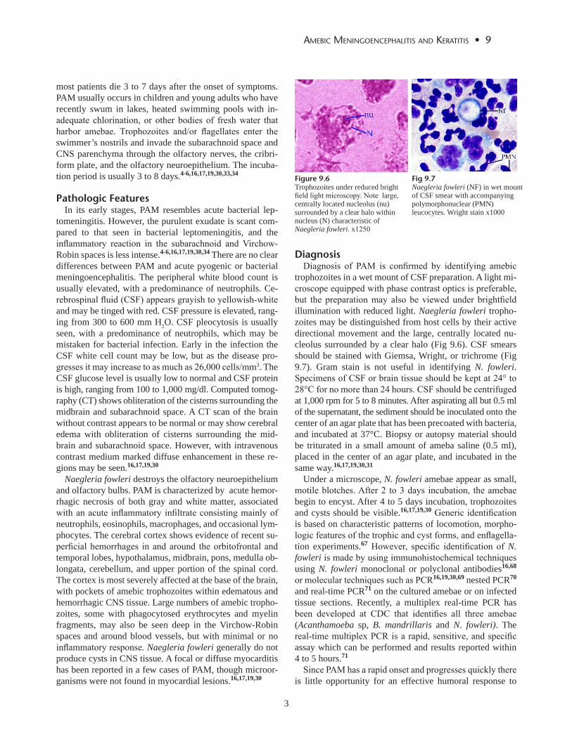
Fig 9.7 Naegleria fowleri (NF) in wet mount of CSF smear with accompanying polymorphonuclear (PMN) leucocytes. Wright stain x1000
Figure 9.6Trophozoites under reduced bright field light microscopy. Note large, centrally located nucleolus (nu) surrounded by a clear halo within nucleus (N) characteristic of Naegleria fowleri. x1250
3
amebic meningoencephaliTis and KeraTiTis • 9
most patients die 3 to 7 days after the onset of symptoms. PAM usually occurs in children and young adults who have recently swum in lakes, heated swimming pools with in-adequate chlorination, or other bodies of fresh water that harbor amebae. Trophozoites and/or flagellates enter the swimmer’s nostrils and invade the subarachnoid space and CNS parenchyma through the olfactory nerves, the cribri-form plate, and the olfactory neuroepithelium. The incuba-tion period is usually 3 to 8 days.4-6,16,17,19,30,33,34
Pathologic FeaturesIn its early stages, PAM resembles acute bacterial lep-
tomeningitis. However, the purulent exudate is scant com-pared to that seen in bacterial leptomeningitis, and the inflammatory reaction in the subarachnoid and Virchow-Robin spaces is less intense.4-6,16,17,19,30,34 There are no clear differences between PAM and acute pyogenic or bacterial meningoencephalitis. The peripheral white blood count is usually elevated, with a predominance of neutrophils. Ce-rebrospinal fluid (CSF) appears grayish to yellowish-white and may be tinged with red. CSF pressure is elevated, rang-ing from 300 to 600 mm H2O. CSF pleocytosis is usually seen, with a predominance of neutrophils, which may be mistaken for bacterial infection. Early in the infection the CSF white cell count may be low, but as the disease pro-gresses it may increase to as much as 26,000 cells/mm3. The CSF glucose level is usually low to normal and CSF protein is high, ranging from 100 to 1,000 mg/dl. Computed tomog-raphy (CT) shows obliteration of the cisterns surrounding the midbrain and subarachnoid space. A CT scan of the brain without contrast appears to be normal or may show cerebral edema with obliteration of cisterns surrounding the mid- brain and subarachnoid space. However, with intravenous contrast medium marked diffuse enhancement in these re-gions may be seen.16,17,19,30
Naegleria fowleri destroys the olfactory neuroepithelium and olfactory bulbs. PAM is characterized by acute hemor-rhagic necrosis of both gray and white matter, associated with an acute inflammatory infiltrate consisting mainly of neutrophils, eosinophils, macrophages, and occasional lym-phocytes. The cerebral cortex shows evidence of recent su-perficial hemorrhages in and around the orbitofrontal and temporal lobes, hypothalamus, midbrain, pons, medulla ob-longata, cerebellum, and upper portion of the spinal cord. The cortex is most severely affected at the base of the brain, with pockets of amebic trophozoites within edematous and hemorrhagic CNS tissue. Large numbers of amebic tropho-zoites, some with phagocytosed erythrocytes and myelin fragments, may also be seen deep in the Virchow-Robin spaces and around blood vessels, but with minimal or no inflammatory response. Naegleria fowleri generally do not produce cysts in CNS tissue. A focal or diffuse myocarditis has been reported in a few cases of PAM, though microor-ganisms were not found in myocardial lesions.16,17,19,30
Diagnosis Diagnosis of PAM is confirmed by identifying amebic
trophozoites in a wet mount of CSF preparation. A light mi-croscope equipped with phase contrast optics is preferable, but the preparation may also be viewed under brightfield illumination with reduced light. Naegleria fowleri tropho-zoites may be distinguished from host cells by their active directional movement and the large, centrally located nu-cleolus surrounded by a clear halo (Fig 9.6). CSF smears should be stained with Giemsa, Wright, or trichrome (Fig 9.7). Gram stain is not useful in identifying N. fowleri. Specimens of CSF or brain tissue should be kept at 24° to 28°C for no more than 24 hours. CSF should be centrifuged at 1,000 rpm for 5 to 8 minutes. After aspirating all but 0.5 ml of the supernatant, the sediment should be inoculated onto the center of an agar plate that has been precoated with bacteria, and incubated at 37°C. Biopsy or autopsy material should be triturated in a small amount of ameba saline (0.5 ml), placed in the center of an agar plate, and incubated in the same way.16,17,19,30,31
Under a microscope, N. fowleri amebae appear as small, motile blotches. After 2 to 3 days incubation, the amebae begin to encyst. After 4 to 5 days incubation, trophozoites and cysts should be visible.16,17,19,30 Generic identification is based on characteristic patterns of locomotion, morpho-logic features of the trophic and cyst forms, and enflagella-tion experiments.67 However, specific identification of N. fowleri is made by using immunohistochemical techniques using N. fowleri monoclonal or polyclonal antibodies16,68 or molecular techniques such as PCR16,19,30,69 nested PCR70 and real-time PCR71 on the cultured amebae or on infected tissue sections. Recently, a multiplex real-time PCR has been developed at CDC that identifies all three amebae (Acanthamoeba sp, B. mandrillaris and N. fowleri). The real-time multiplex PCR is a rapid, sensitive, and specific assay which can be performed and results reported within 4 to 5 hours.71
Since PAM has a rapid onset and progresses quickly there is little opportunity for an effective humoral response to

Figure 9.8 a,b,c,dAcanthamoeba sp have a 2-stage life cycle consisting of trophozoite (a & b) and cyst (c & d), nucleus (N), contractile vacuole (CV), retractile projections (P). Phase contrast, a and c. x1000 Hematoxlin-eosin b and d. x1000
4
9 • Topics on The paThology of proTozoan and invasive arThropod diseases
Table 9.1 Morphologic features of Acanthamoeba sp that cause meningoencephalitis.
Group Species Cyst size Ectocysts Endocysts
I A. astronyxis 18 to 28 µm Nearly circular, gently rippled
Rays if stellate endocyst contact ectocyst in the same plane
II A. castellani, A. divionensis,A. polyphaga, A. rhysodes
<18 µm Prominently wrinkled
Stellate, polygonal,triangular, oval or round
III A. culbertsoni, A. healyi <18 µm Slightlywrinkled
Round or oval
develop against the amebae. However, in one case with a successful outcome, N. fowleri antibody of the IgM class was demonstrated with a titer of 1:4096.34
Treatment Two patients, one from the United States and the other
from Mexico, are known to have survived PAM.27,34 Other reports of survival are unconfirmed.16,17,19,30 The Ameri-can patient was treated aggressively with intrathecal and intravenous amphotericin B (to which N. fowleri is high-ly sensitive) and miconazole, and with oral rifampin.34 A Mexican boy was treated early with intravenous ampho-tericin B and fluconazole, and with oral rifampin.27 Early identification of the etiologic agent and aggressive therapy appear to be key to patient survival.
Acanthamoeba and Balamuthia Granulomatous Amebic Encephalitis (GAE)
Infectious AgentsAcanthamoeba sp have a 2-stage life cycle consisting
of trophozoite and cyst (Figs 9.8a to 9.8d). Acanthamoeba sp trophozoites are slightly larger than those of N. fowl-eri, measuring 15 µm to 45 µm in diameter. A contractile vacuole periodically ruptures on the body surface, which is covered with fine, spine-like, retractile projections (acan-thopodia). Trophozoites usually have a single nucleus with a large, dense nucleolus.67
Acanthamoeba sp divide by conventional mitosis, in which the nucleolus and the nuclear membrane disappear during cell division. The organism does not have a flagellate stage, but it differentiates into a cyst under adverse condi-tions. Cysts are uninucleate and have a proteinaceous, usu-ally wrinkled, ectocyst and an endocyst made of cellulose. At the point of contact between the ectocyst and endocyst there are pores, or ostioles, usually covered by opercula. The cysts are highly resistant to environmental pressures including desiccation.72 As many as 24 Acanthamoeba sp have been described. These species can be divided into 3 groups based on cyst size and morphology as follows: 1) species that produce large cysts (18 µm to 28 µm); 2) species that produce cysts smaller than 18 µm with promi-nently wrinkled ectocysts and stellate, polygonal, triangular, oval, or round endocysts; and 3) species that produce cysts smaller than 18 µm with slightly wrinkled ectocysts and round or oval endocysts 67 (Table 9.1). However, recent de-velopments in molecular analysis has enabled improvements in the taxonomy of this genus and its phylogenetic relation-ships. Further, based on the sequencing of the 18S rRNA gene,
a
a
d
b
c
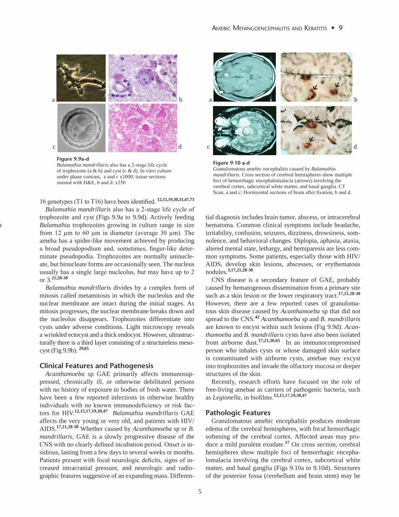
Figure 9.9a-d Balamuthia mandrillaris also has a 2-stage life cycle of trophozoite (a & b) and cyst (c & d). In vitro culture under phase contrast, a and c x1000; tissue sections stained with H&E, b and d. x250
Figure 9.10 a-d Granulomatous amebic encephalitis caused by Balamuthia mandrillaris. Cross section of cerebral hemispheres show multiple foci of hemorrhagic encephalomalacia (arrows) involving the cerebral cortex, subcortical white matter, and basal ganglia. CT Scan, a and c; Horinzontal sections of brain after fixation, b and d.
5
amebic meningoencephaliTis and KeraTiTis • 9
16 genotypes (T1 to T16) have been identified. 12,15,19,30,31,47,73
Balamuthia mandrillaris also has a 2-stage life cycle of trophozoite and cyst (Figs 9.9a to 9.9d). Actively feeding Balamuthia trophozoites growing in culture range in size from 12 µm to 60 µm in diameter (average 30 µm). The ameba has a spider-like movement achieved by producing a broad pseudopodium and, sometimes, finger-like deter-minate pseudopodia. Trophozoites are normally uninucle-ate, but binucleate forms are occasionally seen. The nucleus usually has a single large nucleolus, but may have up to 2 or 3.21,28-30
Balamuthia mandrillaris divides by a complex form of mitosis called metamitosis in which the nucleolus and the nuclear membrane are intact during the initial stages. As mitosis progresses, the nuclear membrane breaks down and the nucleolus disappears. Trophozoites differentiate into cysts under adverse conditions. Light microscopy reveals a wrinkled ectocyst and a thick endocyst. However, ultrastruc-turally there is a third layer consisting of a structureless meso-cyst (Fig 9.9b). 29,65
Clinical Features and PathogenesisAcanthamoeba sp GAE primarily affects immunosup-
pressed, chronically ill, or otherwise debilitated persons with no history of exposure to bodies of fresh water. There have been a few reported infections in otherwise healthy individuals with no known immunodeficiency or risk fac-tors for HIV.12,15,17,19,30,47 Balamuthia mandrillaris GAE affects the very young or very old, and patients with HIV/AIDS.17,21,28-30 Whether caused by Acanthamoeba sp or B. mandrillaris, GAE is a slowly progressive disease of the CNS with no clearly defined incubation period. Onset is in-sidious, lasting from a few days to several weeks or months. Patients present with focal neurologic deficits, signs of in-creased intracranial pressure, and neurologic and radio-graphic features suggestive of an expanding mass. Differen-
tial diagnosis includes brain tumor, abscess, or intracerebral hematoma. Common clinical symptoms include headache, irritability, confusion, seizures, dizziness, drowsiness, som-nolence, and behavioral changes. Diplopia, aphasia, ataxia, altered mental state, lethargy, and hemiparesis are less com-mon symptoms. Some patients, especially those with HIV/AIDS, develop skin lesions, abscesses, or erythematous nodules.3,17,21,28-30
CNS disease is a secondary feature of GAE, probably caused by hematogenous dissemination from a primary site such as a skin lesion or the lower respiratory tract.17,21,28-30 However, there are a few reported cases of granuloma-tous skin disease caused by Acanthamoeba sp that did not spread to the CNS.42 Acanthamoeba sp and B. mandrillaris are known to encyst within such lesions (Fig 9.9d). Acan-thamoeba and B. mandrillaris cysts have also been isolated from airborne dust.17,21,30,65 In an immunocompromised person who inhales cysts or whose damaged skin surface is contaminated with airborne cysts, amebae may excyst into trophozoites and invade the olfactory mucosa or deeper structures of the skin.
Recently, research efforts have focused on the role of free-living amebae as carriers of pathogenic bacteria, such as Legionella, in biofilms.12,15,17,19,30,47
Pathologic FeaturesGranulomatous amebic encephalitis produces moderate
edema of the cerebral hemispheres, with focal hemorrhagic softening of the cerebral cortex. Affected areas may pro-duce a mild purulent exudate.17 On cross section, cerebral hemispheres show multiple foci of hemorrhagic encepha-lomalacia involving the cerebral cortex, subcortical white matter, and basal ganglia (Figs 9.10a to 9.10d). Structures of the posterior fossa (cerebellum and brain stem) may be
a b
c d
a b
c d
a
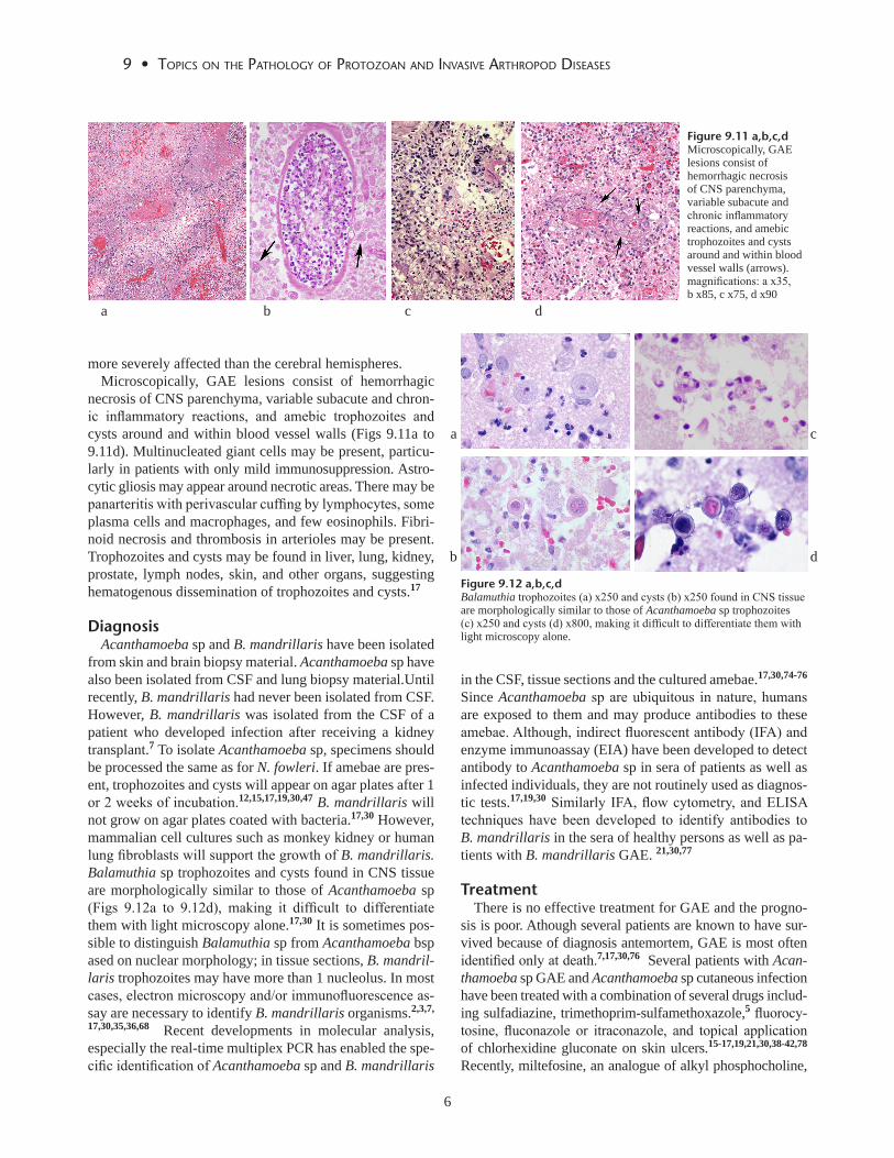
Figure 9.11 a,b,c,d Microscopically, GAE lesions consist of hemorrhagic necrosis of CNS parenchyma, variable subacute and chronic inflammatory reactions, and amebic trophozoites and cysts around and within blood vessel walls (arrows). magnifications: a x35, b x85, c x75, d x90
Figure 9.12 a,b,c,dBalamuthia trophozoites (a) x250 and cysts (b) x250 found in CNS tissue are morphologically similar to those of Acanthamoeba sp trophozoites (c) x250 and cysts (d) x800, making it difficult to differentiate them with light microscopy alone.
a b c d
6
9 • Topics on The paThology of proTozoan and invasive arThropod diseases
more severely affected than the cerebral hemispheres.Microscopically, GAE lesions consist of hemorrhagic
necrosis of CNS parenchyma, variable subacute and chron-ic inflammatory reactions, and amebic trophozoites and cysts around and within blood vessel walls (Figs 9.11a to 9.11d). Multinucleated giant cells may be present, particu-larly in patients with only mild immunosuppression. Astro-cytic gliosis may appear around necrotic areas. There may be panarteritis with perivascular cuffing by lymphocytes, some plasma cells and macrophages, and few eosinophils. Fibri-noid necrosis and thrombosis in arterioles may be present. Trophozoites and cysts may be found in liver, lung, kidney, prostate, lymph nodes, skin, and other organs, suggesting hematogenous dissemination of trophozoites and cysts.17
DiagnosisAcanthamoeba sp and B. mandrillaris have been isolated
from skin and brain biopsy material. Acanthamoeba sp have also been isolated from CSF and lung biopsy material.Until recently, B. mandrillaris had never been isolated from CSF. However, B. mandrillaris was isolated from the CSF of a patient who developed infection after receiving a kidney transplant.7 To isolate Acanthamoeba sp, specimens should be processed the same as for N. fowleri. If amebae are pres-ent, trophozoites and cysts will appear on agar plates after 1 or 2 weeks of incubation.12,15,17,19,30,47 B. mandrillaris will not grow on agar plates coated with bacteria.17,30 However, mammalian cell cultures such as monkey kidney or human lung fibroblasts will support the growth of B. mandrillaris. Balamuthia sp trophozoites and cysts found in CNS tissue are morphologically similar to those of Acanthamoeba sp (Figs 9.12a to 9.12d), making it difficult to differentiate them with light microscopy alone.17,30 It is sometimes pos-sible to distinguish Balamuthia sp from Acanthamoeba bsp ased on nuclear morphology; in tissue sections, B. mandril-laris trophozoites may have more than 1 nucleolus. In most cases, electron microscopy and/or immunofluorescence as-say are necessary to identify B. mandrillaris organisms.2,3,7,
17,30,35,36,68 Recent developments in molecular analysis, especially the real-time multiplex PCR has enabled the spe-cific identification of Acanthamoeba sp and B. mandrillaris
in the CSF, tissue sections and the cultured amebae.17,30,74-76 Since Acanthamoeba sp are ubiquitous in nature, humans are exposed to them and may produce antibodies to these amebae. Although, indirect fluorescent antibody (IFA) and enzyme immunoassay (EIA) have been developed to detect antibody to Acanthamoeba sp in sera of patients as well as infected individuals, they are not routinely used as diagnos-tic tests.17,19,30 Similarly IFA, flow cytometry, and ELISA techniques have been developed to identify antibodies to B. mandrillaris in the sera of healthy persons as well as pa-tients with B. mandrillaris GAE. 21,30,77
Treatment There is no effective treatment for GAE and the progno-
sis is poor. Athough several patients are known to have sur-vived because of diagnosis antemortem, GAE is most often identified only at death.7,17,30,76 Several patients with Acan-thamoeba sp GAE and Acanthamoeba sp cutaneous infection have been treated with a combination of several drugs includ-ing sulfadiazine, trimethoprim-sulfamethoxazole,5 fluorocy-tosine, fluconazole or itraconazole, and topical application of chlorhexidine gluconate on skin ulcers.15-17,19,21,30,38-42,78 Recently, miltefosine, an analogue of alkyl phosphocholine,
a
b
c
d

Figure 9.13Acanthamoeba keratitis, irregular corneal epithelial breakdown, stromal infiltrative keratitis, and edema.H&E x100
Figure 9.14 a,bCorneal biopsy. a. Early stages of Acanthamoeba keratitis showing destruction of the anterior corneal epithelium associated with acute inflammatory cells (mainly neutrophils) into the superficial and middle layers of the corneal stroma. x500 b. Trophozoites and cysts interspersed between the corneal lamellae. x500
7
amebic meningoencephaliTis and KeraTiTis • 9
has been used succefully to cure an HIV negative man with Acanthamoeba sp GAE and disseminated disease.37 Addi-tionally, voriconazole also has been used successfully to treat a lung transplant patient with cutaneous Acanthamoeba sp infection.79 Experimental in vitro studies indicate that both Acanthamoeba sp and B. mandrillaris are sensitive to pent-amidine isethionate, azithromycin/clarithromycin and milt-efosine.19,30,32,80-83 Several cases of B. mandrillaris GAE infection have survived after treatment with pentamidine, clarithromycin/azithromycin, fluconazole, sulfadiazine and 5-fluorocytosine.3,17,19,21,30,35,36,78,84 Recently, miltefosine has also been used successfully to cure B. mandrillaris GAE.7
a particular brand of multipurpose contact lens solution52 that did not have sufficient activity to kill Acanthamoeba sp cysts.85 Subsequently, this brand of contact lens solution was withdrawn by the company.52
AK causes severe ocular pain, a partial or total paracen-tral stromal ring infiltrate (Fig 9.13), recurrent corneal epi-thelial breakdown, and a corneal lesion resistant to common ophthalmic antibacterial medications. In its early stages, AK is frequently misdiagnosed as herpetic keratitis because of the irregular epithelial lesions, stromal infiltrative kerati-tis, and edema common to both forms of the disease. If left untreated, there is a greater risk of corneal perforation and GAE and enucleation. 17,19,30,44,45, 48
Corneal biopsy reveals that in the early stages of AK there is destruction of the anterior corneal epithelium asso-ciated with infiltration of acute inflammatory cells (mainly neutrophils) into the superficial and middle layers of the corneal stroma (Figs 9.14a & 9.14b). As the disease pro-gresses, there is considerable erosion of the corneal stroma, herniation of Descemet’s membrane, and perforation of the cornea. Trophozoites and cysts are interspersed between the corneal lamellae.17, 19,30,44,45,48
DiagnosisDiagnosis is confirmed by finding Acanthamoeba sp in
corneal scrapings fixed with methanol and stained with Gi-emsa, Hemacolor®, or trichrome.17,19,30,44,45,48 Acanthamoe-ba sp can also be isolated into culture on non-nutrient agar plates, as described above. Diagnosis has been established by direct confocal microscopy on the corneal surface.49
Treatment The current treatment of choice for AK is topical appli-
cation of a diamide (propamide isethionate or hexamide) and a cationic antiseptic (polyhexamethyl biguanide or chlorhexidine gluconate) for several weeks.50,53
a b
Acanthamoeba Keratitis
Acanthamoeba sp keratitis (AK) is a painful, vision-threat-ening disease of the cornea that, if not treated promptly, can lead to chronic ulceration of the cornea, loss of visual acuity, blindness, and enucleation. 12,17,19,30,43-45,48,50-52,78
Infectious AgentSpecies reported to cause corneal infections are usually as-
sociated with group II and genotype T4 and include, among others, Acanthamoeba hatchetti, A. polyphaga, A. castellanii, A. culbertsoni, and A. rhysodes, although not all species iso-lated from the environment are clinically relevant. More than 5,000 cases of Acanthamoeba sp keratitis have been reported worldwide, over half of them in the United States. The first case of AK was reported in 1973 in a Texas rancher with a history of trauma to the eye.46 Trophozoites and cysts of A. polyphaga were cultured from corneal scrapings and biopsy specimens. In the 1980s, an in-depth epidemiologic and case-control study revealed that the use of soft contact lenses and homemade saline solution were responsible for an increased incidence of Acanthamoeba sp keratitis.51 A recent outbreak of AK during 2004-2007 occurred in the United States. An in-depth epidemiological study found that it was associated with

Figure 9.15 a,bSappinia sp, Brain. Amebae are large, usually 40 µm or greater characterized by two nuclei (n) tightly apposed to one another. a. Giemsa. x1250 b. Phase contrast. x1250
Figure 9.16Sappinia sp, Brain. Cysts are also binucleate. Two nuclei (N). Phase contrast. x1000
8
9 • Topics on The paThology of proTozoan and invasive arThropod diseases
a b
Sappinia
Sappinia sp have been previously isolated from feces of humans, elk, bison and cattle. A single case of infection with Sappinia pedata, previously described as Sappinia diploidea, causing encephalitis in an immunocompetent male occurred in 2001.59 The patient developed headache, seizure, blurred vision, photophobia and vomiting; an MRI revealed a single space-occupying lesion. On examination of the excised lesion amebic trophozoites but not cysts were seen in the brain tissue. The patient was treated empirically with azithromycin, pentamidine isethionate, itraconazole and flucytosine and recovered with no neurological sequel-ae. Sappinia amebae are large and measure 40 µm or greater and are characterized by the presence of two nuclei tightly apposed to one another (Figs 9.15a & 9.15b). Cysts are also binucleate (Fig 9.16) and can survive adverse conditions in the environment. A recently developed real-time PCR detects both species of Sappinia but not other free-living amebae including Acanthamoeba sp, B. mandrillaris and N. fowleri or human DNA.60
9
amebic meningoencephaliTis and KeraTiTis • 9
References 1. Barete S, Combes A, De Jonckheere JH et al. Fatal disseminated Acanthamoeba
lenticulata infection in a heart transplant recipient. Emerg Infect Dis. 2007; 13:736-738.
2. Bakardjiev A, Azimi PH, Ashouri N et al. Amebic encephalitis caused by Balamuthia mandrillaris: Reporty of four cases. Pediatr Infect Dis. 2003;22:447-452
3. Bravo FG, Cabreara G, Gottuzo E, Visvesvara GS. Cutaneous manifestations of infections by free-living amebas. In: Tropical Dermatology, Tyring SK, Lupin O, Henggae UR, eds. Philadelphia: Elsvier; 2006, pp 49-55.
4. Butt CG. Primary amebic meningoencephalitis. N Engl J Med. 1966;274:1473-1476.
5. Carter RF. Primary amoebic meningo-encephalitis. An appraisal of present knowledge. Trans R Soc Trop Med Hyg. 1972;66:193-213.
6. Centers for Disease Control and Prevention (CDC). Primary amebic meningoencephalitis-Arizona, Florida, and Texas. 2007. Morb Mort Wkly Rep. 2008;57:573-577.
7. Centers for Disease Control and Prevention (CDC). Balamuthia mandrillaris transmitted through organ transplantation – Mississippi, 2009. Morb Mort Wkly Rep. 2010;59:1165-1170.
8. CentersforDiseaseControlandPrevention(CDC).Notesfromthefield:transplant transmitted Balamuthia mandrillaris – Arizona, 2010. Morb Mort Wkly Rep. 2010;59:1182.
9. Cogo PE, Scaglia M, Gatti S, et al. Fatal Naegleria fowleri meningoencephalitis, Italy. Emerg Infect Dis. 2004;10:1835-1837.
10. Gustavo dos Santos Neto. Fatal primary amebic meningoencephalitis. A retrospective study in Richmond, Virginia. Am J Clin Pathol. 1970;54:737-742.
11. Kaul DR, Lowe L, Visvesvara GS, et al. Acanthamoeba infection in a patient with chronic graft-vs-host disease occurring during treatment with voriconazole. Transplant Infect Dis. 2008; 10:437-441.
12. Khan NA. Acanthamoeba spp. In: Emerging Protozoan Pathogens. Khan NA, ed. New York, NY: Taylor and Francis; 2008, pp 71-118.
13. Kinde H, Read DH, Daft BM et al. Infections caused by pathogenic free-living amebas (Balamuthia mandrillaris and Acanthamoeba spp) in horses. J Vet Diagn Invest. 2007;19:317-322.
14. Lozano-Alarcon F, Bradley GA, Houser BS, Visvesvara GS. Primary amebic meningoencephalitis due to Naegleria fowleri in a South American tapir. Vet Pathol. 1997;34:239-243.
15. Marciano-Cabral F, Cabral G. Acanthamoeba spp. as agents of disease in humans. Clin Microbiol Rev. 2003;16:273-307.
16. Marciano-Cabral F, Cabral G. Naegleria fowleri. In: Emerging Protozoan Pathogens, Khan NA, ed. New York, NY: Taylor and Francis; 2008, pp 119-152.
17. Martinez AJ, Visvesvara GS. Free-living, amphizoic and opportunistic amebas. Brain Pathol. 1997;7:583-598.
18. Mckeller M, Mehta L, Greenlee J et al. Granulomatous Acanthamoeba encephalitis mimicking a stroke: correlation with sequential MRI and biopsy, invitroculture,immunofluorescenceandmolecularanalysis.J Clin Microbiol. 2006;44:265-269.
19. Schuster FL, Visvesvara GS. Free-living amoebae as opportunistic and non opportunistic pathogens of humans and animals. Int J Parasitol. 2004;34:1001-1027.
20. Schuster FL, Glaser C, Honarmand S, et al. Balamuthia encephalitis risk, Hispanic Americans. Emerg Infect Dis. 2004;10:1510-1512.
21. Schuster FL, Visvesvara GS. Balamuthia mandrillaris. In: Emerging Protozoan Pathogens, Khan NA, ed. New York, NY: Taylor and Francis; 2008, pp 71-118.
22. Schuster FL, Yagi S, Gavali S,et al. Under the radar: Balamuthia amebic encephalitis. Clin Infect Dis. 2009;48:879-887.
23. Shirwadkar CG, Samant R, Sankhe M, et al. Acanthamoeba encephalitis in a patient with systemic lupus, India. Emerg Infect Dis. 2006;12:984-986.
24. Silva-Vergara ML, Da Cunha Colombo ER, Vissoto E de F, et al. Disseminated Balamuthia mandrillaris amoeba infection in an AIDS patient from Brazil. Am J Trop Med Hyg. 2007;7:1096-1098.
25. Symmers WC. Primary amoebic meningoencephalitis in Britain. Br Med J. 1969;4:449-454.
26. TavaresM,CorreadaCostaJM,CarpenterS,etal.DiagnosisoffirstcaseofBalamuthiaamebicencephalitisinPortugalbyimmunofluorescenceandPCR.JClin Microbiol. 2006; 44:2660-2663.
27. Vargas-Zepeda J, Gomez-Alcala AV, Vasquez-Morales JA, et al. Successful treatment of Naegleria fowleri meningoencephalitis by using intravenous amphotericinB,fluconazoleandrifampicin.Arch Med Res. 2005;36:83-86.
28. Visvesvara GS, Martinez AJ, Schuster FL, et al. Leptomyxid ameba, a new agent of amebic meningoencephalitis in humans and other animals. J Clin Microbiol. 1990;28:2750-2756.
29. Visvesvara GS, Schuster FL, Martinez AJ. Balamuthia mandrillaris, n. g., n. sp., agent of amebic meningoencephalitis in humans and other animals. J Eukaryot Microbiol. 1993;40:504-514.
30. Visvesvara GS, Moura H, Schuster FL. Pathogenic and opportunistic free-living amoebae: Acanthamoeba spp., Balamuthia mandrillaris, Naegleria fowleri and Sappinian diploidea. FEMS Immunol Microbiol. 2007;50:1-26.
31. Visvesvara GS. Free-living amebae as opportunistic agents of human disease. J Neuroparastol. 2010:1:1-13.
32. Walochnik J, Obwaller A, Gruber F et al. Anti Acanthamoebaefficacyandtoxicity of miltefosine in an organotypic skin equivalent. Antimicrob Agents Chemother. 2009;64:539-545.
33. Yoder JS, Eddy BA, Visvesvara GS, et al. The epidemiology of primary amebic meningoencephalitis, 1962-2008. Epidemiol Infect. 2010;138:968-975.
34. Seidel JS, Harmatz P, Visvesvara GS, Cohen A, Edwards J, Turner J. Successful treatment of primary amebic meningoencephalitis. N Engl J Med. 1982;306:346-348.
35. Cary LC, Mauil E, Porter C, et al. Balamuthia mandrillaris meningoencephalitis: survival of a pediatric patient. Pediatrics. 2010;125:e699-e703.
36. Deetz TR, Sawyer MH, Billman G, Schuster FL, Visvesvara GS. Successful treatment of Balamuthia amoebic encephalitis: presentation of 2 cases. Clin Infect Dis. 2003;37:1304-1312.
37. Aichelburg ACV, Walochnik J, Assadian O, et al. Successful treatment of disseminated Acanthamoeba sp. infection with miltefosine. Emerg Infect Dis. 2008;14:1743-1746.
38. Fung KT-T, Dhillon AP, McLaughlin J, et al. Cure of Acanthamoeba cerebral abscess in a liver transplant patient. Liver Transplantation. 2008;14:308-312.
39. Martinez MS, Gonzalez-Madiero G, Santiago P, et al. Granulomatous amebic encephalitis in a patient with AIDS. Isolation of Acanthamoeba sp Group II frombrainandsuccessfultreatmentwithsulfadiazineandfluconazole.J Clin Microbiol. 2000;38:3892-3895.
40. Petry F, Torzewski M, Bohl J, et al. Early diagnosis of Acanthamoeba infection duringroutinecytologicalexaminationofcerebrospinalfluid. J Clin Microbiol. 2006;44:1903-1904.
41. Saxena A, Mittal S, Burman P, Garg P. Acanthamoeba meningitis with successful outcome. Ind J Pediatr. 2009;1063-1064.
42. Slater CA, Sickel JZ, Visvesvara GS, et al. Brief report:successful treatment of disseminated Acanthamoeba infection in an immunocompromised patient. New Engl J Med. 1994;331:85-87.
43. Bang S, Edell E, Eghrari AO, Gottsch JD. Treatment with voriconazole in 3 eyes with resistant Acanthamoeba keratitis. Am J Ophthalmol. 2010;149:66-69.
44. Clarke DW, Niederkorn JY. The pathophysiology of Acanthamoeba keratitis. Trends Parasitol. 2006;22:175-180.
45. Dart JKG, Saw VPJ, Kilvington S. Acanthamoeba keratitis: Diagnosis and Treatment Update 2009. Am J Ophthalmol. 2009;148:487-499.
46. Jones DB, Visvesvara GS, Robinson NM. Acanthamoeba polyphaga keratitis and Acanthamoeba uveitis associated with fatal meningoencephalitis. Trans Ophthalmol Soc U K. 1975;95:221-232.
47. Khan NA. Acanthamoeba spp.: In: Emerging Protozoan Pathogens, Khan NA, ed. New York, NY: Taylor and Francis; 2008; pp 71-118.
48. Panjwani N. Pathogenesis of Acanthamoeba keratitis. Occul Surf. 2010;8:70-79.49. PfisterDR,CameronJD,KrachmerJH,HollandEJ.Confocalmicroscopy
findingsofAcanthamoeba keratitis. Am J Ophthalmol. 1996;121:119-128.50. Seal DV. Acanthamoeba keratitis update – Incidence, molecular epidemiology
and new drugs for treatment. Eye. 2003;17:893-905.51. Stehr-Green JK, Bailey TM, Visvesvara GS. The epidemiology of
Acanthamoeba keratitis in the United States. Am J Ophthalmol. 1989;107:331-336.
52. Verani JR, Lorick SA, Yoder JS, et al. National outbreak of Acanthamoeba keratitis associated with use of contact lens solution, United States. Emerg Infect Dis. 2009;15:1236-1242.
53. Daft BM, Visvesvara GS, Read DH, Kinde H, Uzal FA, Manzer MD. Seasonal meningoencephalitis in Holstein cattle caused by Naegleria fowleri. J Vet Diagn Invest. 2005;17:605-609.
10
9 • Topics on The paThology of proTozoan and invasive arThropod diseases
79. Walia R, Montoya JG, Visvesvara GS, et al. A case of successful trteatment of infection of Acanthamoeba in a lung transplant recipient. Transplant Infect Dis. 2007; 9:51-54.
80. Schuster FL, Visvesvara GS. Axenic growth and drug sensitivity studies of Balamuthia mandrillaris, an agent of amebic meningoencephalitis in humans and other animals. J Clin Microbiol. 1996;34:385-388.
81. SchusterFL,VisvesvaraGS.Efficacyofnovelantimicrobialsagainstclinicalisolates of opportunistic amebas. J Eukaryot Microbiol. 1998;45:612-618.
82. Schuster FL, Guglielmo J, Visvesvara GS. In vitro activity of miltefosine and voriconazole on clinical isolates of free-living amebas: Balamuthia mandrillaris, Acanthamoeba spp., and Naegleria fowleri. J Eukaryot Microbiol. 2006;53:893-905.
83. Walochnik J, Duchene M, Seifert K, et al. Cytotoxic activities of alkylphosphocholines against clinical isolates of Acanthamoeba spp. Antimicrob Agents Chemother. 2002;46:695-701.
84. Jung S, Schelper RL, Visvesvara GS, Chang HT. Balamuthia mandrillaris meningoencephalitis in an immunocompetent patient: an unusual clinical course and a favorable outcome. Arch Pathol Lab Med. 2004;128:466-468.
85. Johnston SP, Sriram S, Qvarnstrom Y, et al. Resistance of Acanthamoeba cysts to disinfection in multiple contact lens solutions. J Clin Microbiol. 2009;47:2040-2045.
54. Finnin PJ, Visvesvara GS, Campbell BE, Fry DR, Gasser RB. Multifocal Balamuthia mandrillaris infection in a dog in Australia. Parasitol Res. 2007;100:423-426
55. Morales JA, Chavez AJ, Visvesvara GS, Dubey JP. Naegleria folwleri –associated encephalitis in a cow from Costa Rica. Vet Parasitol. 2006;139: 221-223.
56. Rideout BA, Gardiner CH, Stalis IH, et al. Fatal infections with Balamuthia mandrillaris (a free-living amoeba) in gorillas and other old world primates. Vet Pathol. 1997;34:15-22.
57. Visvesvara GS, De Jonckheere JF, Sriram R, et al. Isolation and molecular analysis of Naegleria fowleri from a cow brain that died of primary amebic meningoencephalitis. J Clin Microbiol. 2005;43:4203-4204.
58. Visvesvara GS, Shoff ME, Sriram R, et al. Isolation, morphologic, serologic andmolecularidentificationofAcanthamoeba T4 genotype from the liver of a Temminck’s tragopan (Tragopan temminckii). Vet Parasitol. 2010;170:197-200.
59. Gelman BB, Popov V, Chaljub G, et al. Neuropathological and ultrastructural features of amebic encephalitis caused by Sappinia diploidea. J Neuropathol Exp Neurol. 2003;62:990-998.
60. Qvarnstrom Y, da Silva AJ, Schuster FL, Gelman BB, Visvesvara GS. Molecular confirmationofSappinia pedata as causative agent of amebic encephalitis. J Infect Dis. 2009;199:1139-1142.
61. Culbertson CG, Smith JW, Minner JR. Acanthamoeba: observations on animal pathogenicity. Science. 1958;127:1506.
62. Stockman LJ, Wright CJ, Visvesvara GS, et al. Prevalence of Acanthamoeba spp. and other free-living amoebae in household water, Ohio, USA-1990-1992. Parasitol Res. 2010;108:621-627.
63. Bright KR, Marciano-Cabral F, Gerba CP. Occurrence of Naegleria fowleri in Arizona drinking water supply wells. J AWWA. 2009;101:43-50.
64. Dunnebacke TH, Schuster FL, Yagi S, et al. Balamuthia mandrillaris from soil samples. Microbiology. 2004;150:2837-2842.
65. Niyyati M, Lorenzo-Morales J, Rezaeian M, et al. Isolation of Balamuthia mandrillaris from urban dust, free of known infectious involvement. Parasitol Res. 2009;106:279-281.
66. Schuster FL, Dunnebacke TH, Booton CG, et al. Environmental isolation of Balamuthia mandrillaris associated with a case of amebic encephalitis. J Clin Microbiol. 2003:41:3175-3180.
67. Page FC. A New Key to Fresh Water and Soil Gymnamoebae. Cumbria, England: Fresh Water Biological Association; 1985:1-122.
68. Guarner J, Bartlett J, Shieh W-J, Paddock CD, Visvesvara GS, Zaki SR. Histopathologic spectrum and immunohistochemical diagnosis of amebic meningoencephalitis. Modern Pathol. 2007; 20:1230-1237.
69. Zhou L, Sriram S, Visvesvara GS, Xiao L. Genetic variation in the internal transcribed spacer and mitochondrial small subunit rRNA gene of Naegleria spp. J Eukaryot Microbiol. 2003;50:522-526.
70. Reveiller FL, Cabanes PA, Marciano-Cabral F. Development of a nested PCR assay to detect the pathogenic free-living amoeba Naegleria fowleri. Parasitol Res. 2002;88:443-450.
71. Qvarnstrom Y, Visvesvara GS, Sriram R, da Silva AJ. A multiplex real-time PCR assay for simultaneous detection of Acanthamoeba spp., Balamuthia mandrillaris and Naegleria fowleri. J Clin Microbiol. 2006;44:3589-3595.
72. Sriram R, Shoff M, Booton CG, et al. Survival of Acanthamoeba cysts after desiccation for more than 20 years. J Clin Microbiol. 2008;46:4045-4048.
73. BootonGC,VisvesvaraGS,ByersTJ,KellyDJ,FuerstPA.Identificationanddistribution of Acanthamoeba species genotypes associated with nonkeratitis infections. J Clin Microbiol. 2005;43:1689-1693.
74. Booton GC, Carmichael Jr., Visvesvara GS, et al. Genotyping of Balamuthia mandrillaris based on nuclear 18S and mitochondrial 16S rRNA genes. Am J Trop Med Hyg. 2003;68:65-69.
75. BootonGC,CarmichaelJR,VisvesvaraGS,ByersTJ,FuerstPA.Identificationof Balamuthia mandrillaris by PCR assay using the mitochondrial 16S rRNA gene as a target. J Clin Microbiol. 2003;41:453-455.
76. Yagi S, Schuster FL, Visvesvara GS. Demonstration of Balamuthia and Acanthamoeba mitochondrial DNA in sectioned archival brain and other tissues by the polymerase chain reaction. Parasitol Res. 2008;102:211-217.
77. Schuster FL, Yagi S, Wilkins PP, Gavali S, Visvesvara GS, Glaser CA. Balamuthia mandrillaris, agent of amebic encephalitis: detection of serum antibodies and antigenic similarity of isolates by enzyme immunoassay. J Eukaryot Microbiol. 2008;55:313-320.
78. Visvesvara GS. Amebic meningoencephalitideas and keratitis: challenges in diagnosis and treatment. Curr Op Infect Dis. 2010; 23;590-594.