ALTERED ARTERIAL HOMEOSTASIS AND C A :AR …pabrainspine.com/media/articles/Altered Arterial...
14
LITERATURE REVIEW ALTERED ARTERIAL HOMEOSTASIS AND CEREBRAL ANEURYSMS:AREVIEW OF THE LITERATURE AND JUSTIFICATION FOR A SEARCH OF MOLECULAR BIOMARKERS Amin Kassam, M.D. Departments of Neurosurgery and Otolaryngology, University of Pittsburgh, Pittsburgh, Pennsylvania Michael Horowitz, M.D. Departments of Neurosurgery and Radiology, University of Pittsburgh, Pittsburgh, Pennsylvania Yue-Fang Chang, Ph.D. Department of Neurosurgery, University of Pittsburgh, Pittsburgh, Pennsylvania David Peters, Ph.D. Department of Human Genetics, University of Pittsburgh, Pittsburgh, Pennsylvania Reprint requests: Amin Kassam, M.D., Department of Neurological Surgery, Presbyterian University Hospital, Suite B-400, 200 Lothrop Street, Pittsburgh, PA 15213. Email: [email protected] Received, April 17, 2003. Accepted, October 7, 2003. DESPITE THE CATASTROPHIC consequence of ruptured intracranial aneurysms, very little is understood regarding their pathogenesis, and there are no reliable predictive markers for identifying at-risk individuals. Given that intracranial aneurysms have a strong but complex genetic component and well-characterized modifiable risk factors, it seems likely that the most valuable approach to developing minimally invasive diagnostic and prognostic tools will involve a multifactorial model that includes both genetic and environmental risk factors. Unfortunately, the genetic basis of intracranial aneurysms is poorly described, and reports describing the association of nonrandom deoxyribonucleic acid sequence variation with intracranial aneurysms have been limited to a handful of ad hoc studies that have focused on a variety of markers in small populations. One reason for this lack of coordinated analysis of the genetic basis of intracranial aneurysms is that the molecular pathogenesis and pathobiological char- acteristics of the disease are poorly described, so candidate marker selection has been problematic. Few studies have addressed the molecular pathological basis of intracranial aneu- rysms or the possible mechanisms of intracranial aneurysm formation. In this regard, candidate gene selection strategies have relied almost exclusively on limited knowl- edge of monogenic disorders such as Ehlers-Danlos syndrome and Marfan’s syndrome, in which intracranial aneurysm is a feature of a spectrum of syndromic phenotypes. Without exception, these approaches have not affected the clinical identification and/or management of intracranial aneurysms significantly. Therefore, it is imperative that coordinated large-scale efforts in genetics, molecular biology, and genetic epide- miology are implemented to overcome these obstacles and drive developments in the field. In this review, we summarize the current screening modalities for intracranial aneurysms, review the current state of understanding relating to the genetic basis of intracranial aneurysms, and suggest a broader theory of aneurysm pathogenesis to form the foundation of a coordinated molecular search for biological markers that may be associated with aneurysm formation and rupture. KEY WORDS: Aneurysms, Literature review, Molecular pathogenesis Neurosurgery 54:1199-1212, 2004 DOI: 10.1227/01.NEU.0000119708.26886.55 www.neurosurgery-online.com D espite considerable advances in treatment, morbidity and mortality from cerebral aneurysms remain excep- tionally high. In the case of unruptured aneurysms, operative mortality has been reported to be as low as 2.5% and morbidity to be less than 6% (28, 29, 35, 37, 67). Once an aneurysm ruptures, however, subarachnoid hemorrhage (SAH) (89) occurs, and the impact becomes much more dev- astating (21, 70). Aneurysmal SAH involves catastrophic in- tracranial bleeding with immediate and delayed sequelae. Between 30 and 50% of patients die after SAH, and 40 to 50% of survivors have significant neurological deficits resulting in severe disability. To reduce morbidity and mortality rates, it is vital to iden- tify a cost-effective means of effectively screening patients at NEUROSURGERY VOLUME 54 | NUMBER 5 | MAY 2004 | 1199
Transcript of ALTERED ARTERIAL HOMEOSTASIS AND C A :AR …pabrainspine.com/media/articles/Altered Arterial...
LITERATURE REVIEW
ALTERED ARTERIAL HOMEOSTASIS
AND CEREBRAL ANEURYSMS: A REVIEW OF THE
LITERATURE AND JUSTIFICATION FOR A SEARCH
OF MOLECULAR BIOMARKERS
Amin Kassam, M.D.Departments of Neurosurgery andOtolaryngology, University ofPittsburgh,Pittsburgh, Pennsylvania
Michael Horowitz, M.D.Departments of Neurosurgery andRadiology, University of Pittsburgh,Pittsburgh, Pennsylvania
Yue-Fang Chang, Ph.D.Department of Neurosurgery,University of Pittsburgh,Pittsburgh, Pennsylvania
David Peters, Ph.D.Department of Human Genetics,University of Pittsburgh,Pittsburgh, Pennsylvania
Reprint requests:Amin Kassam, M.D.,Department of NeurologicalSurgery,Presbyterian UniversityHospital,Suite B-400,200 Lothrop Street,Pittsburgh, PA 15213.Email: [email protected]
Received, April 17, 2003.
Accepted, October 7, 2003.
DESPITE THE CATASTROPHIC consequence of ruptured intracranial aneurysms, verylittle is understood regarding their pathogenesis, and there are no reliable predictivemarkers for identifying at-risk individuals. Given that intracranial aneurysms have astrong but complex genetic component and well-characterized modifiable risk factors,it seems likely that the most valuable approach to developing minimally invasivediagnostic and prognostic tools will involve a multifactorial model that includes bothgenetic and environmental risk factors. Unfortunately, the genetic basis of intracranialaneurysms is poorly described, and reports describing the association of nonrandomdeoxyribonucleic acid sequence variation with intracranial aneurysms have beenlimited to a handful of ad hoc studies that have focused on a variety of markers in smallpopulations. One reason for this lack of coordinated analysis of the genetic basis ofintracranial aneurysms is that the molecular pathogenesis and pathobiological char-acteristics of the disease are poorly described, so candidate marker selection has beenproblematic.
Few studies have addressed the molecular pathological basis of intracranial aneu-rysms or the possible mechanisms of intracranial aneurysm formation. In this regard,candidate gene selection strategies have relied almost exclusively on limited knowl-edge of monogenic disorders such as Ehlers-Danlos syndrome and Marfan’s syndrome,in which intracranial aneurysm is a feature of a spectrum of syndromic phenotypes.Without exception, these approaches have not affected the clinical identificationand/or management of intracranial aneurysms significantly. Therefore, it is imperativethat coordinated large-scale efforts in genetics, molecular biology, and genetic epide-miology are implemented to overcome these obstacles and drive developments in thefield. In this review, we summarize the current screening modalities for intracranialaneurysms, review the current state of understanding relating to the genetic basis ofintracranial aneurysms, and suggest a broader theory of aneurysm pathogenesis toform the foundation of a coordinated molecular search for biological markers that maybe associated with aneurysm formation and rupture.
KEY WORDS: Aneurysms, Literature review, Molecular pathogenesis
Neurosurgery 54:1199-1212, 2004 DOI: 10.1227/01.NEU.0000119708.26886.55 www.neurosurgery-online.com
Despite considerable advances in treatment, morbidityand mortality from cerebral aneurysms remain excep-tionally high. In the case of unruptured aneurysms,
operative mortality has been reported to be as low as 2.5% andmorbidity to be less than 6% (28, 29, 35, 37, 67). Once ananeurysm ruptures, however, subarachnoid hemorrhage(SAH) (89) occurs, and the impact becomes much more dev-
astating (21, 70). Aneurysmal SAH involves catastrophic in-tracranial bleeding with immediate and delayed sequelae.Between 30 and 50% of patients die after SAH, and 40 to 50%of survivors have significant neurological deficits resulting insevere disability.
To reduce morbidity and mortality rates, it is vital to iden-tify a cost-effective means of effectively screening patients at
NEUROSURGERY VOLUME 54 | NUMBER 5 | MAY 2004 | 1199

risk for the development of intracranial aneurysms. This is anecessary first step toward early intervention and, ideally,prevention. We think that this step probably will occur at amolecular biological level.
There is tremendous variability in the genetic makeup ofeach individual’s organs. In this regard, vascular tissue issimilar to organs and should be considered an organ ratherthan an inanimate conduit of blood. Thus, vascular tissue issubject to the same molecular control mechanisms as anyother tissue in the body. Individuals who develop intracranialaneurysms may be genetically vulnerable because of an im-paired ability to respond at a molecular level to the stressfactors that affect their particular cerebrovasculature.
PREVALENCE, CLINICAL CONSEQUENCES,AND COST OF CEREBRAL ANEURYSMS
Intracranial aneurysms are relatively common, with a prev-alence estimated to be as high as 6% on the basis of autopsyspecimens (11, 81). Each year in the United States, more than28,000 people have SAH from aneurysm rupture (89). Unfor-tunately, 50% of these patients die either at the moment ofrupture or shortly thereafter. Another 25% have devastatingneurological complications, resulting in loss of motor or cog-nitive function that necessitates institutionalization and im-poses enormous costs on both the patients and the system thatmust deliver care for their chronic condition. The remaining25% may experience protracted hospitalization and some de-gree of cognitive or neurological impairment. In contrast, in-tracranial aneurysms that are repaired before they rupturehave a reported mortality as low as 2.5% and morbidity lessthan 6% (68).
From an economic perspective, the cost of acute care forpatients with ruptured cerebral aneurysms is inordinatelyhigh. Hospital expenses to provide acute care for just 112patients after SAH are estimated at $2.8 million. The majorityof these costs are for intensive care (18). Again, the estimatedcosts involved in caring for a patient with a ruptured aneu-rysm far exceed those for the care of a patient whose aneu-rysm is treated before it ruptures ($38,000 versus $12,685) (18).
A patient’s neurological condition at presentation isstrongly associated with outcome (8). Because there is nopredictable way to determine how an individual will respondneurologically to a rupture, clearly it would be advantageousto identify all intracranial aneurysms before rupture. To justifyintervention before rupture, however, we must first under-stand the actual risk of rupture. If an intracranial aneurysmhas been discovered incidentally, what is the true risk ofrupture? The recent results of the International Study of Un-ruptured Aneurysms indicate that intracranial aneurysms lessthan 1 cm in diameter are associated with an exceptionally lowrisk of rupture, which suggests that they should be managedconservatively (33). This conclusion has been met with con-siderable controversy, however, and it has not been accepteduniformly. There is significant evidence to suggest that the
majority of aneurysms that rupture are less than 1 cm indiameter (53, 69).
We think that it is naive to accept that a simple black-and-white image estimating size, such as an arteriogram, is anadequate means of predicting probability of rupture. The dy-namic molecular environment of the vasculature, ultimatelycontrolled by gene expression, may prove to be more predic-tive of aneurysms that are most likely to rupture. Further-more, by understanding the molecular alterations within ananeurysm, we may elucidate the pathogenesis of aneurysmdevelopment and rupture. After the pathophysiology hasbeen elucidated, physicians may develop novel therapies thatgenetically and chemically alter the vessel so that it can repairitself, thus avoiding more invasive therapy such as surgicalclipping or endovascular embolization. We think intracranialaneurysms represent an ideal disease model for screening andprevention, in that effective screening and early interventionmay avert the devastating consequences of rupture.
SCREENING
Current Concepts
Attempts to identify a high-risk patient group with intra-cranial aneurysms that would be amenable for screening be-gan with the 1954 description by Chambers et al. (8) of afamilial cluster. Several radiological studies have emerged tobetter estimate the risk of intracranial aneurysms in familieswithin which at least one relative has a known aneurysm.Much of the defining work analyzed the Finnish population,whose limited migration and well-characterized genetics pro-vide a relatively stable group that is ideal for study (70). In themost comprehensive study to date, Ronkainen et al. (68) usedmagnetic resonance angiography (MRA) to screen 400 patientswho had two or more first-degree relatives (e.g., parents,siblings) with a documented cerebral aneurysm. MRA re-vealed aneurysms in 37 asymptomatic individuals, accountingfor a 9.25% yield in this group (68). Although not ideal, thisfigure is thought to be the best estimate of familial cerebralaneurysms.
It should be noted that the excellent study design ofRonkainen et al. (68) revealed many of the potential limita-tions of MRA, demonstrating it to be far from an optimalscreening modality even in this high-risk group. They used aprospective model with three independent observers to obtaininter- and intrarater reliability with cross-correlation with dig-ital subtraction angiography for all the MRA-positive lesions.On this basis, they identified four patients with MRA evidenceof aneurysms that were revealed as vessel loops by digitalsubtraction angiography, representing a false-positive rate ofapproximately 10%. This rate is very similar to that obtainedin a study by Blatter et al. (3a), who were able to confirm withconventional angiography only 14 of 18 aneurysms that hadbeen identified by MRA. Digital subtraction angiographyidentified four aneurysms that MRA failed to disclose. This10% false-negative rate is probably an underestimation, be-
KASSAM ET AL.
1200 | VOLUME 54 | NUMBER 5 | MAY 2004 www.neurosurgery-online.com

cause not all of the MRA-negative individuals were subjectedto the “gold standard,” invasive angiography. Therefore, someof the MRA-negative patients may have harbored an undetec-ted aneurysm. Furthermore, although the inter-rater reliabilitywas acceptable, ranging from a � value of 0.59 to 0.80 amongthe three readers, Ronkainen et al. astutely recognized thatone reader consistently labeled MRA-questionable lesions asnegative, thus reporting fewer positives overall. This illus-trates the subjective nature implicit in MRA.
Even in the case of familial associations (high-risk popula-tion), controversy exists regarding screening candidates andtimetables. Current recommendations are that individuals bescreened if they have two immediate family members knownto have cerebral aneurysms. However, several studies suggestthat screening be broadened to include patients with only asingle afflicted family member (71). To further complicate thematter, on the basis of an estimated incidence of 2% de novoaneurysm formation (39), there is a question of how often thescreening should be repeated in initially negative patients.Recommendations range from every 6 months to every 5 yearsfor those with a strong family history (71). Furthermore, datafrom the International Study of Unruptured Aneurysms indi-cate that it may not be sufficient to identify a patient withMRA evidence of an intracranial aneurysm; the group most atrisk of rupture also must be known.
Aside from these concerns associated with screening, evenin this selected high-risk population with a family history ofintracranial aneurysm, the high costs and limited availabilityof MRA make it an unrealistic modality for mass screening ofthe general population. Therefore, the normative cost-benefitanalysis mandates a screening program that is convenient,inexpensive, objective, repeatable, and safe. A biologicalmarker seems ideal in this context.
Despite many shortcomings, familial screening studies pro-vide the important first step of definitively demonstrating thatthe disease has an increased tendency to cluster in families,and findings clearly implicate molecular factors in the devel-opment of intracranial aneurysms. The exact pattern of inher-itance is unclear. The consanguinity studies reported from theSaguenay region in Quebec suggest an autosomal dominantmode of transmission, although this has been disputed (15).On the basis of the different models of inheritance, the causefor this predisposition probably is genetically heterogeneous.This heterogeneity is manifest in the various syndromes asso-ciated with increased prevalence of intracranial aneurysms.An analysis of the genetic and molecular biology of theseconditions can narrow the search for the specific abnormalitiesassociated with intracranial aneurysms. This approach hasidentified the extracellular matrix of the arterial wall as themost likely site of molecular alterations implicated in aneu-rysm formation.
Epidemiological and Environmental Risk Factors
The identified epidemiological risk factors for the develop-ment of intracranial aneurysms include increasing age, ciga-
rette smoking, alcohol consumption, sex hormones, and pres-ence of systolic hypertension. Cigarette smoking is the mostconsistent, modifiable risk factor. The risk among currentsmokers is 3- to 10-fold higher than among those who neversmoked, with the risk increasing in a dose-dependent manner(5, 38, 45). The mechanism by which smoking increases risk isnot known, but it may be related to cigarette oxidant inacti-vation of �-1-antitrypsin, a major circulating inhibitor ofserine proteases (64, 65). An imbalance between proteases andantiproteases may lead to increased proteolysis of connectivetissue elements of the arterial wall and extracellular matrixelements supporting the architecture of the cerebral arteries,thus disrupting the normal homeostatic mechanism control-ling vascular repair. Several studies support this theory (71–73), including our own (57, 78), which demonstrated an in-creased frequency of genetically determined �-1-antitrypsindeficiency among patients with SAH. Furthermore, Gaetani etal. (22, 23) observed that although immunologically deter-mined levels of �-1-antitrypsin are elevated in patients withSAH, the collagenase inhibitory capacity in such patients islow, indicating a functional deficiency of �-1-antitrypsin.
Heavy alcohol use also is associated with SAH. Like smok-ing, heavy alcohol consumption acts in a dose-dependentmanner (86), with a relative risk (RR) of 2.8 (95% confidenceinterval [CI], 2.1–3.6) for consumption of less than 150 g perweek and RR of 4.7 (95% CI, 2.1–10.5) for consumption of 150 gper week or more. No mechanism for the increased incidenceof SAH among drinkers is clear, but a significant associationbetween smoking and alcohol use is known, and most studiesfail to adjust for cigarette smoking.
The age-specific rate of SAH is higher in men than in pre-menopausal women, but it is higher in postmenopausalwomen than in men of the same age. Hormone replacementtherapy modestly reduces the risk among postmenopausalwomen (RR range, 0.5–0.6), and oral contraceptive use mod-estly increases the risk (RR range, 1.4–1.5) (36, 46, 58, 79, 87,90).
Hypertension is perhaps the most commonly studied riskfactor for the development and rupture of intracranial aneu-rysms. Longitudinal studies have demonstrated that hyper-tension increases the risk of SAH approximately 2.8-fold (95%CI, 2.1–3.6) (41, 75, 91), and RR is 2.9 (95% CI, 2.4–3.7) inpopulation-based case-control studies (5, 46, 85). The risk ofoccurrence or rupture of an intracranial aneurysm associatedwith hypertension seems to be less than that associated withsmoking.
None of the known epidemiological factors provides thepredictive power to identify individuals with sufficiently ele-vated risk to make presymptomatic screening by current im-aging techniques cost effective (21, 68). Specifically, it wouldnot be cost effective to use MRA to screen every patient whohas hypertension or who smokes. It is possible that epidemi-ological factors are implicated in the pathogenesis of intracra-nial aneurysms via their impact on the preprogrammed mo-lecular environment of vascular tissue.
ARTERIAL HOMEOSTASIS AND ANEURYSMS
NEUROSURGERY VOLUME 54 | NUMBER 5 | MAY 2004 | 1201

Theoretical Impact of Hypertension on MolecularArterial Homeostasis
An example of the interaction between environmental fac-tors and genetic activity is provided by the potential impact ofhypertension on the local cerebrovasculature and the responseat a molecular level. Both Schneider et al. (74) and Sorteberg etal. (77) independently demonstrated that carotid artery clo-sure led to an instantaneous drop in ipsilateral intracranialblood flow velocities, whereas contralateral velocities in-creased. We suggest that this regional state of hypertensionwill initiate a molecular response, on the basis of the individ-ual’s genetic programming, to maintain cerebrovascular ho-meostasis. The response probably involves the activation ofNO pathways in the endothelium and their effects on smoothmuscle cell relaxation through hyperpolarization via potas-sium channel activation (74). As illustrated in Figure 1,changes in flow velocity (secondary to hypertension) causechanges in shear stress, which in turn deform the endothe-lium, leading to an increase in intracellular calcium and acti-vation of nitric oxide synthase and phospholipase A2.
The activation of nitric oxide synthase leads to the synthesisof NO, which diffuses to the smooth muscle cell and activatesthe calcium-dependent K� pump and cyclic guanosine mono-phosphate production. This process also leads to K� pumpactivation and stimulation of cyclic adenosine monophosphateproduction, resulting in adenosine triphosphate-dependentand delayed-rectifier K� channels. The increase in the perme-ability of K� channels causes hyperpolarization of the smoothmuscle cell and relaxation. As a result of activation of phos-pholipase A2, hydrolysis of phospholipids, specifically, phos-
phatidylinositol, occurs. Consequently, arachidonic acid accu-mulates, which leads to the generation of prostacyclin via theaction of cyclooxygenase and prostacyclin synthase. Prostacy-clin diffuses to a smooth muscle cell receptor, which activatescyclic adenosine monophosphate generation, thereby activat-ing adenosine triphosphate-dependent and delayed-rectifierK� channels, and thus contributing to the hyperpolarizationand relaxation of the smooth muscle cell.
This sequence represents the events that normally result inflow-dependent reduction of cerebrovascular tone. In patientswho develop cerebral aneurysms, these biochemical pathwaysmay be altered, exaggerated, and prolonged. There may beexcessive loss of vascular tone and a sustained increase inendothelial intracellular Ca2� levels, which could lead to en-dothelial injury and denuding of the vascular wall. This im-paired, or possibly overwhelmed, ability to maintain vascularintegrity may culminate in the formation of an intracranialaneurysm, particularly when the vasculature is exposed tocritical environmental factors such as hypertension.
The previous discussion exemplifies the potential interac-tion between the molecular environment of the vasculatureand physical factors that can affect this environment. In addi-tion to hypertension, all of the previously described epidemi-ological risk factors identified from prior studies must beconsidered in assessing any potential relationship betweenmolecular alterations and the development of intracranial an-eurysms. On the basis of our review of the literature andbiological considerations, the key epidemiological factorsidentified are smoking, hypertension, and sex.
The current literature does not support the isolated use of any ofthese epidemiological risk factors as a basis for screening aneurysmcandidates. These factors probably do not act alone, but they prob-ably influence the molecular environment of genetically predis-posed patients. The consequent impaired arterial homeostasis mayresult in aneurysm formation. It is hoped that identification of rel-evant molecular alterations of homeostasis will provide a series ofcandidate genetic markers. These will have to be tested for theirpredictive value in identifying individuals at risk for the develop-ment and rupture of cerebral aneurysms.
BIOLOGICAL RATIONALE FOR SELECTINGCANDIDATE MOLECULAR MARKERS
Before embarking on a search for genetic markers, a geneticbasis for the disease must be established. The next step is touse associated genetic syndromes to identify structural abnor-malities that could help guide the search for potential markers.The validity of selected screening markers then can be testedin a prospective controlled fashion. To date, the approach tounderstanding the molecular pathogenesis of intracranial an-eurysms has focused on individual markers on the basis ofpresumed structural relationships. Markers have been consid-ered candidates because of structural abnormalities in geneticsyndromes known to be associated with intracranial aneu-rysms. As discussed below, most of the structural alterations
FIGURE 1. Schematic view of pathways leading to NO and prostacyclin (PGI2)formation in endothelial cells. This diagram depicts these pathways and theireffects on smooth muscle cell tone. KCA, KATP, and Kdr, Ca2�-dependent,adenosine triphosphate-dependent, and delayed-rectifier K� channels, respec-tively; 20:4, arachidonic acid; ACh, acetylcholine; Bk, bradykinin; COX, cyclo-oxygenase; NOS, NO synthase; PCS, prostacyclin synthase; PL, phospholipid;PLA2, phospholipase A2; L-Arg, l-arginine (from, Hashimoto N, Kim C,Kikuchi H, Kojima M, Kang Y, Hazama F: Experimental induction of cerebralaneurysms in monkeys. J Neurosurg 67:903–905, 1987 [25]).
KASSAM ET AL.
1202 | VOLUME 54 | NUMBER 5 | MAY 2004 www.neurosurgery-online.com
associated with these syndromes are at the level of the extra-cellular matrix. This has led to a relatively uncoordinatedeffort to examine the molecular structure of the extracellularmatrix. This in turn has created a confusing body of literatureyielding conflicting reports, in which the same marker is as-sociated with intracranial aneurysms in one study and dis-counted in another (Table 1).
Aneurysms and Associated Conditions
The strongest association between cerebral aneurysms andother syndromes is with polycystic kidney disease. As in thosewho have spontaneous intracranial aneurysms, patients withpolycystic kidney disease have been reported to develop denovo aneurysms (9, 10, 32). The formation of a new aneurysmin these predisposed patients further suggests the possibilityof altered molecular response to some environmental factors.That is, genetic programming of the cerebrovasculature mayimpair the ability to respond to hemodynamic stressors. Anunderlying structural defect thus develops in the cerebralarteries, and new aneurysms form over time. The knownstructural defect in polycystic kidney disease is in polycystin,which is a membrane protein responsible for maintaining thestructural integrity of extracellular matrices of connective tis-sue (24, 76). At least two separate genes, PKD1 and PKD2,have been identified, and they are located on separate chro-mosomes, which suggests a multigenic process.
Neurofibromatosis Type 1 is another associated conditionthat provides further evidence of altered arterial structure. Thedefect in this condition is thought to be in the protein neuro-fibrin, which contains a central guanosine triphosphatase-activating protein domain. In the mouse model, this protein isimportant for cytoplasmic microtubules, and defects lead toarterial thinning and rupture (30).
In Marfan’s syndrome, a defect in the glycoproteinfibrillin-1 forms microfibrils. This protein is an important con-stituent of the extracellular matrix and is distributed through-out elastic tissue.
Fifty percent of individuals with intracranial aneurysmsreportedly have morphological derangements in the structuralproteins that provide mechanical integrity to the arterial walls(81). From the discussion above, alterations in syndromesassociated with intracranial aneurysms seem to be the result ofa multigenic process, thus suggesting genetic heterogeneity.Furthermore, the molecular alterations associated with aneu-rysm formation in these syndromic patients suggest a defect ofthe structural matrix proteins that form the framework of thearterial wall. The next question is: what within the arterialwall should be the focus?
LITERATURE-BASED GENETIC/MOLECULAR MARKERS
Type III Collagen
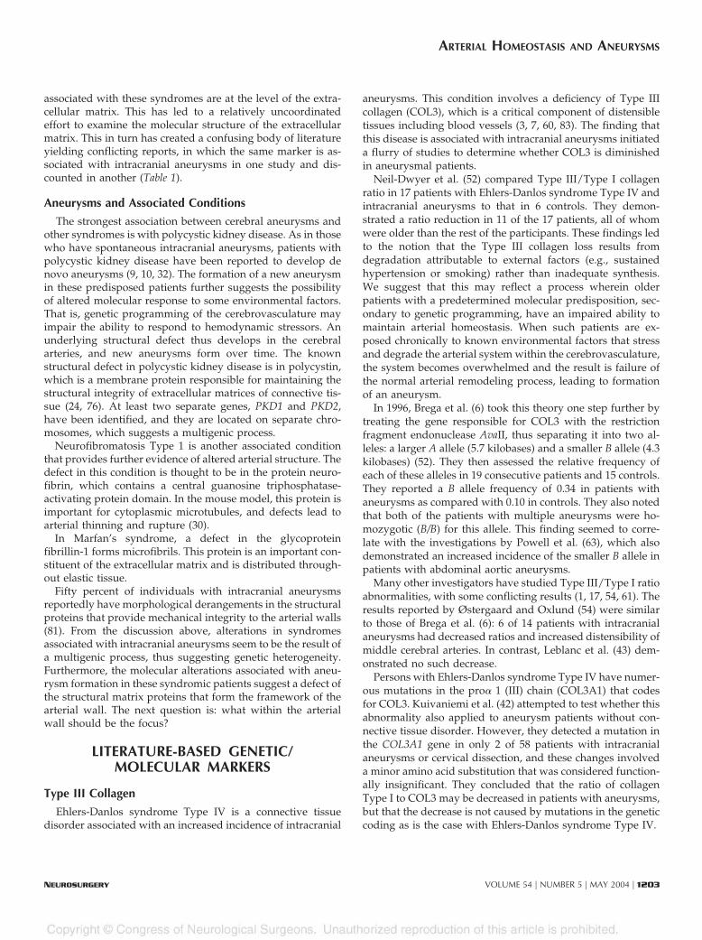
Ehlers-Danlos syndrome Type IV is a connective tissuedisorder associated with an increased incidence of intracranial
aneurysms. This condition involves a deficiency of Type IIIcollagen (COL3), which is a critical component of distensibletissues including blood vessels (3, 7, 60, 83). The finding thatthis disease is associated with intracranial aneurysms initiateda flurry of studies to determine whether COL3 is diminishedin aneurysmal patients.
Neil-Dwyer et al. (52) compared Type III/Type I collagenratio in 17 patients with Ehlers-Danlos syndrome Type IV andintracranial aneurysms to that in 6 controls. They demon-strated a ratio reduction in 11 of the 17 patients, all of whomwere older than the rest of the participants. These findings ledto the notion that the Type III collagen loss results fromdegradation attributable to external factors (e.g., sustainedhypertension or smoking) rather than inadequate synthesis.We suggest that this may reflect a process wherein olderpatients with a predetermined molecular predisposition, sec-ondary to genetic programming, have an impaired ability tomaintain arterial homeostasis. When such patients are ex-posed chronically to known environmental factors that stressand degrade the arterial system within the cerebrovasculature,the system becomes overwhelmed and the result is failure ofthe normal arterial remodeling process, leading to formationof an aneurysm.
In 1996, Brega et al. (6) took this theory one step further bytreating the gene responsible for COL3 with the restrictionfragment endonuclease AvaII, thus separating it into two al-leles: a larger A allele (5.7 kilobases) and a smaller B allele (4.3kilobases) (52). They then assessed the relative frequency ofeach of these alleles in 19 consecutive patients and 15 controls.They reported a B allele frequency of 0.34 in patients withaneurysms as compared with 0.10 in controls. They also notedthat both of the patients with multiple aneurysms were ho-mozygotic (B/B) for this allele. This finding seemed to corre-late with the investigations by Powell et al. (63), which alsodemonstrated an increased incidence of the smaller B allele inpatients with abdominal aortic aneurysms.
Many other investigators have studied Type III/Type I ratioabnormalities, with some conflicting results (1, 17, 54, 61). Theresults reported by Østergaard and Oxlund (54) were similarto those of Brega et al. (6): 6 of 14 patients with intracranialaneurysms had decreased ratios and increased distensibility ofmiddle cerebral arteries. In contrast, Leblanc et al. (43) dem-onstrated no such decrease.
Persons with Ehlers-Danlos syndrome Type IV have numer-ous mutations in the pro� 1 (III) chain (COL3A1) that codesfor COL3. Kuivaniemi et al. (42) attempted to test whether thisabnormality also applied to aneurysm patients without con-nective tissue disorder. However, they detected a mutation inthe COL3A1 gene in only 2 of 58 patients with intracranialaneurysms or cervical dissection, and these changes involveda minor amino acid substitution that was considered function-ally insignificant. They concluded that the ratio of collagenType I to COL3 may be decreased in patients with aneurysms,but that the decrease is not caused by mutations in the geneticcoding as is the case with Ehlers-Danlos syndrome Type IV.
ARTERIAL HOMEOSTASIS AND ANEURYSMS
NEUROSURGERY VOLUME 54 | NUMBER 5 | MAY 2004 | 1203

TABLE 1. Key literature examining molecular alterations associated with cerebral aneurysmsa
Series (ref. no.) Marker Description Analysis Findings Strengths/weaknesses
Neil-Dwyer et al., 1983(52)
Type III collagen Cases: 17 patientswith rupturedaneurysmsControls: 6 age-and sex-matchedcontrols (3gliomas and 3meningiomas)Tissue source:fibroblasts derivedfrom skin/temporalartery biopsies
Protein analysisExpression of collagen byfibroblastsRadioactive labelingCompare Type I/III ratiodeficiencies
Relative Type IIIcollagen deficiency in11 of 17 patientsNot all or none; agraded response withpartial loss in somepatients
First systematic study to analyzemolecular alterationsDid not examine aneurysmaltissueMolecular alterations in skin andsuperficial temporal artery mayhave nothing to do with regionalchanges in cerebrovasculatureadjacent to the aneurysm or inthe domeControls had brain tumors,which are known to havemolecular alterations withaltered genetic expression
Brega et al., 1996 (6) Type III collagen Cases: 19 patientswith aneurysmsControls: 15 fromDNA bankAnalyzeddifferences inexpression of TypeIII collagenSource: DNA fromwhole blood
DNA analysisCases: DNA extractedfrom whole bloodControls: DNA from aknown bankUsing restriction fragmentendonuclease AvaII,separated Type IIIcollagen into a larger Aallele and smaller B allele
B allele frequency of0.34 (cases) versus0.10 (controls)Both patients withmultiple aneurysmswere homozygotic (B/B) for this allele
First effective study at the DNAlevelControls had cystic fibrosis ormuscular dystrophy; no othermedical information knownNo distinction between ruptureand unruptured or adjustment inthe analysisNo adjustments or descriptionfor epidemiological risk factorsBlood-based DNA study assumesthat the regional molecularalterations at the dome andadjacent vasculature arereflected in the blood
Østergaard and Oxlund,1987 (54)
Type III collagen Cases: 14 patientswith fatal rupturedaneurysmsControls: 14 age-and sex-matchedsubjects who diedof other causesTissue source:postmortem tissuefrom the MCA
Mechanical distensibilityand protein analysisProtein analysis usingelectrophoresis forquantitative expression oftype III collagenMechanical analysis usingdistension of the tissue tocorrelate with forcesequivalent to bloodpressure between 100 and200 mm Hg
Protein analysis: 6 of14 patients withdeficiency of Type IIIcollagen comparedwith MCA tissue ofcontrolsMechanical analysis:Significant increase inthe extensibility of theMCA tissue comparedwith controls, but notassociated withalterations inmechanical strength
First study to demonstratealterations in mechanicaldistensibilityNo demographic considerationsor adjustments forepidemiological factors (e.g.,hypertension)No consideration of unrupturedaneurysmsPostmortem study; thereforesubject to secondary artifactsassociated with death; tissueexamined was not viableMCA tissue was harvested fromthe proximal vessel, withhemodynamic properties verydifferent from those at an arterialbifurcation
Leblanc et al., 1989 (43) Type I/III collagen Patient: 1 withpositive familyhistoryTissue source:fibroblast cultures
Fibroblast culturesQuantitative proteinanalysis usingelectrophoresis of TypeI/III collagen productionComparison group wascontrol cell lines
No difference in ratio Single-patient studyStrong family history; thereforemay not be generalizableRegulatory factors not taken intoaccount
Kuivaniemi et al., 1993(42)
Type III collagen Cases only; nocontrols40 patients withaneurysms and 18patients withcervical carotiddissectionTissue source: skinbiopsies, fibroblastcultures, DNAfrom blood
Skin biopsies, fibroblastcultures, RNA extraction,reverse transcriptasecDNA, then PCR analysisGenomic DNA alsoisolated from bloodAnalysis of DNApolymorphismsProtein analysis
No evidence ofpolymorphisms ormutations affectingType III collagen inpatients withaneurysms ordissections
Extremely thorough molecularanalysisSkin fibroblast and blood-basedDNA analysis may not representthe local changes at the circle ofWillis or in the domeNo stratification or adjustment toconsider rupture status orepidemiological dataNo controls
KASSAM ET AL.

Brega et al. (6), however, noted that Kuivaniemi et al. (42)analyzed only the gene’s central domain and did not effec-tively assess related modulating sequences, a concept thatformed the basis of our initial preliminary work, as discussedbelow. Therefore, although the structural domain of theCOL3A1 gene did not demonstrate significant changes in thestudy by Kuivaniemi et al., mutations in regulatory genes still
may lead to increased collagen degradation. Several studieshave demonstrated that the increased susceptibility of COL3to proteases results in increased collagen degradation (47).
Majamaa et al. (47) demonstrated no difference in the TypeIII/Type I ratio in 11 patients with intracranial aneurysms, butthey showed that the COL3 in two of these patients haddecreased thermal stability with increased degradation. Ana-
TABLE 1. Continued
Series (ref. no.) Marker Description Analysis Findings Strengths/weaknesses
Majamaa et al., 1992(47)
Type I/III collagen Cases: 11 patientswith rupturedaneurysms (6 withfamily history)Controls: 9patients; no detailsTissue source:skin-derivedfibroblasts
Protein analysis using cellcultures from fibroblastsfrom skinRNA extracted, Type I andIII collagen isolatedThermal stability assessedusing enzymatic assaysand digestion
No difference in TypeIII/I ratios2 of 11 patients hadincreased thermalinstability in Type IIIcollagen (both hadfamily history)None of the controlshad this increaseNo differences inthermal stability ofType I
Study looking beyond ratios andpossibility of structuralperformance alterationsVery contaminated study withfamily history in some and not inothersNo consideration ofepidemiological factors oradjustments for themNo information on controlsPoor consideration of statisticalanalysis techniques
Chyatte and Lewis, 1997(12)
Type III collagen Cases: 31 patientswith aneurysmsControls: 14craniotomies withMRA evidence ofno aneurysmTissue source: skinfibroblasts
Skin fibroblasts andserum-derived RNAreverse transcription toisolate Type III collagengeneTranslation to produceType III collagen proteinCollagen metabolismestimated using gelatinaseactivity
Type III collagensynthesis same in casesand controlsAneurysm patients had3� increase in serumgelatinase activity,suggesting increasedmetabolism of Type IIIcollagen
Excellent study showing possibleincrease in collagen degradationNot reflective of the vasculartissue in question, i.e., dome/adjacent vessels in the circle ofWillis
Peters et al., 1999 (57) MMP-9 Cases: 76 patientswith aneurysmsControls: pooledgenomic bank,representative ofthe studypopulation basedon demographicsSource: DNAderived fromperipheral blood
DNA analysisPatients were genotypedfor MMP-9 expressionPolymorphisms werenoted based on CA repeatsequences in the promotersequence proximal to thetranscriptional site of thegeneVarying expressedsequences were splicedinto a vector reportersystem using luciferaseassays to reflect thepromoter activity
Constructs bearing thelong (CA) repeatsequence had thegreatest associationwith cerebralaneurysms, suggestingthat this functionalpolymorphism wasassociated withcerebral aneurysms
Another study suggestingpossible increased degradationof structural matrix proteins, thistime based on a polymorphismin a regulatory sequence foundmore commonly in aneurysmpatientsVery little known about thecontrols (presence ofasymptomatic aneurysms)Blood-based DNA study andmay not be reflective of localchanges in the circle of Willis orat the aneurysm dome
Skirgaudas et al., 1996(76)
Cases: 10 patientswith aneurysms ofvarious typesControls: 3postmortem circleof Willis samples
Protein analysisImmunostainingVEGF, bFGF, fibronectin,Type IV collagen, �-smooth muscle actin
9 of 10 aneurysmspositive for bFGF and10 of 10 for VEGFNone of the controlswere positive for eithergrowth factorDomes displayeddisorganized stainingfor the structuralmatrix proteins
Study extended beyondstructural matrix proteins toinclude growth factorsLimited sample sizeMixed population of aneurysmsincluded berry, giant, andmycotic aneurysmsControls represented nonviablepostmortem tissues
a DNA, deoxyribonucleic acid; MCA, middle cerebral artery; RNA, ribonucleic acid; cDNA, complementary DNA; MMP-9, matrix metalloproteinase-9; PCR, polymerase chain reaction; VEGF,vascular endothelial growth factor; bFGF, basic fibroblast growth factor; MRA, magnetic resonance angiography.
ARTERIAL HOMEOSTASIS AND ANEURYSMS
NEUROSURGERY VOLUME 54 | NUMBER 5 | MAY 2004 | 1205

lyzing extracranial vessels, Dobrin et al. (17a) demonstratedthat human iliac arteries treated with elastase dilated but didnot rupture, whereas those pretreated with collagenase di-lated further to the point of rupture. Armed with this infor-mation, Chyatte and Lewis (12) attempted to determinewhether patients with cerebral aneurysms had decreased pro-duction or increased destruction of COL3. They demonstratednormal gene expression in all 31 aneurysm patients examined.However, with their use of gelatinase activity as a marker ofcollagen degradation, they demonstrated that patients withaneurysms had a threefold increase in enzymatic activity com-pared with controls.
Building on this foundation, we conducted a case-controlstudy, which demonstrated that a specific-length polymor-phism [(CA)23) in the promoter of Type IV collagenase (ma-trix metalloproteinase [MMP]-9) was nonrandomly associatedwith the occurrence of intracranial aneurysm (�2; P � 0.02)(57). Variation in the length of this repetitive element modu-lated promoter activity in an in vitro reporter assay, with thehighest promoter activity occurring in constructs bearing thelongest (CA)23 element. Further in vitro reporter analysesused MMP-9 promoter constructs in which the (CA) elementwas deleted or replaced with a nonrepetitive sequence. Thesestudies verified that this repetitive element is important forregulation of the promoter (57). Long MMP-9 alleles [(CA)23]resulted in higher levels of MMP-9 promoter activity and werepresent in greater frequency in patients with an intracranialaneurysm compared with healthy controls. This increasedfrequency may result in subtle differences in MMP-9 activityand consequent degradation of Type IV collagen within thecerebral vasculature, thereby increasing susceptibility to intra-cranial aneurysm formation. To our knowledge, our reportwas the first to describe a functional polymorphism in aregulatory molecule as associated with aneurysm formation.
Finally, when tissue from mice with collagen deficiency(secondary to dietary-induced lathyrism) was subjected invitro to hemodynamic stressors, aneurysm formation reliablyresulted (27, 62). This finding also supports the concept that agenetically heterogeneous process degrades key structuralproteins, thereby potentially predisposing vascular connectivetissue to become aneurysmal when subjected to hemodynamicforces. We think that the alterations observed in these extra-cellular matrix proteins are the result of a complex process ofarterial homeostasis and that they represent the net effect ofthis process. This leads to the question: what are the factorsthat regulate arterial homeostasis and result in the degrada-tion of these matrix proteins?
Other Candidate Molecular Markers
There is evidence of a continuous homeostatic mechanismthat repairs ongoing arterial wall (wear-and-tear) degradationand further evidence that disruption of this process of vascu-lar remodeling results in aneurysm formation (16, 19, 31).Skirgaudas et al. (76) attempted to characterize some of thefactors involved in this process and identify potential markers.
All 10 of their patients with aneurysms had increased levels ofvascular endothelial growth factor, and nine had increases inbasic fibroblast growth factor; none of the three controls hadincreased immunoreactivity for either of these factors. Bothfactors are known to be involved in the sprouting of newvessels from preexisting vascular beds (4, 14, 20). In additionto mediating vascular proliferation, these factors also affectmigration and adhesions to endothelial cells.
Vascular endothelial growth factor is produced by astro-cytes and is an important regulator of vascular permeabilitythrough activation of pathways that cause the enzymaticbreakdown of matrix proteins forming the vessel wall (2, 20,44, 66, 88). Fibrocytes and myocytes release basic fibroblastgrowth factor, which acts synergistically with vascular endo-thelial growth factor to further degrade matrix proteins. Thesetwo angiogenic factors may play an important role in vascularremodeling and maintaining structural integrity in response tostress and injury. Although the increased levels may simply bean epiphenomenon reflecting a general stress response, theimplications of matrix wall degeneration may provide an im-portant clue to understanding aneurysm pathogenesis.
On the basis of this information, Skirgaudas et al. (76)proceeded to the next step of characterizing the changes in thestructural wall proteins in aneurysm patients. They noted aregular band of Type IV collagen in the media and subendo-thelium of all control patients. This feature was absent inpatients with aneurysms. Instead, they saw faint and diffuseexpression of this protein within the media of the patientswith aneurysms, perhaps as a function of increased Type IVcollagenase activity. Fibronectin, another constituent of extra-cellular matrix, also had a disorganized arrangement in theaneurysm wall (76). Disruption of both of these basementmembrane proteins likewise has been described in a rat aneu-rysm model (77). This latter finding further suggests the pos-sibility of an enzymatic degradation process, perhaps associ-ated with increased angiogenesis factors and induction ofproteolysis. The resulting alteration within the matrix wallcompromises structural integrity and may culminate in aneu-rysm formation. If one accepts that there is constant flux ofrepair and degradation, the question arises: what mediatesthis homeostatic process?
MMPs
An important clue may come from the study of MMPs,which are endopeptidases with selective and specific activitiesagainst the extracellular matrix of basement membranes (66).These have been extensively studied in the dissemination ofmetastatic carcinomas, and three subgroups have been char-acterized: interstitial collagenase, stromelysin, and gelatinase(Type IV collagenase). On the basis of tumor invasion models,the metastatic cascade involves the following: 1) attachmentto the extracellular matrix; 2) creation of a proteolytic defect inthe extracellular matrix; and 3) migration through the proteo-lytically modified matrix.
KASSAM ET AL.
1206 | VOLUME 54 | NUMBER 5 | MAY 2004 www.neurosurgery-online.com
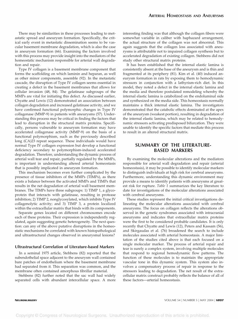
There may be similarities in these processes leading to met-astatic spread and aneurysm formation. Specifically, the crit-ical early event in metastatic dissemination seems to be vas-cular basement membrane degradation, which is also the casein aneurysm formation (66). Examining the factors involvedwith this process may provide insight into the mediators of thehomeostatic mechanism responsible for arterial wall degrada-tion and repair.
Type IV collagen is a basement membrane component thatforms the scaffolding on which laminin and heparan, as wellas other minor components, assemble (92). In the metastaticcascade, the disruption of Type IV collagen seems essential increating a defect in the basement membranes that allows forcellular invasion (48, 84). The gelatinase subgroups of theMMPs are vital for initiating this defect. As discussed earlier,Chyatte and Lewis (12) demonstrated an association betweencollagen degradation and increased gelatinase activity, and wehave confirmed functional polymorphic changes in Type IVcollagenase (MMP-9) in patients with aneurysms (57). Under-standing this process may be critical in finding the factors thatlead to disruption in the structural matrix proteins. Specifi-cally, persons vulnerable to aneurysm formation may haveaccelerated collagenase activity (MMP-9) on the basis of afunctional polymorphism, such as the previously describedlong (CA)23 repeat sequence. These individuals would havenormal Type IV collagen expression but develop a functionaldeficiency secondary to polymorphism-induced accelerateddegradation. Therefore, understanding the dynamic process ofarterial wall tear and repair, partially regulated by the MMPs,is important in understanding altered arterial homeostasisthat is possibly implicated in aneurysm formation.
This mechanism becomes even further complicated by thepresence of tissue inhibitors of the MMPs (TIMPs), as thereexists a balance between the activated MMPs and TIMPs thatresults in the net degradation of arterial wall basement mem-branes. The TIMPs have three subgroups: 1) TIMP 1, a glyco-protein that interacts with gelatinase, resulting in proteaseinhibition; 2) TIMP 2, nonglycosylated, which inhibits Type IVcollagenolytic activity; and 3) TIMP 3, a protein localizedwithin the extracellular matrix that binds with its components.
Separate genes located on different chromosomes encodeeach of these proteins. Their expression is independently reg-ulated, again suggesting genetic heterogeneity. The next ques-tion: can any of the above putative disruptions in the homeo-static mechanisms be correlated with known histopathologicaland ultrastructural changes observed in aneurysmal lesions?
Ultrastructural Correlation of Literature-based Markers
In a seminal 1975 article, Stehbens (82) reported that thesubendothelial space adjacent to the aneurysm wall containedbare patches of endothelium where the basement membranehad separated from it. This separation was common, and themembrane often contained amorphous fibrillar material.
Stehbens (82) further noted that the sac wall had widelyseparated cells with abundant intercellular space. A more
interesting finding was that although the collagen fibers weresomewhat variable in caliber with haphazard arrangement,the actual structure of the collagen was not abnormal. Thisagain suggests that the collagen loss associated with aneu-rysms is attributable not to impaired collagen synthesis but toaccelerated degradation of existing collagen. Stehbens did notstudy other structural matrix proteins.
It has been established that the internal elastic lamina isconsistently absent at the base of the aneurysm and is thin andfragmented at its periphery (81). Kim et al. (40) induced an-eurysm formation in rats by exposing them to hemodynamicstressors in conjunction with a lathyrism-rich diet. In thismodel, they noted a defect in the internal elastic lamina andthe media and therefore postulated remodeling whereby theinternal elastic lamina is catabolized on the endoluminal sideand synthesized on the media side. This homeostasis normallymaintains a thick internal elastic lamina. The investigatorsdemonstrated that the catabolic effects dominated at the apexof the aneurysm (weakest portion), resulting in degradation ofthe internal elastic lamina, which may be related to hemody-namic stressors activating predisposed bifurcation. They wereunable to identify the specific factors that mediate this processto result in an altered structural matrix.
SUMMARY OF THE LITERATURE-BASED MARKERS
By examining the molecular alterations and the mediatorsresponsible for arterial wall degradation and repair (arterialhomeostasis), it may be possible to identify molecular markersto distinguish individuals at high risk for cerebral aneurysms.Furthermore, understanding this dynamic environment mayprovide a means to identify patients with aneurysms at great-est risk for rupture. Table 1 summarizes the key literature todate for investigations of the molecular alterations associatedwith cerebral aneurysms.
These studies represent the initial critical investigations de-lineating the molecular alterations associated with cerebralaneurysms. The focus on collagen reflects the alterations ob-served in the genetic syndromes associated with intracranialaneurysms and indicates that extracellular matrix proteinswere the first to be considered probable candidates. It is onlyrecently that Chyatte and Lewis (12), Peters and Kassam (56),and Skirgaudas et al. (76) broadened the search to includemolecules associated with arterial homeostasis. A major limi-tation of the studies cited above is that each focused on asingle molecular marker. The process of arterial repair andtear is surely a complex system, involving multiple moleculesthat respond to regional hemodynamic flow patterns. Thefunction of these molecules is to maintain the appropriatevascular tone in this dynamic system. This system also in-volves a compensatory process of repair in response to thestressors leading to degradation. The net result of the extra-cellular matrix construct probably reflects the balance of all ofthese factors—arterial homeostasis.
ARTERIAL HOMEOSTASIS AND ANEURYSMS
NEUROSURGERY VOLUME 54 | NUMBER 5 | MAY 2004 | 1207

During the past decade, researchers have increasingly consid-ered molecules other than collagen as putative molecules criticalin aneurysm formation. We have considered the possibility thatthe ability of the cerebrovasculature to repair itself is impairedafter moment-to-moment microinjury. An important mediator inthis process is secreted protein rich in cysteines (SPARC) (os-teonectin), which is a counteradhesive glycoprotein expressed ina variety of tissues, including vascular endothelium, smoothmuscle, and fibroblasts. SPARC is known to inhibit endothelialcell adhesion and proliferation. SPARC and messenger ribonu-cleic acid levels are increased in renal vascular injury (59). It hasbeen proposed that extracellular matrix remodeling is regulatedin part by an interaction between SPARC and Type I collagen(34). We previously reported a differential expression of SPARCwithin aneurysmal tissue as compared with pericranial vascularcontrol tissue (56).
Another notable messenger ribonucleic acid in which ele-vated expression may be characteristic of intracranial aneu-rysms is cdc-rel2a/PNUTL2 (55). This factor belongs to anexpanding family of guanosine triphosphate-binding proteins,
called septins, which are thought to be involved in cytokinesis(13). Vinculin, an adhesion plaque protein, and c-Abl, a non-receptor tyrosine kinase recruited from the nucleus, both mi-grate to focal adhesion sites. Through their interaction withfibronectin, these proteins are implicated in the phosphoryla-tion of the protein paxillin. It is noteworthy that paxillin, anintegrin, is a substrate for SPARC-induced tyrosine phosphor-ylation (56).
A search for molecular markers that can be used for screen-ing purposes must take into account the entire spectrum ofarterial homeostasis that leads to the architectural failure ev-idenced by the well-studied alterations in extracellular matrixproteins. Table 2 summarizes these additional key markers andsuggests some novel markers on the basis of factors critical tomaintaining arterial homeostasis.
CONCLUSION
The need for screening markers that can predict the likeli-hood of aneurysm formation or rupture is obvious. We are at
TABLE 2. Key molecular markers, series, and justificationa
Molecular markerType of molecular (role
in homeostasis)Series (ref. no.) Comments/justification
Type III collagen(COL 3A1)—allele Aand allele B
Structural matrix protein(arterial integrity)
Neil-Dwyer et al., 1983 (52)Brega et al., 1996 (6)Østergaard and Oxlund, 1987 (54)Leblanc et al., 1989 (43)Kuivaniemi et al., 1993 (42)Majamaa et al., 1992 (47)Chyatte and Lewis, 1997 (12)
Initial molecule of interest based onstructural alterations in geneticsyndromes associated with cerebralaneurysmsControversial and conflicting results
Fibronectin, laminin,and heparan
Structural matrix proteins(arterial integrity)
Skirgaudas et al., 1996 (76)Stehbens, 1961 (80)
Poorly studied and implicated based onstructural matrix function
Elastase, �-1-antitrypsin
Proteases (arterialdegradation)
Schievink et al., 1994 (73)Peters et al., 1999 (57)St. Jean et al., 1996 (78)
Association studies linking smoking withaneurysm formation
Type IV collagenase(MMP-9)
Metalloprotease (arterialdegradation)
Peters et al., 1999 (57) Polymorphism with increased activity
VEGF and bFGF Angiogenesis factors(flow responsive andarterial repair)
Skirgaudas et al., 1996 (76) Regulators of arterial homeostasis
PSF Flow-responsivemolecule
Mizushima et al., 1996 (49)Hata et al., 2000 (26)Moncada et al., 1976 (51)Moncada et al., 1978 (50)
Role in flow-dependent vascular tonerelaxationNot yet directly studied in patients withaneurysm
RAI, PNUT, SPARC Tissue-reparativemolecules
Paavola et al., 1999 (55)Cooper and Kiehart, 1996 (13)Iruela-Arispe et al., 1996 (34)Pichler et al., 1996 (59)
Role in homeostasis by repairing arterialwall injuriesNot yet directly studied in patients withaneurysms
a MMP-9, matrix metalloproteinase-9; VEGF, vascular endothelial growth factor; bFGF, basic fibroblast growth factor; PSF, prostacyclin-stimulating factor; SPARC,secreted protein rich in cysteines.
KASSAM ET AL.
1208 | VOLUME 54 | NUMBER 5 | MAY 2004 www.neurosurgery-online.com

a relatively early but critical stage in defining these markers.The progression has been to develop epidemiological riskfactors and then radiological predictors of aneurysm rupture;so far, this has proven to be unreliable. The answer probablywill come from an analysis of the dynamic molecular milieuthat surrounds intracranial aneurysms.
The molecular search has focused predominantly on theextracellular matrix, which is a reflection of the collagen vas-cular syndromes associated with aneurysm formation. Wesuggest a broader search of the entire process of arterial ho-meostasis. There are at least three critical components to ho-meostasis: 1) flow modulation; 2) arterial wall degradationand repair; and 3) the extracellular matrix that results. In thisreview, we have discussed the current screening options; pre-sented the literature regarding the extracellular matrix, whichoften is confusing; and introduced the concept of arterialhomeostasis. A focus on molecular markers associated witharterial homeostasis is critically needed, and it is our hope thatthis review further fosters this pursuit.
REFERENCES
1. Acqui M, Ferrante L, Mastronardi L: Alteration of the collagen type III/type I ratio and intracranial saccular aneurysms in GH-secreting hypophy-seal adenomas. Ital J Neurol Sci 9:356–358, 1988.
2. Barrow DL, Reisner A: Natural history of intracranial aneurysms andvascular malformations. Clin Neurosurg 40:3–39, 1993.
3. Beighton P: The Ehlers-Danlos syndromes, in Beighton P (ed): McKusick’sHeritable Disorders of Connective Tissue. St. Louis, C.V. Mosby, 1993, ed 5, pp189–251.
3a. Blatter DD, Parker DL, Ahn SS, Bahr AL, Robison RO, Schwartz RB, JoleszFA, Boyer RS: Cerebral MR angiography with multiple overlapping thinslab acquisition: Part II—Early clinical experience. Radiology 183:379–389,1992.
4. Bobik A, Campbell JH: Vascular derived growth factors: Cell biology,pathophysiology, and pharmacology. Pharmacol Rev 45:1–42, 1993.
5. Bonita R: Cigarette smoking, hypertension and risk of subarachnoid hem-orrhage: A population-based case-control study. Stroke 17:831–835, 1986.
6. Brega KE, Seltzer WK, Munro LG, Breeze RE: Genotypic variations of typeIII collagen in patients with cerebral aneurysms. Surg Neurol 46:253–257,1996.
7. Byers PH: Ehlers-Danlos syndrome Type IV: A genetic disorder in manyguises. J Invest Dermatol 105:311–313, 1995.
8. Chambers WR, Harper BF, Simpson JR: Familial incidence of congenitalaneurysms of cerebral arteries: Report of cases of ruptured aneurysms infather and son. Jour A M A 155:358–359, 1954.
9. Chauveau D, Pirson Y, Verellen-Dumoulin C, Macnicol A, Gonzalo A,Grunfeld JP: Intracranial aneurysms in autosomal dominant polycystickidney disease. Kidney Int 45:1140–1146, 1994.
10. Chauveau D, Sirieix ME, Schillenger F, Legendre C, Grunfeld JP: Recurrentrupture of intracranial aneurysms in autosomal dominant polycystic kid-ney disease. BMJ 301:966–967, 1990.
11. Chyatte D: The epidemiology, genetics and clinical behavior of intracranialaneurysms, in Awad IA (ed): Current Management of Cerebral Aneurysms.Park Ridge, AANS, 1995, pp 1–20.
12. Chyatte D, Lewis I: Gelatinase activity and the occurrence of cerebralaneurysms. Stroke 28:799–804, 1997.
13. Cooper JA, Kiehart DP: Septins may form a ubiquitous family ofcytoskeletal filaments. J Cell Biol 134:1345–1348, 1996.
14. Criscuolo GR: The genesis of peritumoral vasogenic brain edema andtumor cysts: A hypothetical role for tumor-derived vascular permeabilityfactor. Yale J Biol Med 66:277–314, 1993.
15. de Braekelleer M, Pérusse L, Cantin L, Bouchard J-M, Mathieu J: A studyof inbreeding and kinship in intracranial aneurysms in the SaguenayLac-Saint-Jean region (Quebec, Canada). Ann Hum Genet 60:99–104, 1996.
16. de la Monte S, Moore G, Mong M: Risk factors for the development andrupture of intracranial berry aneurysms. Am J Med 78:957–964, 1985.
17. De Paepe A, van Landegem W, de Keyser F, de Reuck J: Association ofmultiple intracranial aneurysms and collagen type III deficiency. ClinNeurol Neurosurg 90:53–56, 1988.
17a. Dobrin PB, Baker WH, Gley WC: Elastolytic and collagenolytic studies ofarteries: Implications for the mechanical properties of aneurysms. ArchSurg 119:405–409, 1984.
18. Elliot JP, LeRoux PD, Ransom G, Winn R: Predicting length of hospital stayand cost by aneurysm grade on admission. J Neurosurg 85:388–391, 1996.
19. Ferguson G: Physical factors in the initiation, growth, and rupture ofhuman intracranial saccular aneurysms. J Neurosurg 37:666–677, 1972.
20. Ferrara N, Houck KA, Jakeman LB, Winer J, Leung DW: The vascularendothelial growth factor family of peptides. J Cell Biochem 47:211–218,1991.
21. Fogelholm R: Subarachnoid hemorrhage in middle-Finland: Incidence,early prognosis and indications for neurosurgical treatment. Stroke 12:296–301, 1981.
22. Gaetani P, Rodriguez Y, Baena R, Grignani G, Spanu G, Pacchiarini L,Paoletti P: Endothelin and aneurysmal subarachnoid hemorrhage: A studyof subarachnoid cisternal cerebrospinal fluid. J Neurol Neurosurg Psychi-atry 57:66–72, 1994.
23. Gaetani P, Rodriguez Y, Baena R, Quaglini S, Bellazzi R, Cafe C, Torri C,Marzatico F: Experimental subarachnoid hemorrhage: Events related toanti-oxidant enzymatic systems and eicosanoid peroxide enhancement.Neurochem Res 19:839–844, 1994.
24. Harris PC, Ward CJ, Peral B, Hughes J: Autosomal dominant polycystickidney disease: Molecular analysis. Hum Mol Genet 4:1745–1749, 1995.
25. Hashimoto N, Kim C, Kikuchi H, Kojima M, Kang Y, Hazama F: Experi-mental induction of cerebral aneurysms in monkeys. J Neurosurg 67:903–905, 1987.
26. Hata Y, Clermont A, Yamauchi T, Pierce EA, Suzuma I, Kagokawa H,Yoshikawa H, Robinson G, Ishibashi T, Hashimoto T, Umeda F, Bursell SE,Aiello LP: Retinal expression, regulation, and functional bioactivity ofprostacyclin-stimulating factor. J Clin Invest 106:541–550, 2000.
27. Hazama F, Hashimoto N: An animal model of cerebral aneurysms.Neuropathol Appl Neurobiol 13:77–90, 1987.
28. Heiskanen O: Risks of surgery for unruptured intracranial aneurysms.J Neurosurg 65:451–453, 1986.
29. Heiskanen O, Poranen A: Surgery of incidental intracranial aneurysms.Surg Neurol 28:432–436, 1987.
30. Henkemeyer M, Rossi DJ, Holmyard DP, Puri MC, Mbamalu G, Harpal K,Shih TS, Jacks T, Pawson T: Vascular system defects and neuronal apopto-sis in mice lacking RAS GTPase-activating protein. Nature 377:695–701,1995.
31. Holmes B, Harbaugh RE: Traumatic intracranial aneurysms: A contempo-rary review. J Trauma 35:855–860, 1993.
32. Hughes R, Chapman A, Rubinstein D, Stears J, Johnson A, Gabow P:Recurrent intracranial aneurysms (IA) in autosomal dominant polycystickidney disease (ADPKD). Stroke 27:178, 1996 (abstr).
33. International Study of Unruptured Intracranial Aneurysms Investigators:Unruptured intracranial aneurysms: Risk of rupture and risks of surgicalintervention—International Study of Unruptured Intracranial AneurysmsInvestigators. N Engl J Med 339:1725–1733, 1998.
34. Iruela-Arispe ML, Vernon RB, Wu H, Jaenisch R, Sage EH: Type I collagen-deficient Mov-13 mice do not retain SPARC in the extracellular matrix:Implications for fibroblast function. Dev Dyn 207:171–183, 1996.
35. Jain KK: Surgical treatment of unruptured intracranial aneurysms. ActaNeurochir (Wien) 66:187–194, 1982.
36. Johnston SC, Colford JM Jr, Gress DR: Oral contraceptives and the risk ofsubarachnoid hemorrhage: A meta-analysis. Neurology 51:411–418, 1998.
37. Jomin M, Lesoin F, Fawaz A, Villette L: Surgical prognosis of unrupturedintracranial arterial aneurysms: Report of 50 cases. Acta Neurochir (Wien)84:85–88, 1987.
ARTERIAL HOMEOSTASIS AND ANEURYSMS
NEUROSURGERY VOLUME 54 | NUMBER 5 | MAY 2004 | 1209

38. Juvela S, Hillbom M, Numminen H, Koskienen P: Cigarette smoking andalcohol consumption as risk factors for aneurysmal subarachnoid hemor-rhage. Stroke 24:639–640, 1993.
39. Juvela S, Porras M, Heiskanen O: Natural history of unruptured intracra-nial aneurysms: A long-term follow-up study. J Neurosurg 79:174–182,1993.
40. Kim C, Kikuchi H, Hashimoto N, Hayama F, Kataska H: Establishment ofthe experimental conditions for inducing saccular cerebral aneurysms inprimates with special reference to hypertension. Acta Neurochir (Wien)96:132–136, 1989.
41. Knekt P, Reunanen A, Aho K, Heliovaara M, Rissanen A, Aromaa A,Impivaara O: Risk factors for subarachnoid hemorrhage in a longitudinalpopulation study. J Clin Epidemiol 44:933–939, 1991.
42. Kuivaniemi H, Prockop DJ, Wu Y, Madhatheri SL, Kleinert C, Earley JJ,Jokinen A, Stolle C, Majamaa K, Myllylä VV, Norrgård Ö, Schievink WI,Mokri B, Fukawa O, ter Berg JWM, De Paepe A, Lozano AM, Leblanc R,Ryynänen M, Baxter BT, Shikata H, Ferrell RE, Tromp G: Exclusion ofmutations in the gene for type III collagen (COL3A1) as a common cause ofintracranial aneurysms or cervical artery dissections: Results from se-quence analysis of the coding sequences of type III collagen from 55unrelated patients. Neurology 43:2652–2658, 1993.
43. Leblanc R, Lozano AM, van der Rest M, Guttmann RD: Absence of collagendeficiency in familial cerebral aneurysms. J Neurosurg 70:837–840, 1989.
44. Leung DW, Cachianes G, Kuang WJ, Goeddel DV, Ferrara N: Vascularendothelial growth factor is a secreted angiogenic mitogen. Science 246:1306–1309, 1989.
45. Longstreth WT, Nelson LM, Koepsell TD, Van Belle G: Cigarette smoking,alcohol use, and subarachnoid hemorrhage. Stroke 23:1242–1249, 1992.
46. Longstreth WT, Nelson LM, Koepsell TD, Van Belle G: Subarachnoidhemorrahge and hormonal factors in women: A population-based case-control study. Ann Intern Med 121:168–173, 1994.
47. Majamaa K, Savolainen ER, Myllylä VV: Synthesis of structurally unstabletype III procollagen in patients with cerebral artery aneurysm. BiochimBiophys Acta 1138:191–196, 1992.
48. Mignatti P, Rifkin DB: Biology and biochemistry of proteinases in tumourinvasion. Physiol Rev 73:161–195, 1993.
49. Mizushima S, Sato H, Negishi T, Koushima H, Okamoto A, Nii A,Hashimoto T, Umeda F, Nawata H, Kanamori T: Isolation and character-ization of the human chromosomal gene for prostacyclin-stimulating fac-tor. J Biochem 120:929–933, 1996.
50. Moncada S, Vane JR: Unstable metabolites of arachidonic acid and theirrole in haemostasis and thrombosis. Br Med Bull 34:129–135, 1978.
51. Moncada S, Gryglewski R, Bunting S, Vane JR: An enzyme isolated fromarteries transforms prostaglandin endoperoxides to an unstable substancethat inhibits platelet aggregation. Nature 263:663–665, 1976.
52. Neil-Dwyer G, Bartlett JR, Nicholls AC, Narcisi P, Pope FM: Collagendeficiency and ruptured cerebral aneurysms: A clinical and biochemicalstudy. J Neurosurg 59:16–20, 1983.
53. Orz Y, Kobayashi S, Osawa M, Tanaka Y: Aneurysm size: A prognosticfactor for rupture. Br J Neurosurg 11:144–149, 1997.
54. Østergaard JR, Oxlund H: Collagen type III deficiency in patients withrupture of intracranial saccular aneurysms. J Neurosurg 67:690–696, 1987.
55. Paavola P, Horelli-Kuitunen N, Palotie A, Peltonen L: Characterization ofa novel gene, PNUTL2, on human chromosome 17q22-q23 and its exclusionas the Meckel syndrome gene. Genomics 55:122–125, 1999.
56. Peters D, Kassam A, Feingold E, Heidrich-O’Hare E, Yonas H, Ferrell RE,Brufsky A: Molecular anatomy of an intracranial aneurysm: Coordinatedexpression of genes involved in wound healing and tissue remodeling.Stroke 32:1036–1042, 2001.
57. Peters DG, Kassam A, St. Jean PL, Yonas H, Ferrell RE: Functional poly-morphism in the matrix metalloproteinase-9 promoter as a potential riskfactor for intracranial aneurysm. Stroke 30:2612–2616, 1999.
58. Petitti DB, Wingerd J: Use of oral contraceptives, cigarette smoking, andrisk of subarachnoid haemorrhage. Lancet 2:234–235, 1978.
59. Pichler RH, Hugo C, Shankland SJ, Reed MJ, Bassuk JA, Andoh TF,Lombardi DM, Schwartz SM, Bennett WM, Alpers CE, Sage EH, JohnsonRJ, Couser WG: SPARC is expressed in renal interstitial fibrosis and inrenal vascular injury. Kidney Int 50:1978–1989, 1996.
60. Pope FM, Kendall BE, Slapak GI, Kapoor R, McDonald WI, CompstonDAS, Mitchell R, Hope DT, Millar-Craig MW, Dean JCS, Johnston AW,Lynch PG, Sarathchandra P, Narcisi P, Nicholls AC, Richards AJ,Mackenzie JL: Type III collagen mutations cause fragile cerebral arteries.Br J Neurosurg 5:551–574, 1991.
61. Pope FM, Nicholls AC, Narcisi P: Some patients with cerebral aneurysmsare deficient in type III collagen. Lancet 1:973–975, 1981.
62. Powell J: Models of arterial aneurysm: For the investigation of pathogen-esis and pharmacotherapy—A review. Atherosclerosis 87:93–102, 1991.
63. Powell JT, Adamson J, MacSweeney STR: Genetic variants of collagen andabdominal aortic aneurysm. Eur J Vasc Surg 5:145–148, 1991.
64. Pryor WA: Cigarette smoke radicals and the role of free radicals in chem-ical carcinogenicity. Environ Health Perspect 105[Suppl 40]:875–882, 1997.
65. Pryor WA, Stone K: Oxidants in cigarette smoke: Radicals, hydrogenperoxide, peroxynitrate, and peroxynitrite. Ann N Y Acad Sci 686:12–28,1993.
66. Ray JM, Stetler SW: The role of matrix metalloproteinases and their inhib-itors in tumour invasion, metastasis, and angiogenesis. Eur Respir J7:2062–2072, 1994.
67. Rice BJ, Peerless SJ, Drake CG: Surgical treatment of unruptured aneu-rysms of the posterior circulation. J Neurosurg 73:165–173, 1990.
68. Ronkainen A, Puraneu M, Hernesniemi JA: Intracranial aneurysms: MRangiographic screening in 400 asymptomatic individuals with increasedfamilial risk. Radiology 195:35–40, 1995.
69. Rosenorn J, Eskesen V: Patients with unruptured intracranial saccularaneurysms: Clinical features and outcome according to size. Br JNeurosurg 8:73–78, 1994.
70. Sarti C, Tuomilehto J, Salomaa V, Sivenius J, Kaarsalo E, Narva EV, SalmiK, Torppa J: Epidemiology of subarachnoid hemorrhage in Finland from1983 to 1985. Stroke 22:848–853, 1991.
71. Schievink WI: Genetics of intracranial aneurysms. Neurosurgery 40:651–663, 1997.
72. Schievink WI, Katzmann JA, Piepgras DG, Schaid DJ: �-1-antitrypsin phe-notypes among patients with intracranial aneurysms. J Neurosurg 84:781–784, 1996.
73. Schievink WI, Prakash UBS, Piepgras DG, Mokri B: �-1-antitrypsin defi-ciency in intracranial aneurysms and cervical artery dissection. Lancet343:452–453, 1994.
74. Schneider PA, Rossman ME, Bernstein EF, Torem S, Ringelstein EB, OtisSM: Effect of internal carotid artery occlusion on intracranial hemodynam-ics: Transcranial Doppler evaluation and clinical correlation. Stroke 19:589–593, 1988.
75. Shimamoto T, Komachi Y, Inada H, Doi M, Iso H, Sato S, Kitamura A, IidaM, Konishi M, Nakanishi N: Trends for coronary heart disease and strokeand their risk factors in Japan. Circulation 79:503–515, 1989.
76. Skirgaudas M, Awad IA, Kim J, Rothbart D, Criscuolo G: Expression ofangiogenesis factors and selected vascular wall matrix proteins in intracra-nial saccular aneurysms. Neurosurgery 39:537–547, 1996.
77. Sorteberg A, Sorteberg W, Bakke SJ, Lindegaard KF, Boysen M, Nornes H:Cerebral haemodynamics in internal carotid artery trial occlusion. ActaNeurochir (Wien) 139:1066–1073, 1997.
78. St. Jean P, Hart B, Webster M, Steed D, Adamson J, Powell J, Ferrell RE:�-1-antitrypsin deficiency in aneurysmal disease. Hum Hered 46:92–97,1996.
79. Stampfer MJ, Colditz GA, Willett WC, Manson JE, Rosner B, Speizer FE,Hennekens CH: Postmenopausal estrogen therapy and cardiovascular dis-ease: Ten-year follow-up from the nurses’ health study. N Engl J Med325:800–802, 1991.
80. Stehbens WE: Turbulence of blood flow. Q J Exp Physiol Cogn Med Sci44:110–117, 1959.
81. Stehbens WE: Aneurysms and anatomical variations of cerebral arteries.Arch Pathol 75:45–64, 1963.
82. Stehbens WE: Ultrastructure of aneurysms. Arch Neurol 32:798–807, 1975.83. Steinmann B, Royce PM, Superti-Furga A: The Ehlers-Danlos syndromes,
in Royce PM, Steinmann B (eds): Connective Tissue and Its Heritable Disor-ders: Molecular, Genetic, and Medical Aspects. New York, Wiley-Liss, 1993, pp351–407.
KASSAM ET AL.
1210 | VOLUME 54 | NUMBER 5 | MAY 2004 www.neurosurgery-online.com

84. Stetler-Stevenson WG, Aznavoorian S, Liotta LA: Tumour cell interactionswith the extracellular matrix during invasion and metastasis. Annu RevCell Biol 9:541–573, 1993.
85. Taylor CL, Yuan Z, Selman WR, Ratcheson RA, Rimm AA: Cerebral arterialaneurysm formation and rupture in 20,767 elderly patients: Hypertensionand other risk factors. J Neurosurg 83:812–819, 1995.
86. Teunissen LL, Rinkel GJ, Algra A, van Gijn J: Risk factors for subarachnoidhemorrahge: A systematic review. Stroke 27:544–549, 1996.
87. Thorogood M, Mann J, Murphy M, Vessey M: Fatal stroke and use of oralcontraceptives: Findings from a case-control study. Am J Epidemiol 136:35–45, 1992.
88. Unemori EN, Ferrara M, Bauer EA, Amento EP: Vascular endothelialgrowth factor induces interstitial collagenase expression in human endo-thelial cells. J Cell Physiol 153:557–562, 1992.
89. Winn HR, Jane JA, Taylor J, Kaiser D, Britz GW: Prevalence of asymptom-atic incidental aneurysms: Review of 4568 arteriograms. J Neurosurg 96:43–49, 2002.
90. World Health Organization Collaborative Study of Cardiovascular Diseaseand Steroid Hormone Contraception: Hemorrhagic stroke, overall risk, andcombined oral contraceptives: Results of an international, multicentre,case-control study—World Health Organization Collaborative Study ofCardiovascular Disease and Steroid Hormone Contraception. Lancet 348:505–510, 1996.
91. Yano K, Reed DM: MacLean CJ: Serum cholesterol and hemorrhagic strokein the Honolulu Heart Program. Stroke 20:1460–1465, 1989.
92. Yirchenco PD, Cheng YS, Colognato H: Laminin forms an independentnetwork in basement membranes. J Cell Biol 117:1119–1133, 1992.
COMMENTS
In this extensive review of the literature, the authors make acase for a systematic review of molecular markers for the
causes of cerebral intracranial aneurysms. The article includesa summary of the present state of epidemiological knowledgeand the relationship to known risk factors such as smoking,hypertension, and sex. The authors further observe the rela-tively nonspecific nature of these risk factors. Therefore, theyattempt to develop an epidemiological approach leading to amechanical theory regarding the presence of developing an-eurysms as a defect in the repair and remodeling that occurswith flow in all vessels. They then discuss target proteins firstderived from known inherited disorders. The authors rein-force the theory that this is a heterogeneous polymorphicchange, possibly in regulatory molecules, and possibly withdifferent causes and different circumstances. All seem to leadto disruption of the basement membrane or the extracellularmatrix.
The authors make the strong argument that the associatedconditions, multiple causes, and multiple locations suggest amultiplicity of sources with a common mechanism of disrup-tion of vascular remodeling. They recommend a systematicreview for molecular markers of disorders. Given the rela-tively increased access to vessels in this condition, it is sug-gested that such studies may be possible. We look forward tofocused studies that translate information from the basicpathophysiology of disease to potential diagnostic and thera-peutic interventions.
Robert J. DempseyMadison, Wisconsin
In this article, the authors to summarize current screeningmodalities for intracranial aneurysms, review the current
state of understanding of the genetic basis of intracranialaneurysms, and suggest a broader theory of aneurysm patho-genesis to form the foundation of a coordinated molecularsearch of biological markers that may be associated with an-eurysm formation and rupture. The authors present an over-view of the literature in detail, and they provide data regard-ing the prevalence of intracranial aneurysms and clinical andfinancial consequences of aneurysm rupture. Consequently,they elucidate current concepts for screening and review theevidence for the contribution of epidemiological and environ-mental risk factors. The core of the review, however, is thebiological rationale for selecting candidate markers. The sec-tion regarding genetic/molecular markers that have been re-ported in the literature discusses contradictory published re-sults of the role of Type III collagen and mutations of this genein the pathogenesis of intracranial aneurysms. In addition,they discuss vascular endothelial growth factor, matrix met-alloproteinases, and tissue inhibitors of metalloproteinases,which are genes in the center of interest.
This article provides a comprehensive review of these top-ics. In particular, it discusses and clarifies contradictory resultspublished in the field. The identification of genetic markersassociated with aneurysm predisposition is a highly ambitiousgoal. It would be of great interest to find genes and theirvariants involved in aneurysm formation and to use these foridentification of individuals at risk. Furthermore, a better un-derstanding of the molecular pathogenesis of aneurysm de-velopment is a prerequisite for targeted molecular preventionof the disease.
Gabriele SchackertDresden, Germany
This is an innovative article in a series by Kassam et al. Thisarticle, the first in the series, is a review of the literature
supporting the authors’ main hypothesis that alterations invascular homeostasis dependent on genetic factors are impor-tant determinants in the development and eventual rupture ofintracranial aneurysms. The article progresses from the obvi-ous relationship among some genetically based connectivetissue diseases and the incidence of intracranial aneurysms toa review of some of the initial genetic marker studies relatedto aneurysm formation. The authors state that the initial stud-ies looking for genetic markers of aneurysm formation haveconcentrated on extracellular matrix proteins related to colla-gen; their hypothesis is that proteins associated with vascularhomeostasis, i.e., reparative genetic products and those asso-ciated with response to shear stress and remodeling, are im-portant in the pathophysiology of aneurysm formation. Theirstated goal is to find markers that can predict aneurysmformation and allow preemptive treatment as well as to un-derstand the biology of this disease. This article provides athorough review of this topic and services as a useful reposi-tory of this information. Furthermore, the article serves as a
ARTERIAL HOMEOSTASIS AND ANEURYSMS
NEUROSURGERY VOLUME 54 | NUMBER 5 | MAY 2004 | 1211
means by which the authors can present their hypotheses in aformat that fully explains the context of their work.
The second article (1) provides an epidemiological approach toanswering some of the questions posed in the first article. Theauthors evaluated the presence of a variety of vascular homeo-static and structural genes in aneurysm domes (ruptured andunruptured) and superficial temporal or occipital arteries frompatients with and without aneurysms. They measured messen-ger ribonucleic acid (mRNA) associated with modulation of ar-terial flow (prostacyclin-stimulating factor [PSF]), vascular repair(PNUT, secreted protein rich in cysteines [SPARC], and RAI), andstructural strength (fibronectin and collagen Type III). The au-thors demonstrated a decrease of prostacyclin-stimulating factorand RAI in aneurysm domes compared with superficial temporalarteries (STAs). There was no difference in the expression ofmRNA in the STAs of patients with aneurysms versus thosewithout aneurysms. Among the subgroup of smokers, there wasno difference between gene expression in the aneurysm domesand the STAs. In nonsmokers, however, PSF was decreased inthe domes compared with the STAs. Smoking significantly af-fected the odds ratios of the presence of prostacyclin-stimulatingfactor and RAI. This article gives hints at mechanisms associatedwith aneurysm formation and rupture, i.e., the alteration in ge-netic responses to flow and the ability to mount continuing tissuerepair. That smoking influences these factors is, in a way, vali-dation of the findings. It is unfortunate that there is no differencein the STAs of patients with and without aneurysms. Therefore,the idea that the tested mRNA forms the genetic basis for aneu-rysm formation is questionable. It is more likely that the ana-tomic and histological changes associated with aneurysm forma-tion lead to the alterations in mRNA expression. This articleprovides significant insight into some of the biological mecha-nisms associated with aneurysm growth and rupture. This isclearly only the beginning of understanding the biology associ-ated with this disease process.
These articles represent a basic biological approach to adevastating clinical problem. The initial results of some of thestudies were disappointing; for instance, the authors discov-ered no difference in the STAs of patients with and withoutaneurysms. However, these articles provide value in two re-gards. First, this is a beginning to understanding the details ofthe biological basis of aneurysms. Second, and perhaps moreimportant, is the contribution to development of an experi-mental technique template that can point the way towardstudy of other important brain diseases.
Charles J. Hodge, Jr.Syracuse, New York
1. Kassam AB, Horowitz M, Chang Y-F, Peters D: Altered arterial homeostasis andcerebral aneurysms: A molecular epidemiology study. Neurosurgery (in press).
In recent years, there has been an explosion of informationregarding the biological basis of arterial wall formation and
maintenance. There also has been a cogent characterization ofmolecular mechanisms of a number of monogenetic syndromesassociated with vascular wall degeneration and intracranial an-eurysms. However, the problem of berry aneurysms predispos-ing to subarachnoid hemorrhage remains the hallmark of a com-plex genetic disease. It is probable that a complex interplay ofmultiple genetic predispositions interact with host-related factorsand other factors to affect the pathogenesis of these lesions.Hence, monomolecular approaches for markers of intracranialaneurysms have not yielded, and are not likely to yield, thesimple answers desired by clinicians and by patients. Instead,aneurysmal disease is probably the result of a number of differ-ent pathways leading to arterial wall degeneration.
Kassam et al. have succeeded brilliantly at summarizingevolving concepts in this field. They have reviewed a numberof approaches focused toward single genes or molecular sub-strates and synthesized this knowledge into a broader andmore unifying scheme. Although the task seems highly com-plex, emerging bioinformatic and other methods are provid-ing novel and powerful tools to tackle the problem. In a futurearticle (1), the authors propose a molecular screening ap-proach to try to dissect some of the complex factors at play.Other approaches, including genomics and proteomics, alsowill provide information on complex multigenetic and multi-molecular factors operating simultaneously. A problem re-mains, however, that the arterial aneurysm is an end-stagephenotype, and host predisposition is only a pathogenetictrigger. Working on the lesion will reveal a complex set ofsecondary phenomena, all of which are important in the finaldisease phenotype. Working on the host predisposition mayyield a purer link to the original pathogenetic triggers withoutrevealing much information regarding subsequent criticalcontributing factors. Indeed, progress in this field will dependon a combination of integrational and reductionalist biologicalresearch.
Issam A. AwadEvanston, Illinois
1. Kassam AB, Horowitz M, Chang Y-F, Peters D: Altered arterial homeostasis andcerebral aneurysms: A molecular epidemiology study. Neurosurgery (in press).
Customer Service Contact Information
CNS Members: Call Sue Souders at: 301/223-2325.
Non-CNS Members: Call either 301/223-2300 or 1-800/638-3030.
Customer Service fax: 301/223-2400.
Customer Service email: [email protected]
KASSAM ET AL.
1212 | VOLUME 54 | NUMBER 5 | MAY 2004 www.neurosurgery-online.com