ALSO INSIDE THIS ISSUE - Review of Cornea & …...contents Review of Cornea & Contact Lenses | May...
36
MAY 2012 Supplement to May 2012 ALSO INSIDE THIS ISSUE: • Breaking Down CL Epitheliopathies • Ortho-K and Kids: Maximize the Benefits, Minimize the Risk • Technological Advances in Drug Delivery • The Modern RGP Lens Revolution
Transcript of ALSO INSIDE THIS ISSUE - Review of Cornea & …...contents Review of Cornea & Contact Lenses | May...

MAY 2012
Supplement to
May 2012
ALSO INSIDE THIS ISSUE:• Breaking Down CL Epitheliopathies• Ortho-K and Kids: Maximize the Benefi ts,
Minimize the Risk• Technological Advances in Drug Delivery• The Modern RGP Lens Revolution
001_rcl0512.indd 1 4/30/12 3:50 PM

Extreme measures shouldn’t be necessary for all-day lens comfort
© 2012 Novartis 2/12 OPM12039JAD
RO0512_Alcon OptiFree.indd 1 4/24/12 11:35 AM

contentsReview of Cornea & Contact Lenses | May 2012
Departments 4 Editorial
2012 ARVO Explores Microbial KeratitisJoseph P. Shovlin, O.D.
7 News Review
8 Naked EyeAK: Let’s Shift the BlameMark B. Abelson, M.D., C.M., and Caroline Tobey
10 Lens Care UpdateCL Compliance Mistakes in Your PracticeChristine Sindt, O.D.
12 Derail DropoutsThe Argument For Daily Disposable ToricsMile Brujic, O.D., and Jason Miller, O.D., M.B.A.
14 Out of the BoxThe Power of Old WisdomGary Gerber, O.D.
16 Down on the PharmGlaucoma and the New ProstaglandinsJill Autry, R.Ph., O.D., and Elyse L. Chaglasian, O.D.
34 Controversies in CareDaily Disposables
ThePharmaceuticalsIssue
18 The Big Bad Bugs of the Cornea Are Getting WorseThe last several decades have seen the evolution of resistant strains of bacteria and the decline of antibiotic effi cacy. As practi-tioners, it is time to confront the situation.J. James Thimons, O.D.
29 Technological Advances in Drug DeliveryContact lenses can becoming an increas-ingly attractive option for drug delivery in our patients with a variety of ocular disorders.Art Epstein, O.D.
32 The Modern RGP Lens RevolutionLarge-diameter rigid gas-permeable lenses are the future of contacts.
Langis Michaud, O.D., M.Sc.
ON THE COVER
REVIEW OF CORNEA & CONTACT LENSES | MAY 2012 3
26 Ortho-K and Kids: Maximize the Benefi ts, Minimize the Risk While not risk free, orthokeratology allows practitioners to offer new treatment options to our young patients.
Jason Jedlicka, O.D.
Photo: J. James Thim
ons, O.D.
21 CE: Breaking Down CL Epitheliopathies Practitioners should periodically review contact lens-related epitheliopathies and their treatment options to provide their pa-tients with the best lens wear experience.Mile Brujic, O.D., and Crystal Brimer, O.D.
003_rcl0512_toc.indd 3 5/1/12 4:05 PM

Editorial
4 REVIEW OF CORNEA & CONTACT LENSES | MAY 2012
Each year, eye care practitio-ners are provided with an op-portunity to gain insight on
the latest groundbreaking research in vision science. And, this year’s Association for Research in Vi-sion and Ophthalmology (ARVO) annual meeting, held May 6-10 in Ft. Lauderdale, Fla., delivered. Research presented at this annual event helps practitioners formulate best practices through evidence-based science and stimulates researchers to ask, and attempt to answer, additional questions perti-nent to clinical practice.
This year’s abstract review topic was microbial keratitis—an impor-tant conversation due to the dev-astating consequences the disease leaves in its wake. The abstracts presented at ARVO were teeming with helpful tools designed to help the practitioner develop new strate-gies, direct treatment plans and ulti-mately improve patient outcomes.
Program #6132Pseudomonas aeruginosa Keratitis: Pathogen Genotype Impacts Clinical Presentation and Outcomes
Researchers from UC San Francisco, UC Berkeley School of Optometry and the Aravind Eye Hospital in Madurai, India assessed how cytotoxicity and invasiveness, two P. aeruginosa virulence factors, affect clinical outcomes and corneal baseline char-acteristics. Ulcers caused by P. aeruginosawith the invasive genotype presented with better visual acuity, but were associated with less improvement at three months when compared to cytotoxic ulcers. Steroids were associated with better outcomes for invasive P. aeruginosa corneal ulcers but with worse outcomes for cytotoxic ulcers. A tailored
treatment approach by genotypic subtype using PCR testing may be advisable for P. aeruginosa keratitis.
Program #6133Virulence Factors in Pseudomonas aeru-ginosa Keratitis
Researchers in Liverpool, United Kingdom, identified virulence factors in cases of P. aeruginosa keratitis to be used in personal-izing treatments and prognosis. Clinical outcome data showed that clone A, serotype 011 and the presence of exoS were associ-ated with prolonged healing time, larger corneal ulcers and gentamicin resistance. Identification of clonal type and virulence factor exoS is of prognostic significance. When these factors are present in patients with P. aeruginosa keratitis, it may help future therapies and target treatment.
Program #6134The Role of Dendritic Cells in Flagellin-Induced Protection Against Pseudomonas aeruginosa Keratitis
Researchers at Wayne State University and the Kresge Eye Institute in Michigan studied underlying mechanisms for flagellin-induced dendritic cell (DC) recruitment and/ or activation and defined their role in corneal innate defense. Dendritic cells are recruited and/or activated in the cornea’s response to flagellin. Deletion of DCs increased corneal susceptibility to P. aeruginosa and abolished flagellin-induced protection in B6 mice. Dendritic cells play a critical role in corneal innate immunity and defining their role opens a new area of investigation.
Program #6139Genotypic Characterization of Staphylococcus aureus Isolated from Eyes with Keratitis
Researchers at the Ehime University Graduate School of Medicine in Toonishi,
Japan investigated the genotypic character-ization of methicillin-susceptible S. aureus(MSSA) and methicillin-resistant S. aureus(MRSA) isolates from eyes with keratitis and healthy conjunctival sacs. MRSA isolates from eyes with keratitis have genotypic char-acteristics similar to those of commensal MRSA strains, but certain sequences occur more often in MSSA isolates from eyes with keratitis than in commensal MSSA strains.
The results suggest that MSSA lineages with specific genotypic characteristics are more likely to cause keratitis. S. aureus remains a common ocular pathogen with emerging resistance in practice. Additional research looking at the virulence of particular lineages should aid in the management of these common corneal infections.
Program #6140 Molecular Characterization of Virulence Genes Associated with MRSA Keratitis Isolates
Researchers at the Bascom Palmer Eye Institute in Miami identified and character-ized the molecular spectrum and frequency of virulence genes associated with MRSA isolates recovered from keratitis. Findings revealed that MRSA isolates recovered from keratitis are predominantly healthcare-relat-ed (SCCmec II), PVL negative and harbor the quorum-sensing genotype for vancomycin heteroresistance (agrII). This combination of virulence genes may impact efficacy and selection of antibiotic prophylaxis, and thera-peutic management of patients with MRSA keratitis.
Program #6145 Acanthamoeba-Associated Microbial Communities
Researchers at the Bascom Palmer Eye Institute, University of Miami School of Medicine and the University of Washington School of Medicine, used a combination of
This year’s ARVO meeting explores practitioner strategies and treatment options for microbial keratitis.
2012 ARVO Explores Microbial Keratitis
Editorial By Joseph P. Shovlin, O.D.
004_rccl0512_ed.indd 4 4/30/12 2:01 PM

Editorial
REVIEW OF CORNEA & CONTACT LENSES | MAY 2012 5
metagenomics and next-generation sequenc-ing techniques to document the presence, complexity and diversity of Acanthamoeba-associated microbial communities in isolates recovered from patients with amoebic kerati-tis. Culture-independent molecular methods reveal complex and diverse Acanthamoeba-associated microbial communities in clinical isolates. Community complexity and diversity may impact clinical severity, course and time to cure. Acanthamoeba may harbor unique microbiomes that might contribute to the severity of the disease.
Program #6149 Bilateral Herpetic Keratoconjunctivitis in Cancer Patients
This study, conducted at the University of Texas in Houston, is the first in literature to retrospectively review and describe bilateral HSV and HZV keratoconjunctivitis in 90 cancer patients. It was most frequently observed in patients on systemic steroids and an immune-compromised patient popula-tion despite prophylaxis for herpetic disease. Bilateral presentations signal concern for atopy (cited in past research) or an immune-compromised state possibly in occult cancer patients. Future studies are necessary to elucidate predisposing factors for bilateral disease in this population.
Program #6064Boston Type 1 Keratoprosthesis: Microbial Colonization and Antibacterial Resistance
Investigators at the Hospital of the University of Montreal characterized the ocular flora of patients who had the boston keratoprosthesis (KPro) implantation. The KPro implant imparts lifelong risk for keratitis and endophthalmitis. Patients with the KPro were more likely than controls to colonize fluoroquinolone (FQ)-resistant Staphylococci(coagulase negative). The researchers blame the chronic use of FQ for prophylaxis as the
main reason for antibiotic resistance and rec-ommend modifications in prophylaxis in order to prevent emergence of resistant pathogens. One option may be to rotate antibiotics peri-odically.
Program #4690Risk Factors for Contact Lens Related Microbial Keratitis: A Prospective Multicenter Case Control Study
Scientists at the Strasbourg University Hospital in France conducted a study to identify risk factors and the social burden of contact lens related microbial keratitis. They cite concern with the increasing availability of contact lenses through the Internet or local market highlighting the need for professional supervision and the lack of information about the basics of hygiene and handling. Of inter-est, daily disposable lenses and two-week replacement options had a slightly higher relative risk for infection when compared to monthly replacement lenses (maybe due to the lack of basic rules of hygiene, such as lack of hand washing).
Presentation #237The Roles of Epigenetic Factors in the Pathogenesis of Keratitis
Researchers at the Henan Eye Institute in China studied the role of histone deacety-lation (HDAC) in the pathogenesis of fungal keratitis. They found the aberrant expres-sion of HDAC in fungal infected corneas. The expression of HDACs and the loss of balance between histone acetylation and deacetylation are the major features of fungal keratitis. Inhibitors of HDAC, like Trichostatin, may play a vital role in the therapeutic man-agement of fungal keratitis.
Program #1075Treatment of Severe Bacterial Keratitis with Corneal Collagen Crosslinking
Investigators at the Louisiana State
University Department of Ophthalmology in New Orleans looked at characterizing the antibiotic effect and induced histological alterations of corneal crosslinking (CXL) in bacterial keratitis. Thirty minutes after CXL, the experimental cornea was significantly less edematous than the antibiotic treated cornea with severe keratitis. Histological evaluation of the stroma showed a tightly-packed and coherent lamellae.
Colony counts showed statistically signifi-cant differences between the CXL eyes and controls at 30 minutes. CXL appears to be a useful adjunct to treat corneal infection and to maintain structural integrity of the cornea in severe bacterial keratitis.
Program #89Confocal Microscopy: Interpretation of the Clinical Images in Atypical Keratitis
Researchers at the Aarhus University Hospital Department of Ophthalmology in Denmark evaluated the utility of in vivo confocal microscopy (IVCM) in patients with atypical keratitis. They cited an increased frequency of identifying fungal keratitis by localizing highly reflective branching struc-tures, but a decreased ability to recognize Acanthamoeba keratitis (AK) infections. In AK, cysts often lack a visible ring making the organism difficult to recognize from inflammatory cells in the cornea. This finding indicates a higher sensitivity and sensitivity in identifying fungal infections by IVCM than AK.
Program #81The Association Between History and Culture Isolates from Bacterial Keratitis Cases in Shanghai
Scientists at the Eye and Ear Hospital of Fudan University in Shanghai studied the microbiologic characteristics of bacte-rial keratitis cases at their location. They explored the association between history and culture isolates from their patients affected.
004_rccl0512_ed.indd 5 4/30/12 2:01 PM
Editorial
6 REVIEW OF CORNEA & CONTACT LENSES | MAY 2012
Low education levels, ocular surface dis-ease, trauma, cigarette smoking and pres-ence of diabetes were linked to increased odds of gram-positive bacterial keratitis. Contact lens wear was the main risk for gram-negative bacterial infections. They suggest that consideration of risk factors may be useful for choosing empiric antibiotic treatment before pathologic bacteria are identified.
Program #4702Opinions on Bandage Contact Lens Practice in the UK
Researchers at the Royal Victoria Infirmary, United Kingdom, studied the pre-scribing practices of bandage contact lenses (BCL) among members of the Bowman Club (UK Cornea Society). They were surveyed for opinions regarding indications and prescrib-ing patterns, concomitant medication for prophylaxis and complications related to BCL use. The most common lens use was for pain relief, followed by promotion of healing epithelial defects, for wound apposition and mechanical protection of the ocular surface. A high incidence of secondary corneal ulcers was reported and topical antibiotic usage was only found to be used in 42.3% of the consultant’s surveyed. A final recommenda-tion includes sterile lens insertion using forceps and the use of topical antibiotics for prophylaxis.
Program #4691Contact Lens-Associated Microbial Keratitis Trends in South India from 2001-2011 from a Comprehensive Eye Care Centre
Investigators at the Brien Holden Vision Institute in Sydney, Australia conducted a retrospective study to review trends of con-tact lens-associated microbial keratitis (CL-MK) over a decade in an eye health facility in India. Patients were identified with history
of contact lens wear and corneal ulcer between September 2001 and November 2011 using laboratory database and medi-cal screening. Over the decade, the rate of CL-MK increased gradually. The predomi-nant microbe isolated was Pseudomonas. Wearer compliance played a significant role and medical therapy provided a good treat-ment outcome in most cases. There was a gradual increase over the decade in the rate of CL-MK.
Program #4046Incidence of MRSA/MRSE and Co-Existing Ophthalmic Drug Resistance in Refractive Surgery Seeking Patients
Scientists at the U.S. Army Refractive Surgery Research Program in Virginia conducted a study to report the incidence of MRSA colonization in refractive surgery seeking patients, and to evaluate co-existing resistance to normally prescribed ophthalmic medications. Positive cultures for MRSA/MRSE were found in 16.6% of patients in a high-risk group who presented for refrac-tive surgery evaluations. It should be noted that ocular cultures won’t identify MRSA/MRSE carriers. Coexisting ophthalmic drug resistance to medications used in refractive surgery (including latergeneration FQ) is common.
Program #1678Infectious Keratitis Progressing to Endophthalmitis: A 15-year-study of Risk Factors, Microbiology and Clinical Outcomes
At the Bascom Palmer Eye Institute, investigators studied the incidence, microbi-ology, risk factors and clinical outcomes of a consecutive series of patients with infectious keratitis progressing to endophthalmitits. Fifty-two of the 9,934 corneal cultures of patients with infectious keratitis progressed to endophthalmitis. Therefore, progression
of infectious keratitis to endophthalmitis is uncommon. This study suggests that patients using topical corticosteroids, those with a fungal keratitis and those develop-ing infectious keratitis adjacent to a previ-ous surgical wound are patients at higher risk for progression to endophthalmitis. Those patients with sequential keratitis and endophalmitits have generally poor vision outcomes.
Program #14Long-Term Visual Outcomes, Graft Survival and Complications of Deep Anterior Lamellar Keratoplasty in Patients with Herpes Simplex Related Corneal Scarring
This retrospective non-comparative case series study from the United Kingdom inves-tigated long-term visual outcomes, compli-cations and graft survival of patients under-going deep anterior lamellar keratoplasty (DALK) to treat corneal scarring secondary to herpes simplex virus keratitis. Patients undergoing DALK for HSV corneal scarring have a higher rate of complications follow-ing surgery. Graft failure is uncommon with timely and aggressive therapy. There are a large percentage of secondary operations following DALK in this cohort and adequate informed consent is important when coun-seling these patients.
Hope you enjoy this year’s review. If you are interested in read-ing through additional abstracts, visit www.arvo.org. For those who have never attended an ARVO meeting, I hope you can do so next year!
004_rccl0512_ed.indd 6 4/30/12 2:01 PM
REVIEW OF CORNEA & CONTACT LENSES | MAY 2012 7
VOL. 149, No. 4
In The News
• CooperVision announces that the FDA has granted a special 510(k) clearance for Avaira Toric, a two-week silicone hydrogel contact lens for astigmatism. For more information about the early May 2012 distribution, visit www.coopervision.com.
• Eyemaginations launches a new patient education tool to aid in the diagnosis and monitoring of dry eye disease, which will feature tear osmolarity testing. The new content will be available in the upcoming software release for Luma and on all other platforms shortly thereafter. For more information, visit www.eyemaginations.com.
• Hoya Optics has received FDA approval to distribute Clear Optics IOLs to the U.S. market. The two models to be commercially distrib-uted are clear foldable hydrophobic acrylic-optic aspheric intraocular lenses, the iSymm Intraocular Lens (Model FC-60AD) and the preloaded iSert Intraocular Lens (Model PC-60AD). For more information, visit www.hoya.com.
• Alden Optical, Inc. announces that it will extend its series of NovaKone webinars through May 2012 to ad-dress the ongoing interest in the new soft lens for keratoconus. To register for an introductory webinar, visit www.aldenoptical.com/novakone.
• The 36th British Contact Lens Association (BCLA) Clinical Confer-ence and Exhibition will take place in ICC Birmingham, United Kingdom from May 24-27. COPE has approved eight hours of CE credit, with another 4.5 hours pending approval. All CET workshops have been approved for a total of 22.5 CET points, and clinical lectures will provide up to 24.5 CET points. For more information, visit www.blca.org.uk.
ONS Holds Roundtable
ABB Webinar Series is COPE-ApprovedABB Concise announces that its Practice Partnership webinar series,
presented by Patrick Caroline, C.O.T., of Pacifi c University, has continued COPE approval. Sponsored by Paragon Vision Sciences, the series features topics that support successful treatment options and outcomes with gas-permeable lens fi ttings. For a $35 fee, a 10-question online exam is avail-able for CE credit. For more information, visit www.abbconcise.com.
News Review
GPLI Announces 2012 SymposiumThe Gas Permeable Lens Institute (GPLI) will host “GP Lens Practice…
Today and Tomorrow,” on June 3, 2012 in Denver. The symposium will al-low attendees to select from seven courses in fundamental and advanced GP lens education. Speakers include Ed Bennett, O.D., Ms.Ed., Christine Sindt, O.D., Tom Quinn, O.D., M.S., and Shawna Hill Vanderhoof, O.D., to name a few. The conference will be offer up to seven hours of COPE, NCLE and/or JCAHPO credit. For more information, visit www.gpli.info.
Only 18% of baby boomers take supplements to support their eye health, according
to a national survey conducted by the Ocular Nutrition Society (ONS).
Sponsored by Bausch + Lomb, ONS recently held a roundtable event, featuring experts in the fi elds of ophthalmology, optometry, diet and nutrition and primary care, to discuss the state of eye health in the baby boomer population. In a post-event statement, ONS cited scientifi c evidence to support the role of nutrients like zinc, vitamins C and
E, lutein, zeaxanthin and long-chain omega-3 fatty acids, to help promote health in the aging eye. The experts gathered discussed the lack of these nutrients in the American diet and the need for baby boomers to take dietary supplements for eye health.
Survey results also showed that more than 60% of respondents were unaware of the role of omega-3 fatty acids, 66% were unaware of the role of lutein and 89% were not aware of the role of zeaxanthin.
For more information, visit www.ocularnutritionsociety.org.
OcuSoft Releases New MGD EmulsionOcuSoft announces the availability of Retaine MGD ophthalmic
emulsion, a new therapy for individuals suffering from dry eye syndrome and meibomian gland dysfunction. Retaine MGD is a preservative-free oil-in-water emulsion that moisturizes, lubricates and protects moderate to severe dry eyes, using Novasorb technology to prolong corneal contact time. The hypotonicity of the emulsion adds moisture by lowering the salt concentration of tears while the lipid component lubricates and protects the eye surface. For more information, visit www.retainebrand.com.
007_rcl0512_news.indd 7 4/30/12 2:22 PM

Naked EyeBy Mark B. Abelson, M.D., C.M., and Caroline Tobey
8 REVIEW OF CORNEA & CONTACT LENSES | MAY 2012
AK: Let’s Shift the BlameAcanthamoeba keratitis is not limited to contact lens wear.
From fungal keratitis to an update on the 2006 Fusariumoutbreak, we have recently
covered several infectious diseases. Now, it is time to revisit another past culprit—Acanthamoeba kera-titis (AK), a rare and sight-threat-ening disease caused by free-living protozoans. In 2007, the CDC released several public health warn-ings regarding an outbreak thought to be associated with the use of Complete MoisturePlus (Abbott Medical Optics) solution.1 AMO voluntarily recalled the product.
Putting this dangerous pathogen under the public’s microscope was, and continues to be, extremely important. A plethora of literature associating AK with contact lens wear has been published. Much of this literature also details the importance of hygiene and compli-ance, as well as analyzing solutions to determine their effi cacy in killing Acanthamoeba cysts.2
While AK is most commonly associated with contact lens wear, everyone is potentially at risk for in-fection. It is important for practitio-ners to suspect AK when examining contact lens and non-contact lens wearing patients who present with telling symptoms of the disease. In-creasing evidence tying Acantham-oeba to water sources means that all patients and clinicians should be knowledgeable about this danger-ous pathogen.3
The DiseaseAcanthamoeba is one of the most
prevalent and abundant protozoa on earth and has been isolated from treated water, seawater, tap water,
soil, dust, and air. The species’ life cycle consists of trophozoite and cyst stages, and if AK is not caught and treated early, may have sight-threatening consequences.
The correlation between AK and contact lens use is high: In the United States, 85% of cases occur in contact lens wearers.4 However, the disease is still quite rare—the incidence of disease is estimated at approximately one to two cases per million contact lens users.5,6
Contact lens wearers are frequently touching their eyes, therefore put-ting themselves at a greater risk for infection; contaminated lenses have been known to act as “vectors” for Acanthamoeba species.5
Educate Your PatientIt is the practitioner’s responsibil-
ity to ensure that patients who wear contact lenses maintain a healthy and safe care regimen. Fostering open communication with your pa-tient, as well as providing key infor-mation on compliance, is crucial in the hopes of avoiding infection (see “CL Tips to Reinforce” page 9).
Anyone is at RiskWith the brunt of educational
materials focusing on the link between AK and soft contact lens wear, practitioners may be slower to diagnose Acanthamoeba in patients with hard or rigid gas-permeable lenses, or in patients without lenses. Similarly, patients who practice appropriate lens care and hygiene practices are not completely im-mune to AK. However, because of its broad association with contact lens use and similar appearance to other viral, fungal and bacterial infections such as herpes simplex keratitis, AK is easy to misdiagnose. Most often, an accurate diagnosis is made when all treatments fail, or when the parasite is identifi ed through cultures or pathological evaluation.
In a retrospective review of all AK cases diagnosed at Massachusetts Eye and Ear Infi rmary between 1990 to 1994, Emil Chynn, M.D., and colleagues sought to identify potential differences in both time to diagnosis and fi nal visual outcome between contact lens and non-contact lens users with AK.7 Results showed that mean time to diagnosis was signifi cantly longer in non-contact lens wearing patients; 50% of non-contact lens wearing patients had a poor outcome vs. 14% of contact lens patients.7 The lesson: Practitioners should never rule out the possibility of AK in non-contact lens users.
Treatment of AK starts with early identifi cation and diagnosis. Strongly suspect AK in chronic keratitis that does not respond to standard antiviral or antibacterial
A severe case of Acanthamoeba keratitis; notice the ring infiltrate with a central epithelial defect. In cases such as this, a therapeutic corneal transplant may be necessary.
008_rcl0512_NakedEye.indd 8 4/30/12 2:20 PM

JOBSON MEDICAL INFORMATION LLC11 Campus Blvd., Suite 100Newtown Square, PA 19073Telephone (610) 492-1000Fax (610) 492-1049
Editorial inquiries (610) 492-1003Advertising inquiries (610) 492-1011E-mail [email protected]
EDITORIAL STAFFEDITOR-IN-CHIEFAmy Hellem [email protected]
SENIOR EDITORPooja Shah [email protected]
CLINICAL EDITORJoseph P. Shovlin, O.D., [email protected]
EXECUTIVE EDITORArthur B. Epstein, O.D., [email protected]
ASSOCIATE CLINICAL EDITORErnie Bowling, O.D., [email protected]
ASSOCIATE CLINICAL EDITORAlan G. Kabat, O.D., [email protected]
ASSOCIATE CLINICAL EDITORChristine W. Sindt, O.D., [email protected]
CONSULTING EDITORMilton M. Hom, O.D., [email protected]
CONSULTING EDITORStephen M. Cohen, O.D., [email protected]
SENIOR ART/PRODUCTION DIRECTORJoe Morris [email protected]
GRAPHIC DESIGNERAlicia Cairns [email protected]
AD PRODUCTION MANAGERScott Tobin [email protected]
BUSINESS STAFFPRESIDENT/PUBLISHERRichard D. Bay [email protected]
SALES MANAGER, NORTHEAST, MID ATLANTIC, OHIOJames Henne [email protected]
SALES MANAGER, SOUTHEAST, WEST Michele Barrett [email protected]
EDITORIAL BOARDMark B. Abelson, M.D.James V. Aquavella, M.D.Edward S. Bennett, O.D.Brian Chou, O.D.S. Barry Eiden, O.D.Gary Gerber, O.D.Susan Gromacki, O.D.Brien Holden, Ph.D.Bruce Koffler, M.D.Jeffrey Charles Krohn, O.D.Kenneth A. Lebow, O.D.Kelly Nichols, O.D.Robert Ryan, O.D.Jack Schaeffer, O.D.Kirk Smick, O.D.Barry Weissman, O.D.
REVIEW BOARDKenneth Daniels, O.D.Michael DePaolis, O.D.Desmond Fonn, Dip. Optom. M. Optom.Robert M. Grohe, O.D.Patricia Keech, O.D.Jerry Legerton , O.D.Charles B. Slonim, M.D.Mary Jo Stiegemeier, O.D.Loretta B. Szczotka, O.D.Michael A. Ward, F.C.L.S.A.Barry M. Weiner, O.D.
therapy within fi ve to seven days, and in those who report pain to a much worse degree than their clinical appearance suggests. The clinical presentation of AK can vary greatly—patients with AK may present with symptoms of unilateral foreign body sensation,
photophobia, decreased visual acuity, tearing, and pain or red-ness in the eye. A ring-like ulcer-ation of the corneal tissue may be appear upon later presentation, as well as limbitis or anterior stromal infi ltrates and epithelial defects.7 Treating an AK infection is often diffi cult, as the cyst form of Acan-thamoeba is quite resilient.
Current medical treatment usually includes a topical cationic antiseptic agent such as polyhexa-methylene biguanide (0.02%) or chlorhexidine (0.02%), with or without a diamidine such as pro-pamidine (0.1%) or hexamidine (0.1%), and therapy may last from six months to a year. Steroids are usually avoided, and pain control can be helped by topical cyclopegic solutions and oral non-steroidal medications.8 In some severe cases, the patients may require a therapeutic corneal transplant to prevent the infection from spread-ing beyond the cornea.
Education is crucial in prevent-ing patients from AK, and equally crucial for the eye care practitioner. We must properly train and inform our patients while maintaining our guard for the signs of all ocular pathogens, no matter how rare they may be. RCCL
References available at www.reviewofcontactlenses.com.
Advertiser Index
Alcon Laboratories ................................................................... Cover 2Bausch + Lomb ............................................................11, 17, Cover 4
CL Tips to Reinforce
• Wash your hands with soap and water every time before handling lenses, and make sure to thoroughly dry them afterwards.
• Because Acanthamoeba isolates have been found in water sources, such as tap water, swimming pools, hot tubs, lakes and ponds, remember to remove your contact lenses before swimming or showering.
• Never top off. Used solution should be disposed of daily, because the disin-fecting capability of the solution is short lived. Use fresh solution every time you store your lenses in their case.
• Tap water or saline should never be used as a cleansing solution; they are not adequate for disinfection.
• Replace your storage cases at least once every three months.
• Although many solutions may be labeled as no rub, you should gently rub the contact lens in the palm of your hand with fresh solution prior to disinfection.
• When instilling drops, make sure the tip of the bottle does not touch your eyes; this can cause reinfection.
008_rcl0512_NakedEye.indd 9 4/30/12 2:20 PM

Lens Care UpdateBy Christine Sindt, O.D.
10 REVIEW OF CORNEA & CONTACT LENSES | MAY 2012
We complain about and cite poor compliance as a major risk factor for
contact lens infections. Yet, in some of our own practices, we routinely make common compliance mis-takes.
The Importance of Hand Washing
Bacteria and viruses are com-monly transmitted through the hands of health care workers. Hand washing is the single most important intervention to prevent the spread of disease. Keep in mind, I mean hand washing by all offi ce employees. Numerous infectious outbreaks have been traced to contaminated hands of healthcare workers, including reports of epi-demic keratoconjunctivitis.1
In spite of these concerns, compliance with hand washing guidelines remains a problem in most health care settings. Even in controlled study conditions, the hand washing rate usually does not exceed 40%.2 A number of factors are associated with low rates of compliance, including the lack of availability of sinks, skin irritation with repeat exposure, high work volume and low perceived risk.2
Washing hands for 15 seconds achieves a microbial kill of 100.6 – 1.1
and for 30 seconds 101.8-2.8.3 Unfor-tunately, however, many individuals wash their hands for less than 10 seconds, which may not achieve a satisfactory microbial kill rate.
According to the Centers for Disease Control and Prevention, using solutions that contain 60% to 70% alcohol are most effective
on gram-positive, gram-negative and spore-forming bacteria, as well as fungi and viruses.4 Hand disinfectant should be used regu-larly throughout the practice, but use standard hand washing when handling contact lenses.
Contact Lens HygieneIt is vital to properly clean and
store reusable contact lenses. The American National Standards Institute last updated the criteria for in-offi ce disinfection of contact lenses in October 1999. However, the changing lens material and care product market leaves the practi-tioner with questions on how to clean, disinfect and store current non-disposable diagnostic lenses.
• Gas-permeable (GP) lenses are fairly easy to clean and disinfect. Start by using a GP-approved cleaner or polish for a wash. Soak in ophthalmic-grade hydrogen peroxide for 10 minutes. Rinse with saline and store dry. Finally, lubricate with conditioning solu-tion prior to insertion.
• For soft lenses, autoclaving is the only 100%-effective disin-fection method. However, most practitioners use multipurpose so-lutions to disinfect and store lenses.
If a multipurpose solution is used, follow the manufacturer’s recom-mendations on rubbing, rinsing and solution replacement (usually every 30 days).
The Role of Tap Water The use of water with contact
lenses is signifi cantly linked to microbial keratitis infections, specifi cally Acanthamoeba. During the past decade, the Environmental Protection Agency has downregu-lated control of Acanthamoeba in the U.S. tap water supply. They have cited that the critical point identifi cation for the presentation of Acanthamoeba in contact lens wearers is personal hygiene, not drinking water.5 Therefore, regulat-ing Acanthamoeba will not reduce health risk for the general popula-tion.
However, numerous studies have shown that direct contact with water—including tap water, swimming pools, hot tubs and showers—is the primary point of contamination and subsequent infection with the organism.6-9
The lesson: Hands should be thoroughly dried before handling lenses. When rinsing cleaner from GP lenses, switch to sterile saline or multipurpose solution.
Being ever vigilant about our own compliance habits will ensure pa-tient safety in our own practices. RCCL
1. Larson EL, Bryan JL, Adler LM, Blane C. A multifaceted ap-proach to changing handwashing behavior. Am J Infect Control. 1997;25:3-10.2. Larson E, Killien M. Factors influencing handwashing behavior of patient care personnel. Am J Infect Control. 1982;10:93-9.
Additional referemces at www.reviewofcontatlenses.com.
Here are some common compliance mistakes we fi nd in healthcare settings.
CL Compliance Mistakes in Your Practice
Compliance in our own practices can help prevent serious infections.
010_rcl0512_lenscare.indd 10 4/30/12 2:19 PM

RO0911_BL Medical.indd 1 8/23/11 2:02 PM
Derail Dropouts By Mile Brujic, O.D., and Jason Miller, O.D., M.B.A.
12 REVIEW OF CORNEA & CONTACT LENSES | MAY 2012
The Argument For Daily Disposable ToricsPractitioners should consider the newest technologies when picking the right modality for their patients.
We, as eye care practitio-ners, are fortunate to practice in a time when
there are new technologies, mate-rials and modalities consistently emerging in the contact lens market-place. There are many more options available for today’s contact lens wearer, especially in the single-use daily disposable category.
Daily disposable contact lenses, and the latest introduction of the toric designs, offer many advantag-es—starting with convenience. It is important that practitioners discuss the new materials and new modali-ties available with their patients, starting with the daily disposable option for your astigmatic patients.
Consider making a unifi ed ap-proach to growing this market in your practice. The perception of high costs may appear to be a bar-rier, but instead focus on the savings your patient would see in contact lens solution purchases through-out the year and the manufacturer rebates available. Many patients appreciate the convenience and the comfort of having a new lens every day. Many practitioners believe this modality is safer than their two-week and one-month counterparts for a variety of reasons, especially a much higher compliance rate.
For the Allergy SuffererBy utilizing a proactive approach
with diligent allergy history ques-tioning, you may be surprised to learn how many allergy sufferers are currently not wearing their con-tact lenses, not being diagnosed or treated in your practice. A diligent data gathering process will help
reveal these potential contact lens dropouts due to allergy-induced comfort issues.
Studies have shown these allergy patients are excellent one-day dis-posable candidates. Mary Jo Steige-meier, O.D., and Stuart Thomas, O.D., described signifi cant increases in comfort for patients with ocular allergies when they were fi t with daily disposable contact lenses.1 Of-ten times, patients were self-treating during periods of allergy-induced contact lens discomfort by simply not wearing their lenses at all.
As a modality, the daily dispos-able contact lenses may be a per-fect solution for this patient. They have a new, clean lens placed on their eye every day. There are no cleaning, compliance or depositing issues that need to be considered with this modality. (Tip: Don’t ask about allergy symptoms only while collecting the patient’s history, but continue the questioning while you are looking through the slit lamp at the lower lids.)
The Ideal CandidateDaily disposable contact lenses
offer patients the convenience of a fresh lens each time, without the
hassle of cleaning and monitoring a replacement schedule. If a pa-tient is looking for part-time wear, (i.e., while participating in sports, over weekends, while traveling) or simply looking for a more convenient alternative, then daily disposables are probably the best choice. One-day contact lenses are very useful for patients who have had a history of non-compliance, solution or material sensitivity or toxicity. This lens modality may also be the best choice for our less responsible pediatric and teen population. By offering this opportunity to your astigmatic patients, you can potentially add a signifi cant number of contact lens wearers to your offi ce.
The Astigmatic PatientOur astigmatic patients are
typically delayed when it comes to availability of contact lens technolo-gies. In fact, we still occasionally see astigmatic patients who are told they cannot wear lenses due to their astigmatism. In reality, a signifi cant portion of our astigmatic patients wear spherical equivalent contact lenses. This technique is used to mask the uncorrected cylinder, but
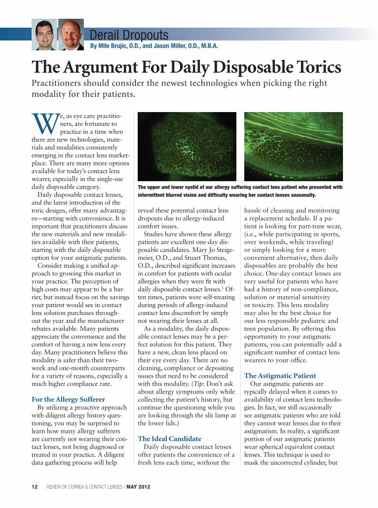
The upper and lower eyelid of our allergy suffering contact lens patient who presented with intermittent blurred vision and difficulty wearing her contact lenses seasonally.
012_rcl0512_Derail.indd 12 4/30/12 2:04 PM
Derail Dropouts
REVIEW OF CORNEA & CONTACT LENSES | MAY 2012 13
often does not provide optimal vi-sion correction for these patients.
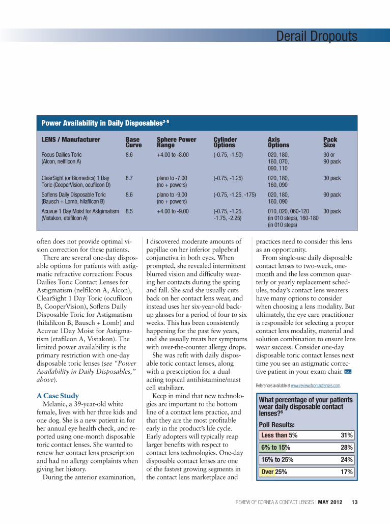
There are several one-day dispos-able options for patients with astig-matic refractive correction: Focus Dailies Toric Contact Lenses for Astigmatism (nelfi lcon A, Alcon), ClearSight 1 Day Toric (ocufi lcon B, CooperVision), Sofl ens Daily Disposable Toric for Astigmatism (hilafi lcon B, Bausch + Lomb) and Acuvue 1Day Moist for Astigma-tism (etafi lcon A, Vistakon). The limited power availability is the primary restriction with one-day disposable toric lenses (see “Power Availability in Daily Disposables,” above).
A Case StudyMelanie, a 39-year-old white
female, lives with her three kids and one dog. She is a new patient in for her annual eye health check, and re-ported using one-month disposable toric contact lenses. She wanted to renew her contact lens prescription and had no allergy complaints when giving her history.
During the anterior examination,
I discovered moderate amounts of papillae on her inferior palpebral conjunctiva in both eyes. When prompted, she revealed intermittent blurred vision and diffi culty wear-ing her contacts during the spring and fall. She said she usually cuts back on her contact lens wear, and instead uses her six-year-old back-up glasses for a period of four to six weeks. This has been consistently happening for the past few years, and she usually treats her symptoms with over-the-counter allergy drops.
She was refi t with daily dispos-able toric contact lenses, along with a prescription for a dual-acting topical antihistamine/mast cell stabilizer.
Keep in mind that new technolo-gies are important to the bottom line of a contact lens practice, and that they are the most profi table early in the product’s life cycle. Early adopters will typically reap larger benefi ts with respect to contact lens technologies. One-day disposable contact lenses are one of the fastest growing segments in the contact lens marketplace and
practices need to consider this lens as an opportunity.
From single-use daily disposable contact lenses to two-week, one-month and the less common quar-terly or yearly replacement sched-ules, today’s contact lens wearers have many options to consider when choosing a lens modality. But ultimately, the eye care practitioner is responsible for selecting a proper contact lens modality, material and solution combination to ensure lens wear success. Consider one-day disposable toric contact lenses next time you see an astigmatic correc-tive patient in your exam chair. RCCL
References available at www.reviewofcontactlenses.com.
What percentage of your patients wear daily disposable contact lenses?6
Poll Results:Less than 5% 31%
6% to 15% 28%
16% to 25% 24%
Over 25% 17%
Power Availability in Daily Disposables2-5
LENS / Manufacturer Base Sphere Power Cylinder Axis Pack Curve Range Options Options SizeFocus Dailies Toric 8.6 +4.00 to -8.00 (-0.75, -1.50) 020, 180, 30 or (Alcon, nelfilcon A) 160, 070, 90 pack 090, 110
ClearSight (or Biomedics) 1 Day 8.7 plano to -7.00 (-0.75, -1.25) 020, 180, 30 packToric (CooperVision, ocufilcon D) (no + powers) 160, 090
Soflens Daily Disposable Toric 8.6 plano to -9.00 (-0.75, -1.25, -175) 020, 180, 90 pack(Bausch + Lomb, hilafilcon B) (no + powers) 160, 090
Acuvue 1 Day Moist for Astgimatism 8.5 +4.00 to -9.00 (-0.75, -1.25, 010, 020, 060-120 30 pack(Vistakon, etafilcon A) -1.75, -2.25) (in 010 steps), 160-180 (in 010 steps)
012_rcl0512_Derail.indd 13 4/30/12 2:04 PM

Out of the Box By Gary Gerber, O.D.
14 REVIEW OF CORNEA & CONTACT LENSES | MAY 2012
I recently met with a practitio-ner to discuss new strategies in practice management. As I have
heard before, this practitioner said he knew it all and that he could summarize practice management in a few words: “Buy smart, hire good staff, be a sharp marketer and focus on customer service.” It was the same old rhetoric.
In response, I asked him if he conducted an inventory of his con-tact lenses. When he looked at me with a blank stare, I repeated the question. He considered the ques-tion and then dismissed it, saying that not only did he not inventory his contact lenses, but also that he didn’t see how my question had anything to do with practice man-agement.
My point was simple. There are a lot of new things in practice man-agement and, in fact, what’s old is new again. Just like the principles of optics haven’t changed, the princi-ples of practice building also haven’t changed. It is the mechanisms and tools to execute those principles that have changed. It makes sense for all of us to periodically make sure that the techniques from our past are revisited, recycled and then recalibrated to current market conditions.
Lens InventoryAs an example, let’s look at a
practice’s inventory of contact lens-es—an interesting notion, because so few of us do this anymore. While it may have been commonplace to keep a stocked inventory in the olden days of vialed lenses, today’s manufacturers and distributors have
made it incredibly easy and effi cient to order lenses online, and to use their fulfi llment systems to do so. For the practitioner, this shifts the need to inventory lenses away from us and onto them. Therefore, in the context of the above discussion, there is room to review contact lens inventories in another way, and like many old practice-building ideas, this too is worth revisiting and recycling.
Revisiting the Old Wisdom• Buy smart. Generally, when you
buy an inventory of lenses, your unit pricing drops. While it’s true you will incur costs for carrying inventory, you should move your in-ventory quickly with good forecast-ed data. With careful planning and smart buying, you can plan around a 30-day schedule and negate nearly any added expenses.
Keep in mind that you do not need to inventory every lens. Start with what you consider to be your go-to lenses in the most common powers. Next, create a list of your second-tier lenses and stop there.
• Hire good staff. Your staff has to understand the reason why—and be able to articulate that reason to your patients. In this case, your staff should understand the benefi ts of having an inventory of lenses, how to dispense ideally a year’s supply of lenses on the spot and how this practice can help with patient com-pliance and pricing.
• Be a sharp marketer. Patients should perceive you as the place to get contact lenses, and having their lenses in stock is one way to support that now uncommon
marketing position. Having a well-researched, carefully planned inventory might be one of the least expensive marketing tools at your disposable.
• Focus on customer service. Think about it, what could be more convenient for a patient than walking out of your offi ce with everything they need in hand—from lenses and solutions to cases and eyeglasses? Being able to have what your customer needs is one of the many hallmarks in great customer service. In our case, serving your patients’ needs when they need them to be served goes along with this mindset.
This article is not specifi cally about inventorying lenses. Rather, it is about realizing that business-building fundamentals have re-mained unchanged from essentially the earliest street markets. Offer great products, value and service in a memorable atmosphere and work with an educated staff—and you’re highly likely to succeed.
The key is rethinking and mod-ernizing your strategy. Instead of a billboard on a highway, use social media. Instead of the Yellow Pages, use search engine optimization. A television commercial may be replaced with your own YouTube channel.
While using these new method-ologies, stick to the core elements of old-school business building. Don’t get into a rut and think that there is nothing new in practice manage-ment. If you get complacent, you’ll fi nd that your practice is the one that suffers. RCCL
The Power of Old WisdomWhile the principles of practice building have not changed, it is time to re-evaluate how you implement those old strategies.
014_rcl0512_otb.indd 14 4/30/12 2:22 PM
What’s the SolutionBy Steve Lowinger, O.D.
Sponsored b y
In today’s society, the phrase “image is everything” rings true. How we present ourselves to our
patient is our biggest asset in the marketing of our practice. This pre-sentation encompasses all facets of the practice: the staff who greet the patients as they enter the offi ce, the lenses and lens care decisions the practitioner makes and the instruc-tions the patients receive before they leave our offi ce. What we say and how we educate our patients in the offi ce will shape their visual health and comfort for the upcoming year.
Often times it is the little things we do for our patients that have the biggest results in growing and maintain-ing our practice. Patients come to us for our expertise, and stay when they can walk away with better vision and comfort.
Our patients have one simple expectation. They wish to see well and be comfortable. Numerous studies cite comfort as the primary reason that patients drop out of contact lens wear.1 Some of these dropouts are vocal, while others simply fade away from our practice. Consider that spending an extra minute with your patients to review their lens care regimen may be the deciding factor in keeping those patients in your practice or losing them entirely. Every contact lens dropout will affect your practice’s bottom line—both in lost revenue and through loss of referrals.
Improve ComfortThe comfort equation has many
variables, the most important being the interaction between the contact lenses and the multipur-pose solutions which disinfect, create wettability and enhance the comfort of the contact lens. One of the latest advances is the introduc-tion of OPTI-FREE® PureMoist®
MPDS and HydraGlyde® Moisture Matrix. HydraGlyde® Moisture Matrix was designed to make the entire contact lens surface hydro-philic and wettable.2
This increased wettability allows a patient to wear their lenses com-fortably for a longer period of time, feel more comfortable at the end of the day and achieve clear vision.3 By providing the patient with a prod-uct that makes their lenses more wettable, we can achieve a better contact lens experience in comfort.
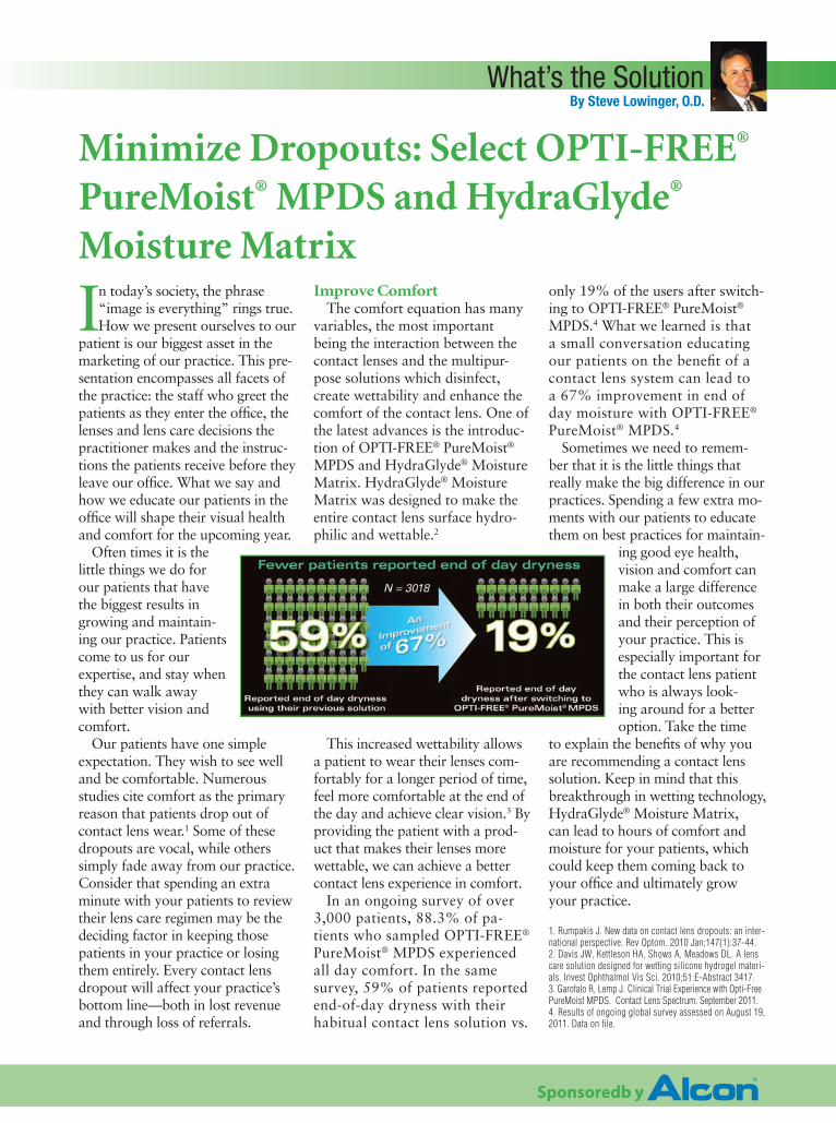
In an ongoing survey of over 3,000 patients, 88.3% of pa-tients who sampled OPTI-FREE® PureMoist® MPDS experienced all day comfort. In the same survey, 59% of patients reported end-of-day dryness with their habitual contact lens solution vs.
only 19% of the users after switch-ing to OPTI-FREE® PureMoist®
MPDS.4 What we learned is that a small conversation educating our patients on the benefi t of a contact lens system can lead to a 67% improvement in end of day moisture with OPTI-FREE® PureMoist® MPDS.4
Sometimes we need to remem-ber that it is the little things that really make the big difference in our practices. Spending a few extra mo-ments with our patients to educate them on best practices for maintain-
ing good eye health, vision and comfort can make a large difference in both their outcomes and their perception of your practice. This is especially important for the contact lens patient who is always look-ing around for a better option. Take the time
to explain the benefi ts of why you are recommending a contact lens solution. Keep in mind that this breakthrough in wetting technology, HydraGlyde® Moisture Matrix, can lead to hours of comfort and moisture for your patients, which could keep them coming back to your offi ce and ultimately grow your practice.
1. Rumpakis J. New data on contact lens dropouts: an inter-national perspective. Rev Optom. 2010 Jan;147(1):37-44.2. Davis JW, Kettleson HA, Shows A, Meadows DL. A lens care solution designed for wetting silicone hydrogel materi-als. Invest Ophthalmol Vis Sci. 2010;51:E-Abstract 3417.3. Garofalo R, Lemp J. Clinical Trial Experience with Opti-Free PureMoist MPDS. Contact Lens Spectrum. September 2011.4. Results of ongoing global survey assessed on August 19, 2011. Data on file.
Minimize Dropouts: Select OPTI-FREE®
PureMoist® MPDS and HydraGlyde® Moisture Matrix
015_rcl0512_alcon.indd 15 5/1/12 10:42 AM

16 REVIEW OF CORNEA & CONTACT LENSES | MAY 2012
Down on the PharmBy Jill Autry, R.Ph., O.D., and Elyse Chaglasian, O.D.
With the advent of the prostaglandin analogues, a paradigm shift in the
treatment of glaucoma and ocular hypertension was realized. Finally, a once-daily, well-tolerated, safe and efficacious medication was introduced into the treatment arma-mentarium for a potentially blind-ing optic neuropathy. Today, this description of the prostaglandin class continues to hold true for the vast majority of patients for whom we daily prescribe either latano-prost (Xalatan, Pfizer), bimatoprost products (Lumigan, Allergan) or travaprost products (Travatan, Alcon). The relative drawbacks have been local side effects such as hyper-emia, iris and periorbital darkening, and eyelash growth, as well as the cost of the medications.1
The New, The Old, The Reformulated
Because the prostaglandin class is indispensable in the treatment of intraocular pressure, the past 15 years have seen various changes in the respective product lines to try to counteract side effects and cost.
LatanoprostLatanoprost, for example, is now
available through multiple generic manufacturers. This substitution opens up access to patients who are self-pay or have high deductibles and/or co-pays for brand name medications. It is important to remember, however, that generic medications for the eye are not required to be tested by the FDA for bioequivalence or therapeutic equivalence, but only require the
active ingredient to be identical to the brand.2 Inactive ingredients can alter the pH, the penetration, the viscosity and other characteristics of ophthalmic medications which can change the clinical effi cacy and side effect profi le of the drug. This fact should be taken into consideration for each individual patient and with each different generic manufacturer.
BimatoprostBimatoprost is generally regard-
ed as the most effi cacious of the prostaglandin analogue ophthalmic class.3 Its original 0.03% formula-tion, however, also demonstrated increased amounts of hyperemia, which discouraged both physi-cians and patients from using it. In response, Allergan lowered the strength of Lumigan from 0.03% to 0.01% and changed the preser-vative concentration, with a resul-tant 65% less moderate to severe hyperemia and equivalent clinical effi cacy.4 The 0.01% formulation allows for the known effi cacy of brimatoprost with a much better side effect profi le.
TravatanThe original Travatan 0.004%
was reformulated as Travatan Z as an option to agents containing benzalkonium chloride (BAK) as a preservative. The concentration of travaprost remained at 0.004% and the BAK was removed and replaced with sofziatim (SofZia), an ionic buffered preservative system. One study found that travoprost with SofZia was less toxic to corneal and conjuncti-val cells than those exposed to
BAK-containing travoprost.5 The amount of hyperemia between Travatan and Travatan Z was unchanged at 30% to 50%.
ZioptanRecently, another prostaglandin
analogue was approved that will further entrench this medica-tion class as the fi rst-line treat-ment of glaucoma and ocular hypertension. The latest agent to be approved is called Zioptan (tafl uprost 0.0015%, Merck) and offers the advantage of being completely preservative-free with the same clinical advantages we have come to expect from the prostaglandin class.6
Zioptan is packaged in unit-dose vials similar to Restasis (cyclosporine, Allergan) and other preservative-free ocular prepara-tions and is recommended for once-daily administration at bed-time. The package insert recom-mends each vial be discarded im-mediately after opening because sterility cannot be maintained without a preservative. There is suffi cient quantity in each vial for both eyes.
Typical of the topical prosta-glandins, Zioptan’s most common adverse reaction is conjuncti-val hyperemia, reported to be between 4% and 20%—which is closer to the hyperemia profi le of latanoprost.6,7 Multiple clinical trials with preservative-contain-ing tafl uprost have illustrated its effi cacy, and at least one study shows the preservative-free formulation to be equivalent in IOP-lowering effect.8,9
Glaucoma and the New ProstaglandinsNew adjustments to commonly prescribed prostaglandins can help practitioners better treat this form of optic neuropathy.
016_rcl0512_dotp.indd 16 4/30/12 3:21 PM

Down on the Pharm
Biotrue® Multi-Purpose Solution Has Proven Biocompatibility
1,560 Patientsused Biotrue multi-purpose solution
daily in the multi-center clinical trials
3,088 Hydrogellens eye examinations in the
multi-center clinical trials
4,124 Siliconehydrogel lens eye examinations
in the multi-center clinical trials
>72,000to Biotrue multi-purpose solution
0 Adverse events*
reported in the multi-center
clinical trials
1 Bausch + Lomb conducted six prospective studies between February 2008 and March 2010. A total of 1,560 subjects used Biotrue multi-purpose solution daily. Eighty-fi ve clinical investigators conducted slit lamp examinations that included grading of corneal infi ltrates, bulbar injection, limbal injection, corneal staining, and tarsal conjunctival abnormalities using numeric grades from 0 to 4. The occurrences of any graded fi nding >Grade 2 at the follow-up visits were negligible (less than 0.1% for each graded fi nding), and there were no adverse events reported.
*Adverse events are defi ned as sight-threatening conditions and include complications such as corneal ulcers.
© 2012 Bausch & Lomb Incorporated. ®/™ are trademarks of Bausch & Lomb Incorporated or its affi liates. PNS06254
Biotrue was extensively tested prior to launch1
Biotrue is biocompatible with hydrogel and silicone hydrogel lenses
Biotrue is proven to keep eyes healthy
EyeExposures
Why Choose Preservative-Free?It is important to remember that
the hyperemia associated with the prostaglandin class is unrelated to the presence or type of preservative. In fact, the original Xalatan had the highest amount of BAK than any of the original three prostaglandin brands, but the lowest amount of reported conjunctival redness. Hyperemia is a side effect of the prostaglandin chemical structure.10
The reason prescribers should consider a preservative-free option is to avoid the toxicity and ocular surface disease inherent with the use of preservatives in general, especially when used chronically or in excess.
Although true allergic reactions are rare, multiple daily applications of BAK and other preservatives have been shown to be toxic to corneal and conjunctival epithelial cells and could compound various ocular surface conditions such as dry eye syndrome.11 Patients who do not have an ocular surface disease and/or use monotherapy to control their intraocular pressure may not need a preservative-free option.
Various statistics indicate that our glaucoma patients have compliance rates as low as 50%.12 The most common causes of non-compliance include forgetfulness, medication
cost, side effect profi le and lack of understanding regarding this asymptomatic disease state. The aforementioned adjustments in the new prostaglandins class and the new tafl uprost has paved the way for more affordable and/or better-tolerated medication, and will give prescribers the ability to use these agents on an even broader range of patients with glaucoma or ocular hypertension. Along with better education on our part, we hope to increase the ease of usage and the compliance of our patients in order to avoid the devastating effects of this optic neuropathy. RCCL
References available at www.reviewofcontactlenses.com.
016_rcl0512_dotp.indd 17 4/30/12 2:23 PM
18 REVIEW OF CORNEA & CONTACT LENSES | MAY 2012
The most noteworthy clinical development of the 21st cen-tury is likely the evolution of
highly resistant bacteria that are being identified in ocular infections.
Traditionally, the ophthalmic arena has been spared most of the concerns associated with resistance of bacteria and efficacy of therapy due to the simple fact that we have the luxury of applying the therapeutic agent directly to the site. To a large degree, this bypasses the concerns associated with GI absorption and access to remote parts of the body. We are also lucky to have a wide array of potential agents available to meet the specific needs of individual patients.
But all that is changing.
History of Bacterial InfectionsOver the last seven years we’ve seen
a rapid decline in the efficacy of antibi-otics and a slowing of the technology pipeline for new drug development. This creates both a current challenge and future concern for all clinicians.1-3
Historically, the most common causes of bacterial infections of the eye have been Staphylococcus aureus, Staphy-lococcus epidermidis, Streptococcus pneumoniae, Haemophilus influenzae, Pseudomonas aeruginosa, Moraxella,
Serratia Marcescens and occasionally, Enterococcus.4 Over the past several decades, these bacteria had shown lit-tle change in susceptibility, which has allowed practitioners to manage most topical infections without significant concern. In particular the third- and fourth-generation fluoroquinolones gave us a powerful weapon to address almost any type of infection.
Ophthalmic InfectionsA 2008 study by Kara Cavuoto,
M.D., looked at bacterial isolates in ophthalmic infections and found an almost three-fold increase in resistance to third-generation fluoroquinolones (ciprofloxacin) relative to S. aureus over a 10-year period (from 1994 to 2003), and an emergence of MRSA from 4.4% to 42.9% of all cultures.5
Additional work by Penny Asbell, M.D., Michael H. Goldstein, M.D., and Fabiana Marangon, M.D., sup-port this concern of increasing resis-tance in ocular therapy.6-8 In 2009, Marguerite McDonald, M.D., identi-fied a remarkable increase in multi-drug resistant isolates to ciprofloxacin in relation to MRSA, which showed that 65% of MRSA isolates and 47% of MRSE isolates also showed cipro-floxacin resistance.9
The last several decades have seen the evolution of resistant strains of bacteria and the decline of antibiotic efficacy. As practitioners, it is time to confront the situation.By J. James Thimons, O.D.
The Big Bad Bugs of the Cornea Are Getting Worse
Dr. Thi-mons grad-uated from Ohio State University
College of Optometry and has served as chief, Optometric Service at the VA Medical Center in Chillicothe, center director and chairman at Omni Eye Services in Fairfax, Va., director at The Glauco-ma Institute at SUNY and executive director at TLC Connecticut. In 2002, he founded the Ophthalmic Consul-tants of Connecticut.
018_rcl0512badbugs.indd 18 4/30/12 2:11 PM

REVIEW OF CORNEA & CONTACT LENSES | MAY 2012 19
There has been a number of iden-tified trends in resistance to agents such as azithromycin, gentamicin and tobramycin. Maria Regina Chalita, M.D., showed a decrease of sensitivity for gentimcin and tobramycin from 88% and 95% respectively, to 50% and 80% over a 15-year period.10,11
Bacterial KeratitisThe more significant issue is the
growth of resistance in the past decade to organisms involved in sight-threatening infectious keratitis and endophthalmitis. Several studies have identified this pattern of rising resistance related primarily to gram-positive bacteria such as S. aureus. Tristan Bourcier, M.D., Ph.D., con-ducted a study of 291 patients with bacterial keratitis, noting that that the predominant culture positive bacteria was gram-positive (83%) with a suprisingly low gram-negative (17%).12 This trend is concerning because the antibiotics used to treat microbial keratitis (MK) are still quite effective against gram-negative bacteria (Pseudomonas specifically). However, the loss of efficacy seen in gram-positive coverage has been both rapid and alarming.
MRSA has seen a rapid rise in bacterial isolates, increasing from 29.5% in 2000 to 41.6% in 2005.13 Additionally, the efficacy patterns also underwent significant change: Fourth-generation fluoroquinolones maintained status against methicillin susceptible (MSSA) bacteria, while there was a marked decline in sus-ceptibility against MRSA isolates. This shifting pattern implies sig-nificant concerns about the clinical protocols used to manage bacterial keratitis in both contact lens and non-contact lens lesions.
Ocular InfectionsIn a seminal work by Dr. Asbell
and colleagues, the Ocular TRUST
(Tracking Resistance in U.S. Today) was developed to address these rapid changes in microbial resistance as they relate to ocular infections.14 The first data report assessed 197 cul-tures from participating centers and looked at S. aureus, S. pneumoniae and H. influenzae isolates from 2005 to 2006. Antibiotics tested were all the available topical agents used in clinical practice.
The outcomes were both interest-ing and concerning. Among S. pnee-moniae isolates, all were susceptible to the fluoroquinolones except five that showed intermediate resistance to ciprofloxacin. The other agents varied from a low of 18.3% with penicillin to a high of 100% with polymixin B. The H. influenzae iso-lates showed overall sensitivity to all agents except trimethoprim. The real concern was with S. aureus—agents that showed good efficacy to MSSA, but demonstrated high-level resis-tance to MRSA. Fourth-generation fluoroquinolones such as moxifloxa-cin, gatifloxacin and levofloxacin demonstrated 15% sensitivity and over 80% resistance to MRSA iso-lates.14
A European study also demon-strated the declining efficacy of the fluoroquinolones against MRSA in a review of 582 isolates.15 This has occurred despite initial marketing that indicated that it was nearly impossible for bacteria to evolve resistance because it would require a simultaneous topoisomerase and DNA gyrase mutation. The clinical implications of this information are
significant: The agents that have been the backbone of therapy for the last decade are exhibiting an Achil-les’ heel that makes treating micro-bial keratitis a moving target.
Post-SurgeryAnother area of concern is the
changing pattern of microbial iso-lates following both cataract and refractive surgery. Eric Donnenfeld, M.D., and colleagues presented data at the 2009 American Society of Cataract and Refractive Surgery meeting showing that 77% of posi-tive lid or conjunctival cultures were either S. epidermidis or S. aureus.16 They also noted that the incidence of colonization of MRSA/MRSE was 33% of all patients assessed, which increased with age to 50% at 80 years old. They identified that colo-nization of the ocular surface is more likely in health care workers (1.25), age (1.27) and glaucoma patients (1.44).16
Darlene Miller, D.H.Sc., M.P.H., and colleagues showed similar pat-terns of microbialization in the surgi-cal population, and also found that 65% of patients with ciprofloxacin-resistant organisms demonstrated in vitro cross resistance to moxifloxa-cin and gatifloxacin.17
Treatment protocols also have begun to change as a result of increasing resistance. The European endophthalmitis trials demonstrated a 78% reduction of incidence with an intraoperative injection of cefu-roxime in addition to all typical pre- and post-operative regimens.18
LASIK and PRK surgery also have shown significant increases in the rate of MRSA infections with similar patterns of antibiotic sensitivity.19
These resistance changes are consistent with evolving patterns of microbial behavior, both systemic and global.20 There is evidence that the emerging patterns of MRSA resistance are independent of the
Post-LASIK MRSA.
018_rcl0512badbugs.indd 19 4/30/12 2:11 PM

20 REVIEW OF CORNEA & CONTACT LENSES | MAY 2012
traditional hospital-based organisms, and instead represent a trans-species infection pattern that is derived from a separate source.
An October 2007 study reviewed the status of MRSA across the Unit-ed States and found an occurrence rate of 95,000 cases with a mortal-ity rate of 19,000.21 Compare that statistic with 12,500 AIDS-related deaths during the same peroid.21 A subsequent November 2007 study identified the likely source of this outbreak as the use of low-dose fluo-roquinolones in the animal husband-ry business to promote faster weight gain in farm animals, specifically swine.22 There is also evidence that this same mechanism may be pro-ducing a ciprofloxacin-resistant S. aureus. The original MRSA is now referred to as HB-MRSA (hospital-based) and the new strain is called CA-MRSA (community-acquired).
For practitioners, the primary impact of this finding is in the man-agement of patients who present with MK, with or without contact lens wear.
A Treatment PlanI still recommend therapy be initi-
ated with a fourth-generation fluo-roquinolone at the appropriate dose rate (qh to q2h) and the patient be seen daily until therapy has demon-strated improvement. If, however, at any time in the first 48 to 72 hours, the patient demonstrates regression or lack of progression of the MK, it is reasonable to assume a
fluoroquinolone failure and to initi-ate additional therapeutic interven-tion to address the issue.
The first step is to culture the patient and send out for laboratory analysis; this is best done via blood and chocolate agars, thioglycollate broth and Sabourauds media. The material should also be plated for Gram and Giemsa stains.
The next step after culture and stain is initiation of fortified antibi-otics. While drugs like Polytrim are effective against MRSA conjunc-tivitis, they are not indicated for suspected MRSA/MK unless they are used only until vancomycin can be obtained. I recommend fortifying the vancomycin to 25mg/ml and dosing at qh, alternating at qh with the flu-oroquinolone that was initially used until culture results are back.
In some contact lens-wearing patients, it is also appropriate to start additional gram-negative cover-age with an agent like tobramycin, fortified to 14mg/ml, on the same alternating dose to address the pos-sibility of Pseudomonas. In most cases, it is reasonable to simultane-ously start doxycycline 100mg p.o. b.i.d. to attempt to delimit the col-lateral damage from collagenase and the recruitment of cytokines and interleukins. The patient must be seen daily until the culture is report-ed, and then the clinician can make adjustments based on results.
Due to the aggressive nature of the more resistant bacteria, it is common to have significant tissue
damage associated with MK, so consider the use of a steroid after the field has been sterilized with sev-eral days of treatment. While each case is unique, the role for the anti-inflammatory properties must be considered but clinicians need to be cautious in implementing it.
Future ConsiderationsUnfortunately, due the aggres-
sive nature of MRSA, the concern regarding the organism mutating against vancomycin is already a reality. Simon R. Bababeygy, M.D., cited two 2009 cases of preseptal cellulites that were vancomycin resistant but responded to rifampin and linezolid therapy.23
The pharmaceutical pipeline to address the rising rate of resistance is not strong at this time. The only new agent recently released is a chloro-fluoroquinolone, Besiv-ance (besifloxacin 0.6%, Bausch + Lomb). It has demonstrated increased sensitivity to MRSA and MRSE in clinical studies in conjunc-tivitis.24-26 Its increased efficacy may be related to the unique structure of the molecule.27 While there is little data related to its role in MK man-agement, animal model studies of endophthalmitis compare it to the other fourth-generation agents.
The World Health Organization (WHO) has begun the process of creating guidelines for the world-wide use of antibiotics in an effort to bolster current sensitivities and decrease future resistance develop-ment. Recently, WHO issued a guideline for the cessation of antibi-otic use in animals, with a complete discontinuation to follow within several years. Given the current issue with trans-species MRSA, this will hopefully begin the process of better balancing our use of antibiotics with our needs. RCCL
References available at www.reviewofcontactlenses.com.
Vancomycin irrigated under the flap in a post-op endophthalmitis eye (left). Four days later (right).
018_rcl0512badbugs.indd 20 4/30/12 2:11 PM

REVIEW OF CORNEA & CONTACT LENSES | MAY 2012 21
CONTINUING EDUCATION
Practitioners should periodically review contact lens-related epitheliopathies and their treatment options to provide their patients with the best lens wear experience.By Mile Brujic, O.D., and Crystal Brimer, O.D.
Breaking Down CL Epitheliopathies
While advancements in contact lenses and lens care technology have
improved the wear experience,
several physiological effects of lens wear continue to contribute to secondary epitheliopathies. There is decreased tear exchange under the lens, which can create a pooling effect of carbon diox-ide, debris, antigens and bacteria. The quality of the tear film, espe-cially the mucin layer as it relates to infection, can be negatively affected with a reduction in lipid layer integrity and increased tear evaporation. Changes in the tear film can then have a secondary effect on the functionality of the epithelium.1-3
In addition to the concerns of con-servative and compliant lens wear, many patients are at even higher risk due to external factors, such as over-night wear, protein and lipid build up, hygiene issues, poorly fit lenses, solution hypersensitivity and lid or ocular surface disease.4,5
In this article, we will explore several epitheliopathies associated with contact lens wear and provide a breakdown of treatment options.
Non-Infectious InfiltratesNon-infectious infiltrates are
an immune-driven response of
Dr. Brujic is a partner of Premier Vision Group, a four-location optometric practice in northwest
Ohio. He lectures extensively in the areas of ocular disease man-agement and contact lenses.
Dr. Brimer is a graduate of UNC-Chapel Hill and Southern College of Optometry. She
has practiced full-scope optom-etry in Wilmington, N.C. for 12 years and has special interest in contact lenses and dry eye management. She is also the owner of Crystal Vision Services, an ophthalmic equipment and practice management consulting company.
Release Date: May 2012Expiration Date: May 1, 2015Goal Statement: This article will offer a review of contact lens-related epitheliopa-thies and provide an outline of their treat-ment options.Faculty/Editorial Board: Mile Brujic, O.D., and Crystal Brimer, O.D.Credit Statement: COPE approval for 1 hour of CE credit is pending for this
course. Check with your local state licensing board to see if this counts toward your CE requirements for relicen-sure.Joint-Sponsorship Statement: This continuing education course is joint-sponsored by the Pennsylvania College of Optometry.Disclosure Statement: The authors have no financial relationships to disclose.
021_rcl0412CEepith.indd 21 4/30/12 4:36 PM
22 REVIEW OF CORNEA & CONTACT LENSES | MAY 2012
CONTINUING EDUCATION
the cornea to antigens. Corneal insult initiates chemotaxis of leukocytes from the surrounding limbal vasculature into the ante-rior stroma.6
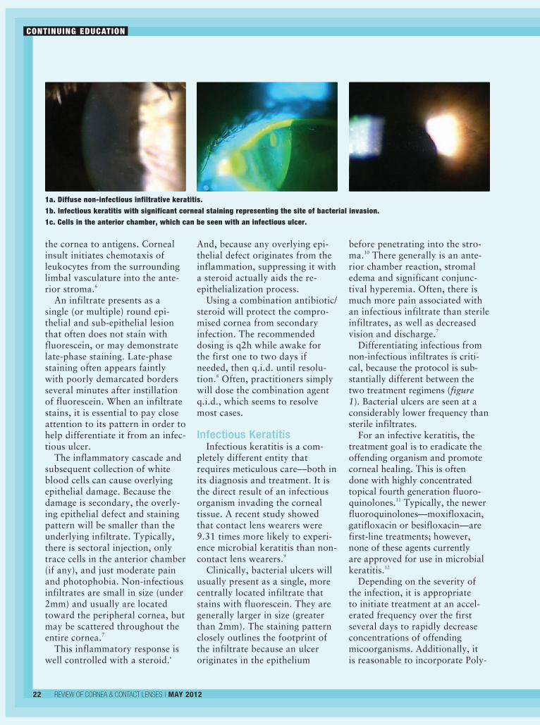
An infiltrate presents as a single (or multiple) round epi-thelial and sub-epithelial lesion that often does not stain with fluorescein, or may demonstrate late-phase staining. Late-phase staining often appears faintly with poorly demarcated borders several minutes after instillation of fluorescein. When an infiltrate stains, it is essential to pay close attention to its pattern in order to help differentiate it from an infec-tious ulcer.
The inflammatory cascade and subsequent collection of white blood cells can cause overlying epithelial damage. Because the damage is secondary, the overly-ing epithelial defect and staining pattern will be smaller than the underlying infiltrate. Typically, there is sectoral injection, only trace cells in the anterior chamber (if any), and just moderate pain and photophobia. Non-infectious infiltrates are small in size (under 2mm) and usually are located toward the peripheral cornea, but may be scattered throughout the entire cornea.7
This inflammatory response is well controlled with a steroid.s
And, because any overlying epi-thelial defect originates from the inflammation, suppressing it with a steroid actually aids the re-epithelialization process.
Using a combination antibiotic/steroid will protect the compro-mised cornea from secondary infection. The recommended dosing is q2h while awake for the first one to two days if needed, then q.i.d. until resolu-tion.8 Often, practitioners simply will dose the combination agent q.i.d., which seems to resolve most cases.
Infectious KeratitisInfectious keratitis is a com-
pletely different entity that requires meticulous care––both in its diagnosis and treatment. It is the direct result of an infectious organism invading the corneal tissue. A recent study showed that contact lens wearers were 9.31 times more likely to experi-ence microbial keratitis than non-contact lens wearers.9
Clinically, bacterial ulcers will usually present as a single, more centrally located infiltrate that stains with fluorescein. They are generally larger in size (greater than 2mm). The staining pattern closely outlines the footprint of the infiltrate because an ulcer originates in the epithelium
before penetrating into the stro-ma.10 There generally is an ante-rior chamber reaction, stromal edema and significant conjunc-tival hyperemia. Often, there is much more pain associated with an infectious infiltrate than sterile infiltrates, as well as decreased vision and discharge.7
Differentiating infectious from non-infectious infiltrates is criti-cal, because the protocol is sub-stantially different between the two treatment regimens (figure 1). Bacterial ulcers are seen at a considerably lower frequency than sterile infiltrates.
For an infective keratitis, the treatment goal is to eradicate the offending organism and promote corneal healing. This is often done with highly concentrated topical fourth generation fluoro-quinolones.11 Typically, the newer fluoroquinolones—moxifloxacin, gatifloxacin or besifloxacin—are first-line treatments; however, none of these agents currently are approved for use in microbial keratitis.12
Depending on the severity of the infection, it is appropriate to initiate treatment at an accel-erated frequency over the first several days to rapidly decrease concentrations of offending micoorganisms. Additionally, it is reasonable to incorporate Poly-
1a. Diffuse non-infectious infiltrative keratitis. 1b. Infectious keratitis with significant corneal staining representing the site of bacterial invasion. 1c. Cells in the anterior chamber, which can be seen with an infectious ulcer.
021_rcl0412CEepith.indd 22 4/30/12 4:37 PM

REVIEW OF CORNEA & CONTACT LENSES | MAY 2012 23
trim (polymyxin b/trimethoprim, Allergan) due to its efficacy against MRSA.
The standard of care for more aggressive or centrally located ulcers is to culture and treat with the appropriate fortified anti-biotics. Some doctors prescribe 30-minute dosing intervals in those cases, alternating fluoroqui-nolones with the fortified antibi-otics.8 However, some research shows equivalent efficacy of fourth-generation fluoroquino-lones to combination fortified antibiotic treatment.13
If the ulcer responds well after 48 hours, the drops can often be decreased to q2h until re-epithelialization and then tapered to q.i.d., according to the corneal response. Cycloplegics can help decrease the patient’s pain signifi-cantly as the infectious infiltrate is healing.
Despite immediate treatment, corneal scarring can threaten the patient’s visual outcome, and concurrent steroid treatment has been questioned as a means to minimize the resultant scarring from these infectious events.
A recent, large-scale study established that there was no ben-efit to the patient’s best-corrected visual acuity or resultant scar size when steroids were incorporated into the treatment protocol.14
However, patients with central ulcers greater than 4mm or those with the worst entering acuities did experience nearly a two-line improvement in visual acuity with adjunctive steroid treatment.
The study also illustrated the safety of concurrent steroid use after 48 hours of fluoroquino-lone treatment in culture positive patients and confirmed that there was no delay in re-epitheliali-zation time in the group using steroids.14
Advancing Wavelike Epitheliopathy
Advancing wavelike epitheli-opathy is a rare condition that consists of centripetally advanc-ing wavelike epithelium that usu-ally extends from the superior limbus towards the visual axis.15
The individual’s visual acuity may be affected, depending on how far the irregular epithelium extends to the visual axis. This can be seen during standard slit lamp examination, but is signifi-cantly easier to identify after the instillation of fluorescein dye while viewing the cornea with a cobalt blue light through a Wrat-ten filter (figure 2). Other signs that may occur include ocular irritation and redness, depending on the severity of the clinical pre-sentation.
In vivo confocal microscopy is remarkable for the presence of atypical, elongated and cetripe-tally oriented cells.16 Additionally, these cells are remarkable for relatively large, hyperreflective nuclei and a loss of visible cel-lular borders in the basal epithe-lium.16,17
The pathophysiology of advancing wavelike epithelium is poorly understood, but is believed to be multifactorial. Risk factors for its development include topical antiglaucoma medications, contact lens solu-tions, contact lens wear, previous ocular surgery and atopic derma-titis.15-17
There have been reports in the literature that describe the effectiveness of an application of 1% silver nitrate to the superior limbus.15-17 Liquid nitrogen also has been documented as an effec-tive treatment for this condition. During the treatment, the corneo-scleral limbus and surface corneal epithelium are exposed to liquid
nitrogen through a double freeze-thaw procedure. The technique is to apply liquid nitrogen to the affected area for one to two sec-onds, wait 30 seconds, and then reapply for another one to two seconds.18
In 2005, Professors Patrick Caroline and Mark Andre report-ed on a case in which advanc-ing wavelike epitheliopathy was included in the differential diag-nosis. The recommended treat-ment called for removal of the offending agents while having the patient utilize non-preserved arti-ficial tears.19
In our office, if visual acuity is not affected, we follow the same treatment plan of removing the suspected offending agent. Initially, we switch contact lens patients who use a multipurpose solution to a hydrogen peroxide system (figure 3). We also look to a daily disposable option, if one is available for their prescription.If the epitheliopathy either is not improving or progressing, it is reasonable to discontinue contact lens wear and have the patient use non-preserved artificial tears. If it continues to progress and/or visual acuity becomes affected, more extreme measures may be warranted.
2. The appearance of advancing wavelike epitheliopathy while viewing the eye with a cobalt blue light through a wratten filter after the instillation of fluorescein dye.
021_rcl0412CEepith.indd 23 4/30/12 4:43 PM
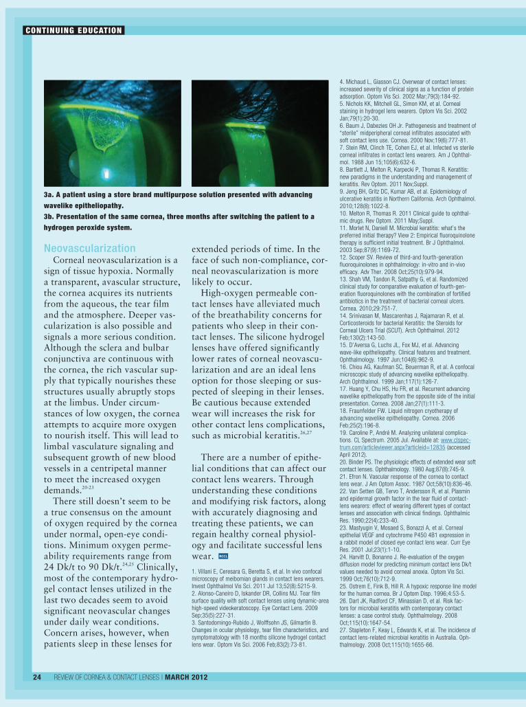
24 REVIEW OF CORNEA & CONTACT LENSES | MAY 2012
CONTINUING EDUCATIONCONTINUING EDUCATION
NeovascularizationCorneal neovascularization is a
sign of tissue hypoxia. Normally a transparent, avascular structure, the cornea acquires its nutrients from the aqueous, the tear film and the atmosphere. Deeper vas-cularization is also possible and signals a more serious condition. Although the sclera and bulbar conjunctiva are continuous with the cornea, the rich vascular sup-ply that typically nourishes these structures usually abruptly stops at the limbus. Under circum-stances of low oxygen, the cornea attempts to acquire more oxygen to nourish itself. This will lead to limbal vasculature signaling and subsequent growth of new blood vessels in a centripetal manner to meet the increased oxygen demands.20-23
There still doesn’t seem to be a true consensus on the amount of oxygen required by the cornea under normal, open-eye condi-tions. Minimum oxygen perme-ability requirements range from 24 Dk/t to 90 Dk/t.24,25 Clinically, most of the contemporary hydro-gel contact lenses utilized in the last two decades seem to avoid significant neovascular changes under daily wear conditions. Concern arises, however, when patients sleep in these lenses for
extended periods of time. In the face of such non-compliance, cor-neal neovascularization is more likely to occur.
High-oxygen permeable con-tact lenses have alleviated much of the breathability concerns for patients who sleep in their con-tact lenses. The silicone hydrogel lenses have offered significantly lower rates of corneal neovascu-larization and are an ideal lens option for those sleeping or sus-pected of sleeping in their lenses. Be cautious because extended wear will increases the risk for other contact lens complications, such as microbial keratitis.26,27
There are a number of epithe-lial conditions that can affect our contact lens wearers. Through understanding these conditions and modifying risk factors, along with accurately diagnosing and treating these patients, we can regain healthy corneal physiol-ogy and facilitate successful lens wear. RCCL
1. Villani E, Ceresara G, Beretta S, et al. In vivo confocal microscopy of meibomian glands in contact lens wearers. Invest Ophthalmol Vis Sci. 2011 Jul 13;52(8):5215-9.2. Alonso-Caneiro D, Iskander DR, Collins MJ. Tear film surface quality with soft contact lenses using dynamic-area high-speed videokeratoscopy. Eye Contact Lens. 2009 Sep;35(5):227-31.3. Santodomingo-Rubido J, Wolffsohn JS, Gilmartin B. Changes in ocular physiology, tear film characteristics, and symptomatology with 18 months silicone hydrogel contact lens wear. Optom Vis Sci. 2006 Feb;83(2):73-81.
4. Michaud L, Giasson CJ. Overwear of contact lenses: increased severity of clinical signs as a function of protein adsorption. Optom Vis Sci. 2002 Mar;79(3):184-92.5. Nichols KK, Mitchell GL, Simon KM, et al. Corneal staining in hydrogel lens wearers. Optom Vis Sci. 2002 Jan;79(1):20-30.6. Baum J, Dabezies OH Jr. Pathogenesis and treatment of “sterile” midperipheral corneal infiltrates associated with soft contact lens use. Cornea. 2000 Nov;19(6):777-81.7. Stein RM, Clinch TE, Cohen EJ, et al. Infected vs sterile corneal infiltrates in contact lens wearers. Am J Ophthal-mol. 1988 Jun 15;105(6):632-6.8. Bartlett J, Melton R, Karpecki P, Thomas R. Keratitis: new paradigms in the understanding and management of keratitis. Rev Optom. 2011 Nov;Suppl. 9. Jeng BH, Gritz DC, Kumar AB, et al. Epidemiology of ulcerative keratitis in Northern California. Arch Ophthalmol. 2010;128(8):1022-8.10. Melton R, Thomas R. 2011 Clinical guide to ophthal-mic drugs. Rev Optom. 2011 May;Suppl. 11. Morlet N, Daniell M. Microbial keratitis: what’s the preferred initial therapy? View 2: Empirical fluoroquinolone therapy is sufficient initial treatment. Br J Ophthalmol. 2003 Sep;87(9):1169-72.12. Scoper SV. Review of third-and fourth-generation fluoroquinolones in ophthalmology: in-vitro and in-vivo efficacy. Adv Ther. 2008 Oct;25(10):979-94.13. Shah VM, Tandon R, Satpathy G, et al. Randomized clinical study for comparative evaluation of fourth-gen-eration fluoroquinolones with the combination of fortified antibiotics in the treatment of bacterial corneal ulcers. Cornea. 2010;29:751-7.14. Srinivasan M, Mascarenhas J, Rajamaran R, et al. Corticosteroids for bacterial Keratitis: the Steroids for Corneal Ulcers Trial (SCUT). Arch Ophthalmol. 2012 Feb;130(2):143-50.15. D’Aversa G, Luchs JL, Fox MJ, et al. Advancing wave-like epitheliopathy. Clinical features and treatment. Ophthalmology. 1997 Jun;104(6):962-9.16. Chiou AG, Kaufman SC, Beuerman R, et al. A confocal microscopic study of advancing wavelike epitheliopathy. Arch Ophthalmol. 1999 Jan;117(1):126-7.17. Huang Y, Chu HS, Hu FR, et al. Recurrent advancing wavelike epitheliopathy from the opposite side of the initial presentation. Cornea. 2008 Jan;27(1):111-3.18. Fraunfelder FW. Liquid nitrogen cryotherapy of advancing wavelike epitheliopathy. Cornea. 2006 Feb;25(2):196-8.19. Caroline P, André M. Analyzing unilateral complica-tions. CL Spectrum. 2005 Jul. Available at: www.clspec-trum.com/articleviewer.aspx?articleid=12835 (accessed April 2012).20. Binder PS. The physiologic effects of extended wear soft contact lenses. Ophthalmology. 1980 Aug;87(8):745-9.21. Efron N. Vascular response of the cornea to contact lens wear. J Am Optom Assoc. 1987 Oct;58(10):836-46.22. Van Setten GB, Tervo T, Andersson R, et al. Plasmin and epidermal growth factor in the tear fluid of contact-lens wearers: effect of wearing different types of contact lenses and association with clinical findings. Ophthalmic Res. 1990;22(4):233-40.23. Mastyugin V, Mosaed S, Bonazzi A, et al. Corneal epithelial VEGF and cytochrome P450 4B1 expression in a rabbit model of closed eye contact lens wear. Curr Eye Res. 2001 Jul;23(1):1-10.24. Harvitt D, Bonanno J. Re-evaluation of the oxygen diffusion model for predicting minimum contact lens Dk/t values needed to avoid corneal anoxia. Optom Vis Sci. 1999 Oct;76(10):712-9.25. Ostrem E, Fink B, Hill R. A hypoxic response line model for the human cornea. Br J Optom Disp. 1996;4:53-5.26. Dart JK, Radford CF, Minassian D, et al. Risk fac-tors for microbial keratitis with contemporary contact lenses: a case control study. Ophthalmology. 2008 Oct;115(10):1647-54.27. Stapleton F, Keay L, Edwards K, et al. The incidence of contact lens-related microbial keratitis in Australia. Oph-thalmology. 2008 Oct;115(10):1655-66.
3a. A patient using a store brand multipurpose solution presented with advancing wavelike epitheliopathy.3b. Presentation of the same cornea, three months after switching the patient to a hydrogen peroxide system.
24 REVIEW OF CORNEA & CONTACT LENSES | MARCH 2012
021_rcl0412CEepith.indd 24 4/30/12 4:45 PM

1. How do non-infectious corneal infiltrates usually present?a. With a significant anterior chamber reaction.b. As a single lesion that causes notable pain.c. As a single or multiple, round sub-epithelial lesions that often
do not stain with fluorescein, or may demonstrate late-phase staining.
d. A dense collection of inflammatory cells with an overlying epithelial defect that mirrors the infiltrate.
2. How do you best protect a compromised cornea from sec-ondary infection?
a. A steroid.b. A bandage contact lens.c. An oral antibiotic.d. A combination antibiotic/steroid.
3. What is an infectious keratitis?a. An inflammatory event.b. A direct result of an infectious organism invading the corneal
tissue.c. A simple break in the epithelium.d. Multiple corneal infiltrates.
4. How do bacterial ulcers usually present?a. As a single or multiple sub-epithelial infiltrates.b. As a single, more centrally located infiltrate that stains with
fluorescein.c. Usually without an anterior chamber reaction.d. With mild pain.
5. Which agents are NOT used to treat corneal infections?a. Alternated fluoroquinolones and fortified antibiotics.b. Topical mast cell stabilizers.c. Fourth-generation fluoroquinolones (e.g., moxifloxacin, gati-
floxacin or besifloxacin).d. Cycloplegics.
6. What is advancing wavelike epitheliopathy?a. An infectious event.b. A poorly understood inflammatory condition.c. A rare condition that consists of centripetally advancing wave-
like epithelium that usually extends from the superior limbus towards the visual axis.
d. A new corneal dystrophy.
7. What is NOT a sign of advancing wavelike epitheliopathy?a. Reduced visual acuity.b. Anterior chamber reaction.c. Ocular irritation.d. Redness.
8. What is a primary risk factor for the development of advancing wavelike epitheliopathy?
a. Contact lens wear.b. Increased age.c. Male sex.d. Family history.
9. What is the correct technique to apply liquid nitrogen?a. Apply five drops, close and rotate eyes for 60 seconds, then rinse.b. Apply one drop every 15 minutes for one hour.c. Bring it very close, but do not allow it to touch the eye. Hold
for one to two seconds, wait 30 seconds and then bring it close again for an additional one to two seconds.
d. Apply to the affected area for one to two seconds, wait 30 seconds and then reapply for another one to two seconds.
10. What is the cornea’s minimum oxygen permeability requirements?
a. 12 Dk/t to 76 Dk/t. b. 24 Dk/t to 90 Dk/t.c. 70 Dk/t to 90 Dk/t.d. 90 Dk/t to 140 Dk/t.
CE TEST Examination Answer Sheet Valid for credit through May 1, 2015
This exam can be taken online at www.reviewofcontactlenses.com. Upon passing the exam, you can view your results immediately. You can also view your test history at any time from the website.
Breaking Down CL Epitheliopathies
Directions: Select one answer for each question in the exam and completely darken the appropriate circle. A minimum score of 70% is required to earn credit.
Mail to: Jobson - Optometric CE, PO Box 488, Canal Street Station, New York, NY 10013
Payment: Remit $20 with this exam. Make check payable to Jobson Medical Information LLC.
COPE approval for 1 hour of CE credit is pending for this course.
This course is joint-sponsored by the Pennsylvania College of Optometry
There is an eight-to-ten week processing time for this exam.
1. A B C D 1 = Excellent 2 = Very Good 3 = Good 4 = Fair 5 = Poor
2. A B C D Rate the effectiveness of how well the activity: 3. A B C D
4. A B C D 11. Met the goal statement: 1 2 3 4 5
5. A B C D 12. Related to your practice needs: 1 2 3 4 5
6. A B C D 13. Will help you improve patient care: 1 2 3 4 5
7. A B C D 14. Avoided commercial bias/influence: 1 2 3 4 5
8. A B C D 15. How would you rate the overall 9. A B C D quality of the material presented? 1 2 3 4 5
10. A B C D 16. Your knowledge of the subject was increased: Greatly Somewhat Little 17. The difficulty of the course was: Complex Appropriate Basic How long did it take to complete this course? Comments on this course:
Suggested topics for future CE articles:
Please retain a copy for your records. Please print clearly.
You must choose and complete one of the following three identifier types:
1 SS # - -
Last 4 digits of your SS # and date of birth State Code and License #: (Example: NY12345678)
2 - 3
First Name
Last Name
The following is your: Home Address Business Address
Business Name
Address
City State
ZIP
Telephone # - -
Fax # - -
By submitting this answer sheet, I certify that I have read the lesson in its entirety and completed the self-assessment exam personally based on the material presented. I have not obtained the answers to this exam by any fraudulent or improper means.
Signature Date
Lesson 108275 RCCL-OSC-0512
REVIEW OF CORNEA & CONTACT LENSES | MAY 2012 25
021_rcl0412CEepith.indd 25 4/30/12 4:38 PM

26 REVIEW OF CORNEA & CONTACT LENSES | MAY 2012
Conceptually, orthokeratology has been around for more than half a century now. But, it is
the improvements in lens materials, design and fabrication over the last decade that have triggered significant changes in how we provide this treat-ment. When you consider the data published over that same 10-year span showed a retarding effect on the progression of myopia, it is clear that orthokeratology has never had greater potential to be a significant factor in the realm of vision correction––par-ticularly for children.1-3 In this regard, it is an exciting time to be able to offer orthokeratology to our patients.
However, there still is much to study and learn about the long-term effects of orthokeratology on the corneal tissue. Reports of ocular infections with lens wear may cause patients to be nervous about pursuing orthokeratology, particularly when parents consider the procedure for their child. It is therefore important that we do our part to alleviate the concerns by knowing what risk factors exist and what problems may arise.
Why Ortho-K?We can all agree that there are
some indisputable benefits of ortho-
keratology—such as convenience and safety. Keep in mind that you should mention both the pros and cons of ortho-k with your patients.
First and foremost, we cannot understate the advantage of expe-riencing freedom from corrective lenses wear, especially for children. How many times have you had frus-trated parents coming in for the fifth time in a month to get their child’s glasses fixed or adjusted? How often do children end up replacing glasses because they are broken or scratched to the point of uselessness? In general, children are harder on eyeglasses than adults—due to their active life-styles and relative carelessness when it comes to their spectacles. Keep in mind that the long-term cost of orthokeratology vs. glasses is not sig-nificantly different when you consider that you are replacing glasses annu-ally, if not more frequently.
Secondly, glasses are awkward to use when playing sports or other physical activities. This is one reason why many parents consider contact lenses in the first place. Many parents, however, are torn between concerns that their child is not ready for contact lenses and the alternative of their child having to wear glasses for athletics.
While not risk free, orthokeratology allows practitioners to offer new treatment options to our young patients.By Jason Jedlicka, O.D.
Ortho-K and Kids: Maximize the Benefits,
Minimize the Risk
Dr. Jedlic-ka is the director of the Cornea and Con-
tact Lens Institute of Minnesota, a referral practice dedicated to specialty contact lens fittings, contact lens research, and education to students and practi-tioners.
026_rcl0512orthok.indd 26 4/30/12 3:58 PM
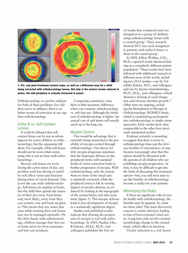
REVIEW OF CORNEA & CONTACT LENSES | MAY 2012 27
Orthokeratology is a perfect solution for both of these problems. For chil-dren active in athletics, there is no better means of correction at any age than orthokeratology.
Ortho-K vs. Soft Contact Lenses
It could be debated that soft contact lenses can be just as advan-tageous for active children as ortho-keratology, but the arguments fall short. For example, while soft lenses should not be worn when swim-ming, this is not an issue with ortho-keratology.
Because soft lenses are worn during the active times of day, any problem with lens drying or stabil-ity will affect vision and function during times of visual demand. This is not the case with orthokeratolo-gy. Soft lenses are applied at home, but the child then spends the major-ity of their day away from home and, most likely, away from their care system, case and back-up glass-es. This means that any issues that arise while wearing their soft lenses may not be managed optimally. On the other hand, with orthokeratol-ogy, children manage their lens use at home never far from assistance and lens care products.
Comparing cumulative costs, there is little monetary difference when you compare orthokeratology vs. soft lens use. Although the initial cost of orthokeratology is higher, the annual cost of soft lenses will usually catch up in the long run.
Myopia ControlOne would-be advantage that is
currently being researched is the pos-sibility of myopia control through orthokeratology. One theory for why myopia progresses stipulates that the hyperopic defocus on the peripheral retina with standard forms of vision correction leads to further progression of myopia. With orthokeratology, only the central 4mm to 5mm of the visual axis is optimally corrected, while the peripheral retina is left in varying degrees of myopic defocus, as evi-denced by looking at the topography of the cornea before and after treat-ment (figure 1). This myopic defocus leads to less development of myopia to a statistically significant degree.
Studies and published results indicate that slowing the progres-sion of myopia is real with ortho-keratology. In 2005, Pauline Cho, B.Optom., M.Ed., Ph.D., and colleagues published the first set
of results that compared axial eye elongation in a group of children using orthokeratology lenses with a control group.1 Their research showed 46% less axial elongation in patients with ortho-k lenses vs. those in the contol group.1
In 2009, Jeffery Walline, O.D., Ph.D., reported nearly identical find-ings in a completely different patient population.2 These results have been followed with additional research in different areas of the world, includ-ing two 2011 studies—one by Tet-suhiko Kakita, M.D., and colleagues and one by Jacinto Santodomingo, Ph.D., M.Sc., and colleagues—which showed a slowing of axial elonga-tion and vitreous chamber growth.3,4
Other tests are ongoing, includ-ing the Retardation of Myopia in Orthokeratology (ROMIO) Study, which is randomizing participants into orthokeratology or single-vision spectacles. Early results show data comparable to the other four previ-ously mentioned studies.5
Evidence certainly is mounting to suggest that there is more to orthokeratology than just the obvi-ous benefits of convenience—it may become increasingly clear that this option should be discussed with the parents of all children who are exhibiting myopia progression. At first, it may be difficult to get into the habit of discussing this treatment option––but, you will soon start to see the benefits of orthokeratology become a reality for your patients.
Minimizing the RisksIf there are significant risks to ocu-
lar health with orthokeratology, the benefits may be negated. So, what are these risks? The main short-term concern is ocular infection, leading to loss of best-corrected visual acu-ity. Long-term risks revolve around morphologic change to the corneal tissue, which affect its function.
Ocular infection is a risk factor
1. Pre- and post-treatment corneal maps, as well as a difference map for a child being corrected with orthokeratology lenses. Not only is the central cornea reduced in power, the mid-periphery is actually increased in power.
026_rcl0512orthok.indd 27 4/30/12 3:58 PM

28 REVIEW OF CORNEA & CONTACT LENSES | MAY 2012
for any type of contact lens wear. Overnight lens wear is associated with a higher risk of ocular infec-tion than daily wear lens use, but gas-permeable (GP) lens wear typ-ically presents a lower risk than soft lenses.6
Additionally, how does the risk for keratitis compare in orthokera-tology lens wearers to that of other lens wearers? The problem we face is that the volume of cases is rela-tively small to make a good esti-mation. We know that infectious keratitis does indeed occur, as sev-eral articles have chronicled these cases.7-10 The American Academy of Ophthalmology last published its stance on the safety of overnight orthokeratology in 2008, suggest-ing that a better-controlled, level 1 study needed to be completed to fully evaluate the risk and the potential for myopia control.11
What can we do in the meantime to minimize that risk to patients, specifically children? Clearly, good lens care practices are of the utmost importance (figure 2). Use of only approved contact lens care products, along with the elimination of tap
water use on lenses, has been recom-mended to reduce the risk; the high-est incidence of infectious keratitis occurs in regions of the world where good care practices are less likely to be followed.7
In addition, provide careful instruction to parents and children about what is to be expected with lens wear, and include comfort upon application, signs and symptoms of problems and after-hours contact information. These steps are vital to minimize the chance of a lens causing serious problems. Written directions and instructions, as well as informed consent, is highly rec-ommended to all orthokeratology lens wearers.
Finally, using only high Dk/t lens materials and FDA-approved designs not only minimizes the risk to patients, but also improves the efficacy of the treatment.12 Requir-ing the highest level of compliance to accepted practices and the high-est quality materials will minimize the risk of short-term complications for patients.
Long-term complications of orthokeratology involve permanent and undesirable changes to corneal shape or function. Some of these changes can be very significant, while others may not necessarily have any impact on ocular function. One study documented residual cor-neal flattening among a group of 28 patients; the impact of this change is still undetermined.13 Certainly, any change to the corneal tissue is both-ersome, but mild flattening of the cornea may or may not create real issues. The aforementioned study evaluated keratometric change two weeks after discontinuation of lens wear vs. pre-treatment levels. It is possible that, with longer time out of lenses, the corneal curvature changes may return to baseline. But, it is also possible that some permanent stro-mal restructuring may occur.13
Reduction in corneal sensation has been documented after three months of overnight orthokeratol-ogy lens wear.14 The significance of this finding has not yet been deter-mined, but it is useful to keep this in mind for our patients. Perhaps of more significance are the 2011 findings of Amelia Nieto-Bona, O.D., M.Sc., and colleagues, which examined the corneal tissue of 15 eyes with confocal microscopy. The researchers reported several changes to corneal tissue––some of which returned to baseline after discontinu-ation of lens wear, and others that showed an increase in endothelial cell polymegathism and a decrease in epithelial layer thickness.15 Again, the significance of these findings is not known, but studies continue to look at the possible long-term mor-phologic changes to the cornea as a result of orthokeratology and their effects on ocular health.
We all want what is best for our children. Good vision and ocular health are important aspects of our children’s lives. To be able to provide vision correction that is convenient, safe and effective at slowing the deterioration of their vision over time is something most of us would want for our children who require vision correc-tion. Orthokeratology seems to be able to provide all these features. Further research will continue to determine the true short- and long-term safety for those children in orthokeratology lenses, as well as the effects it has on slowing the progression of myopia. RCCL
1. Cho P, Cheung, SW, Edwards M. The longitudinal orthokera-tology research in children (LORIC) in Hong Kong: a pilot study on refractive changes and myopia control. Curr Eye Res. 2005 Jan;30(1):71-80.2. Walline JJ, Jones LA, Sinnott LT. Corneal reshaping and myo-pia progression. Br J Ophthalmol. 2009 Sep;93(9):1181-5.3. Kakita T, Hiraoka T, Oshika T. Influence of overnight orthokera-tology on axial elongation in childhood myopia. Invest Ophthalmol Vis Sci. 2011 Apr 6;52(5):2170-4.
Additional references at www.reviewofcontatlenses.com.
2. Using proper solutions and teaching proper technique are crucial to minimizing the risk of complications for children in orthokeratology lenses.
026_rcl0512orthok.indd 28 4/30/12 3:54 PM

REVIEW OF CORNEA & CONTACT LENSES | MAY 2012 29
When treating ocular con-ditions, topical medica-tions can offer many
advantages, including the ability to achieve high tissue concentra-tions and with limited systemic side effects. However, delivering an effective dose to ocular tissues can be a challenge. The bioavail-ability of most topical ophthalmic medications is surprisingly limited. The tears and lacrimal system effi-ciently wash drugs out of the eye before they can be absorbed, and competition with tissues other than intended targets can reduce avail-able drug levels substantially. Even the corneal epithelium itself pres-ents a formidable barrier to drug penetration.
With the high cost and regula-tory burdens associated with new drug development rising rapidly, a variety of strategies and tech-nologies have been developed to increase the effectiveness of exist-ing drugs. The use of engineered drug delivery vehicles increases ophthalmic drug bioavailability
and typically improves therapeu-tic effectiveness. For example, the propriety vehicle DuraSite (InSite Vision, Inc.) is used in the drugs AzaSite (azithromycin 1%, Merck) and Besivance (besifloxacin 0.6%, Bausch + Lomb) to prolong drug-ocular surface contact time, which results in enhanced penetra-tion. DuraSite creates a polymeric mucoadhesive matrix depot, which forms a gel upon instillation into the eye. Xanthan gum, used in both TobraDex ST (tobramycin 0.3%/dexamethasone 0.1%, Alcon) and Moxeza (moxifloxacin 0.5%, Alcon), have a similar mechanism of action.
Why Contact Lenses?Renowned Czech chemist, Otto
Wichterle, Ph.D., invented the first hydrogel material. Quickly recognizing the potential for hydro-gel materials to be manufactured into contact lenses, Dr. Wichterle produced the first soft lenses in his own kitchen using a spin cast-ing apparatus that he devised.1
Contact lenses are becoming an increasingly attractive option for drug delivery in our patients with a variety of ocular disorders. By Art Epstein, O.D.
TechnologicalAdvances
in Drug Delivery
Dr. Epstein is a popu-lar speaker and well-known
author. He serves as an adjunct associate clinical professor at Midwestern Univer-sity Eye Institute in Phoenix, Ariz. and is the Chief Medical Editor of Optometric Physician.
029_rcl0512drugdel.indd 29 4/30/12 2:10 PM
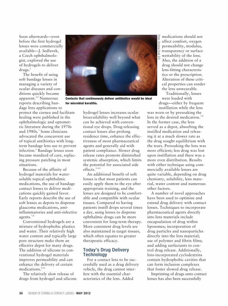
30 REVIEW OF CORNEA & CONTACT LENSES | MAY 2012
Soon afterwards––even before the first hydrogel lenses were commercially available—J. Sedlavek, a Czech ophthalmolo-gist, explored the use of hydrogels to deliver drugs.2
The benefit of using soft bandage lenses in managing a variety of ocular diseases and con-ditions quickly became apparent.3,4 Numerous reports describing ban-dage lens applications to protect the cornea and facilitate healing were published in the ophthalmologic and optomet-ric literature during the 1970s and 1980s.5 Some clinicians advocated the concurrent use of topical antibiotics with long-term bandage lens use to prevent infection.6 Bandage lenses soon became standard of care, replac-ing pressure patching in most situations.
Because of the affinity of hydrogel materials for water-soluble topical ophthalmic medications, the use of bandage contact lenses to deliver medi-cations quickly gained favor. Early reports describe the use of soft lenses as depots to dispense glaucoma medications, anti-inflammatories and anti-infective agents.7,8
Conventional hydrogels are a mixture of hydrophobic plastics and water. Their relatively high water content and typically large pore structure make them an effective depot for many drugs. The addition of silicone to con-ventional hydrogel materials improves permeability and can enhance the delivery of certain medications.9,10
The relatively slow release of drugs from hydrogel and silicone
hydrogel lenses increases ocular bioavailability well beyond what can be achieved with conven-tional eye drops. Drug-releasing contact lenses also prolong residence time, enhance the effec-tiveness of most pharmaceutical agents and generally aid with patient compliance. Slower drug release rates promote diminished systemic absorption, which limits the potential for associated side effects.11,12
An additional benefit of soft lenses is that most patients can easily apply them to the eye after appropriate training, and the lenses are designed to be comfort-able and compatible with ocular tissues. Compared to having patients instill drops several times a day, using lenses to dispense ophthalmic drugs can be more convenient for long-term therapy. More consistent drug levels are also maintained in target tissues, which often equates to greater therapeutic efficacy.
Today’s Drug Delivery Technology
For a contact lens to be suc-cessfully used as a drug delivery vehicle, the drug cannot inter-fere with the essential char-acteristics of the lens. Added
medications should not affect comfort, oxygen permeability, modulus, transparency or surface wettability of the lens. Also, the addition of a drug should not change lens-fitting characteris-tics or the prescription. Alteration of these criti-cal properties can render the lens unwearable.
Traditionally, lenses were loaded with
drugs––either by frequent instillation while the lens
was worn or by presoaking the lens in the desired medication.7,8 In the former case, the lens served as a depot, absorbing the instilled medication and releas-ing it at a much slower rate as the drug sought equilibrium with the tears. Presoaking the lens was more efficient; less drug was lost upon instillation and there was a more even distribution. Results with either technique using com-mercially available lenses are quite variable, depending on drug chemistry, solubility, lens mate-rial, water content and numerous other factors.
A number of novel approaches have been used to optimize and extend drug delivery with contact lenses. Techniques to incorporate pharmaceutical agents directly into lens materials include encapsulation of drug within liposomes; incorporation of drug particles and nanoparticles directly into the lens material; use of polymer and fibrin films; and adding surfactants to con-trol drug release. Additionally, lens-incorporated cyclodextrins contain hydrophobic cavities that can form inclusion complexes that foster slowed drug release.
Imprinting of drugs unto contact lenses has also been successfully
Contacts that continuously deliver antibiotics would be ideal for microbial keratitis.
Photo: Christine Sindt, O.D.
029_rcl0512drugdel.indd 30 4/30/12 2:10 PM

REVIEW OF CORNEA & CONTACT LENSES | MAY 2012 31
accomplished.13 Further, drugs can be impregnated within lens mate-rial matrices through the use of solvents at high temperatures and pressures.14
Incorporating transport barri-ers into lenses is another prom-ising approach for controlled elution of drugs. Barrier incor-poration can be tuned to provide prolonged therapeutic levels of various drugs.15
Clinical Application• Microbial keratitis. Sight-
threatening conditions, such as microbial keratitis, require rapid deployment of high levels of medication to the affected tissues. Currently, patients with severe microbial keratitis are hospital-ized to ensure continuous delivery of therapeutic levels of antibiotic through round-the-clock dosing. Management of these patients could be dramatically improved through the use of an effective drug delivery system, ensuring continuously high antibiotic levels. Contact lens delivery of antibiotics has been investigated, yielding sustained, high therapeu-tic levels with potential utility for managing bacterial keratitis.16,17
• Glaucoma. Chronic ocular conditions, such as glaucoma, require long-term pharmaco-logic treatment. Frequent dos-ing often leads to compliance failures, making this an excel-lent application for the use of extended-release, drug-eluting contact lenses. A feasibility study investigating efficacy and toxicity of contact lenses that were pas-sively impregnated with timolol maleate and brimonidine tartrate found IOP reductions equivalent to conventional therapy and no toxicity.18 This means that we can offer prolonged controlled drug delivery. A recent study found
that contact lenses increased drug bioavailability and decreased sys-temic absorption.19
• Allergy. Patients with ocular allergy also could benefit from continuous delivery of therapeu-tic levels of allergy medication. The use of ketotifen-containing contact lenses for the manage-ment of ocular allergy has been investigated experimentally, and has also undergone several safety and efficacy clinical trials.20,21
• Dry eye. Another promising area for the use of drug-eluting contact lenses is the controlled release of phospholipids. Meibo-mian gland dysfunction (and dry eye in general) has been associ-ated with a lack of phospholipids in the tear film. Phospholipids are an essential component contained in meibum, which acts to stabilize the tear layer. Phospholipid-elut-ing contact lenses may provide an effective treatment for some forms of dry eye, and possibly can improve end-of-day dryness in contact lens wearers by stabi-lizing the tear film and enhancing lens wettability.22 This may be especially helpful in wearers of silicone hydrogel lenses, which sequester lipids due to the hydro-phobic nature of their surfaces.23
In addition to the familiar refractive and protective role soft contact lenses have played for the past 50 years, developments in drug delivery technology are likely to propel soft lenses to even greater heights of clinical significance in the future. Contact lenses are uniquely able to provide sustained effective levels of medi-cation to the eye. Breakthroughs in drug delivery are also likely to help eliminate end-of-day dry-ness in contact lens wearers and offer help to dry eye sufferers. These advances in drug delivery
technology are likely to extend the usefulness of contact lenses for the foreseeable future. RCCL
1. Wichterle O, Lim D, Dreifus M. On the problem of con-tact lenses. Cesk Oftalmol. 1961;17:70-5. Czech.2. Sedlavek J. Possibilities of application of ophthalmic drugs with the aid of gel contact lens. CeskOftalmol. 1965;21:509-14.3. Dohlman CH, Boruchoff A, Mobilia EF. Complications in use of soft contact lenses in corneal disease. Arch Oph-thalmol. 1973 Nov;90(5):367-71.4. Kaufman HE, Uotila MH, Gasset AR, et al. The medical uses of soft contact lenses. Trans Am Acad Ophthalmol Otolaryngol. 1971 Mar-Apr;75(2):361-73.5. Levinson A, Weissman BA, Sachs U. Use of the Bausch & Lomb Soflens Plano T contact lens as a bandage. Am J Optom Physiol Opt. 1977 Feb;54(2):97-103.6. Binder PS, Worthen DM. A continuous-wear hydrophilic lens. Prophylactic topical antibiotics. Arch Ophthalmol. 1976 Dec;94(12):2109-11.7. Ruben M, Watkins R. Pilocarpine dispensation for the soft hydrophilic contact lens. Br J Ophthalmol. 1975 Aug;59(8):455-8.8. Marmion VJ, Jain MR. Role of soft contact lenses and delivery of drugs. Trans Ophthalmol Soc U K. 1976 Jul;96(2):319-21.9. Hui A, Boone A, Jones L. Uptake and release of ciprofloxacin-HCl from conventional and silicone hydro-gel contact lens materials. Eye Contact Lens. 2008 Sep;34(5):266-71.10. Schultz CL, Morck DW. Contact lenses as a drug deliv-ery device for epidermal growth factor in the treatment of ocular wounds. Clin Exp Optom. 2010 Mar;93(2): 61-5.11. Urtti A, Salminen L. Minimizing systemic absorption of topi- cally administered ophthalmic drugs. Surv Ophthal-mol. 1993 May-Jun;37(6):435-56.12. Shell JW. Pharmacokinetics of topically applied ophthal-mic drugs. Surv Ophthalmol. 1982 Jan-Feb;26: 207-18.13. Alvarez-Lorenzo C, Yañez F, Barreiro-Iglesias R, Concheiro A. Imprinted soft contact lenses as nor-floxacin delivery systems. J Control Release. 2006 Jun 20;113(3):236-44.14. Ciolino JB, Hoare TR, Iwata NG, et al. A drug-eluting contact lens.Invest Ophthalmol Vis Sci. 2009 Jul;50(7):3346-52. 15. Kim J, Conway A, Chauhan A. Extended delivery of ophthalmic drugs by silicone hydrogel contact lenses. Bio-materials. 2008 May;29(14):2259-69.16. Tian X, Iwatsu M, Sado K, Kanai A. Studies on the uptake and release of fluoroquinolones by disposable con-tact lenses. CLAO J. 2001 Oct;27(4):216-20.17. Hyatt AJ, Rajan MS, Burling K, et al. Release of vanco-mycin and gentamicin from a contact lens versus a fibrin coating applied to a contact lens. Invest Ophthalmol Vis Sci. 2012 Apr 18;53(4):1946-52.18. Schultz CL, Poling TR, Mint JO. A medical device/drug delivery system for treatment of glaucoma. Clin Exp Optom. 2009 Jul;92(4):343-8.19. Peng CC, Ben-Shlomo A, Mackay EO, et al. Drug deliv-ery by contact lens in spontaneously glaucomatous dogs. Curr Eye Res. 2012 Mar;37(3):204-11.20. Xu J, Li X, Sun F. In vitro and in vivo evaluation of keto-tifen fumarate-loaded silicone hydrogel contact lenses for ocular drug delivery. Drug Deliv. 2011 Feb;18(2):150-8.21. Vistakon Pharmaceuticals. Evaluation of efficacy and safety of an anti-allergy drug with a contact lens in the treatment of allergic conjunctivitis. 2009 Apr 8. Avail-able at: http://clinicaltrials.gov/ct2/show/NCT00445874 (accessed April 2012).22. Pitt WG, Jack DR, Zhao Y, et al. Loading and release of a phospholipid from contact lenses. Optom Vis Sci. 2011 Apr;88(4):502-6.23. Pitt WG, Jack DR, Zhao Y, et al. Transport of phos-pholipid in silicone hydrogel contact lenses. J Biomater Sci Polym Ed. 2012;23(1-4):527-41.
029_rcl0512drugdel.indd 31 4/30/12 2:10 PM
32 REVIEW OF CORNEA & CONTACT LENSES | MAY 2012
Dr. Michaud graduated from the University
of Montreal with a degree in optometry in 1986 and a masters degree in science in 1998. After 15 years in practice, he returned to the University of Montreal, where he now serves as an asso-ciate professor and chief of the contact lens department. He is a diplomate for the Section on Cornea and Contact Lenses of the American Acad-emy of Optomety, a well-known clinical investigator and a worldwide lecturer on contact lenses and anterior segment dis-ease.
A few years ago, my esteemed colleague Nathan Efron, Ph.D., D.Sc., made a strong statement
about the imminent death of rigid gas-permeable (RGP) contact lenses, which were supposed to be eradicated in the subsequent years. His words created a shockwave. However, obviously this scenario never became a reality. In fact, the marketplace is now saturated with RGP lenses to better serve our patients’ needs.
Based on today’s technology and a better understanding of the dynamics between the new lens designs and the cornea, modern large-diameter RGPs are the lenses of the future. In fact, I estimate that they will double their presence in the market within the next five years, competing to replace soft lenses as the go-to option in many-cases. This will happen simultaneously with the launch and development of a new generation of lenses—with large diameters and made from very high-permeable materials.
They will be as comfortable as soft lenses, as easy to fit as soft toric lenses, but with all the benefits of rigid mate-rials, including better visual acuity and less infections or inflammatory events.
Evolution or Revolution?In the last five years, manufactur-
ers have developed interesting RGP lens designs featuring large diameters that exceed the corneal border. The increased availability of material to
produce such large lenses, improved manufacturing processes to customize their design and a better understand-ing of the conjunctival anatomy helped establish new standards in RGP lens fitting.
Nowadays, large-diameter lenses are available from 14mm to 24mm in spherical, toric, reverse geometry and even multifocal designs. In addition, we now have two new types of lenses: corneo-scleral (<15mm diameter lenses partially supported by the cornea) and mini/true scleral (14.3mm to 16mm diameter lenses supported only by the conjunctiva aimed to vault the cor-nea). The latter offers better options to restore and protect the ocular surface.
Clinical applications for large-diam-eter lenses are numerous and exceed the natural niche for RGP lenses (corneal ectasia, irregular corneas, post-surgical correction of ametropia). Modern large-diameter lenses offer a valid option for correcting moder-ate to high spherical refractive errors, moderate to high astigmatism and presbyopia. This represents not only a true evolution in this category of contact lenses, but a real revolution for the entire profession.
Benefits of Large RGPs Large-diameter (14.3mm to 18mm)
RGP lenses offer many advantages over their small-diameter counterparts. For starters, initial discomfort—the primary reason practitioners are not
Large-diameter rigid gas-permeable lenses are the future of contacts.By Langis Michaud, O.D., M.Sc.
The Modern RGP Lens Revolution
032_rcl0512GP.indd 32 4/30/12 2:13 PM
REVIEW OF CORNEA & CONTACT LENSES | MAY 2012 33
fitting small RGP lenses—is no lon-ger an issue because the lenses lie on conjunctiva, a less sensitive tissue. Most of the large-diameter lenses vault the cornea, without touching it. This not only preserves the tissue integrity but alleviates any intoler-ance or corneal sensation associated with contact lens wear. Once prop-erly fitted, large-diameter lenses are as comfortable as a soft lens.
Also consider that there is no invasion of dust and particulates under the surface of a large-diameter lens. Many RGP wearers complain that, especially in the wind, some debris can remain trapped under their lenses, which creates immediate discomfort that ultimately leads to the lens removal. This cannot hap-pen with large-diameter RGP lens-es—once again, offering the same comfort as soft lens wear.
Ocular dryness is the leading cause of contact lens dropout. With large-diameter RGP lenses, the cornea is bathing in fluid during all the wearing hours. Primary ocular dryness can be reduced, and contact lens-induced dryness at the end of the day is eliminated. Therefore, large-diameter lenses can provide a solution to address these issues, which is the reason why many cor-neal specialists look at large-diame-ter RGP lenses to treat even the most severe dryness conditions.
Keep in mind that RGP lenses provide a crisp and sharp visual acuity that cannot be compared to soft lenses. This is especially true when moderate to significant astig-matism is involved. Because RGP materials do not dehydrate over time, and large lenses do not move a lot and do not rotate, these lenses are the optimal choice to correct astigmatism (up to 4D). They also are preferred for the correction of presbyopia and emerging designs of large-diameter lenses are showing promising results.
As an added bonus, large RGP lenses currently are unavailable for purchase on the Internet. This can give practitioners an upper hand in practice sales, as well as in their abil-ity to effectively control the patient’s eye health.
Additional ConsiderationsThe future success of large-diam-
eter RGPs will become a reality if a significant number of practitioners prescribe and fit these lenses. This evolution is not instantaneous.
We need to begin considering large-diameter lenses as our first option. The potential market for these RGP lenses include toric lens patients who are not completely sat-isfied with their vision, patients who report end-of-the-day ocular dryness or RGP lens wearers complaining of periodic discomfort.
Learning how to fit large-diameter RGP lenses is relatively easy. The exact learning time will vary, of course, based on experience. Fortu-nately, the newest lenses in today’s market are user-friendly; the smallest of the large-diameter lenses (14.2mm to 15mm) are as easy to fit as soft lenses. Outside of this range, true scleral lenses remain a challenge due to conjunctival toricity.
The key is to devote enough time initially to learn the fitting process and to be able to troubleshoot the problems as they occur. You can build your expertise by familiarizing yourself with the lenses through regularly fittings.
Learn to Troubleshoot Difficulty handling large-diameter
lenses is the number one reason why many patients cannot wear this modality. For some, such as our arthritic patients, insertion and removal of the lenses can be a chal-lenging task, especially without help.
Another common complaint is tight lenses, a syndrome that is
characterized by the sudden occur-rence of a red and painful eye after only a few hours of wear. This hap-pens when the lens becomes tight on the ocular surface and seals off the corneal periphery. The absence of tear exchange (habitually lim-ited, but present) contributes to trapped debris and toxins under the lens surface, which triggers a severe inflammatory reaction. At that time, the fit has to be revisited, allowing for more tear exchange and increased movement of the lens.
Discomfort with the large-diame-ter lenses is a sign of a bad fit. In the-ory, lens awareness can be present in the first minutes of wear, but true discomfort is certainly not expected.
Keep in mind that large-diameter lenses take time to be fitted. At mini-mum, it takes 20 to 30 minutes for the lens to settle. In some conditions, once the optimal lens is found, a practitioner may first prefer to reas-sess the fit after a few hours of wear before deciding on the modality.
Once stabilized on the eye, a clearance of 125µm to 150µm at the thinnest point is considered optimal for lenses under 15mm. For large lenses, a 200µm clearance is accept-able; although, some practitioners prefer a clearance of 300µm to 400µm. Optical coherence tomogra-phy (OCT) can help to evaluate the lens-to-ocular surface relationship, and to accurately define the appro-priate modifications needed. Some labs offer custom designs based on the OCT measurements.
Large-diameter RGPs represent a true, modern alternative to other modalities. They can compete with soft lenses in both comfort and con-venience. They outperform other lenses for visual acuity and in the treatment of ocular dryness. Con-trary to all predictions, RGP lenses are not dead—in fact, I believe they are taking on a whole new life. RCCL
032_rcl0512GP.indd 33 4/30/12 2:13 PM

34 REVIEW OF CORNEA & CONTACT LENSES | MAY 2012
Proceedings from a live interactive webinar event, attended by several of the industry’s most renowned contact lens practitioners. Moderated by Brian Chou, O.D.
Daily Disposables
On The Web ›› To register for our next event, go to www.reviewofcontactlenses.com/controversies-in-care.CONTROVERSIES IN CARE
Kicking off the April event on daily disposables, moderator Brian Chou, O.D., asked the
audience if they believe all patients should be put into daily disposables. The results were divided: 36% said yes, 39% said no and 25% said yes with some reservations.
Panelists Jason Miller, O.D., M.B.A., and Mile Brujic, O.D., said the survey results refl ected a debate that’s been ongoing between the two of them. “I personally think all patients can be placed in daily disposables, but it’s hard to say yes because we don’t always have avail-able powers to make it the go-to lens for all patients,” said Dr. Miller.
After fi rst acknowledging the benefi ts of daily disposables from a health perspective, Dr. Brujic summed up the challenge: Is the daily disposable technology as com-fortable as two-week or monthly options? Can we create all of the parameters that we currently have available in specialty lenses?
Interestingly, said Dr. Miller, the percentage of eye doctors and opto-metric staff that wear daily dispos-ables is very high, but these same practitioners are not aggressive in putting their patients in this modal-ity. Dr. Brujic said he thinks it is the preconceived notions, like cost for
example, that causes practitioners to fall back to old habits when it comes to fi tting patients.
Dr. Chou provided some his-tory: In 1995, when the modality was fi rst introduced to the U.S., the market share hovered around 5% and went up to 15% in 2011.1 Why the change? Several factors were mentioned starting with the reality that, in the past decade, there have been two major solution recalls, which naturally led practitioners to look at alternative options.
The audience also had a lot to add to this event. Attendees submit-ted several questions throughout the discussion, including one regarding whether daily disposables helped drive referrals to the panelists’ prac-tices. Drs. Miller and Brujic, who were able to answer online in real-time, said they have seen an increase in new patients looking specifi cally for daily disposables and existing patients asking questions about this modality. In response to another au-dience question, Dr. Miller said that an added bonus is the aggressive manufacturer rebates that allow practices to set reasonable prices for the lenses, which generally keeps patients from shopping around.
The audience was then polled on whether the cost of daily
disposables hinders their ability to use the product. The response: 21% strongly agreed, 54% somewhat agreed, 13% some-what disagreed and 13% strongly disagreed. Dr. Brujic said this was interesting since while there is an initial higher purchase cost with daily disposables over two-week or monthly counterparts, the expenses even out over the course of the year.
Dr. Miller disagreed slightly, saying that the patient’s perception is that daily disposables are more expensive and their perception is their truth. While it is important to address the expense factor, it is also important not to dwell on the issue. Rather shift the focus onto the ease, convenience and freshness of the daily option.
From the fi nancial breakdown to the health benefi ts, this month’s talk on daily disposables was high-ly informative. A special thank you to our active audience who partici-pated throughout the evening. We welcome you to join us at our next live conversation, which will focus on steroids. You can register for upcoming online events at www.reviewofcontactlenses.com. RCCL
1. Nichols JJ. Contact Lenses 2011. CL Spectrum. 2012 Jan.
CONTROVERSIES IN CARE POLL
Are daily disposables as good as their two-week and monthly disposable counterparts?
Poll Results:
Yes 84%
No 16%
034_rcl0512_CIC.indd 34 5/1/12 4:44 PM

Meeting and hotel registration opens May 7, 2012www.aaopt.org
Academy 2012 PhoenixOctober 24-27, 2012
Engage in scientific dialogue and debate on the latest practices at Academy 2012 Phoenix. Actively develop your knowledge and skills as a vision scientist or practitioner. As always, the Academy brings you unparalleled education, now in the radiant beauty of Phoenix. See you there!
Save the Date!
RO0512_House AAO.indd 1 4/24/12 3:09 PM
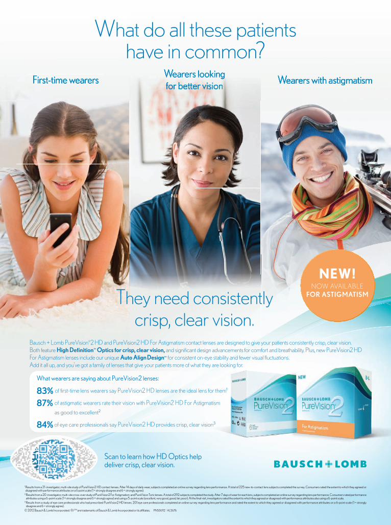
They need consistently
crisp, clear vision.
1 Results from a 21-investigator, multi-site study of PureVision2 HD contact lenses. After 14 days of daily wear, subjects completed an online survey regarding lens performance. A total of 225 new-to-contact lens subjects completed the survey. Consumers rated the extent to which they agreed or
disagreed with performance attributes on a 6 point scale (1 = strongly disagree and 6 = strongly agree).2 Results from a 20-investigator, multi-site cross-over study of PureVision2 For Astigmatism and PureVision Toric lenses. A total of 292 subjects completed the study. After 7 days of wear for each lens, subjects completed an online survey regarding lens performance. Consumers rated performance
attributes using a 6-point scale (1 = strongly disagree and 6 = strongly agree) and using a 5-point scale (excellent, very good, good, fair, poor). At the fi nal visit, investigators rated the extent to which they agreed or disagreed with performance attributes also using a 6-point scale.3 Results from a study of eye care professionals who had prescribed PureVision2 HD lenses. 201 eye care professionals completed an online survey regarding lens performance and rated the extent to which they agreed or disagreed with performance attributes on a 6-point scale (1 = strongly
disagree and 6 = strongly agree).
© 2012 Bausch & Lomb Incorporated. ®/™ are trademarks of Bausch & Lomb Incorporated or its affi liates. PNS06112 HL5676
NOW AVAILABLE
FOR ASTIGMATISM
NEW!
What do all these patientshave in common?
Bausch + Lomb PureVision®2 HD and PureVision2 HD For Astigmatism contact lenses are designed to give your patients consistently crisp, clear vision.
Both feature High Defi nition™ Optics for crisp, clear vision, and signifi cant design advancements for comfort and breathability. Plus, new PureVision2 HD
For Astigmatism lenses include our unique Auto Align Design™ for consistent on-eye stability and fewer visual fl uctuations.
Add it all up, and you’ve got a family of lenses that give your patients more of what they are looking for.
What wearers are saying about PureVision2 lenses:
83% of fi rst-time lens wearers say PureVision2 HD lenses are the ideal lens for them1
87% of astigmatic wearers rate their vision with PureVision2 HD For Astigmatism
as good to excellent2
84% of eye care professionals say PureVision2 HD provides crisp, clear vision3
Scan to learn how HD Optics help
deliver crisp, clear vision.
RCCL0512_BL Pure.indd 1 4/26/12 9:52 AM