Gibberellin DELLA signaling targets the retromer complex ...
Endosperm-limited Brassicaceae Seed Germination: Abscisic Acid Inhibits
Embryo-induced Endosperm Weakening of Lepidium sativum (cress) and
Endosperm Rupture of Cress and Arabidopsis thaliana
Kerstin Muller, Stefanie Tintelnot and Gerhard Leubner-Metzger *
Institute for Biology II, Botany/Plant Physiology, Albert-Ludwigs-University Freiburg, Schanzlestr. 1, D-79104 Freiburg i. Br., Germany
The endosperm is a barrier for radicle protrusion of
many angiosperm seeds. Rupture of the testa (seed coat) and
rupture of the endosperm are two sequential events during the
germination of Lepidium sativum L. and Arabidopsis thaliana
(L.) Heyhn. Abscisic acid (ABA) specifically inhibits the
endosperm rupture of these two closely related Brassicaceae
species. Lepidium seeds are large enough to allow the direct
measurement of endosperm weakening by the puncture force
method. We found that the endosperm weakens prior to
endosperm rupture and that ABA delays the onset and
decreases the rate of this weakening process in a dose-
dependent manner. An early embryo signal is required and
sufficient to induce endosperm weakening, which afterwards
appears to be an organ-autonomous process. Gibberellins can
replace this embryo signal; de novo gibberellin biosynthesis
occurs in the endosperm and weakening is regulated by the
gibberellin/ABA ratio. Our results suggest that the control of
radicle protrusion during the germination of Brassicaceae
seeds is mediated, at least in part, by endosperm weakening.
We propose that Lepidium is an emerging Brassicaceae model
system for endosperm weakening and that the complementary
advantages of Lepidium and Arabidopsis can be used in
parallel experiments to investigate the molecular mechanisms
of endosperm weakening.
Keywords: ABA — Arabidopsis thaliana — Embryo signal
— Endosperm weakening — Gibberellin — Lepidium
sativum.
Abbreviations: ABA, abscisic acid; FLU, flurprimidol;GA4þ7, gibberellin A4þ7; N, Newton; WT, wild type
Introduction
A major reason for the evolutionary success of the
angiosperms is the ‘invention’ of seeds and double
fertilization (Baskin and Baskin 1998; Friedman 1998).
In a typical angiosperm seed, the embryo is surrounded
by two covering layers (Web: ‘The Seed Biology Place’,
http://www.seedbiology.de): the endosperm (nutritive
tissue, living cells) and the testa (seed coat; maternal
tissue, dead cells). The mature seeds of most angiosperms
are endospermic. They have a more or less abundant
endosperm layer, though the evolutionary trend is towards
cotyledon storage and endospermless seeds. This trend is
also evident for the phylogenetically advanced Brassicaceae
family (rosid clade): the mature seeds of species of the
genera Raphanus, Sinapis and Brassica are endospermless
(Schopfer and Plachy 1984, Bergfeld and Schopfer 1986,
Schopfer et al. 2001); the single-celled aleurone layer of
these species is diploid maternal tissue and is part of the
inner testa. The mature seeds of Arabidopsis thaliana,
Lepidium spp. and Cleome spp. have retained a thin
endosperm layer, and the aleurone layer of these species
has been described to be endosperm tissue (O’Brien and
McCully 1969, Vaughan et al. 1971, Corner 1976, Ruiz and
Escale 1995, Nguyen et al. 2000). In the model species A.
thaliana (Arabidopsis), this endosperm is a single cell layer
(Pritchard et al. 2002, Nambara and Marion-Poll 2003,
Liu et al. 2005). The endosperm development of
Arabidopsis and Lepidium spp. is a cellularization process
that begins in the micropylar region and later during
seed development spreads to the central and chalazal
regions (e.g. Brown et al. 1999, Debeaujon et al. 2000,
Nguyen et al. 2000, Windsor et al. 2000). The important
point related to the work presented here is that mature
seeds of Arabidopsis and Lepidium spp. both have been
described to contain cellular endosperm tissue between the
embryo and the testa.
The process of seed germination begins with water
uptake by the dry seed through imbibition and ends when
the radicle has protruded through all covering layers
(Bewley 1997b, Koornneef et al. 2002, Kucera et al. 2005).
In order for a seed to complete germination, the growth
potential of the radicle must overcome the tissue resistance
of the seed covering layers. For many species, including
Arabidopsis, testa rupture and endosperm rupture are two
sequential steps during germination (e.g. Karssen 1976,
*Corresponding author: E-mail, [email protected]; Fax, þ49-761-203-2612.
Plant Cell Physiol. 47(7): 864–877 (2006)doi:10.1093/pcp/pcj059, available online at www.pcp.oxfordjournals.org� The Author 2006. Published by Oxford University Press on behalf of Japanese Society of Plant Physiologists.All rights reserved. For permissions, please email: [email protected]
864

Hepher and Roberts 1985, Leubner-Metzger et al. 1995,
Krock et al. 2002, Petruzzelli et al. 2003, Leubner-Metzger
2003, Liu et al. 2005). The role of the testa as a germination
constraint has been studied by using testa mutants of
Arabidopsis (Debeaujon and Koornneef 2000, Koornneef
et al. 2002). Also the role of the endosperm as a nutritive
tissue has been studied thoroughly in many species. Far less
is known about the role of the endosperm as a constraint to
radicle protrusion during germination.
The original hypothesis that weakening of the micro-
pylar endosperm covering the radicle tip by cell wall-
modifying proteins is a prerequisite for the completion of
germination was proposed based on pioneering work with
lettuce and was published in Plant and Cell Physiology by
Ikuma and Thimann (1963). Subsequent work with seeds
of the asterid clade with either a thin (e.g. lettuce) or a thick
(e.g. tomato, tobacco, coffee) endosperm layer strongly
supports the view that the micropylar endosperm is a
barrier for radicle protrusion (reviewed in Hilhorst 1995,
Bewley 1997a, Leubner-Metzger 2003). The original
work cited in these reviews provided evidence for the
involvement of several cell wall-modifying proteins in
endosperm weakening as well as for the importance of
plant hormones in regulating the resistance of the seed
covering layers. Although some progress has been made, no
definitive conclusions have been reached regarding
the molecular mechanisms that cause endosperm weaken-
ing. Further progress is, at least in part, hampered by the
fact that in these classical asterid seed model systems,
the direct measurement of the endosperm weakening by
the puncture force method is often not possible
(e.g tobacco seeds are too small) or is biased by the
inclusion of additional tissues (e.g. testa for tomato, embryo
for lettuce).
Results produced using the different asterid seed model
systems have led to the conclusion that inhibition of
endosperm rupture by abscisic acid (ABA) appears to be
an evolutionary widespread phenomenon (reviewed in
Bewley 1997a, Leubner-Metzger 2003, Nambara and
Marion-Poll 2003). It is not known whether ABA specifi-
cally inhibits the endosperm rupture of rosid seeds and
whether the thin endosperm of the model species
Arabidopsis and other Brassicaceae weakens and can act
as a barrier for radicle protrusion. In the present work, we
used seeds of Arabidopsis and the much bigger seeds of its
close relative Lepidium sativum (Lepidium, garden cress).
Both species belong to the Brassicoideae subfamily of the
Brassicaceae (Hall et al. 2002, Koch et al. 2003) and are
highly similar in seed structure and physiology. We found
that ABA specifically inhibits endosperm weakening of
Lepidium and endosperm rupture of Lepidium and
Arabidopsis. The close relationship to Arabidopsis, the
bigger seed size and additional advantages make Lepidium
an emerging seed model system for studying endosperm
weakening.
Results
Testa rupture and endosperm rupture are separate events
during the seed germination of Lepidium sativum and
Arabidopsis thaliana
The mature seeds of L. sativum (Lepidium) contain a
fully developed embryo with differentiated meristems,
radicle and cotyledons (Fig. 1). Seeds of Arabidopsis and
of its close relative Lepidium were used in our parallel
experiments. Both species are highly similar in seed
anatomy (Fig. 2B, G), but differ significantly in seed size
(Fig. 2A). In mature seeds of Arabidopsis, the radicle is
covered by a single cell layer of micropylar endosperm
(Fig. 2G; Koornneef et al. 2002, Liu et al. 2005). We found
that the embryo of Lepidium is enclosed by a thin
endosperm, mostly consisting of a single cell layer
(Fig. 1); however, the micropylar endosperm surrounding
the radicle tip has up to two cell layers (Fig. 1C). Lepidium
and Arabidopsis both exhibit a typical two-step germina-
tion: during testa rupture (Fig. 2H for Arabidopsis;
Fig. 2C, D for Lepidium), the micropylar endosperm is
exposed as a cap-like structure that covers the radicle tip.
This stage is followed by endosperm rupture and radicle
emergence (Fig. 2E, I), i.e. the completion of seed
germination. Under our control conditions, 50% rupture
of the Lepidium seed population was evident after 7 h for
the testa and after 16 h for the endosperm (Fig. 3A, B),
while 50% rupture of the Arabidopsis seed population was
evident after 24 h for the testa and after 33 h for the
endosperm (Fig. 3C, D).
ABA inhibits endosperm rupture of Lepidium and
Arabidopsis, but does not appreciably affect testa rupture
of after-ripened seeds
Addition of ABA to the medium does not appreciably
affect testa rupture (Fig. 3A, C), but delays endosperm
rupture of both species in a dose-dependent manner
(Fig. 3B, D). Upon ABA treatment, the intact micropylar
endosperm cap enclosing the radicle tip is visible (Fig. 2F, J)
at incubation times when seedlings are already growing
in control medium. The ABA concentrations used in these
experiments are in the range of the endogenous concentra-
tions known from seeds. For 10 mM ABA, the delayed 50%
endosperm rupture of Lepidium seed populations occurred
at approximately 85 h (i.e. a 5-fold delay), while testa
rupture was not affected (Fig. 3A, B). Fig. 3 shows an ABA
dose-dependent inhibition of Lepidium endosperm rupture,
e.g. for 5mM ABA the 50% value was approximately 65 h.
At the same ABA concentration, delayed 50% endosperm
Brassicaceae endosperm weakening 865

rupture of Arabidopsis seed populations occurred at
approximately 390 h (i.e. a 12-fold delay), while testa
rupture was only delayed by about 10 h (Fig. 3C, D).
Thus, ABA delayed endosperm rupture of Lepidium
and Arabidopsis (Figs. 2, 3), but did not appreciably
affect testa rupture.
For our experiments we used fully after-ripened
Lepidium seeds without a stratification pre-treatment.
Fig. 1 Structure of a mature seed of Lepidium sativum. Bright field microscopy of longitudinal sections of seeds imbibed for 2–3 h stainedwith toluidine blue. (A) Entire seed, showing the mature and fully differentiated embryo, the endosperm and the testa (seed coat). Theboxed letters refer to the positions of the close-up sections. (B) Structure of the seed-covering layers: endosperm, 1 cell layer; and testa(seed coat), composed of inner and outer integument. Note that the mucilage is generated from the outer testa upon imbibition. (C)Structure of the micropylar cap enclosing the radicle tip. The micropylar endosperm has 1–2 cell layers. (D) Structure of the chalazal seedregion. Blue light filter (C, D); differential contrast (D). Size bars are given for each panel.
866 Brassicaceae endosperm weakening

Fig. 2 Seeds of Lepidium sativum and Arabidopsis thaliana have similar seed anatomy and similar germination physiology. (A) The largersize of Lepidium seeds allows methods for which Arabidopsis seeds are too small, e.g. puncture force experiments. (B) Drawing of a matureLepidium seed; the embryo is enclosed by the endosperm and the surrounding testa. (C–F) During the two-step germination of Lepidium,testa rupture (C, D) is followed by endosperm rupture, which occurs after 16 h under control conditions (E). Due to the microphotographicsettings, the transparent outer mucilage layer is not visible. (F) ABA specifically inhibits endosperm rupture; the radicle remains covered bythe micropylar endosperm even after 60 h incubation in the presence of ABA. (G) Drawing of a mature Arabidopsis seed; the seed anatomyis very similar to that of Lepidium. (H–J) Arabidopsis seeds also germinate with testa rupture (H) preceding endosperm rupture (I). Also,during the two-step germination of Arabidopsis, ABA specifically inhibits endosperm rupture (J). Seeds were incubated in continuous lightwithout (control) or with 10mM ABA added to the medium.
Brassicaceae endosperm weakening 867

In contrast, we used a 24 h stratification pre-treatment for
our Arabidopsis experiments. Table 1 shows that the 24 h
stratification treatment promoted the endosperm rupture
of Arabidopsis seeds. Stratification promoted 50% endo-
sperm rupture of a fully after-ripened Arabidopsis seed
population by about 5 h. Testa rupture of fully after-ripened
seeds was not affected by either stratification or ABA
treatment (Table 1). In contrast to after-ripened seeds, the
testa rupture of conditionally dormant seeds was promoted
by stratification (Table 1). The physiological status of the
seed batch and not the stratification treatment per se is
therefore the reason for the differences in the kinetics
of testa rupture. The times for 50% testa rupture of the
different wild type (WT, A. thaliana ecotype Col) seed
Fig. 3 The effect of ABA on thetime courses of testa rupture andendosperm rupture of Lepidiumsativum (A, B) and Arabidopsisthaliana (C, D). ABA inhibitedendosperm rupture (B, D) of bothspecies in a dose-dependentmanner, but did not appreciablyaffect testa rupture (A, C). Note thedifferent time scales. The incidenceof testa and endosperm rupture wasscored with time after the start ofincubation in continuous light at188C (Lepidium) or 248C(Arabidopsis). Mean values� SEfrom one experiment with at leastthree plates with 50 seeds each arepresented.
868 Brassicaceae endosperm weakening
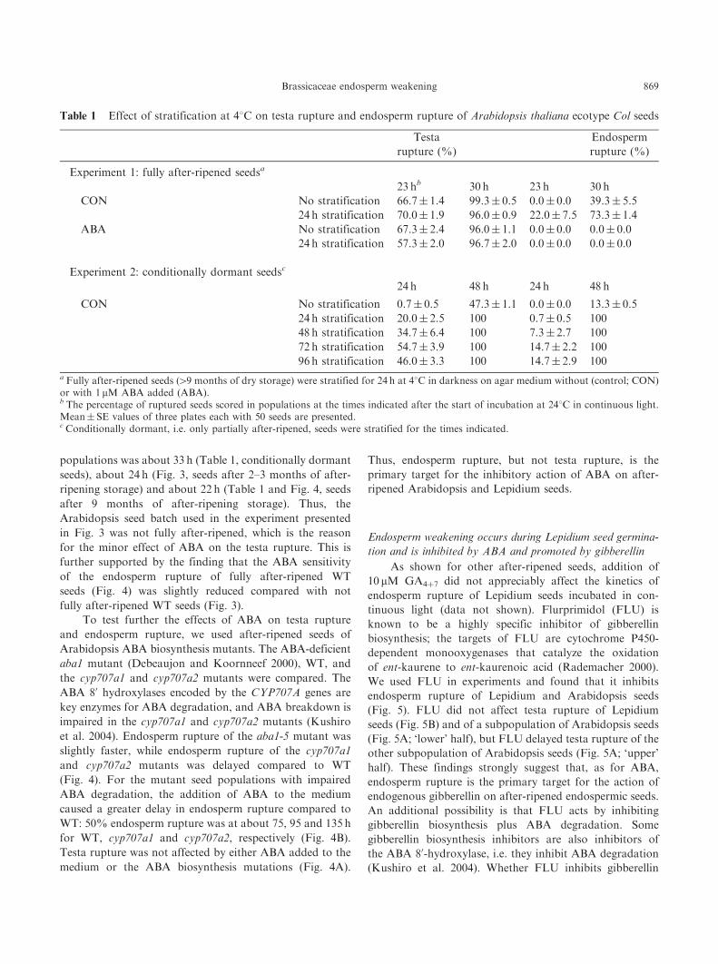
populations was about 33 h (Table 1, conditionally dormant
seeds), about 24 h (Fig. 3, seeds after 2–3 months of after-
ripening storage) and about 22 h (Table 1 and Fig. 4, seeds
after 9 months of after-ripening storage). Thus, the
Arabidopsis seed batch used in the experiment presented
in Fig. 3 was not fully after-ripened, which is the reason
for the minor effect of ABA on the testa rupture. This is
further supported by the finding that the ABA sensitivity
of the endosperm rupture of fully after-ripened WT
seeds (Fig. 4) was slightly reduced compared with not
fully after-ripened WT seeds (Fig. 3).
To test further the effects of ABA on testa rupture
and endosperm rupture, we used after-ripened seeds of
Arabidopsis ABA biosynthesis mutants. The ABA-deficient
aba1 mutant (Debeaujon and Koornneef 2000), WT, and
the cyp707a1 and cyp707a2 mutants were compared. The
ABA 80 hydroxylases encoded by the CYP707A genes are
key enzymes for ABA degradation, and ABA breakdown is
impaired in the cyp707a1 and cyp707a2 mutants (Kushiro
et al. 2004). Endosperm rupture of the aba1-5 mutant was
slightly faster, while endosperm rupture of the cyp707a1
and cyp707a2 mutants was delayed compared to WT
(Fig. 4). For the mutant seed populations with impaired
ABA degradation, the addition of ABA to the medium
caused a greater delay in endosperm rupture compared to
WT: 50% endosperm rupture was at about 75, 95 and 135 h
for WT, cyp707a1 and cyp707a2, respectively (Fig. 4B).
Testa rupture was not affected by either ABA added to the
medium or the ABA biosynthesis mutations (Fig. 4A).
Thus, endosperm rupture, but not testa rupture, is the
primary target for the inhibitory action of ABA on after-
ripened Arabidopsis and Lepidium seeds.
Endosperm weakening occurs during Lepidium seed germina-
tion and is inhibited by ABA and promoted by gibberellin
As shown for other after-ripened seeds, addition of
10 mM GA4þ7 did not appreciably affect the kinetics of
endosperm rupture of Lepidium seeds incubated in con-
tinuous light (data not shown). Flurprimidol (FLU) is
known to be a highly specific inhibitor of gibberellin
biosynthesis; the targets of FLU are cytochrome P450-
dependent monooxygenases that catalyze the oxidation
of ent-kaurene to ent-kaurenoic acid (Rademacher 2000).
We used FLU in experiments and found that it inhibits
endosperm rupture of Lepidium and Arabidopsis seeds
(Fig. 5). FLU did not affect testa rupture of Lepidium
seeds (Fig. 5B) and of a subpopulation of Arabidopsis seeds
(Fig. 5A; ‘lower’ half), but FLU delayed testa rupture of the
other subpopulation of Arabidopsis seeds (Fig. 5A; ‘upper’
half). These findings strongly suggest that, as for ABA,
endosperm rupture is the primary target for the action of
endogenous gibberellin on after-ripened endospermic seeds.
An additional possibility is that FLU acts by inhibiting
gibberellin biosynthesis plus ABA degradation. Some
gibberellin biosynthesis inhibitors are also inhibitors of
the ABA 80-hydroxylase, i.e. they inhibit ABA degradation
(Kushiro et al. 2004). Whether FLU inhibits gibberellin
Table 1 Effect of stratification at 48C on testa rupture and endosperm rupture of Arabidopsis thaliana ecotype Col seeds
Testa
rupture (%)
Endosperm
rupture (%)
Experiment 1: fully after-ripened seedsa
23 hb 30 h 23 h 30 h
CON No stratification 66.7� 1.4 99.3� 0.5 0.0� 0.0 39.3� 5.5
24 h stratification 70.0� 1.9 96.0� 0.9 22.0� 7.5 73.3� 1.4
ABA No stratification 67.3� 2.4 96.0� 1.1 0.0� 0.0 0.0� 0.0
24 h stratification 57.3� 2.0 96.7� 2.0 0.0� 0.0 0.0� 0.0
Experiment 2: conditionally dormant seedsc
24 h 48 h 24 h 48 h
CON No stratification 0.7� 0.5 47.3� 1.1 0.0� 0.0 13.3� 0.5
24 h stratification 20.0� 2.5 100 0.7� 0.5 100
48 h stratification 34.7� 6.4 100 7.3� 2.7 100
72 h stratification 54.7� 3.9 100 14.7� 2.2 100
96 h stratification 46.0� 3.3 100 14.7� 2.9 100
a Fully after-ripened seeds (49 months of dry storage) were stratified for 24 h at 48C in darkness on agar medium without (control; CON)or with 1 mM ABA added (ABA).b The percentage of ruptured seeds scored in populations at the times indicated after the start of incubation at 248C in continuous light.Mean� SE values of three plates each with 50 seeds are presented.c Conditionally dormant, i.e. only partially after-ripened, seeds were stratified for the times indicated.
Brassicaceae endosperm weakening 869

Fig. 5 The effect of gibberellin biosynthesis inhibition on the time courses of testa rupture and endosperm rupture of Arabidopsis thaliana(A) and Lepidium sativum (B). The incidence of testa and endosperm rupture was scored with time after the start of the incubation at 188C(Lepidium) or 248C (Arabidopsis) in continuous light. The medium was either without (control; CON) or with the gibberellin biosynthesisinhibitor flurprimidol (FLU; 10 mM for Arabidopsis, 100mM for Lepidium) added. Mean values� SE from one experiment with at least threeplates with 50 seeds each are presented.
Fig. 4 The effect of ABA biosynthesis mutations on the time courses of testa rupture (A) and endosperm rupture (B) of Arabidopsisthaliana. Wild type (WT) seeds were compared with seeds of the ABA-deficient aba1-5 mutant and of the cyp707a1 and cyp707a2mutants, which are impaired in ABA degradation. The incidence of testa and endosperm rupture was scored with time after the start ofincubation without (control; CON) or with 1 mM ABA added to the medium. The incubation was in continuous light at 248C. Meanvalues� SE from two independent experiments with at least three plates with 50 seeds each are presented.
870 Brassicaceae endosperm weakening

biosynthesis only or gibberellin biosynthesis plus ABA
degradation is not known. In either case, this will cause an
increased ABA/gibberellin ratio that is in agreement with
the observed delay of endosperm rupture.
For the direct measurement of endosperm weakening
by the puncture force method we isolated micropylar
endosperms from Lepidium seeds imbibed for defined
periods of time without (control) and with hormones
(ABA or gibberellin) added to the medium, as indicated
(Fig. 6). While an average force of 37.8� 2.5mN was
necessary to puncture the endosperm at 8 h, which is just
after testa rupture, only 19.5� 1.7mN was necessary at 18 h
when the radicle will protrude through the tissue within
the next 2–4 h (control, Fig. 6). Thus, a 2-fold decrease
in puncture force was evident during germination: the
endosperm tissue weakened prior to its rupture by the
radicle.
The addition of 10 mM ABA to the incubation medium
inhibited endosperm rupture and endosperm weakening
measured at 18 h (Fig. 6). At 120 h, i.e. just prior to the
ABA-delayed endosperm rupture, endosperm weakening
was evident in the ABA-treated seeds and was comparable
in value with the control at 18 h. This inhibition of
endosperm weakening by ABA was dose-dependent,
which is shown by the comparison of puncture force and
endosperm rupture values for different ABA concentrations
at 18 h (Fig. 6) and at 44 h (puncture force/endosperm
rupture: 33.9� 1.0mN/17.4� 4.5% for 5mM ABA,
25.7� 2.8mN/76.5% for 2.5 mM ABA, 100% endosperm
rupture with 1 mM ABA). Thus, ABA inhibits endo-
sperm weakening of Lepidium as well as endosperm
rupture of Lepidium and Arabidopsis in a dose-dependent
manner.
At 18 h, Lepidium seed populations without (control)
and with gibberellin added to the medium had equal
percentages of endosperm rupture (Fig. 6). At this time
point endosperm weakening had caused a decrease in the
puncture force by about 40mN in the gibberellin-treated
seeds compared with about 20mN in the control.
Essentially, gibberellin caused complete weakening to a
near-zero puncture force value. Simultaneous treatment
of Lepidium seeds with 10 mM GA4þ7 plus 10mM ABA
Fig. 6 Direct measurement of Lepidium sativum endosperm weakening of imbibed seeds by the puncture force method. Micropylar seedhalves were dissected from whole seeds imbibed (continuous light, 188C) for 8, 18 or 120 h without (control; CON) or with hormones (ABAand/or GA4þ7 in the concentrations indicated) added to the medium. Mean values� SE of at least 40 endosperm caps are presented.Note that all these seeds had completed testa rupture, but had intact endosperm. The numbers above the columns represent the percentageof seeds with ruptured endosperm in the corresponding seed populations.
Brassicaceae endosperm weakening 871

showed that gibberellin is an antagonist of the ABA
inhibition of endosperm weakening: a decrease in puncture
force was already evident at 18 h (Fig. 6), and at 65 h
a further decrease to 22.7� 2.5mN was associated with
approximately 70% endosperm rupture in the seed popula-
tion treated with gibberellin plus ABA (only 5–10% for
ABA-treated seeds). In agreement with a role for endo-
sperm weakening in the control of endosperm rupture,
gibberellin partially reversed the ABA inhibition of
Lepidium endosperm rupture. The same pattern was
obtained for Arabidopsis: at 84 h, endosperm rupture was
430% in the 10 mM GA4þ7 plus 1 mM ABA-treated
seed populations, whereas it was only 5–10% for the
ABA-treated seeds. Thus, gibberellin is an antagonist of the
ABA inhibition of Lepidium endosperm weakening and
of Lepidium and Arabidopsis endosperm rupture.
Endosperm weakening is an organ-autonomous process, but
requires induction by an early embryo signal, which can be
replaced by gibberellin
It has been proposed that endosperm weakening
requires induction by an embryo signal and that gibberellins
are at least part of this signal (Bewley 1997a). In order to
find out whether and if so in what time frame an embryo
signal induces Lepidium endosperm weakening, we
removed the embryo at different time points during the
germination process. The empty seed halves with the
micropylar endosperms were incubated separately in fresh
medium. Puncture force was measured after 18 h, when
endosperm weakening is evident in whole seeds (Fig. 7).
Embryo removal at 8 h, subsequent incubation of the
isolated seed halves for 10 h and puncture force measure-
ment at the 18 h time point (after the start of seed
Fig. 7 Direct measurement of Lepidium sativum endosperm weakening of isolated micropylar caps by the puncture force method.(A) Embryos were removed from whole seeds imbibed for 2 and 5 h, respectively. The dissected micropylar seed halves were incubateduntil 18 h (i.e. for 16 or 13 h starting from the 2 or 5 h time point, respectively) in fresh medium without (control; CON) or with 10 mMABA or 10 mM GA4þ7 added. Note that the puncture force of endosperm plus testa was measured when 2 or 5 h caps were used.(B) Embryos were removed from whole seeds imbibed for 8 h, and the dissected micropylar seed halves were incubated until 18 or 24 h(i.e. for 10 or 16 h, respectively, starting from the 8 h time point) in fresh medium without (control; CON) or with 10mM ABA, 10 mMGA4þ7
or 100mM flurprimidol (FLU; gibberellin biosynthesis inhibitor) added. Note that the puncture force of the endosperm was measuredsince 8 h caps from seeds after testa rupture were used. For direct comparison, puncture force values from seeds without embryoremoved are given. Imbibition and incubation were in continuous light at 188C. Mean values� SE of at least 40 endosperm caps arepresented.
872 Brassicaceae endosperm weakening
imbibition) resulted in the same degree of endosperm
weakening as found in whole seeds at 18 h (Fig. 7B). The
change in puncture force of isolated endosperms, i.e. a
decrease of approximately 20mN, corresponded to that
found in whole seeds. The remaining puncture force at
18 h of approximately 20mN decreased further when the
isolated endosperms were incubated for a longer time;
at 24 h, approximately 3mN was measured (Fig. 7B).
No endosperm weakening took place if the embryo
was removed after 2 h, but weakening of about 20mN was
measured at the 18 h time point if the embryo was removed
after 5 h of seed incubation (Fig. 7A). A further decrease of
approximately 10mN was measured at the 24 h time point.
An embryo–endosperm interaction in the time frame
between 2 and 5 h is thus sufficient to induce weakening.
After this period, the endosperm weakening does not
require the presence of the embryo. The addition of
10 mM GA4þ7 replaced the embryo signal and caused
complete weakening of micropylar endosperms isolated at
2 h. Thus, an embryo signal is evident in Lepidium seeds
between 2 and 5 h after the start of imbibition. It induces
micropylar endosperm weakening and can be replaced by
gibberellin.
Addition of ABA or the gibberellin biosynthesis
inhibitor FLU to the incubation medium of endosperm
caps isolated at the 8 h time point inhibited endosperm
weakening, while simultaneous treatment with FLU plus
gibberellin fully restored it (Fig. 7B). Endosperm weakening
therefore appears to require de novo gibberellin biosyn-
thesis in the micropylar endosperm cap. Thus, after the
induction by an embryo signal, endosperm weakening is
an organ-autonomous process evident in isolated micro-
pylar endosperm caps and is controlled by the ABA/
gibberellin ratio.
Discussion
The endosperm is known to act as a barrier for radicle
protrusion and thereby the completion of germination in
seeds from several angiosperm clades. Examples for this
include Trollius (basal eudicots; Hepher and Roberts 1985),
Chenopodium (core eudicots, caryophyllids; Karssen 1976),
and several well studied species of the Solanaceae
(e.g. tomato and tobacco) and Asteraceae (lettuce) (core
eudicots, asterids; Hilhorst 1995, Bewley 1997a, Leubner-
Metzger 2003). In contrast, only very little is known
about species of the rosid clade of the core eudicots. The
rosid clade includes the Brassicaceae with the model
plant Arabidopsis, but it is not known whether the thin
endosperm present in mature Arabidopsis seeds acts as a
barrier for radicle protrusion. If it does, one would expect
the endosperm to weaken before the radicle can protrude.
At this point, Arabidopsis has its limits as a model system,
because of its small seed size, which makes it impossible
to quantify endosperm weakening directly by measuring
puncture force. We demonstrated by direct quantification
of the puncture force that endosperm weakening occurs in
the much bigger seeds of L. sativum (garden cress).
Lepidium is a very close relative of Arabidopsis from the
same subfamily (Brassicoideae, Brassicaceae; Hall et al.
2002, Koch et al. 2003). We showed that the seeds
of Lepidium are highly similar in structure to those of
Arabidopsis: a thin layer of endosperm cells is retained in
the mature seeds and completely encloses the fully devel-
oped embryo. The micropylar endosperm surrounding the
radicle tip is composed of one cell layer in A. thaliana
(e.g. Brown et al. 1999, Debeaujon et al. 2000, Windsor
et al. 2000, Liu et al. 2005), 1–2 cell layers in L. sativum
(this work, Vaughan et al. 1971, Corner 1976) and a few cell
layers in Lepidium virginicum (O’Brien and McCully 1969,
Nguyen et al. 2000). Earlier work with Arabidopsis
(reviewed in Koornneef et al. 2002, Yamaguchi and
Kamiya 2002, Kucera et al. 2005) and Lepidium (e.g.
Asami et al. 1998) together with our parallel experiments
with both species (this work) strongly support the view
that the seed germination physiology of Arabidopsis and
Lepidium is highly similar.
Arabidopsis (Liu et al. 2005) and Lepidium (this work)
exhibit a two-step germination, in which testa rupture
and endosperm rupture are sequential events. Such two-step
germination is widespread over the entire phylogenetic tree
and has been described for many species, e.g. for Trollius
(Ranunculaceae; Hepher and Roberts 1985), Chenopodium
(Amaranthaceae; Karssen 1968, Karssen 1976), Nicotiana
and Petunia (Cestroideae subfamily of the Solanaceae,
Leubner-Metzger et al. 1995, Krock et al. 2002, Petruzzelli
et al. 2003). In our present work, we utilized this distinction
to demonstrate that endosperm rupture, but not testa
rupture, of Arabidopsis and Lepidium is inhibited by the
plant hormone ABA. This inhibitory effect of ABA is
counteracted by gibberellin supporting the view that
endosperm rupture is under the control of an ABA–
gibberellin antagonism (Koornneef et al. 2002, Yamaguchi
and Kamiya 2002, Leubner-Metzger 2003, Kucera et al.
2005). Experiments in lettuce with radioactive ABA showed
that this substance is readily taken up from the medium
within the initial 2–4 h of seed imbibition (McWha and
Hillman 1973). We therefore can assume for our experi-
ments with Arabidopsis that the 24 h stratification pre-
treatment ensures significant ABA uptake early during
imbibition. We also utilized the permeation technique of
dry seeds with dichloromethane (Meyer and Mayer 1971).
Permeation of dry Lepidium seeds with ABA delayed
endosperm rupture, but did not affect testa rupture; and,
permeation of gibberellin had no effect (data not shown).
The finding that endosperm rupture, but not testa rupture,
Brassicaceae endosperm weakening 873
is the target site of the inhibition by ABA treatment is
further supported by the fact that ABA does not inhibit
testa rupture of endospermless Brassica seeds (Schopfer
and Plachy 1984).
Further support for this conclusion is evident from
our experiments with Arabidopsis mutants with altered
endogenous ABA contents. Mutants such as aba1-5 are
ABA deficient and non-dormant (Debeaujon et al. 2000).
Mutants that are impaired in ABA degradation such as
cyp707a1 and cyp707a2 are characterized by seeds with
enhanced dormancy, increased ABA contents and a longer
period required for after-ripening (Kushiro et al. 2004,
Millar et al. 2006). Although the endogenous ABA contents
of dry cyp707a1 and cyp707a2 mutant seeds are increased
5- to 10-fold (Kushiro et al. 2004, Okamoto et al. 2006),
testa rupture of after-ripened seeds of these mutants was
not affected. ABA biosynthesis and ABA degradation in
Arabidopsis seeds is localized in the embryo as well as in
the endosperm (Lefebvre et al. 2006, Okamoto et al. 2006).
The high abundance of CYP707A2 mRNA in the dry seeds,
and its transient expression pattern during early imbibition
(6 h), suggest that ABA degradation in seeds is mainly
achieved by the CYP707A2 isoform (Kushiro et al. 2004,
Millar et al. 2006, Okamoto et al. 2006). This CYP707A2
mRNA expression during early imbibition is localized in
the radicle tip and the micropylar endosperm, suggesting
that the ABA degradation during early imbibition is
mediated by the CYP707A2 enzyme expressed in these
tissues. These results are in agreement with our finding that
ABA caused a stronger delay in cyp707a2 endosperm
rupture compared with WT and cyp707a1. We speculate
that endosperm weakening is delayed in the Arabidopsis
cyp707a2 mutant due to impaired ABA degradation and
that this is, at least in part, the reason for the higher ABA
sensitivity of the cyp707a2 endosperm rupture.
Sequential testa rupture and endosperm rupture is a
major experimental advantage making it easier to assign
enzymes, transcription factors and plant hormones to their
target sites during germination. This is an important
experimental feature of our system compared with tomato
or pepper seeds (Solanoideae subfamily of the Solanaceae),
where no visible distinction between the two landmarks is
possible (Watkins and Cantliffe 1983, Groot and Karssen
1987, Petruzzelli et al. 2003). The micropylar caps of tomato
and pepper consist of endosperm plus testa tissue covering
the radicle tip. ABA treatment does not inhibit germination
scored as initial radicle extension growth of detipped
(surgical removal of the micropylar cap) tomato seeds
(Liptay and Schopfer 1983, Groot and Karssen 1987, Groot
and Karssen 1992). Detipping can also replace the require-
ment for treatment with gibberellin of gibberellin-deficient
mutant seeds of tomato. These results support the hypoth-
esis that the primary control of timing radicle emergence
resides in the micropylar endosperm cap, and that
endosperm weakening is promoted by gibberellin and
inhibited by ABA (Groot and Karssen 1987, Groot and
Karssen 1992, Ni and Bradford 1993). Direct measurement
of the weakening using the puncture force method demon-
strated that it occurs prior to radicle protrusion and is
promoted by gibberellin in tomato (Groot and Karssen
1987, Groot and Karssen 1992, Wu et al. 2000), pepper
(Watkins and Cantliffe 1983), coffee (da Silva et al. 2005),
Fraxinus excelsior (Finch-Savage and Clay 1997) and
Syringa spp. (Junttila 1973). All of these species belong
to the asterid clade and none exhibits a visible two-step
germination. In Lepidium, endosperm weakening can be
measured without interference of the testa. We found that
endosperm weakening occurs prior to endosperm rupture
of Lepidium seeds and that it is promoted by gibberellin.
Thus, gibberellin-promoted endosperm weakening is also
evident in endospermic Brassicaceae seeds, i.e. in seeds
of the rosid clade.
Tomato and coffee endosperm weakening seems to
be biphasic (Groot and Karssen 1992, Toorop et al. 2000,
Wu et al. 2000, da Silva et al. 2004): the first phase of
endosperm weakening is ABA insensitive; the second phase
is inhibited by ABA. This suggests that in these seeds
weakening is achieved by at least two distinct sets
of molecular mechanisms. In contrast to these rosid species,
we found only one phase of endosperm weakening
in Lepidium, which can be inhibited by ABA in a dose-
dependent manner. Interestingly, the overall change
in puncture force of the Lepidium endosperm caps was
equal at about 20mN with and without ABA. The
association of the percentage of endosperm rupture with
equal puncture force values in control and ABA-treated
seeds supports our working hypothesis that endosperm
weakening is required for radicle protrusion and contributes
to its timing.
The endosperm layers of mature seeds of Lepidium,
Arabidopsis, Chenopodium and lettuce are thin compared
with those of tomato, pepper and coffee. Chenopodium
seeds are too small for puncture force measurements, but
experiments with ABA and scarification are in agreement
with the hypothesis that the endosperm weakens prior to
endosperm rupture (Karssen 1968, Karssen 1976, Bewley
1997a). Numerous publications (e.g. Ikuma and Thimann
1963, Pavlista and Haber 1970, Jones 1974, Halmer et al.
1975, Pavlista and Valdovinos 1978, Tao and Khan 1979,
Psaras and Paragamian 1984, Abeles 1986, Brooks and
Mitchell 1988, Nijsse et al. 1998, Toorop et al. 1999) are
indirect support for the hypothesis that weakening of the
thin lettuce endosperm occurs and is a prerequisite for
endosperm rupture. Structural modifications in the endo-
sperm of lettuce opposite the radicle tip were observed in
whole seeds prior to radicle emergence and are in agreement
874 Brassicaceae endosperm weakening

with the occurrence of endosperm weakening caused by
cell wall-modifying proteins (Jones 1974). These structural
modifications may occur as very local weakening in
the micropylar endosperm and may include cell wall
loosening, loss of cell-to-cell adhesion and cell autolysis
(e.g. Pavlista and Haber 1970, Jones 1974, Pavlista and
Valdovinos 1978, Psaras and Paragamian 1984, Nijsse et al.
1998). ABA delays endosperm rupture of lettuce (Toorop
et al. 1999), but the effect of ABA on lettuce endosperm
weakening has not been measured directly.
Our results demonstrate that early during imbibition
(between 2 and 5 h) an embryo signal is necessary and
sufficient to induce Lepidium endosperm weakening,
but that the subsequent molecular mechanisms are organ
autonomous and under gibberellin–ABA control.
Gibberellin treatment of early isolated endosperm caps
can replace the embryo signal and causes endosperm
weakening. This appears to be different from isolated
lettuce endosperm, where gibberellin only modifies the
endosperm when it is isolated from seeds during late
imbibition (Psaras and Paragamian 1984). In agreement
with our results for gibberellin, gibberellin induced and
ABA inhibited the weakening of isolated micropylar caps
of tomato that were dissected early during imbibition, i.e.
at 3 h, and measured at 24 h (Groot and Karssen 1992).
The hypothesis that gibberellin is an embryo signal for
the induction of Lepidium endosperm weakening is
consistent with published work on gibberellin biosynthesis
and response during Arabidopsis seed germination
(Yamaguchi et al. 2001, Yamaguchi and Kamiya 2002,
Ogawa et al. 2003, Yamauchi et al. 2004). Gibberellin
biosynthesis in the embryo is induced at specific sites during
imbibition, and bioactive gibberellins accumulate just
prior to radicle protrusion. This gibberellin also induces
gibberellin-responsive genes at other locations including the
Arabidopsis endosperm. These findings strongly suggest
(i) that gibberellin moves as an embryo signal to the
endosperm; (ii) that gibberellin induces gene expression in
the endosperm; and (iii) that these genes might facilitate
gibberellin-controlled endosperm weakening. Furthermore,
low temperature pre-incubation, used to promote and
synchronize Arabidopsis seed germination, further
enhanced the expression of some gibberellin-responsive
genes and caused additional expression of gibberellin
biosynthesis genes in the endosperm (Ogawa et al. 2003,
Yamauchi et al. 2004). In agreement with this, our
experiments with isolated endosperm caps of Lepidium
strongly suggest that de novo gibberellin biosynthesis is
evident in the micropylar endosperm and is required for
endosperm weakening. Taken together, these results show
that gibberellin is not only an embryo signal, but also
a locally generated signal for endosperm weakening of
Arabidopsis and Lepidium.
In conclusion, our results clearly demonstrate that
Lepidium and Arabidopsis endosperm rupture is promoted
by gibberellin and inhibited by ABA, and that Lepidium
endosperm weakening is promoted by gibberellin and
inhibited by ABA. These results support the hypothesis
that the gibberellin–ABA control of endosperm rupture is
mediated, at least in part, by the antagonistic effects on
endosperm weakening. The molecular mechanisms that
control endosperm weakening might differ among seeds
from distinct angiosperm clades. A ‘one-phase’ ABA-
inhibited endosperm weakening is evident in Lepidium
seeds. We speculate that during evolution the endospermic
Brassicaceae seeds have retained ABA-inhibitable molecu-
lar mechanisms also found in asterid seeds (second phase
of endosperm weakening), whereas the ABA-insensitive
phase of endosperm weakening was lost. Lepidium, a close
relative of Arabidopsis, is a new rosid seed model system for
endosperm weakening. The complementary advantages
of both systems will be exploited in future experiments
to investigate the molecular mechanisms of endosperm
weakening.
Materials and Methods
Light microscopy
Seeds were imbibed for 2–3 h and fixed in a buffer containing4% (w/v) para-formaldehyde, 0.25% (v/v) glutaraldehyde, 10mMsodium phosphate and 100mM NaCl overnight at 48C. Fixationwas followed by an ethanol dilution series and subsequent stepwiseexchange of ethanol with xylol. Seeds were embedded in paraffinand cut into 10mm sections. To remove the xylol, sections wereincubated in a decreasing ethanol series. Staining was carried outin 0.05% (w/v) toluidine blue solution followed by an exchange ofethanol with xylol. An Axioplan 2 microscope (Zeiss, Oberkochen,Germany) was used for bright field microscopy. Images were takenwith an EOS D30 digital camera (Canon, Krefeld, Germany).
Seeds and germination assays
Lepidium sativum L. ‘Gartenkresse, einfache’ seeds (Juliwa,Heidelberg, Germany) were incubated in Petri dishes containing6ml of 1/10 Murashige–Skoog medium without hormones orvitamins (Duchefa, Haarlem, The Netherlands), adjusted topH7.0, and two layers of filter paper. The Petri dishes weresealed with parafilm, placed in a Sanyo Versatile EnviromentalTest Chamber (MLR-350) and incubated at 188C in continuouswhite light (8.4 mmol s–1m–2). Germination experiments wereperformed with fully after-ripened seeds of L. sativum. Arabidopsisthaliana (L.) Heyhn. seeds were incubated on the same mediumsolidified with 1% (w/v) agar–agar for 24 h at 48C andsubsequently in an MLR-350 chamber at 248C in continuouswhite light (8.4 mmol s–1m–2). Arabidopsis thaliana ecotypeColumbia (WT) and mutant seeds in the Columbia backgroundwere used. Homozygous seeds of the aba1-5 (CS155, TheNottingham Arabidopsis Stock Centre), cyp707a1 and cyp707a2(Dr. Eiji Nambara, RIKEN Institute, Japan) mutants werecompared with WT from the same harvest. Germination experi-ments were performed with conditionally dormant (Table 1), notfully after-ripened (2–3 months of dry storage; Fig. 3) and fully
Brassicaceae endosperm weakening 875

after-ripened (49 months of dry storage; all other experiments)A. thaliana seed batches. Testa rupture and endosperm rupturewere scored using a binocular microscope. If indicated, ABA(Sigma, Taufkirchen, Germany), GA4þ7 (Duchefa) or thegibberellin biosynthesis inhibitor FLU (Duchefa) was added tothe medium. Seeds were photographed using a Leica DCF480digital camera (Bensheim, Germany) attached to a stereomicro-scope (Leica Mz 12.5). The software used was IM 1000 (Leica) andAdobe Photoshop.
Puncture force measurements
Puncture force was measured using a custom-made machine.Lepidium seeds were cut in half after the indicated incubationperiod, the embryo carefully removed and the empty,intact endosperm cap placed in a seed-shaped mould. A metalprobe (diameter 0.3mm) was driven into the endosperm cap(2mm min–1), and the force it took to rupture the tissue registeredas a peak on an attached recorder. The corresponding forcewas calculated based on a calibration curve using defined massesof water. For the experiments on the embryo signal (Fig. 7), seedswere incubated under control conditions (MLR-350 chamber asdescribed above) for the indicated periods of time and dissected.The empty seed halves with the micropylar endosperm were thentransferred to fresh medium and further incubated under theindicated conditions. Puncture force was measured on the seedhalves at the times indicated.
Acknowledgments
Our research is supported by the DeutscheForschungsgemeinschaft (DFG grant LE 720/6-1). We thank EijiNambara (RIKEN Institute, Japan) for kindly providing thecyp707a mutant seeds, Ada Linkies for critical reading of themanuscript, and Kai Graber for expert technical assistance.
References
Abeles, F.B. (1986) Role of ethylene in Lactuca sativa cv ‘GrandRapids’ seed germination. Plant Physiol 81: 780–787.
Asami, T., Robertson, M., Yamamoto, S., Yoneyama, K.,Takeuchi, Y. and Yoshida, S. (1998) Biological activities of anabscisic acid analog in barley, cress, and rice. Plant Cell Physiol39: 342–348.
Baskin, C.C. and Baskin, J.M. (1998) Seeds—Ecology,Biogeography, and Evolution of Dormancy and Germination.
Bergfeld, R. and Schopfer, P. (1986) Differentiation of a functionalaleurone layer within the seed coat of Sinapis alba L. Ann Bot 57:25–33.
Bewley, J.D. (1997a) Breaking down the walls—a role for endo-b-mannanase in release from seed dormancy? Trends Plant Sci 2:464–469.
Bewley, J.D. (1997b) Seed germination and dormancy. Plant Cell9: 1055–1066.
Brooks, C.A. and Mitchell, C.A. (1988) Effect of salicylhydroxa-mic acid on endosperm strength and embryo growth of Lactucasativa L. cv Waldmann’s Green seeds. Plant Physiol 86: 826–829.
Brown, R.C., Lemmon, B.E., Nguyen, H. and Olsen, O.A. (1999)Development of endosperm in Arabidopsis thaliana. Sex PlantReprod 12: 32–42.
Corner, E.J.H. (1976) The Seeds of Dicotyledons.da Silva, E.A.A., Toorop, P.E., Nijsse, J., Bewley, J.D. andHilhorst, H.W.M. (2005) Exogenous gibberellins inhibit coffee
(Coffea arabica cv. Rubi) seed germination and cause cell deathin the embryo. J Exp Bot 413: 1029–1038.
Silva, E.A.A., Toorop, P.E., van Aelst, A.C. and Hilhorst,H.W.M. (2004) Abscisic acid controls embryo growth potentialand endosperm cap weakening during coffee (Coffea arabica cv.Rubi) seed germination. Planta 220: 251–261.
Debeaujon, I. and Koornneef, M. (2000) Gibberellin requirementfor Arabidopsis seed germination is determined both by testacharacteristics and embryonic abscisic acid. Plant Physiol 122:415–424.
Debeaujon, I., Leon-Kloosterziel, K.M. and Koornneef, M. (2000)Influence of the testa on seed dormancy, germination, andlongevity in Arabidopsis. Plant Physiol 122: 403–413.
Finch-Savage, W.E. and Clay, H.A. (1997) The influence of embryorestraint during dormancy loss and germination of Fraxinusexcelsior seeds. In Basic and Applied Aspects of Seed Biology.Edited by Ellis, R.H., Black, M., Murdoch, A.J. and Hong, T.D.Kluwer Academic Publishers, Dordrecht, The Netherlands,pp. 245–253.
Friedman, W.E. (1998) The evolution of double fertilizationand endosperm: an ‘historical’ perspective. Sex Plant Reprod11: 6–16.
Groot, S.P.C. and Karssen, C.M. (1987) Gibberellins regulate seedgermination in tomato by endosperm weakening: a study withgibberellin-deficient mutants. Planta 171: 525–531.
Groot, S.P.C. andKarssen,C.M. (1992)Dormancy and germinationof abscisic acid-deficient tomato seeds. Plant Physiol 99: 952–958.
Hall, A.E., Fiebig, A. and Preuss, D. (2002) Beyond theArabidopsis genome: opportunities for comparative genomics.Plant Physiol 129: 1439–1447.
Halmer, P., Bewley, J.D. and Thorpe, T.A. (1975) Enzyme tobreak down lettuce endosperm cell wall during gibberellin- andlight-induced germination. Nature 258: 716–718.
Hepher, A. and Roberts, J.A. (1985) The control of seedgermination in Trollius ledebouri: the breaking of dormancy.Planta 166: 314–320.
Hilhorst, H.W.M. (1995) A critical update on seed dormancy.I. Primary dormancy. Seed Sci Res 5: 61–73.
Ikuma, H. and Thimann, K.V. (1963) The role of the seed-coats ingermination of photosensitive lettuce seeds. Plant Cell Physiol 4:169–185.
Jones, R.L. (1974) The structure of lettuce endosperm. Planta 121:133–146.
Junttila, O. (1973) The mechanism of low temperature dormancyin mature seeds of Syringa species. Plant Physiol 29: 256–263.
Karssen, C.M. (1968) The light promoted germination of the seedsof Chenopodium album L.; II. Effects of (RS)—abscisic acid.Acta Bot Neerl 17: 293–308.
Karssen, C.M. (1976) Uptake and effect of abscisic acid duringinduction and progress of radicle growth in seeds ofChenopodium album. Physiol Plant 36: 259–263.
Koch,M., Al-Shehbaz, I.A. andMummenhoff, K. (2003)Molecularsystematics, evolution, and population biology in the mustardfamily (Brassicaceae). Ann Missouri Bot Gard 90: 151–171.
Koornneef, M., Bentsink, L. and Hilhorst, H. (2002) Seeddormancy and germination. Curr Opin Plant Biol 5: 33–36.
Krock, B., Schmidt, S., Hertweck, C. and Baldwin, I.T. (2002)Vegetation-derived abscisic acid and four terpenes enforcedormancy in seeds of the post-fire annual, Nicotiana attenuata.Seed Sci Res 12: 239–252.
Kucera, B., Cohn, M.A. and Leubner-Metzger, G. (2005) Planthormone interactions during seed dormancy release andgermination. Seed Sci Res 15: 281–307.
876 Brassicaceae endosperm weakening

Kushiro, T., Okamoto, M., Nakabayashi, K., Yamagishi, K.,Kitamura, S., Asami, T., Hirai, N., Koshiba, T., Kamiya, Y. andNambara, E. (2004) The Arabidopsis cytochrome P450CYP707A encodes ABA 80-hydroxylases: key enzymes in ABAcatabolism. EMBO J 23: 1647–1656.
Lefebvre, V., North, H., Frey, A., Sotta, B., Seo, M., Okamoto, M.,Nambara, E. and Marion-Poll, A. (2006) Functional analysis ofArabidopsis NCED6 and NCED9 genes indicates that ABAsynthesized in the endosperm is involved in the induction of seeddormancy. Plant J 45: 309–319.
Leubner-Metzger, G. (2003) Functions and regulation ofb-1,3-glucanase during seed germination, dormancy release andafter-ripening. Seed Sci Res 13: 17–34.
Leubner-Metzger, G., Frundt, C., Vogeli-Lange, R. and Meins,F.Jr (1995) Class I b-1,3-glucanase in the endosperm of tobaccoduring germination. Plant Physiol 109: 751–759.
Liptay, A. and Schopfer, P. (1983) Effect of water stress, seed coatrestraint, and abscisic acid upon different germination capabil-ities of two tomato lines at low temperature. Plant Physiol 73:935–938.
Liu, P.-P., Koizuka, N., Homrichhausen, T.M., Hewitt, J.R.,Martin, R.C. and Nonogaki, H. (2005) Large-scale screening ofArabidopsis enhancer-trap lines for seed germination-associatedgenes. Plant J 41: 936–944.
McWha, J.A. and Hillman, J.R. (1973) Uptake and metabolism of2-[14C]abscisic acid. Planta 110: 345–351.
Meyer, H. and Mayer, A.M. (1971) Permeation of dry seeds withchemicals: use of dichloromethane. Science 171: 583–584.
Millar, A.A., Jacobsen, J.V., Ross, J.J., Helliwell, C.A., Poole, A.T.,Scofield, G., Reid, J.B. and Gubler, F. (2006) Seed dormancyand ABAmetabolism in Arabidopsis and barley: the role of ABA80-hydroxylase. Plant J 45: 942–954.
Nambara, E. and Marion-Poll, A. (2003) ABA action andinteractions in seeds. Trends Plant Sci 8: 213–217.
Nguyen, H., Brown, R.C. and Lemmon, B.E. (2000) Thespecialized chalazal endosperm in Arabidopsis thaliana andLepidium virginicum (Brassicaceae). Protoplasma 212: 99–110.
Ni, B.R. and Bradford, K.J. (1993) Germination and dormancy ofabscisic acid-deficient and gibberellin-deficient mutant tomato(Lycopersicon esculentum) seeds—sensitivity of germination toabscisic acid, gibberellin, and water potential. Plant Physiol 101:607–617.
Nijsse, J., Erbe, E., Brantjes, N.B.M., Schel, J.H.N. and Wergin,W.P. (1998) Low-temperature scanning electron microscopicobservations on endosperm in imbibed and germinated lettuceseeds. Can J Bot 76: 509–516.
O’Brien, T.P. and McCully, E. (1969) Plant Structure andDevelopment: A Pictorial and Physiological Approach.
Ogawa,M., Hanada, A., Yamauchi, Y., Kuwahara, A., Kamiya, Y.and Yamaguchi, S. (2003) Gibberellin biosynthesis andresponse during Arabidopsis seed germination. Plant Cell 15:1591–1604.
Okamoto, M., Kuwahara, A., Seo, M., Kushiro, T., Asami, T.,Hirai, N., Kamiya, Y., Koshiba, T. and Nambara, E. (2006)CYP707A1 and CYP707A2, which encode ABA 80-hydroxylases,are indispensable for a proper control of seed dormancy andgermination in Arabidopsis. Plant Physiol 141: 97–107.
Pavlista, A.D. and Haber, A.H. (1970) Embryo expansion withoutprotrusion in lettuce seeds. Plant Physiol 45: 636–637.
Pavlista, A.D. and Valdovinos, J.G. (1978) Changes in the surfaceappearance of the endosperm during lettuce achene germination.Bot Gaz 139: 171–179.
Petruzzelli, L., Muller, K., Hermann, K. and Leubner-Metzger, G.(2003) Distinct expression patterns of b-1,3-glucanases andchitinases during the germination of solanaceous seeds. SeedSci Res 13: 139–153.
Pritchard, S.L., Charlton, W.L., Baker, A. and Graham, I.A.(2002) Germination and storage reserve mobilization areregulated independently in Arabidopsis. Plant J 31: 639–647.
Psaras, G.K. and Paragamian, K. (1984) Structural alterations inisolated endosperms of Lactuca sativa L. achenes. J Plant Physiol117: 93–96.
Rademacher, W. (2000) Growth retardants: effects on gibberellinbiosynthesis and other. metabolic pathways. Annu Rev PlantPhysiol Mol Biol 51: 501–531.
Ruiz, Z.T. and Escale, M. (1995) Ultramicroscopic seed morphol-ogy of Cleome L. (Capparidaceae) in relation to their taxonomyand dispersal syndromes. Ernstia 5: 139–160.
Schopfer, P. and Plachy, C. (1984) Control of seed germination byabscisic acid. II. Effect on embryo water uptake in Brassicanapus L. Plant Physiol 76: 155–160.
Schopfer, P., Plachy, C. and Frahry, G. (2001) Release of reactiveoxygen intermediates (superoxide radicals, hydrogen peroxide,and hydroxyl radicals) and peroxidase in germinating radishseeds controlled by light, gibberellin, and abscisic acid. PlantPhysiol 125: 1591–1602.
Tao, K.-L. andKhan, A.A. (1979) Changes in the strength of lettuceendosperm during germination. Plant Physiol 63: 126–128.
Toorop, P.E., Bewley, J.D., Abrams, S.R. and Hilhorst, H.W.M.(1999) Structure–activity studies with ABA analogs on germina-tion and endo- b-mannanase activity in tomato and lettuce seeds.J. Plant Physiol 154: 679–685.
Toorop, P.E., van Aelst, A.C. and Hilhorst, H.W.M. (2000) Thesecond step of the biphasic endosperm cap weakening thatmediates tomato (Lycopersicon esculentum) seed germination isunder control of ABA. J Exp Bot 51: 1371–1379.
Vaughan, J.G., Whitehouse, F.L.S. and Whitehouse, J.M. (1971)Seed structure and the taxonomy of the Cruciferae. Bot J LinnSoc 64: 383–409.
Watkins, J.T. and Cantliffe, D.J. (1983) Mechanical resistance ofthe seed coat and endosperm during germination of Capsicumannuum at low temperatures. Plant Physiol 72: 146–150.
Windsor, J.B., Symonds, V.V., Mendenhall, J. and Lloyd, A.M.(2000) Arabidopsis seed coat development: morphologicaldifferentiation of the outer integument. Plant J 22: 483–493.
Wu, C.-T., Leubner-Metzger, G., Meins, F., Jr and Bradford, K.J.(2000) Class I b-1,3-glucanase and chitinase are expressed inthe micropylar endosperm of tomato seeds prior to radicleemergence. Plant Physiol 126: 1299–1313.
Yamaguchi, S. and Kamiya, Y. (2002) Gibberellins and light-stimulated seed germination. J Plant Growth Regul 20: 369–376.
Yamaguchi, S., Kamiya, Y. and Sun, T.P. (2001) Distinct cell-specific expression patterns of early and late gibberellinbiosynthetic genes during Arabidopsis seed germination. PlantJ 28: 443–453.
Yamauchi, Y., Ogawa,M., Kuwahara, A., Hanada, A., Kamiya, Y.and Yamaguchi, S. (2004) Activation of gibberellin biosynthesisand response pathways by low temperature during imbibitionof Arabidopsis thaliana seeds. Plant Cell 16: 367–378.
(Received April 3, 2006; Accepted May 3, 2006)
Brassicaceae endosperm weakening 877


![A Study of Gibberellin Homeostasis and Cryptochrome ......A Study of Gibberellin Homeostasis and Cryptochrome-Mediated Blue Light Inhibition of Hypocotyl Elongation1[W][OA] Xiaoying](https://static.fdocuments.us/doc/165x107/60ccdc6ac14e006de60f656c/a-study-of-gibberellin-homeostasis-and-cryptochrome-a-study-of-gibberellin.jpg)