Alimentary System - Imperial College Union | companion text I have predominantly used to compile...
50
Guide for use of these notes First of all thank you for choosing to download these notes to study from I hope you find them useful, please feel free to email me if you have any problems with the notes or if you notice any errors. I don't promise to respond to all emails but I'll do my best. The companion text I have predominantly used to compile these notes is "The Digestive System" by Margaret E. Smith in the system of the body series. The recommended text is Vander's but due to its smaller size and consistent clinical cases its quite a useful little book to have next to you when writing notes. I organise my notes so that you should read the learning objectives on the left then proceed down the right hand side for a few learning objectives and then cross back over to the left and continue like that. Anything in this highlighted green is a definition or explains basically something's function. Text highlighted in yellow or with a star is what I would deem important and key to your information. Italics and bold just help to make certain terms stand out. The notes are a bit quirky but I hope you like them and find some of the memory aides strange enough so that they stick in your head. I provide them to you in OneNote format as that is how I created them, they can be saved as PDF but the formatting is not as nice. The one caveat with this is that these notes are freely copy able and editable. I would prefer if you didn't copy and paste my notes into your own but used them as a reference or preferably instead embellished these already existing notes by adding to them. Good luck with first year And listen when Kevin Murphy says somatostatin Stuart Taylor Alimentary System 21 February 2012 12:46 Stuart's Alimentary System Page 1
Transcript of Alimentary System - Imperial College Union | companion text I have predominantly used to compile...

Guide for use of these notes
First of all thank you for choosing to download these notes to study from I hope you find them useful please feel free to email me if you have any problems with the notes or if you notice any errors I dont promise to respond to all emails but Ill do my best
The companion text I have predominantly used to compile these notes is The Digestive System by Margaret E Smith in the system of the body series The recommended text is Vanders but due to its smaller size and consistent clinical cases its quite a useful little book to have next to you when writing notes
I organise my notes so that you should read the learning objectives on the left then proceed down the right hand side for a few learning objectives and then cross back over to the left and continue like that
Anything in this highlighted green is a definition or explains basically somethings functionText highlighted in yellow or with a star is what I would deem important and key to your informationItalics and bold just help to make certain terms stand out
The notes are a bit quirky but I hope you like them and find some of the memory aides strange enough so that they stick in your head
I provide them to you in OneNote format as that is how I created them they can be saved as PDF but the formatting is not as nice The one caveat with this is that these notes are freely copy able and editable I would prefer if you didnt copy and paste my notes into your own but used them as a reference or preferably instead embellished these already existing notes by adding to them
Good luck with first yearAnd listen when Kevin Murphy says somatostatin
Stuart Taylor
Alimentary System21 February 20121246
Stuarts Alimentary System Page 1
Learning Objectives
List the names of the organs of the alimentary tractDescribe symptoms and signs of alimentary tract disease List the main diseases of the GI tract and liverBe aware of the economic burden of GI and liver diseases
List the names of the organs of the alimentary tract
Mouth and oesophagusbullStomachbullDuodenumbullLiverbullBiliary SystembullPancreasbullSmall intestine consisting of duodenum jejunum and ileumbullLarge intestine consisting of colon rectum and anusbullDescribe symptoms and signs of alimentary tract disease
Symptoms
AnorexiabullWeight lossbullAnaemia- due to poor iron absorptionbull
General
Haematemesis and Melaena- black tarry stool associated with GI bleed and thus haemoglobin oxidation
bull
Nausea and vomitingbullDysphagia- difficulty swallowing and Odynophagia- pain on swallowingbullHeartburn acid regurgitation and belchingbullChest painbullEpigastric painbull
Upper GI tract
RUQ painbullBiliary Colic- no relief no matter how they lie due to irritation of the viscerabullJaundice Icterus (of eyes)bullDark urine pale stoolbullAbdominal distensions (Ascites)bull
Liver and biliary
Abdo painbullDiarrhoea steatorrhoeabullDistensionbull
Mid GI Tract and Pancreas
Abdominal PainbullBleedingbullConstipationbullDiarrhoeabullIncontinencebull
Lower GI Tract
Signs
Cachexia- Severe weight lossbullObesitybullLymphadenopathy- swollen enlarged lymph nodesbullAnaemiabullStigmata of chronic liver diseasebull
General
Koilonychia- spoon shaped nails due to iron-deficient anaemiabullLeuconychia- white nails caused by liver failurebullClubbing- chronic inflammation or neoplasiabullDupytrens contracture- Alcoholic liver diseasebullTachycardiabullTremorbull
Hands
Organ enlargementbullMassbullTendernessbullDistensionsbull
Abdomen
HaemorrhoidsbullFistulabullFissurebullRectal massesbullProctitisbull
Anus and Rectum
Epidemiology
Malnutrition
Enteric infections
Viral hepatitis and consequences
Gastric cancer
Worldwidebull
Dyspepsia
Liver disease due to alcohol and obesity
Colon cancer
UKbull
Chronic hepatitis B (05-2)
Chronic hepatitis C (04-1)
Alcohol related steato-hepatitis()
Obesity related steato-hepatitis ()
4-6 of population have abnormal LFTs
Main causes of chronic liver disease in UKbull
350 million chronically infected with 1 million deaths per yearbull90 is self-limiting infection (immune system is able to clear up)bull
Of this 30 have cirrhosis +- HCC
70 asymptomatic carriage
10 is persistent infectionbull
1 fulminant infectionbull
Hepatitis B
Has 80 persistent infection with then 20 having a progressive diseasebullAsymptomatic -gt Cirrhosis -gt CancerbullApparently spread iatrogenically by doctorsbull
Hepatitis C
List the main diseases of the GI tract and liver
Indigestion characterised by chronic recurrent pain in the upper abdomenbullCommon reason for primary and secondary care consultationsbullNHS costs bullQuality of lifebullRisk of complicationsbull
Dyspepsia
Ulceration caused by low pH of stomach contents entering the oesophagusbullIncreased risk of adenocarcinomabull
Gastro Oesophageal Reflux Disease
Found within 50 of the world population geographic distribution is closely linked to socio-economic development
bull
Chronic gastritis is related to helicobacter pylori with 85 having no long term effects 14 have peptic ulceration and 1 have gastric adenocarcinoma or lymphoma
bull
Helicobacter pylori- Gram negative spirilli bacteria that colonises the gastric mucosa whose infection persists for life unless treated
Peptic ulceration affects up to 10 of population and is estimated to causes 16500 deaths per year in USA due to them bleeding
bull
Gastro-intestinal cancers
Duodenal ulcer
This is mostly metastatic from other cancers in the body
Primary liver cancer (Hepatocellular and cholangio carcinomas have 2000 cases per annum
Primary liver cell cancer (HCC) is higher in cirrhosis
Can be detected at an early stage by ultrasound scanning
50 5 year survival
Cholangiocarcinoma increases 20 fold in last 20 years but unfortunately there is no treatment
Liver cancerbull
95 adenocarcinoma of pancreatic duct
Difficult to diagnose early
Pancreatic cancerbull
Be aware of the economic burden of GI and liver diseases
12 of all deaths in UK are due to digestive diseasesbull1 in 8 admissions to hospitalbull1 in 4 main operations within general hospitalsbullLiver deaths are increasing in England and Walesbull
Mortality and lost yearsbullAbsence from workbullMorbiditybull
The Burden of GI Diseases01 May 20121759
Stuarts Alimentary System Page 2
no treatment
95 adenocarcinoma of pancreatic duct
Difficult to diagnose early
One of the poorest survival rates of 2 at 5 years
Pancreatic cancerbull
Inflammatory conditions
Affects 150000 people in the UK and 8500 new cases are diagnosed every yearbullUlcerative colitis and Crohns diseasebull
Common in the West but virtually unknown elsewhere in the worldbullUK 1 in 1000bullCaused by gluten sensitivity which is particularly bad in Western Ireland (1 in 200250)
bull
Coeliac
disease
Normal
Coeliac diseasebull
Ulcerative colitis can causes toxic megacolonbull
Biliary disease and conditions
About 1 in 10 in Britain have gallstones especially women overweight people and those who are middle aged or over
bull
Female fat and fortybull
Pancreatic Diseases
Mild to life threateningbullBlockage of pancreatic ductbullBack up of pancreatic enzymes causing severe inflammationbullEthanol and gallstones in 80bull
Acute Pancreatitisbull
Permanent damage to pancreasbullAlcohol excess main causesbullCan greatly impair quality of lifebull
Chronic Pancreatitisbull
Intestinal Diseases and Conditions
Water and foodborne infections can be due to viruses bacteria and parasitesbull
Irritable bowel syndrome is a very common condition in our society It affects a third of our population at one time or another About 1 in 10 have symptoms bad enough to go to doctor
bullLarge bowels diseases and conditions
Faecal incontinence (soiling) may affect 1 in 20 peoplebullAbout half the population has haemorrhoids by age 50bullOver half the over 70s population of the UK have diverticula of the large intestinebullDiverticula- Small outpouchings of the colonbull
Anal disease and conditions
1 in 4 main operations within general hospitalsbullLiver deaths are increasing in England and Walesbull
Mortality and lost yearsbullAbsence from workbull
1994-1995 England- GI disease accounted for 551 million bed days a cost of pound136 billion pa
69 GI diseases
pound028 billion pa
Out patient care
385
pound024 billion
GP visits
3
pound032 billion
Community health and social services
Morbiditybull
Antacids
Antispasmodics
Ulcer-Healing
Chronic diarrhoeal agents
Laxatives
Haemorrhoid treatment
Stoma care
Intestinal secretion drugs
Total cost to NHS in 19956 was pound083 billion
NHS prescription costsbull
Total overall economic burden of GI disease is approximately pound8billion
Stuarts Alimentary System Page 3
Learning Objectives
List the main functions of the oesophagus
Define the epithelial type that lines the oesophagus and explain how this is adapted to its function
Define the anatomical levels and relations of the oesophagus
Summarize the organization of muscle types and function within the oesophagus
Define the structural basis for the gastro-oesophageal sphincter
List the main functions of the stomach
Demarcate the functionally distinct regions of the gastric mucosa
Sketch and label a typical acid- and enzyme-secreting gastric gland
Summarize the functions of acid and enzyme secreting cells
Using simple diagrams explain the mechanism of secretion of pepsinogen by the chief cells
Using simple diagrams explain the mechanism of secretion of HCl by the oxyntic cells
Summarize the control of gastric secretion and outline the basis for the use of H2 inhibitors and proton pump inhibitors in pharmacological control of gastric acid secretion
List the main functions of the oesophagusDefine the epithelial type that lines the oesophagus and explain how this is adapted to its function
The main function of the oesophagus is to act as a conduit for food drink and swallowed secretions from pharynx to stomach
bull
These epithelial cells are joined by desmosomes (cytokeratin filaments) and thus good mechanical continuity is ensured
Surface replacement by stem cells at base takes several weeks
The oesophagus is lined by non-keratinizing stratified squamous epithelium This is important because the oesophagus is constantly under wear and tear due to extremes of temperature and texture and thus SSE is good for protection Furthermore mucus secreting glands lubricate the oesophagus
bull
Pressing of food on soft palate pushes it upwards so food cant enter nasal cavity which is combined with the epiglottis sealing off the trachealarynx
bull
Define the anatomical levels and relations of the oesophagus
It begins at the inferior border of the cricoid cartilage opposite vertebrae CVI and ends at the cardiac opening of the stomach opposite TXI
bull
It descends anterior to the vertebrae in a midline position and posterior to the tracheabullDue to its positioning any surgery that is carried out on it can potentially sever the recurrent laryngeal nerve or perforate the pericardium
bull
Trachea
Aorta
Diaphragm
Start
End
Impt
Recurrent laryngeal
nerves
Pericardium
Summarize the organization of muscle types and function within the oesophagus
Upper oesophageal sphincter is formed by skeletal musclebullLower oesophageal sphincter is formed from a combination of skeletal (diaphragm) and smooth muscle
bull
Muscle coats of oesophagus are continuous with the laryngopharynxbullLowest part of inferior constrictor (cricopharyngeus) is upper oesophageal sphincterbullLongitudinal and circular layers of muscle really helices of various pitchesbullMuscle undergoes transition from skeletal to smooth between pharynx and stomachbullMuscular contractions (peristalsis) moves food down the oesophagusbull
Circular
Longitudinal
List the main functions of the stomach
Break food down into smaller particles (due to acid and pepsin)bullHold food and release at a controlled steady rate into duodenumbullKill parasites and certain bacteriabull
NB - Stomach is lined by simple columnar epithelium with very tight junctions which acts to restrict bacterial entry
Link to anatomy
Cardia and pyloric regions which are associated with only mucus secretionsbullBody and fundus regions secrete mucus HCl pepsinogenbull
Stimulate histamine release from chromaffin cells (also known as ECL cells-enterochromaffin like cells)(lamina propia)
Antrum secretes gastrinbull
There are a number of regions of the stomach which includebull
Oesophagus enters at the cardia region on lesser curvature between fundus and bodybullAntrum tapers to pyloric sphincterbullSuspended in mesenteries (lesser and greater omenta and gastrosplenic ligament)bullFunctional gastro-oesophageal sphincter depends on skeletal muscle of the diaphragmbullForegut derivative receiving blood from all 3 branches of coeliac axisbullParasympathetic nerves from vagus sympathetic from T6-T8 via coeliac plexusbull
Demarcate the functionally distinct regions of the gastric mucosaSketch and label a typical acid- and enzyme-secreting gastric glandSummarize the functions of acid and enzyme secreting cells
Antrum Body amp Fundus Cardia amp Pyloric
Pepsinogen HCl
Mucus
Gastrin
HCO3-
Mucus
Acid
2Lday
150mM H+
(3 mill x that in blood)
Mucins = gel
Coating
HCO3- trapped
in mucus gel
pH atEpithelial surface = 6-7
Lumen = 1-2
Oesophagus and Stomach01 May 20122136
Stuarts Alimentary System Page 4
Circular
Longitudinal
Define the structural basis for the gastro-oesophageal sphincter
The Zigzag Line (Z line) is the junction between the stomach and oesophagus epithelium which is clear anatomically as the oesophagus are pink whereas the stomachs are more red in colour
bull
Gastric folds (rugae) allows an increase in the size of the stomach when food enters but not an increased pressure (which would happen if it was purely stretching of stomach mucosa)
bull
Antrum Body amp Fundus Cardia amp Pyloric
Pepsinogen HCl
Mucus
Gastrin
HCO3-
Mucus
Acid
2Lday
150mM H+
(3 mill x that in blood)
Mucins = gel
Coating
HCO3- trapped
in mucus gel
pH atEpithelial surface = 6-7
Lumen = 1-2
Using simple diagrams explain the mechanism of secretion of pepsinogen by the chief cells
Protein-secreting epithelial cellbullAbundant RERbullGolgi packaging and modifying for exportbullMass of apical secretion granulesbullSecretes pepsinogenbull
Gastric Chief Cell
Using simple diagrams explain the mechanism of secretion of HCl by the oxyntic cells
Many mitochondria are needed to provide the large amount of ATP required for secretion
bull
Cytoplasmic tubulovesicles contain H+K+ ATPasebullInternal canaliculi extend to apical surfacebullWhilst secreting the tubulovesicles fuse with membrane and microvilli project into canaliculi
bull
A canaliculus is an adaptation found on gastric parietal cells It is a deep infolding or little channel which serves to increase the surface area eg for secretion The membrane of parietal cells is dynamic the numbers of canaliculi rise and fall according to secretory need This is accomplished by the fusion of canalicular precursors or tubulovesicles with the membrane to increase surface area and the reciprocal endocytosis of the canaliculi (reforming the tubulovesicles) to decrease
bull
Stomach ndash Parietal Cell
Parietal cellNa+
K+
Carbonic
Anhydrase
H+
CO2
Cl-
Cannaliculi
(fused with TV
H+K+ ATPase)
Gastric lumen
Interstitial fluidCapillaries
+H2O
HCO3-
ATP
Summarize the control of gastric secretion and outline the basis for the use of H2 inhibitors and proton pump inhibitors in pharmacological control of gastric acid secretion
There are 3 phrases of gastric secretionbull
Cephalic phase-thought sight smell and taste of food1)
Page 65 The Digestive System by Margaret E SmithbullMotility is reduced which means that the pyloric sphincter is closed this is so that food remains in stomach so it can be digested
bull
Three Phases of Gastric Secretion
1 ndash Cephalic PhaseThought sight smell amp taste of food (conditioned)
LO12
Fundus
Middle
Pylorus
Pyloric
sphincter
Cardiac
sphincter
Vagus
HClpepsin
gastrin
Parietal cell
Na+
Carbonic
Anhydrase
H+
CO2
Cl-Cannaliculi
(fused with TV
H+K+ ATPase)
Gastric lumen
Interstitial fluidCapillaries
+H2O
HCO3-
ATP
K+
Stuarts Alimentary System Page 5
Page 65 The Digestive System by Margaret E SmithbullMotility is reduced which means that the pyloric sphincter is closed this is so that food remains in stomach so it can be digested
bull
Gastric phase- food in stomach-stretch and chemo receptors2
Gastric phase accounts for more than 50 of the acid secreted during a mealbullDistension is not as powerful a stimulant as the chemical constituents of foodbullDuring the gastric phase the stomach empties at a rate proportional to the volume of the material within it
bull
Vagus nerve input increases peristalsisbull
Intestinal phase-3
Three Phases of Gastric Secretion
3 ndash Intestinal Phase
LO12
HClpepsin
Chyme
Fundus
Middle
Pylorus
Pyloric
sphincter
pH lt 2
or lipids
1
2
Gastric inhibitory peptide
Cholecystokinin
Secretin
3
X Enterogastrones
Enterogastric reflex
Excitatory intestinal phase
Protein concentration in
Duodenum
Stimulates gastrin secretion
The intestinal phase is largely inhibitory with gastric secretion and motility both being inhibited
bull
Acid in the duodenum causes the release of Secretin and fat within the duodenum causes the release of Cholecystokinin (CCK) and Gastric Inhibitor peptide (GIP) into the blood
bull
All of these are responsible for decreasing gastric juice secretionbullMotility is inhibited by distension of the duodenum via a quick enterogastric reflex via the vagus nerve and via a slower humoral mechanism utilising enterogastrones such as secretin and CCK
bull
Three Phases of Gastric Secretion
2 ndash Gastric PhaseFood in stomach ndash stretch - and chemo-receptors
LO12
Pyloric
sphincter
Vagus
HClpepsin
DistensionVagus
Local reflexes
Fundus
Middle
Pylorus
Potassium enters via a sodium potassium exchanger and is pumped into the lumen so that it can then be exchanged for H+ via the H+K+ ATPase It then leaves the lumen and enters back into the cell as shown above
bull
CO2 enters and is combined with water to form Hydrogen Carbonate and a hydrogen ion by the enzyme carbonic anhydrase
bull
Hydrogen carbonate is then exchanged for chloride ions HCO3- ends up in the blood
and causes a phenomenon known as the alkaline tidebull
Energy from ATP is used to pump hydrogen into the cannaliculi which then will enter the gastric lumen
bull
Explanation of control of gastric acid secretion
Stimulated by thought of food tastesmell food in stomachbullNeural (vagal parasympathetic) and endocrine (gastrin) inputsbullFinal common pathway is intramucosal histamine release from ECL cellsbullHistamine acts on parietal cells through H receptorsbullInhibited by H+ receptors (Ranitidine) or by proton pump inhibitors (Omeprazole)bull
Basic Plan of Gut Wall
Oesophagus
Mucosa epithelium
lamina propria
(loose connective tissue)
muscularis mucosae
Submucosa connective tissue
(containing nerve plexus)
Muscularis smooth muscle
(containing nerve plexus)
SerosaAdventitia
connective tissue +-
epithelium
Basic Plan of Gut Wall
1 = Mucosa
1
1
1
2 = Sub-mucosa
3 = Muscularis
4 = Serosa
Adventitia3
32
2 3
2
4
Parietal cell
Na+
Carbonic
Anhydrase
H+
CO2
Cl-Cannaliculi
(fused with TV
H+K+ ATPase)
Gastric lumen
Interstitial fluidCapillaries
+H2O
HCO3-
ATP
K+
Circular muscle is closer to the lumenbullMyenteric plexus lies between the longitudinal and the circular musclesbull
Qu 1 Which of the following statements about the generalised structure of the gut wall is incorrect
The myenteric plexus lies between the circular muscle and the longitudinal muscle
The submucosal plexus is closer to the lumen than the myenteric plexus
radic The longitudinal muscle is closer to the lumen than the circular muscle
The submucosa contains nerve plexuses and blood vessels
The myenteric plexus particularly regulates gastrointestinal motility
radic Mark = 3 (conf=3 )
Best Option The longitudinal muscle is closer to the lumen than the circular muscle
Pasted from lthttpswwwuclacuklaptlaptlitesysrunhtmicl08_alimentaryf=cleari=icl1k=1u=_st1511i=Imperial gt
Qu 2 Which of the following statements about the gastrointestinal tract is correct
The rate of fluid secretion is typically approximately 1 litre day
Most of the fluid secreted is reabsorbed in the large intestine
Mixing movements depend primarily on contraction of the circular muscle of the gut wall
X Absorption of ions glucose amino acids from the gastrointestinal tract is dependent on an Na+K+ATPase in the apical membrane
The cephalic phase in the control of gastrointestinal function depends on the presence of chyme in the small intestine
X Mark = -2 (conf=2 )
Best Option Mixing movements depend primarily on contraction of the circular muscle of the gut wall The rate of secretion is normally much higher than this (typically about 7 litres) and most of it is reabsorbed in the small intestine The sodium potassium pump is on the basolateral not the apical membrane (absorption at the apical membrane is secondary active) The cephalic phase (cephalic = pertaining to the head) depends on the sightsmelltaste of food
Pasted from lthttpswwwuclacuklaptlaptlitesysrunhtmicl08_alimentaryf=cleari=icl1k=1u=_st1511i=Imperialgt
Stuarts Alimentary System Page 6
Learning Objectives
List the main functions of the small intestine
Distinguish between the duodenum jejunum and ileum
Describe the nature of villi and crypts
Describe the source amp process of enterocyte renewal in the small intestine
Explain how enterocytes are adapted for absorption
Compare turnover time of intestinal epithelium with epithelia from other sites
Describe the structurefunction relationship of the digestive epithelium
Describe the structurefunction relationship of the circular muscles
Explain digestion and absorption of carbohydrates proteins and lipids in the small intestine
List the main functions of the small intestine
To absorb nutrients salt and waterbull
Distinguish between the duodenum jejunum and ileum
Approximately 6m long and 35cm in diameterbullDuodenum 25cmbullJejunum 25mbullIleum 375mbull
Mesentery is fan shaped and throws the small intestine into folds and supports blood supply
bull
The circular folds become progressively less prominent from proximal to distalbull
Digestive Epithelium
External wall has longitudinal and circular muscles (important for motility)bullInternal mucosa arranged in circular foldsbullMucosa covered in villi (1mm tall)bullInvaginations known as Crypts of Lieberkuhnbull
Structures and cell types
Describe the source amp process of enterocyte renewal in the small intestine
Explain how enterocytes are adapted for absorptionDescribe the nature of villi and crypts
Cell types
Most abundant in small intestine
Tall columnar with microvilli and a basal nucleus
Specialised for absorption and transport
Short lifespan of 1-6 days
Tight junctions whose permeability changes as you move down the alimentary tract
Picket fence model of proteins in cell membrane holds important proteins in place
Primary enterocytes
LUMEN
absorption
2nd most abundant epithelial cell type
Mucous containing granules accumulate at the apical end of the cell causing goblet cell
Mucous is a large glycoprotein that facilitates passage of material through the bowel
Ie there are relatively few in the duodenum but very many in the
Abundance of goblet cells along the entire length of the bowel increases
Scattered goblet cells
Mucosa lined withbull
Compare turnover time of intestinal epithelium with epithelia from other sites
Enterocytes and goblet cells of the small intestine have a short life span (36 hours)bullHuge energy resource to constantly recycle new cellsbullEnterocytes are the first line of defence against GI pathogens and may be directly affected by toxic substances in the diet
bull
Effects of agents which interfere with cell function metabolic rate etc which will be diminished
bull
Any lesions will be short-livedbullIf escalator like transit of enterocytes is interrupted through impaired production of new cells (eg radiation) severe intestinal dysfunction will occur Causes gut wall to fall apart
bull
Cholera
Cholera enterotoxin results in prolonged opening of the chloride channels in the small intestine allowing uncontrolled secretion of water
bull
Bodily fluid moves freely into the lumen and hence out through the intestine leading to rapid massive dehydration and death
bull
Treatment is rehydration Cholera bacteria will clear out and epithelium will be replaced
bull
Describe the structurefunction relationship of the digestive epithelium
Distinguished by Brunners glandsbullSubmucosal coiled tubular mucous glands secreting alkaline fluidbullOpen into the base of the cryptsbullAlkaline secretions neutralize acidic chyme and help optimise pH for action of pancreatic digestive enzymes
bull
Duodenum
Characterised by the presence of numerous large folds in the submucosa called plicae circulares (valves of Kerckring)
bull
NB these are also present in the duodenum and ileum but plicae in the jejunum tend to be taller thinner and more frequent
bull
Jejunum
Shares some features with the large intestinebullThe ileum has a lot of Peyers patches - large clusters of lymph nodules in the submucosa
bull
Prime immune response against intestinal bacteriabullWell positioned to prevent sneaky bacteria from colon migrating up into small intestine
bull
Ileum
Describe the structurefunction relationship of the circular muscles
To mix ingested food with digestive secretions and enzymesaTo facilitate contact between intestinal contents and the intestinal mucosabTo propel intestinal contents along alimentary tractc
Functions of small intestine motility
Mixes the contents of the lumenbullSegmentation occurs by stationary contraction of circular muscles at intervalsbullMore frequent contractions in duodenum compared to ileum and allows pancreatic enzymes and bile to mix with chyme
bull
Although moves in both directions net effect is downwardsbull
Segmentation
Duodenum jejunum and ileum02 May 20120857
Stuarts Alimentary System Page 7
Mucous containing granules accumulate at the apical end of the cell causing goblet cell
Mucous is a large glycoprotein that facilitates passage of material through the bowel
Ie there are relatively few in the duodenum but very many in the colon
Abundance of goblet cells along the entire length of the bowel increases
Columnar epithelial cells scattered among the absorptive cells
In the intestines they are found in the lower part of the crypt
Hormone secreting cells which influence gut motility
They sense what is going on in the gut (little finger of cytoplasmmembrane) release hormones from their basal side into the blood stream
Known as Chromaffin cells
Enteroendocrine cells
Glycocalyx is a carbohydrate layer on top of the villi that attracts a layer of water and mucous known as the unstirred layer
The carbohydrate layer on the apical membrane also serves as protection from the digestional lumen
Make up the brush border
Several thousand per cell each ~015-15 micrometres
Microvilli
Found only in the bases of the crypts
Antibacterial enzyme lysozyme (protect stem cells)
Glycoproteins and zinc (essential trace metal for a number of enzymes)
Contain very large acidophilic granules which contain
Also able to engulf some bacteria and protozoa and have a role in regulating intestinal gut flora
Paneth cells
Epithelial cells are essential in the GI tract to continually repair the surface epithelium
Creates a constant escalator of new cells
Continually divide by mitosis and migrate up to top of villus replacing older cells that die by apoptosis
At villus tips cells become senescent (aged) sloughed into the lumen of the intestine and are digested and reabsorbed
Stem cells
In crypts of Lieberkuhnbull
Motility
Villi only occur in small intestinebullMotile have a rich blood supply and lymph drainage for absorption of digested nutrients
bull
Have good innervation from the submucosal plexusbullHave simple epithelium (1 cell thick like the rest of the intestine) dominated by enterocytes (columnar absorptive cells)
bull
Digestion and absorption
Involves sequential contraction of adjacent rings of smooth musclebullPropels chyme towards the colonbullMost waves of peristalsis travel about 10cm (not the full length of intestine)bullCNS is needed to be involvedbull
Peristalsis
Cycles of smooth muscle contraction that mainly occurs in fastingbullEach cycle = contraction of adjacent segments of small intestinebullBegin in stomach migrate through small intestine towards colon On reaching terminal ileum next contraction starts in the duodenum
bull
Prevents migration of colonic bacteria into the ileum and may clean the ileum of residual food
bull
Also occurs in fed state but is less ordered more frequent and harder to distinguish from the other motile movements
bull
Migrating Motor Complex
Explain digestion and absorption of carbohydrates proteins and lipids in the small intestine
In the small intestine digestion occurs in an alkaline environmentbullDigestive enzymes and bile enter the duodenum from the pancreatic duct and bile ductbullUsually the enzymes are associated with the brush borderbull
Type of Transport
Carrier Proteins
AgainstWith Gradient
Energy Required
Passive Diffusion
No With No
Facilitated Diffusion
Yes With No
Primary Active Transport
Yes Against Yes (hydrolysis of ATP)
Secondary Active
Transport
Yes Against Yes (Electrochemical
Gradient)
Because a lot of food is water soluble which doesnt pass well across membranes transporter proteins are needed
bull
Secondary active transport- Coupling of sodium influx to export of other moleculesbull
Duodenum
~50 of ingested calories in Western dietbullDigestion beings in the mouth by salivary alpha amylase but is destroyed in the stomach bull
Carbohydrates
Explain digestion and absorption of carbohydrates proteins and lipids in the small intestine
Digestion of lipids
Lipids are poorly soluble in water which makes them more complicated to digestbull
Secretion of bile and lipases1)Emulsification2)Enzymatic hydrolysis and ester linkages3)Solubilisation of lipolytic products in bile salts micelles4)
Four stage process in the small intestinebull
Emulsification of lipidsBile and lipases are secreted into the duodenumbullBile salts facilitate the emulsification of fat into a suspension of lipid droplets (1 micrometre diameter)
bull
The function of emulsification is to increase the surface area for digestionbullAllows pancreatic lipase to split triglyceridesbull
Bile salt moleculeSteroid nucleus planar- two faces amphipathicbullHydrophobic nucleus and a methyl face dissolves in fatbullHydrophilic hydroxyl and carboxyl face dissolves in waterbull
Stuarts Alimentary System Page 8
transporter proteins are neededSecondary active transport- Coupling of sodium influx to export of other moleculesbull
~50 of ingested calories in Western dietbullDigestion beings in the mouth by salivary alpha amylase but is destroyed in the stomach (acid pH)
bull
Most of the digestion of carbohydrates occurs in the small intestinebullPossible evolutionary function to determine contains energy and therefore tastes nicebullCarbohydrates are either simple or complex Simple contains monosaccharides and disaccharides Complex contains sugars bound together to form a chain such as amylose and amylopectin
bull
Digestion of carbohydrates
Carbohydrates
Secreted into duodenum in response to a mealbullContinues digestion of starch and glycogen which is started by salivary amylasebullNeeds Cl- for optimum activity and neutralslightly alkaline pH (Brunners glands)bullActs mainly in the lumenbullBreaks down large carbohydratesbull
Disaccharides and oligosaccharides are broken down to monosaccharides by enzymes on the brush border such as maltase lactase and sucrase
bull
Pancreatic alpha amylase
Absorption of glucose and galactose is by 2o active transport (carrier protein and electrochemical gradient) Carrier proteins = SGLT-1 on apical membrane
bull
Absorption of fructose is by facilitated diffusion Carrier protein = Glut-5 on apical membrane
bull
Glut 2 facilitates exit at the basolateral membranebullThe human small intestine can absorb 10kg of simple sugars per daybull
Basolateral
Enterocyte of brush
border
Absorption of carbohydrates
Protein digestion begins in the stomach by pepsin but pepsin is inactivated by the alkaline duodenum
bull
Trypsin is activated by enterokinase an enzyme located on the duodenal brush border trypsin then activates other proteases by autocatalysis
bull
Brush border peptidases break down the largest peptides prior to absorptionbullAmino acids are absorbed by facilitated diffusion and secondary active transport (similar to sugars)
bull
Di and tri peptides are absorbed using carrier proteins distinct from single amino acidsbullCytoplasmic peptidases break down most of the di and tri peptides before they cross the basolateral membrane
bull
Digestion of proteins
Colipase prevents bile salts from displacing lipase from the fat dropletbullPhospholipase A2 bullPancreatic cholesterol esterasebull
Micelles are important in absorption they are absorbed much quicker than the emulsion
bull
Micelles allow transport across the unstirred layer and present the fatty acids and monoglycerides to the brush border
bull
Absorption of lipids
Lipid metabolism
Fatty acids bind to the apical membrane bullFatty acid binding proteins (FABP) facilitate transfer of fatty acids from apical membrane to the smooth ER
bull
In the smooth ER- fatty acids esterified into diglycerides and triglyceridesbull
Monoglyceride acylation
Triglycerides are synthesised from CoA fatty acid and alpha glycerophosphatebullPhosphatidic acid
These are lipoprotein particles synthesised in enterocytes as an emulsionbull80-90 triglycerides 8-9 phospholipids 2 cholesterol 2 protein trace carbohydrate
bull
Chylomicrons are transported to the Golgi and secreted across the basement membrane by exocytosis
bull
They are too big to enter blood capillaries of the villi so instead they enter the lacteals
bull
Extra infoThe ileum is separated from the colon by the ileocaecal sphincterbullRelaxation and contraction controls the passage of material into the colonbullAlso prevents the back flow of bacteria into the ileumbull
Chylomicrons
Monoglycerides and free fattyacids absorbed by enterocytesare resynthesized into triglycerides
Monoglyceride acylation1(major)Phosphatidic acid pathway2(minor)
via 2 different pathways
Stuarts Alimentary System Page 9
Learning Objectives
List three mechanisms of infectious diarrhoea
List the innate functions of the alimentary system which are part of our defence systems
Define MALT and GALT
Describe a Peyerrsquos patch
Define the role of IgA in the GI tract
Describe the importance of colonic flora
List the innate functions of the alimentary system which are part of our defence systems
Our immune system needs to be tolerogenic of food because it is basically just a foreign antigen but it still needs to be able to mount an appropriate immune response when a proper pathogen invades
bull
Mucous layers
Peristalsis
Enterocyte membrane
Anatomicalbull
Gastric acid
Enzyme (proteases)
Oral flora
Peristalsis
Chemicalbull
Oral and gut flora
Commensal bacteriabull
Non-immune mechanisms
List three mechanisms of infectious diarrhoea
Enterotoxigenic which produces a cholera like toxin which results in watery diarrhoea
Enterohaemorrhagic- Causes lysis of red blood cells which clot and form micro clots- Haemolytic uraemic syndrome
Routes of infection- school trips to farms or unpasteurised milk and burgers Severe infection may require antibiotics
E coli
Shigella
Faeco-oral route
Acute Gastroenteritis lt3days
Incubation 24-48 hours
May be infectious for up to 2 weeks
Norovirus
Mild to full blown infection
Liquid stool coma in 4-12hours
Peristalsis gurgling
Rice water stools
Salt and water loss
Hypovolaemic shock
Treated by fluid replacement salt water sugar IV fluids
Cholera
Produce A+B toxins
Treatment begins with isolation Stop current antibiotics and change to vancomycin and metronidazole
Clostridium difficile- overuse of antibiotics creates a selective pressure for bacteria and destroys commensal bacteria Therefore there are less bacteria to compete with it which means you are more likely to be infected by it
Nuts
Hen egg white
Cows milk
Wheat
Soya
Intolerance of food
Travellers diarrhoea whose main causes are
Define MALT and GALT
Mucosa associated lymphoid tissue and Gut associated lymphoid tissuebull
GALTNot organisedIntra-epithelial lymphocytesbullLamina propria lymphocytesbull
IgA secretory and interstitial
IgG
IgM
Cell mediated immunity
Generates lymphoid cells and antibodiesbull
M cells have an important role within the GALT in that they are responsible for antigen sampling
bull
GALT
CryptopatchesbullPeyers patchesbullIsolated lymphoid folliclesbullMesenteric lymph nodesbull
Organised
Describe a Peyerrsquos patchDefine the role of IgA in the GI tract
IgA binds to pathogens and neutralises it this is important because it stops the pathogen being able to penetrate into the gut mucosa
bull
M cells take up antigen and contain pores on their surface to do sobull
Immunological mechanisms of the alimentary tract02 May 20121001
Stuarts Alimentary System Page 10
IgA is transported from submucosa to lumen by transcytosisbull
Describe the importance of colonic flora
Coeliac disease is an autoimmune diseasebullA lot more immune cells destruction of villi demarcationbull
Caused by an immune response to wheat in particular glutenbull
Common screening mechanism is for antibody testbull
Oral toleranceNutsbullHen egg whitebullCows milkbullWheatbullSesame seedsbullSoyabullShell fishbull
Immunological cells
Crohns disease
NOD2 is involved in potentially causing Crohns diseasebullDysregulation and immune activationbull
Primary sclerosing cholangitisInflammatory condition of the biliary treebullAssociated with IBDbullLeads to cholangiocarcinomabull
IgA binds to pathogens and neutralises it this is important because it stops the pathogen being able to penetrate into the gut mucosa
bull
M cells take up antigen and contain pores on their surface to do sobull
Intra-epithelial lymphocytesMake up one fifth of the intestinal epitheliumbull
Migrated from other tissues
Conventional T cells (also lamina propria)bull
Resident
Express unusual combination of CD4 CD8 or gamma delta TCR
Unconventional T cells (innate)bull
Stuarts Alimentary System Page 11
SoyabullShell fishbull
Stuarts Alimentary System Page 12
Learning Objectives
List the main functions of the liver
Review the organisation of the liver and biliary system at the level of gross anatomy
Describe the main features of the blood supply to the liver
Outline the embryological origins of the liver
Explain the organisation of liver tissue in relation to its microcirculation making correct use of the terms portal triad central vein sinusoidal capillary hepatocyte lobule periportal region and centrilobular region
Summarise the functional importance of the main structural features of hepatocytes (rough ER Golgi complex secretion granules glycogen granules mitochondria smooth ER junctional complexes)
Draw a simple diagram outlining the relationships of hepatocytes to bile canaliculi and sinusoidal capillaries and use this to explain major hepatic functions
Define the position and main roles of the fixed macrophages (Kupffer cells)
Explain the main structural and functional changes in the liver between the embryonic period and the postnatal period
Review the organisation of the liver and biliary system at the level of gross anatomy
The liver lies in the right upper quadrant predominantly and crosses the midline and extends into the superior medial segment of the ULQ
bull
Bare edge of liver is where there is no peritoneum because it anastomoses directly with the diaphragm
bull
Anatomical lobes based on the attachment of the mesenteriesbullBoundary between the territories of the left and right branches of the hepatic artery is important
bull
This put the small lobes (caudate and quadrate) in with the functional left lobebullCouinaud classification divides the liver into 8 functionally independent segments bullCentrally there is the portal vein hepatic artery and bile ductbullPeripherally there is the hepatic veinbullEach segment can be resected without damaging those remainingbull
Lobes of the liver
Biliary tract
Outline the embryological origins of the liver
The liver and biliary system share a common origin with the ventral part of the pancreas at the beginning of the midgut
bull
The septum transversum is a thick mass of cranial mesenchyme that gives rise to parts of the thoracic diaphragm and the ventral mesentery of the foregut in a developed human being
bull
The septum transversum arises at an embryonic junctional site This is where the ectoderm of the amnion meets the endoderm of the yolk sac (externally) and where the foregut meets the midgut (internally)
bull
Mesenchymal structure of the septum transversum provides support upon which the liver and blood supply can develop
bull
List the main functions of the liver
Secretes bile into duodenum (via gall bladder where bile is stored and concentrated) ndash bile salts are needed to emulsify dietary fats for efficient digestion and absorption
1
Phagocytoses and breaks down over-date red cells2Excretes bile pigments (Hb breakdown products) into bile3Metabolises many natural and synthetic molecules to prepare them for excretion4Synthesises and secretes key blood proteins (eg albumin and fibrinogen)5Key site of insulin dependent glycogen storage (ldquoglucostatrdquo) and of intermediary metabolism of nutrients
6
Most functions are carried out by the hepatocytes whereas the breakdown and recycling of red cells is carried about by the Kupffer cells in the endothelial lining of the blood sinusoids
bull
The lobules of the liver consist of cords (sheets) of hepatocytes These radiate from a central vein (draining via the main hepatic veins to the IVC)
bull
An arteriole
A branch of the portal vein
A bile duct
All come from the main triad entering the liver at the porta or hilum
Round the edges of adjoining lobules are portal triads consisting ofbull
Explain the organisation of liver tissue in relation to its microcirculation making correct use of the terms portal triad central vein sinusoidal capillary hepatocyte lobule periportal region and centrilobular region
Organisation of the Liver03 May 20122241
Stuarts Alimentary System Page 13
The organ that stores the bile produced by the liver and releases it into the duodenum via the Sphincter of Oddi
bull
Describe the main features of the blood supply to the liver
There is a rich blood supply which is ~25 of the resting cardiac outputbull
20 arterial blood from the hepatic artery (left and right branches)
80 venous blood draining from the gut through the hepatic portal vein (HPV)
HPV drains the gut which means that the liver is the first organ to have nutrient rich blood which is important for its function
There is a dual blood supply bull
Blood supply
80 of cells in the liver are hepatocytesbullEndothelial cells line blood vessels and sinusoidsbullCholangiocytes line biliary structuresbullKupffer cells are fixed phagocytes aka liver macrophagesbullHepatic stellate cells- These are Vitamin A storage cells (Ito cells) and may be activated to a fibrogenic myofibroblastic phenotype
bull
Hepatocyte sheets near the central vein of a lobulebullFlattened dense cell nuclei belong to phagocytic Kupffer cellsbullHepatocyte nuclei are paler and roundedbull
Lobule Acinus
Histological (morphological unit) Functional unit
Easily identified Less clearly identified
Hepatic vein at centre Unit of hepatocytes aligned around hepatic arterioles and portal venules
Acinus divided into zones dependent on proximity to arterial blood supply
Summarise the functional importance of the main structural features of hepatocytes (rough ER Golgi complex secretion granules glycogen granules mitochondria smooth ER junctional complexes)
The plasmalemma of adjacent hepatocytes shows irregularities with tight junctions spot desmosomes and gap junctions These separate the canaliculus from the rest of the intercellular space
bull
Mitochondria are needed to provide the energy required for endocytosis and membrane ruffling
bull
Instead of forming simple sheets or glands facing a lumen on one side and the capillary bed on the other hepatocytes are arranged as complex anastomosing sheets separated by wide sinusoidal capillaries
bull
The apical part of the hepatocyte is reduced to a narrow band surrounding the cell within the plane of the sheet of the hepatocytes These apical domains bound a meshwork of narrow intercellular spaces called bile canaliculi Bile is secreted into the canaliculi tight junction on each side of the canaliculi prevent leak back to the circulation
bull
Draw a simple diagram outlining the relationships of hepatocytes to bile canaliculi and sinusoidal capillaries and use this to explain major hepatic functions
Terminal Bile Duct
Terminal Hepatic Arteriole
Terminal Hepatic Venule
Stuarts Alimentary System Page 14
The portal vein hepatic artery and bile ducts that enter the liver together retain their relationship even after multiple branching these microscopic vascular bundles within the liver are termed portal triads A region of liver parenchyma surrounded by a ring of about 5 or 6 portal triads is called a lobule A tributary of the hepatic veins at the centre of each lobule receives the blood draining from the sinusoidal capillaries In other words the lobule is a unit of vascular supply within the liver the blood circulating from the peripheral triads to the central vein
bull
However the bile canalicular network drains towards the bile ductules within the triad and the term acinus is used for a unit of bile secretion These are simply different ways of looking at the same way
bull
Bile secreted from the apical (canalicular) surfaces of the hepatocytes drains through the canalicular network to the bile ductules in the triads
bull
All other liver secretion products such as albumin fibrinogen and glucose are released at the non- canalicular surfaces of the hepatocytes into perivascular space (of Disse) and thus enter the very permeable sinusoidal capillaries
bull
Canaliculus lumen is only 075 microns in diameterbullMicrovilli project from the canalicular membrane into the lumen providing a large surface area for secretion
bull
Leaky tight junctions between hepatocytes permits paracellular exchange between the plasma and the canaliculus
bull
Atony (lack of contraction of bile canaliculus) results in reduced bile flow (cholestasis)bull
Terminal Bile Duct
Terminal Hepatic Arteriole
Terminal Hepatic Venule
One major function of the liver is not attributable to the hepatocytes This is the phagocytosis and lysosomal breakdown of old red cells which is handled by fixed macrophages called Kupffer cells that form part of the lining of the sinusoids
bull
Define the position and main roles of the fixed macrophages (Kupffer cells)
I
Sinusoid
Stuarts Alimentary System Page 15
Learning Objectives
Briefly describe how the liver is supplied with blood
Describe how the liver ldquobuffersrdquo the blood glucose concentration in terms of glycogen storagebreakdown and glucose synthesis from non-carbohydrate sources (gluconeogenesis)
Describe the role of liver in protein and fat metabolism
Describe how bile is stored and concentrated in the gall bladder and reabsorbed in the ileum the main contents of bile and the role of bile in the digestion of fats are bile salts bilirubin cholesterol phosphlipids bicarbonate ions and water
Define the term jaundice Explain the difference between haemolytic and obstructive jaundice
Briefly describe the role of the liver in metabolisinginactivating steroid and peptide hormones and various ldquoforeignrdquo chemicals (drugs) which are then excreted in bile the storage of fat soluble vitamins (ADEK) vitamin B12 iron (as ferritin)
Describe how Kupffer cells in liver sinusoids destroy any bacteria which have entered the blood from the gut lumen
Describe how the liver performs the first hydroxylation step on vitamin D necessary to convert it to the biologically active form
Briefly describe how the liver is supplied with blood
There is a rich blood supply which is ~25 of the resting cardiac output
20 arterial blood from the hepatic artery (left and right branches)
80 venous blood draining from the gut through the hepatic portal vein (HPV)
HPV drains the gut which means that the liver is the first organ to have nutrient rich blood which is important for its function
There is a dual blood supply
Blood supply
Describe how the liver ldquobuffersrdquo the blood glucose concentration in terms of glycogen storagebreakdown and glucose synthesis from non-carbohydrate sources (gluconeogenesis)
Important to control blood glucose levels which rise after a meal and are taken up by tissues
bull
Stored as glycogen mainly in muscle and liverbullThe breakdown of liver glycogen maintains blood glucose concentration between meals (muscle cannot release glucose back into blood)
bull
24h fast will exhaust liver glycogen (80g) Then whatbull
The process of synthesising glucose from non-carbohydrate sourcesbullLactate-gt Pyruvate -gt Glucose (lactate is produced in rbc metabolism anaerobic and also in muscle)
bull
Also amino acids via deamination can be used to make glucose via the pathway Alanine-gtPyruvate-gt Glucose
bull
From triglycerides-gtGlycerol-gt Glucosebull
Gluconeogenesis
Cori Cycle
1 3
G lu co seG lu cose
2 P yru vate
2 L a cta te
2AT P
L iv er M u sc le
2 P yru vate
2 L a cta te
6AT P
C o ri C y cle
L a cta te
D eh y d ro g en a se
Describe the role of liver in protein and fat metabolism
Protein metabolism
Synthesises 90 of plasma proteins (remainder are gamma globulins) Makes 15-50gday
bull
Importance of plasma proteins- bindingcarrier function plasma colloid osmotic pressure- oedema
bull
Synthesis of blood clotting factorsbullSynthesis of dietary non essential amino acids by transaminationbull
TransaminationStart with appropriate alpha keto acid precursorbullExchange of an amine group on one acid with a ketone group on another acidbull
Eg Pyruvate+ glutamate -gt Alanine+ alpha ketogluterate
Essential amino acids (lysine leucine isoleucine methionine threonine tyrosine valine and phenylalanine) do not have appropriate keto acid precursors
bull
ILLMTTVPbullGlutamic acid is a common intermediate in transamination reactionsbull
DeaminationDe-aminate amino acids prior to use as an energy sourcebullDeamination is the conversion of an amino acid into the corresponding keto acid by the removal of the amine group as ammonia and replacing it with a ketone group
bull
Deamination occurs primarily on glutamic acid because glutamic acid is the end product of many transamination reactions
bull
Oxygen from water is used to create the ketone groupbullReducing agent co factor is neededbull
ExamplesTransamination- AA + alpha-ketogluteric acid ---gt A-keto acid + Glutamic acidDeamination- Glutamic acid + NAD+ + H2O ---gt NADH + H+ + NH3 + α ketogluterate
Metabolism of NH2 leads to formation of NH3bullAmmonia is NH3bull
Urea Synthesis
Describe how bile is stored and concentrated in the gall bladder and reabsorbed in the ileum the main contents of bile and the role of bile in the digestion of fats are bile salts bilirubin cholesterol phospholipids bicarbonate ions and water
Bile is continually secreted by the liver and stored and concentrated in the gall bladder which holds 15-60ml and concentrates bile salts
bull
The liver uses 2 pathways for secretion and absorptionbull
Secretion of bile through narrow canaliculi between adjoining hepatocytes into the biliary system
1
In or out of the blood sinusoids (all transactions except bile)2
Major components are bile salts (50 dry weight) cholesterol phospholipids (lecithin) bile pigments (bilirubin biliverdin) bicarbonate ions and water
bull
Separately some components would be insoluble but together bile is a stable solutionbull
Primarily bile acids are made from the oxidation of cholesterol -gt Cholic and chenodeoxycholic acids -gt addition of carboxyl and hydroxyl groups to make it water soluble
bull
Conjugated with taurine or glycine -gt taurocholic and glycocholic acids increases water solubility
bull
Liver Function04 May 20121001
Stuarts Alimentary System Page 16
Metabolism of NH2 leads to formation of NH3bullAmmonia is NH3bullNH3 is toxic particularly to the CNSbullLiver converts NH3 to ureabullUrea is very water soluble metabolically inert non toxic Excreted in waterbull2 molecules of ammonia + 1 molecule of CO2 gives water and Ureabull
Urea Synthesis
Fat metabolism
Fat is the main energy store in the body 100x glycogen levels Stored in adipose and the liver When glycogen stores are full liver can convert excess glucose and amino acids to fat for storage
bull
Metabolises fats as energy source- converts FAs to AcetylCoA -gt TCA cycle in liverbullAlternatively liver can convert 2 Acetyl CoA-gt Acetoacetic acid for transport in blood -gt other tissues -gt Acetyl CoA -gt Energy
bull
Synthesises lipoproteins cholesterol phospholipidsbull
Lipoprotein synthesisThere is a problem with transporting lipids in an aqueous medium therefore lipoproteins are formed which are triglycerides cholesterol esters phospholipid encased in a protein coat
bull
Numerous types of phospholipidbull
Cholesteryl ester transferase protein (CETP) shuttles cholesterol from HDL to LDLsbullInhibiting CETP of interest to drug companiesbullMost promising was Pfizrs CETP inhibitor torcetrapibbullHowever all development stopped due to increased systolic blood pressure deaths and cardiovascular events
bull
Are the bad and good labels too simplisticbull
Phospholipid- Fatty acid phosphoric acid and a nitrogen containing basebullCholesterol - Synthesis of sterol nucleus from Acetyl CoA (also in diet) Used in synthesis various compounds including steroid hormones and bile salts
bull
Phospholipids and cholesterol important role in structure of membranes of cells and intracellular organelles
bull
Cholesterol and phospholipid synthesis
Primarily bile acids are made from the oxidation of cholesterol -gt Cholic and chenodeoxycholic acids -gt addition of carboxyl and hydroxyl groups to make it water soluble
bull
Conjugated with taurine or glycine -gt taurocholic and glycocholic acids increases water solubility
bull
Ratio of glycocholates to taurocholates is usually approximately 31 but the ratio depends on the relative amount of the two amino acids
bull
NB bacteria in the ileum de-conjugate and dehydroxylate primary bile salts to form secondary bile salts
bull
Cholesterol
(Chenodeoxy) cholic acid (primary bile acids)
Conjugation w taurine
or glycine
addition of carboxyl and
hydroxyl groups
Bile acid
conjugates
Bile salt (ionised) conjugates
Bile acid
conjugates
GALL
BLADDER
DUODENUM
Digest absorb fat
Excrete a variety of substances via GI tract
Neutralise acid chyme from stomach
The functions of bile are tobull
Bile is released into the duodenum during digestion Small amounts during cephalic gastric phases due to vagal nerve and gastrin action
bull
Intestinal phase Cholecystokinin (CCK) causes contraction gall bladder and relaxation of Sphincter of Oddi
bull
Digestion of lipidsSee Duodenum jejunum and ileumbull
Enterohepatic circulation
Active reabsorption bile salts in terminal ileum In addition deconjugation and de-hydroxylation by bacteria make bile salts lipid soluble Lose lt5 (15-35 per day)
bull
Recirculate via HPV back to liver Hepatocytes avidly extract bile salts- one pass clears allbullBile salts are reconjugated and some-rehydroxylate before reusebullBile salt pool secreted twice per mealbull
53
DUODENUM ILEUM
LIVER
GALL BLADDER
Products of lipid
digestion absorbed
Bile Salts
Bile Acids
Bile Salts
(1ry and 2ry)
Active
absorption
Deconjugation and
dehydroxylation
LARGE
INTESTINE
Deconjugation and
dehydroxylation
Passive
absorption
95
Reconjugation and
Rehydroxylation
Briefly describe the role of the liver in metabolisinginactivating steroid and peptide hormones and various ldquoforeignrdquo chemicals (drugs) which are then excreted in bile the storage of fat soluble vitamins (ADEK) vitamin B12 iron (as ferritin)Describe how Kupffer cells in liver sinusoids destroy any bacteria which have entered the blood from the gut lumenDescribe how the liver performs the first hydroxylation step on vitamin D necessary to convert it to the biologically active form
Liver breaks down inactivates steroid and peptide hormones Secreted into bile for excretion
bull
Dont forget bile is a method of excreting waste material - anything greater than 500Da is excreted in bile rather than out of the kidneys
bull
Also performs similar role with variety of foreign compounds - usually drugsbullExcretory route for excess cholesterol lecithin allows more cholesterol in micelles Too much can cause gall stones Also lost in salts
bull
Bilirubin is excreted in bile pigments and is created from the breakdown of haem from old red blood cells (15 from other proteins) Iron from haem is removed in spleen and conserved
bull
Porphyrin group reduced to bilirubin and conjugated to glucoronic acid in liverbullLive disease- bile pigments gallstonesbull
StorageThe liver stores the fat soluble vitamins (ADEK) Stores are sufficient for 6months apart from Vitamin K (Think special K= abnormal) where the store is small Vit K is essential for blood clotting
bull
Storage of iron as ferritin which is necessary for erythropoiesisbullStorage of Vit b12 - pernicious (megaloblastic) anaemia nerve demyelinationbullGlycogen and fat storebull
Other functions
Protection- liver sinusoids contain tissue macrophages (Kupffer cells) Bacteria may cross from gut lumen to blood Kupffer cells destroy these and prevent bacteria entering the rest of the body
bull
Ca2+ metabolism- UV light converts cholesterol to Vitamin D precursor which requires a double hydroxylation at position 25 and 1 to convert it to the active form First is in the liver and second is in the kidneys Liver disease therefore can cause rickets
bull
Stuarts Alimentary System Page 17
53
DUODENUM ILEUM
LIVER
GALL BLADDER
Products of lipid
digestion absorbed
Bile Salts
Bile Acids
Bile Salts
(1ry and 2ry)
Active
absorption
Deconjugation and
dehydroxylation
LARGE
INTESTINE
Deconjugation and
dehydroxylation
Passive
absorption
95
Reconjugation and
Rehydroxylation
Protection- liver sinusoids contain tissue macrophages (Kupffer cells) Bacteria may cross from gut lumen to blood Kupffer cells destroy these and prevent bacteria entering the rest of the body
bull
Ca2+ metabolism- UV light converts cholesterol to Vitamin D precursor which requires a double hydroxylation at position 25 and 1 to convert it to the active form First is in the liver and second is in the kidneys Liver disease therefore can cause rickets
bull
Qu 4 Which of the following statements about the liver is incorrect
The digestion products of proteins carbohydrates and lipids all travel from the gut to the liver primarily in the portal vein
X Glycogen metabolism in the liver maintains blood glucose levels between meals
The liver can use amino acids glycerol or lactate as a substrate for gluconeogenesis
Glutamic acid is a common intermediate in transamination reactions
The liver is the main organ in which ammonia is converted to urea
X Mark = -2 (conf=2 )
Best Option The digestion products of proteins carbohydrates and lipids all travel from the gut to the liver primarily in the portal veinChylomicrons (including the breakdown products of lipids) enter the lacteals and are transferred to the bloodstream via the lymphatic system
Pasted from lthttpswwwuclacuklaptlaptlitesysrunhtmicl08_alimentaryf=cleari=icl1k=1u=_st1511i=Imperial gt
Stuarts Alimentary System Page 18
Learning Objectives
Define Liver Failure and its main types
Understand the important underlying pathophysiology
Name important causes of acute and chronic liver failure
Know the clinical features and complications of liver failure
Be aware of possible treatments for liver failure
Define Liver Failure and its main types
Rate of onset- manifestations and outcome
Cause
Clinical features
Characterised by bull
Rate of decline of function determines the way the syndrome manifests and the likely outcome
bull
Main features are the consequence of the accumulation of toxins resulting from the loss of the detoxifying function of the liver
bull
To understand liver failure one must remind oneself of the functions of the liver
Excretion bilirubin cholesterol hormones drugsbullEnzyme activationbullStorage glycogen fat vitamins (ADEK) and regulation of glucosebullSynthesis plasma proteins (eg albumin clotting factors) bilebullDetoxificationbullImmune regulation- Kupffer cells- antigens immune complexesbull
Main types are acute and chronic
Liver Failure- Insufficient hepatocyte function to maintain homeostasis
Understand the important underlying pathophysiology
Centrilobular necrosis of hepatocytesbullMononuclear cell infiltrate portal tract and lobulesbullFatty changebullActivation of macrophagesbullRelease of cytokines- TNF interleukin 16bull
Name important causes of acute and chronic liver failure
Hyperacute- le7 days between jaundice and encephalopathy
Acute- 1-4 weeks
Subacute- 5-12 weeks
Develops in a previously normal liver and is subdivided according to interval between the onset of jaundice and encephalopathy
bull
Acute liver failure- specific aetiologies
Aetiology= main influence on rate of deterioration and prognosis
Time from jaundice to encephalopathy Common aetiology
Hyperacute le7 days Paracetamol
Hepatitis A B
Acute 1 ndash 4 weeks Hepatitis A B E
Idiosyncratic drug toxicity
Subacute 5-28 weeks NANB hepatitis
Infection HSV (herpes simplex) EBV varicellabullDrugs Isoniazid ecstasy halothanebullMetabolic Wilsons ReyesbullVascular Budd-Chiari ischaemicbullOther Fatty liver of pregnancy lymphoma amanita phalloidesbull
Other causes
This is a mushroom that tastes pleasant but the fatal dose is only 30gbullRisk is not decreased by cooking and there is delayed presentation of liver and kidney failure and no known antidote
bull
Amanita phalloides
Khat- this is a leaf that is chewed in eastern African and the Arabian peninsula and contains cathionone- amphetamine like substance May cause chronic hepatitis
Rarer Causes
Budd-Chiari syndromeObstruction of hepatic veins at any site from lobule of IVC to right atriumbullPresent with abdominal pain hepatomegaly ascites and histologically shows sinusoidal distension
bull
Causes include thrombophilia webs veno-occlusive diseasebull
Wilsons DiseaseAutosomal recessive 130000bullCopper accumulation in liver basal ganglia and corneabull
Know the clinical features and complications of liver failure
Clinical features of acute liver failure include
This is a reversible neuropsychiatric state which impairs cognition and consciousness
It is caused by hepatocellular dysfunction and portal-systemic shunting Shunting leads to loss of metabolism and detox process
Brain is exposed to increasing levels of ammonia and other neurotransmitters
Grade 1 - Confused
Grade 2 - Drowsy
Grade 3- Sleeping but can be roused
Grade 4- Coma unrousable
There are four Grades of Hepatic Encephalopathy
Hepatic encephalopathy1
Cause of death in 30-50 with acute liver failure
Present in 80 with grade 4 encephalopathy
Blood supply in the brain is in a balance with carotid artery pressure vs intracerebral pressure
Disruption of blood brain barrier and increased osmosis into brain
Death actually results from brain stem herniation and cerebral hypoxia
Multifactorial- vasogenic leads to disruption of blood brain barrier cytotoxic mechanisms results in increased uptake of water into brain cells
Systolic hypertensioniIncreased muscle toneiiMyoclonus- (Muscle twitching)iiiDecerbrate posturingivDysconjugate eye movementsvLoss of pupillary reflexesviRespiratory arrestvii
Clinical features include
Cerebral oedema2
Liver synthesises all coagulation apart from Factor VIII It also synthesises inhibitors of coagulation and proteins involved on the fibrinolytic system so coagulopathy is very complex
Platelet count falls and platelets dysfunctional
Bleeding mucous membranes GIT brain
Measure the prothrombin time
Coagulopathy 3
Hypoglycaemia due to high insulin and low liver uptake
Decreased gluconeogenesis potassium levels due to increased urinary loss
Decreased sodium and metabolic acidosis
Metabolic acidosis especially with paracetamol toxicity also lactic acidosis due to reduced tissue perfusion by time of grade 3 encephalopathy Also hypo(phosphataemia calcaemia and magnesaemia)
Metabolic glucose potassium sodium and potential metabolic acidosis4
Common in both acute and chronic liver failure
Poor host defence can be caused by Kupffer cell failure and polymorphic dysfunction Which in turn causes a reduced conscious state
There may also possibly be increased access for infection such as with endotracheal tubes lines catheters and ascites
There may be both bacterial and fungal infections Bacteria are mainly gram positive especially Staphylococcus but it can be any Fungal infection is unrecognised in up to 13 of cases
Infection5
Know the clinical features and complications of liver failure
Portal hypertension1Clinical features of acute liver failure include
Liver Failure07 May 20121513
Stuarts Alimentary System Page 19
Wilsons DiseaseAutosomal recessive 130000bullCopper accumulation in liver basal ganglia and corneabullCan present with acute or chronic liver diseasebullTreatment is with penicillamine a copper chelatorbullIt is due to failure of copper excretion into bilebull
Acute Fatty Liver of PregnancyIncidence 11000 UKbullAetiology is inherited defects of fatty oxidationbullPresents in the 3rd trimester with RUQ pain vomiting then later with jaundice encephalopathy ascites and bleeding
bull
Treatment is urgent deliverybull
Chronic liver failure- can complicate virtually all forms of chronic liver disease
CausesAlcoholic liver diseasebullChronic viral hepatitis (CB)bullAutoimmune chronic active hepatitisbullPrimary biliary cirrhosisbullPrimary sclerosing cholangitisbullHaemochromatosisbull
Child-Pugh grade
Stage A B C
Bilirubin
umoll
lt17 17-34 gt34
Albumin
gl
gt35 30-35 lt30
Ascites none Easily controlled Poorly controlled
Encephalopathy None Grade 1 Grade 2-4
Clotting (PT) normal lt14 gt14
Non alcoholic fatty liver diseasebull
Be aware of possible treatments for liver failure
Remove toxic drug N-acetylcysteine used to combat paracetamol overdose
Underlying causes1
Preventioncontrol of infection and or bleeding
Nutrition
Early renal support
Recognitionmanagement of raised intracranial pressure
Supportive2
In acute liver failure - Grade 3 or 4 encephalopathy
Chronic liver failure- Child-Pugh grade BC
60 for acute
90 for chronic
Survival- 12 month survival
Transplantation3
Artificial support is an attractive option due to the scarcity of organs and delays associated with transplantations
There can be the potential for full recovery especially with paracetamol
Can also be used as a bridge to either a transplant or recovery
Replace necessary liver function synthetic eliminatory and metabolici
eg Albumin and clotting factors in addition to other plasma proteins
Metabolise fats proteins cholesterol and excrete drugs
Counter adverse effects of necrotic liverii
The system must
Human or animal
Biological- live hepatocytesi
There are 2 approaches
Artificial liver support4
Know the clinical features and complications of liver failure
Carries blood from abdominal alimentary tract (ie spleen gall bladder pancreas bowel) to the liver
Normal portal pressure is 7mm Hg
Portal venous system
Collaterals develop
Encephalopathy- lack of ammonia processing
Septicaemia
Impaired liver regeneration
Portal systemic shunting
Portal circulation can be blocked inside or outside of liver therefore collaterals develop to carry portal blood into the systemic circulation
Portal pressure correlates particularly with nodule formation
Portal hypertension block (liver nodule development)
Portal vein runs posterior to pancreas- formed by union of superior mesenteric and splenic vein
Inferior mesenteric brings blood from left colon and rectum
Portal hypertension1
Supplied by left gastric vein and drain into azygos system
Deviation of blood into these channels leads to varicosities in lower end of oesophagus
Bleeding leads to haematemesis melaena encephalopathy
Oesophageal varices
Sodium retention
Portal hypertension
Hypoalbuminaemia
Extracellular fluid within the peritoneal cavity
Spontaneous bacterial peritonitis
Renal failure encephalopathy
Complications
Ascites2
Sodium retention due to kidney response to perceived reduced circulating plasma volume (peripheral vasodilation AV shunting) sodium retention mediated by renin-angiotensin-aldosterone system
bull
Acute tubular necrosis-acute liver failure
Renal vasoconstriction
Decreased renal prostaglandins
Sepsis
Bleeding and hypotension
Hepatorenal syndrome (functional renal failure)- chronic liver disease
Renal failure3
Clinical features of acute liver failure include
Stuarts Alimentary System Page 20
Human or animal
Immortal cell line
Eg mars system- selectively eliminates toxins bound to albumin
Logistically easier and cheaper to target toxins that accumulate in liver
Non-biological- blood purification - adsorption and dialysis techniquesii
Or auxillary liver transplant- donor liver placed alongside native liver
Stuarts Alimentary System Page 21
Learning Objectives
Understand the production and excretion of bilirubinDescribe the features of pre-hepatic hepatic and post-hepatic jaundiceGive two examples of each of these types of jaundiceDescribe the pathogenesis of the symptoms and signs associated with jaundice
Bile Overview Features
Bile production is necessary for cholesterol homeostasis dietary lipid vitamin absorption and for the removal of xenobioticsdrugsendogenous waste products eg cholesterol metabolites adrenocortical and other steroid hormones
bull
Bile Production
Cholesterol and Alkaline Phosphatase (ALP) will be raised if there is an obstructionbull500-600ml of bile is produced daily It is golden yellow in colour resulting from the glucoronides of bile pigments
bull
60 bile secreted by hepatocytes (liver cells)bullUp to 40 secreted by cholangiocytes (biliary epithelial cells)bullBile drains from liver through bile ducts and into duodenum at duodenal papillabullBiliary tree called the bile ducts they enlarge as they become further away from their origin
bull
Role of biliary treeAlters pH fluidity and modifies bile as it flows throughbullH20 drawn into bile (osmosis through paracellular junctions)bullGlutathione split into constituent amino acids and reabsorbedbullLuminal glucose and some organic acids also reabsorbedbullHCO3
- and Cl- actively secreted into bile by CFTR mechanismbullCholangiocytes contribute IgA by exocytosisbull
8
Bile
flow
Lipids
Bile acids
Organic anions
Cl-
Cl-
HCO3-
H2O
Hepatocyte
Cholangiocyte
Bile Formation
Bile FlowBile flow closely related to concentration of bile acids and salt in bloodbullBiliary excretion of bile salts and toxins is performed by transporters expressed on apical surface of hepatocytes and cholangiocytes
bull
These biliary transporters also govern rate of bile flowbullDysfunction of the transporters is a major cause of cholestasisbull
Biliary salt excretory pump (BSEP) responsible for active transport of bile acids across hepatocyte canalicular membranes into bile and secretion of bile acids is a major determinant of bile flow
MDR related proteins (MRP1 and 3)
Products of the familial intrahepatic cholestasis gene (FIC1) and multidrug resistance genes (MDR1 and 3)
MDR1 mediates the canalicular excretion of xenobiotics and cytotoxins
MDR3 encodes a phospholipid transporter protein that translocates phosphatidylcholine from the inner to outer leaflet of the canalicular membrane
Main transporters include the bull
When people have high levels of bile salts they can get high levels of inflammation in the bile duct due to bile salt detergent properties
bull
Na and K salts of bile acids are conjugated in the liver to glycine and taurine (cysteine derivative)
bull
Bile acids synthesised from cholesterolbull4 acids in humans bullTwo primary ones are Cholic acid and Chenodeoxycholic acid which are converted by colonic bacteria to Deoxycholic aid and Lithocholic acid which are secondary acids
bull
Remember that bile salts form micelles and are amphipathic with hydrophilic domain facing out and hydrophobic domain facing in which means that free fatty acids and cholesterol are inside
bull
Detergent like actions make bile salts potentially cytotoxic in high concentrations but cell membranes are protected by other intraluminal lipids eg phosphatidylcholine in biliary tree and fatty acids in GIT and also by their own plasmalemma content of cholesterol and glycolipids
bull
Anatomy of the biliary system
Canal of Hering- Hepatocytes of extreme periportal zone make contact with bile duct lining cells and there is a short stretch where bile flows in channels lined by a mixture of bile duct lining cells and hepatocytes
bull
Sphincter of Oddi controls the release of bile when were fasting bile cant pass into the gut and go up into the gall bladder Cholecystekinin opens the sphincter causes contraction of the gall bladder and subsequent excretion of bile into the duodenum
bull
Bile salts
Understand the production and excretion of bilirubin
Bilirubin is a H20 insoluble yellow pigmentbull75 BR from Hb breakdownbull22 from catabolism of other haem proteinsbull3 from ineffective bone marrow erythropoiesisbull
BR is bound to albumin with most dissociating in the liverbullThere is more free than conjugatedbullFree BR enters hepatocytes binds cytoplasmic proteins and becomes conjugated to glucoronic acid (UDPGT from smooth ER) then to diglucoronide-BR which is more soluble and greater than free BR It is transported across concentration gradient into bile canaliculi into the GIT
bull
Urobilinogens - H2O soluble colourless derivatives of BR formed by action of GIT bacteria
bull
GIT mucosa is relatively impermeable to conjugated BR but is permeable to unconjugated BR and Urobilinogens
bull
Therefore some unconjugated BR enters enterohepatic circulation and some forms urobilinogens
bull
Some bilirubin goes to the kidney (urobilinogen)bull20 urobilinogens reabsorbed into general circulation and is excreted in urinebullSome urobilinogens passed in stool as Stercobilinogen bull
Oxidation of stercobilinogen to stercobilin causes brown colouration of faeces bull
Describe the features of pre-hepatic hepatic and post-hepatic jaundice
Cholestasis is a cause of jaundice and means the cessation of bile flowbullJaundice- Excess bilirubin in blood (gt34-50 microML)bullYellow tinge to skin sclera mucous membranesbull
Jaundice07 May 20122024
Stuarts Alimentary System Page 22
of bile duct lining cells and hepatocytesSphincter of Oddi controls the release of bile when were fasting bile cant pass into the gut and go up into the gall bladder Cholecystekinin opens the sphincter causes contraction of the gall bladder and subsequent excretion of bile into the duodenum
bull
Functions of Gall BladderStores bile (50ml) released after meal for fat digestionbullAcidifies bilebullConcentrates bile by H20 diffusion following net absorption of Na+ Cl- Ca2+ HCO3
-
(decreased intra-cystic pH)bull
Describe the features of pre-hepatic hepatic and post-hepatic jaundice
Cholestasis is a cause of jaundice and means the cessation of bile flowbullJaundice- Excess bilirubin in blood (gt34-50 microML)bullYellow tinge to skin sclera mucous membranesbullCholestasis normally results in jaundicebullHowever jaundice does not necessarily mean there is cholestasisbull
Haemolysis
Massive transfusion
Haematoma resorption
Ineffective erythropoiesis
Increased quantity of BR frombull
Look for Hb drop without overt bleeding where BRgtgtgt LFTsbullIx blood film haepatoglobins LDHbull
Pre-hepatic jaundice
Hepatocytes are malfunctioningbullDefective uptake and conjugationbullDefective BR excretionbull
Liver FailureAcute FulminantbullAcute or ChronicbullViral hepatitis EtOH AID PBC PSCbullIntraheptic cholestasis sepsis TPN drugsbull
Amount of BR being made is normal and blood flow is normal The problem is with the conjugation of the BR to make it bound and therefore inactive
HepaticHepatocellular
Defective transport of BR by biliary duct system eg common bile duct stones HepPancBil malignancy local Lnpathy
bull
Lookout for sepsis (cholangitis)bullCholangiocarcinoma is cancer of the bile duct themselvesbull
4141
Post hepatic Obstructive
Give two examples of each of these types of jaundiceDescribe the pathogenesis of the symptoms and signs associated with
jaundice
Pre-hepatic- rate of formation of bilirubingt removal
Damage to liver cells stop conjugation and or excretion Associated with severe hepatocellular damage
a
Failure to uptake bilebReflux of bile between hepatocytesc
Hepatic-
Damage to intrahepatic bile ductsaObstruction of intra or extrahepatic bile ductsb
Post hepatic-
Pre-Hepatic Hepatic Post-Hepatic
Inherited disorders of BR metabolism
Hepatitis due to Intrahep cholestasis 2ndary to drugs or virus
- Crigler-Najjar -Viral Primary sclerosing cholangitis
- Gilbertrsquos -Autoimmune Primary biliary cirrhosis
- Rotor -Drugs Gallstones in CBD
- Dubin-Johnson - Alcohol Cholangiocarcinoma
Carcinoma head of pancreas
Haemolysis Metabolic liver disease
Carcinoma ampulla
Obstructive jaundice is associated with a pale stool and dark urinebull
Stuarts Alimentary System Page 23
Learning Objectives
Outline the embryonic development of the pancreas
Review the anatomical regions and main anatomical relations of the pancreas
Distinguish between the exocrine and endocrine parts of the pancreas in structural and functional terms
Sketch the duct system of the pancreas
Define a pancreatic acinus
Describe the subcellular organisation of synthesis and secretion by the pancreatic acinar cells
List the most important components of the pancreatic (exocrine) secretions and define their roles in digestion
Explain the mechanism for bicarbonate secretion in terms of ion exchange pumps and membrane ion channels and the dependence on active transport
Understand that acinar cells synthesise enzymes for the digestion of carbohydrate lipids and proteins and store these in an inactive form in zymogen granules and explain how these enzymes are activated when they enter the duodenum
Explain how nervous stimulation and the hormones secretin and CCK regulate the release of pancreatic juice
Outline the embryonic development of the pancreas
The pancreas develops as a part of the ventral foregut mesentery arising at the foregut-midgut junction
bull
Composed of both a dorsal and ventral bud with the ventral bud being part of the hepatobiliary bud
bull
Ventral and dorsal pancreatic buds fuse with what was originally the ventral bud now being called the uncinate process
bull
The development also explains why there is an accessory pancreatic duct because the pancreas originally was two separate structures
bull
Ventral bud duct becomes main pancreatic ductbull
Review the anatomical regions and main anatomical relations of the pancreas
Uncinate- Hooklike in LatinbullEndocrine function is more concentrated in the tailbullPortal vein forms behind the head of the pancreas and gets trapped between uncinate process and neck region
bull
Portal vein formed by SMV + Splenic VeinbullAssociated with the 2nd part of duodenum at the ampulla of Vaterbull
Lies mainly on the posterior abdominal wall extending from C-shaped duodenum to hilum of spleen
bull
Pancreatic juice reaches duodenum via the main and accessory pancreatic glands
bull
Distinguish between the exocrine and endocrine parts of the pancreas in structural and functional terms
Endocrine- Secretion into the blood stream to have effect on distant target organ (AutocrineParacrine)- Ductless GlandsExocrine- Secretion into a duct to have direct local effect
Insulin- Anabolic hormone promotes glucose transport into cells and storage as glycogen reduces blood glucose promotes protein synthesis and lipogenesisGlucagon- Increases gluconeogenesis and glycogenolysisSomatostatin- Endocrine cyanide- BRUTAL
Islets of LangerhansbullSecretes hormones into blood- Insulin and Glucagon in addition to somatostatin and pancreatic polypeptide
bull
Endocrine- 2 of the gland
Secretes (Pancreatic Juice) into duodenum via pancreatic ductcommon bile duct in addition to having a digestive function
bullExocrine- 98 of the gland
Sketch the duct system of the pancreas
Exocrine Endocrine
Ducts Derived from the branching duct system
Acini are grape-like clusters of secretory units
Lose contact with ducts-become islets
Acinar cells secrete pro-enzymes into ducts
Differentiate into alpha and beta cells secreting into the blood
More endocrine function from the tail of the pancreas than the head
Pancreas structure and function11 May 20121357
Stuarts Alimentary System Page 24
List the most important components of the pancreatic (exocrine) secretions and define their roles in digestion
Duct and centroacinar cellbullJuice = rich in bicarbonate ~120mM where the plasmas is only 25mM pH 75-8bull
Prevents damage to duodenal mucosa
Raises pH to optimum range for pancreatic enzymes to work
Neutralises acid chyme from the stomachbull
Washes low volume enzyme secretion out of pancreas into duodenumbull
38
Effect of Duodenal pH on Bicarbonate
Secretion Rate
Duodenal pH lt3
= not much more
increase in
bicarbonate
secretion
Duodenal pH lt 5
= significant linear
increase in
pancreatic
bicarbonate
secretion
It may seem strange that bicarbonate secretion stops at pH which is still acid Howver bile also contains bicarbonate and helps neutralise the acid chyme
bull
Brunners glands secrete alkaline fluidbull
Bicarbonate-
H2O + CO2 lt=gt H2CO3 lt=gt H+ + HCO3-bull
Blood coming away from stomach is quite alkali- alkali tidebullBlood coming away from pancreas is relatively acidic- acid tidebull
Explain the mechanism for bicarbonate secretion in terms of ion exchange pumps and membrane ion channels and the dependence on active transport
Simplified
Main posterior relations are IVC abdominal aorta and the left kidney with the pancreas also having close relations with (and supply from) the coeliac and superior mesenteric arteries
bull
More endocrine function from the tail of the pancreas than the head
Define a pancreatic acinus
A small sac like cavity in the pancreas containing acinar cells that secrete low volume high viscous enzyme rich fluid there are also duct and centroacinar cells that secrete high volume watery HCO3
- into the lumen
Understand that acinar cells synthesise enzymes for the digestion of carbohydrate lipids and proteins and store these in an inactive form in zymogen granules and explain how these enzymes are activated when they enter the duodenum
Trypsinogen a precursor of a powerful protease which is converted to trypsin by enterokinase an enzyme present on the brush border of the small intestine
Chymotrypsinogen which can be activated by trypsin
Proelastase
Procarboxypeptidase
Prophospholipase A
Some of the main enzymes that the pancreas releases arebull
Zymogen means pro-enzymebullProteases are secreted as inactive pro-enzymes which protects acini against bull
CentroAcinar
Stuarts Alimentary System Page 25
What this does show well is the role of the CFTR chlorine channels in that it is important for chlorine to be able to be pass out of cell into the lumen so that it can then be exchanged later for the bicarbonate
bull
Detailed
Na+
CO2 + H2O H+ + HCO3-
CO2
HCO3-
Na+
H+
BLOOD DUCT CELL LUMEN
Catalysed by carbonic anhydrasebullSeparation of H+ and HCO3
-bullNa moves down concentration gradient via paracellular tight junctionsbullH20 followsbull
Na+
CO2 + H2O H+ + HCO3-
CO2
HCO3-
Na+
H+
BLOOD DUCT CELL LUMEN
Na+ Na+
Cl-
Cl-
At basolateral membrane sodium enters down concentration gradient resulting in H+ ion export into the blood- acidic tide
bull
Hydrogen carbonate is exchanged for Cl- which is in lumen due to CFTR channelsbullExchange is driven by electrochemical gradientsbullHigh ec (blood) Na compared to ic (duct cells)- ec= extracellularbullHigh Cl in lumen compared to ic (duct cell)bull
Na+
CO2 + H2O H+ + HCO3-
CO2
HCO3-
Na+
H+
BLOOD DUCT CELL LUMEN
Na+ Na+
Cl-
Cl-
K+K+
Na gradient into the cell maintained by sodium potassium ATPase Uses ATP therefore Primary Active Transport
bull
Na+
CO2 + H2O H+ + HCO3-
CO2
HCO3-
Na+
H+
BLOOD DUCT CELL LUMEN
Na+ Na+
Cl-
Cl-
K+K+
Potassium returns to blood via K channelsbull
Explain how nervous stimulation and the hormones secretin and CCK regulate the release of pancreatic juice
Gut is stimulated by the vagus nerve which is parasympatheticbull
Control of secretion
Cephalic phaseMediated by cholinergic vagus nerve The juice secreted during this phase is mainly enzyme rich containing very little HCO3
-bull
In response to vagal stimulation the acinar cells also secrete kallikreins which catalyse the production of bradykinin a vasodilator This results in increased blood flow to the pancreas and increased volume of secretion
bull
Gastric PhaseThe presence of food in the stomach stimulates the secretion of pancreatic juice via a hormonal mechanism
bull
Activation of chemoreceptors in the walls of the stomach by peptides and the activation of mechanoreceptors causes the release of the hormone gastrin from G cells and the local circulation Stimulation of cholinergic is also involved in this phase of control During the gastric phase the secretion of both the enzyme-rich and the alkaline components of pancreatic juice is increased
bull
Intestinal phase- 70-80 of pancreatic secretionFood material in the duodenum stimulates both the secretion of the alkaline and enzymatic components of the pancreatic juice
bull
Acid stimulates the release of secretin in the walls of the intestine and this hormone stimulates the duct cells to secrete the alkaline fluid Note this is a feedback control mechanism which helps control the pH of the contents in the duodenum
bull
The enzyme rich juice is released during the intestinal phase in response to fat and peptides in the food The fats and peptides cause release of CCK from the walls of the duodenum into the blood CCK also stimulates the acinar cells to secrete enzymes
bull
Trypsin in the duodenum inhibits the release of enzymes via inhibition of CCK releasebullSecretin exerts a permissive effect on the secretion of enzymes it does not stimulate enzyme secretion on its own but it enhances the effect of CCK
bull
Similarly CCK exerts a permissive effect on the secretion of the alkaline fluid by secretin
bull
Vagus stimulation causes release of mainly the enzyme rich secretion but if the vagi are sectioned the alkali secretion elicited in response to secretin is reduced by 50 indicating a functional overlap between the effects of vagal stimulation and secretin Thus the vagal mechanism may enhance the effect of secretin
bull
Extra informationBicarbonate rich component is controlled by release of a hormone Secretin (cAMP)bullEnzyme rich component secretion is controlled by vagal reflex and by a hormone Cholecystokinin (CCK) (Ca2+PLC)
bull
CCK also stimulates bile secretionbull
Switching off CCKCephalic phase ends when meal eatenbullAbsorption of fats and peptides removes local luminal stimulus for CCK release from mucosa
bull
Possibly other mechanismsbull
Stimulus Interaction
CCK alone has no effect on bicarbonate secretion
bull
CCK can markedly increase bicarbonate secretion that has been stimulated by secretin
bull
Vagus nerve has similar effect to CCK
bull
Secretin no effect on enzyme secretion
bull
Proelastase
Procarboxypeptidase
Prophospholipase A
Zymogen means pro-enzymebullProteases are secreted as inactive pro-enzymes which protects acini against autodigestion
bull
Pancreatic juice contains a trypsin inhibitor to prevent activationbullEnzymes only become active in the duodenumbullThe problem is that a blockage of the pancreatic duct may overload protection mechanisms and result in auto-digestion= acute pancreatitis
bull
Pancreatic secretions adapt to diet eg high protein low carbs increases proportion of proteases decreases proportion of amylases
bull
Pancreatic enzymes (+bile) are essential for normal digestion of a meal Lack of these can lead to malnutrition even if the dietary input is ok (Unlike salivary gastric enzymes)
bull
Increased faecal fat-occurs when pancreatic lipase secretion significantly reduced
Social anus cannot differentiate between oil and air
Orlistat-
Stuarts Alimentary System Page 26
Potassium returns to blood via K channelsbullCl- returns to lumen via CFTRbull
Stuarts Alimentary System Page 27
Learning Objectives
Define acute and chronic pancreatitis
List the symptoms and signs of acute pancreatitis
List four causes of acute pancreatitis
List blood tests and imaging modalities which are useful for patients with pancreatitis
List the complications of acute and chronic pancreatitis
List three causes of chronic pancreatitis
Define acute and chronic pancreatitis
Acute Pancreatitis- Is an acute inflammatory process of the pancreas with variable involvement of other regional tissues or remote organ systems
bull
150-420 per million populationbullSevere pancreatitis has mortality rate of 20bullAetiology of acute pancreatitis should be determined in 80 of patients (20 as idiopathic)
bull
Pancreas is retroperitoneal apart from tailbullLocated in C shape of duodenumbullSecretions from accessory duct enter duodenum at Ampulla of Vaterbull
List four causes of acute pancreatitis
The most common causes of pancreatitis are alcoholism and cholelithiasis Other causes are metabolic infections medications and vasculitis
List the symptoms and signs of acute pancreatitis
Abdominal pain radiating to backbullVomiting- near to stomach and duodenum inflammation can make you vomitbullOrgan failure (chest kidney)bullJaundice cholangitis- due to proximity to duct and gall stonesbull
Severe acute pancreatitisaMild acute pancreatitisbAcute fluid collectioncPancreatic necrosis and infected necrosisdAcute pseudocystePancreatic abscessf
The scope of clinical presentation also includesbull
Clinically (pain and vomiting) and elevated pancreatic enzymes (lipase lasts longer and has more specificity than amylase)
bull
CXR (to exclude perforated ulcer)bullUS (for gall stones)bullCT (in doubtful cases and to exclude other pathology)bull
Diagnosis of acute pancreatitis
Previous gall stonesbullAlcohol intakebullFamily historybullDrug intakebullExposure to viral causes or prodromalbull
History
List blood tests and imaging modalities which are useful for patients with pancreatitis
Pancreatic enzymesbullLiver function testsbullUS of gall bladderbull
Initial investigations
Fasting plasma lipidsbullFasting plasma calciumbullViral antibody titresbullRepeated biliary USbullMRCP- MRI scan of the liver and the pancreasbullCTbull
Subsequent investigations
Another USbullEUS- endoscopic ultrasoundbullERCP (bile for crystal bile pancreatic cytology sphincter of Oddi manometry) Itrsquos a type of endoscopy
bull
Pancreatic function testsbull
Dilatation of the pancreatic duct can occur if a bile stone obstructs the ductbull
Further investigations
Clinical impressions of severitybullBMIgt30 bullPleural effusion on CXR- usually on the left sidebullAPACHE II scoregt8bull
Determining severity of pancreatitis
Glasgow score 3 or morebullCRPgt150 (Normal range is about 5)bull
After 48 hours you may be able to detect organ failurebullBlack on CT = necroticbull
24H after admission
List the complications of acute and chronic pancreatitis
Multiple organ failurebullNecrotising pancreatitis abscessbullHaemorrhagebullBiliary complicationsbullPseudocyst- fluid collected in gland forms a cystbull
Management of pancreatitis
IV fluids respiratory active monitoring
Supportivebull
Severe attack due to gall stones
Jaundice cholangitis dilated CBD (cystic bile duct
ERCPbull
Conflicting evidence
Maximum 14 days
Prophylactic antibodiesbull
FNA (fine needle aspiration) for 30 people with necrosis bull
Embolisation for haemorrhage
Drainage of abscess
Interventional radiologybull
Cholecystectomy during the same admission
Necrescetomy
Drainage of pseuodcyst
Surgicalbull
Define acute and chronic pancreatitisList three causes of chronic pancreatitis
Chronic pancreatitis
Pancreatitis11 May 20121526
Stuarts Alimentary System Page 28
The most common causes of chronic pancreatitis are alcoholism microlithisasis and idiopathic
bull
Rare causes are hereditary pancreatitis hyperparathyroidism and obstruction of the main pancreatic duct caused by stenosis stones or cancer
bull
The picture above shows that in chronic pancreatitis episodes of acute pancreatitis are not cleared and they have repeated attacks of inflammation
bull
Symptoms may be very similar to acute pancreatitis but occasionally there is no pain or there may be severe epigastric pain that can last for many hours or multiple days
bull
When lipase and proteases secretions are reduced to lt10 of normal level the patient will develop steatorrhoea
bull
Management of chronic pancreatitis
Enzyme replacementbullAdequate nutritionbullInuslin supplementbullManagement of secondary complications eg jaundicebull
Chronic relapsing casesPain managementbullRehabilitation of the alcoholic patientbull
Intractable pain not responsive otherwise
Management of a secondary complication eg stricture CBD
Strechnic nerves- coeliac plexus- cut the nerves so pain cannot be felt
Surgical intervention (pancreatectomy)bull
Diffuse calcium stones= calcification of pancreas and thus chronic pancreatitisbull
Acute pancreatitis CT scan demonstrating a pancreatic abscess
Chronic pancreatitis An example of a focal area of compression of the main pancreatic duct with signs of distal obstruction 34- chronic obstructive pancreatitis
Stuarts Alimentary System Page 29
Learning Objectives
To describe principles of energy balance
To describe methods of assessment of energy stores and risks
To understand mechanisms of obesity
To describe complications of obesity
To understand the basics of obesity management
To describe principles of energy balance
In a steady state energy in must equal energy out1Overweightobese individual has increased total energy expenditure2
REE
thermogenesis
Physical activity
CHO
FAT
PROTEIN
ENERGY
INTAKEENERGY
EXPENDITURE
70kg Weight
56Kg FFM
14kg Fat Mass
20
REE= Resting energy expenditurebullSomebody who eats more just develops a new set point for their weight-they do not gain weight indefinitely
bull
Dietary Energy
REE
Exercise
Thermogenesis
Faclultative T
We are very efficient at exercise so that not much energy is wastedbullThermogenesis is the cost of storing foodbull
Lean body mass (FFM) is proportional to resting
energy expenditure
1000
1500
2000
2500
3000
30 50 70 90 110LBM( kg)
RE
E (
kc
ald
ay)
FFM (lean body mass) is proportional to our energy expenditurebullReally tells us how many mitochondria somebody hasbull
To describe methods of assessment of energy stores and risks
Weight and height1Regional adiposityenergy partitioning2
Weight- definition of morbidity and mortality Not defined as standard deviation from the norm Weight is defined as increased risk of early death
bull
BMI= Weight Height2bullWaist to hip ratio the metabolic syndrome Better predictor of ischaemic heart disease
bull
Metabolic syndromeAssociated with too much omental fatbull
Fasting glucose
gt60mmoll
Waist circumference
Mengt102
Womengt88
Microalbumin
Insulin resistance
Hypertension
BPgt13580
HDL
Menlt10
Womenlt13
Metabolic
Syndrome
BMI is not as good as a predictor of cardiac events as waist to hip ratiobullOmental fat is more metabolically active and drains directly through the liverbull
To understand mechanisms of obesity
Physiologically designed to protect against starvationbullHumans with opposable thumbs were able to break open bones and suck out the marrow inside
bull
1st half of century energy expenditure was driving energy intakebullGradually in 1960s food became readily available reduced in cost driving decreased energy consumption
bull
Causes of obesity
Energy intake per person of food sold fairly constant Increase in energy intake is not necessarily the main cause of obesity
Although we are eating the same amount of calories we are eating more fat in comparison to carbohydrate
Autoregulation following fat ingestion is difficult
Satiety involves complex endocrine regulation
Energy intakebull
Big reduction in energy expenditure over recent years
Energy Usagebull
To describe complications of obesity
MortalitybullMorbiditybullEconomicbull
Life span was reduced with a BMI of 35 or 30bull
Psychological morbidity
Diabetes and metabolic
syndrome
Cardiovascular disease
Gynaecologic disease Pregnancy
bullFetus
bullMother
bullPregnancy
bullObesity
Chest disease
Malignancy
Rheumatological
disease
PsychologicalbullCardiovascularbullGastrointestinalbullChest diseasebullEndocrinebullGynaecological- Poly cystic ovary syndrome
bull
Obstetricbull
Obesity and Malnutrition14 May 20120856
Stuarts Alimentary System Page 30
comparison to carbohydrateAutoregulation following fat ingestion is difficult
Satiety involves complex endocrine regulation
Big reduction in energy expenditure over recent years
Today we walk 3000 steps a day Amish community walk 18-20000 a day
Energy Usagebull
Most obesity is not monogenic
However obesity does have some major genetic factors Weight gain and storage is predicted well by genetics eg twin pairs
FTO gene - homozygous for this gene means you are 16 more likely to be obese
Genetics loads the gun the environment pulls the trigger- BRUTAL
Genesbull
White adipose tissue- Stores most fat and produces leptin
Leptin tells hypothalamus you have enough energy
White adipose tissue is an endocrine gland in its own right
Leptin isnt about regulating excess calories itrsquos the means by which low energy stores tell the body to stop menstruation and antibody production
Insulin may have an effect on satiety intake
Ghrelin from stomach tells us we are hungry
PYY (Polypeptide Y) produced by small intestine that tells hypothalamus you have eaten a big meal
Endocrinologybull
Energy (or fat) homeostasis
LEPTIN
White adipose tissue
Energy
expenditure
(Thermogenesis)
Satiety
(intake)
Hypothalamus
INSULIN
GhrelinPYYhunger
You have eaten big meal
Life span was reduced with a BMI of 35 or 30bull
Psychological morbidity
Diabetes and metabolic
syndrome
Cardiovascular disease
Gynaecologic disease Pregnancy
bullFetus
bullMother
bullPregnancy
bullObesity
Chest disease
Malignancy
Rheumatological
disease
High diabetes risk may be due to the fact that genes predispose to hyperglycaemia and omental fat levels
bull
To understand the basics of obesity management
WhybullFollow evidence basebullLifestyle diet and exercisebullPharmacologicalbullSurgicalbull
100kggtgt10kg weight lossPCOSbullOesophagitisbullCHDbullOsteoathritisbullLiver functionbullFunctionbull
Atkins associated with greatest weight loss over 12 months compared to Zone Ornish and LEARN diets
bull
n=16
n=21n=19
Role of diet and physical activity
in the management of obesity
0
ndash2
ndash4
ndash6
ndash8
Diet only
Change in b
odyw
eig
ht at w
eek 1
2 (
)
Adapted from Bond Brill et al Int J Obes 2002 26 1484ndash93
Diet +
30-minute walk
Diet +
60-minute walk
ndash46
ndash62ndash66
902 kg 936 kg 884 kg Baseline
p005
People are quite likely to put weight back onbull
Pharmacotherapy
Orlistat is a GI lipase inhibitorbullSibutramine- NA uptake had other effectsbullRimonabant CBI- Cannabinoid receptor can cause depressionbull
Surgery
Diets
Stomach is reduced to 20ml in sizebullBoth very safe and effective means of weight loss recommended for people with a bull
Every kg = 1mmHG less in BP
In diabetesbull
Mortality 20bull
09mmHg per kg bull
HbA1c 15 bull
Risk of DM 40 bull
LDL 15 bull
HDL 8bull
Stuarts Alimentary System Page 31
Both very safe and effective means of weight loss recommended for people with a BMI gt40
bull
Endobarrier- tube 30cm long and fits into the pylorus bypassing the duodenum and enters the small intestine Food contact with the duodenum is important in satiety
bull
Lifestyle change had best outcome for people who were going on to develop diabetes
bull
Denmark taxed saturated fat in foodbull
Stuarts Alimentary System Page 32
Learning Objectives
To demonstrate a basic understanding of the role of nutrition in health and disease
To demonstrate a basic understanding of the role of the gastrointestinal track in maintaining nutritional status
To have a basic understanding of the role of the macro-nutrients in health and disease
To demonstrate a basic understanding of the role of nutrition in health and disease
Although the gut has many functions its main purpose is to act as a barrier that allows the movement of nutrients across it and it is highly adapted for this function
bull
Most nutrients are efficiently absorbed in the small bowelbullStates of malabsorption have disastrous effects on functionbullCoeliac disease causes flattening of villibull
Below 1200kcal
A third of the global population live below recommended nutritional needsbull
Most of Type 2 diabetes
30-40 CVD
Largest risk factor for many cancers
Half of western society is overweight and 25 are obese Accounts forbull
MASLOWS HIERARCHY OF NEEDS- GCSE BUSINESS STUDIES
Physical needs- supply of water and foodbullDensity of malnutrition is directly related to low life expectancybullGambia- obesity and malnutrition side by sidebull
UnderweightMonitoring body weight can be complicated by fluid balancebullArm circumference- can be used to measure subcutaneous fatbullUndernourishment in children can be determined by using growth chartsbull
Reference Nutrient Intake- This is 25SD above the Estimated Average Intake (the amount actually needed)
bullDietary reference values
Nutrient Requirement= Metabolic demand Efficiency of utilisationbullAlso can be thought of as the intake associated with absence of disease needed to cure sign of deficiency or needed to maintain circulating levels or tissue concentration
bull
Amount which must be consumed by an individual to maintain optimal health and function and or avoid deficiency
bull
Observation of intakes
Balance studies
Physiological estimates
Clinical studies
Functional tests
Methods used to determine requirementsbull
EssentialbullRoles as an anti oxidant (Fe3+ to Fe2+)bullImportant in formation of collagenbullDeficiency is scurvybull
Vitamin C- Ascorbic Acid
There are different needs for each nutrient which also vary before individuals and life stages eg women of childbearing age need more iron than men
bull
Only absorb 10 of iron from diet
bull
If you dont supply the body with enough energy then nutrients will be misused eg deamination of amino acids to generate glucose and energy
bull
Obligatory energy expenditure= Basal Metabolic RatebullAdaptive thermogenesis- Amount of energy needed to keep warm and absorb nutrientsbullAs you get older you lose muscle mass which drives down energy intakebull
Energy Metabolism
Glycogen Trigylcerides Protein
Aceytl
CoA
Glucose Free Fatty Acids Amino Acids
Pyruvate
Glycogenolysis
Glycolysis
Lipolysis
-Oxidation
Proteinolysis
Deamination
and oxidation
Energy Metabolism
Energy
PyruvateAmino
Acid
Fatty
Acid
ACETYL CoA
TCAKetone
Bodies
Fatty
Acids
Fates of Acetyl CoA
To have a basic understanding of the role of the macro-nutrients in health and disease
Under nutrition14 May 20121006
Stuarts Alimentary System Page 33
Deficiency is scurvybull
PyruvateAmino
Acid
Fatty
Acid
ACETYL CoA
TCAKetone
Bodies
Fatty
Acids
Fates of Acetyl CoA
Glycogen- Stored in the liver and muscleAdipose tissue- Major storeMuscle- Prolonged starvation
The human body will increase in weight twenty fold from a baby to an adultbullAll material in weight gain enters the human body as food and drinkbullGrossly abnormal diets will cause changes in body weight configuration and composition
bull
Example height 100 years ago limit related to energy intake now we possibly reach our genetic maximum
bull
Growth
Average human eats 900000 kcals per yearbullA 3 error would result in an extra 27000 kcals per yearbullThis would result in an extra 10lb weight gain per yearbullLeptin levels are high in the morningbullPYY- tells you to stop eatingbullThere are far more signals that tell you to eat then to stop eating in the human bodybull
GHRELIN
PYY3-36
HYPOTHALAMUS
PERIPHERAL SIGNALS REGULATING APPETITE
LEPTIN
Changes in AgeingDecline in body size liver and kidney massbullIncrease in body fatbullDecline in muscle massbullDecline in total body waterbull
Causes of under nutrition in the developing worldPoliticsbullClimatebullPoor waterbullPoor agricultural policybullDemand of the developed worldbullFood security will become the biggest public health issue in the coming yearsbull
Regulation of feeding
To have a basic understanding of the role of the macro-nutrients in health and disease
Thiamine occurs in the human body as free thiamine and as various phosphorylated forms
bull
Beriberi is a nervous system ailment caused by a deficiency of thiamine in the dietbullThiamine is involved in the breakdown of energy molecules such as glucose and is also found on the membranes of neurons
bull
Symptoms of beriberi include severe lethargy and fatigue together with complications affecting the cardiovascular nervous muscular and gastrointestinal symptoms
bull
Beriberi can almost be man made because thiamine bearing husks of white rice have been removed in polished white rice
bull
Thiamine- Vitamin B1
Nicotinamide is the derivative of niacin and used by the body to form the coenzymes NAD and NADP
bull
Deficiency state is PellagrabullThought initially to be an infectious disease as it spread rapidly around far east and Europe It was noted this occurred as staple crops were replaced with maize apart from in Mexico where crops were softened with lime
bull
Niacin- Vitamin B3
Severe malnutrition in children can affect IQbull7 of people over age of 70 are in malnutritionbull
Signs of under nutritionWeight lossbullLoss of subcutaneous fatbullMuscle wastingbullPeripheral oedemabullGlossitis cracking edges of mouthbullHair lossbullChronic infectionsbullPoor wound healing chronic wounds pressure soresbullListless apatheticbullRecurrent pulmonary infectionsbull
Complications relative to loss of LBM
Lean Body Mass
( loss of total)
Complications
(related to lost LBM)
Associated mortality
()
10 Impaired immunity
increased infection
10
20 Decreased healing
weakness infection
30
30 Too weak to sit
pressure sores
pneumonia no healing
50
40 Death usually from
pneumonia
100
Energy restrictionstarvation
Weight loss andchange in body
composition
Decrease in metabolic
activity of the FFM
Reduction in RMR
activepassive
Response to negative
energy balance
Response to reduced
energy flux
T3
Hormonal events InsulinT4T3=
GlucagonGH
Substrate mobilisation
FFA and amino acids
SNS activity and
catecholamines
metabolic flux
energy expenditure
MECHANISMS
Metabolism Response to Starvation (Short Term)No injury or lsquoStressrsquo (Protective Adaptation Occurs)
Lower metabolic rate
20-25kcalkgd
ENERGY DEPOT
FAT FATTY ACID
ENERGY
PRODUCTION
LEAN MASS
Minimal catabolism to
meet glucose needs
EROSION MINIMAL
Hormone adaptation Protein synthesis
preserves protein
Intact skin
90 kcal Ketones
Energy for To tissues
protein synthesis
Gluconeogenesis (10 kcal)
Oxygen
Glucose
For obligate users (brain)
Micronutrients needs
Amino Acids
Alanine
UREA
Heat loss blocked
Stuarts Alimentary System Page 34
Normal Metabolismnonstressed normal nutrient intake
CHO 55-60kcal Pathway to energy
oxygen used
FAT 25-30kcal Micronutrients
PROTEIN 5-10kcal Pathway to protein synthesis
ENERGY DEPOT
FAT MASS
ENERGY
PRODUCTION
For Cell function
Muscle function
Tissue repair
Normal metabolic rate
25-30kcalkgd
LEAN MASS
COMPARTMENT
Protein synthesis
adequate to maintain
physical and metabolic
machinery
Hormones are
balanced
Anabolic and
catabolic stimuli
Intact skin
Prevents heat amp
water loss
Stuarts Alimentary System Page 35
Learning Objectives
To recognise the clinical presentation of malabsorption
To understand the disease mechanisms leading to malabsorption
To learn about the clinical importance presentation complications and treatment of coeliac disease
To recognise the clinical presentation of malabsorption
Change in consistency of stools wateryexplosivefloating
Increase volume weight of stool with an increased watery texture
Normal frequency is 3day - 3week
Normal weight = lt200ml per day
Rarely lasts longer than 2 weeks
Osmotic
Secretory
Inflammatory
Chronic diarrhoea has many causes
Diarrhoeabull
Pale poorly formed stools with an offensive smell which is difficult to flush away Faecal fat content should be less than 6g a day
Steatorrhoeabull
Weight lossbullSigns and symptoms of nutrient deficienciesbullAnd in children growth failurebull
BMI scorebullWeight loss scorebullAcute disease effect scorebullScores are assigned to each individual section and divides people into low medium and high risk categories
bull
Muscle wastingbullLoss of fatbullOedemaascites low albuminbullAnaemia skin lesions poor wound healingbullAnthropometrybull
Malnutrition Universal Screening Tool
FatsbullAmino acids and proteinsbullCarbohydratesbullElectrolytes and waterbull
Nutrition
MineralsbullTrace elementsbullVitaminsbull
Reduced gastric tissuesecretion
Loss of pancreatic tissue
Impaired bile secretion
Abnormal digestionbullMaldigestion and malabsorption
Loss of functioning enterocytes
Pre and post mucosal effects
Single gene disorders
Abnormal absorptionbull
Fats- faecal elastasebullCommon tests
To understand the disease mechanisms leading to malabsorption
Causes of malabsorption
Common Coeliac disease Gluten sensitive enteropathy
Small bowel bacterial overgrowth
Gastric surgery and achlorhydria Intestinal blind loops post-surgery Jejunal diverticulaIntestinal stricturesFistulae (as in Crohns disease)Impaired peristalsis (fibrosis)
Pancreatic insufficiency Chronic pancreatitisCystic fibrosis
Less common
Short bowel syndrome Intestinal resection for Crohns diseasemesenteric vascular disease or injury
Chronic infections Tropical sprue Giardiasis Other parasites (eg Strongyloides)Tuberculosis AIDS Whipplersquos
Lymphoma IPSID EATL refractory sprue amp ulcerative jejuno-ileitis
Radiation enteritis Fibrosis Atrophy Strictures Lymphangiectasia
Intestinal lymphangiectasia Congenital Infective Fibrotic Malignant Cardiac
Drugs Orlistat Laxatives Neomycin Cholestyramine (and certain others with specific interactions)
Allergic Eosinophilic enteritis Milk and soya enteropathyAutoimmune enteropathy
Immunodeficiency Congenital acquired
Usually after surgery for Crohns diseasebullLess than 200cm of small intestine causes malabsorption malnutrition electrolyte imbalance progressive weight loss
bull
Short bowel syndrome
Malabsorption of specific nutrients
Nutrient Condition
Lactose Lactase non-persistence
Vitamin B12 Pernicious anaemia (loss of intrinsic factor)
Bile salts Bile salt diarrhoea
Hypolactasia in the small intestinal brush border membrane is usual adult human phenotype (non-persistence of neonatal lactase)
bull
Malabsorption of lactose in small intestine gives symptoms from breakdown in colon (H2Co2SCFAlactate)
bull
Iron deficiency- Hyper or hypothyroidism can cause pretty much any GI conditionGI tumour could cause an intestinal bleeding but does not necessarily exit from the rectum
To learn about the clinical importance presentation complications and treatment of coeliac disease
Frequent Clinical featuresMalaisebull
Coeliac disease- Inflammatory disease of upper small intestine resulting from gluten ingestion from genetically susceptible individuals
Common Clinical Features
Malabsorption21 May 20120857
Stuarts Alimentary System Page 36
Loss of functioning enterocytes
Pre and post mucosal effects
Single gene disorders
Fats- faecal elastasebullProtein and nitrogen- urinary excretion albuminbull
Also xylose absorption test of intestinal integrityfunction
Pancreolauryl test of pancreatic esterase activity
Minerals- ions calciumbull
Common tests
Enzyme stable during passage through GI tract and thus a small sample of stool is tested
bull
High sensitivity and specificity for diagnosing pancreatic insufficiencybull
Faecal Pancreatic Elastase
Frequent Clinical featuresMalaisebullFatiguebullSteattorhoeabullDiarrhoeabullWeight lossbullAnaemiabull
Coeliac disease- Inflammatory disease of upper small intestine resulting from gluten ingestion from genetically susceptible individuals
Possible decline in severity of illness
Less reliance on wheat-based diet
Under-diagnosis in the community may be related to change in presentation in coeliac disease
bullChanging presentation of coeliac disease
IgA classbullTissue transglutaminase is the autoantigen recognised by endomysial antibodies and allows the production of anti-tTG antibodies
bull
Autoantibodies
Intense pruritus
Blisters rarely present
Vesicular rashbull
IgA
Skin biopsybull
Dermatitis herpetiformis
Fraction of cereals that bindsbullProteins found in wheat barley ryebullThere are HLA association- HLA-A1 and HLA-BBbullEnteropathy- Inflammatory damage with cell death Hyperplastic crypts results from stimulated regeneration
bull
Main method of treatment is to avoid gluten and may be combined with nutritional supplements
bull
Gluten
Nutrient malabsorption and impaired nutritional status- slow growth anaemia neurological disorders
bull
Lymphoma- enteropathy associated T-cell lymphoma
Adenocarcinoma
Osteoporosis osteopenia
Small bowel malignancybull
Complications
AnorexiabullAbdominal painbullOral ulcersbullOsteopeniabull
Common Clinical Features
TetanybullOedemabullRashesbull
Rare Clinical Features
Stuarts Alimentary System Page 37
Learning Objectives
Describe the major features of the enteric nervous system
Explain how the autonomic nervous system and the enteric nervous system interact
Describe how gut hormones control GI function
Describe the major features of the enteric nervous system
The wall of the GI tract contains many neurons- 2nd only to CNS (10-100 million)bullRich plexus (network) of ganglia (nerve calls + glial cells) interconnected by tract of fine unmyelinated nerve fibres Doesnt really matter if you start digestion a few seconds late which is why the lack of myelination is not an issue
bull
The enteric nervous system integrates the motor and secretory activities of the GI system
bull
Can function independently of central controlbullIf the sympathetic and parasympathetic nerves to the gut are cut many motor and secretory activities continue as they are controlled by the enteric nervous system
bull
Inflammation (ulcerative colitis Crohns disease)bullPost-operative injurybullIrritable bowel syndromebullAgeing (constipation)bull
Enteric neural dysfunctiondegeneration
MotilitybullBlood flowbullWater and electrolyte transportbullSecretionbullAbsorptionbull
Enteric nervous system regulates
Multipolar- one axon but with multiple dendritesbullInterneurons- neurons between neurones that integrate the sensory input and effector output this allows fine control
bull
Myenteric plexus(Auerbachs)- is to regulate the circular and longitudinal muscle layers- controls peristalsis
bull
Gut secretionsaGut epithelial and endocrine functionbBlood flowc
The submucosal plexus mainly sense luminal conditions and controlsbull
Neurons
Submucosa is closer to the surface than the muscle layersbullMyenteric and submucosal plexuses respond and co-ordinate each other Allows a specific type of response
bull
Migrating myoelectric complex
Waves of electrical impulses moving through the gut Trying to sweep the undigested food out Begins at stomach then gradually moves down the GI tract Explained well in Vanders page 542
bull
Myenteric Plexus
Submucosal Plexus
Circular Muscle
Longitudinal
Muscle
MucosaMuscularis Mucosa
mMyenteric plexus (Auerbachrsquos plexus)
located between the circular and longitudinal smooth
muscle layers Controls activity of muscularis externa
Motility
Submucosal plexus (Meissnerrsquos plexus)
Sensing environment within lumen
Blood flow epithelial and enocrine cell function
Minor plexuses
including deep muscular plexus (inside
circular muscle) and the ganglia supplying
biliary system and pancreas
Explain how the autonomic nervous system and the enteric nervous system interact
Regulates smooth muscle cardiac muscle and glandsbullHas both sympathetic and parasympathetic nervous systembull
ANS
Sympathetic
Cell bodies of preganglionic neurons in the thoracic and lumbar spinal cord Cell bodies of postganglionic neurons in the
pre vertebral and para vertebral ganglia
Cell bodies of Preganglionic parasympathetic neurons are in the brainstem and sacral spinal cord
bull
Preganglionic neurons interact directly with enteric nervous systembull
Majority of sympathetic fibres do not directly innervate structures in the GI tract instead they terminate on neurons in the intramural plexuses
bull
Vasoconstrictor sympathetic fibres do directly innervate the blood vessels of the GI tract- coeliac superior and inferior mesenteric
bull
Sympathetic innervation
Autonomic nervous system
Vagal nuclei
Sacral spinal
cord
Vagus nerve
Pelvic nerves
Parasympathetic division Sympathetic division
Pre ganglionic fibers
Sympathetic
ganglia
Postganglionic fibers
Enteric nervous system
Myenteric
plexus
Submucosal
plexus
Smooth muscle Secretory
cells
Endocrine
cells Blood vessels
Thoracic and Lumbar SC
Central nervous system
Sympathetic and parasympathetic efferents
Myenteric plexus
Submucosal plexus
Chemo and
mechanoreceptors in
wall of GI tract
Smooth muscle
Endocrine cells
Secretory cells
Blood vessels
Splanchnic
and vagal
afferents
Local
afferents
Describe how gut hormones control GI function
Pretty major endocrine glandbull
Gastrin
Ghrelin
Somatostatin
Histamine
Secretin
CCK
Somatostatin
PYY
GIP (gastric
inhibitory peptide)
GLP-1
GLP-2
Oxyntomodulin
Neurotensin
Somatostatin
PYY
GLP1
Oxyntomodulin
Neurotensin
Somatostatin
The Gut Hormones
Insulin
Glucagon
Somatostatin
Pancreatic Polypeptide
Enteric Nervous System21 May 20120957
Stuarts Alimentary System Page 38
Short afferents for when it can be handled within the gut itselfbullInterestingly the nausea associated with travel sickness is part of an adaptation to stop you being poisoned The brain thinks that you are hallucinating because you are going so fast which must have resulted from poisoning so as a result you vomit up your last meal to try and clear the toxin
bull
Central nervous system
Sympathetic and parasympathetic efferents
Myenteric plexus
Submucosal plexus
Chemo and
mechanoreceptors in
wall of GI tract
Smooth muscle
Endocrine cells
Secretory cells
Blood vessels
Splanchnic
and vagal
afferents
Local
afferents
Gastrin
Ghrelin
Somatostatin
Histamine
Secretin
CCK
Somatostatin
PYY
GIP (gastric
inhibitory peptide)
GLP-1
GLP-2
Oxyntomodulin
Neurotensin
Somatostatin
PYY
GLP1
Oxyntomodulin
Neurotensin
Somatostatin
The Gut Hormones
Insulin
Glucagon
Somatostatin
Pancreatic Polypeptide
These are produced predominantly in the mucosa layer but with some also made in the submucosa
bull
Act on secretory cells located in the wall of the GI tract pancreas and liver to alter the rate or the composition their secretions
bull
Histamine released from stomach wall cells is a key physiological stimulus to HCl secretion by gastric parietal cells
bull
Somatostatin from the stomach can inhibit acid secretion by paracrine mechanismsbull
Regulation of the mechanical processes of digestion
Regulation of the chemical and enzymatic processes of digestion
Control of post absorptive processes involved in the assimilation of digested food and CNS feedback regulating intake
Effects on the growth and development of the GI tract
Functions of the gastrointestinal endocrine system includebull
Paracrine
Synthesised in gastric antrum and upper small intestinebull
Amino acids and peptides in the lumen of the stomach
Gastric distension
Vagus nerve directly-acetylcholine
Release stimulated bybull
Gastrin stimulates gastric acid secretionbullRelease inhibited when pH of stomach falls below pH 3bull
Gastrin
Endocrine cyanidebullSynthesised in endocrine D cells of the gastric and duodenal mucosa pancreas (also hypothalamus)
bull
Somatostatin is a universal inhibitorbullRelease in response to a mixed mealbull
Gastric intestinal and pancreatic secretions
Growth and proliferation
Release of gut hormones
Intestinal nutrient and electrolyte transport
G thatrsquos GRIM
Motility
Inhibition ofbull
Analogues used to treat neuroendocrine tumours- Octreotide- has a much longer half life
bull
Gastric hormones have a very short half life less than 10 minutesbull
Somatostatin
Describe how gut hormones control GI function
Secreted by the S cells of the upper duodenum and jejunumbullMajor stimulus is the presence of acid in the duodenum (pH falls below 45)bullStimulates pancreatic bicarbonate secretion (effect potentiated by CCK)bullHigh concentrations inhibition of gastric acid and gastric emptyingbull
Secretin
Secreted by cells most densely located in the small intestinebullRelease stimulated by fat and peptides in upper small intestinebull
Stimulates pancreatic enzyme release
Delays gastric emptying
Stimulates gall bladder contraction
Decreases food intake and meal size
Actionsbull
Cholecystokinin
Secreted by mucosal K cells (predominant in the duodenum and jejunum)bullGIP released following ingestion of a mixed mealbullFat is more potent stimulus than carbohydratebullStimulates insulin secretionbullGIP receptor antagonists reduce postprandial insulin releasebull
Gastric inhibitory peptide
Cells found throughout the mucosa of the terminal ileum colon and rectumbullReleased from L cells post prandially (particularly fat)bull
PYY reduces intestinal motility
Reduces gall bladder contraction
Reduces pancreatic exocrine secretion
Inhibitor of intestinal fluid and electrolyte secretion
Actionsbull
Peptide YY (PYY)
Hormone Location Stimulated by Actions
Gastrin G cells of gastric antrum and upper duodenal mucosa
Mechanical distension of stomachDirect vagal stimulationPeptides and amino acids in stomach
Release of gastric acid from oxynticparietal cellsRelease inhibited when pH falls below 3 in the stomach
Somatostatin D cells of duodenal and jejunal mucosa pancreas and hypothalamus
Released in response to a mixed meal InhibitsGastric intestinal and pancreatic secretionsGrowth and proliferationRelease of GI hormonesIntestinal nutrient and electrolyte transportMotility
Secretin S cells of upper duodenum and jejunum
pH below 45 in the duodenum Stimulates the release of bicarbonateHigh levels can reduce gastric acid and gastric emptying
CCK Cells located mostly densely in small intestine
Stimulated by fat and peptides in the small intestine
Stimulates pancreatic enzymesDelays gastric emptyingStimulates gall bladder contractionDecreases food intake and size of meals
GIP K cells of upper duodenum and jejunum
Ingestion of a mixed meal- fat is a large stimulus
Stimulates Insulin secretion
PYY L cells present from terminal ileum to colon and rectum
DecreasesMotilityGall bladder contractionPancreatic exocrine secretionsFluid and electrolyte transport
Stuarts Alimentary System Page 39
Learning Objectives
Review the anatomical sections and main anatomical relations of the large intestine and related structures
Compare the structure and functions of the small and large intestines
Describe how the motility of the large intestine is regulated
Describe the control of defaecation
Describe the main functions of the large intestine
Review the anatomical sections and main anatomical relations of the large intestine and related structures
Consists of the colon cecum appendix rectum and anal canalbullThe cecum is a blind pouch just distal to the ileocaecal valve- larger in herbivore for the breakdown of food with bacteria
bull
The appendix is a thin finger-like extension of the cecum- not physiologically relevant in humans Escape pod for good bacteria- hide in their during a diarrhoeal infection and helps with repopulation
bull
ColonThe principal functions of the colon are the reabsorption of electrolytes and water and the elimination of undigested food and waste
bull
15m long 6cm in diameterbullThe ascending colon is on the right side of the abdomen runs from the cecum to the hepatic flexure (the turn of the colon by the liver)
bull
The transverse colon runs from the hepatic flexure to the splenic flexure (turn of the colon by the spleen)
bull
The descending colon runs from the splenic flexure to the sigmoid colonbullSigmoid (s-shaped) colon runs from descending colon to the rectumbull
Taeniea coli- Bands of LONGITUDINAL muscle that leads to a pouched appearancebullContains fatty tags- appendices epiploicae suggested to have a protective function against intra-abdominal infections but still relatively unsure about true function
bull
Taeniea coli shorter than small intestinebullThey cause the formation of pouched ovoid segments called haustra (singular haustrum)
bull
Compare the structure and functions of the small and large intestines
Like the small intestine enterocytes and goblet cells are abundantbullAbundant cryptsbullStem cells found in cryptsbull
Mucosa appears smooth at the gross level because it has no villi (smaller SA than small intestine)
bull
Enterocytes have short irregular microvilli and primarily concerned with resorption of salts
bull
Water is absorbed as it passively follows the electrolytes resulting in more solid gut contents
bull
Large intestine
Higher number of goblet cells than small intestinebullMore prevalent in the crypts than along the surface number increases distally toward the rectum
bull
The mucus facilitates the passage of the increasingly solid colonic contents and covers bacteria and particulate matter
bull
Acetylcholine (parasympathetic and enteric nervous system) stimulates Goblet Cell secretion
bull
Goblet Cells
No Paneth cells in the large intestine this is because we want to keep the commensal bacterial
bull
Reminder that the function of Paneth cells is to release acidophilic granules containing bacterial lysozyme glycoproteins and zinc and to engulf some bacteria
bull
Glycocalyx does not contain digestive enzymesbull
Paneth cells
~05-15um high and they make up the brush borderbullThere are several thousand microvilli per cellbullThe surface of the microvilli are covered with glycocalyxbull
Rich carbohydrate layer on apical membrane
Protection from digestional lumen on apical membrane yet allows for absorption
Traps a layer of water and mucous known as the unstirred layer which regulates rate of absorption from intestinal lumen
Glycocalyxbull
Microvilli
Describe how the motility of the large intestine is regulated
Like the small intestine muscularis externa consists of an inner circular and outer longitudinal layer
bull
Circular muscles are segmentally thickenedbullLongitudinal layer concentrated in three bands known as the taenia colibullBetween the taenia coli the longitudinal layer is thinbullBundles of muscle from the taenia coli penetrate the circular layer at irregular intervals
bull
Taenia coli- shorter than circular muscle layer ovoid segments called haustra can contract individually
bull
Apart from rectum and anal canal the muscle layers are substantial and continuousbull
Muscle layers
Large Intestine21 May 20121051
Stuarts Alimentary System Page 40
Dilated distal portion of the alimentary canalbullHistologically similar to the colon but distinguished by transverse rectal folds in its submucosa and the absence of taenia coli
bull
Terminal portion is the anal canal Surrounded by internal (circular muscle) and external (striated muscle) anal sphincters
bull
Rectum
Describe the main functions of the large intestine
Colon absorbs electrolytes and waterbullMore is absorbed in the proximal colonbullNa and Cl absorbed by exchange mechanisms and ion channelsbullWater follows by osmosisbullK+ moves passively into lumen- loses potassium into the gutbullLarge intestine can reabsorb approx 45 litres water (usually 15 litres) Above this threshold you have diarrhoea
bull
Reabsorption
All mammals have symbiotic relationships with their gut microbial communitybullStomach and small intestine have few bacteriabullLarge intestine contains many essential to normal functionbullAverage weight of live bacteria is 15kgbull
Large intestine Flora
Synthesis and excretion of Vitamin K-germ free animals can have clotting problems
bull
Prevent colonization by pathogens by competing for attachment sites or for essential nutrients
bull
Antagonise other bacteria through the production of substances which inhibit or kill non-indigenous species Stops bad bacteria getting a foothold
bull
Stimulate the production of cross-reactive antibodies Antibodies produced against components of the normal flora can cross react with certain related pathogens and thereby prevent infection or invasion
bull
Stimulate the development of certain tissues including cecum and lymphatic tissues
bull
Change in gut morphologybull
Roles
Bacteroides- Gram negative anaerobic non-sporeforming bacteriabullBifidobacteria are gram positive non-sporeforming lactic acid bacteria Thought tostop colonisation by pathogens
bull
Species
1) Drug metabolism
2) Insulin resistance
3) Bile acid metabolism
4) Lipid metabolism
5) Obesity
Yakult- Just executing a bottle full of bacteriabull
Links between gut bacteria and
Between the taenia coli the longitudinal layer is thinbullBundles of muscle from the taenia coli penetrate the circular layer at irregular intervals
bull
Taenia coli- shorter than circular muscle layer ovoid segments called haustra can contract individually
bull
Apart from rectum and anal canal the muscle layers are substantial and continuousbullMovements of large intestine more complicated then small intestinebull
5-10cm per hour moving along the large intestinebullKneading process designed to absorb salt and waterbullTry to keep it up in the top of the large intestine due to the top being able to absorb more
bull
Haustral contraction- thickened circular muscle causes back and forth mixingbullShort propulsive movements every 30 mins which increase in frequency following a meal
bull
Large Intestine Motility
1-3 times daily- mass movement which resembles a peristaltic wavebullCan propel contents 13-34 of length of large intestine in few secondsbullFood that contains fibre promotes rapid transport through the colonbullStimulated to some degree whilst eatingbull
Mass movement
Parasympathetic vagus nerve controls most of large intestinebullExternal anal sphincter controlled by somatic motor fibres in the pudendal nervesbullAfferent sensory neurons detect pressurebullHirschsprungs disease- No enteric intramural gangliabullMyenteric plexus ganglia concentrated below taenia colibullHormonalparacrine control eg aldosterone can affect sodium and water absorption
bull
Control
Rectum filled with faeces by mass movement in the sigmoid colonbullStores until is convenient to voidbullDefecation reflex controlled primarily by the sacral spinal cord-both reflex and voluntary actions
bull
Describe the control of defaecation
Reflex to sudden distension of walls of rectumbullPressure receptors send signals via myenteric plexus to initiate peristaltic waves in descending sigmoid colon and rectum Internal anal sphincter inhibited
bull
Weak intrinsic signal augmented by autonomic reflexbullExternal anal sphincter under voluntary controlbullUrge resisted sensation subsidesbull
Reflex
Social rectum- Can distinguish between solid liquid and gas Cannot tell the difference between gas and oil very well
150gday adultbullTwo thirds waterbullSolids cellulose bacteria cell debris bile pigments salts (K+)bullBile pigments give colour- oxidation of Stercobilinogen to stercobilinbullBacterial fermentation gives odourbull
Faeces
Stuarts Alimentary System Page 41
Learning Objectives
Briefly describe the processes of diffusion and osmosis
Describe the different protein-mediated transport systems that move substances across membranes
Explain the mechanisms involved in absorbing water Na+ K+ Ca++ Cl- HCO3-
Iron and vitamins in the alimentary tract
Briefly describe the processes of diffusion and osmosis
Occurs rapidly over microscopic distances but slowly over macroscopic distancesbullCell membrane acts as a diffusion barrier enabling cells to be selectivebull
Diffusion
Movement of water across a semi-permeable from a hypotonic to a hypertonic solution
bull
Primary active transport uses hydrolysis of ATPbullSecondary active transport is coupled to sodium importbull
Osmosis
Explain the mechanisms involved in absorbing water Na+ K+ Ca++ Cl- HCO3-
Iron and vitamins in the alimentary tract
99 of H2O presented to the GI tract is absorbedbullGreatest amount of water is absorbed in the jejunumbull
Water
Alimentary SystemIngest 2L Saliva 12L
Gastric
secretions
2L
Pancreas
12L
Bile 07L
Intestinal
24L
Small Intestine
absorbs 8L
Colon
absorbs 14L
Paracellular = between red tight junctionsbullTranscellular transport= through the cells themselvesbull
Driven by Na+bull
Counter transport in exchange for H+ (proximal bowel)
Co-transport with amino acids monosaccharaides (jejunum)
Co-transport with Cl- (ileum)
Restricted movement through ion channels (colon)
Transport of Na from lumen into enterocyte complex and varies between species Becomes more efficient as you travel down the intestine
bull
Standing Gradient Osmosis
Jejunum
IleumColon
Tight junctionsProximal
bowel
Intercellular space
Hypertonic
Explain the mechanisms involved in absorbing water Na+ K+ Ca++ Cl-
HCO3- Iron and vitamins in the alimentary tract
Think DIC
Duodenum and Ileum absorb Ca2+bull
Ca2+ deficient diet increases guts ability to absorbbullVit D and parathyroid hormone stimulate absorptionbullDiet 1-6gday secretion 06g Absorb 07g bull
Calcium is a secondary messenger lies on top of sodium channels etcbull
MethodCalcium carried across apical membrane by Intestinal calcium-bindingprotein (IMcal) facilitated diffusion
bull
Binds to calbindin in cytosolbullCalcium pumped across basolateral membrane by Plasma Membrane Calcium ATPase (PMCA) and NaCa exchanger
bull
PMCA has a high affinity for Ca2+ but low capacity Maintains the very low concentrations of calcium normally observed within a cell
bull
The NaCa exchanger has a low affinity for Ca but a high capacity Requires large concentrations of calcium to be effective
bull
Calcium
Essential for normal Ca absorptionbullDeficiency causes rickets osteoporosisbullIncreases transport of Ca across the brush border although the exact mechanism is unclear
bull
Enhances the transport of calcium ions across the cytosolbullIncreases the levels of calbindinbullIncreases rate of extrusion across basolateral membrane by increasing the level of Ca ATPase in the membrane
bull
Vitamin D
Essentially iron is toxic- if over absorbed there are serious pathophysiological effects
bull
But only absorbs 05-15 mgs
Adults ingest approx 15-20mgdaybull
Hydroxide
Phosphate
Bicarbonate ions
Iron forms insoluble salts withbull
Vitamin C increases iron absorption by converting Fe3+ to Fe2+ which we can absorb (It prevents formation of insoluble complexes)
bull
Inorganic iron (Fe3 = ferric and Fe2= ferrous)aAs a heme group (haemoglobin myoglobin and cytochromes)b
Iron is present asbull
Heme is smaller part of diet but more easily absorbed (20 of presented rather than 5)
bull
Cannot absorb Fe3+ only Fe2+ can be absorbedbull
HemeDietary heme is highly bioavailablebullHeme is absorbed intact into the enterocytebull
Process of absorption
Takes place in duodenum- Duodenum + Iron= DI= Die of too much ironbullHeme is internalised into the enterocyte via Heme Carrier Protein 1 (HCP-1) bull
Iron
IMcal= Intestinal calcium binding proteinPMCA= Plasma Membrane Calcium ATPase
Specific issues in alimentary absorption21 May 20121351
Stuarts Alimentary System Page 42
Active transport of Na+ into lateral intercellular spaces by Na+K+ATPase transport in the lateral plasma membrane
bull
Cl and HCO3 are transported into the intercellular spaces due to electrical potential created by the Na+ transport
bull
High conc of ions in the intercellular spaces causes the fluid there to be hypertonic
bull
Osmotic flow of water from the gut lumen via adjacent cells and tight junctions into the intercellular space
bull
Water distends the intercellular channels and causes increased hydrostatic pressure
bull
Ions and water move across the basement membrane of the epithelium and are carried away by the capillaries
bull
Cl- is co-transported with Na+ in the ileum and exchanged with HCO3- in the
colons Both are types of secondary active transportbull
K+ diffuses in via paracellular pathways in the small intestine leaks out of cells in colon Passive transport
bull
In secretory diarrhoea the secretion of Cl Na and water into the intestinal lumen by cells in the crypts of Lieberkuhn is elevated
bull
Cholera toxin permanently activates adenylyl cyclase elevating cAMP in the crypt cells and thus enhances the secretion of Cl (and therefore also of Na and water)
bull
Cholera patients may produce up to 20 litres per day of watery stool Such patients are likely to die unless they are promptly and adequately rehydrated
bull
Diarrhoea
Intercellular space
Hypertonic
Heme is absorbed intact into the enterocytebull
Process of absorption
Takes place in duodenum- Duodenum + Iron= DI= Die of too much ironbullHeme is internalised into the enterocyte via Heme Carrier Protein 1 (HCP-1) and via receptor mediated endocytosis
bull
Fe2+ liberated by Heme oxygenasebullDuodenal Cytochrome B (Dcytb) catalyses the reduction of Fe3+ to Fe2+ in the process of iron absorption in the duodenum of mammals
bull
Fe2+ is transported via Divalent Metal Transporter 1 (DMT-1) a H+ coupled cotransporter
bull
Fe2+ binds to mobileferrin and is carried to the basolateral membrane and moves via Ferroportin ion channel into blood
bull
Hepcidin the major iron regulating protein suppresses ferroportin function to decreases iron absorption
bull
Hephaestin is a transmembrane copper-dependent ferroxidase that converts Fe2+ to Fe3+
bull
Binds to apoferritin in cytosol to form ferritin micelle
Ferritin is globular protein complex Fe2+ is oxidised to Fe3+ which crystallises within protein shell
A single ferritin molecule can store up to 4000 iron ions
In excess dietary iron absorption you produce more ferritin
Fe3+ travels in blood bound to transferrin ORbull
Prevention of iron toxicityIrreversible binding of iron to ferritin in the epithelial cells stops too much absorption of iron
bull
Ironferritin is not available for transport into plasmabullIronferritin is lost in the intestinal lumen and excreted in the faecesbullIncrease in iron concentration in the cytosol increases ferritin synthesis and decreases its synthesis of transferrin receptors
bull
Organic compounds that cannot be manufactured by the body but vital to metabolism
bull
Passive diffusion predominant mechanismbullFat soluble (ADEK) transported to brush border in micelles K taken up by active transport
bull
Specific transport mechanisms for vitamin C (ascorbic acid) folic acid vitamin B1 (thiamine) vitamin B12
bull
Vitamins
Takes place in ileum- B12 + Ileum= BIbullLiver contains a large store (2-5mg)bullMost Vitamin B12 in food is bound to proteinsbullLow pH and the digestion of proteins by pepsin releases free vit B12bullBinds to R proteinsbullCan actually be destroyed in GI tract therefore bound to R proteins which are later digested in duodenum
bull
Vitamin B12
Intrinsic factor
Vitamin B12 binding glycoproteinbullSecreted by Gastric parietal cellsbullVitB12IF is resistant to digestionbullNo IF then no absorption of Vit B12bullVitB12IF complex binds to cubulin receptor taken up in distal ileum-mechanism unknown but thought to involve receptor mediated endocytosis
bull
Once in cell Vit B12IF complex broken down-possibly in mitochondriabullB12 binds to protein Transcobalamin II (TCII) crosses basolateral membrane by unknown mechanism
bull
Travels to liver bound to TCIIbullTCII receptors on cells allow them to uptake complexbullProteolysis then breaks down TCII inside of cellsbull
Mechanism
Impaired absorption of vit B12 retards the maturation of red blood cellsbullPernicious anaemiabullSchilling test used to be used to check for B12 reabsorptionbull
Vitamin B12 deficiency
Divalent Metal Transporter
Heme Carrier Protein 1
Summary- Water
Ingests about 2Ldaybull7L enters the gastrointestinal tract in various secretionbull99 of the H20 presented to the GI tract is absorbedbullThe absorption of water is powered by the absorption of ionsbullThe greatest amount of water is absorbed in the small intestine esp the jejunumbull
Absorbed in the small intestinebullCalbindin (a calcium binding protein) facilitates the transport of Ca++ through the cytosol of the intestinal epithelial
bull
Ca++ is transported across the basolateral membrane by Ca ATPase and the NaCA++ exchange protein
bull
Vitamin D stimulates the absorption of CA++ by enhancing the synthesis of calbindin and Ca-ATPase
bull
Summary- Calcium
About 5 of inorganic iron ingested is absorbed by the small intestinebullApproximately 20 of the heme iron is absorbedbullInorganic iron is transported across the brush border via DMT-1 (Divalent Metal Transporter-1)
bull
In the epithelial cells majority of iron is bound to ferritin and is unavailable for absorptionbullTransported into blood by ferroportin converted to Fe3+ by Hephaestin so it can bind to transferrin
bull
Summary- Iron
Receptors on the ileal brush border membrane bind the IF B12 complex and allow Vitamin B12 to be taken up into epithelial cells
bull
Vitamin 12 appear in the plasma bound to Transcobalamin IIbull
Summary- Vit B12
Hephaestin
Transcobalamin II
IntrinsicFactor
Ferroportin
Cubulin
Qu 4 Which of the following statements about transport across the gut wall is incorrect
Stuarts Alimentary System Page 43
All other things being equal net osmotic water movement will occur from a solution of low tonicity to a solution of high tonicity
X Potassium is actively transported into enterocytes across the lateral membrane
Paracellular transport involves movement through tight junctions
Absorption of galactose across the apical membrane takes place by secondary active transport
Ca++ crosses the basolateral membrane by facilitated diffusion
X Mark = -2 (conf=2 )
Best Option Ca++ crosses the basolateral membrane by facilitated diffusionCa++ cross the apical membrane via a facilitated diffusion mechanism involving intestinal calcium-binding protein and is then pumped across the basolateral membrane The sodiumpotassium pump on the lateral membrane both pumps Na+ ions out of the cell and K+ ions in Galactose (and glucose) enter the cell via a Nasugar co-transporter This depends on a sodium gradient developed as a result of active transport elsewhere (ie the basolateral membrane pump) and is therefore secondary active paracellular transport is between cells and therefore must pass though the tight junctions
Pasted from lthttpswwwuclacuklaptlaptlitesysrunhtmicl08_alimentaryf=cleari=icl1k=1u=_st1511i=Imperialgt
Stuarts Alimentary System Page 44
Learning Objectives
To understand the biochemistry and metabolism of ethanol
To understand the physiological effects and disease processes that alcohol can bring about in the various organ systems with particular reference to liver disease and cirrhosis
To appreciate the psychological and material impact of alcohol on both the individual and society as a whole
To develop the knowledge and skills to advise on how to enjoy alcohol responsibly and to identify and deal with problem drinking
To understand the biochemistry and metabolism of ethanol
One of the simplest organic chemicals- C2H5OHbullInsoluble in fats and oilsbullThere is no plasma protein binding for ethanolbull
Metabolism of Ethanol
ALDH
CATALASEADH
ETHANOL
ACETALDEHYDE
CO2 + H20
CYP2E1
Cytochrome
P450-2e1
Alcohol-dehydrogenasebullAldehyde is very toxic- increases blood pressure nausea headachebullAldehyde dehydrogenase (ALDH)- genetic polymorphisms for this genebullAsia- capacity to breakdown alcohol is significantly reduced- a lot more side effects and more prone to liver disease
bull
Cytochrome P452E1- it is an inducible enzyme so people who drink heavily can still metabolise effectively
bull
Reactive Oxygen Species are generated via the CYP2E1 pathway tissue damage inflammation and stimulation of the immune system which leads to fibrosis and cirrhosis
bull
To understand the physiological effects and disease processes that alcohol can bring about in the various organ systems with particular reference to liver disease and cirrhosis
Ethanol and break down products are able to damage hepatocytesbull
Normal liver-gt Fatty liver-gt Fibrosis-gt Cirrhosis-gt Hepatocellular carcinomabullProgression of ALD
Fat droplets deposited in the liverbullOccurs in up to 60 of heavy drinkersbullLeads to steatohepatitis and abnormal LFTsbullReversible if alcohol reducedbullFat sets up an inflammatory reaction which stimulates the immune system and cytokines causing damage
bull
Fatty Liver
Space of Diss- migrating macrophages chronic inflammation causes them to develop into fixed hepatic stellate which lay down collagen fibres
bullHepatic Fibrosis
AsymptomaticbullIrreversible scarring of liver with fibrous bands and regenerative nodulesbullEventually develops in 20 after 15 yearsbullMorbidity common associated with jaundice ascites bleeding cachexia infections and encephalopathy
bull
3-5 per annum risk of developing liver cancerbullDeath in most within 10 yearsbull
Cirrhosis
To appreciate the psychological and material impact of alcohol on both the individual and society as a whole
Addictive element that is associated with alcoholbullAlcohol misuse (Problem drinking)bullAlcoholic liver diseasebull
These three are arranged in a Venn diaphragm
Alcohol addiction (Alcoholism)bull
Addiction is dealt by psychiatrists and drug and alcohol workersbull
Failure to carry out major obligation at work home or school because of repeated alcohol use
bull
Repeated use of alcohol even when it is physically dangerous to do sobullContinued use of alcohol despite knowing that it has caused or worsened social or interpersonal problems
bull
Alcohol misuse
Evidence of tolerance andor withdrawalbullAmount of duration of use often greater than intendedbullRepeated failure to control or reduce alcohol usebullWithdrawal symptomsbull
Alcohol dependence
Nature vs nurture
Suggestion that there must be a genetic component to alcohol addictionbullAlcohol and heroin addicted mice get clever- sleep with dirty old men mice to get their fix
bull
Adoption studies- birth parents had a much bigger affect then adopted parents which would indicate that there is a genetic component
bull
To understand the physiological effects and disease processes that alcohol can bring about in the various organ systems with particular reference to liver disease and cirrhosis
Cancer
Reflux
Oesophagitis
Oesophagusbull
Gastritis
Ulcers
Cancer
Stomachbull
Evidence that no alcohol whatsoever leads to a slight increased risk of strokebull
Alcohol and the upper GI tract
Wernickes encephalopathybullKorsakoffs psychosisbullOptic toxicitybullAutonomic dysfunctionbullPeripheral neuropathybull
Neurological effects
Increased incidence of infectious disease
Bacterial pneumonia Septicaemia TB Hepatitis C
Alcohol leads to immunosuppressionbull
Contributes to liver disease and renal dysfunction
Alcoholic hepatitis liver cirrhosis renal disease associated with IgA deposition
Alcohol leads to an increased incidence of autoimmunitybull
Alcohol and Immunity
Fetal alcohol syndrome is among the most commonly known causes of mental retardation and is a major public health problem
bullFetal alcohol syndrome
Other interesting information
Significantly improves life expectancy if you detox alcoholicsbull
EXAMINABLE
Up to 45 due to alcoholbull
Steatorrhoea vitamin deficiencies hypocalcaemia
Exocrine insufficiencybull
Diabetes
Endocrine insufficiencybull
Chronic Pancreatitis
To develop the knowledge and skills to advise on how to enjoy alcohol responsibly and to identify and deal with problem drinking
A unit= 8g of alcoholbullUseful link to check you are doing the calculations rightbullhttpwwwdrinkawarecouktips-and-toolsdrink-diarybull
Calculating units by mass
alcohol is the number of g of alcohol in 100mlbullGs of alcohol = percentage mass of alcohol x volume drunk100bullAnnoyingly to make it more complicated most drinks dont give the percentage of alcohol by mass they instead do it by volume
bull
Calculating units by volumeGs of alcohol= (Percentage volume x 0789 x volume drunk)100bullThe problem is when calculating percentage by volume is that ethanols density is not the same as water Therefore 10g of alcohol does not have a volume of 10ml In order to be able to do the calculation we need to multiply by ethanols density which is 0789
bull
Worked exampleLast night I drank 1 pint of lager at 5 alcohol by volume Calculate how many units I consumed
bull
Gs of alcohol= (5x 0789x 568)100=224076 g of alcoholbull8g of alcohol = 1 unit -gt 2240768=2801 units bull
Clinical features of Acute Alcohol Poisoning
Ataxia and anaesthesia leading to accidental injurybull
Alcohol Lifestyle and you25 May 20121358
Stuarts Alimentary System Page 45
Worked exampleLast night I drank 1 pint of lager at 5 alcohol by volume Calculate how many units I consumed
bull
Gs of alcohol= (5x 0789x 568)100=224076 g of alcoholbull8g of alcohol = 1 unit -gt 2240768=2801 units bull
I wouldnt think that they would ask you to do it by volume as they didnt teach us about the density of ethanol causing problems however it may crop up and is useful to know
bull
Food in the stomach dramatically reduces the amount that gets absorbed into the blood
bull
You can have liver disease without getting drunkbullAlcohol is very rapidly absorbed into the duodenumbull
First Pass Metabolism of Ethanol
0
10
20
30
40
50
60
70
80
0
20
40
60
80
105
135
165
210
270
Minutes
Blo
od
Alc
oh
ol
Co
ncen
tra
tio
n
Oral Alcohol
IV Alcohol
Alcohol induced hypoglycaemia is one of the serious complications of acute alcohol poisoning
bull
Clinical features of Acute Alcohol Poisoning
Ataxia and anaesthesia leading to accidental injurybullDysarthria (slurring of words) and nystagmus- weird eye twitchingbullDrowsiness which may progress to comabullInhalation of vomit which can be fatal- maintain airway by keeping patient in the recovery position
bull
Alcohol is a strong Leydig cell toxin it can have an adverse effect on the synthesis and secretion of testosterone and is one of the commonest causes of male impotence
bullEffects of Alcohol on Male Reproductive System
Hangover- end up dehydrating yourself Altered osmolarity of endolymphatics
Stuarts Alimentary System Page 46
Learning Objectives
To understand that diarrhoea is a very significant contributor to global morbidity and mortality in children under 5 years You should be familiar with the size of the problem
To understand the critical differences between acute and chronic presentations of diarrhoea in children
To be aware of the major advances in prevention and management that have lead to reductions in mortality from diarrhoea in the last 25 years
To understand that diarrhoea is a very significant contributor to global morbidity and mortality in children under 5 years You should be familiar with the size of the problem
One billion people worldwide lack access to clean waterbull25 billion do without adequate sanitationbullMedian episodes per year of diarrhoea = 32bull
Diarrhoea- Increase in stool frequency with change to loose or watery stool
To be aware of the major advances in prevention and management that have lead to reductions in mortality from diarrhoea in the last 25 years
Do not use anti-diarrhoea drugsbull
Virtually every child infected by age of 5 years
Faecal-oral spread
Enterocyte destruction osmotic diarrhoea
Rehydration is key
Rotavirusbull
Norovirsues
Sapporo virus
Calicivirusesbull
AdenovirusbullEnterovirusbull
Viral causes of diarrhoea
Enterotoxigenic EColi (ETEC) travellers diarrhoeabullShigellae spp Invasive DysenterybullVibrio choleraebullSalmonellaebullCampylobacterbullYersiniabull
Bacterial causes
Always look for viruses bullFever look for salmonella shigella campylobacterbull
Investigations
Female educationbullImproved supplemental feedingnutritionbullPromotion of breast-feedingbullEffect of free soap in a community meant that there was a 53 reduction in incidence of diarrhoea and pneumonia
bull
Reasons for declining mortality
Diarrhoea accounts for 20 of mortality lt 5 yearsbullSummary
Chronic Diarrhoea
Use heightweight measurementbullCoeliac disease- destruction of enterocytes brush borderbullInfections and post infection syndromesbullCarbohydrate intolerancebull
Permanent (eg Coeliac disease)
Temporary (eg Cows milk protein intolerance)
Mother can pass on enough cows milk protein just by drinking it herself
Food sensitivitiesbull
ComplicationsMalnutritionbullPoor growth delayed pubertybullAnaemia ricketsbullCognitive impairmentbull
To understand the critical differences between acute and chronic presentations of diarrhoea in children
lt2 weeksbull
Viral
Bacterial
Protozoal
Infectionbull
Acute diarrhoea
Types of diarrhoea
Cholera and some Ecoli toxins switches on chloride pump
Secretory- active secretion or inhibited absorption without structural damagebull
Maldigestion (eg pancreatic disease)
Osmotic laxatives
Osmotic- water drawn to gut lumen passivelybull
Infections
Inflammatory disease eg Crohns
Inflammatory- damaged mucosa cannot absorbbull
Still eatingfeedingbullStill passing urinebullPoo-blood Mucus FreqbullFood eaten- New OffbulltravelbullSwimmingbullPetsbullImmunodeficiencybull
History
Clinical
features
Dark sunken eyes
No tearsLow turgor
Sunken
fontanelle
Dry mucous
membranes
Delayed capillary refill time
Moderate 5-10 Severe gt10
General Drowsy Limp sweaty cold cyanotic
Pulse Tachycardic Rapid weak
BP May be normal Low
Urine output Reduced Absent
Percent of body weight water lossbull
Oral rehydration needs glucose in order to help with sodium absorptionbullIntra-osseus- if you cannot get the vein you go for flat bit of the tibia as this will be rapidly become part of the circulation
bull
ABC (airway breathing circulation)bullAny other focus for infectionbullDegree of dehydrationbull
Examination
1g of faeces may contain 10000000 virus particlesbull
CryptosporidiumbullGiardiabullEnteamoeba histolyticabull
Protozoa
Diarrhoea in Children25 May 20121358
Stuarts Alimentary System Page 47
Stuarts Alimentary System Page 48
Learning Objectives
How blood osmolality and pressure regulate thirst
The major hypothalamic circuits regulating appetite
The role of leptin in the regulation of body weight
How gut hormones can influence short term appetite
How blood osmolality and pressure regulate thirst
Body fluid osmolality is increasedaBlood volume is reducedbBlood pressure is reducedc
A person feels thirsty whenbull
The increase of the plasma osmolality is the more potent stimulus as just a 2-3 change induces a strong desire to drink
bull
A decrease of 10-15 in blood volume or arterial pressure is required to produce the same response
bull
As covered in other sections antidiuretic hormone acts on the kidneys to regulate the volume and osmolality of urine High levels of ADH cause the kidneys to retain more water and a small volume of urine is excreted
bull
Osmoreceptors that measure the osmolality of the blood are located in the Organum vasculosum (of the lamina terminalis (OVLT) or supraoptic crest) and the Subfornical organ (SFO)
bull
These osmoreceptor cells shrink or swell in response in changes to osmolality They then send signals to the ADH producing cells of the hypothalamus to alter ADH release
bull
Sensation of thirst
The feeling of thirst is decreased by drinking even before sufficient water has been absorbed by the GI tract to correct plasma osmolality (PO)
bull
Receptors in the mouth pharynx oesophagus seem to be involvedbullRelief of thirst sensation via these receptors is short livedbullThirst is only completely satisfied once PO is decreased or blood volume or arterial pressure is corrected
bull
Once again the renin-angiotensin-aldosterone system Angiotensin II invokes the sensation of thirst this is important because one of its important functions is to raise blood pressure and therefore it is released when blood pressure falls Angiotensin II actives SFO neurons to contribute to the homeostatic response to maintain body fluids at the correct levels
bull
The major hypothalamic circuits regulating appetite
3V
PARAVENTRICULAR NUCLEUS
VENTROMEDIAL HYPO
ARCUATE NUCLEUS
THE HYPOTHALAMUS
LATERAL HYPO
Agouti-related peptide GABA and neuropeptide Y are important orexigenic agents
bull
Leptin and Melanin stimulating hormone are important anorexigenic agentsbull
Orexigenic action
Anorexigenic action
Agouti-related peptide (AgRP) -amino butyric acid (GABA) Galanin Growth hormone releasing hormone (GHRH) Melanin concentrating hormone (MCH) Neuropeptide Y (NPY) Opioid peptides ( endorphin)
-Melanocyte stimulating hormone (-MSH) Amylin (islet amyloid polypeptide) Ciliary neurotrophic factor (CNTF) Cocaine and amphetamine regulated transcript (CART) Corticotrophin releasing hormone (CRH) Cholecystokinin (CCK) Glucagon-like peptide-1 amp 2 (GLP-12) Insulin Leptin Neurotensin Proinflammatory cytokines (IL1- TNF) Serotonin Urocortin
Arcuate Nucleus
This is a key brain area involved in the regulation of food intake Here there is an incomplete blood brain barrier which allows access to peripheral hormones that regulate food intake
bull
Stimulatory (NPYAgrp neuron)
Inhibitory (POMC neuron)
There are two neural populationbull
My Drawing
The role of leptin in the regulation of body weight
Leptin is a circulating hormone produced by adipose cells bullThey hypothalamus sense the concentration of the hormone then alters neuropeptides to increase or decrease food intake
bull
Leptin is low when there is low body fatbullLeptin levels are high when there is high body fatbullHormone that decreases food intake and increases thermogenesisbull
Can leptin cure obesitybull 3 ways in which leptin regulatory loop could
lead to obesity
Hunger and thirst29 August 20120038
Stuarts Alimentary System Page 49
No NPY or Agrp mutations associated with appetite have been discovered in humans
bull
POMC deficiency and MC4-R mutations cause morbid obesitybull
Can leptin cure obesitybull 3 ways in which leptin regulatory loop could
lead to obesity
A drug company paid a lot of money to commercial rights to leptin hoping that mechanism A or B would be leptins involvement in obesity Instead obese people have high levels of leptin because they have leptin resistance
bull
There are a small number of cases of congenital leptin deficiency which results in severely hyperphagic and obese individuals
bull
How gut hormones can influence short term appetite
We feel less hungry after a meal not due to the bulk in the stomach or the nutrients in the circulation but because of the release of hormones from the gut
bull
36 amino acid peptide with a tyrosine at both ends that is released into the circulation after a meal
Inhibits NPY release
Stimulates POMC neurons
Directly modulates neurons in arcuate nucleus
Decreases appetite
Peptide YY
These hormones are bull
Ghrelin is a gastric hormone that is 28 amino acids long (peptide hormone) and has an octanyl at the 3rd position High when fasting and falls after eating
Stimulates NPYAgrp neurons
Inhibits POMC neurons
Increases appetite
Ghrelin
Infusion of
ghrelin in nine
volunteers ndash
hunger scores
0 120 240Time (min)
2
3
4
5
6
7
8ghrelin saline
breakfast
hunger
lunch
start
infusion
Visual
Analogue
Hunger
Score
Gut hormones may represent a novel treatment for obesitybull
12 Volunteers lunch
PYY3-36 reduced food intake 36
Depression
Bowel cancer
Osteoarthritis
Gout
Sleep apnoea
Obesity is Associated with
Comorbidities
Stroke
Diabetes
Peripheral
vascular disease
Hypertension
Myocardial Infarction
Thrifty Gene Hypothesis
James Neel 1962bullSpecific genes have been selected for to increase metabolic efficiency and fat storage In the context of plentiful food and little exercise these genes predispose their carriers to obesity and diabetes
bull
Evolutionary it is sensible to put on weight as thin humans didnt survive famines so didnt pass their genes on to modern humans
bull
Evidence Populations historically prone to starvation become most obese when exposed to Western diet and sedentary life-style (eg Pima Indians and Pacific islanders)
bull
Adaptive Drift Hypothesis
In most animal species the fatter slower animals are eaten by predators and the thin starve in hard times
bull
10-20k years ago humans learned to defend themselves against predatorsbullThus obesity is not selected againstbullPutting on body fat then is a neutral change (genetic drift) However unlikely because of circumstances to put on much weight
bull
Stuarts Alimentary System Page 50

Learning Objectives
List the names of the organs of the alimentary tractDescribe symptoms and signs of alimentary tract disease List the main diseases of the GI tract and liverBe aware of the economic burden of GI and liver diseases
List the names of the organs of the alimentary tract
Mouth and oesophagusbullStomachbullDuodenumbullLiverbullBiliary SystembullPancreasbullSmall intestine consisting of duodenum jejunum and ileumbullLarge intestine consisting of colon rectum and anusbullDescribe symptoms and signs of alimentary tract disease
Symptoms
AnorexiabullWeight lossbullAnaemia- due to poor iron absorptionbull
General
Haematemesis and Melaena- black tarry stool associated with GI bleed and thus haemoglobin oxidation
bull
Nausea and vomitingbullDysphagia- difficulty swallowing and Odynophagia- pain on swallowingbullHeartburn acid regurgitation and belchingbullChest painbullEpigastric painbull
Upper GI tract
RUQ painbullBiliary Colic- no relief no matter how they lie due to irritation of the viscerabullJaundice Icterus (of eyes)bullDark urine pale stoolbullAbdominal distensions (Ascites)bull
Liver and biliary
Abdo painbullDiarrhoea steatorrhoeabullDistensionbull
Mid GI Tract and Pancreas
Abdominal PainbullBleedingbullConstipationbullDiarrhoeabullIncontinencebull
Lower GI Tract
Signs
Cachexia- Severe weight lossbullObesitybullLymphadenopathy- swollen enlarged lymph nodesbullAnaemiabullStigmata of chronic liver diseasebull
General
Koilonychia- spoon shaped nails due to iron-deficient anaemiabullLeuconychia- white nails caused by liver failurebullClubbing- chronic inflammation or neoplasiabullDupytrens contracture- Alcoholic liver diseasebullTachycardiabullTremorbull
Hands
Organ enlargementbullMassbullTendernessbullDistensionsbull
Abdomen
HaemorrhoidsbullFistulabullFissurebullRectal massesbullProctitisbull
Anus and Rectum
Epidemiology
Malnutrition
Enteric infections
Viral hepatitis and consequences
Gastric cancer
Worldwidebull
Dyspepsia
Liver disease due to alcohol and obesity
Colon cancer
UKbull
Chronic hepatitis B (05-2)
Chronic hepatitis C (04-1)
Alcohol related steato-hepatitis()
Obesity related steato-hepatitis ()
4-6 of population have abnormal LFTs
Main causes of chronic liver disease in UKbull
350 million chronically infected with 1 million deaths per yearbull90 is self-limiting infection (immune system is able to clear up)bull
Of this 30 have cirrhosis +- HCC
70 asymptomatic carriage
10 is persistent infectionbull
1 fulminant infectionbull
Hepatitis B
Has 80 persistent infection with then 20 having a progressive diseasebullAsymptomatic -gt Cirrhosis -gt CancerbullApparently spread iatrogenically by doctorsbull
Hepatitis C
List the main diseases of the GI tract and liver
Indigestion characterised by chronic recurrent pain in the upper abdomenbullCommon reason for primary and secondary care consultationsbullNHS costs bullQuality of lifebullRisk of complicationsbull
Dyspepsia
Ulceration caused by low pH of stomach contents entering the oesophagusbullIncreased risk of adenocarcinomabull
Gastro Oesophageal Reflux Disease
Found within 50 of the world population geographic distribution is closely linked to socio-economic development
bull
Chronic gastritis is related to helicobacter pylori with 85 having no long term effects 14 have peptic ulceration and 1 have gastric adenocarcinoma or lymphoma
bull
Helicobacter pylori- Gram negative spirilli bacteria that colonises the gastric mucosa whose infection persists for life unless treated
Peptic ulceration affects up to 10 of population and is estimated to causes 16500 deaths per year in USA due to them bleeding
bull
Gastro-intestinal cancers
Duodenal ulcer
This is mostly metastatic from other cancers in the body
Primary liver cancer (Hepatocellular and cholangio carcinomas have 2000 cases per annum
Primary liver cell cancer (HCC) is higher in cirrhosis
Can be detected at an early stage by ultrasound scanning
50 5 year survival
Cholangiocarcinoma increases 20 fold in last 20 years but unfortunately there is no treatment
Liver cancerbull
95 adenocarcinoma of pancreatic duct
Difficult to diagnose early
Pancreatic cancerbull
Be aware of the economic burden of GI and liver diseases
12 of all deaths in UK are due to digestive diseasesbull1 in 8 admissions to hospitalbull1 in 4 main operations within general hospitalsbullLiver deaths are increasing in England and Walesbull
Mortality and lost yearsbullAbsence from workbullMorbiditybull
The Burden of GI Diseases01 May 20121759
Stuarts Alimentary System Page 2
no treatment
95 adenocarcinoma of pancreatic duct
Difficult to diagnose early
One of the poorest survival rates of 2 at 5 years
Pancreatic cancerbull
Inflammatory conditions
Affects 150000 people in the UK and 8500 new cases are diagnosed every yearbullUlcerative colitis and Crohns diseasebull
Common in the West but virtually unknown elsewhere in the worldbullUK 1 in 1000bullCaused by gluten sensitivity which is particularly bad in Western Ireland (1 in 200250)
bull
Coeliac
disease
Normal
Coeliac diseasebull
Ulcerative colitis can causes toxic megacolonbull
Biliary disease and conditions
About 1 in 10 in Britain have gallstones especially women overweight people and those who are middle aged or over
bull
Female fat and fortybull
Pancreatic Diseases
Mild to life threateningbullBlockage of pancreatic ductbullBack up of pancreatic enzymes causing severe inflammationbullEthanol and gallstones in 80bull
Acute Pancreatitisbull
Permanent damage to pancreasbullAlcohol excess main causesbullCan greatly impair quality of lifebull
Chronic Pancreatitisbull
Intestinal Diseases and Conditions
Water and foodborne infections can be due to viruses bacteria and parasitesbull
Irritable bowel syndrome is a very common condition in our society It affects a third of our population at one time or another About 1 in 10 have symptoms bad enough to go to doctor
bullLarge bowels diseases and conditions
Faecal incontinence (soiling) may affect 1 in 20 peoplebullAbout half the population has haemorrhoids by age 50bullOver half the over 70s population of the UK have diverticula of the large intestinebullDiverticula- Small outpouchings of the colonbull
Anal disease and conditions
1 in 4 main operations within general hospitalsbullLiver deaths are increasing in England and Walesbull
Mortality and lost yearsbullAbsence from workbull
1994-1995 England- GI disease accounted for 551 million bed days a cost of pound136 billion pa
69 GI diseases
pound028 billion pa
Out patient care
385
pound024 billion
GP visits
3
pound032 billion
Community health and social services
Morbiditybull
Antacids
Antispasmodics
Ulcer-Healing
Chronic diarrhoeal agents
Laxatives
Haemorrhoid treatment
Stoma care
Intestinal secretion drugs
Total cost to NHS in 19956 was pound083 billion
NHS prescription costsbull
Total overall economic burden of GI disease is approximately pound8billion
Stuarts Alimentary System Page 3
Learning Objectives
List the main functions of the oesophagus
Define the epithelial type that lines the oesophagus and explain how this is adapted to its function
Define the anatomical levels and relations of the oesophagus
Summarize the organization of muscle types and function within the oesophagus
Define the structural basis for the gastro-oesophageal sphincter
List the main functions of the stomach
Demarcate the functionally distinct regions of the gastric mucosa
Sketch and label a typical acid- and enzyme-secreting gastric gland
Summarize the functions of acid and enzyme secreting cells
Using simple diagrams explain the mechanism of secretion of pepsinogen by the chief cells
Using simple diagrams explain the mechanism of secretion of HCl by the oxyntic cells
Summarize the control of gastric secretion and outline the basis for the use of H2 inhibitors and proton pump inhibitors in pharmacological control of gastric acid secretion
List the main functions of the oesophagusDefine the epithelial type that lines the oesophagus and explain how this is adapted to its function
The main function of the oesophagus is to act as a conduit for food drink and swallowed secretions from pharynx to stomach
bull
These epithelial cells are joined by desmosomes (cytokeratin filaments) and thus good mechanical continuity is ensured
Surface replacement by stem cells at base takes several weeks
The oesophagus is lined by non-keratinizing stratified squamous epithelium This is important because the oesophagus is constantly under wear and tear due to extremes of temperature and texture and thus SSE is good for protection Furthermore mucus secreting glands lubricate the oesophagus
bull
Pressing of food on soft palate pushes it upwards so food cant enter nasal cavity which is combined with the epiglottis sealing off the trachealarynx
bull
Define the anatomical levels and relations of the oesophagus
It begins at the inferior border of the cricoid cartilage opposite vertebrae CVI and ends at the cardiac opening of the stomach opposite TXI
bull
It descends anterior to the vertebrae in a midline position and posterior to the tracheabullDue to its positioning any surgery that is carried out on it can potentially sever the recurrent laryngeal nerve or perforate the pericardium
bull
Trachea
Aorta
Diaphragm
Start
End
Impt
Recurrent laryngeal
nerves
Pericardium
Summarize the organization of muscle types and function within the oesophagus
Upper oesophageal sphincter is formed by skeletal musclebullLower oesophageal sphincter is formed from a combination of skeletal (diaphragm) and smooth muscle
bull
Muscle coats of oesophagus are continuous with the laryngopharynxbullLowest part of inferior constrictor (cricopharyngeus) is upper oesophageal sphincterbullLongitudinal and circular layers of muscle really helices of various pitchesbullMuscle undergoes transition from skeletal to smooth between pharynx and stomachbullMuscular contractions (peristalsis) moves food down the oesophagusbull
Circular
Longitudinal
List the main functions of the stomach
Break food down into smaller particles (due to acid and pepsin)bullHold food and release at a controlled steady rate into duodenumbullKill parasites and certain bacteriabull
NB - Stomach is lined by simple columnar epithelium with very tight junctions which acts to restrict bacterial entry
Link to anatomy
Cardia and pyloric regions which are associated with only mucus secretionsbullBody and fundus regions secrete mucus HCl pepsinogenbull
Stimulate histamine release from chromaffin cells (also known as ECL cells-enterochromaffin like cells)(lamina propia)
Antrum secretes gastrinbull
There are a number of regions of the stomach which includebull
Oesophagus enters at the cardia region on lesser curvature between fundus and bodybullAntrum tapers to pyloric sphincterbullSuspended in mesenteries (lesser and greater omenta and gastrosplenic ligament)bullFunctional gastro-oesophageal sphincter depends on skeletal muscle of the diaphragmbullForegut derivative receiving blood from all 3 branches of coeliac axisbullParasympathetic nerves from vagus sympathetic from T6-T8 via coeliac plexusbull
Demarcate the functionally distinct regions of the gastric mucosaSketch and label a typical acid- and enzyme-secreting gastric glandSummarize the functions of acid and enzyme secreting cells
Antrum Body amp Fundus Cardia amp Pyloric
Pepsinogen HCl
Mucus
Gastrin
HCO3-
Mucus
Acid
2Lday
150mM H+
(3 mill x that in blood)
Mucins = gel
Coating
HCO3- trapped
in mucus gel
pH atEpithelial surface = 6-7
Lumen = 1-2
Oesophagus and Stomach01 May 20122136
Stuarts Alimentary System Page 4
Circular
Longitudinal
Define the structural basis for the gastro-oesophageal sphincter
The Zigzag Line (Z line) is the junction between the stomach and oesophagus epithelium which is clear anatomically as the oesophagus are pink whereas the stomachs are more red in colour
bull
Gastric folds (rugae) allows an increase in the size of the stomach when food enters but not an increased pressure (which would happen if it was purely stretching of stomach mucosa)
bull
Antrum Body amp Fundus Cardia amp Pyloric
Pepsinogen HCl
Mucus
Gastrin
HCO3-
Mucus
Acid
2Lday
150mM H+
(3 mill x that in blood)
Mucins = gel
Coating
HCO3- trapped
in mucus gel
pH atEpithelial surface = 6-7
Lumen = 1-2
Using simple diagrams explain the mechanism of secretion of pepsinogen by the chief cells
Protein-secreting epithelial cellbullAbundant RERbullGolgi packaging and modifying for exportbullMass of apical secretion granulesbullSecretes pepsinogenbull
Gastric Chief Cell
Using simple diagrams explain the mechanism of secretion of HCl by the oxyntic cells
Many mitochondria are needed to provide the large amount of ATP required for secretion
bull
Cytoplasmic tubulovesicles contain H+K+ ATPasebullInternal canaliculi extend to apical surfacebullWhilst secreting the tubulovesicles fuse with membrane and microvilli project into canaliculi
bull
A canaliculus is an adaptation found on gastric parietal cells It is a deep infolding or little channel which serves to increase the surface area eg for secretion The membrane of parietal cells is dynamic the numbers of canaliculi rise and fall according to secretory need This is accomplished by the fusion of canalicular precursors or tubulovesicles with the membrane to increase surface area and the reciprocal endocytosis of the canaliculi (reforming the tubulovesicles) to decrease
bull
Stomach ndash Parietal Cell
Parietal cellNa+
K+
Carbonic
Anhydrase
H+
CO2
Cl-
Cannaliculi
(fused with TV
H+K+ ATPase)
Gastric lumen
Interstitial fluidCapillaries
+H2O
HCO3-
ATP
Summarize the control of gastric secretion and outline the basis for the use of H2 inhibitors and proton pump inhibitors in pharmacological control of gastric acid secretion
There are 3 phrases of gastric secretionbull
Cephalic phase-thought sight smell and taste of food1)
Page 65 The Digestive System by Margaret E SmithbullMotility is reduced which means that the pyloric sphincter is closed this is so that food remains in stomach so it can be digested
bull
Three Phases of Gastric Secretion
1 ndash Cephalic PhaseThought sight smell amp taste of food (conditioned)
LO12
Fundus
Middle
Pylorus
Pyloric
sphincter
Cardiac
sphincter
Vagus
HClpepsin
gastrin
Parietal cell
Na+
Carbonic
Anhydrase
H+
CO2
Cl-Cannaliculi
(fused with TV
H+K+ ATPase)
Gastric lumen
Interstitial fluidCapillaries
+H2O
HCO3-
ATP
K+
Stuarts Alimentary System Page 5
Page 65 The Digestive System by Margaret E SmithbullMotility is reduced which means that the pyloric sphincter is closed this is so that food remains in stomach so it can be digested
bull
Gastric phase- food in stomach-stretch and chemo receptors2
Gastric phase accounts for more than 50 of the acid secreted during a mealbullDistension is not as powerful a stimulant as the chemical constituents of foodbullDuring the gastric phase the stomach empties at a rate proportional to the volume of the material within it
bull
Vagus nerve input increases peristalsisbull
Intestinal phase-3
Three Phases of Gastric Secretion
3 ndash Intestinal Phase
LO12
HClpepsin
Chyme
Fundus
Middle
Pylorus
Pyloric
sphincter
pH lt 2
or lipids
1
2
Gastric inhibitory peptide
Cholecystokinin
Secretin
3
X Enterogastrones
Enterogastric reflex
Excitatory intestinal phase
Protein concentration in
Duodenum
Stimulates gastrin secretion
The intestinal phase is largely inhibitory with gastric secretion and motility both being inhibited
bull
Acid in the duodenum causes the release of Secretin and fat within the duodenum causes the release of Cholecystokinin (CCK) and Gastric Inhibitor peptide (GIP) into the blood
bull
All of these are responsible for decreasing gastric juice secretionbullMotility is inhibited by distension of the duodenum via a quick enterogastric reflex via the vagus nerve and via a slower humoral mechanism utilising enterogastrones such as secretin and CCK
bull
Three Phases of Gastric Secretion
2 ndash Gastric PhaseFood in stomach ndash stretch - and chemo-receptors
LO12
Pyloric
sphincter
Vagus
HClpepsin
DistensionVagus
Local reflexes
Fundus
Middle
Pylorus
Potassium enters via a sodium potassium exchanger and is pumped into the lumen so that it can then be exchanged for H+ via the H+K+ ATPase It then leaves the lumen and enters back into the cell as shown above
bull
CO2 enters and is combined with water to form Hydrogen Carbonate and a hydrogen ion by the enzyme carbonic anhydrase
bull
Hydrogen carbonate is then exchanged for chloride ions HCO3- ends up in the blood
and causes a phenomenon known as the alkaline tidebull
Energy from ATP is used to pump hydrogen into the cannaliculi which then will enter the gastric lumen
bull
Explanation of control of gastric acid secretion
Stimulated by thought of food tastesmell food in stomachbullNeural (vagal parasympathetic) and endocrine (gastrin) inputsbullFinal common pathway is intramucosal histamine release from ECL cellsbullHistamine acts on parietal cells through H receptorsbullInhibited by H+ receptors (Ranitidine) or by proton pump inhibitors (Omeprazole)bull
Basic Plan of Gut Wall
Oesophagus
Mucosa epithelium
lamina propria
(loose connective tissue)
muscularis mucosae
Submucosa connective tissue
(containing nerve plexus)
Muscularis smooth muscle
(containing nerve plexus)
SerosaAdventitia
connective tissue +-
epithelium
Basic Plan of Gut Wall
1 = Mucosa
1
1
1
2 = Sub-mucosa
3 = Muscularis
4 = Serosa
Adventitia3
32
2 3
2
4
Parietal cell
Na+
Carbonic
Anhydrase
H+
CO2
Cl-Cannaliculi
(fused with TV
H+K+ ATPase)
Gastric lumen
Interstitial fluidCapillaries
+H2O
HCO3-
ATP
K+
Circular muscle is closer to the lumenbullMyenteric plexus lies between the longitudinal and the circular musclesbull
Qu 1 Which of the following statements about the generalised structure of the gut wall is incorrect
The myenteric plexus lies between the circular muscle and the longitudinal muscle
The submucosal plexus is closer to the lumen than the myenteric plexus
radic The longitudinal muscle is closer to the lumen than the circular muscle
The submucosa contains nerve plexuses and blood vessels
The myenteric plexus particularly regulates gastrointestinal motility
radic Mark = 3 (conf=3 )
Best Option The longitudinal muscle is closer to the lumen than the circular muscle
Pasted from lthttpswwwuclacuklaptlaptlitesysrunhtmicl08_alimentaryf=cleari=icl1k=1u=_st1511i=Imperial gt
Qu 2 Which of the following statements about the gastrointestinal tract is correct
The rate of fluid secretion is typically approximately 1 litre day
Most of the fluid secreted is reabsorbed in the large intestine
Mixing movements depend primarily on contraction of the circular muscle of the gut wall
X Absorption of ions glucose amino acids from the gastrointestinal tract is dependent on an Na+K+ATPase in the apical membrane
The cephalic phase in the control of gastrointestinal function depends on the presence of chyme in the small intestine
X Mark = -2 (conf=2 )
Best Option Mixing movements depend primarily on contraction of the circular muscle of the gut wall The rate of secretion is normally much higher than this (typically about 7 litres) and most of it is reabsorbed in the small intestine The sodium potassium pump is on the basolateral not the apical membrane (absorption at the apical membrane is secondary active) The cephalic phase (cephalic = pertaining to the head) depends on the sightsmelltaste of food
Pasted from lthttpswwwuclacuklaptlaptlitesysrunhtmicl08_alimentaryf=cleari=icl1k=1u=_st1511i=Imperialgt
Stuarts Alimentary System Page 6
Learning Objectives
List the main functions of the small intestine
Distinguish between the duodenum jejunum and ileum
Describe the nature of villi and crypts
Describe the source amp process of enterocyte renewal in the small intestine
Explain how enterocytes are adapted for absorption
Compare turnover time of intestinal epithelium with epithelia from other sites
Describe the structurefunction relationship of the digestive epithelium
Describe the structurefunction relationship of the circular muscles
Explain digestion and absorption of carbohydrates proteins and lipids in the small intestine
List the main functions of the small intestine
To absorb nutrients salt and waterbull
Distinguish between the duodenum jejunum and ileum
Approximately 6m long and 35cm in diameterbullDuodenum 25cmbullJejunum 25mbullIleum 375mbull
Mesentery is fan shaped and throws the small intestine into folds and supports blood supply
bull
The circular folds become progressively less prominent from proximal to distalbull
Digestive Epithelium
External wall has longitudinal and circular muscles (important for motility)bullInternal mucosa arranged in circular foldsbullMucosa covered in villi (1mm tall)bullInvaginations known as Crypts of Lieberkuhnbull
Structures and cell types
Describe the source amp process of enterocyte renewal in the small intestine
Explain how enterocytes are adapted for absorptionDescribe the nature of villi and crypts
Cell types
Most abundant in small intestine
Tall columnar with microvilli and a basal nucleus
Specialised for absorption and transport
Short lifespan of 1-6 days
Tight junctions whose permeability changes as you move down the alimentary tract
Picket fence model of proteins in cell membrane holds important proteins in place
Primary enterocytes
LUMEN
absorption
2nd most abundant epithelial cell type
Mucous containing granules accumulate at the apical end of the cell causing goblet cell
Mucous is a large glycoprotein that facilitates passage of material through the bowel
Ie there are relatively few in the duodenum but very many in the
Abundance of goblet cells along the entire length of the bowel increases
Scattered goblet cells
Mucosa lined withbull
Compare turnover time of intestinal epithelium with epithelia from other sites
Enterocytes and goblet cells of the small intestine have a short life span (36 hours)bullHuge energy resource to constantly recycle new cellsbullEnterocytes are the first line of defence against GI pathogens and may be directly affected by toxic substances in the diet
bull
Effects of agents which interfere with cell function metabolic rate etc which will be diminished
bull
Any lesions will be short-livedbullIf escalator like transit of enterocytes is interrupted through impaired production of new cells (eg radiation) severe intestinal dysfunction will occur Causes gut wall to fall apart
bull
Cholera
Cholera enterotoxin results in prolonged opening of the chloride channels in the small intestine allowing uncontrolled secretion of water
bull
Bodily fluid moves freely into the lumen and hence out through the intestine leading to rapid massive dehydration and death
bull
Treatment is rehydration Cholera bacteria will clear out and epithelium will be replaced
bull
Describe the structurefunction relationship of the digestive epithelium
Distinguished by Brunners glandsbullSubmucosal coiled tubular mucous glands secreting alkaline fluidbullOpen into the base of the cryptsbullAlkaline secretions neutralize acidic chyme and help optimise pH for action of pancreatic digestive enzymes
bull
Duodenum
Characterised by the presence of numerous large folds in the submucosa called plicae circulares (valves of Kerckring)
bull
NB these are also present in the duodenum and ileum but plicae in the jejunum tend to be taller thinner and more frequent
bull
Jejunum
Shares some features with the large intestinebullThe ileum has a lot of Peyers patches - large clusters of lymph nodules in the submucosa
bull
Prime immune response against intestinal bacteriabullWell positioned to prevent sneaky bacteria from colon migrating up into small intestine
bull
Ileum
Describe the structurefunction relationship of the circular muscles
To mix ingested food with digestive secretions and enzymesaTo facilitate contact between intestinal contents and the intestinal mucosabTo propel intestinal contents along alimentary tractc
Functions of small intestine motility
Mixes the contents of the lumenbullSegmentation occurs by stationary contraction of circular muscles at intervalsbullMore frequent contractions in duodenum compared to ileum and allows pancreatic enzymes and bile to mix with chyme
bull
Although moves in both directions net effect is downwardsbull
Segmentation
Duodenum jejunum and ileum02 May 20120857
Stuarts Alimentary System Page 7
Mucous containing granules accumulate at the apical end of the cell causing goblet cell
Mucous is a large glycoprotein that facilitates passage of material through the bowel
Ie there are relatively few in the duodenum but very many in the colon
Abundance of goblet cells along the entire length of the bowel increases
Columnar epithelial cells scattered among the absorptive cells
In the intestines they are found in the lower part of the crypt
Hormone secreting cells which influence gut motility
They sense what is going on in the gut (little finger of cytoplasmmembrane) release hormones from their basal side into the blood stream
Known as Chromaffin cells
Enteroendocrine cells
Glycocalyx is a carbohydrate layer on top of the villi that attracts a layer of water and mucous known as the unstirred layer
The carbohydrate layer on the apical membrane also serves as protection from the digestional lumen
Make up the brush border
Several thousand per cell each ~015-15 micrometres
Microvilli
Found only in the bases of the crypts
Antibacterial enzyme lysozyme (protect stem cells)
Glycoproteins and zinc (essential trace metal for a number of enzymes)
Contain very large acidophilic granules which contain
Also able to engulf some bacteria and protozoa and have a role in regulating intestinal gut flora
Paneth cells
Epithelial cells are essential in the GI tract to continually repair the surface epithelium
Creates a constant escalator of new cells
Continually divide by mitosis and migrate up to top of villus replacing older cells that die by apoptosis
At villus tips cells become senescent (aged) sloughed into the lumen of the intestine and are digested and reabsorbed
Stem cells
In crypts of Lieberkuhnbull
Motility
Villi only occur in small intestinebullMotile have a rich blood supply and lymph drainage for absorption of digested nutrients
bull
Have good innervation from the submucosal plexusbullHave simple epithelium (1 cell thick like the rest of the intestine) dominated by enterocytes (columnar absorptive cells)
bull
Digestion and absorption
Involves sequential contraction of adjacent rings of smooth musclebullPropels chyme towards the colonbullMost waves of peristalsis travel about 10cm (not the full length of intestine)bullCNS is needed to be involvedbull
Peristalsis
Cycles of smooth muscle contraction that mainly occurs in fastingbullEach cycle = contraction of adjacent segments of small intestinebullBegin in stomach migrate through small intestine towards colon On reaching terminal ileum next contraction starts in the duodenum
bull
Prevents migration of colonic bacteria into the ileum and may clean the ileum of residual food
bull
Also occurs in fed state but is less ordered more frequent and harder to distinguish from the other motile movements
bull
Migrating Motor Complex
Explain digestion and absorption of carbohydrates proteins and lipids in the small intestine
In the small intestine digestion occurs in an alkaline environmentbullDigestive enzymes and bile enter the duodenum from the pancreatic duct and bile ductbullUsually the enzymes are associated with the brush borderbull
Type of Transport
Carrier Proteins
AgainstWith Gradient
Energy Required
Passive Diffusion
No With No
Facilitated Diffusion
Yes With No
Primary Active Transport
Yes Against Yes (hydrolysis of ATP)
Secondary Active
Transport
Yes Against Yes (Electrochemical
Gradient)
Because a lot of food is water soluble which doesnt pass well across membranes transporter proteins are needed
bull
Secondary active transport- Coupling of sodium influx to export of other moleculesbull
Duodenum
~50 of ingested calories in Western dietbullDigestion beings in the mouth by salivary alpha amylase but is destroyed in the stomach bull
Carbohydrates
Explain digestion and absorption of carbohydrates proteins and lipids in the small intestine
Digestion of lipids
Lipids are poorly soluble in water which makes them more complicated to digestbull
Secretion of bile and lipases1)Emulsification2)Enzymatic hydrolysis and ester linkages3)Solubilisation of lipolytic products in bile salts micelles4)
Four stage process in the small intestinebull
Emulsification of lipidsBile and lipases are secreted into the duodenumbullBile salts facilitate the emulsification of fat into a suspension of lipid droplets (1 micrometre diameter)
bull
The function of emulsification is to increase the surface area for digestionbullAllows pancreatic lipase to split triglyceridesbull
Bile salt moleculeSteroid nucleus planar- two faces amphipathicbullHydrophobic nucleus and a methyl face dissolves in fatbullHydrophilic hydroxyl and carboxyl face dissolves in waterbull
Stuarts Alimentary System Page 8
transporter proteins are neededSecondary active transport- Coupling of sodium influx to export of other moleculesbull
~50 of ingested calories in Western dietbullDigestion beings in the mouth by salivary alpha amylase but is destroyed in the stomach (acid pH)
bull
Most of the digestion of carbohydrates occurs in the small intestinebullPossible evolutionary function to determine contains energy and therefore tastes nicebullCarbohydrates are either simple or complex Simple contains monosaccharides and disaccharides Complex contains sugars bound together to form a chain such as amylose and amylopectin
bull
Digestion of carbohydrates
Carbohydrates
Secreted into duodenum in response to a mealbullContinues digestion of starch and glycogen which is started by salivary amylasebullNeeds Cl- for optimum activity and neutralslightly alkaline pH (Brunners glands)bullActs mainly in the lumenbullBreaks down large carbohydratesbull
Disaccharides and oligosaccharides are broken down to monosaccharides by enzymes on the brush border such as maltase lactase and sucrase
bull
Pancreatic alpha amylase
Absorption of glucose and galactose is by 2o active transport (carrier protein and electrochemical gradient) Carrier proteins = SGLT-1 on apical membrane
bull
Absorption of fructose is by facilitated diffusion Carrier protein = Glut-5 on apical membrane
bull
Glut 2 facilitates exit at the basolateral membranebullThe human small intestine can absorb 10kg of simple sugars per daybull
Basolateral
Enterocyte of brush
border
Absorption of carbohydrates
Protein digestion begins in the stomach by pepsin but pepsin is inactivated by the alkaline duodenum
bull
Trypsin is activated by enterokinase an enzyme located on the duodenal brush border trypsin then activates other proteases by autocatalysis
bull
Brush border peptidases break down the largest peptides prior to absorptionbullAmino acids are absorbed by facilitated diffusion and secondary active transport (similar to sugars)
bull
Di and tri peptides are absorbed using carrier proteins distinct from single amino acidsbullCytoplasmic peptidases break down most of the di and tri peptides before they cross the basolateral membrane
bull
Digestion of proteins
Colipase prevents bile salts from displacing lipase from the fat dropletbullPhospholipase A2 bullPancreatic cholesterol esterasebull
Micelles are important in absorption they are absorbed much quicker than the emulsion
bull
Micelles allow transport across the unstirred layer and present the fatty acids and monoglycerides to the brush border
bull
Absorption of lipids
Lipid metabolism
Fatty acids bind to the apical membrane bullFatty acid binding proteins (FABP) facilitate transfer of fatty acids from apical membrane to the smooth ER
bull
In the smooth ER- fatty acids esterified into diglycerides and triglyceridesbull
Monoglyceride acylation
Triglycerides are synthesised from CoA fatty acid and alpha glycerophosphatebullPhosphatidic acid
These are lipoprotein particles synthesised in enterocytes as an emulsionbull80-90 triglycerides 8-9 phospholipids 2 cholesterol 2 protein trace carbohydrate
bull
Chylomicrons are transported to the Golgi and secreted across the basement membrane by exocytosis
bull
They are too big to enter blood capillaries of the villi so instead they enter the lacteals
bull
Extra infoThe ileum is separated from the colon by the ileocaecal sphincterbullRelaxation and contraction controls the passage of material into the colonbullAlso prevents the back flow of bacteria into the ileumbull
Chylomicrons
Monoglycerides and free fattyacids absorbed by enterocytesare resynthesized into triglycerides
Monoglyceride acylation1(major)Phosphatidic acid pathway2(minor)
via 2 different pathways
Stuarts Alimentary System Page 9
Learning Objectives
List three mechanisms of infectious diarrhoea
List the innate functions of the alimentary system which are part of our defence systems
Define MALT and GALT
Describe a Peyerrsquos patch
Define the role of IgA in the GI tract
Describe the importance of colonic flora
List the innate functions of the alimentary system which are part of our defence systems
Our immune system needs to be tolerogenic of food because it is basically just a foreign antigen but it still needs to be able to mount an appropriate immune response when a proper pathogen invades
bull
Mucous layers
Peristalsis
Enterocyte membrane
Anatomicalbull
Gastric acid
Enzyme (proteases)
Oral flora
Peristalsis
Chemicalbull
Oral and gut flora
Commensal bacteriabull
Non-immune mechanisms
List three mechanisms of infectious diarrhoea
Enterotoxigenic which produces a cholera like toxin which results in watery diarrhoea
Enterohaemorrhagic- Causes lysis of red blood cells which clot and form micro clots- Haemolytic uraemic syndrome
Routes of infection- school trips to farms or unpasteurised milk and burgers Severe infection may require antibiotics
E coli
Shigella
Faeco-oral route
Acute Gastroenteritis lt3days
Incubation 24-48 hours
May be infectious for up to 2 weeks
Norovirus
Mild to full blown infection
Liquid stool coma in 4-12hours
Peristalsis gurgling
Rice water stools
Salt and water loss
Hypovolaemic shock
Treated by fluid replacement salt water sugar IV fluids
Cholera
Produce A+B toxins
Treatment begins with isolation Stop current antibiotics and change to vancomycin and metronidazole
Clostridium difficile- overuse of antibiotics creates a selective pressure for bacteria and destroys commensal bacteria Therefore there are less bacteria to compete with it which means you are more likely to be infected by it
Nuts
Hen egg white
Cows milk
Wheat
Soya
Intolerance of food
Travellers diarrhoea whose main causes are
Define MALT and GALT
Mucosa associated lymphoid tissue and Gut associated lymphoid tissuebull
GALTNot organisedIntra-epithelial lymphocytesbullLamina propria lymphocytesbull
IgA secretory and interstitial
IgG
IgM
Cell mediated immunity
Generates lymphoid cells and antibodiesbull
M cells have an important role within the GALT in that they are responsible for antigen sampling
bull
GALT
CryptopatchesbullPeyers patchesbullIsolated lymphoid folliclesbullMesenteric lymph nodesbull
Organised
Describe a Peyerrsquos patchDefine the role of IgA in the GI tract
IgA binds to pathogens and neutralises it this is important because it stops the pathogen being able to penetrate into the gut mucosa
bull
M cells take up antigen and contain pores on their surface to do sobull
Immunological mechanisms of the alimentary tract02 May 20121001
Stuarts Alimentary System Page 10
IgA is transported from submucosa to lumen by transcytosisbull
Describe the importance of colonic flora
Coeliac disease is an autoimmune diseasebullA lot more immune cells destruction of villi demarcationbull
Caused by an immune response to wheat in particular glutenbull
Common screening mechanism is for antibody testbull
Oral toleranceNutsbullHen egg whitebullCows milkbullWheatbullSesame seedsbullSoyabullShell fishbull
Immunological cells
Crohns disease
NOD2 is involved in potentially causing Crohns diseasebullDysregulation and immune activationbull
Primary sclerosing cholangitisInflammatory condition of the biliary treebullAssociated with IBDbullLeads to cholangiocarcinomabull
IgA binds to pathogens and neutralises it this is important because it stops the pathogen being able to penetrate into the gut mucosa
bull
M cells take up antigen and contain pores on their surface to do sobull
Intra-epithelial lymphocytesMake up one fifth of the intestinal epitheliumbull
Migrated from other tissues
Conventional T cells (also lamina propria)bull
Resident
Express unusual combination of CD4 CD8 or gamma delta TCR
Unconventional T cells (innate)bull
Stuarts Alimentary System Page 11
SoyabullShell fishbull
Stuarts Alimentary System Page 12
Learning Objectives
List the main functions of the liver
Review the organisation of the liver and biliary system at the level of gross anatomy
Describe the main features of the blood supply to the liver
Outline the embryological origins of the liver
Explain the organisation of liver tissue in relation to its microcirculation making correct use of the terms portal triad central vein sinusoidal capillary hepatocyte lobule periportal region and centrilobular region
Summarise the functional importance of the main structural features of hepatocytes (rough ER Golgi complex secretion granules glycogen granules mitochondria smooth ER junctional complexes)
Draw a simple diagram outlining the relationships of hepatocytes to bile canaliculi and sinusoidal capillaries and use this to explain major hepatic functions
Define the position and main roles of the fixed macrophages (Kupffer cells)
Explain the main structural and functional changes in the liver between the embryonic period and the postnatal period
Review the organisation of the liver and biliary system at the level of gross anatomy
The liver lies in the right upper quadrant predominantly and crosses the midline and extends into the superior medial segment of the ULQ
bull
Bare edge of liver is where there is no peritoneum because it anastomoses directly with the diaphragm
bull
Anatomical lobes based on the attachment of the mesenteriesbullBoundary between the territories of the left and right branches of the hepatic artery is important
bull
This put the small lobes (caudate and quadrate) in with the functional left lobebullCouinaud classification divides the liver into 8 functionally independent segments bullCentrally there is the portal vein hepatic artery and bile ductbullPeripherally there is the hepatic veinbullEach segment can be resected without damaging those remainingbull
Lobes of the liver
Biliary tract
Outline the embryological origins of the liver
The liver and biliary system share a common origin with the ventral part of the pancreas at the beginning of the midgut
bull
The septum transversum is a thick mass of cranial mesenchyme that gives rise to parts of the thoracic diaphragm and the ventral mesentery of the foregut in a developed human being
bull
The septum transversum arises at an embryonic junctional site This is where the ectoderm of the amnion meets the endoderm of the yolk sac (externally) and where the foregut meets the midgut (internally)
bull
Mesenchymal structure of the septum transversum provides support upon which the liver and blood supply can develop
bull
List the main functions of the liver
Secretes bile into duodenum (via gall bladder where bile is stored and concentrated) ndash bile salts are needed to emulsify dietary fats for efficient digestion and absorption
1
Phagocytoses and breaks down over-date red cells2Excretes bile pigments (Hb breakdown products) into bile3Metabolises many natural and synthetic molecules to prepare them for excretion4Synthesises and secretes key blood proteins (eg albumin and fibrinogen)5Key site of insulin dependent glycogen storage (ldquoglucostatrdquo) and of intermediary metabolism of nutrients
6
Most functions are carried out by the hepatocytes whereas the breakdown and recycling of red cells is carried about by the Kupffer cells in the endothelial lining of the blood sinusoids
bull
The lobules of the liver consist of cords (sheets) of hepatocytes These radiate from a central vein (draining via the main hepatic veins to the IVC)
bull
An arteriole
A branch of the portal vein
A bile duct
All come from the main triad entering the liver at the porta or hilum
Round the edges of adjoining lobules are portal triads consisting ofbull
Explain the organisation of liver tissue in relation to its microcirculation making correct use of the terms portal triad central vein sinusoidal capillary hepatocyte lobule periportal region and centrilobular region
Organisation of the Liver03 May 20122241
Stuarts Alimentary System Page 13
The organ that stores the bile produced by the liver and releases it into the duodenum via the Sphincter of Oddi
bull
Describe the main features of the blood supply to the liver
There is a rich blood supply which is ~25 of the resting cardiac outputbull
20 arterial blood from the hepatic artery (left and right branches)
80 venous blood draining from the gut through the hepatic portal vein (HPV)
HPV drains the gut which means that the liver is the first organ to have nutrient rich blood which is important for its function
There is a dual blood supply bull
Blood supply
80 of cells in the liver are hepatocytesbullEndothelial cells line blood vessels and sinusoidsbullCholangiocytes line biliary structuresbullKupffer cells are fixed phagocytes aka liver macrophagesbullHepatic stellate cells- These are Vitamin A storage cells (Ito cells) and may be activated to a fibrogenic myofibroblastic phenotype
bull
Hepatocyte sheets near the central vein of a lobulebullFlattened dense cell nuclei belong to phagocytic Kupffer cellsbullHepatocyte nuclei are paler and roundedbull
Lobule Acinus
Histological (morphological unit) Functional unit
Easily identified Less clearly identified
Hepatic vein at centre Unit of hepatocytes aligned around hepatic arterioles and portal venules
Acinus divided into zones dependent on proximity to arterial blood supply
Summarise the functional importance of the main structural features of hepatocytes (rough ER Golgi complex secretion granules glycogen granules mitochondria smooth ER junctional complexes)
The plasmalemma of adjacent hepatocytes shows irregularities with tight junctions spot desmosomes and gap junctions These separate the canaliculus from the rest of the intercellular space
bull
Mitochondria are needed to provide the energy required for endocytosis and membrane ruffling
bull
Instead of forming simple sheets or glands facing a lumen on one side and the capillary bed on the other hepatocytes are arranged as complex anastomosing sheets separated by wide sinusoidal capillaries
bull
The apical part of the hepatocyte is reduced to a narrow band surrounding the cell within the plane of the sheet of the hepatocytes These apical domains bound a meshwork of narrow intercellular spaces called bile canaliculi Bile is secreted into the canaliculi tight junction on each side of the canaliculi prevent leak back to the circulation
bull
Draw a simple diagram outlining the relationships of hepatocytes to bile canaliculi and sinusoidal capillaries and use this to explain major hepatic functions
Terminal Bile Duct
Terminal Hepatic Arteriole
Terminal Hepatic Venule
Stuarts Alimentary System Page 14
The portal vein hepatic artery and bile ducts that enter the liver together retain their relationship even after multiple branching these microscopic vascular bundles within the liver are termed portal triads A region of liver parenchyma surrounded by a ring of about 5 or 6 portal triads is called a lobule A tributary of the hepatic veins at the centre of each lobule receives the blood draining from the sinusoidal capillaries In other words the lobule is a unit of vascular supply within the liver the blood circulating from the peripheral triads to the central vein
bull
However the bile canalicular network drains towards the bile ductules within the triad and the term acinus is used for a unit of bile secretion These are simply different ways of looking at the same way
bull
Bile secreted from the apical (canalicular) surfaces of the hepatocytes drains through the canalicular network to the bile ductules in the triads
bull
All other liver secretion products such as albumin fibrinogen and glucose are released at the non- canalicular surfaces of the hepatocytes into perivascular space (of Disse) and thus enter the very permeable sinusoidal capillaries
bull
Canaliculus lumen is only 075 microns in diameterbullMicrovilli project from the canalicular membrane into the lumen providing a large surface area for secretion
bull
Leaky tight junctions between hepatocytes permits paracellular exchange between the plasma and the canaliculus
bull
Atony (lack of contraction of bile canaliculus) results in reduced bile flow (cholestasis)bull
Terminal Bile Duct
Terminal Hepatic Arteriole
Terminal Hepatic Venule
One major function of the liver is not attributable to the hepatocytes This is the phagocytosis and lysosomal breakdown of old red cells which is handled by fixed macrophages called Kupffer cells that form part of the lining of the sinusoids
bull
Define the position and main roles of the fixed macrophages (Kupffer cells)
I
Sinusoid
Stuarts Alimentary System Page 15
Learning Objectives
Briefly describe how the liver is supplied with blood
Describe how the liver ldquobuffersrdquo the blood glucose concentration in terms of glycogen storagebreakdown and glucose synthesis from non-carbohydrate sources (gluconeogenesis)
Describe the role of liver in protein and fat metabolism
Describe how bile is stored and concentrated in the gall bladder and reabsorbed in the ileum the main contents of bile and the role of bile in the digestion of fats are bile salts bilirubin cholesterol phosphlipids bicarbonate ions and water
Define the term jaundice Explain the difference between haemolytic and obstructive jaundice
Briefly describe the role of the liver in metabolisinginactivating steroid and peptide hormones and various ldquoforeignrdquo chemicals (drugs) which are then excreted in bile the storage of fat soluble vitamins (ADEK) vitamin B12 iron (as ferritin)
Describe how Kupffer cells in liver sinusoids destroy any bacteria which have entered the blood from the gut lumen
Describe how the liver performs the first hydroxylation step on vitamin D necessary to convert it to the biologically active form
Briefly describe how the liver is supplied with blood
There is a rich blood supply which is ~25 of the resting cardiac output
20 arterial blood from the hepatic artery (left and right branches)
80 venous blood draining from the gut through the hepatic portal vein (HPV)
HPV drains the gut which means that the liver is the first organ to have nutrient rich blood which is important for its function
There is a dual blood supply
Blood supply
Describe how the liver ldquobuffersrdquo the blood glucose concentration in terms of glycogen storagebreakdown and glucose synthesis from non-carbohydrate sources (gluconeogenesis)
Important to control blood glucose levels which rise after a meal and are taken up by tissues
bull
Stored as glycogen mainly in muscle and liverbullThe breakdown of liver glycogen maintains blood glucose concentration between meals (muscle cannot release glucose back into blood)
bull
24h fast will exhaust liver glycogen (80g) Then whatbull
The process of synthesising glucose from non-carbohydrate sourcesbullLactate-gt Pyruvate -gt Glucose (lactate is produced in rbc metabolism anaerobic and also in muscle)
bull
Also amino acids via deamination can be used to make glucose via the pathway Alanine-gtPyruvate-gt Glucose
bull
From triglycerides-gtGlycerol-gt Glucosebull
Gluconeogenesis
Cori Cycle
1 3
G lu co seG lu cose
2 P yru vate
2 L a cta te
2AT P
L iv er M u sc le
2 P yru vate
2 L a cta te
6AT P
C o ri C y cle
L a cta te
D eh y d ro g en a se
Describe the role of liver in protein and fat metabolism
Protein metabolism
Synthesises 90 of plasma proteins (remainder are gamma globulins) Makes 15-50gday
bull
Importance of plasma proteins- bindingcarrier function plasma colloid osmotic pressure- oedema
bull
Synthesis of blood clotting factorsbullSynthesis of dietary non essential amino acids by transaminationbull
TransaminationStart with appropriate alpha keto acid precursorbullExchange of an amine group on one acid with a ketone group on another acidbull
Eg Pyruvate+ glutamate -gt Alanine+ alpha ketogluterate
Essential amino acids (lysine leucine isoleucine methionine threonine tyrosine valine and phenylalanine) do not have appropriate keto acid precursors
bull
ILLMTTVPbullGlutamic acid is a common intermediate in transamination reactionsbull
DeaminationDe-aminate amino acids prior to use as an energy sourcebullDeamination is the conversion of an amino acid into the corresponding keto acid by the removal of the amine group as ammonia and replacing it with a ketone group
bull
Deamination occurs primarily on glutamic acid because glutamic acid is the end product of many transamination reactions
bull
Oxygen from water is used to create the ketone groupbullReducing agent co factor is neededbull
ExamplesTransamination- AA + alpha-ketogluteric acid ---gt A-keto acid + Glutamic acidDeamination- Glutamic acid + NAD+ + H2O ---gt NADH + H+ + NH3 + α ketogluterate
Metabolism of NH2 leads to formation of NH3bullAmmonia is NH3bull
Urea Synthesis
Describe how bile is stored and concentrated in the gall bladder and reabsorbed in the ileum the main contents of bile and the role of bile in the digestion of fats are bile salts bilirubin cholesterol phospholipids bicarbonate ions and water
Bile is continually secreted by the liver and stored and concentrated in the gall bladder which holds 15-60ml and concentrates bile salts
bull
The liver uses 2 pathways for secretion and absorptionbull
Secretion of bile through narrow canaliculi between adjoining hepatocytes into the biliary system
1
In or out of the blood sinusoids (all transactions except bile)2
Major components are bile salts (50 dry weight) cholesterol phospholipids (lecithin) bile pigments (bilirubin biliverdin) bicarbonate ions and water
bull
Separately some components would be insoluble but together bile is a stable solutionbull
Primarily bile acids are made from the oxidation of cholesterol -gt Cholic and chenodeoxycholic acids -gt addition of carboxyl and hydroxyl groups to make it water soluble
bull
Conjugated with taurine or glycine -gt taurocholic and glycocholic acids increases water solubility
bull
Liver Function04 May 20121001
Stuarts Alimentary System Page 16
Metabolism of NH2 leads to formation of NH3bullAmmonia is NH3bullNH3 is toxic particularly to the CNSbullLiver converts NH3 to ureabullUrea is very water soluble metabolically inert non toxic Excreted in waterbull2 molecules of ammonia + 1 molecule of CO2 gives water and Ureabull
Urea Synthesis
Fat metabolism
Fat is the main energy store in the body 100x glycogen levels Stored in adipose and the liver When glycogen stores are full liver can convert excess glucose and amino acids to fat for storage
bull
Metabolises fats as energy source- converts FAs to AcetylCoA -gt TCA cycle in liverbullAlternatively liver can convert 2 Acetyl CoA-gt Acetoacetic acid for transport in blood -gt other tissues -gt Acetyl CoA -gt Energy
bull
Synthesises lipoproteins cholesterol phospholipidsbull
Lipoprotein synthesisThere is a problem with transporting lipids in an aqueous medium therefore lipoproteins are formed which are triglycerides cholesterol esters phospholipid encased in a protein coat
bull
Numerous types of phospholipidbull
Cholesteryl ester transferase protein (CETP) shuttles cholesterol from HDL to LDLsbullInhibiting CETP of interest to drug companiesbullMost promising was Pfizrs CETP inhibitor torcetrapibbullHowever all development stopped due to increased systolic blood pressure deaths and cardiovascular events
bull
Are the bad and good labels too simplisticbull
Phospholipid- Fatty acid phosphoric acid and a nitrogen containing basebullCholesterol - Synthesis of sterol nucleus from Acetyl CoA (also in diet) Used in synthesis various compounds including steroid hormones and bile salts
bull
Phospholipids and cholesterol important role in structure of membranes of cells and intracellular organelles
bull
Cholesterol and phospholipid synthesis
Primarily bile acids are made from the oxidation of cholesterol -gt Cholic and chenodeoxycholic acids -gt addition of carboxyl and hydroxyl groups to make it water soluble
bull
Conjugated with taurine or glycine -gt taurocholic and glycocholic acids increases water solubility
bull
Ratio of glycocholates to taurocholates is usually approximately 31 but the ratio depends on the relative amount of the two amino acids
bull
NB bacteria in the ileum de-conjugate and dehydroxylate primary bile salts to form secondary bile salts
bull
Cholesterol
(Chenodeoxy) cholic acid (primary bile acids)
Conjugation w taurine
or glycine
addition of carboxyl and
hydroxyl groups
Bile acid
conjugates
Bile salt (ionised) conjugates
Bile acid
conjugates
GALL
BLADDER
DUODENUM
Digest absorb fat
Excrete a variety of substances via GI tract
Neutralise acid chyme from stomach
The functions of bile are tobull
Bile is released into the duodenum during digestion Small amounts during cephalic gastric phases due to vagal nerve and gastrin action
bull
Intestinal phase Cholecystokinin (CCK) causes contraction gall bladder and relaxation of Sphincter of Oddi
bull
Digestion of lipidsSee Duodenum jejunum and ileumbull
Enterohepatic circulation
Active reabsorption bile salts in terminal ileum In addition deconjugation and de-hydroxylation by bacteria make bile salts lipid soluble Lose lt5 (15-35 per day)
bull
Recirculate via HPV back to liver Hepatocytes avidly extract bile salts- one pass clears allbullBile salts are reconjugated and some-rehydroxylate before reusebullBile salt pool secreted twice per mealbull
53
DUODENUM ILEUM
LIVER
GALL BLADDER
Products of lipid
digestion absorbed
Bile Salts
Bile Acids
Bile Salts
(1ry and 2ry)
Active
absorption
Deconjugation and
dehydroxylation
LARGE
INTESTINE
Deconjugation and
dehydroxylation
Passive
absorption
95
Reconjugation and
Rehydroxylation
Briefly describe the role of the liver in metabolisinginactivating steroid and peptide hormones and various ldquoforeignrdquo chemicals (drugs) which are then excreted in bile the storage of fat soluble vitamins (ADEK) vitamin B12 iron (as ferritin)Describe how Kupffer cells in liver sinusoids destroy any bacteria which have entered the blood from the gut lumenDescribe how the liver performs the first hydroxylation step on vitamin D necessary to convert it to the biologically active form
Liver breaks down inactivates steroid and peptide hormones Secreted into bile for excretion
bull
Dont forget bile is a method of excreting waste material - anything greater than 500Da is excreted in bile rather than out of the kidneys
bull
Also performs similar role with variety of foreign compounds - usually drugsbullExcretory route for excess cholesterol lecithin allows more cholesterol in micelles Too much can cause gall stones Also lost in salts
bull
Bilirubin is excreted in bile pigments and is created from the breakdown of haem from old red blood cells (15 from other proteins) Iron from haem is removed in spleen and conserved
bull
Porphyrin group reduced to bilirubin and conjugated to glucoronic acid in liverbullLive disease- bile pigments gallstonesbull
StorageThe liver stores the fat soluble vitamins (ADEK) Stores are sufficient for 6months apart from Vitamin K (Think special K= abnormal) where the store is small Vit K is essential for blood clotting
bull
Storage of iron as ferritin which is necessary for erythropoiesisbullStorage of Vit b12 - pernicious (megaloblastic) anaemia nerve demyelinationbullGlycogen and fat storebull
Other functions
Protection- liver sinusoids contain tissue macrophages (Kupffer cells) Bacteria may cross from gut lumen to blood Kupffer cells destroy these and prevent bacteria entering the rest of the body
bull
Ca2+ metabolism- UV light converts cholesterol to Vitamin D precursor which requires a double hydroxylation at position 25 and 1 to convert it to the active form First is in the liver and second is in the kidneys Liver disease therefore can cause rickets
bull
Stuarts Alimentary System Page 17
53
DUODENUM ILEUM
LIVER
GALL BLADDER
Products of lipid
digestion absorbed
Bile Salts
Bile Acids
Bile Salts
(1ry and 2ry)
Active
absorption
Deconjugation and
dehydroxylation
LARGE
INTESTINE
Deconjugation and
dehydroxylation
Passive
absorption
95
Reconjugation and
Rehydroxylation
Protection- liver sinusoids contain tissue macrophages (Kupffer cells) Bacteria may cross from gut lumen to blood Kupffer cells destroy these and prevent bacteria entering the rest of the body
bull
Ca2+ metabolism- UV light converts cholesterol to Vitamin D precursor which requires a double hydroxylation at position 25 and 1 to convert it to the active form First is in the liver and second is in the kidneys Liver disease therefore can cause rickets
bull
Qu 4 Which of the following statements about the liver is incorrect
The digestion products of proteins carbohydrates and lipids all travel from the gut to the liver primarily in the portal vein
X Glycogen metabolism in the liver maintains blood glucose levels between meals
The liver can use amino acids glycerol or lactate as a substrate for gluconeogenesis
Glutamic acid is a common intermediate in transamination reactions
The liver is the main organ in which ammonia is converted to urea
X Mark = -2 (conf=2 )
Best Option The digestion products of proteins carbohydrates and lipids all travel from the gut to the liver primarily in the portal veinChylomicrons (including the breakdown products of lipids) enter the lacteals and are transferred to the bloodstream via the lymphatic system
Pasted from lthttpswwwuclacuklaptlaptlitesysrunhtmicl08_alimentaryf=cleari=icl1k=1u=_st1511i=Imperial gt
Stuarts Alimentary System Page 18
Learning Objectives
Define Liver Failure and its main types
Understand the important underlying pathophysiology
Name important causes of acute and chronic liver failure
Know the clinical features and complications of liver failure
Be aware of possible treatments for liver failure
Define Liver Failure and its main types
Rate of onset- manifestations and outcome
Cause
Clinical features
Characterised by bull
Rate of decline of function determines the way the syndrome manifests and the likely outcome
bull
Main features are the consequence of the accumulation of toxins resulting from the loss of the detoxifying function of the liver
bull
To understand liver failure one must remind oneself of the functions of the liver
Excretion bilirubin cholesterol hormones drugsbullEnzyme activationbullStorage glycogen fat vitamins (ADEK) and regulation of glucosebullSynthesis plasma proteins (eg albumin clotting factors) bilebullDetoxificationbullImmune regulation- Kupffer cells- antigens immune complexesbull
Main types are acute and chronic
Liver Failure- Insufficient hepatocyte function to maintain homeostasis
Understand the important underlying pathophysiology
Centrilobular necrosis of hepatocytesbullMononuclear cell infiltrate portal tract and lobulesbullFatty changebullActivation of macrophagesbullRelease of cytokines- TNF interleukin 16bull
Name important causes of acute and chronic liver failure
Hyperacute- le7 days between jaundice and encephalopathy
Acute- 1-4 weeks
Subacute- 5-12 weeks
Develops in a previously normal liver and is subdivided according to interval between the onset of jaundice and encephalopathy
bull
Acute liver failure- specific aetiologies
Aetiology= main influence on rate of deterioration and prognosis
Time from jaundice to encephalopathy Common aetiology
Hyperacute le7 days Paracetamol
Hepatitis A B
Acute 1 ndash 4 weeks Hepatitis A B E
Idiosyncratic drug toxicity
Subacute 5-28 weeks NANB hepatitis
Infection HSV (herpes simplex) EBV varicellabullDrugs Isoniazid ecstasy halothanebullMetabolic Wilsons ReyesbullVascular Budd-Chiari ischaemicbullOther Fatty liver of pregnancy lymphoma amanita phalloidesbull
Other causes
This is a mushroom that tastes pleasant but the fatal dose is only 30gbullRisk is not decreased by cooking and there is delayed presentation of liver and kidney failure and no known antidote
bull
Amanita phalloides
Khat- this is a leaf that is chewed in eastern African and the Arabian peninsula and contains cathionone- amphetamine like substance May cause chronic hepatitis
Rarer Causes
Budd-Chiari syndromeObstruction of hepatic veins at any site from lobule of IVC to right atriumbullPresent with abdominal pain hepatomegaly ascites and histologically shows sinusoidal distension
bull
Causes include thrombophilia webs veno-occlusive diseasebull
Wilsons DiseaseAutosomal recessive 130000bullCopper accumulation in liver basal ganglia and corneabull
Know the clinical features and complications of liver failure
Clinical features of acute liver failure include
This is a reversible neuropsychiatric state which impairs cognition and consciousness
It is caused by hepatocellular dysfunction and portal-systemic shunting Shunting leads to loss of metabolism and detox process
Brain is exposed to increasing levels of ammonia and other neurotransmitters
Grade 1 - Confused
Grade 2 - Drowsy
Grade 3- Sleeping but can be roused
Grade 4- Coma unrousable
There are four Grades of Hepatic Encephalopathy
Hepatic encephalopathy1
Cause of death in 30-50 with acute liver failure
Present in 80 with grade 4 encephalopathy
Blood supply in the brain is in a balance with carotid artery pressure vs intracerebral pressure
Disruption of blood brain barrier and increased osmosis into brain
Death actually results from brain stem herniation and cerebral hypoxia
Multifactorial- vasogenic leads to disruption of blood brain barrier cytotoxic mechanisms results in increased uptake of water into brain cells
Systolic hypertensioniIncreased muscle toneiiMyoclonus- (Muscle twitching)iiiDecerbrate posturingivDysconjugate eye movementsvLoss of pupillary reflexesviRespiratory arrestvii
Clinical features include
Cerebral oedema2
Liver synthesises all coagulation apart from Factor VIII It also synthesises inhibitors of coagulation and proteins involved on the fibrinolytic system so coagulopathy is very complex
Platelet count falls and platelets dysfunctional
Bleeding mucous membranes GIT brain
Measure the prothrombin time
Coagulopathy 3
Hypoglycaemia due to high insulin and low liver uptake
Decreased gluconeogenesis potassium levels due to increased urinary loss
Decreased sodium and metabolic acidosis
Metabolic acidosis especially with paracetamol toxicity also lactic acidosis due to reduced tissue perfusion by time of grade 3 encephalopathy Also hypo(phosphataemia calcaemia and magnesaemia)
Metabolic glucose potassium sodium and potential metabolic acidosis4
Common in both acute and chronic liver failure
Poor host defence can be caused by Kupffer cell failure and polymorphic dysfunction Which in turn causes a reduced conscious state
There may also possibly be increased access for infection such as with endotracheal tubes lines catheters and ascites
There may be both bacterial and fungal infections Bacteria are mainly gram positive especially Staphylococcus but it can be any Fungal infection is unrecognised in up to 13 of cases
Infection5
Know the clinical features and complications of liver failure
Portal hypertension1Clinical features of acute liver failure include
Liver Failure07 May 20121513
Stuarts Alimentary System Page 19
Wilsons DiseaseAutosomal recessive 130000bullCopper accumulation in liver basal ganglia and corneabullCan present with acute or chronic liver diseasebullTreatment is with penicillamine a copper chelatorbullIt is due to failure of copper excretion into bilebull
Acute Fatty Liver of PregnancyIncidence 11000 UKbullAetiology is inherited defects of fatty oxidationbullPresents in the 3rd trimester with RUQ pain vomiting then later with jaundice encephalopathy ascites and bleeding
bull
Treatment is urgent deliverybull
Chronic liver failure- can complicate virtually all forms of chronic liver disease
CausesAlcoholic liver diseasebullChronic viral hepatitis (CB)bullAutoimmune chronic active hepatitisbullPrimary biliary cirrhosisbullPrimary sclerosing cholangitisbullHaemochromatosisbull
Child-Pugh grade
Stage A B C
Bilirubin
umoll
lt17 17-34 gt34
Albumin
gl
gt35 30-35 lt30
Ascites none Easily controlled Poorly controlled
Encephalopathy None Grade 1 Grade 2-4
Clotting (PT) normal lt14 gt14
Non alcoholic fatty liver diseasebull
Be aware of possible treatments for liver failure
Remove toxic drug N-acetylcysteine used to combat paracetamol overdose
Underlying causes1
Preventioncontrol of infection and or bleeding
Nutrition
Early renal support
Recognitionmanagement of raised intracranial pressure
Supportive2
In acute liver failure - Grade 3 or 4 encephalopathy
Chronic liver failure- Child-Pugh grade BC
60 for acute
90 for chronic
Survival- 12 month survival
Transplantation3
Artificial support is an attractive option due to the scarcity of organs and delays associated with transplantations
There can be the potential for full recovery especially with paracetamol
Can also be used as a bridge to either a transplant or recovery
Replace necessary liver function synthetic eliminatory and metabolici
eg Albumin and clotting factors in addition to other plasma proteins
Metabolise fats proteins cholesterol and excrete drugs
Counter adverse effects of necrotic liverii
The system must
Human or animal
Biological- live hepatocytesi
There are 2 approaches
Artificial liver support4
Know the clinical features and complications of liver failure
Carries blood from abdominal alimentary tract (ie spleen gall bladder pancreas bowel) to the liver
Normal portal pressure is 7mm Hg
Portal venous system
Collaterals develop
Encephalopathy- lack of ammonia processing
Septicaemia
Impaired liver regeneration
Portal systemic shunting
Portal circulation can be blocked inside or outside of liver therefore collaterals develop to carry portal blood into the systemic circulation
Portal pressure correlates particularly with nodule formation
Portal hypertension block (liver nodule development)
Portal vein runs posterior to pancreas- formed by union of superior mesenteric and splenic vein
Inferior mesenteric brings blood from left colon and rectum
Portal hypertension1
Supplied by left gastric vein and drain into azygos system
Deviation of blood into these channels leads to varicosities in lower end of oesophagus
Bleeding leads to haematemesis melaena encephalopathy
Oesophageal varices
Sodium retention
Portal hypertension
Hypoalbuminaemia
Extracellular fluid within the peritoneal cavity
Spontaneous bacterial peritonitis
Renal failure encephalopathy
Complications
Ascites2
Sodium retention due to kidney response to perceived reduced circulating plasma volume (peripheral vasodilation AV shunting) sodium retention mediated by renin-angiotensin-aldosterone system
bull
Acute tubular necrosis-acute liver failure
Renal vasoconstriction
Decreased renal prostaglandins
Sepsis
Bleeding and hypotension
Hepatorenal syndrome (functional renal failure)- chronic liver disease
Renal failure3
Clinical features of acute liver failure include
Stuarts Alimentary System Page 20
Human or animal
Immortal cell line
Eg mars system- selectively eliminates toxins bound to albumin
Logistically easier and cheaper to target toxins that accumulate in liver
Non-biological- blood purification - adsorption and dialysis techniquesii
Or auxillary liver transplant- donor liver placed alongside native liver
Stuarts Alimentary System Page 21
Learning Objectives
Understand the production and excretion of bilirubinDescribe the features of pre-hepatic hepatic and post-hepatic jaundiceGive two examples of each of these types of jaundiceDescribe the pathogenesis of the symptoms and signs associated with jaundice
Bile Overview Features
Bile production is necessary for cholesterol homeostasis dietary lipid vitamin absorption and for the removal of xenobioticsdrugsendogenous waste products eg cholesterol metabolites adrenocortical and other steroid hormones
bull
Bile Production
Cholesterol and Alkaline Phosphatase (ALP) will be raised if there is an obstructionbull500-600ml of bile is produced daily It is golden yellow in colour resulting from the glucoronides of bile pigments
bull
60 bile secreted by hepatocytes (liver cells)bullUp to 40 secreted by cholangiocytes (biliary epithelial cells)bullBile drains from liver through bile ducts and into duodenum at duodenal papillabullBiliary tree called the bile ducts they enlarge as they become further away from their origin
bull
Role of biliary treeAlters pH fluidity and modifies bile as it flows throughbullH20 drawn into bile (osmosis through paracellular junctions)bullGlutathione split into constituent amino acids and reabsorbedbullLuminal glucose and some organic acids also reabsorbedbullHCO3
- and Cl- actively secreted into bile by CFTR mechanismbullCholangiocytes contribute IgA by exocytosisbull
8
Bile
flow
Lipids
Bile acids
Organic anions
Cl-
Cl-
HCO3-
H2O
Hepatocyte
Cholangiocyte
Bile Formation
Bile FlowBile flow closely related to concentration of bile acids and salt in bloodbullBiliary excretion of bile salts and toxins is performed by transporters expressed on apical surface of hepatocytes and cholangiocytes
bull
These biliary transporters also govern rate of bile flowbullDysfunction of the transporters is a major cause of cholestasisbull
Biliary salt excretory pump (BSEP) responsible for active transport of bile acids across hepatocyte canalicular membranes into bile and secretion of bile acids is a major determinant of bile flow
MDR related proteins (MRP1 and 3)
Products of the familial intrahepatic cholestasis gene (FIC1) and multidrug resistance genes (MDR1 and 3)
MDR1 mediates the canalicular excretion of xenobiotics and cytotoxins
MDR3 encodes a phospholipid transporter protein that translocates phosphatidylcholine from the inner to outer leaflet of the canalicular membrane
Main transporters include the bull
When people have high levels of bile salts they can get high levels of inflammation in the bile duct due to bile salt detergent properties
bull
Na and K salts of bile acids are conjugated in the liver to glycine and taurine (cysteine derivative)
bull
Bile acids synthesised from cholesterolbull4 acids in humans bullTwo primary ones are Cholic acid and Chenodeoxycholic acid which are converted by colonic bacteria to Deoxycholic aid and Lithocholic acid which are secondary acids
bull
Remember that bile salts form micelles and are amphipathic with hydrophilic domain facing out and hydrophobic domain facing in which means that free fatty acids and cholesterol are inside
bull
Detergent like actions make bile salts potentially cytotoxic in high concentrations but cell membranes are protected by other intraluminal lipids eg phosphatidylcholine in biliary tree and fatty acids in GIT and also by their own plasmalemma content of cholesterol and glycolipids
bull
Anatomy of the biliary system
Canal of Hering- Hepatocytes of extreme periportal zone make contact with bile duct lining cells and there is a short stretch where bile flows in channels lined by a mixture of bile duct lining cells and hepatocytes
bull
Sphincter of Oddi controls the release of bile when were fasting bile cant pass into the gut and go up into the gall bladder Cholecystekinin opens the sphincter causes contraction of the gall bladder and subsequent excretion of bile into the duodenum
bull
Bile salts
Understand the production and excretion of bilirubin
Bilirubin is a H20 insoluble yellow pigmentbull75 BR from Hb breakdownbull22 from catabolism of other haem proteinsbull3 from ineffective bone marrow erythropoiesisbull
BR is bound to albumin with most dissociating in the liverbullThere is more free than conjugatedbullFree BR enters hepatocytes binds cytoplasmic proteins and becomes conjugated to glucoronic acid (UDPGT from smooth ER) then to diglucoronide-BR which is more soluble and greater than free BR It is transported across concentration gradient into bile canaliculi into the GIT
bull
Urobilinogens - H2O soluble colourless derivatives of BR formed by action of GIT bacteria
bull
GIT mucosa is relatively impermeable to conjugated BR but is permeable to unconjugated BR and Urobilinogens
bull
Therefore some unconjugated BR enters enterohepatic circulation and some forms urobilinogens
bull
Some bilirubin goes to the kidney (urobilinogen)bull20 urobilinogens reabsorbed into general circulation and is excreted in urinebullSome urobilinogens passed in stool as Stercobilinogen bull
Oxidation of stercobilinogen to stercobilin causes brown colouration of faeces bull
Describe the features of pre-hepatic hepatic and post-hepatic jaundice
Cholestasis is a cause of jaundice and means the cessation of bile flowbullJaundice- Excess bilirubin in blood (gt34-50 microML)bullYellow tinge to skin sclera mucous membranesbull
Jaundice07 May 20122024
Stuarts Alimentary System Page 22
of bile duct lining cells and hepatocytesSphincter of Oddi controls the release of bile when were fasting bile cant pass into the gut and go up into the gall bladder Cholecystekinin opens the sphincter causes contraction of the gall bladder and subsequent excretion of bile into the duodenum
bull
Functions of Gall BladderStores bile (50ml) released after meal for fat digestionbullAcidifies bilebullConcentrates bile by H20 diffusion following net absorption of Na+ Cl- Ca2+ HCO3
-
(decreased intra-cystic pH)bull
Describe the features of pre-hepatic hepatic and post-hepatic jaundice
Cholestasis is a cause of jaundice and means the cessation of bile flowbullJaundice- Excess bilirubin in blood (gt34-50 microML)bullYellow tinge to skin sclera mucous membranesbullCholestasis normally results in jaundicebullHowever jaundice does not necessarily mean there is cholestasisbull
Haemolysis
Massive transfusion
Haematoma resorption
Ineffective erythropoiesis
Increased quantity of BR frombull
Look for Hb drop without overt bleeding where BRgtgtgt LFTsbullIx blood film haepatoglobins LDHbull
Pre-hepatic jaundice
Hepatocytes are malfunctioningbullDefective uptake and conjugationbullDefective BR excretionbull
Liver FailureAcute FulminantbullAcute or ChronicbullViral hepatitis EtOH AID PBC PSCbullIntraheptic cholestasis sepsis TPN drugsbull
Amount of BR being made is normal and blood flow is normal The problem is with the conjugation of the BR to make it bound and therefore inactive
HepaticHepatocellular
Defective transport of BR by biliary duct system eg common bile duct stones HepPancBil malignancy local Lnpathy
bull
Lookout for sepsis (cholangitis)bullCholangiocarcinoma is cancer of the bile duct themselvesbull
4141
Post hepatic Obstructive
Give two examples of each of these types of jaundiceDescribe the pathogenesis of the symptoms and signs associated with
jaundice
Pre-hepatic- rate of formation of bilirubingt removal
Damage to liver cells stop conjugation and or excretion Associated with severe hepatocellular damage
a
Failure to uptake bilebReflux of bile between hepatocytesc
Hepatic-
Damage to intrahepatic bile ductsaObstruction of intra or extrahepatic bile ductsb
Post hepatic-
Pre-Hepatic Hepatic Post-Hepatic
Inherited disorders of BR metabolism
Hepatitis due to Intrahep cholestasis 2ndary to drugs or virus
- Crigler-Najjar -Viral Primary sclerosing cholangitis
- Gilbertrsquos -Autoimmune Primary biliary cirrhosis
- Rotor -Drugs Gallstones in CBD
- Dubin-Johnson - Alcohol Cholangiocarcinoma
Carcinoma head of pancreas
Haemolysis Metabolic liver disease
Carcinoma ampulla
Obstructive jaundice is associated with a pale stool and dark urinebull
Stuarts Alimentary System Page 23
Learning Objectives
Outline the embryonic development of the pancreas
Review the anatomical regions and main anatomical relations of the pancreas
Distinguish between the exocrine and endocrine parts of the pancreas in structural and functional terms
Sketch the duct system of the pancreas
Define a pancreatic acinus
Describe the subcellular organisation of synthesis and secretion by the pancreatic acinar cells
List the most important components of the pancreatic (exocrine) secretions and define their roles in digestion
Explain the mechanism for bicarbonate secretion in terms of ion exchange pumps and membrane ion channels and the dependence on active transport
Understand that acinar cells synthesise enzymes for the digestion of carbohydrate lipids and proteins and store these in an inactive form in zymogen granules and explain how these enzymes are activated when they enter the duodenum
Explain how nervous stimulation and the hormones secretin and CCK regulate the release of pancreatic juice
Outline the embryonic development of the pancreas
The pancreas develops as a part of the ventral foregut mesentery arising at the foregut-midgut junction
bull
Composed of both a dorsal and ventral bud with the ventral bud being part of the hepatobiliary bud
bull
Ventral and dorsal pancreatic buds fuse with what was originally the ventral bud now being called the uncinate process
bull
The development also explains why there is an accessory pancreatic duct because the pancreas originally was two separate structures
bull
Ventral bud duct becomes main pancreatic ductbull
Review the anatomical regions and main anatomical relations of the pancreas
Uncinate- Hooklike in LatinbullEndocrine function is more concentrated in the tailbullPortal vein forms behind the head of the pancreas and gets trapped between uncinate process and neck region
bull
Portal vein formed by SMV + Splenic VeinbullAssociated with the 2nd part of duodenum at the ampulla of Vaterbull
Lies mainly on the posterior abdominal wall extending from C-shaped duodenum to hilum of spleen
bull
Pancreatic juice reaches duodenum via the main and accessory pancreatic glands
bull
Distinguish between the exocrine and endocrine parts of the pancreas in structural and functional terms
Endocrine- Secretion into the blood stream to have effect on distant target organ (AutocrineParacrine)- Ductless GlandsExocrine- Secretion into a duct to have direct local effect
Insulin- Anabolic hormone promotes glucose transport into cells and storage as glycogen reduces blood glucose promotes protein synthesis and lipogenesisGlucagon- Increases gluconeogenesis and glycogenolysisSomatostatin- Endocrine cyanide- BRUTAL
Islets of LangerhansbullSecretes hormones into blood- Insulin and Glucagon in addition to somatostatin and pancreatic polypeptide
bull
Endocrine- 2 of the gland
Secretes (Pancreatic Juice) into duodenum via pancreatic ductcommon bile duct in addition to having a digestive function
bullExocrine- 98 of the gland
Sketch the duct system of the pancreas
Exocrine Endocrine
Ducts Derived from the branching duct system
Acini are grape-like clusters of secretory units
Lose contact with ducts-become islets
Acinar cells secrete pro-enzymes into ducts
Differentiate into alpha and beta cells secreting into the blood
More endocrine function from the tail of the pancreas than the head
Pancreas structure and function11 May 20121357
Stuarts Alimentary System Page 24
List the most important components of the pancreatic (exocrine) secretions and define their roles in digestion
Duct and centroacinar cellbullJuice = rich in bicarbonate ~120mM where the plasmas is only 25mM pH 75-8bull
Prevents damage to duodenal mucosa
Raises pH to optimum range for pancreatic enzymes to work
Neutralises acid chyme from the stomachbull
Washes low volume enzyme secretion out of pancreas into duodenumbull
38
Effect of Duodenal pH on Bicarbonate
Secretion Rate
Duodenal pH lt3
= not much more
increase in
bicarbonate
secretion
Duodenal pH lt 5
= significant linear
increase in
pancreatic
bicarbonate
secretion
It may seem strange that bicarbonate secretion stops at pH which is still acid Howver bile also contains bicarbonate and helps neutralise the acid chyme
bull
Brunners glands secrete alkaline fluidbull
Bicarbonate-
H2O + CO2 lt=gt H2CO3 lt=gt H+ + HCO3-bull
Blood coming away from stomach is quite alkali- alkali tidebullBlood coming away from pancreas is relatively acidic- acid tidebull
Explain the mechanism for bicarbonate secretion in terms of ion exchange pumps and membrane ion channels and the dependence on active transport
Simplified
Main posterior relations are IVC abdominal aorta and the left kidney with the pancreas also having close relations with (and supply from) the coeliac and superior mesenteric arteries
bull
More endocrine function from the tail of the pancreas than the head
Define a pancreatic acinus
A small sac like cavity in the pancreas containing acinar cells that secrete low volume high viscous enzyme rich fluid there are also duct and centroacinar cells that secrete high volume watery HCO3
- into the lumen
Understand that acinar cells synthesise enzymes for the digestion of carbohydrate lipids and proteins and store these in an inactive form in zymogen granules and explain how these enzymes are activated when they enter the duodenum
Trypsinogen a precursor of a powerful protease which is converted to trypsin by enterokinase an enzyme present on the brush border of the small intestine
Chymotrypsinogen which can be activated by trypsin
Proelastase
Procarboxypeptidase
Prophospholipase A
Some of the main enzymes that the pancreas releases arebull
Zymogen means pro-enzymebullProteases are secreted as inactive pro-enzymes which protects acini against bull
CentroAcinar
Stuarts Alimentary System Page 25
What this does show well is the role of the CFTR chlorine channels in that it is important for chlorine to be able to be pass out of cell into the lumen so that it can then be exchanged later for the bicarbonate
bull
Detailed
Na+
CO2 + H2O H+ + HCO3-
CO2
HCO3-
Na+
H+
BLOOD DUCT CELL LUMEN
Catalysed by carbonic anhydrasebullSeparation of H+ and HCO3
-bullNa moves down concentration gradient via paracellular tight junctionsbullH20 followsbull
Na+
CO2 + H2O H+ + HCO3-
CO2
HCO3-
Na+
H+
BLOOD DUCT CELL LUMEN
Na+ Na+
Cl-
Cl-
At basolateral membrane sodium enters down concentration gradient resulting in H+ ion export into the blood- acidic tide
bull
Hydrogen carbonate is exchanged for Cl- which is in lumen due to CFTR channelsbullExchange is driven by electrochemical gradientsbullHigh ec (blood) Na compared to ic (duct cells)- ec= extracellularbullHigh Cl in lumen compared to ic (duct cell)bull
Na+
CO2 + H2O H+ + HCO3-
CO2
HCO3-
Na+
H+
BLOOD DUCT CELL LUMEN
Na+ Na+
Cl-
Cl-
K+K+
Na gradient into the cell maintained by sodium potassium ATPase Uses ATP therefore Primary Active Transport
bull
Na+
CO2 + H2O H+ + HCO3-
CO2
HCO3-
Na+
H+
BLOOD DUCT CELL LUMEN
Na+ Na+
Cl-
Cl-
K+K+
Potassium returns to blood via K channelsbull
Explain how nervous stimulation and the hormones secretin and CCK regulate the release of pancreatic juice
Gut is stimulated by the vagus nerve which is parasympatheticbull
Control of secretion
Cephalic phaseMediated by cholinergic vagus nerve The juice secreted during this phase is mainly enzyme rich containing very little HCO3
-bull
In response to vagal stimulation the acinar cells also secrete kallikreins which catalyse the production of bradykinin a vasodilator This results in increased blood flow to the pancreas and increased volume of secretion
bull
Gastric PhaseThe presence of food in the stomach stimulates the secretion of pancreatic juice via a hormonal mechanism
bull
Activation of chemoreceptors in the walls of the stomach by peptides and the activation of mechanoreceptors causes the release of the hormone gastrin from G cells and the local circulation Stimulation of cholinergic is also involved in this phase of control During the gastric phase the secretion of both the enzyme-rich and the alkaline components of pancreatic juice is increased
bull
Intestinal phase- 70-80 of pancreatic secretionFood material in the duodenum stimulates both the secretion of the alkaline and enzymatic components of the pancreatic juice
bull
Acid stimulates the release of secretin in the walls of the intestine and this hormone stimulates the duct cells to secrete the alkaline fluid Note this is a feedback control mechanism which helps control the pH of the contents in the duodenum
bull
The enzyme rich juice is released during the intestinal phase in response to fat and peptides in the food The fats and peptides cause release of CCK from the walls of the duodenum into the blood CCK also stimulates the acinar cells to secrete enzymes
bull
Trypsin in the duodenum inhibits the release of enzymes via inhibition of CCK releasebullSecretin exerts a permissive effect on the secretion of enzymes it does not stimulate enzyme secretion on its own but it enhances the effect of CCK
bull
Similarly CCK exerts a permissive effect on the secretion of the alkaline fluid by secretin
bull
Vagus stimulation causes release of mainly the enzyme rich secretion but if the vagi are sectioned the alkali secretion elicited in response to secretin is reduced by 50 indicating a functional overlap between the effects of vagal stimulation and secretin Thus the vagal mechanism may enhance the effect of secretin
bull
Extra informationBicarbonate rich component is controlled by release of a hormone Secretin (cAMP)bullEnzyme rich component secretion is controlled by vagal reflex and by a hormone Cholecystokinin (CCK) (Ca2+PLC)
bull
CCK also stimulates bile secretionbull
Switching off CCKCephalic phase ends when meal eatenbullAbsorption of fats and peptides removes local luminal stimulus for CCK release from mucosa
bull
Possibly other mechanismsbull
Stimulus Interaction
CCK alone has no effect on bicarbonate secretion
bull
CCK can markedly increase bicarbonate secretion that has been stimulated by secretin
bull
Vagus nerve has similar effect to CCK
bull
Secretin no effect on enzyme secretion
bull
Proelastase
Procarboxypeptidase
Prophospholipase A
Zymogen means pro-enzymebullProteases are secreted as inactive pro-enzymes which protects acini against autodigestion
bull
Pancreatic juice contains a trypsin inhibitor to prevent activationbullEnzymes only become active in the duodenumbullThe problem is that a blockage of the pancreatic duct may overload protection mechanisms and result in auto-digestion= acute pancreatitis
bull
Pancreatic secretions adapt to diet eg high protein low carbs increases proportion of proteases decreases proportion of amylases
bull
Pancreatic enzymes (+bile) are essential for normal digestion of a meal Lack of these can lead to malnutrition even if the dietary input is ok (Unlike salivary gastric enzymes)
bull
Increased faecal fat-occurs when pancreatic lipase secretion significantly reduced
Social anus cannot differentiate between oil and air
Orlistat-
Stuarts Alimentary System Page 26
Potassium returns to blood via K channelsbullCl- returns to lumen via CFTRbull
Stuarts Alimentary System Page 27
Learning Objectives
Define acute and chronic pancreatitis
List the symptoms and signs of acute pancreatitis
List four causes of acute pancreatitis
List blood tests and imaging modalities which are useful for patients with pancreatitis
List the complications of acute and chronic pancreatitis
List three causes of chronic pancreatitis
Define acute and chronic pancreatitis
Acute Pancreatitis- Is an acute inflammatory process of the pancreas with variable involvement of other regional tissues or remote organ systems
bull
150-420 per million populationbullSevere pancreatitis has mortality rate of 20bullAetiology of acute pancreatitis should be determined in 80 of patients (20 as idiopathic)
bull
Pancreas is retroperitoneal apart from tailbullLocated in C shape of duodenumbullSecretions from accessory duct enter duodenum at Ampulla of Vaterbull
List four causes of acute pancreatitis
The most common causes of pancreatitis are alcoholism and cholelithiasis Other causes are metabolic infections medications and vasculitis
List the symptoms and signs of acute pancreatitis
Abdominal pain radiating to backbullVomiting- near to stomach and duodenum inflammation can make you vomitbullOrgan failure (chest kidney)bullJaundice cholangitis- due to proximity to duct and gall stonesbull
Severe acute pancreatitisaMild acute pancreatitisbAcute fluid collectioncPancreatic necrosis and infected necrosisdAcute pseudocystePancreatic abscessf
The scope of clinical presentation also includesbull
Clinically (pain and vomiting) and elevated pancreatic enzymes (lipase lasts longer and has more specificity than amylase)
bull
CXR (to exclude perforated ulcer)bullUS (for gall stones)bullCT (in doubtful cases and to exclude other pathology)bull
Diagnosis of acute pancreatitis
Previous gall stonesbullAlcohol intakebullFamily historybullDrug intakebullExposure to viral causes or prodromalbull
History
List blood tests and imaging modalities which are useful for patients with pancreatitis
Pancreatic enzymesbullLiver function testsbullUS of gall bladderbull
Initial investigations
Fasting plasma lipidsbullFasting plasma calciumbullViral antibody titresbullRepeated biliary USbullMRCP- MRI scan of the liver and the pancreasbullCTbull
Subsequent investigations
Another USbullEUS- endoscopic ultrasoundbullERCP (bile for crystal bile pancreatic cytology sphincter of Oddi manometry) Itrsquos a type of endoscopy
bull
Pancreatic function testsbull
Dilatation of the pancreatic duct can occur if a bile stone obstructs the ductbull
Further investigations
Clinical impressions of severitybullBMIgt30 bullPleural effusion on CXR- usually on the left sidebullAPACHE II scoregt8bull
Determining severity of pancreatitis
Glasgow score 3 or morebullCRPgt150 (Normal range is about 5)bull
After 48 hours you may be able to detect organ failurebullBlack on CT = necroticbull
24H after admission
List the complications of acute and chronic pancreatitis
Multiple organ failurebullNecrotising pancreatitis abscessbullHaemorrhagebullBiliary complicationsbullPseudocyst- fluid collected in gland forms a cystbull
Management of pancreatitis
IV fluids respiratory active monitoring
Supportivebull
Severe attack due to gall stones
Jaundice cholangitis dilated CBD (cystic bile duct
ERCPbull
Conflicting evidence
Maximum 14 days
Prophylactic antibodiesbull
FNA (fine needle aspiration) for 30 people with necrosis bull
Embolisation for haemorrhage
Drainage of abscess
Interventional radiologybull
Cholecystectomy during the same admission
Necrescetomy
Drainage of pseuodcyst
Surgicalbull
Define acute and chronic pancreatitisList three causes of chronic pancreatitis
Chronic pancreatitis
Pancreatitis11 May 20121526
Stuarts Alimentary System Page 28
The most common causes of chronic pancreatitis are alcoholism microlithisasis and idiopathic
bull
Rare causes are hereditary pancreatitis hyperparathyroidism and obstruction of the main pancreatic duct caused by stenosis stones or cancer
bull
The picture above shows that in chronic pancreatitis episodes of acute pancreatitis are not cleared and they have repeated attacks of inflammation
bull
Symptoms may be very similar to acute pancreatitis but occasionally there is no pain or there may be severe epigastric pain that can last for many hours or multiple days
bull
When lipase and proteases secretions are reduced to lt10 of normal level the patient will develop steatorrhoea
bull
Management of chronic pancreatitis
Enzyme replacementbullAdequate nutritionbullInuslin supplementbullManagement of secondary complications eg jaundicebull
Chronic relapsing casesPain managementbullRehabilitation of the alcoholic patientbull
Intractable pain not responsive otherwise
Management of a secondary complication eg stricture CBD
Strechnic nerves- coeliac plexus- cut the nerves so pain cannot be felt
Surgical intervention (pancreatectomy)bull
Diffuse calcium stones= calcification of pancreas and thus chronic pancreatitisbull
Acute pancreatitis CT scan demonstrating a pancreatic abscess
Chronic pancreatitis An example of a focal area of compression of the main pancreatic duct with signs of distal obstruction 34- chronic obstructive pancreatitis
Stuarts Alimentary System Page 29
Learning Objectives
To describe principles of energy balance
To describe methods of assessment of energy stores and risks
To understand mechanisms of obesity
To describe complications of obesity
To understand the basics of obesity management
To describe principles of energy balance
In a steady state energy in must equal energy out1Overweightobese individual has increased total energy expenditure2
REE
thermogenesis
Physical activity
CHO
FAT
PROTEIN
ENERGY
INTAKEENERGY
EXPENDITURE
70kg Weight
56Kg FFM
14kg Fat Mass
20
REE= Resting energy expenditurebullSomebody who eats more just develops a new set point for their weight-they do not gain weight indefinitely
bull
Dietary Energy
REE
Exercise
Thermogenesis
Faclultative T
We are very efficient at exercise so that not much energy is wastedbullThermogenesis is the cost of storing foodbull
Lean body mass (FFM) is proportional to resting
energy expenditure
1000
1500
2000
2500
3000
30 50 70 90 110LBM( kg)
RE
E (
kc
ald
ay)
FFM (lean body mass) is proportional to our energy expenditurebullReally tells us how many mitochondria somebody hasbull
To describe methods of assessment of energy stores and risks
Weight and height1Regional adiposityenergy partitioning2
Weight- definition of morbidity and mortality Not defined as standard deviation from the norm Weight is defined as increased risk of early death
bull
BMI= Weight Height2bullWaist to hip ratio the metabolic syndrome Better predictor of ischaemic heart disease
bull
Metabolic syndromeAssociated with too much omental fatbull
Fasting glucose
gt60mmoll
Waist circumference
Mengt102
Womengt88
Microalbumin
Insulin resistance
Hypertension
BPgt13580
HDL
Menlt10
Womenlt13
Metabolic
Syndrome
BMI is not as good as a predictor of cardiac events as waist to hip ratiobullOmental fat is more metabolically active and drains directly through the liverbull
To understand mechanisms of obesity
Physiologically designed to protect against starvationbullHumans with opposable thumbs were able to break open bones and suck out the marrow inside
bull
1st half of century energy expenditure was driving energy intakebullGradually in 1960s food became readily available reduced in cost driving decreased energy consumption
bull
Causes of obesity
Energy intake per person of food sold fairly constant Increase in energy intake is not necessarily the main cause of obesity
Although we are eating the same amount of calories we are eating more fat in comparison to carbohydrate
Autoregulation following fat ingestion is difficult
Satiety involves complex endocrine regulation
Energy intakebull
Big reduction in energy expenditure over recent years
Energy Usagebull
To describe complications of obesity
MortalitybullMorbiditybullEconomicbull
Life span was reduced with a BMI of 35 or 30bull
Psychological morbidity
Diabetes and metabolic
syndrome
Cardiovascular disease
Gynaecologic disease Pregnancy
bullFetus
bullMother
bullPregnancy
bullObesity
Chest disease
Malignancy
Rheumatological
disease
PsychologicalbullCardiovascularbullGastrointestinalbullChest diseasebullEndocrinebullGynaecological- Poly cystic ovary syndrome
bull
Obstetricbull
Obesity and Malnutrition14 May 20120856
Stuarts Alimentary System Page 30
comparison to carbohydrateAutoregulation following fat ingestion is difficult
Satiety involves complex endocrine regulation
Big reduction in energy expenditure over recent years
Today we walk 3000 steps a day Amish community walk 18-20000 a day
Energy Usagebull
Most obesity is not monogenic
However obesity does have some major genetic factors Weight gain and storage is predicted well by genetics eg twin pairs
FTO gene - homozygous for this gene means you are 16 more likely to be obese
Genetics loads the gun the environment pulls the trigger- BRUTAL
Genesbull
White adipose tissue- Stores most fat and produces leptin
Leptin tells hypothalamus you have enough energy
White adipose tissue is an endocrine gland in its own right
Leptin isnt about regulating excess calories itrsquos the means by which low energy stores tell the body to stop menstruation and antibody production
Insulin may have an effect on satiety intake
Ghrelin from stomach tells us we are hungry
PYY (Polypeptide Y) produced by small intestine that tells hypothalamus you have eaten a big meal
Endocrinologybull
Energy (or fat) homeostasis
LEPTIN
White adipose tissue
Energy
expenditure
(Thermogenesis)
Satiety
(intake)
Hypothalamus
INSULIN
GhrelinPYYhunger
You have eaten big meal
Life span was reduced with a BMI of 35 or 30bull
Psychological morbidity
Diabetes and metabolic
syndrome
Cardiovascular disease
Gynaecologic disease Pregnancy
bullFetus
bullMother
bullPregnancy
bullObesity
Chest disease
Malignancy
Rheumatological
disease
High diabetes risk may be due to the fact that genes predispose to hyperglycaemia and omental fat levels
bull
To understand the basics of obesity management
WhybullFollow evidence basebullLifestyle diet and exercisebullPharmacologicalbullSurgicalbull
100kggtgt10kg weight lossPCOSbullOesophagitisbullCHDbullOsteoathritisbullLiver functionbullFunctionbull
Atkins associated with greatest weight loss over 12 months compared to Zone Ornish and LEARN diets
bull
n=16
n=21n=19
Role of diet and physical activity
in the management of obesity
0
ndash2
ndash4
ndash6
ndash8
Diet only
Change in b
odyw
eig
ht at w
eek 1
2 (
)
Adapted from Bond Brill et al Int J Obes 2002 26 1484ndash93
Diet +
30-minute walk
Diet +
60-minute walk
ndash46
ndash62ndash66
902 kg 936 kg 884 kg Baseline
p005
People are quite likely to put weight back onbull
Pharmacotherapy
Orlistat is a GI lipase inhibitorbullSibutramine- NA uptake had other effectsbullRimonabant CBI- Cannabinoid receptor can cause depressionbull
Surgery
Diets
Stomach is reduced to 20ml in sizebullBoth very safe and effective means of weight loss recommended for people with a bull
Every kg = 1mmHG less in BP
In diabetesbull
Mortality 20bull
09mmHg per kg bull
HbA1c 15 bull
Risk of DM 40 bull
LDL 15 bull
HDL 8bull
Stuarts Alimentary System Page 31
Both very safe and effective means of weight loss recommended for people with a BMI gt40
bull
Endobarrier- tube 30cm long and fits into the pylorus bypassing the duodenum and enters the small intestine Food contact with the duodenum is important in satiety
bull
Lifestyle change had best outcome for people who were going on to develop diabetes
bull
Denmark taxed saturated fat in foodbull
Stuarts Alimentary System Page 32
Learning Objectives
To demonstrate a basic understanding of the role of nutrition in health and disease
To demonstrate a basic understanding of the role of the gastrointestinal track in maintaining nutritional status
To have a basic understanding of the role of the macro-nutrients in health and disease
To demonstrate a basic understanding of the role of nutrition in health and disease
Although the gut has many functions its main purpose is to act as a barrier that allows the movement of nutrients across it and it is highly adapted for this function
bull
Most nutrients are efficiently absorbed in the small bowelbullStates of malabsorption have disastrous effects on functionbullCoeliac disease causes flattening of villibull
Below 1200kcal
A third of the global population live below recommended nutritional needsbull
Most of Type 2 diabetes
30-40 CVD
Largest risk factor for many cancers
Half of western society is overweight and 25 are obese Accounts forbull
MASLOWS HIERARCHY OF NEEDS- GCSE BUSINESS STUDIES
Physical needs- supply of water and foodbullDensity of malnutrition is directly related to low life expectancybullGambia- obesity and malnutrition side by sidebull
UnderweightMonitoring body weight can be complicated by fluid balancebullArm circumference- can be used to measure subcutaneous fatbullUndernourishment in children can be determined by using growth chartsbull
Reference Nutrient Intake- This is 25SD above the Estimated Average Intake (the amount actually needed)
bullDietary reference values
Nutrient Requirement= Metabolic demand Efficiency of utilisationbullAlso can be thought of as the intake associated with absence of disease needed to cure sign of deficiency or needed to maintain circulating levels or tissue concentration
bull
Amount which must be consumed by an individual to maintain optimal health and function and or avoid deficiency
bull
Observation of intakes
Balance studies
Physiological estimates
Clinical studies
Functional tests
Methods used to determine requirementsbull
EssentialbullRoles as an anti oxidant (Fe3+ to Fe2+)bullImportant in formation of collagenbullDeficiency is scurvybull
Vitamin C- Ascorbic Acid
There are different needs for each nutrient which also vary before individuals and life stages eg women of childbearing age need more iron than men
bull
Only absorb 10 of iron from diet
bull
If you dont supply the body with enough energy then nutrients will be misused eg deamination of amino acids to generate glucose and energy
bull
Obligatory energy expenditure= Basal Metabolic RatebullAdaptive thermogenesis- Amount of energy needed to keep warm and absorb nutrientsbullAs you get older you lose muscle mass which drives down energy intakebull
Energy Metabolism
Glycogen Trigylcerides Protein
Aceytl
CoA
Glucose Free Fatty Acids Amino Acids
Pyruvate
Glycogenolysis
Glycolysis
Lipolysis
-Oxidation
Proteinolysis
Deamination
and oxidation
Energy Metabolism
Energy
PyruvateAmino
Acid
Fatty
Acid
ACETYL CoA
TCAKetone
Bodies
Fatty
Acids
Fates of Acetyl CoA
To have a basic understanding of the role of the macro-nutrients in health and disease
Under nutrition14 May 20121006
Stuarts Alimentary System Page 33
Deficiency is scurvybull
PyruvateAmino
Acid
Fatty
Acid
ACETYL CoA
TCAKetone
Bodies
Fatty
Acids
Fates of Acetyl CoA
Glycogen- Stored in the liver and muscleAdipose tissue- Major storeMuscle- Prolonged starvation
The human body will increase in weight twenty fold from a baby to an adultbullAll material in weight gain enters the human body as food and drinkbullGrossly abnormal diets will cause changes in body weight configuration and composition
bull
Example height 100 years ago limit related to energy intake now we possibly reach our genetic maximum
bull
Growth
Average human eats 900000 kcals per yearbullA 3 error would result in an extra 27000 kcals per yearbullThis would result in an extra 10lb weight gain per yearbullLeptin levels are high in the morningbullPYY- tells you to stop eatingbullThere are far more signals that tell you to eat then to stop eating in the human bodybull
GHRELIN
PYY3-36
HYPOTHALAMUS
PERIPHERAL SIGNALS REGULATING APPETITE
LEPTIN
Changes in AgeingDecline in body size liver and kidney massbullIncrease in body fatbullDecline in muscle massbullDecline in total body waterbull
Causes of under nutrition in the developing worldPoliticsbullClimatebullPoor waterbullPoor agricultural policybullDemand of the developed worldbullFood security will become the biggest public health issue in the coming yearsbull
Regulation of feeding
To have a basic understanding of the role of the macro-nutrients in health and disease
Thiamine occurs in the human body as free thiamine and as various phosphorylated forms
bull
Beriberi is a nervous system ailment caused by a deficiency of thiamine in the dietbullThiamine is involved in the breakdown of energy molecules such as glucose and is also found on the membranes of neurons
bull
Symptoms of beriberi include severe lethargy and fatigue together with complications affecting the cardiovascular nervous muscular and gastrointestinal symptoms
bull
Beriberi can almost be man made because thiamine bearing husks of white rice have been removed in polished white rice
bull
Thiamine- Vitamin B1
Nicotinamide is the derivative of niacin and used by the body to form the coenzymes NAD and NADP
bull
Deficiency state is PellagrabullThought initially to be an infectious disease as it spread rapidly around far east and Europe It was noted this occurred as staple crops were replaced with maize apart from in Mexico where crops were softened with lime
bull
Niacin- Vitamin B3
Severe malnutrition in children can affect IQbull7 of people over age of 70 are in malnutritionbull
Signs of under nutritionWeight lossbullLoss of subcutaneous fatbullMuscle wastingbullPeripheral oedemabullGlossitis cracking edges of mouthbullHair lossbullChronic infectionsbullPoor wound healing chronic wounds pressure soresbullListless apatheticbullRecurrent pulmonary infectionsbull
Complications relative to loss of LBM
Lean Body Mass
( loss of total)
Complications
(related to lost LBM)
Associated mortality
()
10 Impaired immunity
increased infection
10
20 Decreased healing
weakness infection
30
30 Too weak to sit
pressure sores
pneumonia no healing
50
40 Death usually from
pneumonia
100
Energy restrictionstarvation
Weight loss andchange in body
composition
Decrease in metabolic
activity of the FFM
Reduction in RMR
activepassive
Response to negative
energy balance
Response to reduced
energy flux
T3
Hormonal events InsulinT4T3=
GlucagonGH
Substrate mobilisation
FFA and amino acids
SNS activity and
catecholamines
metabolic flux
energy expenditure
MECHANISMS
Metabolism Response to Starvation (Short Term)No injury or lsquoStressrsquo (Protective Adaptation Occurs)
Lower metabolic rate
20-25kcalkgd
ENERGY DEPOT
FAT FATTY ACID
ENERGY
PRODUCTION
LEAN MASS
Minimal catabolism to
meet glucose needs
EROSION MINIMAL
Hormone adaptation Protein synthesis
preserves protein
Intact skin
90 kcal Ketones
Energy for To tissues
protein synthesis
Gluconeogenesis (10 kcal)
Oxygen
Glucose
For obligate users (brain)
Micronutrients needs
Amino Acids
Alanine
UREA
Heat loss blocked
Stuarts Alimentary System Page 34
Normal Metabolismnonstressed normal nutrient intake
CHO 55-60kcal Pathway to energy
oxygen used
FAT 25-30kcal Micronutrients
PROTEIN 5-10kcal Pathway to protein synthesis
ENERGY DEPOT
FAT MASS
ENERGY
PRODUCTION
For Cell function
Muscle function
Tissue repair
Normal metabolic rate
25-30kcalkgd
LEAN MASS
COMPARTMENT
Protein synthesis
adequate to maintain
physical and metabolic
machinery
Hormones are
balanced
Anabolic and
catabolic stimuli
Intact skin
Prevents heat amp
water loss
Stuarts Alimentary System Page 35
Learning Objectives
To recognise the clinical presentation of malabsorption
To understand the disease mechanisms leading to malabsorption
To learn about the clinical importance presentation complications and treatment of coeliac disease
To recognise the clinical presentation of malabsorption
Change in consistency of stools wateryexplosivefloating
Increase volume weight of stool with an increased watery texture
Normal frequency is 3day - 3week
Normal weight = lt200ml per day
Rarely lasts longer than 2 weeks
Osmotic
Secretory
Inflammatory
Chronic diarrhoea has many causes
Diarrhoeabull
Pale poorly formed stools with an offensive smell which is difficult to flush away Faecal fat content should be less than 6g a day
Steatorrhoeabull
Weight lossbullSigns and symptoms of nutrient deficienciesbullAnd in children growth failurebull
BMI scorebullWeight loss scorebullAcute disease effect scorebullScores are assigned to each individual section and divides people into low medium and high risk categories
bull
Muscle wastingbullLoss of fatbullOedemaascites low albuminbullAnaemia skin lesions poor wound healingbullAnthropometrybull
Malnutrition Universal Screening Tool
FatsbullAmino acids and proteinsbullCarbohydratesbullElectrolytes and waterbull
Nutrition
MineralsbullTrace elementsbullVitaminsbull
Reduced gastric tissuesecretion
Loss of pancreatic tissue
Impaired bile secretion
Abnormal digestionbullMaldigestion and malabsorption
Loss of functioning enterocytes
Pre and post mucosal effects
Single gene disorders
Abnormal absorptionbull
Fats- faecal elastasebullCommon tests
To understand the disease mechanisms leading to malabsorption
Causes of malabsorption
Common Coeliac disease Gluten sensitive enteropathy
Small bowel bacterial overgrowth
Gastric surgery and achlorhydria Intestinal blind loops post-surgery Jejunal diverticulaIntestinal stricturesFistulae (as in Crohns disease)Impaired peristalsis (fibrosis)
Pancreatic insufficiency Chronic pancreatitisCystic fibrosis
Less common
Short bowel syndrome Intestinal resection for Crohns diseasemesenteric vascular disease or injury
Chronic infections Tropical sprue Giardiasis Other parasites (eg Strongyloides)Tuberculosis AIDS Whipplersquos
Lymphoma IPSID EATL refractory sprue amp ulcerative jejuno-ileitis
Radiation enteritis Fibrosis Atrophy Strictures Lymphangiectasia
Intestinal lymphangiectasia Congenital Infective Fibrotic Malignant Cardiac
Drugs Orlistat Laxatives Neomycin Cholestyramine (and certain others with specific interactions)
Allergic Eosinophilic enteritis Milk and soya enteropathyAutoimmune enteropathy
Immunodeficiency Congenital acquired
Usually after surgery for Crohns diseasebullLess than 200cm of small intestine causes malabsorption malnutrition electrolyte imbalance progressive weight loss
bull
Short bowel syndrome
Malabsorption of specific nutrients
Nutrient Condition
Lactose Lactase non-persistence
Vitamin B12 Pernicious anaemia (loss of intrinsic factor)
Bile salts Bile salt diarrhoea
Hypolactasia in the small intestinal brush border membrane is usual adult human phenotype (non-persistence of neonatal lactase)
bull
Malabsorption of lactose in small intestine gives symptoms from breakdown in colon (H2Co2SCFAlactate)
bull
Iron deficiency- Hyper or hypothyroidism can cause pretty much any GI conditionGI tumour could cause an intestinal bleeding but does not necessarily exit from the rectum
To learn about the clinical importance presentation complications and treatment of coeliac disease
Frequent Clinical featuresMalaisebull
Coeliac disease- Inflammatory disease of upper small intestine resulting from gluten ingestion from genetically susceptible individuals
Common Clinical Features
Malabsorption21 May 20120857
Stuarts Alimentary System Page 36
Loss of functioning enterocytes
Pre and post mucosal effects
Single gene disorders
Fats- faecal elastasebullProtein and nitrogen- urinary excretion albuminbull
Also xylose absorption test of intestinal integrityfunction
Pancreolauryl test of pancreatic esterase activity
Minerals- ions calciumbull
Common tests
Enzyme stable during passage through GI tract and thus a small sample of stool is tested
bull
High sensitivity and specificity for diagnosing pancreatic insufficiencybull
Faecal Pancreatic Elastase
Frequent Clinical featuresMalaisebullFatiguebullSteattorhoeabullDiarrhoeabullWeight lossbullAnaemiabull
Coeliac disease- Inflammatory disease of upper small intestine resulting from gluten ingestion from genetically susceptible individuals
Possible decline in severity of illness
Less reliance on wheat-based diet
Under-diagnosis in the community may be related to change in presentation in coeliac disease
bullChanging presentation of coeliac disease
IgA classbullTissue transglutaminase is the autoantigen recognised by endomysial antibodies and allows the production of anti-tTG antibodies
bull
Autoantibodies
Intense pruritus
Blisters rarely present
Vesicular rashbull
IgA
Skin biopsybull
Dermatitis herpetiformis
Fraction of cereals that bindsbullProteins found in wheat barley ryebullThere are HLA association- HLA-A1 and HLA-BBbullEnteropathy- Inflammatory damage with cell death Hyperplastic crypts results from stimulated regeneration
bull
Main method of treatment is to avoid gluten and may be combined with nutritional supplements
bull
Gluten
Nutrient malabsorption and impaired nutritional status- slow growth anaemia neurological disorders
bull
Lymphoma- enteropathy associated T-cell lymphoma
Adenocarcinoma
Osteoporosis osteopenia
Small bowel malignancybull
Complications
AnorexiabullAbdominal painbullOral ulcersbullOsteopeniabull
Common Clinical Features
TetanybullOedemabullRashesbull
Rare Clinical Features
Stuarts Alimentary System Page 37
Learning Objectives
Describe the major features of the enteric nervous system
Explain how the autonomic nervous system and the enteric nervous system interact
Describe how gut hormones control GI function
Describe the major features of the enteric nervous system
The wall of the GI tract contains many neurons- 2nd only to CNS (10-100 million)bullRich plexus (network) of ganglia (nerve calls + glial cells) interconnected by tract of fine unmyelinated nerve fibres Doesnt really matter if you start digestion a few seconds late which is why the lack of myelination is not an issue
bull
The enteric nervous system integrates the motor and secretory activities of the GI system
bull
Can function independently of central controlbullIf the sympathetic and parasympathetic nerves to the gut are cut many motor and secretory activities continue as they are controlled by the enteric nervous system
bull
Inflammation (ulcerative colitis Crohns disease)bullPost-operative injurybullIrritable bowel syndromebullAgeing (constipation)bull
Enteric neural dysfunctiondegeneration
MotilitybullBlood flowbullWater and electrolyte transportbullSecretionbullAbsorptionbull
Enteric nervous system regulates
Multipolar- one axon but with multiple dendritesbullInterneurons- neurons between neurones that integrate the sensory input and effector output this allows fine control
bull
Myenteric plexus(Auerbachs)- is to regulate the circular and longitudinal muscle layers- controls peristalsis
bull
Gut secretionsaGut epithelial and endocrine functionbBlood flowc
The submucosal plexus mainly sense luminal conditions and controlsbull
Neurons
Submucosa is closer to the surface than the muscle layersbullMyenteric and submucosal plexuses respond and co-ordinate each other Allows a specific type of response
bull
Migrating myoelectric complex
Waves of electrical impulses moving through the gut Trying to sweep the undigested food out Begins at stomach then gradually moves down the GI tract Explained well in Vanders page 542
bull
Myenteric Plexus
Submucosal Plexus
Circular Muscle
Longitudinal
Muscle
MucosaMuscularis Mucosa
mMyenteric plexus (Auerbachrsquos plexus)
located between the circular and longitudinal smooth
muscle layers Controls activity of muscularis externa
Motility
Submucosal plexus (Meissnerrsquos plexus)
Sensing environment within lumen
Blood flow epithelial and enocrine cell function
Minor plexuses
including deep muscular plexus (inside
circular muscle) and the ganglia supplying
biliary system and pancreas
Explain how the autonomic nervous system and the enteric nervous system interact
Regulates smooth muscle cardiac muscle and glandsbullHas both sympathetic and parasympathetic nervous systembull
ANS
Sympathetic
Cell bodies of preganglionic neurons in the thoracic and lumbar spinal cord Cell bodies of postganglionic neurons in the
pre vertebral and para vertebral ganglia
Cell bodies of Preganglionic parasympathetic neurons are in the brainstem and sacral spinal cord
bull
Preganglionic neurons interact directly with enteric nervous systembull
Majority of sympathetic fibres do not directly innervate structures in the GI tract instead they terminate on neurons in the intramural plexuses
bull
Vasoconstrictor sympathetic fibres do directly innervate the blood vessels of the GI tract- coeliac superior and inferior mesenteric
bull
Sympathetic innervation
Autonomic nervous system
Vagal nuclei
Sacral spinal
cord
Vagus nerve
Pelvic nerves
Parasympathetic division Sympathetic division
Pre ganglionic fibers
Sympathetic
ganglia
Postganglionic fibers
Enteric nervous system
Myenteric
plexus
Submucosal
plexus
Smooth muscle Secretory
cells
Endocrine
cells Blood vessels
Thoracic and Lumbar SC
Central nervous system
Sympathetic and parasympathetic efferents
Myenteric plexus
Submucosal plexus
Chemo and
mechanoreceptors in
wall of GI tract
Smooth muscle
Endocrine cells
Secretory cells
Blood vessels
Splanchnic
and vagal
afferents
Local
afferents
Describe how gut hormones control GI function
Pretty major endocrine glandbull
Gastrin
Ghrelin
Somatostatin
Histamine
Secretin
CCK
Somatostatin
PYY
GIP (gastric
inhibitory peptide)
GLP-1
GLP-2
Oxyntomodulin
Neurotensin
Somatostatin
PYY
GLP1
Oxyntomodulin
Neurotensin
Somatostatin
The Gut Hormones
Insulin
Glucagon
Somatostatin
Pancreatic Polypeptide
Enteric Nervous System21 May 20120957
Stuarts Alimentary System Page 38
Short afferents for when it can be handled within the gut itselfbullInterestingly the nausea associated with travel sickness is part of an adaptation to stop you being poisoned The brain thinks that you are hallucinating because you are going so fast which must have resulted from poisoning so as a result you vomit up your last meal to try and clear the toxin
bull
Central nervous system
Sympathetic and parasympathetic efferents
Myenteric plexus
Submucosal plexus
Chemo and
mechanoreceptors in
wall of GI tract
Smooth muscle
Endocrine cells
Secretory cells
Blood vessels
Splanchnic
and vagal
afferents
Local
afferents
Gastrin
Ghrelin
Somatostatin
Histamine
Secretin
CCK
Somatostatin
PYY
GIP (gastric
inhibitory peptide)
GLP-1
GLP-2
Oxyntomodulin
Neurotensin
Somatostatin
PYY
GLP1
Oxyntomodulin
Neurotensin
Somatostatin
The Gut Hormones
Insulin
Glucagon
Somatostatin
Pancreatic Polypeptide
These are produced predominantly in the mucosa layer but with some also made in the submucosa
bull
Act on secretory cells located in the wall of the GI tract pancreas and liver to alter the rate or the composition their secretions
bull
Histamine released from stomach wall cells is a key physiological stimulus to HCl secretion by gastric parietal cells
bull
Somatostatin from the stomach can inhibit acid secretion by paracrine mechanismsbull
Regulation of the mechanical processes of digestion
Regulation of the chemical and enzymatic processes of digestion
Control of post absorptive processes involved in the assimilation of digested food and CNS feedback regulating intake
Effects on the growth and development of the GI tract
Functions of the gastrointestinal endocrine system includebull
Paracrine
Synthesised in gastric antrum and upper small intestinebull
Amino acids and peptides in the lumen of the stomach
Gastric distension
Vagus nerve directly-acetylcholine
Release stimulated bybull
Gastrin stimulates gastric acid secretionbullRelease inhibited when pH of stomach falls below pH 3bull
Gastrin
Endocrine cyanidebullSynthesised in endocrine D cells of the gastric and duodenal mucosa pancreas (also hypothalamus)
bull
Somatostatin is a universal inhibitorbullRelease in response to a mixed mealbull
Gastric intestinal and pancreatic secretions
Growth and proliferation
Release of gut hormones
Intestinal nutrient and electrolyte transport
G thatrsquos GRIM
Motility
Inhibition ofbull
Analogues used to treat neuroendocrine tumours- Octreotide- has a much longer half life
bull
Gastric hormones have a very short half life less than 10 minutesbull
Somatostatin
Describe how gut hormones control GI function
Secreted by the S cells of the upper duodenum and jejunumbullMajor stimulus is the presence of acid in the duodenum (pH falls below 45)bullStimulates pancreatic bicarbonate secretion (effect potentiated by CCK)bullHigh concentrations inhibition of gastric acid and gastric emptyingbull
Secretin
Secreted by cells most densely located in the small intestinebullRelease stimulated by fat and peptides in upper small intestinebull
Stimulates pancreatic enzyme release
Delays gastric emptying
Stimulates gall bladder contraction
Decreases food intake and meal size
Actionsbull
Cholecystokinin
Secreted by mucosal K cells (predominant in the duodenum and jejunum)bullGIP released following ingestion of a mixed mealbullFat is more potent stimulus than carbohydratebullStimulates insulin secretionbullGIP receptor antagonists reduce postprandial insulin releasebull
Gastric inhibitory peptide
Cells found throughout the mucosa of the terminal ileum colon and rectumbullReleased from L cells post prandially (particularly fat)bull
PYY reduces intestinal motility
Reduces gall bladder contraction
Reduces pancreatic exocrine secretion
Inhibitor of intestinal fluid and electrolyte secretion
Actionsbull
Peptide YY (PYY)
Hormone Location Stimulated by Actions
Gastrin G cells of gastric antrum and upper duodenal mucosa
Mechanical distension of stomachDirect vagal stimulationPeptides and amino acids in stomach
Release of gastric acid from oxynticparietal cellsRelease inhibited when pH falls below 3 in the stomach
Somatostatin D cells of duodenal and jejunal mucosa pancreas and hypothalamus
Released in response to a mixed meal InhibitsGastric intestinal and pancreatic secretionsGrowth and proliferationRelease of GI hormonesIntestinal nutrient and electrolyte transportMotility
Secretin S cells of upper duodenum and jejunum
pH below 45 in the duodenum Stimulates the release of bicarbonateHigh levels can reduce gastric acid and gastric emptying
CCK Cells located mostly densely in small intestine
Stimulated by fat and peptides in the small intestine
Stimulates pancreatic enzymesDelays gastric emptyingStimulates gall bladder contractionDecreases food intake and size of meals
GIP K cells of upper duodenum and jejunum
Ingestion of a mixed meal- fat is a large stimulus
Stimulates Insulin secretion
PYY L cells present from terminal ileum to colon and rectum
DecreasesMotilityGall bladder contractionPancreatic exocrine secretionsFluid and electrolyte transport
Stuarts Alimentary System Page 39
Learning Objectives
Review the anatomical sections and main anatomical relations of the large intestine and related structures
Compare the structure and functions of the small and large intestines
Describe how the motility of the large intestine is regulated
Describe the control of defaecation
Describe the main functions of the large intestine
Review the anatomical sections and main anatomical relations of the large intestine and related structures
Consists of the colon cecum appendix rectum and anal canalbullThe cecum is a blind pouch just distal to the ileocaecal valve- larger in herbivore for the breakdown of food with bacteria
bull
The appendix is a thin finger-like extension of the cecum- not physiologically relevant in humans Escape pod for good bacteria- hide in their during a diarrhoeal infection and helps with repopulation
bull
ColonThe principal functions of the colon are the reabsorption of electrolytes and water and the elimination of undigested food and waste
bull
15m long 6cm in diameterbullThe ascending colon is on the right side of the abdomen runs from the cecum to the hepatic flexure (the turn of the colon by the liver)
bull
The transverse colon runs from the hepatic flexure to the splenic flexure (turn of the colon by the spleen)
bull
The descending colon runs from the splenic flexure to the sigmoid colonbullSigmoid (s-shaped) colon runs from descending colon to the rectumbull
Taeniea coli- Bands of LONGITUDINAL muscle that leads to a pouched appearancebullContains fatty tags- appendices epiploicae suggested to have a protective function against intra-abdominal infections but still relatively unsure about true function
bull
Taeniea coli shorter than small intestinebullThey cause the formation of pouched ovoid segments called haustra (singular haustrum)
bull
Compare the structure and functions of the small and large intestines
Like the small intestine enterocytes and goblet cells are abundantbullAbundant cryptsbullStem cells found in cryptsbull
Mucosa appears smooth at the gross level because it has no villi (smaller SA than small intestine)
bull
Enterocytes have short irregular microvilli and primarily concerned with resorption of salts
bull
Water is absorbed as it passively follows the electrolytes resulting in more solid gut contents
bull
Large intestine
Higher number of goblet cells than small intestinebullMore prevalent in the crypts than along the surface number increases distally toward the rectum
bull
The mucus facilitates the passage of the increasingly solid colonic contents and covers bacteria and particulate matter
bull
Acetylcholine (parasympathetic and enteric nervous system) stimulates Goblet Cell secretion
bull
Goblet Cells
No Paneth cells in the large intestine this is because we want to keep the commensal bacterial
bull
Reminder that the function of Paneth cells is to release acidophilic granules containing bacterial lysozyme glycoproteins and zinc and to engulf some bacteria
bull
Glycocalyx does not contain digestive enzymesbull
Paneth cells
~05-15um high and they make up the brush borderbullThere are several thousand microvilli per cellbullThe surface of the microvilli are covered with glycocalyxbull
Rich carbohydrate layer on apical membrane
Protection from digestional lumen on apical membrane yet allows for absorption
Traps a layer of water and mucous known as the unstirred layer which regulates rate of absorption from intestinal lumen
Glycocalyxbull
Microvilli
Describe how the motility of the large intestine is regulated
Like the small intestine muscularis externa consists of an inner circular and outer longitudinal layer
bull
Circular muscles are segmentally thickenedbullLongitudinal layer concentrated in three bands known as the taenia colibullBetween the taenia coli the longitudinal layer is thinbullBundles of muscle from the taenia coli penetrate the circular layer at irregular intervals
bull
Taenia coli- shorter than circular muscle layer ovoid segments called haustra can contract individually
bull
Apart from rectum and anal canal the muscle layers are substantial and continuousbull
Muscle layers
Large Intestine21 May 20121051
Stuarts Alimentary System Page 40
Dilated distal portion of the alimentary canalbullHistologically similar to the colon but distinguished by transverse rectal folds in its submucosa and the absence of taenia coli
bull
Terminal portion is the anal canal Surrounded by internal (circular muscle) and external (striated muscle) anal sphincters
bull
Rectum
Describe the main functions of the large intestine
Colon absorbs electrolytes and waterbullMore is absorbed in the proximal colonbullNa and Cl absorbed by exchange mechanisms and ion channelsbullWater follows by osmosisbullK+ moves passively into lumen- loses potassium into the gutbullLarge intestine can reabsorb approx 45 litres water (usually 15 litres) Above this threshold you have diarrhoea
bull
Reabsorption
All mammals have symbiotic relationships with their gut microbial communitybullStomach and small intestine have few bacteriabullLarge intestine contains many essential to normal functionbullAverage weight of live bacteria is 15kgbull
Large intestine Flora
Synthesis and excretion of Vitamin K-germ free animals can have clotting problems
bull
Prevent colonization by pathogens by competing for attachment sites or for essential nutrients
bull
Antagonise other bacteria through the production of substances which inhibit or kill non-indigenous species Stops bad bacteria getting a foothold
bull
Stimulate the production of cross-reactive antibodies Antibodies produced against components of the normal flora can cross react with certain related pathogens and thereby prevent infection or invasion
bull
Stimulate the development of certain tissues including cecum and lymphatic tissues
bull
Change in gut morphologybull
Roles
Bacteroides- Gram negative anaerobic non-sporeforming bacteriabullBifidobacteria are gram positive non-sporeforming lactic acid bacteria Thought tostop colonisation by pathogens
bull
Species
1) Drug metabolism
2) Insulin resistance
3) Bile acid metabolism
4) Lipid metabolism
5) Obesity
Yakult- Just executing a bottle full of bacteriabull
Links between gut bacteria and
Between the taenia coli the longitudinal layer is thinbullBundles of muscle from the taenia coli penetrate the circular layer at irregular intervals
bull
Taenia coli- shorter than circular muscle layer ovoid segments called haustra can contract individually
bull
Apart from rectum and anal canal the muscle layers are substantial and continuousbullMovements of large intestine more complicated then small intestinebull
5-10cm per hour moving along the large intestinebullKneading process designed to absorb salt and waterbullTry to keep it up in the top of the large intestine due to the top being able to absorb more
bull
Haustral contraction- thickened circular muscle causes back and forth mixingbullShort propulsive movements every 30 mins which increase in frequency following a meal
bull
Large Intestine Motility
1-3 times daily- mass movement which resembles a peristaltic wavebullCan propel contents 13-34 of length of large intestine in few secondsbullFood that contains fibre promotes rapid transport through the colonbullStimulated to some degree whilst eatingbull
Mass movement
Parasympathetic vagus nerve controls most of large intestinebullExternal anal sphincter controlled by somatic motor fibres in the pudendal nervesbullAfferent sensory neurons detect pressurebullHirschsprungs disease- No enteric intramural gangliabullMyenteric plexus ganglia concentrated below taenia colibullHormonalparacrine control eg aldosterone can affect sodium and water absorption
bull
Control
Rectum filled with faeces by mass movement in the sigmoid colonbullStores until is convenient to voidbullDefecation reflex controlled primarily by the sacral spinal cord-both reflex and voluntary actions
bull
Describe the control of defaecation
Reflex to sudden distension of walls of rectumbullPressure receptors send signals via myenteric plexus to initiate peristaltic waves in descending sigmoid colon and rectum Internal anal sphincter inhibited
bull
Weak intrinsic signal augmented by autonomic reflexbullExternal anal sphincter under voluntary controlbullUrge resisted sensation subsidesbull
Reflex
Social rectum- Can distinguish between solid liquid and gas Cannot tell the difference between gas and oil very well
150gday adultbullTwo thirds waterbullSolids cellulose bacteria cell debris bile pigments salts (K+)bullBile pigments give colour- oxidation of Stercobilinogen to stercobilinbullBacterial fermentation gives odourbull
Faeces
Stuarts Alimentary System Page 41
Learning Objectives
Briefly describe the processes of diffusion and osmosis
Describe the different protein-mediated transport systems that move substances across membranes
Explain the mechanisms involved in absorbing water Na+ K+ Ca++ Cl- HCO3-
Iron and vitamins in the alimentary tract
Briefly describe the processes of diffusion and osmosis
Occurs rapidly over microscopic distances but slowly over macroscopic distancesbullCell membrane acts as a diffusion barrier enabling cells to be selectivebull
Diffusion
Movement of water across a semi-permeable from a hypotonic to a hypertonic solution
bull
Primary active transport uses hydrolysis of ATPbullSecondary active transport is coupled to sodium importbull
Osmosis
Explain the mechanisms involved in absorbing water Na+ K+ Ca++ Cl- HCO3-
Iron and vitamins in the alimentary tract
99 of H2O presented to the GI tract is absorbedbullGreatest amount of water is absorbed in the jejunumbull
Water
Alimentary SystemIngest 2L Saliva 12L
Gastric
secretions
2L
Pancreas
12L
Bile 07L
Intestinal
24L
Small Intestine
absorbs 8L
Colon
absorbs 14L
Paracellular = between red tight junctionsbullTranscellular transport= through the cells themselvesbull
Driven by Na+bull
Counter transport in exchange for H+ (proximal bowel)
Co-transport with amino acids monosaccharaides (jejunum)
Co-transport with Cl- (ileum)
Restricted movement through ion channels (colon)
Transport of Na from lumen into enterocyte complex and varies between species Becomes more efficient as you travel down the intestine
bull
Standing Gradient Osmosis
Jejunum
IleumColon
Tight junctionsProximal
bowel
Intercellular space
Hypertonic
Explain the mechanisms involved in absorbing water Na+ K+ Ca++ Cl-
HCO3- Iron and vitamins in the alimentary tract
Think DIC
Duodenum and Ileum absorb Ca2+bull
Ca2+ deficient diet increases guts ability to absorbbullVit D and parathyroid hormone stimulate absorptionbullDiet 1-6gday secretion 06g Absorb 07g bull
Calcium is a secondary messenger lies on top of sodium channels etcbull
MethodCalcium carried across apical membrane by Intestinal calcium-bindingprotein (IMcal) facilitated diffusion
bull
Binds to calbindin in cytosolbullCalcium pumped across basolateral membrane by Plasma Membrane Calcium ATPase (PMCA) and NaCa exchanger
bull
PMCA has a high affinity for Ca2+ but low capacity Maintains the very low concentrations of calcium normally observed within a cell
bull
The NaCa exchanger has a low affinity for Ca but a high capacity Requires large concentrations of calcium to be effective
bull
Calcium
Essential for normal Ca absorptionbullDeficiency causes rickets osteoporosisbullIncreases transport of Ca across the brush border although the exact mechanism is unclear
bull
Enhances the transport of calcium ions across the cytosolbullIncreases the levels of calbindinbullIncreases rate of extrusion across basolateral membrane by increasing the level of Ca ATPase in the membrane
bull
Vitamin D
Essentially iron is toxic- if over absorbed there are serious pathophysiological effects
bull
But only absorbs 05-15 mgs
Adults ingest approx 15-20mgdaybull
Hydroxide
Phosphate
Bicarbonate ions
Iron forms insoluble salts withbull
Vitamin C increases iron absorption by converting Fe3+ to Fe2+ which we can absorb (It prevents formation of insoluble complexes)
bull
Inorganic iron (Fe3 = ferric and Fe2= ferrous)aAs a heme group (haemoglobin myoglobin and cytochromes)b
Iron is present asbull
Heme is smaller part of diet but more easily absorbed (20 of presented rather than 5)
bull
Cannot absorb Fe3+ only Fe2+ can be absorbedbull
HemeDietary heme is highly bioavailablebullHeme is absorbed intact into the enterocytebull
Process of absorption
Takes place in duodenum- Duodenum + Iron= DI= Die of too much ironbullHeme is internalised into the enterocyte via Heme Carrier Protein 1 (HCP-1) bull
Iron
IMcal= Intestinal calcium binding proteinPMCA= Plasma Membrane Calcium ATPase
Specific issues in alimentary absorption21 May 20121351
Stuarts Alimentary System Page 42
Active transport of Na+ into lateral intercellular spaces by Na+K+ATPase transport in the lateral plasma membrane
bull
Cl and HCO3 are transported into the intercellular spaces due to electrical potential created by the Na+ transport
bull
High conc of ions in the intercellular spaces causes the fluid there to be hypertonic
bull
Osmotic flow of water from the gut lumen via adjacent cells and tight junctions into the intercellular space
bull
Water distends the intercellular channels and causes increased hydrostatic pressure
bull
Ions and water move across the basement membrane of the epithelium and are carried away by the capillaries
bull
Cl- is co-transported with Na+ in the ileum and exchanged with HCO3- in the
colons Both are types of secondary active transportbull
K+ diffuses in via paracellular pathways in the small intestine leaks out of cells in colon Passive transport
bull
In secretory diarrhoea the secretion of Cl Na and water into the intestinal lumen by cells in the crypts of Lieberkuhn is elevated
bull
Cholera toxin permanently activates adenylyl cyclase elevating cAMP in the crypt cells and thus enhances the secretion of Cl (and therefore also of Na and water)
bull
Cholera patients may produce up to 20 litres per day of watery stool Such patients are likely to die unless they are promptly and adequately rehydrated
bull
Diarrhoea
Intercellular space
Hypertonic
Heme is absorbed intact into the enterocytebull
Process of absorption
Takes place in duodenum- Duodenum + Iron= DI= Die of too much ironbullHeme is internalised into the enterocyte via Heme Carrier Protein 1 (HCP-1) and via receptor mediated endocytosis
bull
Fe2+ liberated by Heme oxygenasebullDuodenal Cytochrome B (Dcytb) catalyses the reduction of Fe3+ to Fe2+ in the process of iron absorption in the duodenum of mammals
bull
Fe2+ is transported via Divalent Metal Transporter 1 (DMT-1) a H+ coupled cotransporter
bull
Fe2+ binds to mobileferrin and is carried to the basolateral membrane and moves via Ferroportin ion channel into blood
bull
Hepcidin the major iron regulating protein suppresses ferroportin function to decreases iron absorption
bull
Hephaestin is a transmembrane copper-dependent ferroxidase that converts Fe2+ to Fe3+
bull
Binds to apoferritin in cytosol to form ferritin micelle
Ferritin is globular protein complex Fe2+ is oxidised to Fe3+ which crystallises within protein shell
A single ferritin molecule can store up to 4000 iron ions
In excess dietary iron absorption you produce more ferritin
Fe3+ travels in blood bound to transferrin ORbull
Prevention of iron toxicityIrreversible binding of iron to ferritin in the epithelial cells stops too much absorption of iron
bull
Ironferritin is not available for transport into plasmabullIronferritin is lost in the intestinal lumen and excreted in the faecesbullIncrease in iron concentration in the cytosol increases ferritin synthesis and decreases its synthesis of transferrin receptors
bull
Organic compounds that cannot be manufactured by the body but vital to metabolism
bull
Passive diffusion predominant mechanismbullFat soluble (ADEK) transported to brush border in micelles K taken up by active transport
bull
Specific transport mechanisms for vitamin C (ascorbic acid) folic acid vitamin B1 (thiamine) vitamin B12
bull
Vitamins
Takes place in ileum- B12 + Ileum= BIbullLiver contains a large store (2-5mg)bullMost Vitamin B12 in food is bound to proteinsbullLow pH and the digestion of proteins by pepsin releases free vit B12bullBinds to R proteinsbullCan actually be destroyed in GI tract therefore bound to R proteins which are later digested in duodenum
bull
Vitamin B12
Intrinsic factor
Vitamin B12 binding glycoproteinbullSecreted by Gastric parietal cellsbullVitB12IF is resistant to digestionbullNo IF then no absorption of Vit B12bullVitB12IF complex binds to cubulin receptor taken up in distal ileum-mechanism unknown but thought to involve receptor mediated endocytosis
bull
Once in cell Vit B12IF complex broken down-possibly in mitochondriabullB12 binds to protein Transcobalamin II (TCII) crosses basolateral membrane by unknown mechanism
bull
Travels to liver bound to TCIIbullTCII receptors on cells allow them to uptake complexbullProteolysis then breaks down TCII inside of cellsbull
Mechanism
Impaired absorption of vit B12 retards the maturation of red blood cellsbullPernicious anaemiabullSchilling test used to be used to check for B12 reabsorptionbull
Vitamin B12 deficiency
Divalent Metal Transporter
Heme Carrier Protein 1
Summary- Water
Ingests about 2Ldaybull7L enters the gastrointestinal tract in various secretionbull99 of the H20 presented to the GI tract is absorbedbullThe absorption of water is powered by the absorption of ionsbullThe greatest amount of water is absorbed in the small intestine esp the jejunumbull
Absorbed in the small intestinebullCalbindin (a calcium binding protein) facilitates the transport of Ca++ through the cytosol of the intestinal epithelial
bull
Ca++ is transported across the basolateral membrane by Ca ATPase and the NaCA++ exchange protein
bull
Vitamin D stimulates the absorption of CA++ by enhancing the synthesis of calbindin and Ca-ATPase
bull
Summary- Calcium
About 5 of inorganic iron ingested is absorbed by the small intestinebullApproximately 20 of the heme iron is absorbedbullInorganic iron is transported across the brush border via DMT-1 (Divalent Metal Transporter-1)
bull
In the epithelial cells majority of iron is bound to ferritin and is unavailable for absorptionbullTransported into blood by ferroportin converted to Fe3+ by Hephaestin so it can bind to transferrin
bull
Summary- Iron
Receptors on the ileal brush border membrane bind the IF B12 complex and allow Vitamin B12 to be taken up into epithelial cells
bull
Vitamin 12 appear in the plasma bound to Transcobalamin IIbull
Summary- Vit B12
Hephaestin
Transcobalamin II
IntrinsicFactor
Ferroportin
Cubulin
Qu 4 Which of the following statements about transport across the gut wall is incorrect
Stuarts Alimentary System Page 43
All other things being equal net osmotic water movement will occur from a solution of low tonicity to a solution of high tonicity
X Potassium is actively transported into enterocytes across the lateral membrane
Paracellular transport involves movement through tight junctions
Absorption of galactose across the apical membrane takes place by secondary active transport
Ca++ crosses the basolateral membrane by facilitated diffusion
X Mark = -2 (conf=2 )
Best Option Ca++ crosses the basolateral membrane by facilitated diffusionCa++ cross the apical membrane via a facilitated diffusion mechanism involving intestinal calcium-binding protein and is then pumped across the basolateral membrane The sodiumpotassium pump on the lateral membrane both pumps Na+ ions out of the cell and K+ ions in Galactose (and glucose) enter the cell via a Nasugar co-transporter This depends on a sodium gradient developed as a result of active transport elsewhere (ie the basolateral membrane pump) and is therefore secondary active paracellular transport is between cells and therefore must pass though the tight junctions
Pasted from lthttpswwwuclacuklaptlaptlitesysrunhtmicl08_alimentaryf=cleari=icl1k=1u=_st1511i=Imperialgt
Stuarts Alimentary System Page 44
Learning Objectives
To understand the biochemistry and metabolism of ethanol
To understand the physiological effects and disease processes that alcohol can bring about in the various organ systems with particular reference to liver disease and cirrhosis
To appreciate the psychological and material impact of alcohol on both the individual and society as a whole
To develop the knowledge and skills to advise on how to enjoy alcohol responsibly and to identify and deal with problem drinking
To understand the biochemistry and metabolism of ethanol
One of the simplest organic chemicals- C2H5OHbullInsoluble in fats and oilsbullThere is no plasma protein binding for ethanolbull
Metabolism of Ethanol
ALDH
CATALASEADH
ETHANOL
ACETALDEHYDE
CO2 + H20
CYP2E1
Cytochrome
P450-2e1
Alcohol-dehydrogenasebullAldehyde is very toxic- increases blood pressure nausea headachebullAldehyde dehydrogenase (ALDH)- genetic polymorphisms for this genebullAsia- capacity to breakdown alcohol is significantly reduced- a lot more side effects and more prone to liver disease
bull
Cytochrome P452E1- it is an inducible enzyme so people who drink heavily can still metabolise effectively
bull
Reactive Oxygen Species are generated via the CYP2E1 pathway tissue damage inflammation and stimulation of the immune system which leads to fibrosis and cirrhosis
bull
To understand the physiological effects and disease processes that alcohol can bring about in the various organ systems with particular reference to liver disease and cirrhosis
Ethanol and break down products are able to damage hepatocytesbull
Normal liver-gt Fatty liver-gt Fibrosis-gt Cirrhosis-gt Hepatocellular carcinomabullProgression of ALD
Fat droplets deposited in the liverbullOccurs in up to 60 of heavy drinkersbullLeads to steatohepatitis and abnormal LFTsbullReversible if alcohol reducedbullFat sets up an inflammatory reaction which stimulates the immune system and cytokines causing damage
bull
Fatty Liver
Space of Diss- migrating macrophages chronic inflammation causes them to develop into fixed hepatic stellate which lay down collagen fibres
bullHepatic Fibrosis
AsymptomaticbullIrreversible scarring of liver with fibrous bands and regenerative nodulesbullEventually develops in 20 after 15 yearsbullMorbidity common associated with jaundice ascites bleeding cachexia infections and encephalopathy
bull
3-5 per annum risk of developing liver cancerbullDeath in most within 10 yearsbull
Cirrhosis
To appreciate the psychological and material impact of alcohol on both the individual and society as a whole
Addictive element that is associated with alcoholbullAlcohol misuse (Problem drinking)bullAlcoholic liver diseasebull
These three are arranged in a Venn diaphragm
Alcohol addiction (Alcoholism)bull
Addiction is dealt by psychiatrists and drug and alcohol workersbull
Failure to carry out major obligation at work home or school because of repeated alcohol use
bull
Repeated use of alcohol even when it is physically dangerous to do sobullContinued use of alcohol despite knowing that it has caused or worsened social or interpersonal problems
bull
Alcohol misuse
Evidence of tolerance andor withdrawalbullAmount of duration of use often greater than intendedbullRepeated failure to control or reduce alcohol usebullWithdrawal symptomsbull
Alcohol dependence
Nature vs nurture
Suggestion that there must be a genetic component to alcohol addictionbullAlcohol and heroin addicted mice get clever- sleep with dirty old men mice to get their fix
bull
Adoption studies- birth parents had a much bigger affect then adopted parents which would indicate that there is a genetic component
bull
To understand the physiological effects and disease processes that alcohol can bring about in the various organ systems with particular reference to liver disease and cirrhosis
Cancer
Reflux
Oesophagitis
Oesophagusbull
Gastritis
Ulcers
Cancer
Stomachbull
Evidence that no alcohol whatsoever leads to a slight increased risk of strokebull
Alcohol and the upper GI tract
Wernickes encephalopathybullKorsakoffs psychosisbullOptic toxicitybullAutonomic dysfunctionbullPeripheral neuropathybull
Neurological effects
Increased incidence of infectious disease
Bacterial pneumonia Septicaemia TB Hepatitis C
Alcohol leads to immunosuppressionbull
Contributes to liver disease and renal dysfunction
Alcoholic hepatitis liver cirrhosis renal disease associated with IgA deposition
Alcohol leads to an increased incidence of autoimmunitybull
Alcohol and Immunity
Fetal alcohol syndrome is among the most commonly known causes of mental retardation and is a major public health problem
bullFetal alcohol syndrome
Other interesting information
Significantly improves life expectancy if you detox alcoholicsbull
EXAMINABLE
Up to 45 due to alcoholbull
Steatorrhoea vitamin deficiencies hypocalcaemia
Exocrine insufficiencybull
Diabetes
Endocrine insufficiencybull
Chronic Pancreatitis
To develop the knowledge and skills to advise on how to enjoy alcohol responsibly and to identify and deal with problem drinking
A unit= 8g of alcoholbullUseful link to check you are doing the calculations rightbullhttpwwwdrinkawarecouktips-and-toolsdrink-diarybull
Calculating units by mass
alcohol is the number of g of alcohol in 100mlbullGs of alcohol = percentage mass of alcohol x volume drunk100bullAnnoyingly to make it more complicated most drinks dont give the percentage of alcohol by mass they instead do it by volume
bull
Calculating units by volumeGs of alcohol= (Percentage volume x 0789 x volume drunk)100bullThe problem is when calculating percentage by volume is that ethanols density is not the same as water Therefore 10g of alcohol does not have a volume of 10ml In order to be able to do the calculation we need to multiply by ethanols density which is 0789
bull
Worked exampleLast night I drank 1 pint of lager at 5 alcohol by volume Calculate how many units I consumed
bull
Gs of alcohol= (5x 0789x 568)100=224076 g of alcoholbull8g of alcohol = 1 unit -gt 2240768=2801 units bull
Clinical features of Acute Alcohol Poisoning
Ataxia and anaesthesia leading to accidental injurybull
Alcohol Lifestyle and you25 May 20121358
Stuarts Alimentary System Page 45
Worked exampleLast night I drank 1 pint of lager at 5 alcohol by volume Calculate how many units I consumed
bull
Gs of alcohol= (5x 0789x 568)100=224076 g of alcoholbull8g of alcohol = 1 unit -gt 2240768=2801 units bull
I wouldnt think that they would ask you to do it by volume as they didnt teach us about the density of ethanol causing problems however it may crop up and is useful to know
bull
Food in the stomach dramatically reduces the amount that gets absorbed into the blood
bull
You can have liver disease without getting drunkbullAlcohol is very rapidly absorbed into the duodenumbull
First Pass Metabolism of Ethanol
0
10
20
30
40
50
60
70
80
0
20
40
60
80
105
135
165
210
270
Minutes
Blo
od
Alc
oh
ol
Co
ncen
tra
tio
n
Oral Alcohol
IV Alcohol
Alcohol induced hypoglycaemia is one of the serious complications of acute alcohol poisoning
bull
Clinical features of Acute Alcohol Poisoning
Ataxia and anaesthesia leading to accidental injurybullDysarthria (slurring of words) and nystagmus- weird eye twitchingbullDrowsiness which may progress to comabullInhalation of vomit which can be fatal- maintain airway by keeping patient in the recovery position
bull
Alcohol is a strong Leydig cell toxin it can have an adverse effect on the synthesis and secretion of testosterone and is one of the commonest causes of male impotence
bullEffects of Alcohol on Male Reproductive System
Hangover- end up dehydrating yourself Altered osmolarity of endolymphatics
Stuarts Alimentary System Page 46
Learning Objectives
To understand that diarrhoea is a very significant contributor to global morbidity and mortality in children under 5 years You should be familiar with the size of the problem
To understand the critical differences between acute and chronic presentations of diarrhoea in children
To be aware of the major advances in prevention and management that have lead to reductions in mortality from diarrhoea in the last 25 years
To understand that diarrhoea is a very significant contributor to global morbidity and mortality in children under 5 years You should be familiar with the size of the problem
One billion people worldwide lack access to clean waterbull25 billion do without adequate sanitationbullMedian episodes per year of diarrhoea = 32bull
Diarrhoea- Increase in stool frequency with change to loose or watery stool
To be aware of the major advances in prevention and management that have lead to reductions in mortality from diarrhoea in the last 25 years
Do not use anti-diarrhoea drugsbull
Virtually every child infected by age of 5 years
Faecal-oral spread
Enterocyte destruction osmotic diarrhoea
Rehydration is key
Rotavirusbull
Norovirsues
Sapporo virus
Calicivirusesbull
AdenovirusbullEnterovirusbull
Viral causes of diarrhoea
Enterotoxigenic EColi (ETEC) travellers diarrhoeabullShigellae spp Invasive DysenterybullVibrio choleraebullSalmonellaebullCampylobacterbullYersiniabull
Bacterial causes
Always look for viruses bullFever look for salmonella shigella campylobacterbull
Investigations
Female educationbullImproved supplemental feedingnutritionbullPromotion of breast-feedingbullEffect of free soap in a community meant that there was a 53 reduction in incidence of diarrhoea and pneumonia
bull
Reasons for declining mortality
Diarrhoea accounts for 20 of mortality lt 5 yearsbullSummary
Chronic Diarrhoea
Use heightweight measurementbullCoeliac disease- destruction of enterocytes brush borderbullInfections and post infection syndromesbullCarbohydrate intolerancebull
Permanent (eg Coeliac disease)
Temporary (eg Cows milk protein intolerance)
Mother can pass on enough cows milk protein just by drinking it herself
Food sensitivitiesbull
ComplicationsMalnutritionbullPoor growth delayed pubertybullAnaemia ricketsbullCognitive impairmentbull
To understand the critical differences between acute and chronic presentations of diarrhoea in children
lt2 weeksbull
Viral
Bacterial
Protozoal
Infectionbull
Acute diarrhoea
Types of diarrhoea
Cholera and some Ecoli toxins switches on chloride pump
Secretory- active secretion or inhibited absorption without structural damagebull
Maldigestion (eg pancreatic disease)
Osmotic laxatives
Osmotic- water drawn to gut lumen passivelybull
Infections
Inflammatory disease eg Crohns
Inflammatory- damaged mucosa cannot absorbbull
Still eatingfeedingbullStill passing urinebullPoo-blood Mucus FreqbullFood eaten- New OffbulltravelbullSwimmingbullPetsbullImmunodeficiencybull
History
Clinical
features
Dark sunken eyes
No tearsLow turgor
Sunken
fontanelle
Dry mucous
membranes
Delayed capillary refill time
Moderate 5-10 Severe gt10
General Drowsy Limp sweaty cold cyanotic
Pulse Tachycardic Rapid weak
BP May be normal Low
Urine output Reduced Absent
Percent of body weight water lossbull
Oral rehydration needs glucose in order to help with sodium absorptionbullIntra-osseus- if you cannot get the vein you go for flat bit of the tibia as this will be rapidly become part of the circulation
bull
ABC (airway breathing circulation)bullAny other focus for infectionbullDegree of dehydrationbull
Examination
1g of faeces may contain 10000000 virus particlesbull
CryptosporidiumbullGiardiabullEnteamoeba histolyticabull
Protozoa
Diarrhoea in Children25 May 20121358
Stuarts Alimentary System Page 47
Stuarts Alimentary System Page 48
Learning Objectives
How blood osmolality and pressure regulate thirst
The major hypothalamic circuits regulating appetite
The role of leptin in the regulation of body weight
How gut hormones can influence short term appetite
How blood osmolality and pressure regulate thirst
Body fluid osmolality is increasedaBlood volume is reducedbBlood pressure is reducedc
A person feels thirsty whenbull
The increase of the plasma osmolality is the more potent stimulus as just a 2-3 change induces a strong desire to drink
bull
A decrease of 10-15 in blood volume or arterial pressure is required to produce the same response
bull
As covered in other sections antidiuretic hormone acts on the kidneys to regulate the volume and osmolality of urine High levels of ADH cause the kidneys to retain more water and a small volume of urine is excreted
bull
Osmoreceptors that measure the osmolality of the blood are located in the Organum vasculosum (of the lamina terminalis (OVLT) or supraoptic crest) and the Subfornical organ (SFO)
bull
These osmoreceptor cells shrink or swell in response in changes to osmolality They then send signals to the ADH producing cells of the hypothalamus to alter ADH release
bull
Sensation of thirst
The feeling of thirst is decreased by drinking even before sufficient water has been absorbed by the GI tract to correct plasma osmolality (PO)
bull
Receptors in the mouth pharynx oesophagus seem to be involvedbullRelief of thirst sensation via these receptors is short livedbullThirst is only completely satisfied once PO is decreased or blood volume or arterial pressure is corrected
bull
Once again the renin-angiotensin-aldosterone system Angiotensin II invokes the sensation of thirst this is important because one of its important functions is to raise blood pressure and therefore it is released when blood pressure falls Angiotensin II actives SFO neurons to contribute to the homeostatic response to maintain body fluids at the correct levels
bull
The major hypothalamic circuits regulating appetite
3V
PARAVENTRICULAR NUCLEUS
VENTROMEDIAL HYPO
ARCUATE NUCLEUS
THE HYPOTHALAMUS
LATERAL HYPO
Agouti-related peptide GABA and neuropeptide Y are important orexigenic agents
bull
Leptin and Melanin stimulating hormone are important anorexigenic agentsbull
Orexigenic action
Anorexigenic action
Agouti-related peptide (AgRP) -amino butyric acid (GABA) Galanin Growth hormone releasing hormone (GHRH) Melanin concentrating hormone (MCH) Neuropeptide Y (NPY) Opioid peptides ( endorphin)
-Melanocyte stimulating hormone (-MSH) Amylin (islet amyloid polypeptide) Ciliary neurotrophic factor (CNTF) Cocaine and amphetamine regulated transcript (CART) Corticotrophin releasing hormone (CRH) Cholecystokinin (CCK) Glucagon-like peptide-1 amp 2 (GLP-12) Insulin Leptin Neurotensin Proinflammatory cytokines (IL1- TNF) Serotonin Urocortin
Arcuate Nucleus
This is a key brain area involved in the regulation of food intake Here there is an incomplete blood brain barrier which allows access to peripheral hormones that regulate food intake
bull
Stimulatory (NPYAgrp neuron)
Inhibitory (POMC neuron)
There are two neural populationbull
My Drawing
The role of leptin in the regulation of body weight
Leptin is a circulating hormone produced by adipose cells bullThey hypothalamus sense the concentration of the hormone then alters neuropeptides to increase or decrease food intake
bull
Leptin is low when there is low body fatbullLeptin levels are high when there is high body fatbullHormone that decreases food intake and increases thermogenesisbull
Can leptin cure obesitybull 3 ways in which leptin regulatory loop could
lead to obesity
Hunger and thirst29 August 20120038
Stuarts Alimentary System Page 49
No NPY or Agrp mutations associated with appetite have been discovered in humans
bull
POMC deficiency and MC4-R mutations cause morbid obesitybull
Can leptin cure obesitybull 3 ways in which leptin regulatory loop could
lead to obesity
A drug company paid a lot of money to commercial rights to leptin hoping that mechanism A or B would be leptins involvement in obesity Instead obese people have high levels of leptin because they have leptin resistance
bull
There are a small number of cases of congenital leptin deficiency which results in severely hyperphagic and obese individuals
bull
How gut hormones can influence short term appetite
We feel less hungry after a meal not due to the bulk in the stomach or the nutrients in the circulation but because of the release of hormones from the gut
bull
36 amino acid peptide with a tyrosine at both ends that is released into the circulation after a meal
Inhibits NPY release
Stimulates POMC neurons
Directly modulates neurons in arcuate nucleus
Decreases appetite
Peptide YY
These hormones are bull
Ghrelin is a gastric hormone that is 28 amino acids long (peptide hormone) and has an octanyl at the 3rd position High when fasting and falls after eating
Stimulates NPYAgrp neurons
Inhibits POMC neurons
Increases appetite
Ghrelin
Infusion of
ghrelin in nine
volunteers ndash
hunger scores
0 120 240Time (min)
2
3
4
5
6
7
8ghrelin saline
breakfast
hunger
lunch
start
infusion
Visual
Analogue
Hunger
Score
Gut hormones may represent a novel treatment for obesitybull
12 Volunteers lunch
PYY3-36 reduced food intake 36
Depression
Bowel cancer
Osteoarthritis
Gout
Sleep apnoea
Obesity is Associated with
Comorbidities
Stroke
Diabetes
Peripheral
vascular disease
Hypertension
Myocardial Infarction
Thrifty Gene Hypothesis
James Neel 1962bullSpecific genes have been selected for to increase metabolic efficiency and fat storage In the context of plentiful food and little exercise these genes predispose their carriers to obesity and diabetes
bull
Evolutionary it is sensible to put on weight as thin humans didnt survive famines so didnt pass their genes on to modern humans
bull
Evidence Populations historically prone to starvation become most obese when exposed to Western diet and sedentary life-style (eg Pima Indians and Pacific islanders)
bull
Adaptive Drift Hypothesis
In most animal species the fatter slower animals are eaten by predators and the thin starve in hard times
bull
10-20k years ago humans learned to defend themselves against predatorsbullThus obesity is not selected againstbullPutting on body fat then is a neutral change (genetic drift) However unlikely because of circumstances to put on much weight
bull
Stuarts Alimentary System Page 50

Learning Objectives
List the main functions of the oesophagus
Define the epithelial type that lines the oesophagus and explain how this is adapted to its function
Define the anatomical levels and relations of the oesophagus
Summarize the organization of muscle types and function within the oesophagus
Define the structural basis for the gastro-oesophageal sphincter
List the main functions of the stomach
Demarcate the functionally distinct regions of the gastric mucosa
Sketch and label a typical acid- and enzyme-secreting gastric gland
Summarize the functions of acid and enzyme secreting cells
Using simple diagrams explain the mechanism of secretion of pepsinogen by the chief cells
Using simple diagrams explain the mechanism of secretion of HCl by the oxyntic cells
Summarize the control of gastric secretion and outline the basis for the use of H2 inhibitors and proton pump inhibitors in pharmacological control of gastric acid secretion
List the main functions of the oesophagusDefine the epithelial type that lines the oesophagus and explain how this is adapted to its function
The main function of the oesophagus is to act as a conduit for food drink and swallowed secretions from pharynx to stomach
bull
These epithelial cells are joined by desmosomes (cytokeratin filaments) and thus good mechanical continuity is ensured
Surface replacement by stem cells at base takes several weeks
The oesophagus is lined by non-keratinizing stratified squamous epithelium This is important because the oesophagus is constantly under wear and tear due to extremes of temperature and texture and thus SSE is good for protection Furthermore mucus secreting glands lubricate the oesophagus
bull
Pressing of food on soft palate pushes it upwards so food cant enter nasal cavity which is combined with the epiglottis sealing off the trachealarynx
bull
Define the anatomical levels and relations of the oesophagus
It begins at the inferior border of the cricoid cartilage opposite vertebrae CVI and ends at the cardiac opening of the stomach opposite TXI
bull
It descends anterior to the vertebrae in a midline position and posterior to the tracheabullDue to its positioning any surgery that is carried out on it can potentially sever the recurrent laryngeal nerve or perforate the pericardium
bull
Trachea
Aorta
Diaphragm
Start
End
Impt
Recurrent laryngeal
nerves
Pericardium
Summarize the organization of muscle types and function within the oesophagus
Upper oesophageal sphincter is formed by skeletal musclebullLower oesophageal sphincter is formed from a combination of skeletal (diaphragm) and smooth muscle
bull
Muscle coats of oesophagus are continuous with the laryngopharynxbullLowest part of inferior constrictor (cricopharyngeus) is upper oesophageal sphincterbullLongitudinal and circular layers of muscle really helices of various pitchesbullMuscle undergoes transition from skeletal to smooth between pharynx and stomachbullMuscular contractions (peristalsis) moves food down the oesophagusbull
Circular
Longitudinal
List the main functions of the stomach
Break food down into smaller particles (due to acid and pepsin)bullHold food and release at a controlled steady rate into duodenumbullKill parasites and certain bacteriabull
NB - Stomach is lined by simple columnar epithelium with very tight junctions which acts to restrict bacterial entry
Link to anatomy
Cardia and pyloric regions which are associated with only mucus secretionsbullBody and fundus regions secrete mucus HCl pepsinogenbull
Stimulate histamine release from chromaffin cells (also known as ECL cells-enterochromaffin like cells)(lamina propia)
Antrum secretes gastrinbull
There are a number of regions of the stomach which includebull
Oesophagus enters at the cardia region on lesser curvature between fundus and bodybullAntrum tapers to pyloric sphincterbullSuspended in mesenteries (lesser and greater omenta and gastrosplenic ligament)bullFunctional gastro-oesophageal sphincter depends on skeletal muscle of the diaphragmbullForegut derivative receiving blood from all 3 branches of coeliac axisbullParasympathetic nerves from vagus sympathetic from T6-T8 via coeliac plexusbull
Demarcate the functionally distinct regions of the gastric mucosaSketch and label a typical acid- and enzyme-secreting gastric glandSummarize the functions of acid and enzyme secreting cells
Antrum Body amp Fundus Cardia amp Pyloric
Pepsinogen HCl
Mucus
Gastrin
HCO3-
Mucus
Acid
2Lday
150mM H+
(3 mill x that in blood)
Mucins = gel
Coating
HCO3- trapped
in mucus gel
pH atEpithelial surface = 6-7
Lumen = 1-2
Oesophagus and Stomach01 May 20122136
Stuarts Alimentary System Page 4
Circular
Longitudinal
Define the structural basis for the gastro-oesophageal sphincter
The Zigzag Line (Z line) is the junction between the stomach and oesophagus epithelium which is clear anatomically as the oesophagus are pink whereas the stomachs are more red in colour
bull
Gastric folds (rugae) allows an increase in the size of the stomach when food enters but not an increased pressure (which would happen if it was purely stretching of stomach mucosa)
bull
Antrum Body amp Fundus Cardia amp Pyloric
Pepsinogen HCl
Mucus
Gastrin
HCO3-
Mucus
Acid
2Lday
150mM H+
(3 mill x that in blood)
Mucins = gel
Coating
HCO3- trapped
in mucus gel
pH atEpithelial surface = 6-7
Lumen = 1-2
Using simple diagrams explain the mechanism of secretion of pepsinogen by the chief cells
Protein-secreting epithelial cellbullAbundant RERbullGolgi packaging and modifying for exportbullMass of apical secretion granulesbullSecretes pepsinogenbull
Gastric Chief Cell
Using simple diagrams explain the mechanism of secretion of HCl by the oxyntic cells
Many mitochondria are needed to provide the large amount of ATP required for secretion
bull
Cytoplasmic tubulovesicles contain H+K+ ATPasebullInternal canaliculi extend to apical surfacebullWhilst secreting the tubulovesicles fuse with membrane and microvilli project into canaliculi
bull
A canaliculus is an adaptation found on gastric parietal cells It is a deep infolding or little channel which serves to increase the surface area eg for secretion The membrane of parietal cells is dynamic the numbers of canaliculi rise and fall according to secretory need This is accomplished by the fusion of canalicular precursors or tubulovesicles with the membrane to increase surface area and the reciprocal endocytosis of the canaliculi (reforming the tubulovesicles) to decrease
bull
Stomach ndash Parietal Cell
Parietal cellNa+
K+
Carbonic
Anhydrase
H+
CO2
Cl-
Cannaliculi
(fused with TV
H+K+ ATPase)
Gastric lumen
Interstitial fluidCapillaries
+H2O
HCO3-
ATP
Summarize the control of gastric secretion and outline the basis for the use of H2 inhibitors and proton pump inhibitors in pharmacological control of gastric acid secretion
There are 3 phrases of gastric secretionbull
Cephalic phase-thought sight smell and taste of food1)
Page 65 The Digestive System by Margaret E SmithbullMotility is reduced which means that the pyloric sphincter is closed this is so that food remains in stomach so it can be digested
bull
Three Phases of Gastric Secretion
1 ndash Cephalic PhaseThought sight smell amp taste of food (conditioned)
LO12
Fundus
Middle
Pylorus
Pyloric
sphincter
Cardiac
sphincter
Vagus
HClpepsin
gastrin
Parietal cell
Na+
Carbonic
Anhydrase
H+
CO2
Cl-Cannaliculi
(fused with TV
H+K+ ATPase)
Gastric lumen
Interstitial fluidCapillaries
+H2O
HCO3-
ATP
K+
Stuarts Alimentary System Page 5
Page 65 The Digestive System by Margaret E SmithbullMotility is reduced which means that the pyloric sphincter is closed this is so that food remains in stomach so it can be digested
bull
Gastric phase- food in stomach-stretch and chemo receptors2
Gastric phase accounts for more than 50 of the acid secreted during a mealbullDistension is not as powerful a stimulant as the chemical constituents of foodbullDuring the gastric phase the stomach empties at a rate proportional to the volume of the material within it
bull
Vagus nerve input increases peristalsisbull
Intestinal phase-3
Three Phases of Gastric Secretion
3 ndash Intestinal Phase
LO12
HClpepsin
Chyme
Fundus
Middle
Pylorus
Pyloric
sphincter
pH lt 2
or lipids
1
2
Gastric inhibitory peptide
Cholecystokinin
Secretin
3
X Enterogastrones
Enterogastric reflex
Excitatory intestinal phase
Protein concentration in
Duodenum
Stimulates gastrin secretion
The intestinal phase is largely inhibitory with gastric secretion and motility both being inhibited
bull
Acid in the duodenum causes the release of Secretin and fat within the duodenum causes the release of Cholecystokinin (CCK) and Gastric Inhibitor peptide (GIP) into the blood
bull
All of these are responsible for decreasing gastric juice secretionbullMotility is inhibited by distension of the duodenum via a quick enterogastric reflex via the vagus nerve and via a slower humoral mechanism utilising enterogastrones such as secretin and CCK
bull
Three Phases of Gastric Secretion
2 ndash Gastric PhaseFood in stomach ndash stretch - and chemo-receptors
LO12
Pyloric
sphincter
Vagus
HClpepsin
DistensionVagus
Local reflexes
Fundus
Middle
Pylorus
Potassium enters via a sodium potassium exchanger and is pumped into the lumen so that it can then be exchanged for H+ via the H+K+ ATPase It then leaves the lumen and enters back into the cell as shown above
bull
CO2 enters and is combined with water to form Hydrogen Carbonate and a hydrogen ion by the enzyme carbonic anhydrase
bull
Hydrogen carbonate is then exchanged for chloride ions HCO3- ends up in the blood
and causes a phenomenon known as the alkaline tidebull
Energy from ATP is used to pump hydrogen into the cannaliculi which then will enter the gastric lumen
bull
Explanation of control of gastric acid secretion
Stimulated by thought of food tastesmell food in stomachbullNeural (vagal parasympathetic) and endocrine (gastrin) inputsbullFinal common pathway is intramucosal histamine release from ECL cellsbullHistamine acts on parietal cells through H receptorsbullInhibited by H+ receptors (Ranitidine) or by proton pump inhibitors (Omeprazole)bull
Basic Plan of Gut Wall
Oesophagus
Mucosa epithelium
lamina propria
(loose connective tissue)
muscularis mucosae
Submucosa connective tissue
(containing nerve plexus)
Muscularis smooth muscle
(containing nerve plexus)
SerosaAdventitia
connective tissue +-
epithelium
Basic Plan of Gut Wall
1 = Mucosa
1
1
1
2 = Sub-mucosa
3 = Muscularis
4 = Serosa
Adventitia3
32
2 3
2
4
Parietal cell
Na+
Carbonic
Anhydrase
H+
CO2
Cl-Cannaliculi
(fused with TV
H+K+ ATPase)
Gastric lumen
Interstitial fluidCapillaries
+H2O
HCO3-
ATP
K+
Circular muscle is closer to the lumenbullMyenteric plexus lies between the longitudinal and the circular musclesbull
Qu 1 Which of the following statements about the generalised structure of the gut wall is incorrect
The myenteric plexus lies between the circular muscle and the longitudinal muscle
The submucosal plexus is closer to the lumen than the myenteric plexus
radic The longitudinal muscle is closer to the lumen than the circular muscle
The submucosa contains nerve plexuses and blood vessels
The myenteric plexus particularly regulates gastrointestinal motility
radic Mark = 3 (conf=3 )
Best Option The longitudinal muscle is closer to the lumen than the circular muscle
Pasted from lthttpswwwuclacuklaptlaptlitesysrunhtmicl08_alimentaryf=cleari=icl1k=1u=_st1511i=Imperial gt
Qu 2 Which of the following statements about the gastrointestinal tract is correct
The rate of fluid secretion is typically approximately 1 litre day
Most of the fluid secreted is reabsorbed in the large intestine
Mixing movements depend primarily on contraction of the circular muscle of the gut wall
X Absorption of ions glucose amino acids from the gastrointestinal tract is dependent on an Na+K+ATPase in the apical membrane
The cephalic phase in the control of gastrointestinal function depends on the presence of chyme in the small intestine
X Mark = -2 (conf=2 )
Best Option Mixing movements depend primarily on contraction of the circular muscle of the gut wall The rate of secretion is normally much higher than this (typically about 7 litres) and most of it is reabsorbed in the small intestine The sodium potassium pump is on the basolateral not the apical membrane (absorption at the apical membrane is secondary active) The cephalic phase (cephalic = pertaining to the head) depends on the sightsmelltaste of food
Pasted from lthttpswwwuclacuklaptlaptlitesysrunhtmicl08_alimentaryf=cleari=icl1k=1u=_st1511i=Imperialgt
Stuarts Alimentary System Page 6
Learning Objectives
List the main functions of the small intestine
Distinguish between the duodenum jejunum and ileum
Describe the nature of villi and crypts
Describe the source amp process of enterocyte renewal in the small intestine
Explain how enterocytes are adapted for absorption
Compare turnover time of intestinal epithelium with epithelia from other sites
Describe the structurefunction relationship of the digestive epithelium
Describe the structurefunction relationship of the circular muscles
Explain digestion and absorption of carbohydrates proteins and lipids in the small intestine
List the main functions of the small intestine
To absorb nutrients salt and waterbull
Distinguish between the duodenum jejunum and ileum
Approximately 6m long and 35cm in diameterbullDuodenum 25cmbullJejunum 25mbullIleum 375mbull
Mesentery is fan shaped and throws the small intestine into folds and supports blood supply
bull
The circular folds become progressively less prominent from proximal to distalbull
Digestive Epithelium
External wall has longitudinal and circular muscles (important for motility)bullInternal mucosa arranged in circular foldsbullMucosa covered in villi (1mm tall)bullInvaginations known as Crypts of Lieberkuhnbull
Structures and cell types
Describe the source amp process of enterocyte renewal in the small intestine
Explain how enterocytes are adapted for absorptionDescribe the nature of villi and crypts
Cell types
Most abundant in small intestine
Tall columnar with microvilli and a basal nucleus
Specialised for absorption and transport
Short lifespan of 1-6 days
Tight junctions whose permeability changes as you move down the alimentary tract
Picket fence model of proteins in cell membrane holds important proteins in place
Primary enterocytes
LUMEN
absorption
2nd most abundant epithelial cell type
Mucous containing granules accumulate at the apical end of the cell causing goblet cell
Mucous is a large glycoprotein that facilitates passage of material through the bowel
Ie there are relatively few in the duodenum but very many in the
Abundance of goblet cells along the entire length of the bowel increases
Scattered goblet cells
Mucosa lined withbull
Compare turnover time of intestinal epithelium with epithelia from other sites
Enterocytes and goblet cells of the small intestine have a short life span (36 hours)bullHuge energy resource to constantly recycle new cellsbullEnterocytes are the first line of defence against GI pathogens and may be directly affected by toxic substances in the diet
bull
Effects of agents which interfere with cell function metabolic rate etc which will be diminished
bull
Any lesions will be short-livedbullIf escalator like transit of enterocytes is interrupted through impaired production of new cells (eg radiation) severe intestinal dysfunction will occur Causes gut wall to fall apart
bull
Cholera
Cholera enterotoxin results in prolonged opening of the chloride channels in the small intestine allowing uncontrolled secretion of water
bull
Bodily fluid moves freely into the lumen and hence out through the intestine leading to rapid massive dehydration and death
bull
Treatment is rehydration Cholera bacteria will clear out and epithelium will be replaced
bull
Describe the structurefunction relationship of the digestive epithelium
Distinguished by Brunners glandsbullSubmucosal coiled tubular mucous glands secreting alkaline fluidbullOpen into the base of the cryptsbullAlkaline secretions neutralize acidic chyme and help optimise pH for action of pancreatic digestive enzymes
bull
Duodenum
Characterised by the presence of numerous large folds in the submucosa called plicae circulares (valves of Kerckring)
bull
NB these are also present in the duodenum and ileum but plicae in the jejunum tend to be taller thinner and more frequent
bull
Jejunum
Shares some features with the large intestinebullThe ileum has a lot of Peyers patches - large clusters of lymph nodules in the submucosa
bull
Prime immune response against intestinal bacteriabullWell positioned to prevent sneaky bacteria from colon migrating up into small intestine
bull
Ileum
Describe the structurefunction relationship of the circular muscles
To mix ingested food with digestive secretions and enzymesaTo facilitate contact between intestinal contents and the intestinal mucosabTo propel intestinal contents along alimentary tractc
Functions of small intestine motility
Mixes the contents of the lumenbullSegmentation occurs by stationary contraction of circular muscles at intervalsbullMore frequent contractions in duodenum compared to ileum and allows pancreatic enzymes and bile to mix with chyme
bull
Although moves in both directions net effect is downwardsbull
Segmentation
Duodenum jejunum and ileum02 May 20120857
Stuarts Alimentary System Page 7
Mucous containing granules accumulate at the apical end of the cell causing goblet cell
Mucous is a large glycoprotein that facilitates passage of material through the bowel
Ie there are relatively few in the duodenum but very many in the colon
Abundance of goblet cells along the entire length of the bowel increases
Columnar epithelial cells scattered among the absorptive cells
In the intestines they are found in the lower part of the crypt
Hormone secreting cells which influence gut motility
They sense what is going on in the gut (little finger of cytoplasmmembrane) release hormones from their basal side into the blood stream
Known as Chromaffin cells
Enteroendocrine cells
Glycocalyx is a carbohydrate layer on top of the villi that attracts a layer of water and mucous known as the unstirred layer
The carbohydrate layer on the apical membrane also serves as protection from the digestional lumen
Make up the brush border
Several thousand per cell each ~015-15 micrometres
Microvilli
Found only in the bases of the crypts
Antibacterial enzyme lysozyme (protect stem cells)
Glycoproteins and zinc (essential trace metal for a number of enzymes)
Contain very large acidophilic granules which contain
Also able to engulf some bacteria and protozoa and have a role in regulating intestinal gut flora
Paneth cells
Epithelial cells are essential in the GI tract to continually repair the surface epithelium
Creates a constant escalator of new cells
Continually divide by mitosis and migrate up to top of villus replacing older cells that die by apoptosis
At villus tips cells become senescent (aged) sloughed into the lumen of the intestine and are digested and reabsorbed
Stem cells
In crypts of Lieberkuhnbull
Motility
Villi only occur in small intestinebullMotile have a rich blood supply and lymph drainage for absorption of digested nutrients
bull
Have good innervation from the submucosal plexusbullHave simple epithelium (1 cell thick like the rest of the intestine) dominated by enterocytes (columnar absorptive cells)
bull
Digestion and absorption
Involves sequential contraction of adjacent rings of smooth musclebullPropels chyme towards the colonbullMost waves of peristalsis travel about 10cm (not the full length of intestine)bullCNS is needed to be involvedbull
Peristalsis
Cycles of smooth muscle contraction that mainly occurs in fastingbullEach cycle = contraction of adjacent segments of small intestinebullBegin in stomach migrate through small intestine towards colon On reaching terminal ileum next contraction starts in the duodenum
bull
Prevents migration of colonic bacteria into the ileum and may clean the ileum of residual food
bull
Also occurs in fed state but is less ordered more frequent and harder to distinguish from the other motile movements
bull
Migrating Motor Complex
Explain digestion and absorption of carbohydrates proteins and lipids in the small intestine
In the small intestine digestion occurs in an alkaline environmentbullDigestive enzymes and bile enter the duodenum from the pancreatic duct and bile ductbullUsually the enzymes are associated with the brush borderbull
Type of Transport
Carrier Proteins
AgainstWith Gradient
Energy Required
Passive Diffusion
No With No
Facilitated Diffusion
Yes With No
Primary Active Transport
Yes Against Yes (hydrolysis of ATP)
Secondary Active
Transport
Yes Against Yes (Electrochemical
Gradient)
Because a lot of food is water soluble which doesnt pass well across membranes transporter proteins are needed
bull
Secondary active transport- Coupling of sodium influx to export of other moleculesbull
Duodenum
~50 of ingested calories in Western dietbullDigestion beings in the mouth by salivary alpha amylase but is destroyed in the stomach bull
Carbohydrates
Explain digestion and absorption of carbohydrates proteins and lipids in the small intestine
Digestion of lipids
Lipids are poorly soluble in water which makes them more complicated to digestbull
Secretion of bile and lipases1)Emulsification2)Enzymatic hydrolysis and ester linkages3)Solubilisation of lipolytic products in bile salts micelles4)
Four stage process in the small intestinebull
Emulsification of lipidsBile and lipases are secreted into the duodenumbullBile salts facilitate the emulsification of fat into a suspension of lipid droplets (1 micrometre diameter)
bull
The function of emulsification is to increase the surface area for digestionbullAllows pancreatic lipase to split triglyceridesbull
Bile salt moleculeSteroid nucleus planar- two faces amphipathicbullHydrophobic nucleus and a methyl face dissolves in fatbullHydrophilic hydroxyl and carboxyl face dissolves in waterbull
Stuarts Alimentary System Page 8
transporter proteins are neededSecondary active transport- Coupling of sodium influx to export of other moleculesbull
~50 of ingested calories in Western dietbullDigestion beings in the mouth by salivary alpha amylase but is destroyed in the stomach (acid pH)
bull
Most of the digestion of carbohydrates occurs in the small intestinebullPossible evolutionary function to determine contains energy and therefore tastes nicebullCarbohydrates are either simple or complex Simple contains monosaccharides and disaccharides Complex contains sugars bound together to form a chain such as amylose and amylopectin
bull
Digestion of carbohydrates
Carbohydrates
Secreted into duodenum in response to a mealbullContinues digestion of starch and glycogen which is started by salivary amylasebullNeeds Cl- for optimum activity and neutralslightly alkaline pH (Brunners glands)bullActs mainly in the lumenbullBreaks down large carbohydratesbull
Disaccharides and oligosaccharides are broken down to monosaccharides by enzymes on the brush border such as maltase lactase and sucrase
bull
Pancreatic alpha amylase
Absorption of glucose and galactose is by 2o active transport (carrier protein and electrochemical gradient) Carrier proteins = SGLT-1 on apical membrane
bull
Absorption of fructose is by facilitated diffusion Carrier protein = Glut-5 on apical membrane
bull
Glut 2 facilitates exit at the basolateral membranebullThe human small intestine can absorb 10kg of simple sugars per daybull
Basolateral
Enterocyte of brush
border
Absorption of carbohydrates
Protein digestion begins in the stomach by pepsin but pepsin is inactivated by the alkaline duodenum
bull
Trypsin is activated by enterokinase an enzyme located on the duodenal brush border trypsin then activates other proteases by autocatalysis
bull
Brush border peptidases break down the largest peptides prior to absorptionbullAmino acids are absorbed by facilitated diffusion and secondary active transport (similar to sugars)
bull
Di and tri peptides are absorbed using carrier proteins distinct from single amino acidsbullCytoplasmic peptidases break down most of the di and tri peptides before they cross the basolateral membrane
bull
Digestion of proteins
Colipase prevents bile salts from displacing lipase from the fat dropletbullPhospholipase A2 bullPancreatic cholesterol esterasebull
Micelles are important in absorption they are absorbed much quicker than the emulsion
bull
Micelles allow transport across the unstirred layer and present the fatty acids and monoglycerides to the brush border
bull
Absorption of lipids
Lipid metabolism
Fatty acids bind to the apical membrane bullFatty acid binding proteins (FABP) facilitate transfer of fatty acids from apical membrane to the smooth ER
bull
In the smooth ER- fatty acids esterified into diglycerides and triglyceridesbull
Monoglyceride acylation
Triglycerides are synthesised from CoA fatty acid and alpha glycerophosphatebullPhosphatidic acid
These are lipoprotein particles synthesised in enterocytes as an emulsionbull80-90 triglycerides 8-9 phospholipids 2 cholesterol 2 protein trace carbohydrate
bull
Chylomicrons are transported to the Golgi and secreted across the basement membrane by exocytosis
bull
They are too big to enter blood capillaries of the villi so instead they enter the lacteals
bull
Extra infoThe ileum is separated from the colon by the ileocaecal sphincterbullRelaxation and contraction controls the passage of material into the colonbullAlso prevents the back flow of bacteria into the ileumbull
Chylomicrons
Monoglycerides and free fattyacids absorbed by enterocytesare resynthesized into triglycerides
Monoglyceride acylation1(major)Phosphatidic acid pathway2(minor)
via 2 different pathways
Stuarts Alimentary System Page 9
Learning Objectives
List three mechanisms of infectious diarrhoea
List the innate functions of the alimentary system which are part of our defence systems
Define MALT and GALT
Describe a Peyerrsquos patch
Define the role of IgA in the GI tract
Describe the importance of colonic flora
List the innate functions of the alimentary system which are part of our defence systems
Our immune system needs to be tolerogenic of food because it is basically just a foreign antigen but it still needs to be able to mount an appropriate immune response when a proper pathogen invades
bull
Mucous layers
Peristalsis
Enterocyte membrane
Anatomicalbull
Gastric acid
Enzyme (proteases)
Oral flora
Peristalsis
Chemicalbull
Oral and gut flora
Commensal bacteriabull
Non-immune mechanisms
List three mechanisms of infectious diarrhoea
Enterotoxigenic which produces a cholera like toxin which results in watery diarrhoea
Enterohaemorrhagic- Causes lysis of red blood cells which clot and form micro clots- Haemolytic uraemic syndrome
Routes of infection- school trips to farms or unpasteurised milk and burgers Severe infection may require antibiotics
E coli
Shigella
Faeco-oral route
Acute Gastroenteritis lt3days
Incubation 24-48 hours
May be infectious for up to 2 weeks
Norovirus
Mild to full blown infection
Liquid stool coma in 4-12hours
Peristalsis gurgling
Rice water stools
Salt and water loss
Hypovolaemic shock
Treated by fluid replacement salt water sugar IV fluids
Cholera
Produce A+B toxins
Treatment begins with isolation Stop current antibiotics and change to vancomycin and metronidazole
Clostridium difficile- overuse of antibiotics creates a selective pressure for bacteria and destroys commensal bacteria Therefore there are less bacteria to compete with it which means you are more likely to be infected by it
Nuts
Hen egg white
Cows milk
Wheat
Soya
Intolerance of food
Travellers diarrhoea whose main causes are
Define MALT and GALT
Mucosa associated lymphoid tissue and Gut associated lymphoid tissuebull
GALTNot organisedIntra-epithelial lymphocytesbullLamina propria lymphocytesbull
IgA secretory and interstitial
IgG
IgM
Cell mediated immunity
Generates lymphoid cells and antibodiesbull
M cells have an important role within the GALT in that they are responsible for antigen sampling
bull
GALT
CryptopatchesbullPeyers patchesbullIsolated lymphoid folliclesbullMesenteric lymph nodesbull
Organised
Describe a Peyerrsquos patchDefine the role of IgA in the GI tract
IgA binds to pathogens and neutralises it this is important because it stops the pathogen being able to penetrate into the gut mucosa
bull
M cells take up antigen and contain pores on their surface to do sobull
Immunological mechanisms of the alimentary tract02 May 20121001
Stuarts Alimentary System Page 10
IgA is transported from submucosa to lumen by transcytosisbull
Describe the importance of colonic flora
Coeliac disease is an autoimmune diseasebullA lot more immune cells destruction of villi demarcationbull
Caused by an immune response to wheat in particular glutenbull
Common screening mechanism is for antibody testbull
Oral toleranceNutsbullHen egg whitebullCows milkbullWheatbullSesame seedsbullSoyabullShell fishbull
Immunological cells
Crohns disease
NOD2 is involved in potentially causing Crohns diseasebullDysregulation and immune activationbull
Primary sclerosing cholangitisInflammatory condition of the biliary treebullAssociated with IBDbullLeads to cholangiocarcinomabull
IgA binds to pathogens and neutralises it this is important because it stops the pathogen being able to penetrate into the gut mucosa
bull
M cells take up antigen and contain pores on their surface to do sobull
Intra-epithelial lymphocytesMake up one fifth of the intestinal epitheliumbull
Migrated from other tissues
Conventional T cells (also lamina propria)bull
Resident
Express unusual combination of CD4 CD8 or gamma delta TCR
Unconventional T cells (innate)bull
Stuarts Alimentary System Page 11
SoyabullShell fishbull
Stuarts Alimentary System Page 12
Learning Objectives
List the main functions of the liver
Review the organisation of the liver and biliary system at the level of gross anatomy
Describe the main features of the blood supply to the liver
Outline the embryological origins of the liver
Explain the organisation of liver tissue in relation to its microcirculation making correct use of the terms portal triad central vein sinusoidal capillary hepatocyte lobule periportal region and centrilobular region
Summarise the functional importance of the main structural features of hepatocytes (rough ER Golgi complex secretion granules glycogen granules mitochondria smooth ER junctional complexes)
Draw a simple diagram outlining the relationships of hepatocytes to bile canaliculi and sinusoidal capillaries and use this to explain major hepatic functions
Define the position and main roles of the fixed macrophages (Kupffer cells)
Explain the main structural and functional changes in the liver between the embryonic period and the postnatal period
Review the organisation of the liver and biliary system at the level of gross anatomy
The liver lies in the right upper quadrant predominantly and crosses the midline and extends into the superior medial segment of the ULQ
bull
Bare edge of liver is where there is no peritoneum because it anastomoses directly with the diaphragm
bull
Anatomical lobes based on the attachment of the mesenteriesbullBoundary between the territories of the left and right branches of the hepatic artery is important
bull
This put the small lobes (caudate and quadrate) in with the functional left lobebullCouinaud classification divides the liver into 8 functionally independent segments bullCentrally there is the portal vein hepatic artery and bile ductbullPeripherally there is the hepatic veinbullEach segment can be resected without damaging those remainingbull
Lobes of the liver
Biliary tract
Outline the embryological origins of the liver
The liver and biliary system share a common origin with the ventral part of the pancreas at the beginning of the midgut
bull
The septum transversum is a thick mass of cranial mesenchyme that gives rise to parts of the thoracic diaphragm and the ventral mesentery of the foregut in a developed human being
bull
The septum transversum arises at an embryonic junctional site This is where the ectoderm of the amnion meets the endoderm of the yolk sac (externally) and where the foregut meets the midgut (internally)
bull
Mesenchymal structure of the septum transversum provides support upon which the liver and blood supply can develop
bull
List the main functions of the liver
Secretes bile into duodenum (via gall bladder where bile is stored and concentrated) ndash bile salts are needed to emulsify dietary fats for efficient digestion and absorption
1
Phagocytoses and breaks down over-date red cells2Excretes bile pigments (Hb breakdown products) into bile3Metabolises many natural and synthetic molecules to prepare them for excretion4Synthesises and secretes key blood proteins (eg albumin and fibrinogen)5Key site of insulin dependent glycogen storage (ldquoglucostatrdquo) and of intermediary metabolism of nutrients
6
Most functions are carried out by the hepatocytes whereas the breakdown and recycling of red cells is carried about by the Kupffer cells in the endothelial lining of the blood sinusoids
bull
The lobules of the liver consist of cords (sheets) of hepatocytes These radiate from a central vein (draining via the main hepatic veins to the IVC)
bull
An arteriole
A branch of the portal vein
A bile duct
All come from the main triad entering the liver at the porta or hilum
Round the edges of adjoining lobules are portal triads consisting ofbull
Explain the organisation of liver tissue in relation to its microcirculation making correct use of the terms portal triad central vein sinusoidal capillary hepatocyte lobule periportal region and centrilobular region
Organisation of the Liver03 May 20122241
Stuarts Alimentary System Page 13
The organ that stores the bile produced by the liver and releases it into the duodenum via the Sphincter of Oddi
bull
Describe the main features of the blood supply to the liver
There is a rich blood supply which is ~25 of the resting cardiac outputbull
20 arterial blood from the hepatic artery (left and right branches)
80 venous blood draining from the gut through the hepatic portal vein (HPV)
HPV drains the gut which means that the liver is the first organ to have nutrient rich blood which is important for its function
There is a dual blood supply bull
Blood supply
80 of cells in the liver are hepatocytesbullEndothelial cells line blood vessels and sinusoidsbullCholangiocytes line biliary structuresbullKupffer cells are fixed phagocytes aka liver macrophagesbullHepatic stellate cells- These are Vitamin A storage cells (Ito cells) and may be activated to a fibrogenic myofibroblastic phenotype
bull
Hepatocyte sheets near the central vein of a lobulebullFlattened dense cell nuclei belong to phagocytic Kupffer cellsbullHepatocyte nuclei are paler and roundedbull
Lobule Acinus
Histological (morphological unit) Functional unit
Easily identified Less clearly identified
Hepatic vein at centre Unit of hepatocytes aligned around hepatic arterioles and portal venules
Acinus divided into zones dependent on proximity to arterial blood supply
Summarise the functional importance of the main structural features of hepatocytes (rough ER Golgi complex secretion granules glycogen granules mitochondria smooth ER junctional complexes)
The plasmalemma of adjacent hepatocytes shows irregularities with tight junctions spot desmosomes and gap junctions These separate the canaliculus from the rest of the intercellular space
bull
Mitochondria are needed to provide the energy required for endocytosis and membrane ruffling
bull
Instead of forming simple sheets or glands facing a lumen on one side and the capillary bed on the other hepatocytes are arranged as complex anastomosing sheets separated by wide sinusoidal capillaries
bull
The apical part of the hepatocyte is reduced to a narrow band surrounding the cell within the plane of the sheet of the hepatocytes These apical domains bound a meshwork of narrow intercellular spaces called bile canaliculi Bile is secreted into the canaliculi tight junction on each side of the canaliculi prevent leak back to the circulation
bull
Draw a simple diagram outlining the relationships of hepatocytes to bile canaliculi and sinusoidal capillaries and use this to explain major hepatic functions
Terminal Bile Duct
Terminal Hepatic Arteriole
Terminal Hepatic Venule
Stuarts Alimentary System Page 14
The portal vein hepatic artery and bile ducts that enter the liver together retain their relationship even after multiple branching these microscopic vascular bundles within the liver are termed portal triads A region of liver parenchyma surrounded by a ring of about 5 or 6 portal triads is called a lobule A tributary of the hepatic veins at the centre of each lobule receives the blood draining from the sinusoidal capillaries In other words the lobule is a unit of vascular supply within the liver the blood circulating from the peripheral triads to the central vein
bull
However the bile canalicular network drains towards the bile ductules within the triad and the term acinus is used for a unit of bile secretion These are simply different ways of looking at the same way
bull
Bile secreted from the apical (canalicular) surfaces of the hepatocytes drains through the canalicular network to the bile ductules in the triads
bull
All other liver secretion products such as albumin fibrinogen and glucose are released at the non- canalicular surfaces of the hepatocytes into perivascular space (of Disse) and thus enter the very permeable sinusoidal capillaries
bull
Canaliculus lumen is only 075 microns in diameterbullMicrovilli project from the canalicular membrane into the lumen providing a large surface area for secretion
bull
Leaky tight junctions between hepatocytes permits paracellular exchange between the plasma and the canaliculus
bull
Atony (lack of contraction of bile canaliculus) results in reduced bile flow (cholestasis)bull
Terminal Bile Duct
Terminal Hepatic Arteriole
Terminal Hepatic Venule
One major function of the liver is not attributable to the hepatocytes This is the phagocytosis and lysosomal breakdown of old red cells which is handled by fixed macrophages called Kupffer cells that form part of the lining of the sinusoids
bull
Define the position and main roles of the fixed macrophages (Kupffer cells)
I
Sinusoid
Stuarts Alimentary System Page 15
Learning Objectives
Briefly describe how the liver is supplied with blood
Describe how the liver ldquobuffersrdquo the blood glucose concentration in terms of glycogen storagebreakdown and glucose synthesis from non-carbohydrate sources (gluconeogenesis)
Describe the role of liver in protein and fat metabolism
Describe how bile is stored and concentrated in the gall bladder and reabsorbed in the ileum the main contents of bile and the role of bile in the digestion of fats are bile salts bilirubin cholesterol phosphlipids bicarbonate ions and water
Define the term jaundice Explain the difference between haemolytic and obstructive jaundice
Briefly describe the role of the liver in metabolisinginactivating steroid and peptide hormones and various ldquoforeignrdquo chemicals (drugs) which are then excreted in bile the storage of fat soluble vitamins (ADEK) vitamin B12 iron (as ferritin)
Describe how Kupffer cells in liver sinusoids destroy any bacteria which have entered the blood from the gut lumen
Describe how the liver performs the first hydroxylation step on vitamin D necessary to convert it to the biologically active form
Briefly describe how the liver is supplied with blood
There is a rich blood supply which is ~25 of the resting cardiac output
20 arterial blood from the hepatic artery (left and right branches)
80 venous blood draining from the gut through the hepatic portal vein (HPV)
HPV drains the gut which means that the liver is the first organ to have nutrient rich blood which is important for its function
There is a dual blood supply
Blood supply
Describe how the liver ldquobuffersrdquo the blood glucose concentration in terms of glycogen storagebreakdown and glucose synthesis from non-carbohydrate sources (gluconeogenesis)
Important to control blood glucose levels which rise after a meal and are taken up by tissues
bull
Stored as glycogen mainly in muscle and liverbullThe breakdown of liver glycogen maintains blood glucose concentration between meals (muscle cannot release glucose back into blood)
bull
24h fast will exhaust liver glycogen (80g) Then whatbull
The process of synthesising glucose from non-carbohydrate sourcesbullLactate-gt Pyruvate -gt Glucose (lactate is produced in rbc metabolism anaerobic and also in muscle)
bull
Also amino acids via deamination can be used to make glucose via the pathway Alanine-gtPyruvate-gt Glucose
bull
From triglycerides-gtGlycerol-gt Glucosebull
Gluconeogenesis
Cori Cycle
1 3
G lu co seG lu cose
2 P yru vate
2 L a cta te
2AT P
L iv er M u sc le
2 P yru vate
2 L a cta te
6AT P
C o ri C y cle
L a cta te
D eh y d ro g en a se
Describe the role of liver in protein and fat metabolism
Protein metabolism
Synthesises 90 of plasma proteins (remainder are gamma globulins) Makes 15-50gday
bull
Importance of plasma proteins- bindingcarrier function plasma colloid osmotic pressure- oedema
bull
Synthesis of blood clotting factorsbullSynthesis of dietary non essential amino acids by transaminationbull
TransaminationStart with appropriate alpha keto acid precursorbullExchange of an amine group on one acid with a ketone group on another acidbull
Eg Pyruvate+ glutamate -gt Alanine+ alpha ketogluterate
Essential amino acids (lysine leucine isoleucine methionine threonine tyrosine valine and phenylalanine) do not have appropriate keto acid precursors
bull
ILLMTTVPbullGlutamic acid is a common intermediate in transamination reactionsbull
DeaminationDe-aminate amino acids prior to use as an energy sourcebullDeamination is the conversion of an amino acid into the corresponding keto acid by the removal of the amine group as ammonia and replacing it with a ketone group
bull
Deamination occurs primarily on glutamic acid because glutamic acid is the end product of many transamination reactions
bull
Oxygen from water is used to create the ketone groupbullReducing agent co factor is neededbull
ExamplesTransamination- AA + alpha-ketogluteric acid ---gt A-keto acid + Glutamic acidDeamination- Glutamic acid + NAD+ + H2O ---gt NADH + H+ + NH3 + α ketogluterate
Metabolism of NH2 leads to formation of NH3bullAmmonia is NH3bull
Urea Synthesis
Describe how bile is stored and concentrated in the gall bladder and reabsorbed in the ileum the main contents of bile and the role of bile in the digestion of fats are bile salts bilirubin cholesterol phospholipids bicarbonate ions and water
Bile is continually secreted by the liver and stored and concentrated in the gall bladder which holds 15-60ml and concentrates bile salts
bull
The liver uses 2 pathways for secretion and absorptionbull
Secretion of bile through narrow canaliculi between adjoining hepatocytes into the biliary system
1
In or out of the blood sinusoids (all transactions except bile)2
Major components are bile salts (50 dry weight) cholesterol phospholipids (lecithin) bile pigments (bilirubin biliverdin) bicarbonate ions and water
bull
Separately some components would be insoluble but together bile is a stable solutionbull
Primarily bile acids are made from the oxidation of cholesterol -gt Cholic and chenodeoxycholic acids -gt addition of carboxyl and hydroxyl groups to make it water soluble
bull
Conjugated with taurine or glycine -gt taurocholic and glycocholic acids increases water solubility
bull
Liver Function04 May 20121001
Stuarts Alimentary System Page 16
Metabolism of NH2 leads to formation of NH3bullAmmonia is NH3bullNH3 is toxic particularly to the CNSbullLiver converts NH3 to ureabullUrea is very water soluble metabolically inert non toxic Excreted in waterbull2 molecules of ammonia + 1 molecule of CO2 gives water and Ureabull
Urea Synthesis
Fat metabolism
Fat is the main energy store in the body 100x glycogen levels Stored in adipose and the liver When glycogen stores are full liver can convert excess glucose and amino acids to fat for storage
bull
Metabolises fats as energy source- converts FAs to AcetylCoA -gt TCA cycle in liverbullAlternatively liver can convert 2 Acetyl CoA-gt Acetoacetic acid for transport in blood -gt other tissues -gt Acetyl CoA -gt Energy
bull
Synthesises lipoproteins cholesterol phospholipidsbull
Lipoprotein synthesisThere is a problem with transporting lipids in an aqueous medium therefore lipoproteins are formed which are triglycerides cholesterol esters phospholipid encased in a protein coat
bull
Numerous types of phospholipidbull
Cholesteryl ester transferase protein (CETP) shuttles cholesterol from HDL to LDLsbullInhibiting CETP of interest to drug companiesbullMost promising was Pfizrs CETP inhibitor torcetrapibbullHowever all development stopped due to increased systolic blood pressure deaths and cardiovascular events
bull
Are the bad and good labels too simplisticbull
Phospholipid- Fatty acid phosphoric acid and a nitrogen containing basebullCholesterol - Synthesis of sterol nucleus from Acetyl CoA (also in diet) Used in synthesis various compounds including steroid hormones and bile salts
bull
Phospholipids and cholesterol important role in structure of membranes of cells and intracellular organelles
bull
Cholesterol and phospholipid synthesis
Primarily bile acids are made from the oxidation of cholesterol -gt Cholic and chenodeoxycholic acids -gt addition of carboxyl and hydroxyl groups to make it water soluble
bull
Conjugated with taurine or glycine -gt taurocholic and glycocholic acids increases water solubility
bull
Ratio of glycocholates to taurocholates is usually approximately 31 but the ratio depends on the relative amount of the two amino acids
bull
NB bacteria in the ileum de-conjugate and dehydroxylate primary bile salts to form secondary bile salts
bull
Cholesterol
(Chenodeoxy) cholic acid (primary bile acids)
Conjugation w taurine
or glycine
addition of carboxyl and
hydroxyl groups
Bile acid
conjugates
Bile salt (ionised) conjugates
Bile acid
conjugates
GALL
BLADDER
DUODENUM
Digest absorb fat
Excrete a variety of substances via GI tract
Neutralise acid chyme from stomach
The functions of bile are tobull
Bile is released into the duodenum during digestion Small amounts during cephalic gastric phases due to vagal nerve and gastrin action
bull
Intestinal phase Cholecystokinin (CCK) causes contraction gall bladder and relaxation of Sphincter of Oddi
bull
Digestion of lipidsSee Duodenum jejunum and ileumbull
Enterohepatic circulation
Active reabsorption bile salts in terminal ileum In addition deconjugation and de-hydroxylation by bacteria make bile salts lipid soluble Lose lt5 (15-35 per day)
bull
Recirculate via HPV back to liver Hepatocytes avidly extract bile salts- one pass clears allbullBile salts are reconjugated and some-rehydroxylate before reusebullBile salt pool secreted twice per mealbull
53
DUODENUM ILEUM
LIVER
GALL BLADDER
Products of lipid
digestion absorbed
Bile Salts
Bile Acids
Bile Salts
(1ry and 2ry)
Active
absorption
Deconjugation and
dehydroxylation
LARGE
INTESTINE
Deconjugation and
dehydroxylation
Passive
absorption
95
Reconjugation and
Rehydroxylation
Briefly describe the role of the liver in metabolisinginactivating steroid and peptide hormones and various ldquoforeignrdquo chemicals (drugs) which are then excreted in bile the storage of fat soluble vitamins (ADEK) vitamin B12 iron (as ferritin)Describe how Kupffer cells in liver sinusoids destroy any bacteria which have entered the blood from the gut lumenDescribe how the liver performs the first hydroxylation step on vitamin D necessary to convert it to the biologically active form
Liver breaks down inactivates steroid and peptide hormones Secreted into bile for excretion
bull
Dont forget bile is a method of excreting waste material - anything greater than 500Da is excreted in bile rather than out of the kidneys
bull
Also performs similar role with variety of foreign compounds - usually drugsbullExcretory route for excess cholesterol lecithin allows more cholesterol in micelles Too much can cause gall stones Also lost in salts
bull
Bilirubin is excreted in bile pigments and is created from the breakdown of haem from old red blood cells (15 from other proteins) Iron from haem is removed in spleen and conserved
bull
Porphyrin group reduced to bilirubin and conjugated to glucoronic acid in liverbullLive disease- bile pigments gallstonesbull
StorageThe liver stores the fat soluble vitamins (ADEK) Stores are sufficient for 6months apart from Vitamin K (Think special K= abnormal) where the store is small Vit K is essential for blood clotting
bull
Storage of iron as ferritin which is necessary for erythropoiesisbullStorage of Vit b12 - pernicious (megaloblastic) anaemia nerve demyelinationbullGlycogen and fat storebull
Other functions
Protection- liver sinusoids contain tissue macrophages (Kupffer cells) Bacteria may cross from gut lumen to blood Kupffer cells destroy these and prevent bacteria entering the rest of the body
bull
Ca2+ metabolism- UV light converts cholesterol to Vitamin D precursor which requires a double hydroxylation at position 25 and 1 to convert it to the active form First is in the liver and second is in the kidneys Liver disease therefore can cause rickets
bull
Stuarts Alimentary System Page 17
53
DUODENUM ILEUM
LIVER
GALL BLADDER
Products of lipid
digestion absorbed
Bile Salts
Bile Acids
Bile Salts
(1ry and 2ry)
Active
absorption
Deconjugation and
dehydroxylation
LARGE
INTESTINE
Deconjugation and
dehydroxylation
Passive
absorption
95
Reconjugation and
Rehydroxylation
Protection- liver sinusoids contain tissue macrophages (Kupffer cells) Bacteria may cross from gut lumen to blood Kupffer cells destroy these and prevent bacteria entering the rest of the body
bull
Ca2+ metabolism- UV light converts cholesterol to Vitamin D precursor which requires a double hydroxylation at position 25 and 1 to convert it to the active form First is in the liver and second is in the kidneys Liver disease therefore can cause rickets
bull
Qu 4 Which of the following statements about the liver is incorrect
The digestion products of proteins carbohydrates and lipids all travel from the gut to the liver primarily in the portal vein
X Glycogen metabolism in the liver maintains blood glucose levels between meals
The liver can use amino acids glycerol or lactate as a substrate for gluconeogenesis
Glutamic acid is a common intermediate in transamination reactions
The liver is the main organ in which ammonia is converted to urea
X Mark = -2 (conf=2 )
Best Option The digestion products of proteins carbohydrates and lipids all travel from the gut to the liver primarily in the portal veinChylomicrons (including the breakdown products of lipids) enter the lacteals and are transferred to the bloodstream via the lymphatic system
Pasted from lthttpswwwuclacuklaptlaptlitesysrunhtmicl08_alimentaryf=cleari=icl1k=1u=_st1511i=Imperial gt
Stuarts Alimentary System Page 18
Learning Objectives
Define Liver Failure and its main types
Understand the important underlying pathophysiology
Name important causes of acute and chronic liver failure
Know the clinical features and complications of liver failure
Be aware of possible treatments for liver failure
Define Liver Failure and its main types
Rate of onset- manifestations and outcome
Cause
Clinical features
Characterised by bull
Rate of decline of function determines the way the syndrome manifests and the likely outcome
bull
Main features are the consequence of the accumulation of toxins resulting from the loss of the detoxifying function of the liver
bull
To understand liver failure one must remind oneself of the functions of the liver
Excretion bilirubin cholesterol hormones drugsbullEnzyme activationbullStorage glycogen fat vitamins (ADEK) and regulation of glucosebullSynthesis plasma proteins (eg albumin clotting factors) bilebullDetoxificationbullImmune regulation- Kupffer cells- antigens immune complexesbull
Main types are acute and chronic
Liver Failure- Insufficient hepatocyte function to maintain homeostasis
Understand the important underlying pathophysiology
Centrilobular necrosis of hepatocytesbullMononuclear cell infiltrate portal tract and lobulesbullFatty changebullActivation of macrophagesbullRelease of cytokines- TNF interleukin 16bull
Name important causes of acute and chronic liver failure
Hyperacute- le7 days between jaundice and encephalopathy
Acute- 1-4 weeks
Subacute- 5-12 weeks
Develops in a previously normal liver and is subdivided according to interval between the onset of jaundice and encephalopathy
bull
Acute liver failure- specific aetiologies
Aetiology= main influence on rate of deterioration and prognosis
Time from jaundice to encephalopathy Common aetiology
Hyperacute le7 days Paracetamol
Hepatitis A B
Acute 1 ndash 4 weeks Hepatitis A B E
Idiosyncratic drug toxicity
Subacute 5-28 weeks NANB hepatitis
Infection HSV (herpes simplex) EBV varicellabullDrugs Isoniazid ecstasy halothanebullMetabolic Wilsons ReyesbullVascular Budd-Chiari ischaemicbullOther Fatty liver of pregnancy lymphoma amanita phalloidesbull
Other causes
This is a mushroom that tastes pleasant but the fatal dose is only 30gbullRisk is not decreased by cooking and there is delayed presentation of liver and kidney failure and no known antidote
bull
Amanita phalloides
Khat- this is a leaf that is chewed in eastern African and the Arabian peninsula and contains cathionone- amphetamine like substance May cause chronic hepatitis
Rarer Causes
Budd-Chiari syndromeObstruction of hepatic veins at any site from lobule of IVC to right atriumbullPresent with abdominal pain hepatomegaly ascites and histologically shows sinusoidal distension
bull
Causes include thrombophilia webs veno-occlusive diseasebull
Wilsons DiseaseAutosomal recessive 130000bullCopper accumulation in liver basal ganglia and corneabull
Know the clinical features and complications of liver failure
Clinical features of acute liver failure include
This is a reversible neuropsychiatric state which impairs cognition and consciousness
It is caused by hepatocellular dysfunction and portal-systemic shunting Shunting leads to loss of metabolism and detox process
Brain is exposed to increasing levels of ammonia and other neurotransmitters
Grade 1 - Confused
Grade 2 - Drowsy
Grade 3- Sleeping but can be roused
Grade 4- Coma unrousable
There are four Grades of Hepatic Encephalopathy
Hepatic encephalopathy1
Cause of death in 30-50 with acute liver failure
Present in 80 with grade 4 encephalopathy
Blood supply in the brain is in a balance with carotid artery pressure vs intracerebral pressure
Disruption of blood brain barrier and increased osmosis into brain
Death actually results from brain stem herniation and cerebral hypoxia
Multifactorial- vasogenic leads to disruption of blood brain barrier cytotoxic mechanisms results in increased uptake of water into brain cells
Systolic hypertensioniIncreased muscle toneiiMyoclonus- (Muscle twitching)iiiDecerbrate posturingivDysconjugate eye movementsvLoss of pupillary reflexesviRespiratory arrestvii
Clinical features include
Cerebral oedema2
Liver synthesises all coagulation apart from Factor VIII It also synthesises inhibitors of coagulation and proteins involved on the fibrinolytic system so coagulopathy is very complex
Platelet count falls and platelets dysfunctional
Bleeding mucous membranes GIT brain
Measure the prothrombin time
Coagulopathy 3
Hypoglycaemia due to high insulin and low liver uptake
Decreased gluconeogenesis potassium levels due to increased urinary loss
Decreased sodium and metabolic acidosis
Metabolic acidosis especially with paracetamol toxicity also lactic acidosis due to reduced tissue perfusion by time of grade 3 encephalopathy Also hypo(phosphataemia calcaemia and magnesaemia)
Metabolic glucose potassium sodium and potential metabolic acidosis4
Common in both acute and chronic liver failure
Poor host defence can be caused by Kupffer cell failure and polymorphic dysfunction Which in turn causes a reduced conscious state
There may also possibly be increased access for infection such as with endotracheal tubes lines catheters and ascites
There may be both bacterial and fungal infections Bacteria are mainly gram positive especially Staphylococcus but it can be any Fungal infection is unrecognised in up to 13 of cases
Infection5
Know the clinical features and complications of liver failure
Portal hypertension1Clinical features of acute liver failure include
Liver Failure07 May 20121513
Stuarts Alimentary System Page 19
Wilsons DiseaseAutosomal recessive 130000bullCopper accumulation in liver basal ganglia and corneabullCan present with acute or chronic liver diseasebullTreatment is with penicillamine a copper chelatorbullIt is due to failure of copper excretion into bilebull
Acute Fatty Liver of PregnancyIncidence 11000 UKbullAetiology is inherited defects of fatty oxidationbullPresents in the 3rd trimester with RUQ pain vomiting then later with jaundice encephalopathy ascites and bleeding
bull
Treatment is urgent deliverybull
Chronic liver failure- can complicate virtually all forms of chronic liver disease
CausesAlcoholic liver diseasebullChronic viral hepatitis (CB)bullAutoimmune chronic active hepatitisbullPrimary biliary cirrhosisbullPrimary sclerosing cholangitisbullHaemochromatosisbull
Child-Pugh grade
Stage A B C
Bilirubin
umoll
lt17 17-34 gt34
Albumin
gl
gt35 30-35 lt30
Ascites none Easily controlled Poorly controlled
Encephalopathy None Grade 1 Grade 2-4
Clotting (PT) normal lt14 gt14
Non alcoholic fatty liver diseasebull
Be aware of possible treatments for liver failure
Remove toxic drug N-acetylcysteine used to combat paracetamol overdose
Underlying causes1
Preventioncontrol of infection and or bleeding
Nutrition
Early renal support
Recognitionmanagement of raised intracranial pressure
Supportive2
In acute liver failure - Grade 3 or 4 encephalopathy
Chronic liver failure- Child-Pugh grade BC
60 for acute
90 for chronic
Survival- 12 month survival
Transplantation3
Artificial support is an attractive option due to the scarcity of organs and delays associated with transplantations
There can be the potential for full recovery especially with paracetamol
Can also be used as a bridge to either a transplant or recovery
Replace necessary liver function synthetic eliminatory and metabolici
eg Albumin and clotting factors in addition to other plasma proteins
Metabolise fats proteins cholesterol and excrete drugs
Counter adverse effects of necrotic liverii
The system must
Human or animal
Biological- live hepatocytesi
There are 2 approaches
Artificial liver support4
Know the clinical features and complications of liver failure
Carries blood from abdominal alimentary tract (ie spleen gall bladder pancreas bowel) to the liver
Normal portal pressure is 7mm Hg
Portal venous system
Collaterals develop
Encephalopathy- lack of ammonia processing
Septicaemia
Impaired liver regeneration
Portal systemic shunting
Portal circulation can be blocked inside or outside of liver therefore collaterals develop to carry portal blood into the systemic circulation
Portal pressure correlates particularly with nodule formation
Portal hypertension block (liver nodule development)
Portal vein runs posterior to pancreas- formed by union of superior mesenteric and splenic vein
Inferior mesenteric brings blood from left colon and rectum
Portal hypertension1
Supplied by left gastric vein and drain into azygos system
Deviation of blood into these channels leads to varicosities in lower end of oesophagus
Bleeding leads to haematemesis melaena encephalopathy
Oesophageal varices
Sodium retention
Portal hypertension
Hypoalbuminaemia
Extracellular fluid within the peritoneal cavity
Spontaneous bacterial peritonitis
Renal failure encephalopathy
Complications
Ascites2
Sodium retention due to kidney response to perceived reduced circulating plasma volume (peripheral vasodilation AV shunting) sodium retention mediated by renin-angiotensin-aldosterone system
bull
Acute tubular necrosis-acute liver failure
Renal vasoconstriction
Decreased renal prostaglandins
Sepsis
Bleeding and hypotension
Hepatorenal syndrome (functional renal failure)- chronic liver disease
Renal failure3
Clinical features of acute liver failure include
Stuarts Alimentary System Page 20
Human or animal
Immortal cell line
Eg mars system- selectively eliminates toxins bound to albumin
Logistically easier and cheaper to target toxins that accumulate in liver
Non-biological- blood purification - adsorption and dialysis techniquesii
Or auxillary liver transplant- donor liver placed alongside native liver
Stuarts Alimentary System Page 21
Learning Objectives
Understand the production and excretion of bilirubinDescribe the features of pre-hepatic hepatic and post-hepatic jaundiceGive two examples of each of these types of jaundiceDescribe the pathogenesis of the symptoms and signs associated with jaundice
Bile Overview Features
Bile production is necessary for cholesterol homeostasis dietary lipid vitamin absorption and for the removal of xenobioticsdrugsendogenous waste products eg cholesterol metabolites adrenocortical and other steroid hormones
bull
Bile Production
Cholesterol and Alkaline Phosphatase (ALP) will be raised if there is an obstructionbull500-600ml of bile is produced daily It is golden yellow in colour resulting from the glucoronides of bile pigments
bull
60 bile secreted by hepatocytes (liver cells)bullUp to 40 secreted by cholangiocytes (biliary epithelial cells)bullBile drains from liver through bile ducts and into duodenum at duodenal papillabullBiliary tree called the bile ducts they enlarge as they become further away from their origin
bull
Role of biliary treeAlters pH fluidity and modifies bile as it flows throughbullH20 drawn into bile (osmosis through paracellular junctions)bullGlutathione split into constituent amino acids and reabsorbedbullLuminal glucose and some organic acids also reabsorbedbullHCO3
- and Cl- actively secreted into bile by CFTR mechanismbullCholangiocytes contribute IgA by exocytosisbull
8
Bile
flow
Lipids
Bile acids
Organic anions
Cl-
Cl-
HCO3-
H2O
Hepatocyte
Cholangiocyte
Bile Formation
Bile FlowBile flow closely related to concentration of bile acids and salt in bloodbullBiliary excretion of bile salts and toxins is performed by transporters expressed on apical surface of hepatocytes and cholangiocytes
bull
These biliary transporters also govern rate of bile flowbullDysfunction of the transporters is a major cause of cholestasisbull
Biliary salt excretory pump (BSEP) responsible for active transport of bile acids across hepatocyte canalicular membranes into bile and secretion of bile acids is a major determinant of bile flow
MDR related proteins (MRP1 and 3)
Products of the familial intrahepatic cholestasis gene (FIC1) and multidrug resistance genes (MDR1 and 3)
MDR1 mediates the canalicular excretion of xenobiotics and cytotoxins
MDR3 encodes a phospholipid transporter protein that translocates phosphatidylcholine from the inner to outer leaflet of the canalicular membrane
Main transporters include the bull
When people have high levels of bile salts they can get high levels of inflammation in the bile duct due to bile salt detergent properties
bull
Na and K salts of bile acids are conjugated in the liver to glycine and taurine (cysteine derivative)
bull
Bile acids synthesised from cholesterolbull4 acids in humans bullTwo primary ones are Cholic acid and Chenodeoxycholic acid which are converted by colonic bacteria to Deoxycholic aid and Lithocholic acid which are secondary acids
bull
Remember that bile salts form micelles and are amphipathic with hydrophilic domain facing out and hydrophobic domain facing in which means that free fatty acids and cholesterol are inside
bull
Detergent like actions make bile salts potentially cytotoxic in high concentrations but cell membranes are protected by other intraluminal lipids eg phosphatidylcholine in biliary tree and fatty acids in GIT and also by their own plasmalemma content of cholesterol and glycolipids
bull
Anatomy of the biliary system
Canal of Hering- Hepatocytes of extreme periportal zone make contact with bile duct lining cells and there is a short stretch where bile flows in channels lined by a mixture of bile duct lining cells and hepatocytes
bull
Sphincter of Oddi controls the release of bile when were fasting bile cant pass into the gut and go up into the gall bladder Cholecystekinin opens the sphincter causes contraction of the gall bladder and subsequent excretion of bile into the duodenum
bull
Bile salts
Understand the production and excretion of bilirubin
Bilirubin is a H20 insoluble yellow pigmentbull75 BR from Hb breakdownbull22 from catabolism of other haem proteinsbull3 from ineffective bone marrow erythropoiesisbull
BR is bound to albumin with most dissociating in the liverbullThere is more free than conjugatedbullFree BR enters hepatocytes binds cytoplasmic proteins and becomes conjugated to glucoronic acid (UDPGT from smooth ER) then to diglucoronide-BR which is more soluble and greater than free BR It is transported across concentration gradient into bile canaliculi into the GIT
bull
Urobilinogens - H2O soluble colourless derivatives of BR formed by action of GIT bacteria
bull
GIT mucosa is relatively impermeable to conjugated BR but is permeable to unconjugated BR and Urobilinogens
bull
Therefore some unconjugated BR enters enterohepatic circulation and some forms urobilinogens
bull
Some bilirubin goes to the kidney (urobilinogen)bull20 urobilinogens reabsorbed into general circulation and is excreted in urinebullSome urobilinogens passed in stool as Stercobilinogen bull
Oxidation of stercobilinogen to stercobilin causes brown colouration of faeces bull
Describe the features of pre-hepatic hepatic and post-hepatic jaundice
Cholestasis is a cause of jaundice and means the cessation of bile flowbullJaundice- Excess bilirubin in blood (gt34-50 microML)bullYellow tinge to skin sclera mucous membranesbull
Jaundice07 May 20122024
Stuarts Alimentary System Page 22
of bile duct lining cells and hepatocytesSphincter of Oddi controls the release of bile when were fasting bile cant pass into the gut and go up into the gall bladder Cholecystekinin opens the sphincter causes contraction of the gall bladder and subsequent excretion of bile into the duodenum
bull
Functions of Gall BladderStores bile (50ml) released after meal for fat digestionbullAcidifies bilebullConcentrates bile by H20 diffusion following net absorption of Na+ Cl- Ca2+ HCO3
-
(decreased intra-cystic pH)bull
Describe the features of pre-hepatic hepatic and post-hepatic jaundice
Cholestasis is a cause of jaundice and means the cessation of bile flowbullJaundice- Excess bilirubin in blood (gt34-50 microML)bullYellow tinge to skin sclera mucous membranesbullCholestasis normally results in jaundicebullHowever jaundice does not necessarily mean there is cholestasisbull
Haemolysis
Massive transfusion
Haematoma resorption
Ineffective erythropoiesis
Increased quantity of BR frombull
Look for Hb drop without overt bleeding where BRgtgtgt LFTsbullIx blood film haepatoglobins LDHbull
Pre-hepatic jaundice
Hepatocytes are malfunctioningbullDefective uptake and conjugationbullDefective BR excretionbull
Liver FailureAcute FulminantbullAcute or ChronicbullViral hepatitis EtOH AID PBC PSCbullIntraheptic cholestasis sepsis TPN drugsbull
Amount of BR being made is normal and blood flow is normal The problem is with the conjugation of the BR to make it bound and therefore inactive
HepaticHepatocellular
Defective transport of BR by biliary duct system eg common bile duct stones HepPancBil malignancy local Lnpathy
bull
Lookout for sepsis (cholangitis)bullCholangiocarcinoma is cancer of the bile duct themselvesbull
4141
Post hepatic Obstructive
Give two examples of each of these types of jaundiceDescribe the pathogenesis of the symptoms and signs associated with
jaundice
Pre-hepatic- rate of formation of bilirubingt removal
Damage to liver cells stop conjugation and or excretion Associated with severe hepatocellular damage
a
Failure to uptake bilebReflux of bile between hepatocytesc
Hepatic-
Damage to intrahepatic bile ductsaObstruction of intra or extrahepatic bile ductsb
Post hepatic-
Pre-Hepatic Hepatic Post-Hepatic
Inherited disorders of BR metabolism
Hepatitis due to Intrahep cholestasis 2ndary to drugs or virus
- Crigler-Najjar -Viral Primary sclerosing cholangitis
- Gilbertrsquos -Autoimmune Primary biliary cirrhosis
- Rotor -Drugs Gallstones in CBD
- Dubin-Johnson - Alcohol Cholangiocarcinoma
Carcinoma head of pancreas
Haemolysis Metabolic liver disease
Carcinoma ampulla
Obstructive jaundice is associated with a pale stool and dark urinebull
Stuarts Alimentary System Page 23
Learning Objectives
Outline the embryonic development of the pancreas
Review the anatomical regions and main anatomical relations of the pancreas
Distinguish between the exocrine and endocrine parts of the pancreas in structural and functional terms
Sketch the duct system of the pancreas
Define a pancreatic acinus
Describe the subcellular organisation of synthesis and secretion by the pancreatic acinar cells
List the most important components of the pancreatic (exocrine) secretions and define their roles in digestion
Explain the mechanism for bicarbonate secretion in terms of ion exchange pumps and membrane ion channels and the dependence on active transport
Understand that acinar cells synthesise enzymes for the digestion of carbohydrate lipids and proteins and store these in an inactive form in zymogen granules and explain how these enzymes are activated when they enter the duodenum
Explain how nervous stimulation and the hormones secretin and CCK regulate the release of pancreatic juice
Outline the embryonic development of the pancreas
The pancreas develops as a part of the ventral foregut mesentery arising at the foregut-midgut junction
bull
Composed of both a dorsal and ventral bud with the ventral bud being part of the hepatobiliary bud
bull
Ventral and dorsal pancreatic buds fuse with what was originally the ventral bud now being called the uncinate process
bull
The development also explains why there is an accessory pancreatic duct because the pancreas originally was two separate structures
bull
Ventral bud duct becomes main pancreatic ductbull
Review the anatomical regions and main anatomical relations of the pancreas
Uncinate- Hooklike in LatinbullEndocrine function is more concentrated in the tailbullPortal vein forms behind the head of the pancreas and gets trapped between uncinate process and neck region
bull
Portal vein formed by SMV + Splenic VeinbullAssociated with the 2nd part of duodenum at the ampulla of Vaterbull
Lies mainly on the posterior abdominal wall extending from C-shaped duodenum to hilum of spleen
bull
Pancreatic juice reaches duodenum via the main and accessory pancreatic glands
bull
Distinguish between the exocrine and endocrine parts of the pancreas in structural and functional terms
Endocrine- Secretion into the blood stream to have effect on distant target organ (AutocrineParacrine)- Ductless GlandsExocrine- Secretion into a duct to have direct local effect
Insulin- Anabolic hormone promotes glucose transport into cells and storage as glycogen reduces blood glucose promotes protein synthesis and lipogenesisGlucagon- Increases gluconeogenesis and glycogenolysisSomatostatin- Endocrine cyanide- BRUTAL
Islets of LangerhansbullSecretes hormones into blood- Insulin and Glucagon in addition to somatostatin and pancreatic polypeptide
bull
Endocrine- 2 of the gland
Secretes (Pancreatic Juice) into duodenum via pancreatic ductcommon bile duct in addition to having a digestive function
bullExocrine- 98 of the gland
Sketch the duct system of the pancreas
Exocrine Endocrine
Ducts Derived from the branching duct system
Acini are grape-like clusters of secretory units
Lose contact with ducts-become islets
Acinar cells secrete pro-enzymes into ducts
Differentiate into alpha and beta cells secreting into the blood
More endocrine function from the tail of the pancreas than the head
Pancreas structure and function11 May 20121357
Stuarts Alimentary System Page 24
List the most important components of the pancreatic (exocrine) secretions and define their roles in digestion
Duct and centroacinar cellbullJuice = rich in bicarbonate ~120mM where the plasmas is only 25mM pH 75-8bull
Prevents damage to duodenal mucosa
Raises pH to optimum range for pancreatic enzymes to work
Neutralises acid chyme from the stomachbull
Washes low volume enzyme secretion out of pancreas into duodenumbull
38
Effect of Duodenal pH on Bicarbonate
Secretion Rate
Duodenal pH lt3
= not much more
increase in
bicarbonate
secretion
Duodenal pH lt 5
= significant linear
increase in
pancreatic
bicarbonate
secretion
It may seem strange that bicarbonate secretion stops at pH which is still acid Howver bile also contains bicarbonate and helps neutralise the acid chyme
bull
Brunners glands secrete alkaline fluidbull
Bicarbonate-
H2O + CO2 lt=gt H2CO3 lt=gt H+ + HCO3-bull
Blood coming away from stomach is quite alkali- alkali tidebullBlood coming away from pancreas is relatively acidic- acid tidebull
Explain the mechanism for bicarbonate secretion in terms of ion exchange pumps and membrane ion channels and the dependence on active transport
Simplified
Main posterior relations are IVC abdominal aorta and the left kidney with the pancreas also having close relations with (and supply from) the coeliac and superior mesenteric arteries
bull
More endocrine function from the tail of the pancreas than the head
Define a pancreatic acinus
A small sac like cavity in the pancreas containing acinar cells that secrete low volume high viscous enzyme rich fluid there are also duct and centroacinar cells that secrete high volume watery HCO3
- into the lumen
Understand that acinar cells synthesise enzymes for the digestion of carbohydrate lipids and proteins and store these in an inactive form in zymogen granules and explain how these enzymes are activated when they enter the duodenum
Trypsinogen a precursor of a powerful protease which is converted to trypsin by enterokinase an enzyme present on the brush border of the small intestine
Chymotrypsinogen which can be activated by trypsin
Proelastase
Procarboxypeptidase
Prophospholipase A
Some of the main enzymes that the pancreas releases arebull
Zymogen means pro-enzymebullProteases are secreted as inactive pro-enzymes which protects acini against bull
CentroAcinar
Stuarts Alimentary System Page 25
What this does show well is the role of the CFTR chlorine channels in that it is important for chlorine to be able to be pass out of cell into the lumen so that it can then be exchanged later for the bicarbonate
bull
Detailed
Na+
CO2 + H2O H+ + HCO3-
CO2
HCO3-
Na+
H+
BLOOD DUCT CELL LUMEN
Catalysed by carbonic anhydrasebullSeparation of H+ and HCO3
-bullNa moves down concentration gradient via paracellular tight junctionsbullH20 followsbull
Na+
CO2 + H2O H+ + HCO3-
CO2
HCO3-
Na+
H+
BLOOD DUCT CELL LUMEN
Na+ Na+
Cl-
Cl-
At basolateral membrane sodium enters down concentration gradient resulting in H+ ion export into the blood- acidic tide
bull
Hydrogen carbonate is exchanged for Cl- which is in lumen due to CFTR channelsbullExchange is driven by electrochemical gradientsbullHigh ec (blood) Na compared to ic (duct cells)- ec= extracellularbullHigh Cl in lumen compared to ic (duct cell)bull
Na+
CO2 + H2O H+ + HCO3-
CO2
HCO3-
Na+
H+
BLOOD DUCT CELL LUMEN
Na+ Na+
Cl-
Cl-
K+K+
Na gradient into the cell maintained by sodium potassium ATPase Uses ATP therefore Primary Active Transport
bull
Na+
CO2 + H2O H+ + HCO3-
CO2
HCO3-
Na+
H+
BLOOD DUCT CELL LUMEN
Na+ Na+
Cl-
Cl-
K+K+
Potassium returns to blood via K channelsbull
Explain how nervous stimulation and the hormones secretin and CCK regulate the release of pancreatic juice
Gut is stimulated by the vagus nerve which is parasympatheticbull
Control of secretion
Cephalic phaseMediated by cholinergic vagus nerve The juice secreted during this phase is mainly enzyme rich containing very little HCO3
-bull
In response to vagal stimulation the acinar cells also secrete kallikreins which catalyse the production of bradykinin a vasodilator This results in increased blood flow to the pancreas and increased volume of secretion
bull
Gastric PhaseThe presence of food in the stomach stimulates the secretion of pancreatic juice via a hormonal mechanism
bull
Activation of chemoreceptors in the walls of the stomach by peptides and the activation of mechanoreceptors causes the release of the hormone gastrin from G cells and the local circulation Stimulation of cholinergic is also involved in this phase of control During the gastric phase the secretion of both the enzyme-rich and the alkaline components of pancreatic juice is increased
bull
Intestinal phase- 70-80 of pancreatic secretionFood material in the duodenum stimulates both the secretion of the alkaline and enzymatic components of the pancreatic juice
bull
Acid stimulates the release of secretin in the walls of the intestine and this hormone stimulates the duct cells to secrete the alkaline fluid Note this is a feedback control mechanism which helps control the pH of the contents in the duodenum
bull
The enzyme rich juice is released during the intestinal phase in response to fat and peptides in the food The fats and peptides cause release of CCK from the walls of the duodenum into the blood CCK also stimulates the acinar cells to secrete enzymes
bull
Trypsin in the duodenum inhibits the release of enzymes via inhibition of CCK releasebullSecretin exerts a permissive effect on the secretion of enzymes it does not stimulate enzyme secretion on its own but it enhances the effect of CCK
bull
Similarly CCK exerts a permissive effect on the secretion of the alkaline fluid by secretin
bull
Vagus stimulation causes release of mainly the enzyme rich secretion but if the vagi are sectioned the alkali secretion elicited in response to secretin is reduced by 50 indicating a functional overlap between the effects of vagal stimulation and secretin Thus the vagal mechanism may enhance the effect of secretin
bull
Extra informationBicarbonate rich component is controlled by release of a hormone Secretin (cAMP)bullEnzyme rich component secretion is controlled by vagal reflex and by a hormone Cholecystokinin (CCK) (Ca2+PLC)
bull
CCK also stimulates bile secretionbull
Switching off CCKCephalic phase ends when meal eatenbullAbsorption of fats and peptides removes local luminal stimulus for CCK release from mucosa
bull
Possibly other mechanismsbull
Stimulus Interaction
CCK alone has no effect on bicarbonate secretion
bull
CCK can markedly increase bicarbonate secretion that has been stimulated by secretin
bull
Vagus nerve has similar effect to CCK
bull
Secretin no effect on enzyme secretion
bull
Proelastase
Procarboxypeptidase
Prophospholipase A
Zymogen means pro-enzymebullProteases are secreted as inactive pro-enzymes which protects acini against autodigestion
bull
Pancreatic juice contains a trypsin inhibitor to prevent activationbullEnzymes only become active in the duodenumbullThe problem is that a blockage of the pancreatic duct may overload protection mechanisms and result in auto-digestion= acute pancreatitis
bull
Pancreatic secretions adapt to diet eg high protein low carbs increases proportion of proteases decreases proportion of amylases
bull
Pancreatic enzymes (+bile) are essential for normal digestion of a meal Lack of these can lead to malnutrition even if the dietary input is ok (Unlike salivary gastric enzymes)
bull
Increased faecal fat-occurs when pancreatic lipase secretion significantly reduced
Social anus cannot differentiate between oil and air
Orlistat-
Stuarts Alimentary System Page 26
Potassium returns to blood via K channelsbullCl- returns to lumen via CFTRbull
Stuarts Alimentary System Page 27
Learning Objectives
Define acute and chronic pancreatitis
List the symptoms and signs of acute pancreatitis
List four causes of acute pancreatitis
List blood tests and imaging modalities which are useful for patients with pancreatitis
List the complications of acute and chronic pancreatitis
List three causes of chronic pancreatitis
Define acute and chronic pancreatitis
Acute Pancreatitis- Is an acute inflammatory process of the pancreas with variable involvement of other regional tissues or remote organ systems
bull
150-420 per million populationbullSevere pancreatitis has mortality rate of 20bullAetiology of acute pancreatitis should be determined in 80 of patients (20 as idiopathic)
bull
Pancreas is retroperitoneal apart from tailbullLocated in C shape of duodenumbullSecretions from accessory duct enter duodenum at Ampulla of Vaterbull
List four causes of acute pancreatitis
The most common causes of pancreatitis are alcoholism and cholelithiasis Other causes are metabolic infections medications and vasculitis
List the symptoms and signs of acute pancreatitis
Abdominal pain radiating to backbullVomiting- near to stomach and duodenum inflammation can make you vomitbullOrgan failure (chest kidney)bullJaundice cholangitis- due to proximity to duct and gall stonesbull
Severe acute pancreatitisaMild acute pancreatitisbAcute fluid collectioncPancreatic necrosis and infected necrosisdAcute pseudocystePancreatic abscessf
The scope of clinical presentation also includesbull
Clinically (pain and vomiting) and elevated pancreatic enzymes (lipase lasts longer and has more specificity than amylase)
bull
CXR (to exclude perforated ulcer)bullUS (for gall stones)bullCT (in doubtful cases and to exclude other pathology)bull
Diagnosis of acute pancreatitis
Previous gall stonesbullAlcohol intakebullFamily historybullDrug intakebullExposure to viral causes or prodromalbull
History
List blood tests and imaging modalities which are useful for patients with pancreatitis
Pancreatic enzymesbullLiver function testsbullUS of gall bladderbull
Initial investigations
Fasting plasma lipidsbullFasting plasma calciumbullViral antibody titresbullRepeated biliary USbullMRCP- MRI scan of the liver and the pancreasbullCTbull
Subsequent investigations
Another USbullEUS- endoscopic ultrasoundbullERCP (bile for crystal bile pancreatic cytology sphincter of Oddi manometry) Itrsquos a type of endoscopy
bull
Pancreatic function testsbull
Dilatation of the pancreatic duct can occur if a bile stone obstructs the ductbull
Further investigations
Clinical impressions of severitybullBMIgt30 bullPleural effusion on CXR- usually on the left sidebullAPACHE II scoregt8bull
Determining severity of pancreatitis
Glasgow score 3 or morebullCRPgt150 (Normal range is about 5)bull
After 48 hours you may be able to detect organ failurebullBlack on CT = necroticbull
24H after admission
List the complications of acute and chronic pancreatitis
Multiple organ failurebullNecrotising pancreatitis abscessbullHaemorrhagebullBiliary complicationsbullPseudocyst- fluid collected in gland forms a cystbull
Management of pancreatitis
IV fluids respiratory active monitoring
Supportivebull
Severe attack due to gall stones
Jaundice cholangitis dilated CBD (cystic bile duct
ERCPbull
Conflicting evidence
Maximum 14 days
Prophylactic antibodiesbull
FNA (fine needle aspiration) for 30 people with necrosis bull
Embolisation for haemorrhage
Drainage of abscess
Interventional radiologybull
Cholecystectomy during the same admission
Necrescetomy
Drainage of pseuodcyst
Surgicalbull
Define acute and chronic pancreatitisList three causes of chronic pancreatitis
Chronic pancreatitis
Pancreatitis11 May 20121526
Stuarts Alimentary System Page 28
The most common causes of chronic pancreatitis are alcoholism microlithisasis and idiopathic
bull
Rare causes are hereditary pancreatitis hyperparathyroidism and obstruction of the main pancreatic duct caused by stenosis stones or cancer
bull
The picture above shows that in chronic pancreatitis episodes of acute pancreatitis are not cleared and they have repeated attacks of inflammation
bull
Symptoms may be very similar to acute pancreatitis but occasionally there is no pain or there may be severe epigastric pain that can last for many hours or multiple days
bull
When lipase and proteases secretions are reduced to lt10 of normal level the patient will develop steatorrhoea
bull
Management of chronic pancreatitis
Enzyme replacementbullAdequate nutritionbullInuslin supplementbullManagement of secondary complications eg jaundicebull
Chronic relapsing casesPain managementbullRehabilitation of the alcoholic patientbull
Intractable pain not responsive otherwise
Management of a secondary complication eg stricture CBD
Strechnic nerves- coeliac plexus- cut the nerves so pain cannot be felt
Surgical intervention (pancreatectomy)bull
Diffuse calcium stones= calcification of pancreas and thus chronic pancreatitisbull
Acute pancreatitis CT scan demonstrating a pancreatic abscess
Chronic pancreatitis An example of a focal area of compression of the main pancreatic duct with signs of distal obstruction 34- chronic obstructive pancreatitis
Stuarts Alimentary System Page 29
Learning Objectives
To describe principles of energy balance
To describe methods of assessment of energy stores and risks
To understand mechanisms of obesity
To describe complications of obesity
To understand the basics of obesity management
To describe principles of energy balance
In a steady state energy in must equal energy out1Overweightobese individual has increased total energy expenditure2
REE
thermogenesis
Physical activity
CHO
FAT
PROTEIN
ENERGY
INTAKEENERGY
EXPENDITURE
70kg Weight
56Kg FFM
14kg Fat Mass
20
REE= Resting energy expenditurebullSomebody who eats more just develops a new set point for their weight-they do not gain weight indefinitely
bull
Dietary Energy
REE
Exercise
Thermogenesis
Faclultative T
We are very efficient at exercise so that not much energy is wastedbullThermogenesis is the cost of storing foodbull
Lean body mass (FFM) is proportional to resting
energy expenditure
1000
1500
2000
2500
3000
30 50 70 90 110LBM( kg)
RE
E (
kc
ald
ay)
FFM (lean body mass) is proportional to our energy expenditurebullReally tells us how many mitochondria somebody hasbull
To describe methods of assessment of energy stores and risks
Weight and height1Regional adiposityenergy partitioning2
Weight- definition of morbidity and mortality Not defined as standard deviation from the norm Weight is defined as increased risk of early death
bull
BMI= Weight Height2bullWaist to hip ratio the metabolic syndrome Better predictor of ischaemic heart disease
bull
Metabolic syndromeAssociated with too much omental fatbull
Fasting glucose
gt60mmoll
Waist circumference
Mengt102
Womengt88
Microalbumin
Insulin resistance
Hypertension
BPgt13580
HDL
Menlt10
Womenlt13
Metabolic
Syndrome
BMI is not as good as a predictor of cardiac events as waist to hip ratiobullOmental fat is more metabolically active and drains directly through the liverbull
To understand mechanisms of obesity
Physiologically designed to protect against starvationbullHumans with opposable thumbs were able to break open bones and suck out the marrow inside
bull
1st half of century energy expenditure was driving energy intakebullGradually in 1960s food became readily available reduced in cost driving decreased energy consumption
bull
Causes of obesity
Energy intake per person of food sold fairly constant Increase in energy intake is not necessarily the main cause of obesity
Although we are eating the same amount of calories we are eating more fat in comparison to carbohydrate
Autoregulation following fat ingestion is difficult
Satiety involves complex endocrine regulation
Energy intakebull
Big reduction in energy expenditure over recent years
Energy Usagebull
To describe complications of obesity
MortalitybullMorbiditybullEconomicbull
Life span was reduced with a BMI of 35 or 30bull
Psychological morbidity
Diabetes and metabolic
syndrome
Cardiovascular disease
Gynaecologic disease Pregnancy
bullFetus
bullMother
bullPregnancy
bullObesity
Chest disease
Malignancy
Rheumatological
disease
PsychologicalbullCardiovascularbullGastrointestinalbullChest diseasebullEndocrinebullGynaecological- Poly cystic ovary syndrome
bull
Obstetricbull
Obesity and Malnutrition14 May 20120856
Stuarts Alimentary System Page 30
comparison to carbohydrateAutoregulation following fat ingestion is difficult
Satiety involves complex endocrine regulation
Big reduction in energy expenditure over recent years
Today we walk 3000 steps a day Amish community walk 18-20000 a day
Energy Usagebull
Most obesity is not monogenic
However obesity does have some major genetic factors Weight gain and storage is predicted well by genetics eg twin pairs
FTO gene - homozygous for this gene means you are 16 more likely to be obese
Genetics loads the gun the environment pulls the trigger- BRUTAL
Genesbull
White adipose tissue- Stores most fat and produces leptin
Leptin tells hypothalamus you have enough energy
White adipose tissue is an endocrine gland in its own right
Leptin isnt about regulating excess calories itrsquos the means by which low energy stores tell the body to stop menstruation and antibody production
Insulin may have an effect on satiety intake
Ghrelin from stomach tells us we are hungry
PYY (Polypeptide Y) produced by small intestine that tells hypothalamus you have eaten a big meal
Endocrinologybull
Energy (or fat) homeostasis
LEPTIN
White adipose tissue
Energy
expenditure
(Thermogenesis)
Satiety
(intake)
Hypothalamus
INSULIN
GhrelinPYYhunger
You have eaten big meal
Life span was reduced with a BMI of 35 or 30bull
Psychological morbidity
Diabetes and metabolic
syndrome
Cardiovascular disease
Gynaecologic disease Pregnancy
bullFetus
bullMother
bullPregnancy
bullObesity
Chest disease
Malignancy
Rheumatological
disease
High diabetes risk may be due to the fact that genes predispose to hyperglycaemia and omental fat levels
bull
To understand the basics of obesity management
WhybullFollow evidence basebullLifestyle diet and exercisebullPharmacologicalbullSurgicalbull
100kggtgt10kg weight lossPCOSbullOesophagitisbullCHDbullOsteoathritisbullLiver functionbullFunctionbull
Atkins associated with greatest weight loss over 12 months compared to Zone Ornish and LEARN diets
bull
n=16
n=21n=19
Role of diet and physical activity
in the management of obesity
0
ndash2
ndash4
ndash6
ndash8
Diet only
Change in b
odyw
eig
ht at w
eek 1
2 (
)
Adapted from Bond Brill et al Int J Obes 2002 26 1484ndash93
Diet +
30-minute walk
Diet +
60-minute walk
ndash46
ndash62ndash66
902 kg 936 kg 884 kg Baseline
p005
People are quite likely to put weight back onbull
Pharmacotherapy
Orlistat is a GI lipase inhibitorbullSibutramine- NA uptake had other effectsbullRimonabant CBI- Cannabinoid receptor can cause depressionbull
Surgery
Diets
Stomach is reduced to 20ml in sizebullBoth very safe and effective means of weight loss recommended for people with a bull
Every kg = 1mmHG less in BP
In diabetesbull
Mortality 20bull
09mmHg per kg bull
HbA1c 15 bull
Risk of DM 40 bull
LDL 15 bull
HDL 8bull
Stuarts Alimentary System Page 31
Both very safe and effective means of weight loss recommended for people with a BMI gt40
bull
Endobarrier- tube 30cm long and fits into the pylorus bypassing the duodenum and enters the small intestine Food contact with the duodenum is important in satiety
bull
Lifestyle change had best outcome for people who were going on to develop diabetes
bull
Denmark taxed saturated fat in foodbull
Stuarts Alimentary System Page 32
Learning Objectives
To demonstrate a basic understanding of the role of nutrition in health and disease
To demonstrate a basic understanding of the role of the gastrointestinal track in maintaining nutritional status
To have a basic understanding of the role of the macro-nutrients in health and disease
To demonstrate a basic understanding of the role of nutrition in health and disease
Although the gut has many functions its main purpose is to act as a barrier that allows the movement of nutrients across it and it is highly adapted for this function
bull
Most nutrients are efficiently absorbed in the small bowelbullStates of malabsorption have disastrous effects on functionbullCoeliac disease causes flattening of villibull
Below 1200kcal
A third of the global population live below recommended nutritional needsbull
Most of Type 2 diabetes
30-40 CVD
Largest risk factor for many cancers
Half of western society is overweight and 25 are obese Accounts forbull
MASLOWS HIERARCHY OF NEEDS- GCSE BUSINESS STUDIES
Physical needs- supply of water and foodbullDensity of malnutrition is directly related to low life expectancybullGambia- obesity and malnutrition side by sidebull
UnderweightMonitoring body weight can be complicated by fluid balancebullArm circumference- can be used to measure subcutaneous fatbullUndernourishment in children can be determined by using growth chartsbull
Reference Nutrient Intake- This is 25SD above the Estimated Average Intake (the amount actually needed)
bullDietary reference values
Nutrient Requirement= Metabolic demand Efficiency of utilisationbullAlso can be thought of as the intake associated with absence of disease needed to cure sign of deficiency or needed to maintain circulating levels or tissue concentration
bull
Amount which must be consumed by an individual to maintain optimal health and function and or avoid deficiency
bull
Observation of intakes
Balance studies
Physiological estimates
Clinical studies
Functional tests
Methods used to determine requirementsbull
EssentialbullRoles as an anti oxidant (Fe3+ to Fe2+)bullImportant in formation of collagenbullDeficiency is scurvybull
Vitamin C- Ascorbic Acid
There are different needs for each nutrient which also vary before individuals and life stages eg women of childbearing age need more iron than men
bull
Only absorb 10 of iron from diet
bull
If you dont supply the body with enough energy then nutrients will be misused eg deamination of amino acids to generate glucose and energy
bull
Obligatory energy expenditure= Basal Metabolic RatebullAdaptive thermogenesis- Amount of energy needed to keep warm and absorb nutrientsbullAs you get older you lose muscle mass which drives down energy intakebull
Energy Metabolism
Glycogen Trigylcerides Protein
Aceytl
CoA
Glucose Free Fatty Acids Amino Acids
Pyruvate
Glycogenolysis
Glycolysis
Lipolysis
-Oxidation
Proteinolysis
Deamination
and oxidation
Energy Metabolism
Energy
PyruvateAmino
Acid
Fatty
Acid
ACETYL CoA
TCAKetone
Bodies
Fatty
Acids
Fates of Acetyl CoA
To have a basic understanding of the role of the macro-nutrients in health and disease
Under nutrition14 May 20121006
Stuarts Alimentary System Page 33
Deficiency is scurvybull
PyruvateAmino
Acid
Fatty
Acid
ACETYL CoA
TCAKetone
Bodies
Fatty
Acids
Fates of Acetyl CoA
Glycogen- Stored in the liver and muscleAdipose tissue- Major storeMuscle- Prolonged starvation
The human body will increase in weight twenty fold from a baby to an adultbullAll material in weight gain enters the human body as food and drinkbullGrossly abnormal diets will cause changes in body weight configuration and composition
bull
Example height 100 years ago limit related to energy intake now we possibly reach our genetic maximum
bull
Growth
Average human eats 900000 kcals per yearbullA 3 error would result in an extra 27000 kcals per yearbullThis would result in an extra 10lb weight gain per yearbullLeptin levels are high in the morningbullPYY- tells you to stop eatingbullThere are far more signals that tell you to eat then to stop eating in the human bodybull
GHRELIN
PYY3-36
HYPOTHALAMUS
PERIPHERAL SIGNALS REGULATING APPETITE
LEPTIN
Changes in AgeingDecline in body size liver and kidney massbullIncrease in body fatbullDecline in muscle massbullDecline in total body waterbull
Causes of under nutrition in the developing worldPoliticsbullClimatebullPoor waterbullPoor agricultural policybullDemand of the developed worldbullFood security will become the biggest public health issue in the coming yearsbull
Regulation of feeding
To have a basic understanding of the role of the macro-nutrients in health and disease
Thiamine occurs in the human body as free thiamine and as various phosphorylated forms
bull
Beriberi is a nervous system ailment caused by a deficiency of thiamine in the dietbullThiamine is involved in the breakdown of energy molecules such as glucose and is also found on the membranes of neurons
bull
Symptoms of beriberi include severe lethargy and fatigue together with complications affecting the cardiovascular nervous muscular and gastrointestinal symptoms
bull
Beriberi can almost be man made because thiamine bearing husks of white rice have been removed in polished white rice
bull
Thiamine- Vitamin B1
Nicotinamide is the derivative of niacin and used by the body to form the coenzymes NAD and NADP
bull
Deficiency state is PellagrabullThought initially to be an infectious disease as it spread rapidly around far east and Europe It was noted this occurred as staple crops were replaced with maize apart from in Mexico where crops were softened with lime
bull
Niacin- Vitamin B3
Severe malnutrition in children can affect IQbull7 of people over age of 70 are in malnutritionbull
Signs of under nutritionWeight lossbullLoss of subcutaneous fatbullMuscle wastingbullPeripheral oedemabullGlossitis cracking edges of mouthbullHair lossbullChronic infectionsbullPoor wound healing chronic wounds pressure soresbullListless apatheticbullRecurrent pulmonary infectionsbull
Complications relative to loss of LBM
Lean Body Mass
( loss of total)
Complications
(related to lost LBM)
Associated mortality
()
10 Impaired immunity
increased infection
10
20 Decreased healing
weakness infection
30
30 Too weak to sit
pressure sores
pneumonia no healing
50
40 Death usually from
pneumonia
100
Energy restrictionstarvation
Weight loss andchange in body
composition
Decrease in metabolic
activity of the FFM
Reduction in RMR
activepassive
Response to negative
energy balance
Response to reduced
energy flux
T3
Hormonal events InsulinT4T3=
GlucagonGH
Substrate mobilisation
FFA and amino acids
SNS activity and
catecholamines
metabolic flux
energy expenditure
MECHANISMS
Metabolism Response to Starvation (Short Term)No injury or lsquoStressrsquo (Protective Adaptation Occurs)
Lower metabolic rate
20-25kcalkgd
ENERGY DEPOT
FAT FATTY ACID
ENERGY
PRODUCTION
LEAN MASS
Minimal catabolism to
meet glucose needs
EROSION MINIMAL
Hormone adaptation Protein synthesis
preserves protein
Intact skin
90 kcal Ketones
Energy for To tissues
protein synthesis
Gluconeogenesis (10 kcal)
Oxygen
Glucose
For obligate users (brain)
Micronutrients needs
Amino Acids
Alanine
UREA
Heat loss blocked
Stuarts Alimentary System Page 34
Normal Metabolismnonstressed normal nutrient intake
CHO 55-60kcal Pathway to energy
oxygen used
FAT 25-30kcal Micronutrients
PROTEIN 5-10kcal Pathway to protein synthesis
ENERGY DEPOT
FAT MASS
ENERGY
PRODUCTION
For Cell function
Muscle function
Tissue repair
Normal metabolic rate
25-30kcalkgd
LEAN MASS
COMPARTMENT
Protein synthesis
adequate to maintain
physical and metabolic
machinery
Hormones are
balanced
Anabolic and
catabolic stimuli
Intact skin
Prevents heat amp
water loss
Stuarts Alimentary System Page 35
Learning Objectives
To recognise the clinical presentation of malabsorption
To understand the disease mechanisms leading to malabsorption
To learn about the clinical importance presentation complications and treatment of coeliac disease
To recognise the clinical presentation of malabsorption
Change in consistency of stools wateryexplosivefloating
Increase volume weight of stool with an increased watery texture
Normal frequency is 3day - 3week
Normal weight = lt200ml per day
Rarely lasts longer than 2 weeks
Osmotic
Secretory
Inflammatory
Chronic diarrhoea has many causes
Diarrhoeabull
Pale poorly formed stools with an offensive smell which is difficult to flush away Faecal fat content should be less than 6g a day
Steatorrhoeabull
Weight lossbullSigns and symptoms of nutrient deficienciesbullAnd in children growth failurebull
BMI scorebullWeight loss scorebullAcute disease effect scorebullScores are assigned to each individual section and divides people into low medium and high risk categories
bull
Muscle wastingbullLoss of fatbullOedemaascites low albuminbullAnaemia skin lesions poor wound healingbullAnthropometrybull
Malnutrition Universal Screening Tool
FatsbullAmino acids and proteinsbullCarbohydratesbullElectrolytes and waterbull
Nutrition
MineralsbullTrace elementsbullVitaminsbull
Reduced gastric tissuesecretion
Loss of pancreatic tissue
Impaired bile secretion
Abnormal digestionbullMaldigestion and malabsorption
Loss of functioning enterocytes
Pre and post mucosal effects
Single gene disorders
Abnormal absorptionbull
Fats- faecal elastasebullCommon tests
To understand the disease mechanisms leading to malabsorption
Causes of malabsorption
Common Coeliac disease Gluten sensitive enteropathy
Small bowel bacterial overgrowth
Gastric surgery and achlorhydria Intestinal blind loops post-surgery Jejunal diverticulaIntestinal stricturesFistulae (as in Crohns disease)Impaired peristalsis (fibrosis)
Pancreatic insufficiency Chronic pancreatitisCystic fibrosis
Less common
Short bowel syndrome Intestinal resection for Crohns diseasemesenteric vascular disease or injury
Chronic infections Tropical sprue Giardiasis Other parasites (eg Strongyloides)Tuberculosis AIDS Whipplersquos
Lymphoma IPSID EATL refractory sprue amp ulcerative jejuno-ileitis
Radiation enteritis Fibrosis Atrophy Strictures Lymphangiectasia
Intestinal lymphangiectasia Congenital Infective Fibrotic Malignant Cardiac
Drugs Orlistat Laxatives Neomycin Cholestyramine (and certain others with specific interactions)
Allergic Eosinophilic enteritis Milk and soya enteropathyAutoimmune enteropathy
Immunodeficiency Congenital acquired
Usually after surgery for Crohns diseasebullLess than 200cm of small intestine causes malabsorption malnutrition electrolyte imbalance progressive weight loss
bull
Short bowel syndrome
Malabsorption of specific nutrients
Nutrient Condition
Lactose Lactase non-persistence
Vitamin B12 Pernicious anaemia (loss of intrinsic factor)
Bile salts Bile salt diarrhoea
Hypolactasia in the small intestinal brush border membrane is usual adult human phenotype (non-persistence of neonatal lactase)
bull
Malabsorption of lactose in small intestine gives symptoms from breakdown in colon (H2Co2SCFAlactate)
bull
Iron deficiency- Hyper or hypothyroidism can cause pretty much any GI conditionGI tumour could cause an intestinal bleeding but does not necessarily exit from the rectum
To learn about the clinical importance presentation complications and treatment of coeliac disease
Frequent Clinical featuresMalaisebull
Coeliac disease- Inflammatory disease of upper small intestine resulting from gluten ingestion from genetically susceptible individuals
Common Clinical Features
Malabsorption21 May 20120857
Stuarts Alimentary System Page 36
Loss of functioning enterocytes
Pre and post mucosal effects
Single gene disorders
Fats- faecal elastasebullProtein and nitrogen- urinary excretion albuminbull
Also xylose absorption test of intestinal integrityfunction
Pancreolauryl test of pancreatic esterase activity
Minerals- ions calciumbull
Common tests
Enzyme stable during passage through GI tract and thus a small sample of stool is tested
bull
High sensitivity and specificity for diagnosing pancreatic insufficiencybull
Faecal Pancreatic Elastase
Frequent Clinical featuresMalaisebullFatiguebullSteattorhoeabullDiarrhoeabullWeight lossbullAnaemiabull
Coeliac disease- Inflammatory disease of upper small intestine resulting from gluten ingestion from genetically susceptible individuals
Possible decline in severity of illness
Less reliance on wheat-based diet
Under-diagnosis in the community may be related to change in presentation in coeliac disease
bullChanging presentation of coeliac disease
IgA classbullTissue transglutaminase is the autoantigen recognised by endomysial antibodies and allows the production of anti-tTG antibodies
bull
Autoantibodies
Intense pruritus
Blisters rarely present
Vesicular rashbull
IgA
Skin biopsybull
Dermatitis herpetiformis
Fraction of cereals that bindsbullProteins found in wheat barley ryebullThere are HLA association- HLA-A1 and HLA-BBbullEnteropathy- Inflammatory damage with cell death Hyperplastic crypts results from stimulated regeneration
bull
Main method of treatment is to avoid gluten and may be combined with nutritional supplements
bull
Gluten
Nutrient malabsorption and impaired nutritional status- slow growth anaemia neurological disorders
bull
Lymphoma- enteropathy associated T-cell lymphoma
Adenocarcinoma
Osteoporosis osteopenia
Small bowel malignancybull
Complications
AnorexiabullAbdominal painbullOral ulcersbullOsteopeniabull
Common Clinical Features
TetanybullOedemabullRashesbull
Rare Clinical Features
Stuarts Alimentary System Page 37
Learning Objectives
Describe the major features of the enteric nervous system
Explain how the autonomic nervous system and the enteric nervous system interact
Describe how gut hormones control GI function
Describe the major features of the enteric nervous system
The wall of the GI tract contains many neurons- 2nd only to CNS (10-100 million)bullRich plexus (network) of ganglia (nerve calls + glial cells) interconnected by tract of fine unmyelinated nerve fibres Doesnt really matter if you start digestion a few seconds late which is why the lack of myelination is not an issue
bull
The enteric nervous system integrates the motor and secretory activities of the GI system
bull
Can function independently of central controlbullIf the sympathetic and parasympathetic nerves to the gut are cut many motor and secretory activities continue as they are controlled by the enteric nervous system
bull
Inflammation (ulcerative colitis Crohns disease)bullPost-operative injurybullIrritable bowel syndromebullAgeing (constipation)bull
Enteric neural dysfunctiondegeneration
MotilitybullBlood flowbullWater and electrolyte transportbullSecretionbullAbsorptionbull
Enteric nervous system regulates
Multipolar- one axon but with multiple dendritesbullInterneurons- neurons between neurones that integrate the sensory input and effector output this allows fine control
bull
Myenteric plexus(Auerbachs)- is to regulate the circular and longitudinal muscle layers- controls peristalsis
bull
Gut secretionsaGut epithelial and endocrine functionbBlood flowc
The submucosal plexus mainly sense luminal conditions and controlsbull
Neurons
Submucosa is closer to the surface than the muscle layersbullMyenteric and submucosal plexuses respond and co-ordinate each other Allows a specific type of response
bull
Migrating myoelectric complex
Waves of electrical impulses moving through the gut Trying to sweep the undigested food out Begins at stomach then gradually moves down the GI tract Explained well in Vanders page 542
bull
Myenteric Plexus
Submucosal Plexus
Circular Muscle
Longitudinal
Muscle
MucosaMuscularis Mucosa
mMyenteric plexus (Auerbachrsquos plexus)
located between the circular and longitudinal smooth
muscle layers Controls activity of muscularis externa
Motility
Submucosal plexus (Meissnerrsquos plexus)
Sensing environment within lumen
Blood flow epithelial and enocrine cell function
Minor plexuses
including deep muscular plexus (inside
circular muscle) and the ganglia supplying
biliary system and pancreas
Explain how the autonomic nervous system and the enteric nervous system interact
Regulates smooth muscle cardiac muscle and glandsbullHas both sympathetic and parasympathetic nervous systembull
ANS
Sympathetic
Cell bodies of preganglionic neurons in the thoracic and lumbar spinal cord Cell bodies of postganglionic neurons in the
pre vertebral and para vertebral ganglia
Cell bodies of Preganglionic parasympathetic neurons are in the brainstem and sacral spinal cord
bull
Preganglionic neurons interact directly with enteric nervous systembull
Majority of sympathetic fibres do not directly innervate structures in the GI tract instead they terminate on neurons in the intramural plexuses
bull
Vasoconstrictor sympathetic fibres do directly innervate the blood vessels of the GI tract- coeliac superior and inferior mesenteric
bull
Sympathetic innervation
Autonomic nervous system
Vagal nuclei
Sacral spinal
cord
Vagus nerve
Pelvic nerves
Parasympathetic division Sympathetic division
Pre ganglionic fibers
Sympathetic
ganglia
Postganglionic fibers
Enteric nervous system
Myenteric
plexus
Submucosal
plexus
Smooth muscle Secretory
cells
Endocrine
cells Blood vessels
Thoracic and Lumbar SC
Central nervous system
Sympathetic and parasympathetic efferents
Myenteric plexus
Submucosal plexus
Chemo and
mechanoreceptors in
wall of GI tract
Smooth muscle
Endocrine cells
Secretory cells
Blood vessels
Splanchnic
and vagal
afferents
Local
afferents
Describe how gut hormones control GI function
Pretty major endocrine glandbull
Gastrin
Ghrelin
Somatostatin
Histamine
Secretin
CCK
Somatostatin
PYY
GIP (gastric
inhibitory peptide)
GLP-1
GLP-2
Oxyntomodulin
Neurotensin
Somatostatin
PYY
GLP1
Oxyntomodulin
Neurotensin
Somatostatin
The Gut Hormones
Insulin
Glucagon
Somatostatin
Pancreatic Polypeptide
Enteric Nervous System21 May 20120957
Stuarts Alimentary System Page 38
Short afferents for when it can be handled within the gut itselfbullInterestingly the nausea associated with travel sickness is part of an adaptation to stop you being poisoned The brain thinks that you are hallucinating because you are going so fast which must have resulted from poisoning so as a result you vomit up your last meal to try and clear the toxin
bull
Central nervous system
Sympathetic and parasympathetic efferents
Myenteric plexus
Submucosal plexus
Chemo and
mechanoreceptors in
wall of GI tract
Smooth muscle
Endocrine cells
Secretory cells
Blood vessels
Splanchnic
and vagal
afferents
Local
afferents
Gastrin
Ghrelin
Somatostatin
Histamine
Secretin
CCK
Somatostatin
PYY
GIP (gastric
inhibitory peptide)
GLP-1
GLP-2
Oxyntomodulin
Neurotensin
Somatostatin
PYY
GLP1
Oxyntomodulin
Neurotensin
Somatostatin
The Gut Hormones
Insulin
Glucagon
Somatostatin
Pancreatic Polypeptide
These are produced predominantly in the mucosa layer but with some also made in the submucosa
bull
Act on secretory cells located in the wall of the GI tract pancreas and liver to alter the rate or the composition their secretions
bull
Histamine released from stomach wall cells is a key physiological stimulus to HCl secretion by gastric parietal cells
bull
Somatostatin from the stomach can inhibit acid secretion by paracrine mechanismsbull
Regulation of the mechanical processes of digestion
Regulation of the chemical and enzymatic processes of digestion
Control of post absorptive processes involved in the assimilation of digested food and CNS feedback regulating intake
Effects on the growth and development of the GI tract
Functions of the gastrointestinal endocrine system includebull
Paracrine
Synthesised in gastric antrum and upper small intestinebull
Amino acids and peptides in the lumen of the stomach
Gastric distension
Vagus nerve directly-acetylcholine
Release stimulated bybull
Gastrin stimulates gastric acid secretionbullRelease inhibited when pH of stomach falls below pH 3bull
Gastrin
Endocrine cyanidebullSynthesised in endocrine D cells of the gastric and duodenal mucosa pancreas (also hypothalamus)
bull
Somatostatin is a universal inhibitorbullRelease in response to a mixed mealbull
Gastric intestinal and pancreatic secretions
Growth and proliferation
Release of gut hormones
Intestinal nutrient and electrolyte transport
G thatrsquos GRIM
Motility
Inhibition ofbull
Analogues used to treat neuroendocrine tumours- Octreotide- has a much longer half life
bull
Gastric hormones have a very short half life less than 10 minutesbull
Somatostatin
Describe how gut hormones control GI function
Secreted by the S cells of the upper duodenum and jejunumbullMajor stimulus is the presence of acid in the duodenum (pH falls below 45)bullStimulates pancreatic bicarbonate secretion (effect potentiated by CCK)bullHigh concentrations inhibition of gastric acid and gastric emptyingbull
Secretin
Secreted by cells most densely located in the small intestinebullRelease stimulated by fat and peptides in upper small intestinebull
Stimulates pancreatic enzyme release
Delays gastric emptying
Stimulates gall bladder contraction
Decreases food intake and meal size
Actionsbull
Cholecystokinin
Secreted by mucosal K cells (predominant in the duodenum and jejunum)bullGIP released following ingestion of a mixed mealbullFat is more potent stimulus than carbohydratebullStimulates insulin secretionbullGIP receptor antagonists reduce postprandial insulin releasebull
Gastric inhibitory peptide
Cells found throughout the mucosa of the terminal ileum colon and rectumbullReleased from L cells post prandially (particularly fat)bull
PYY reduces intestinal motility
Reduces gall bladder contraction
Reduces pancreatic exocrine secretion
Inhibitor of intestinal fluid and electrolyte secretion
Actionsbull
Peptide YY (PYY)
Hormone Location Stimulated by Actions
Gastrin G cells of gastric antrum and upper duodenal mucosa
Mechanical distension of stomachDirect vagal stimulationPeptides and amino acids in stomach
Release of gastric acid from oxynticparietal cellsRelease inhibited when pH falls below 3 in the stomach
Somatostatin D cells of duodenal and jejunal mucosa pancreas and hypothalamus
Released in response to a mixed meal InhibitsGastric intestinal and pancreatic secretionsGrowth and proliferationRelease of GI hormonesIntestinal nutrient and electrolyte transportMotility
Secretin S cells of upper duodenum and jejunum
pH below 45 in the duodenum Stimulates the release of bicarbonateHigh levels can reduce gastric acid and gastric emptying
CCK Cells located mostly densely in small intestine
Stimulated by fat and peptides in the small intestine
Stimulates pancreatic enzymesDelays gastric emptyingStimulates gall bladder contractionDecreases food intake and size of meals
GIP K cells of upper duodenum and jejunum
Ingestion of a mixed meal- fat is a large stimulus
Stimulates Insulin secretion
PYY L cells present from terminal ileum to colon and rectum
DecreasesMotilityGall bladder contractionPancreatic exocrine secretionsFluid and electrolyte transport
Stuarts Alimentary System Page 39
Learning Objectives
Review the anatomical sections and main anatomical relations of the large intestine and related structures
Compare the structure and functions of the small and large intestines
Describe how the motility of the large intestine is regulated
Describe the control of defaecation
Describe the main functions of the large intestine
Review the anatomical sections and main anatomical relations of the large intestine and related structures
Consists of the colon cecum appendix rectum and anal canalbullThe cecum is a blind pouch just distal to the ileocaecal valve- larger in herbivore for the breakdown of food with bacteria
bull
The appendix is a thin finger-like extension of the cecum- not physiologically relevant in humans Escape pod for good bacteria- hide in their during a diarrhoeal infection and helps with repopulation
bull
ColonThe principal functions of the colon are the reabsorption of electrolytes and water and the elimination of undigested food and waste
bull
15m long 6cm in diameterbullThe ascending colon is on the right side of the abdomen runs from the cecum to the hepatic flexure (the turn of the colon by the liver)
bull
The transverse colon runs from the hepatic flexure to the splenic flexure (turn of the colon by the spleen)
bull
The descending colon runs from the splenic flexure to the sigmoid colonbullSigmoid (s-shaped) colon runs from descending colon to the rectumbull
Taeniea coli- Bands of LONGITUDINAL muscle that leads to a pouched appearancebullContains fatty tags- appendices epiploicae suggested to have a protective function against intra-abdominal infections but still relatively unsure about true function
bull
Taeniea coli shorter than small intestinebullThey cause the formation of pouched ovoid segments called haustra (singular haustrum)
bull
Compare the structure and functions of the small and large intestines
Like the small intestine enterocytes and goblet cells are abundantbullAbundant cryptsbullStem cells found in cryptsbull
Mucosa appears smooth at the gross level because it has no villi (smaller SA than small intestine)
bull
Enterocytes have short irregular microvilli and primarily concerned with resorption of salts
bull
Water is absorbed as it passively follows the electrolytes resulting in more solid gut contents
bull
Large intestine
Higher number of goblet cells than small intestinebullMore prevalent in the crypts than along the surface number increases distally toward the rectum
bull
The mucus facilitates the passage of the increasingly solid colonic contents and covers bacteria and particulate matter
bull
Acetylcholine (parasympathetic and enteric nervous system) stimulates Goblet Cell secretion
bull
Goblet Cells
No Paneth cells in the large intestine this is because we want to keep the commensal bacterial
bull
Reminder that the function of Paneth cells is to release acidophilic granules containing bacterial lysozyme glycoproteins and zinc and to engulf some bacteria
bull
Glycocalyx does not contain digestive enzymesbull
Paneth cells
~05-15um high and they make up the brush borderbullThere are several thousand microvilli per cellbullThe surface of the microvilli are covered with glycocalyxbull
Rich carbohydrate layer on apical membrane
Protection from digestional lumen on apical membrane yet allows for absorption
Traps a layer of water and mucous known as the unstirred layer which regulates rate of absorption from intestinal lumen
Glycocalyxbull
Microvilli
Describe how the motility of the large intestine is regulated
Like the small intestine muscularis externa consists of an inner circular and outer longitudinal layer
bull
Circular muscles are segmentally thickenedbullLongitudinal layer concentrated in three bands known as the taenia colibullBetween the taenia coli the longitudinal layer is thinbullBundles of muscle from the taenia coli penetrate the circular layer at irregular intervals
bull
Taenia coli- shorter than circular muscle layer ovoid segments called haustra can contract individually
bull
Apart from rectum and anal canal the muscle layers are substantial and continuousbull
Muscle layers
Large Intestine21 May 20121051
Stuarts Alimentary System Page 40
Dilated distal portion of the alimentary canalbullHistologically similar to the colon but distinguished by transverse rectal folds in its submucosa and the absence of taenia coli
bull
Terminal portion is the anal canal Surrounded by internal (circular muscle) and external (striated muscle) anal sphincters
bull
Rectum
Describe the main functions of the large intestine
Colon absorbs electrolytes and waterbullMore is absorbed in the proximal colonbullNa and Cl absorbed by exchange mechanisms and ion channelsbullWater follows by osmosisbullK+ moves passively into lumen- loses potassium into the gutbullLarge intestine can reabsorb approx 45 litres water (usually 15 litres) Above this threshold you have diarrhoea
bull
Reabsorption
All mammals have symbiotic relationships with their gut microbial communitybullStomach and small intestine have few bacteriabullLarge intestine contains many essential to normal functionbullAverage weight of live bacteria is 15kgbull
Large intestine Flora
Synthesis and excretion of Vitamin K-germ free animals can have clotting problems
bull
Prevent colonization by pathogens by competing for attachment sites or for essential nutrients
bull
Antagonise other bacteria through the production of substances which inhibit or kill non-indigenous species Stops bad bacteria getting a foothold
bull
Stimulate the production of cross-reactive antibodies Antibodies produced against components of the normal flora can cross react with certain related pathogens and thereby prevent infection or invasion
bull
Stimulate the development of certain tissues including cecum and lymphatic tissues
bull
Change in gut morphologybull
Roles
Bacteroides- Gram negative anaerobic non-sporeforming bacteriabullBifidobacteria are gram positive non-sporeforming lactic acid bacteria Thought tostop colonisation by pathogens
bull
Species
1) Drug metabolism
2) Insulin resistance
3) Bile acid metabolism
4) Lipid metabolism
5) Obesity
Yakult- Just executing a bottle full of bacteriabull
Links between gut bacteria and
Between the taenia coli the longitudinal layer is thinbullBundles of muscle from the taenia coli penetrate the circular layer at irregular intervals
bull
Taenia coli- shorter than circular muscle layer ovoid segments called haustra can contract individually
bull
Apart from rectum and anal canal the muscle layers are substantial and continuousbullMovements of large intestine more complicated then small intestinebull
5-10cm per hour moving along the large intestinebullKneading process designed to absorb salt and waterbullTry to keep it up in the top of the large intestine due to the top being able to absorb more
bull
Haustral contraction- thickened circular muscle causes back and forth mixingbullShort propulsive movements every 30 mins which increase in frequency following a meal
bull
Large Intestine Motility
1-3 times daily- mass movement which resembles a peristaltic wavebullCan propel contents 13-34 of length of large intestine in few secondsbullFood that contains fibre promotes rapid transport through the colonbullStimulated to some degree whilst eatingbull
Mass movement
Parasympathetic vagus nerve controls most of large intestinebullExternal anal sphincter controlled by somatic motor fibres in the pudendal nervesbullAfferent sensory neurons detect pressurebullHirschsprungs disease- No enteric intramural gangliabullMyenteric plexus ganglia concentrated below taenia colibullHormonalparacrine control eg aldosterone can affect sodium and water absorption
bull
Control
Rectum filled with faeces by mass movement in the sigmoid colonbullStores until is convenient to voidbullDefecation reflex controlled primarily by the sacral spinal cord-both reflex and voluntary actions
bull
Describe the control of defaecation
Reflex to sudden distension of walls of rectumbullPressure receptors send signals via myenteric plexus to initiate peristaltic waves in descending sigmoid colon and rectum Internal anal sphincter inhibited
bull
Weak intrinsic signal augmented by autonomic reflexbullExternal anal sphincter under voluntary controlbullUrge resisted sensation subsidesbull
Reflex
Social rectum- Can distinguish between solid liquid and gas Cannot tell the difference between gas and oil very well
150gday adultbullTwo thirds waterbullSolids cellulose bacteria cell debris bile pigments salts (K+)bullBile pigments give colour- oxidation of Stercobilinogen to stercobilinbullBacterial fermentation gives odourbull
Faeces
Stuarts Alimentary System Page 41
Learning Objectives
Briefly describe the processes of diffusion and osmosis
Describe the different protein-mediated transport systems that move substances across membranes
Explain the mechanisms involved in absorbing water Na+ K+ Ca++ Cl- HCO3-
Iron and vitamins in the alimentary tract
Briefly describe the processes of diffusion and osmosis
Occurs rapidly over microscopic distances but slowly over macroscopic distancesbullCell membrane acts as a diffusion barrier enabling cells to be selectivebull
Diffusion
Movement of water across a semi-permeable from a hypotonic to a hypertonic solution
bull
Primary active transport uses hydrolysis of ATPbullSecondary active transport is coupled to sodium importbull
Osmosis
Explain the mechanisms involved in absorbing water Na+ K+ Ca++ Cl- HCO3-
Iron and vitamins in the alimentary tract
99 of H2O presented to the GI tract is absorbedbullGreatest amount of water is absorbed in the jejunumbull
Water
Alimentary SystemIngest 2L Saliva 12L
Gastric
secretions
2L
Pancreas
12L
Bile 07L
Intestinal
24L
Small Intestine
absorbs 8L
Colon
absorbs 14L
Paracellular = between red tight junctionsbullTranscellular transport= through the cells themselvesbull
Driven by Na+bull
Counter transport in exchange for H+ (proximal bowel)
Co-transport with amino acids monosaccharaides (jejunum)
Co-transport with Cl- (ileum)
Restricted movement through ion channels (colon)
Transport of Na from lumen into enterocyte complex and varies between species Becomes more efficient as you travel down the intestine
bull
Standing Gradient Osmosis
Jejunum
IleumColon
Tight junctionsProximal
bowel
Intercellular space
Hypertonic
Explain the mechanisms involved in absorbing water Na+ K+ Ca++ Cl-
HCO3- Iron and vitamins in the alimentary tract
Think DIC
Duodenum and Ileum absorb Ca2+bull
Ca2+ deficient diet increases guts ability to absorbbullVit D and parathyroid hormone stimulate absorptionbullDiet 1-6gday secretion 06g Absorb 07g bull
Calcium is a secondary messenger lies on top of sodium channels etcbull
MethodCalcium carried across apical membrane by Intestinal calcium-bindingprotein (IMcal) facilitated diffusion
bull
Binds to calbindin in cytosolbullCalcium pumped across basolateral membrane by Plasma Membrane Calcium ATPase (PMCA) and NaCa exchanger
bull
PMCA has a high affinity for Ca2+ but low capacity Maintains the very low concentrations of calcium normally observed within a cell
bull
The NaCa exchanger has a low affinity for Ca but a high capacity Requires large concentrations of calcium to be effective
bull
Calcium
Essential for normal Ca absorptionbullDeficiency causes rickets osteoporosisbullIncreases transport of Ca across the brush border although the exact mechanism is unclear
bull
Enhances the transport of calcium ions across the cytosolbullIncreases the levels of calbindinbullIncreases rate of extrusion across basolateral membrane by increasing the level of Ca ATPase in the membrane
bull
Vitamin D
Essentially iron is toxic- if over absorbed there are serious pathophysiological effects
bull
But only absorbs 05-15 mgs
Adults ingest approx 15-20mgdaybull
Hydroxide
Phosphate
Bicarbonate ions
Iron forms insoluble salts withbull
Vitamin C increases iron absorption by converting Fe3+ to Fe2+ which we can absorb (It prevents formation of insoluble complexes)
bull
Inorganic iron (Fe3 = ferric and Fe2= ferrous)aAs a heme group (haemoglobin myoglobin and cytochromes)b
Iron is present asbull
Heme is smaller part of diet but more easily absorbed (20 of presented rather than 5)
bull
Cannot absorb Fe3+ only Fe2+ can be absorbedbull
HemeDietary heme is highly bioavailablebullHeme is absorbed intact into the enterocytebull
Process of absorption
Takes place in duodenum- Duodenum + Iron= DI= Die of too much ironbullHeme is internalised into the enterocyte via Heme Carrier Protein 1 (HCP-1) bull
Iron
IMcal= Intestinal calcium binding proteinPMCA= Plasma Membrane Calcium ATPase
Specific issues in alimentary absorption21 May 20121351
Stuarts Alimentary System Page 42
Active transport of Na+ into lateral intercellular spaces by Na+K+ATPase transport in the lateral plasma membrane
bull
Cl and HCO3 are transported into the intercellular spaces due to electrical potential created by the Na+ transport
bull
High conc of ions in the intercellular spaces causes the fluid there to be hypertonic
bull
Osmotic flow of water from the gut lumen via adjacent cells and tight junctions into the intercellular space
bull
Water distends the intercellular channels and causes increased hydrostatic pressure
bull
Ions and water move across the basement membrane of the epithelium and are carried away by the capillaries
bull
Cl- is co-transported with Na+ in the ileum and exchanged with HCO3- in the
colons Both are types of secondary active transportbull
K+ diffuses in via paracellular pathways in the small intestine leaks out of cells in colon Passive transport
bull
In secretory diarrhoea the secretion of Cl Na and water into the intestinal lumen by cells in the crypts of Lieberkuhn is elevated
bull
Cholera toxin permanently activates adenylyl cyclase elevating cAMP in the crypt cells and thus enhances the secretion of Cl (and therefore also of Na and water)
bull
Cholera patients may produce up to 20 litres per day of watery stool Such patients are likely to die unless they are promptly and adequately rehydrated
bull
Diarrhoea
Intercellular space
Hypertonic
Heme is absorbed intact into the enterocytebull
Process of absorption
Takes place in duodenum- Duodenum + Iron= DI= Die of too much ironbullHeme is internalised into the enterocyte via Heme Carrier Protein 1 (HCP-1) and via receptor mediated endocytosis
bull
Fe2+ liberated by Heme oxygenasebullDuodenal Cytochrome B (Dcytb) catalyses the reduction of Fe3+ to Fe2+ in the process of iron absorption in the duodenum of mammals
bull
Fe2+ is transported via Divalent Metal Transporter 1 (DMT-1) a H+ coupled cotransporter
bull
Fe2+ binds to mobileferrin and is carried to the basolateral membrane and moves via Ferroportin ion channel into blood
bull
Hepcidin the major iron regulating protein suppresses ferroportin function to decreases iron absorption
bull
Hephaestin is a transmembrane copper-dependent ferroxidase that converts Fe2+ to Fe3+
bull
Binds to apoferritin in cytosol to form ferritin micelle
Ferritin is globular protein complex Fe2+ is oxidised to Fe3+ which crystallises within protein shell
A single ferritin molecule can store up to 4000 iron ions
In excess dietary iron absorption you produce more ferritin
Fe3+ travels in blood bound to transferrin ORbull
Prevention of iron toxicityIrreversible binding of iron to ferritin in the epithelial cells stops too much absorption of iron
bull
Ironferritin is not available for transport into plasmabullIronferritin is lost in the intestinal lumen and excreted in the faecesbullIncrease in iron concentration in the cytosol increases ferritin synthesis and decreases its synthesis of transferrin receptors
bull
Organic compounds that cannot be manufactured by the body but vital to metabolism
bull
Passive diffusion predominant mechanismbullFat soluble (ADEK) transported to brush border in micelles K taken up by active transport
bull
Specific transport mechanisms for vitamin C (ascorbic acid) folic acid vitamin B1 (thiamine) vitamin B12
bull
Vitamins
Takes place in ileum- B12 + Ileum= BIbullLiver contains a large store (2-5mg)bullMost Vitamin B12 in food is bound to proteinsbullLow pH and the digestion of proteins by pepsin releases free vit B12bullBinds to R proteinsbullCan actually be destroyed in GI tract therefore bound to R proteins which are later digested in duodenum
bull
Vitamin B12
Intrinsic factor
Vitamin B12 binding glycoproteinbullSecreted by Gastric parietal cellsbullVitB12IF is resistant to digestionbullNo IF then no absorption of Vit B12bullVitB12IF complex binds to cubulin receptor taken up in distal ileum-mechanism unknown but thought to involve receptor mediated endocytosis
bull
Once in cell Vit B12IF complex broken down-possibly in mitochondriabullB12 binds to protein Transcobalamin II (TCII) crosses basolateral membrane by unknown mechanism
bull
Travels to liver bound to TCIIbullTCII receptors on cells allow them to uptake complexbullProteolysis then breaks down TCII inside of cellsbull
Mechanism
Impaired absorption of vit B12 retards the maturation of red blood cellsbullPernicious anaemiabullSchilling test used to be used to check for B12 reabsorptionbull
Vitamin B12 deficiency
Divalent Metal Transporter
Heme Carrier Protein 1
Summary- Water
Ingests about 2Ldaybull7L enters the gastrointestinal tract in various secretionbull99 of the H20 presented to the GI tract is absorbedbullThe absorption of water is powered by the absorption of ionsbullThe greatest amount of water is absorbed in the small intestine esp the jejunumbull
Absorbed in the small intestinebullCalbindin (a calcium binding protein) facilitates the transport of Ca++ through the cytosol of the intestinal epithelial
bull
Ca++ is transported across the basolateral membrane by Ca ATPase and the NaCA++ exchange protein
bull
Vitamin D stimulates the absorption of CA++ by enhancing the synthesis of calbindin and Ca-ATPase
bull
Summary- Calcium
About 5 of inorganic iron ingested is absorbed by the small intestinebullApproximately 20 of the heme iron is absorbedbullInorganic iron is transported across the brush border via DMT-1 (Divalent Metal Transporter-1)
bull
In the epithelial cells majority of iron is bound to ferritin and is unavailable for absorptionbullTransported into blood by ferroportin converted to Fe3+ by Hephaestin so it can bind to transferrin
bull
Summary- Iron
Receptors on the ileal brush border membrane bind the IF B12 complex and allow Vitamin B12 to be taken up into epithelial cells
bull
Vitamin 12 appear in the plasma bound to Transcobalamin IIbull
Summary- Vit B12
Hephaestin
Transcobalamin II
IntrinsicFactor
Ferroportin
Cubulin
Qu 4 Which of the following statements about transport across the gut wall is incorrect
Stuarts Alimentary System Page 43
All other things being equal net osmotic water movement will occur from a solution of low tonicity to a solution of high tonicity
X Potassium is actively transported into enterocytes across the lateral membrane
Paracellular transport involves movement through tight junctions
Absorption of galactose across the apical membrane takes place by secondary active transport
Ca++ crosses the basolateral membrane by facilitated diffusion
X Mark = -2 (conf=2 )
Best Option Ca++ crosses the basolateral membrane by facilitated diffusionCa++ cross the apical membrane via a facilitated diffusion mechanism involving intestinal calcium-binding protein and is then pumped across the basolateral membrane The sodiumpotassium pump on the lateral membrane both pumps Na+ ions out of the cell and K+ ions in Galactose (and glucose) enter the cell via a Nasugar co-transporter This depends on a sodium gradient developed as a result of active transport elsewhere (ie the basolateral membrane pump) and is therefore secondary active paracellular transport is between cells and therefore must pass though the tight junctions
Pasted from lthttpswwwuclacuklaptlaptlitesysrunhtmicl08_alimentaryf=cleari=icl1k=1u=_st1511i=Imperialgt
Stuarts Alimentary System Page 44
Learning Objectives
To understand the biochemistry and metabolism of ethanol
To understand the physiological effects and disease processes that alcohol can bring about in the various organ systems with particular reference to liver disease and cirrhosis
To appreciate the psychological and material impact of alcohol on both the individual and society as a whole
To develop the knowledge and skills to advise on how to enjoy alcohol responsibly and to identify and deal with problem drinking
To understand the biochemistry and metabolism of ethanol
One of the simplest organic chemicals- C2H5OHbullInsoluble in fats and oilsbullThere is no plasma protein binding for ethanolbull
Metabolism of Ethanol
ALDH
CATALASEADH
ETHANOL
ACETALDEHYDE
CO2 + H20
CYP2E1
Cytochrome
P450-2e1
Alcohol-dehydrogenasebullAldehyde is very toxic- increases blood pressure nausea headachebullAldehyde dehydrogenase (ALDH)- genetic polymorphisms for this genebullAsia- capacity to breakdown alcohol is significantly reduced- a lot more side effects and more prone to liver disease
bull
Cytochrome P452E1- it is an inducible enzyme so people who drink heavily can still metabolise effectively
bull
Reactive Oxygen Species are generated via the CYP2E1 pathway tissue damage inflammation and stimulation of the immune system which leads to fibrosis and cirrhosis
bull
To understand the physiological effects and disease processes that alcohol can bring about in the various organ systems with particular reference to liver disease and cirrhosis
Ethanol and break down products are able to damage hepatocytesbull
Normal liver-gt Fatty liver-gt Fibrosis-gt Cirrhosis-gt Hepatocellular carcinomabullProgression of ALD
Fat droplets deposited in the liverbullOccurs in up to 60 of heavy drinkersbullLeads to steatohepatitis and abnormal LFTsbullReversible if alcohol reducedbullFat sets up an inflammatory reaction which stimulates the immune system and cytokines causing damage
bull
Fatty Liver
Space of Diss- migrating macrophages chronic inflammation causes them to develop into fixed hepatic stellate which lay down collagen fibres
bullHepatic Fibrosis
AsymptomaticbullIrreversible scarring of liver with fibrous bands and regenerative nodulesbullEventually develops in 20 after 15 yearsbullMorbidity common associated with jaundice ascites bleeding cachexia infections and encephalopathy
bull
3-5 per annum risk of developing liver cancerbullDeath in most within 10 yearsbull
Cirrhosis
To appreciate the psychological and material impact of alcohol on both the individual and society as a whole
Addictive element that is associated with alcoholbullAlcohol misuse (Problem drinking)bullAlcoholic liver diseasebull
These three are arranged in a Venn diaphragm
Alcohol addiction (Alcoholism)bull
Addiction is dealt by psychiatrists and drug and alcohol workersbull
Failure to carry out major obligation at work home or school because of repeated alcohol use
bull
Repeated use of alcohol even when it is physically dangerous to do sobullContinued use of alcohol despite knowing that it has caused or worsened social or interpersonal problems
bull
Alcohol misuse
Evidence of tolerance andor withdrawalbullAmount of duration of use often greater than intendedbullRepeated failure to control or reduce alcohol usebullWithdrawal symptomsbull
Alcohol dependence
Nature vs nurture
Suggestion that there must be a genetic component to alcohol addictionbullAlcohol and heroin addicted mice get clever- sleep with dirty old men mice to get their fix
bull
Adoption studies- birth parents had a much bigger affect then adopted parents which would indicate that there is a genetic component
bull
To understand the physiological effects and disease processes that alcohol can bring about in the various organ systems with particular reference to liver disease and cirrhosis
Cancer
Reflux
Oesophagitis
Oesophagusbull
Gastritis
Ulcers
Cancer
Stomachbull
Evidence that no alcohol whatsoever leads to a slight increased risk of strokebull
Alcohol and the upper GI tract
Wernickes encephalopathybullKorsakoffs psychosisbullOptic toxicitybullAutonomic dysfunctionbullPeripheral neuropathybull
Neurological effects
Increased incidence of infectious disease
Bacterial pneumonia Septicaemia TB Hepatitis C
Alcohol leads to immunosuppressionbull
Contributes to liver disease and renal dysfunction
Alcoholic hepatitis liver cirrhosis renal disease associated with IgA deposition
Alcohol leads to an increased incidence of autoimmunitybull
Alcohol and Immunity
Fetal alcohol syndrome is among the most commonly known causes of mental retardation and is a major public health problem
bullFetal alcohol syndrome
Other interesting information
Significantly improves life expectancy if you detox alcoholicsbull
EXAMINABLE
Up to 45 due to alcoholbull
Steatorrhoea vitamin deficiencies hypocalcaemia
Exocrine insufficiencybull
Diabetes
Endocrine insufficiencybull
Chronic Pancreatitis
To develop the knowledge and skills to advise on how to enjoy alcohol responsibly and to identify and deal with problem drinking
A unit= 8g of alcoholbullUseful link to check you are doing the calculations rightbullhttpwwwdrinkawarecouktips-and-toolsdrink-diarybull
Calculating units by mass
alcohol is the number of g of alcohol in 100mlbullGs of alcohol = percentage mass of alcohol x volume drunk100bullAnnoyingly to make it more complicated most drinks dont give the percentage of alcohol by mass they instead do it by volume
bull
Calculating units by volumeGs of alcohol= (Percentage volume x 0789 x volume drunk)100bullThe problem is when calculating percentage by volume is that ethanols density is not the same as water Therefore 10g of alcohol does not have a volume of 10ml In order to be able to do the calculation we need to multiply by ethanols density which is 0789
bull
Worked exampleLast night I drank 1 pint of lager at 5 alcohol by volume Calculate how many units I consumed
bull
Gs of alcohol= (5x 0789x 568)100=224076 g of alcoholbull8g of alcohol = 1 unit -gt 2240768=2801 units bull
Clinical features of Acute Alcohol Poisoning
Ataxia and anaesthesia leading to accidental injurybull
Alcohol Lifestyle and you25 May 20121358
Stuarts Alimentary System Page 45
Worked exampleLast night I drank 1 pint of lager at 5 alcohol by volume Calculate how many units I consumed
bull
Gs of alcohol= (5x 0789x 568)100=224076 g of alcoholbull8g of alcohol = 1 unit -gt 2240768=2801 units bull
I wouldnt think that they would ask you to do it by volume as they didnt teach us about the density of ethanol causing problems however it may crop up and is useful to know
bull
Food in the stomach dramatically reduces the amount that gets absorbed into the blood
bull
You can have liver disease without getting drunkbullAlcohol is very rapidly absorbed into the duodenumbull
First Pass Metabolism of Ethanol
0
10
20
30
40
50
60
70
80
0
20
40
60
80
105
135
165
210
270
Minutes
Blo
od
Alc
oh
ol
Co
ncen
tra
tio
n
Oral Alcohol
IV Alcohol
Alcohol induced hypoglycaemia is one of the serious complications of acute alcohol poisoning
bull
Clinical features of Acute Alcohol Poisoning
Ataxia and anaesthesia leading to accidental injurybullDysarthria (slurring of words) and nystagmus- weird eye twitchingbullDrowsiness which may progress to comabullInhalation of vomit which can be fatal- maintain airway by keeping patient in the recovery position
bull
Alcohol is a strong Leydig cell toxin it can have an adverse effect on the synthesis and secretion of testosterone and is one of the commonest causes of male impotence
bullEffects of Alcohol on Male Reproductive System
Hangover- end up dehydrating yourself Altered osmolarity of endolymphatics
Stuarts Alimentary System Page 46
Learning Objectives
To understand that diarrhoea is a very significant contributor to global morbidity and mortality in children under 5 years You should be familiar with the size of the problem
To understand the critical differences between acute and chronic presentations of diarrhoea in children
To be aware of the major advances in prevention and management that have lead to reductions in mortality from diarrhoea in the last 25 years
To understand that diarrhoea is a very significant contributor to global morbidity and mortality in children under 5 years You should be familiar with the size of the problem
One billion people worldwide lack access to clean waterbull25 billion do without adequate sanitationbullMedian episodes per year of diarrhoea = 32bull
Diarrhoea- Increase in stool frequency with change to loose or watery stool
To be aware of the major advances in prevention and management that have lead to reductions in mortality from diarrhoea in the last 25 years
Do not use anti-diarrhoea drugsbull
Virtually every child infected by age of 5 years
Faecal-oral spread
Enterocyte destruction osmotic diarrhoea
Rehydration is key
Rotavirusbull
Norovirsues
Sapporo virus
Calicivirusesbull
AdenovirusbullEnterovirusbull
Viral causes of diarrhoea
Enterotoxigenic EColi (ETEC) travellers diarrhoeabullShigellae spp Invasive DysenterybullVibrio choleraebullSalmonellaebullCampylobacterbullYersiniabull
Bacterial causes
Always look for viruses bullFever look for salmonella shigella campylobacterbull
Investigations
Female educationbullImproved supplemental feedingnutritionbullPromotion of breast-feedingbullEffect of free soap in a community meant that there was a 53 reduction in incidence of diarrhoea and pneumonia
bull
Reasons for declining mortality
Diarrhoea accounts for 20 of mortality lt 5 yearsbullSummary
Chronic Diarrhoea
Use heightweight measurementbullCoeliac disease- destruction of enterocytes brush borderbullInfections and post infection syndromesbullCarbohydrate intolerancebull
Permanent (eg Coeliac disease)
Temporary (eg Cows milk protein intolerance)
Mother can pass on enough cows milk protein just by drinking it herself
Food sensitivitiesbull
ComplicationsMalnutritionbullPoor growth delayed pubertybullAnaemia ricketsbullCognitive impairmentbull
To understand the critical differences between acute and chronic presentations of diarrhoea in children
lt2 weeksbull
Viral
Bacterial
Protozoal
Infectionbull
Acute diarrhoea
Types of diarrhoea
Cholera and some Ecoli toxins switches on chloride pump
Secretory- active secretion or inhibited absorption without structural damagebull
Maldigestion (eg pancreatic disease)
Osmotic laxatives
Osmotic- water drawn to gut lumen passivelybull
Infections
Inflammatory disease eg Crohns
Inflammatory- damaged mucosa cannot absorbbull
Still eatingfeedingbullStill passing urinebullPoo-blood Mucus FreqbullFood eaten- New OffbulltravelbullSwimmingbullPetsbullImmunodeficiencybull
History
Clinical
features
Dark sunken eyes
No tearsLow turgor
Sunken
fontanelle
Dry mucous
membranes
Delayed capillary refill time
Moderate 5-10 Severe gt10
General Drowsy Limp sweaty cold cyanotic
Pulse Tachycardic Rapid weak
BP May be normal Low
Urine output Reduced Absent
Percent of body weight water lossbull
Oral rehydration needs glucose in order to help with sodium absorptionbullIntra-osseus- if you cannot get the vein you go for flat bit of the tibia as this will be rapidly become part of the circulation
bull
ABC (airway breathing circulation)bullAny other focus for infectionbullDegree of dehydrationbull
Examination
1g of faeces may contain 10000000 virus particlesbull
CryptosporidiumbullGiardiabullEnteamoeba histolyticabull
Protozoa
Diarrhoea in Children25 May 20121358
Stuarts Alimentary System Page 47
Stuarts Alimentary System Page 48
Learning Objectives
How blood osmolality and pressure regulate thirst
The major hypothalamic circuits regulating appetite
The role of leptin in the regulation of body weight
How gut hormones can influence short term appetite
How blood osmolality and pressure regulate thirst
Body fluid osmolality is increasedaBlood volume is reducedbBlood pressure is reducedc
A person feels thirsty whenbull
The increase of the plasma osmolality is the more potent stimulus as just a 2-3 change induces a strong desire to drink
bull
A decrease of 10-15 in blood volume or arterial pressure is required to produce the same response
bull
As covered in other sections antidiuretic hormone acts on the kidneys to regulate the volume and osmolality of urine High levels of ADH cause the kidneys to retain more water and a small volume of urine is excreted
bull
Osmoreceptors that measure the osmolality of the blood are located in the Organum vasculosum (of the lamina terminalis (OVLT) or supraoptic crest) and the Subfornical organ (SFO)
bull
These osmoreceptor cells shrink or swell in response in changes to osmolality They then send signals to the ADH producing cells of the hypothalamus to alter ADH release
bull
Sensation of thirst
The feeling of thirst is decreased by drinking even before sufficient water has been absorbed by the GI tract to correct plasma osmolality (PO)
bull
Receptors in the mouth pharynx oesophagus seem to be involvedbullRelief of thirst sensation via these receptors is short livedbullThirst is only completely satisfied once PO is decreased or blood volume or arterial pressure is corrected
bull
Once again the renin-angiotensin-aldosterone system Angiotensin II invokes the sensation of thirst this is important because one of its important functions is to raise blood pressure and therefore it is released when blood pressure falls Angiotensin II actives SFO neurons to contribute to the homeostatic response to maintain body fluids at the correct levels
bull
The major hypothalamic circuits regulating appetite
3V
PARAVENTRICULAR NUCLEUS
VENTROMEDIAL HYPO
ARCUATE NUCLEUS
THE HYPOTHALAMUS
LATERAL HYPO
Agouti-related peptide GABA and neuropeptide Y are important orexigenic agents
bull
Leptin and Melanin stimulating hormone are important anorexigenic agentsbull
Orexigenic action
Anorexigenic action
Agouti-related peptide (AgRP) -amino butyric acid (GABA) Galanin Growth hormone releasing hormone (GHRH) Melanin concentrating hormone (MCH) Neuropeptide Y (NPY) Opioid peptides ( endorphin)
-Melanocyte stimulating hormone (-MSH) Amylin (islet amyloid polypeptide) Ciliary neurotrophic factor (CNTF) Cocaine and amphetamine regulated transcript (CART) Corticotrophin releasing hormone (CRH) Cholecystokinin (CCK) Glucagon-like peptide-1 amp 2 (GLP-12) Insulin Leptin Neurotensin Proinflammatory cytokines (IL1- TNF) Serotonin Urocortin
Arcuate Nucleus
This is a key brain area involved in the regulation of food intake Here there is an incomplete blood brain barrier which allows access to peripheral hormones that regulate food intake
bull
Stimulatory (NPYAgrp neuron)
Inhibitory (POMC neuron)
There are two neural populationbull
My Drawing
The role of leptin in the regulation of body weight
Leptin is a circulating hormone produced by adipose cells bullThey hypothalamus sense the concentration of the hormone then alters neuropeptides to increase or decrease food intake
bull
Leptin is low when there is low body fatbullLeptin levels are high when there is high body fatbullHormone that decreases food intake and increases thermogenesisbull
Can leptin cure obesitybull 3 ways in which leptin regulatory loop could
lead to obesity
Hunger and thirst29 August 20120038
Stuarts Alimentary System Page 49
No NPY or Agrp mutations associated with appetite have been discovered in humans
bull
POMC deficiency and MC4-R mutations cause morbid obesitybull
Can leptin cure obesitybull 3 ways in which leptin regulatory loop could
lead to obesity
A drug company paid a lot of money to commercial rights to leptin hoping that mechanism A or B would be leptins involvement in obesity Instead obese people have high levels of leptin because they have leptin resistance
bull
There are a small number of cases of congenital leptin deficiency which results in severely hyperphagic and obese individuals
bull
How gut hormones can influence short term appetite
We feel less hungry after a meal not due to the bulk in the stomach or the nutrients in the circulation but because of the release of hormones from the gut
bull
36 amino acid peptide with a tyrosine at both ends that is released into the circulation after a meal
Inhibits NPY release
Stimulates POMC neurons
Directly modulates neurons in arcuate nucleus
Decreases appetite
Peptide YY
These hormones are bull
Ghrelin is a gastric hormone that is 28 amino acids long (peptide hormone) and has an octanyl at the 3rd position High when fasting and falls after eating
Stimulates NPYAgrp neurons
Inhibits POMC neurons
Increases appetite
Ghrelin
Infusion of
ghrelin in nine
volunteers ndash
hunger scores
0 120 240Time (min)
2
3
4
5
6
7
8ghrelin saline
breakfast
hunger
lunch
start
infusion
Visual
Analogue
Hunger
Score
Gut hormones may represent a novel treatment for obesitybull
12 Volunteers lunch
PYY3-36 reduced food intake 36
Depression
Bowel cancer
Osteoarthritis
Gout
Sleep apnoea
Obesity is Associated with
Comorbidities
Stroke
Diabetes
Peripheral
vascular disease
Hypertension
Myocardial Infarction
Thrifty Gene Hypothesis
James Neel 1962bullSpecific genes have been selected for to increase metabolic efficiency and fat storage In the context of plentiful food and little exercise these genes predispose their carriers to obesity and diabetes
bull
Evolutionary it is sensible to put on weight as thin humans didnt survive famines so didnt pass their genes on to modern humans
bull
Evidence Populations historically prone to starvation become most obese when exposed to Western diet and sedentary life-style (eg Pima Indians and Pacific islanders)
bull
Adaptive Drift Hypothesis
In most animal species the fatter slower animals are eaten by predators and the thin starve in hard times
bull
10-20k years ago humans learned to defend themselves against predatorsbullThus obesity is not selected againstbullPutting on body fat then is a neutral change (genetic drift) However unlikely because of circumstances to put on much weight
bull
Stuarts Alimentary System Page 50

Learning Objectives
List the main functions of the small intestine
Distinguish between the duodenum jejunum and ileum
Describe the nature of villi and crypts
Describe the source amp process of enterocyte renewal in the small intestine
Explain how enterocytes are adapted for absorption
Compare turnover time of intestinal epithelium with epithelia from other sites
Describe the structurefunction relationship of the digestive epithelium
Describe the structurefunction relationship of the circular muscles
Explain digestion and absorption of carbohydrates proteins and lipids in the small intestine
List the main functions of the small intestine
To absorb nutrients salt and waterbull
Distinguish between the duodenum jejunum and ileum
Approximately 6m long and 35cm in diameterbullDuodenum 25cmbullJejunum 25mbullIleum 375mbull
Mesentery is fan shaped and throws the small intestine into folds and supports blood supply
bull
The circular folds become progressively less prominent from proximal to distalbull
Digestive Epithelium
External wall has longitudinal and circular muscles (important for motility)bullInternal mucosa arranged in circular foldsbullMucosa covered in villi (1mm tall)bullInvaginations known as Crypts of Lieberkuhnbull
Structures and cell types
Describe the source amp process of enterocyte renewal in the small intestine
Explain how enterocytes are adapted for absorptionDescribe the nature of villi and crypts
Cell types
Most abundant in small intestine
Tall columnar with microvilli and a basal nucleus
Specialised for absorption and transport
Short lifespan of 1-6 days
Tight junctions whose permeability changes as you move down the alimentary tract
Picket fence model of proteins in cell membrane holds important proteins in place
Primary enterocytes
LUMEN
absorption
2nd most abundant epithelial cell type
Mucous containing granules accumulate at the apical end of the cell causing goblet cell
Mucous is a large glycoprotein that facilitates passage of material through the bowel
Ie there are relatively few in the duodenum but very many in the
Abundance of goblet cells along the entire length of the bowel increases
Scattered goblet cells
Mucosa lined withbull
Compare turnover time of intestinal epithelium with epithelia from other sites
Enterocytes and goblet cells of the small intestine have a short life span (36 hours)bullHuge energy resource to constantly recycle new cellsbullEnterocytes are the first line of defence against GI pathogens and may be directly affected by toxic substances in the diet
bull
Effects of agents which interfere with cell function metabolic rate etc which will be diminished
bull
Any lesions will be short-livedbullIf escalator like transit of enterocytes is interrupted through impaired production of new cells (eg radiation) severe intestinal dysfunction will occur Causes gut wall to fall apart
bull
Cholera
Cholera enterotoxin results in prolonged opening of the chloride channels in the small intestine allowing uncontrolled secretion of water
bull
Bodily fluid moves freely into the lumen and hence out through the intestine leading to rapid massive dehydration and death
bull
Treatment is rehydration Cholera bacteria will clear out and epithelium will be replaced
bull
Describe the structurefunction relationship of the digestive epithelium
Distinguished by Brunners glandsbullSubmucosal coiled tubular mucous glands secreting alkaline fluidbullOpen into the base of the cryptsbullAlkaline secretions neutralize acidic chyme and help optimise pH for action of pancreatic digestive enzymes
bull
Duodenum
Characterised by the presence of numerous large folds in the submucosa called plicae circulares (valves of Kerckring)
bull
NB these are also present in the duodenum and ileum but plicae in the jejunum tend to be taller thinner and more frequent
bull
Jejunum
Shares some features with the large intestinebullThe ileum has a lot of Peyers patches - large clusters of lymph nodules in the submucosa
bull
Prime immune response against intestinal bacteriabullWell positioned to prevent sneaky bacteria from colon migrating up into small intestine
bull
Ileum
Describe the structurefunction relationship of the circular muscles
To mix ingested food with digestive secretions and enzymesaTo facilitate contact between intestinal contents and the intestinal mucosabTo propel intestinal contents along alimentary tractc
Functions of small intestine motility
Mixes the contents of the lumenbullSegmentation occurs by stationary contraction of circular muscles at intervalsbullMore frequent contractions in duodenum compared to ileum and allows pancreatic enzymes and bile to mix with chyme
bull
Although moves in both directions net effect is downwardsbull
Segmentation
Duodenum jejunum and ileum02 May 20120857
Stuarts Alimentary System Page 7
Mucous containing granules accumulate at the apical end of the cell causing goblet cell
Mucous is a large glycoprotein that facilitates passage of material through the bowel
Ie there are relatively few in the duodenum but very many in the colon
Abundance of goblet cells along the entire length of the bowel increases
Columnar epithelial cells scattered among the absorptive cells
In the intestines they are found in the lower part of the crypt
Hormone secreting cells which influence gut motility
They sense what is going on in the gut (little finger of cytoplasmmembrane) release hormones from their basal side into the blood stream
Known as Chromaffin cells
Enteroendocrine cells
Glycocalyx is a carbohydrate layer on top of the villi that attracts a layer of water and mucous known as the unstirred layer
The carbohydrate layer on the apical membrane also serves as protection from the digestional lumen
Make up the brush border
Several thousand per cell each ~015-15 micrometres
Microvilli
Found only in the bases of the crypts
Antibacterial enzyme lysozyme (protect stem cells)
Glycoproteins and zinc (essential trace metal for a number of enzymes)
Contain very large acidophilic granules which contain
Also able to engulf some bacteria and protozoa and have a role in regulating intestinal gut flora
Paneth cells
Epithelial cells are essential in the GI tract to continually repair the surface epithelium
Creates a constant escalator of new cells
Continually divide by mitosis and migrate up to top of villus replacing older cells that die by apoptosis
At villus tips cells become senescent (aged) sloughed into the lumen of the intestine and are digested and reabsorbed
Stem cells
In crypts of Lieberkuhnbull
Motility
Villi only occur in small intestinebullMotile have a rich blood supply and lymph drainage for absorption of digested nutrients
bull
Have good innervation from the submucosal plexusbullHave simple epithelium (1 cell thick like the rest of the intestine) dominated by enterocytes (columnar absorptive cells)
bull
Digestion and absorption
Involves sequential contraction of adjacent rings of smooth musclebullPropels chyme towards the colonbullMost waves of peristalsis travel about 10cm (not the full length of intestine)bullCNS is needed to be involvedbull
Peristalsis
Cycles of smooth muscle contraction that mainly occurs in fastingbullEach cycle = contraction of adjacent segments of small intestinebullBegin in stomach migrate through small intestine towards colon On reaching terminal ileum next contraction starts in the duodenum
bull
Prevents migration of colonic bacteria into the ileum and may clean the ileum of residual food
bull
Also occurs in fed state but is less ordered more frequent and harder to distinguish from the other motile movements
bull
Migrating Motor Complex
Explain digestion and absorption of carbohydrates proteins and lipids in the small intestine
In the small intestine digestion occurs in an alkaline environmentbullDigestive enzymes and bile enter the duodenum from the pancreatic duct and bile ductbullUsually the enzymes are associated with the brush borderbull
Type of Transport
Carrier Proteins
AgainstWith Gradient
Energy Required
Passive Diffusion
No With No
Facilitated Diffusion
Yes With No
Primary Active Transport
Yes Against Yes (hydrolysis of ATP)
Secondary Active
Transport
Yes Against Yes (Electrochemical
Gradient)
Because a lot of food is water soluble which doesnt pass well across membranes transporter proteins are needed
bull
Secondary active transport- Coupling of sodium influx to export of other moleculesbull
Duodenum
~50 of ingested calories in Western dietbullDigestion beings in the mouth by salivary alpha amylase but is destroyed in the stomach bull
Carbohydrates
Explain digestion and absorption of carbohydrates proteins and lipids in the small intestine
Digestion of lipids
Lipids are poorly soluble in water which makes them more complicated to digestbull
Secretion of bile and lipases1)Emulsification2)Enzymatic hydrolysis and ester linkages3)Solubilisation of lipolytic products in bile salts micelles4)
Four stage process in the small intestinebull
Emulsification of lipidsBile and lipases are secreted into the duodenumbullBile salts facilitate the emulsification of fat into a suspension of lipid droplets (1 micrometre diameter)
bull
The function of emulsification is to increase the surface area for digestionbullAllows pancreatic lipase to split triglyceridesbull
Bile salt moleculeSteroid nucleus planar- two faces amphipathicbullHydrophobic nucleus and a methyl face dissolves in fatbullHydrophilic hydroxyl and carboxyl face dissolves in waterbull
Stuarts Alimentary System Page 8
transporter proteins are neededSecondary active transport- Coupling of sodium influx to export of other moleculesbull
~50 of ingested calories in Western dietbullDigestion beings in the mouth by salivary alpha amylase but is destroyed in the stomach (acid pH)
bull
Most of the digestion of carbohydrates occurs in the small intestinebullPossible evolutionary function to determine contains energy and therefore tastes nicebullCarbohydrates are either simple or complex Simple contains monosaccharides and disaccharides Complex contains sugars bound together to form a chain such as amylose and amylopectin
bull
Digestion of carbohydrates
Carbohydrates
Secreted into duodenum in response to a mealbullContinues digestion of starch and glycogen which is started by salivary amylasebullNeeds Cl- for optimum activity and neutralslightly alkaline pH (Brunners glands)bullActs mainly in the lumenbullBreaks down large carbohydratesbull
Disaccharides and oligosaccharides are broken down to monosaccharides by enzymes on the brush border such as maltase lactase and sucrase
bull
Pancreatic alpha amylase
Absorption of glucose and galactose is by 2o active transport (carrier protein and electrochemical gradient) Carrier proteins = SGLT-1 on apical membrane
bull
Absorption of fructose is by facilitated diffusion Carrier protein = Glut-5 on apical membrane
bull
Glut 2 facilitates exit at the basolateral membranebullThe human small intestine can absorb 10kg of simple sugars per daybull
Basolateral
Enterocyte of brush
border
Absorption of carbohydrates
Protein digestion begins in the stomach by pepsin but pepsin is inactivated by the alkaline duodenum
bull
Trypsin is activated by enterokinase an enzyme located on the duodenal brush border trypsin then activates other proteases by autocatalysis
bull
Brush border peptidases break down the largest peptides prior to absorptionbullAmino acids are absorbed by facilitated diffusion and secondary active transport (similar to sugars)
bull
Di and tri peptides are absorbed using carrier proteins distinct from single amino acidsbullCytoplasmic peptidases break down most of the di and tri peptides before they cross the basolateral membrane
bull
Digestion of proteins
Colipase prevents bile salts from displacing lipase from the fat dropletbullPhospholipase A2 bullPancreatic cholesterol esterasebull
Micelles are important in absorption they are absorbed much quicker than the emulsion
bull
Micelles allow transport across the unstirred layer and present the fatty acids and monoglycerides to the brush border
bull
Absorption of lipids
Lipid metabolism
Fatty acids bind to the apical membrane bullFatty acid binding proteins (FABP) facilitate transfer of fatty acids from apical membrane to the smooth ER
bull
In the smooth ER- fatty acids esterified into diglycerides and triglyceridesbull
Monoglyceride acylation
Triglycerides are synthesised from CoA fatty acid and alpha glycerophosphatebullPhosphatidic acid
These are lipoprotein particles synthesised in enterocytes as an emulsionbull80-90 triglycerides 8-9 phospholipids 2 cholesterol 2 protein trace carbohydrate
bull
Chylomicrons are transported to the Golgi and secreted across the basement membrane by exocytosis
bull
They are too big to enter blood capillaries of the villi so instead they enter the lacteals
bull
Extra infoThe ileum is separated from the colon by the ileocaecal sphincterbullRelaxation and contraction controls the passage of material into the colonbullAlso prevents the back flow of bacteria into the ileumbull
Chylomicrons
Monoglycerides and free fattyacids absorbed by enterocytesare resynthesized into triglycerides
Monoglyceride acylation1(major)Phosphatidic acid pathway2(minor)
via 2 different pathways
Stuarts Alimentary System Page 9
Learning Objectives
List three mechanisms of infectious diarrhoea
List the innate functions of the alimentary system which are part of our defence systems
Define MALT and GALT
Describe a Peyerrsquos patch
Define the role of IgA in the GI tract
Describe the importance of colonic flora
List the innate functions of the alimentary system which are part of our defence systems
Our immune system needs to be tolerogenic of food because it is basically just a foreign antigen but it still needs to be able to mount an appropriate immune response when a proper pathogen invades
bull
Mucous layers
Peristalsis
Enterocyte membrane
Anatomicalbull
Gastric acid
Enzyme (proteases)
Oral flora
Peristalsis
Chemicalbull
Oral and gut flora
Commensal bacteriabull
Non-immune mechanisms
List three mechanisms of infectious diarrhoea
Enterotoxigenic which produces a cholera like toxin which results in watery diarrhoea
Enterohaemorrhagic- Causes lysis of red blood cells which clot and form micro clots- Haemolytic uraemic syndrome
Routes of infection- school trips to farms or unpasteurised milk and burgers Severe infection may require antibiotics
E coli
Shigella
Faeco-oral route
Acute Gastroenteritis lt3days
Incubation 24-48 hours
May be infectious for up to 2 weeks
Norovirus
Mild to full blown infection
Liquid stool coma in 4-12hours
Peristalsis gurgling
Rice water stools
Salt and water loss
Hypovolaemic shock
Treated by fluid replacement salt water sugar IV fluids
Cholera
Produce A+B toxins
Treatment begins with isolation Stop current antibiotics and change to vancomycin and metronidazole
Clostridium difficile- overuse of antibiotics creates a selective pressure for bacteria and destroys commensal bacteria Therefore there are less bacteria to compete with it which means you are more likely to be infected by it
Nuts
Hen egg white
Cows milk
Wheat
Soya
Intolerance of food
Travellers diarrhoea whose main causes are
Define MALT and GALT
Mucosa associated lymphoid tissue and Gut associated lymphoid tissuebull
GALTNot organisedIntra-epithelial lymphocytesbullLamina propria lymphocytesbull
IgA secretory and interstitial
IgG
IgM
Cell mediated immunity
Generates lymphoid cells and antibodiesbull
M cells have an important role within the GALT in that they are responsible for antigen sampling
bull
GALT
CryptopatchesbullPeyers patchesbullIsolated lymphoid folliclesbullMesenteric lymph nodesbull
Organised
Describe a Peyerrsquos patchDefine the role of IgA in the GI tract
IgA binds to pathogens and neutralises it this is important because it stops the pathogen being able to penetrate into the gut mucosa
bull
M cells take up antigen and contain pores on their surface to do sobull
Immunological mechanisms of the alimentary tract02 May 20121001
Stuarts Alimentary System Page 10
IgA is transported from submucosa to lumen by transcytosisbull
Describe the importance of colonic flora
Coeliac disease is an autoimmune diseasebullA lot more immune cells destruction of villi demarcationbull
Caused by an immune response to wheat in particular glutenbull
Common screening mechanism is for antibody testbull
Oral toleranceNutsbullHen egg whitebullCows milkbullWheatbullSesame seedsbullSoyabullShell fishbull
Immunological cells
Crohns disease
NOD2 is involved in potentially causing Crohns diseasebullDysregulation and immune activationbull
Primary sclerosing cholangitisInflammatory condition of the biliary treebullAssociated with IBDbullLeads to cholangiocarcinomabull
IgA binds to pathogens and neutralises it this is important because it stops the pathogen being able to penetrate into the gut mucosa
bull
M cells take up antigen and contain pores on their surface to do sobull
Intra-epithelial lymphocytesMake up one fifth of the intestinal epitheliumbull
Migrated from other tissues
Conventional T cells (also lamina propria)bull
Resident
Express unusual combination of CD4 CD8 or gamma delta TCR
Unconventional T cells (innate)bull
Stuarts Alimentary System Page 11
SoyabullShell fishbull
Stuarts Alimentary System Page 12
Learning Objectives
List the main functions of the liver
Review the organisation of the liver and biliary system at the level of gross anatomy
Describe the main features of the blood supply to the liver
Outline the embryological origins of the liver
Explain the organisation of liver tissue in relation to its microcirculation making correct use of the terms portal triad central vein sinusoidal capillary hepatocyte lobule periportal region and centrilobular region
Summarise the functional importance of the main structural features of hepatocytes (rough ER Golgi complex secretion granules glycogen granules mitochondria smooth ER junctional complexes)
Draw a simple diagram outlining the relationships of hepatocytes to bile canaliculi and sinusoidal capillaries and use this to explain major hepatic functions
Define the position and main roles of the fixed macrophages (Kupffer cells)
Explain the main structural and functional changes in the liver between the embryonic period and the postnatal period
Review the organisation of the liver and biliary system at the level of gross anatomy
The liver lies in the right upper quadrant predominantly and crosses the midline and extends into the superior medial segment of the ULQ
bull
Bare edge of liver is where there is no peritoneum because it anastomoses directly with the diaphragm
bull
Anatomical lobes based on the attachment of the mesenteriesbullBoundary between the territories of the left and right branches of the hepatic artery is important
bull
This put the small lobes (caudate and quadrate) in with the functional left lobebullCouinaud classification divides the liver into 8 functionally independent segments bullCentrally there is the portal vein hepatic artery and bile ductbullPeripherally there is the hepatic veinbullEach segment can be resected without damaging those remainingbull
Lobes of the liver
Biliary tract
Outline the embryological origins of the liver
The liver and biliary system share a common origin with the ventral part of the pancreas at the beginning of the midgut
bull
The septum transversum is a thick mass of cranial mesenchyme that gives rise to parts of the thoracic diaphragm and the ventral mesentery of the foregut in a developed human being
bull
The septum transversum arises at an embryonic junctional site This is where the ectoderm of the amnion meets the endoderm of the yolk sac (externally) and where the foregut meets the midgut (internally)
bull
Mesenchymal structure of the septum transversum provides support upon which the liver and blood supply can develop
bull
List the main functions of the liver
Secretes bile into duodenum (via gall bladder where bile is stored and concentrated) ndash bile salts are needed to emulsify dietary fats for efficient digestion and absorption
1
Phagocytoses and breaks down over-date red cells2Excretes bile pigments (Hb breakdown products) into bile3Metabolises many natural and synthetic molecules to prepare them for excretion4Synthesises and secretes key blood proteins (eg albumin and fibrinogen)5Key site of insulin dependent glycogen storage (ldquoglucostatrdquo) and of intermediary metabolism of nutrients
6
Most functions are carried out by the hepatocytes whereas the breakdown and recycling of red cells is carried about by the Kupffer cells in the endothelial lining of the blood sinusoids
bull
The lobules of the liver consist of cords (sheets) of hepatocytes These radiate from a central vein (draining via the main hepatic veins to the IVC)
bull
An arteriole
A branch of the portal vein
A bile duct
All come from the main triad entering the liver at the porta or hilum
Round the edges of adjoining lobules are portal triads consisting ofbull
Explain the organisation of liver tissue in relation to its microcirculation making correct use of the terms portal triad central vein sinusoidal capillary hepatocyte lobule periportal region and centrilobular region
Organisation of the Liver03 May 20122241
Stuarts Alimentary System Page 13
The organ that stores the bile produced by the liver and releases it into the duodenum via the Sphincter of Oddi
bull
Describe the main features of the blood supply to the liver
There is a rich blood supply which is ~25 of the resting cardiac outputbull
20 arterial blood from the hepatic artery (left and right branches)
80 venous blood draining from the gut through the hepatic portal vein (HPV)
HPV drains the gut which means that the liver is the first organ to have nutrient rich blood which is important for its function
There is a dual blood supply bull
Blood supply
80 of cells in the liver are hepatocytesbullEndothelial cells line blood vessels and sinusoidsbullCholangiocytes line biliary structuresbullKupffer cells are fixed phagocytes aka liver macrophagesbullHepatic stellate cells- These are Vitamin A storage cells (Ito cells) and may be activated to a fibrogenic myofibroblastic phenotype
bull
Hepatocyte sheets near the central vein of a lobulebullFlattened dense cell nuclei belong to phagocytic Kupffer cellsbullHepatocyte nuclei are paler and roundedbull
Lobule Acinus
Histological (morphological unit) Functional unit
Easily identified Less clearly identified
Hepatic vein at centre Unit of hepatocytes aligned around hepatic arterioles and portal venules
Acinus divided into zones dependent on proximity to arterial blood supply
Summarise the functional importance of the main structural features of hepatocytes (rough ER Golgi complex secretion granules glycogen granules mitochondria smooth ER junctional complexes)
The plasmalemma of adjacent hepatocytes shows irregularities with tight junctions spot desmosomes and gap junctions These separate the canaliculus from the rest of the intercellular space
bull
Mitochondria are needed to provide the energy required for endocytosis and membrane ruffling
bull
Instead of forming simple sheets or glands facing a lumen on one side and the capillary bed on the other hepatocytes are arranged as complex anastomosing sheets separated by wide sinusoidal capillaries
bull
The apical part of the hepatocyte is reduced to a narrow band surrounding the cell within the plane of the sheet of the hepatocytes These apical domains bound a meshwork of narrow intercellular spaces called bile canaliculi Bile is secreted into the canaliculi tight junction on each side of the canaliculi prevent leak back to the circulation
bull
Draw a simple diagram outlining the relationships of hepatocytes to bile canaliculi and sinusoidal capillaries and use this to explain major hepatic functions
Terminal Bile Duct
Terminal Hepatic Arteriole
Terminal Hepatic Venule
Stuarts Alimentary System Page 14
The portal vein hepatic artery and bile ducts that enter the liver together retain their relationship even after multiple branching these microscopic vascular bundles within the liver are termed portal triads A region of liver parenchyma surrounded by a ring of about 5 or 6 portal triads is called a lobule A tributary of the hepatic veins at the centre of each lobule receives the blood draining from the sinusoidal capillaries In other words the lobule is a unit of vascular supply within the liver the blood circulating from the peripheral triads to the central vein
bull
However the bile canalicular network drains towards the bile ductules within the triad and the term acinus is used for a unit of bile secretion These are simply different ways of looking at the same way
bull
Bile secreted from the apical (canalicular) surfaces of the hepatocytes drains through the canalicular network to the bile ductules in the triads
bull
All other liver secretion products such as albumin fibrinogen and glucose are released at the non- canalicular surfaces of the hepatocytes into perivascular space (of Disse) and thus enter the very permeable sinusoidal capillaries
bull
Canaliculus lumen is only 075 microns in diameterbullMicrovilli project from the canalicular membrane into the lumen providing a large surface area for secretion
bull
Leaky tight junctions between hepatocytes permits paracellular exchange between the plasma and the canaliculus
bull
Atony (lack of contraction of bile canaliculus) results in reduced bile flow (cholestasis)bull
Terminal Bile Duct
Terminal Hepatic Arteriole
Terminal Hepatic Venule
One major function of the liver is not attributable to the hepatocytes This is the phagocytosis and lysosomal breakdown of old red cells which is handled by fixed macrophages called Kupffer cells that form part of the lining of the sinusoids
bull
Define the position and main roles of the fixed macrophages (Kupffer cells)
I
Sinusoid
Stuarts Alimentary System Page 15
Learning Objectives
Briefly describe how the liver is supplied with blood
Describe how the liver ldquobuffersrdquo the blood glucose concentration in terms of glycogen storagebreakdown and glucose synthesis from non-carbohydrate sources (gluconeogenesis)
Describe the role of liver in protein and fat metabolism
Describe how bile is stored and concentrated in the gall bladder and reabsorbed in the ileum the main contents of bile and the role of bile in the digestion of fats are bile salts bilirubin cholesterol phosphlipids bicarbonate ions and water
Define the term jaundice Explain the difference between haemolytic and obstructive jaundice
Briefly describe the role of the liver in metabolisinginactivating steroid and peptide hormones and various ldquoforeignrdquo chemicals (drugs) which are then excreted in bile the storage of fat soluble vitamins (ADEK) vitamin B12 iron (as ferritin)
Describe how Kupffer cells in liver sinusoids destroy any bacteria which have entered the blood from the gut lumen
Describe how the liver performs the first hydroxylation step on vitamin D necessary to convert it to the biologically active form
Briefly describe how the liver is supplied with blood
There is a rich blood supply which is ~25 of the resting cardiac output
20 arterial blood from the hepatic artery (left and right branches)
80 venous blood draining from the gut through the hepatic portal vein (HPV)
HPV drains the gut which means that the liver is the first organ to have nutrient rich blood which is important for its function
There is a dual blood supply
Blood supply
Describe how the liver ldquobuffersrdquo the blood glucose concentration in terms of glycogen storagebreakdown and glucose synthesis from non-carbohydrate sources (gluconeogenesis)
Important to control blood glucose levels which rise after a meal and are taken up by tissues
bull
Stored as glycogen mainly in muscle and liverbullThe breakdown of liver glycogen maintains blood glucose concentration between meals (muscle cannot release glucose back into blood)
bull
24h fast will exhaust liver glycogen (80g) Then whatbull
The process of synthesising glucose from non-carbohydrate sourcesbullLactate-gt Pyruvate -gt Glucose (lactate is produced in rbc metabolism anaerobic and also in muscle)
bull
Also amino acids via deamination can be used to make glucose via the pathway Alanine-gtPyruvate-gt Glucose
bull
From triglycerides-gtGlycerol-gt Glucosebull
Gluconeogenesis
Cori Cycle
1 3
G lu co seG lu cose
2 P yru vate
2 L a cta te
2AT P
L iv er M u sc le
2 P yru vate
2 L a cta te
6AT P
C o ri C y cle
L a cta te
D eh y d ro g en a se
Describe the role of liver in protein and fat metabolism
Protein metabolism
Synthesises 90 of plasma proteins (remainder are gamma globulins) Makes 15-50gday
bull
Importance of plasma proteins- bindingcarrier function plasma colloid osmotic pressure- oedema
bull
Synthesis of blood clotting factorsbullSynthesis of dietary non essential amino acids by transaminationbull
TransaminationStart with appropriate alpha keto acid precursorbullExchange of an amine group on one acid with a ketone group on another acidbull
Eg Pyruvate+ glutamate -gt Alanine+ alpha ketogluterate
Essential amino acids (lysine leucine isoleucine methionine threonine tyrosine valine and phenylalanine) do not have appropriate keto acid precursors
bull
ILLMTTVPbullGlutamic acid is a common intermediate in transamination reactionsbull
DeaminationDe-aminate amino acids prior to use as an energy sourcebullDeamination is the conversion of an amino acid into the corresponding keto acid by the removal of the amine group as ammonia and replacing it with a ketone group
bull
Deamination occurs primarily on glutamic acid because glutamic acid is the end product of many transamination reactions
bull
Oxygen from water is used to create the ketone groupbullReducing agent co factor is neededbull
ExamplesTransamination- AA + alpha-ketogluteric acid ---gt A-keto acid + Glutamic acidDeamination- Glutamic acid + NAD+ + H2O ---gt NADH + H+ + NH3 + α ketogluterate
Metabolism of NH2 leads to formation of NH3bullAmmonia is NH3bull
Urea Synthesis
Describe how bile is stored and concentrated in the gall bladder and reabsorbed in the ileum the main contents of bile and the role of bile in the digestion of fats are bile salts bilirubin cholesterol phospholipids bicarbonate ions and water
Bile is continually secreted by the liver and stored and concentrated in the gall bladder which holds 15-60ml and concentrates bile salts
bull
The liver uses 2 pathways for secretion and absorptionbull
Secretion of bile through narrow canaliculi between adjoining hepatocytes into the biliary system
1
In or out of the blood sinusoids (all transactions except bile)2
Major components are bile salts (50 dry weight) cholesterol phospholipids (lecithin) bile pigments (bilirubin biliverdin) bicarbonate ions and water
bull
Separately some components would be insoluble but together bile is a stable solutionbull
Primarily bile acids are made from the oxidation of cholesterol -gt Cholic and chenodeoxycholic acids -gt addition of carboxyl and hydroxyl groups to make it water soluble
bull
Conjugated with taurine or glycine -gt taurocholic and glycocholic acids increases water solubility
bull
Liver Function04 May 20121001
Stuarts Alimentary System Page 16
Metabolism of NH2 leads to formation of NH3bullAmmonia is NH3bullNH3 is toxic particularly to the CNSbullLiver converts NH3 to ureabullUrea is very water soluble metabolically inert non toxic Excreted in waterbull2 molecules of ammonia + 1 molecule of CO2 gives water and Ureabull
Urea Synthesis
Fat metabolism
Fat is the main energy store in the body 100x glycogen levels Stored in adipose and the liver When glycogen stores are full liver can convert excess glucose and amino acids to fat for storage
bull
Metabolises fats as energy source- converts FAs to AcetylCoA -gt TCA cycle in liverbullAlternatively liver can convert 2 Acetyl CoA-gt Acetoacetic acid for transport in blood -gt other tissues -gt Acetyl CoA -gt Energy
bull
Synthesises lipoproteins cholesterol phospholipidsbull
Lipoprotein synthesisThere is a problem with transporting lipids in an aqueous medium therefore lipoproteins are formed which are triglycerides cholesterol esters phospholipid encased in a protein coat
bull
Numerous types of phospholipidbull
Cholesteryl ester transferase protein (CETP) shuttles cholesterol from HDL to LDLsbullInhibiting CETP of interest to drug companiesbullMost promising was Pfizrs CETP inhibitor torcetrapibbullHowever all development stopped due to increased systolic blood pressure deaths and cardiovascular events
bull
Are the bad and good labels too simplisticbull
Phospholipid- Fatty acid phosphoric acid and a nitrogen containing basebullCholesterol - Synthesis of sterol nucleus from Acetyl CoA (also in diet) Used in synthesis various compounds including steroid hormones and bile salts
bull
Phospholipids and cholesterol important role in structure of membranes of cells and intracellular organelles
bull
Cholesterol and phospholipid synthesis
Primarily bile acids are made from the oxidation of cholesterol -gt Cholic and chenodeoxycholic acids -gt addition of carboxyl and hydroxyl groups to make it water soluble
bull
Conjugated with taurine or glycine -gt taurocholic and glycocholic acids increases water solubility
bull
Ratio of glycocholates to taurocholates is usually approximately 31 but the ratio depends on the relative amount of the two amino acids
bull
NB bacteria in the ileum de-conjugate and dehydroxylate primary bile salts to form secondary bile salts
bull
Cholesterol
(Chenodeoxy) cholic acid (primary bile acids)
Conjugation w taurine
or glycine
addition of carboxyl and
hydroxyl groups
Bile acid
conjugates
Bile salt (ionised) conjugates
Bile acid
conjugates
GALL
BLADDER
DUODENUM
Digest absorb fat
Excrete a variety of substances via GI tract
Neutralise acid chyme from stomach
The functions of bile are tobull
Bile is released into the duodenum during digestion Small amounts during cephalic gastric phases due to vagal nerve and gastrin action
bull
Intestinal phase Cholecystokinin (CCK) causes contraction gall bladder and relaxation of Sphincter of Oddi
bull
Digestion of lipidsSee Duodenum jejunum and ileumbull
Enterohepatic circulation
Active reabsorption bile salts in terminal ileum In addition deconjugation and de-hydroxylation by bacteria make bile salts lipid soluble Lose lt5 (15-35 per day)
bull
Recirculate via HPV back to liver Hepatocytes avidly extract bile salts- one pass clears allbullBile salts are reconjugated and some-rehydroxylate before reusebullBile salt pool secreted twice per mealbull
53
DUODENUM ILEUM
LIVER
GALL BLADDER
Products of lipid
digestion absorbed
Bile Salts
Bile Acids
Bile Salts
(1ry and 2ry)
Active
absorption
Deconjugation and
dehydroxylation
LARGE
INTESTINE
Deconjugation and
dehydroxylation
Passive
absorption
95
Reconjugation and
Rehydroxylation
Briefly describe the role of the liver in metabolisinginactivating steroid and peptide hormones and various ldquoforeignrdquo chemicals (drugs) which are then excreted in bile the storage of fat soluble vitamins (ADEK) vitamin B12 iron (as ferritin)Describe how Kupffer cells in liver sinusoids destroy any bacteria which have entered the blood from the gut lumenDescribe how the liver performs the first hydroxylation step on vitamin D necessary to convert it to the biologically active form
Liver breaks down inactivates steroid and peptide hormones Secreted into bile for excretion
bull
Dont forget bile is a method of excreting waste material - anything greater than 500Da is excreted in bile rather than out of the kidneys
bull
Also performs similar role with variety of foreign compounds - usually drugsbullExcretory route for excess cholesterol lecithin allows more cholesterol in micelles Too much can cause gall stones Also lost in salts
bull
Bilirubin is excreted in bile pigments and is created from the breakdown of haem from old red blood cells (15 from other proteins) Iron from haem is removed in spleen and conserved
bull
Porphyrin group reduced to bilirubin and conjugated to glucoronic acid in liverbullLive disease- bile pigments gallstonesbull
StorageThe liver stores the fat soluble vitamins (ADEK) Stores are sufficient for 6months apart from Vitamin K (Think special K= abnormal) where the store is small Vit K is essential for blood clotting
bull
Storage of iron as ferritin which is necessary for erythropoiesisbullStorage of Vit b12 - pernicious (megaloblastic) anaemia nerve demyelinationbullGlycogen and fat storebull
Other functions
Protection- liver sinusoids contain tissue macrophages (Kupffer cells) Bacteria may cross from gut lumen to blood Kupffer cells destroy these and prevent bacteria entering the rest of the body
bull
Ca2+ metabolism- UV light converts cholesterol to Vitamin D precursor which requires a double hydroxylation at position 25 and 1 to convert it to the active form First is in the liver and second is in the kidneys Liver disease therefore can cause rickets
bull
Stuarts Alimentary System Page 17
53
DUODENUM ILEUM
LIVER
GALL BLADDER
Products of lipid
digestion absorbed
Bile Salts
Bile Acids
Bile Salts
(1ry and 2ry)
Active
absorption
Deconjugation and
dehydroxylation
LARGE
INTESTINE
Deconjugation and
dehydroxylation
Passive
absorption
95
Reconjugation and
Rehydroxylation
Protection- liver sinusoids contain tissue macrophages (Kupffer cells) Bacteria may cross from gut lumen to blood Kupffer cells destroy these and prevent bacteria entering the rest of the body
bull
Ca2+ metabolism- UV light converts cholesterol to Vitamin D precursor which requires a double hydroxylation at position 25 and 1 to convert it to the active form First is in the liver and second is in the kidneys Liver disease therefore can cause rickets
bull
Qu 4 Which of the following statements about the liver is incorrect
The digestion products of proteins carbohydrates and lipids all travel from the gut to the liver primarily in the portal vein
X Glycogen metabolism in the liver maintains blood glucose levels between meals
The liver can use amino acids glycerol or lactate as a substrate for gluconeogenesis
Glutamic acid is a common intermediate in transamination reactions
The liver is the main organ in which ammonia is converted to urea
X Mark = -2 (conf=2 )
Best Option The digestion products of proteins carbohydrates and lipids all travel from the gut to the liver primarily in the portal veinChylomicrons (including the breakdown products of lipids) enter the lacteals and are transferred to the bloodstream via the lymphatic system
Pasted from lthttpswwwuclacuklaptlaptlitesysrunhtmicl08_alimentaryf=cleari=icl1k=1u=_st1511i=Imperial gt
Stuarts Alimentary System Page 18
Learning Objectives
Define Liver Failure and its main types
Understand the important underlying pathophysiology
Name important causes of acute and chronic liver failure
Know the clinical features and complications of liver failure
Be aware of possible treatments for liver failure
Define Liver Failure and its main types
Rate of onset- manifestations and outcome
Cause
Clinical features
Characterised by bull
Rate of decline of function determines the way the syndrome manifests and the likely outcome
bull
Main features are the consequence of the accumulation of toxins resulting from the loss of the detoxifying function of the liver
bull
To understand liver failure one must remind oneself of the functions of the liver
Excretion bilirubin cholesterol hormones drugsbullEnzyme activationbullStorage glycogen fat vitamins (ADEK) and regulation of glucosebullSynthesis plasma proteins (eg albumin clotting factors) bilebullDetoxificationbullImmune regulation- Kupffer cells- antigens immune complexesbull
Main types are acute and chronic
Liver Failure- Insufficient hepatocyte function to maintain homeostasis
Understand the important underlying pathophysiology
Centrilobular necrosis of hepatocytesbullMononuclear cell infiltrate portal tract and lobulesbullFatty changebullActivation of macrophagesbullRelease of cytokines- TNF interleukin 16bull
Name important causes of acute and chronic liver failure
Hyperacute- le7 days between jaundice and encephalopathy
Acute- 1-4 weeks
Subacute- 5-12 weeks
Develops in a previously normal liver and is subdivided according to interval between the onset of jaundice and encephalopathy
bull
Acute liver failure- specific aetiologies
Aetiology= main influence on rate of deterioration and prognosis
Time from jaundice to encephalopathy Common aetiology
Hyperacute le7 days Paracetamol
Hepatitis A B
Acute 1 ndash 4 weeks Hepatitis A B E
Idiosyncratic drug toxicity
Subacute 5-28 weeks NANB hepatitis
Infection HSV (herpes simplex) EBV varicellabullDrugs Isoniazid ecstasy halothanebullMetabolic Wilsons ReyesbullVascular Budd-Chiari ischaemicbullOther Fatty liver of pregnancy lymphoma amanita phalloidesbull
Other causes
This is a mushroom that tastes pleasant but the fatal dose is only 30gbullRisk is not decreased by cooking and there is delayed presentation of liver and kidney failure and no known antidote
bull
Amanita phalloides
Khat- this is a leaf that is chewed in eastern African and the Arabian peninsula and contains cathionone- amphetamine like substance May cause chronic hepatitis
Rarer Causes
Budd-Chiari syndromeObstruction of hepatic veins at any site from lobule of IVC to right atriumbullPresent with abdominal pain hepatomegaly ascites and histologically shows sinusoidal distension
bull
Causes include thrombophilia webs veno-occlusive diseasebull
Wilsons DiseaseAutosomal recessive 130000bullCopper accumulation in liver basal ganglia and corneabull
Know the clinical features and complications of liver failure
Clinical features of acute liver failure include
This is a reversible neuropsychiatric state which impairs cognition and consciousness
It is caused by hepatocellular dysfunction and portal-systemic shunting Shunting leads to loss of metabolism and detox process
Brain is exposed to increasing levels of ammonia and other neurotransmitters
Grade 1 - Confused
Grade 2 - Drowsy
Grade 3- Sleeping but can be roused
Grade 4- Coma unrousable
There are four Grades of Hepatic Encephalopathy
Hepatic encephalopathy1
Cause of death in 30-50 with acute liver failure
Present in 80 with grade 4 encephalopathy
Blood supply in the brain is in a balance with carotid artery pressure vs intracerebral pressure
Disruption of blood brain barrier and increased osmosis into brain
Death actually results from brain stem herniation and cerebral hypoxia
Multifactorial- vasogenic leads to disruption of blood brain barrier cytotoxic mechanisms results in increased uptake of water into brain cells
Systolic hypertensioniIncreased muscle toneiiMyoclonus- (Muscle twitching)iiiDecerbrate posturingivDysconjugate eye movementsvLoss of pupillary reflexesviRespiratory arrestvii
Clinical features include
Cerebral oedema2
Liver synthesises all coagulation apart from Factor VIII It also synthesises inhibitors of coagulation and proteins involved on the fibrinolytic system so coagulopathy is very complex
Platelet count falls and platelets dysfunctional
Bleeding mucous membranes GIT brain
Measure the prothrombin time
Coagulopathy 3
Hypoglycaemia due to high insulin and low liver uptake
Decreased gluconeogenesis potassium levels due to increased urinary loss
Decreased sodium and metabolic acidosis
Metabolic acidosis especially with paracetamol toxicity also lactic acidosis due to reduced tissue perfusion by time of grade 3 encephalopathy Also hypo(phosphataemia calcaemia and magnesaemia)
Metabolic glucose potassium sodium and potential metabolic acidosis4
Common in both acute and chronic liver failure
Poor host defence can be caused by Kupffer cell failure and polymorphic dysfunction Which in turn causes a reduced conscious state
There may also possibly be increased access for infection such as with endotracheal tubes lines catheters and ascites
There may be both bacterial and fungal infections Bacteria are mainly gram positive especially Staphylococcus but it can be any Fungal infection is unrecognised in up to 13 of cases
Infection5
Know the clinical features and complications of liver failure
Portal hypertension1Clinical features of acute liver failure include
Liver Failure07 May 20121513
Stuarts Alimentary System Page 19
Wilsons DiseaseAutosomal recessive 130000bullCopper accumulation in liver basal ganglia and corneabullCan present with acute or chronic liver diseasebullTreatment is with penicillamine a copper chelatorbullIt is due to failure of copper excretion into bilebull
Acute Fatty Liver of PregnancyIncidence 11000 UKbullAetiology is inherited defects of fatty oxidationbullPresents in the 3rd trimester with RUQ pain vomiting then later with jaundice encephalopathy ascites and bleeding
bull
Treatment is urgent deliverybull
Chronic liver failure- can complicate virtually all forms of chronic liver disease
CausesAlcoholic liver diseasebullChronic viral hepatitis (CB)bullAutoimmune chronic active hepatitisbullPrimary biliary cirrhosisbullPrimary sclerosing cholangitisbullHaemochromatosisbull
Child-Pugh grade
Stage A B C
Bilirubin
umoll
lt17 17-34 gt34
Albumin
gl
gt35 30-35 lt30
Ascites none Easily controlled Poorly controlled
Encephalopathy None Grade 1 Grade 2-4
Clotting (PT) normal lt14 gt14
Non alcoholic fatty liver diseasebull
Be aware of possible treatments for liver failure
Remove toxic drug N-acetylcysteine used to combat paracetamol overdose
Underlying causes1
Preventioncontrol of infection and or bleeding
Nutrition
Early renal support
Recognitionmanagement of raised intracranial pressure
Supportive2
In acute liver failure - Grade 3 or 4 encephalopathy
Chronic liver failure- Child-Pugh grade BC
60 for acute
90 for chronic
Survival- 12 month survival
Transplantation3
Artificial support is an attractive option due to the scarcity of organs and delays associated with transplantations
There can be the potential for full recovery especially with paracetamol
Can also be used as a bridge to either a transplant or recovery
Replace necessary liver function synthetic eliminatory and metabolici
eg Albumin and clotting factors in addition to other plasma proteins
Metabolise fats proteins cholesterol and excrete drugs
Counter adverse effects of necrotic liverii
The system must
Human or animal
Biological- live hepatocytesi
There are 2 approaches
Artificial liver support4
Know the clinical features and complications of liver failure
Carries blood from abdominal alimentary tract (ie spleen gall bladder pancreas bowel) to the liver
Normal portal pressure is 7mm Hg
Portal venous system
Collaterals develop
Encephalopathy- lack of ammonia processing
Septicaemia
Impaired liver regeneration
Portal systemic shunting
Portal circulation can be blocked inside or outside of liver therefore collaterals develop to carry portal blood into the systemic circulation
Portal pressure correlates particularly with nodule formation
Portal hypertension block (liver nodule development)
Portal vein runs posterior to pancreas- formed by union of superior mesenteric and splenic vein
Inferior mesenteric brings blood from left colon and rectum
Portal hypertension1
Supplied by left gastric vein and drain into azygos system
Deviation of blood into these channels leads to varicosities in lower end of oesophagus
Bleeding leads to haematemesis melaena encephalopathy
Oesophageal varices
Sodium retention
Portal hypertension
Hypoalbuminaemia
Extracellular fluid within the peritoneal cavity
Spontaneous bacterial peritonitis
Renal failure encephalopathy
Complications
Ascites2
Sodium retention due to kidney response to perceived reduced circulating plasma volume (peripheral vasodilation AV shunting) sodium retention mediated by renin-angiotensin-aldosterone system
bull
Acute tubular necrosis-acute liver failure
Renal vasoconstriction
Decreased renal prostaglandins
Sepsis
Bleeding and hypotension
Hepatorenal syndrome (functional renal failure)- chronic liver disease
Renal failure3
Clinical features of acute liver failure include
Stuarts Alimentary System Page 20
Human or animal
Immortal cell line
Eg mars system- selectively eliminates toxins bound to albumin
Logistically easier and cheaper to target toxins that accumulate in liver
Non-biological- blood purification - adsorption and dialysis techniquesii
Or auxillary liver transplant- donor liver placed alongside native liver
Stuarts Alimentary System Page 21
Learning Objectives
Understand the production and excretion of bilirubinDescribe the features of pre-hepatic hepatic and post-hepatic jaundiceGive two examples of each of these types of jaundiceDescribe the pathogenesis of the symptoms and signs associated with jaundice
Bile Overview Features
Bile production is necessary for cholesterol homeostasis dietary lipid vitamin absorption and for the removal of xenobioticsdrugsendogenous waste products eg cholesterol metabolites adrenocortical and other steroid hormones
bull
Bile Production
Cholesterol and Alkaline Phosphatase (ALP) will be raised if there is an obstructionbull500-600ml of bile is produced daily It is golden yellow in colour resulting from the glucoronides of bile pigments
bull
60 bile secreted by hepatocytes (liver cells)bullUp to 40 secreted by cholangiocytes (biliary epithelial cells)bullBile drains from liver through bile ducts and into duodenum at duodenal papillabullBiliary tree called the bile ducts they enlarge as they become further away from their origin
bull
Role of biliary treeAlters pH fluidity and modifies bile as it flows throughbullH20 drawn into bile (osmosis through paracellular junctions)bullGlutathione split into constituent amino acids and reabsorbedbullLuminal glucose and some organic acids also reabsorbedbullHCO3
- and Cl- actively secreted into bile by CFTR mechanismbullCholangiocytes contribute IgA by exocytosisbull
8
Bile
flow
Lipids
Bile acids
Organic anions
Cl-
Cl-
HCO3-
H2O
Hepatocyte
Cholangiocyte
Bile Formation
Bile FlowBile flow closely related to concentration of bile acids and salt in bloodbullBiliary excretion of bile salts and toxins is performed by transporters expressed on apical surface of hepatocytes and cholangiocytes
bull
These biliary transporters also govern rate of bile flowbullDysfunction of the transporters is a major cause of cholestasisbull
Biliary salt excretory pump (BSEP) responsible for active transport of bile acids across hepatocyte canalicular membranes into bile and secretion of bile acids is a major determinant of bile flow
MDR related proteins (MRP1 and 3)
Products of the familial intrahepatic cholestasis gene (FIC1) and multidrug resistance genes (MDR1 and 3)
MDR1 mediates the canalicular excretion of xenobiotics and cytotoxins
MDR3 encodes a phospholipid transporter protein that translocates phosphatidylcholine from the inner to outer leaflet of the canalicular membrane
Main transporters include the bull
When people have high levels of bile salts they can get high levels of inflammation in the bile duct due to bile salt detergent properties
bull
Na and K salts of bile acids are conjugated in the liver to glycine and taurine (cysteine derivative)
bull
Bile acids synthesised from cholesterolbull4 acids in humans bullTwo primary ones are Cholic acid and Chenodeoxycholic acid which are converted by colonic bacteria to Deoxycholic aid and Lithocholic acid which are secondary acids
bull
Remember that bile salts form micelles and are amphipathic with hydrophilic domain facing out and hydrophobic domain facing in which means that free fatty acids and cholesterol are inside
bull
Detergent like actions make bile salts potentially cytotoxic in high concentrations but cell membranes are protected by other intraluminal lipids eg phosphatidylcholine in biliary tree and fatty acids in GIT and also by their own plasmalemma content of cholesterol and glycolipids
bull
Anatomy of the biliary system
Canal of Hering- Hepatocytes of extreme periportal zone make contact with bile duct lining cells and there is a short stretch where bile flows in channels lined by a mixture of bile duct lining cells and hepatocytes
bull
Sphincter of Oddi controls the release of bile when were fasting bile cant pass into the gut and go up into the gall bladder Cholecystekinin opens the sphincter causes contraction of the gall bladder and subsequent excretion of bile into the duodenum
bull
Bile salts
Understand the production and excretion of bilirubin
Bilirubin is a H20 insoluble yellow pigmentbull75 BR from Hb breakdownbull22 from catabolism of other haem proteinsbull3 from ineffective bone marrow erythropoiesisbull
BR is bound to albumin with most dissociating in the liverbullThere is more free than conjugatedbullFree BR enters hepatocytes binds cytoplasmic proteins and becomes conjugated to glucoronic acid (UDPGT from smooth ER) then to diglucoronide-BR which is more soluble and greater than free BR It is transported across concentration gradient into bile canaliculi into the GIT
bull
Urobilinogens - H2O soluble colourless derivatives of BR formed by action of GIT bacteria
bull
GIT mucosa is relatively impermeable to conjugated BR but is permeable to unconjugated BR and Urobilinogens
bull
Therefore some unconjugated BR enters enterohepatic circulation and some forms urobilinogens
bull
Some bilirubin goes to the kidney (urobilinogen)bull20 urobilinogens reabsorbed into general circulation and is excreted in urinebullSome urobilinogens passed in stool as Stercobilinogen bull
Oxidation of stercobilinogen to stercobilin causes brown colouration of faeces bull
Describe the features of pre-hepatic hepatic and post-hepatic jaundice
Cholestasis is a cause of jaundice and means the cessation of bile flowbullJaundice- Excess bilirubin in blood (gt34-50 microML)bullYellow tinge to skin sclera mucous membranesbull
Jaundice07 May 20122024
Stuarts Alimentary System Page 22
of bile duct lining cells and hepatocytesSphincter of Oddi controls the release of bile when were fasting bile cant pass into the gut and go up into the gall bladder Cholecystekinin opens the sphincter causes contraction of the gall bladder and subsequent excretion of bile into the duodenum
bull
Functions of Gall BladderStores bile (50ml) released after meal for fat digestionbullAcidifies bilebullConcentrates bile by H20 diffusion following net absorption of Na+ Cl- Ca2+ HCO3
-
(decreased intra-cystic pH)bull
Describe the features of pre-hepatic hepatic and post-hepatic jaundice
Cholestasis is a cause of jaundice and means the cessation of bile flowbullJaundice- Excess bilirubin in blood (gt34-50 microML)bullYellow tinge to skin sclera mucous membranesbullCholestasis normally results in jaundicebullHowever jaundice does not necessarily mean there is cholestasisbull
Haemolysis
Massive transfusion
Haematoma resorption
Ineffective erythropoiesis
Increased quantity of BR frombull
Look for Hb drop without overt bleeding where BRgtgtgt LFTsbullIx blood film haepatoglobins LDHbull
Pre-hepatic jaundice
Hepatocytes are malfunctioningbullDefective uptake and conjugationbullDefective BR excretionbull
Liver FailureAcute FulminantbullAcute or ChronicbullViral hepatitis EtOH AID PBC PSCbullIntraheptic cholestasis sepsis TPN drugsbull
Amount of BR being made is normal and blood flow is normal The problem is with the conjugation of the BR to make it bound and therefore inactive
HepaticHepatocellular
Defective transport of BR by biliary duct system eg common bile duct stones HepPancBil malignancy local Lnpathy
bull
Lookout for sepsis (cholangitis)bullCholangiocarcinoma is cancer of the bile duct themselvesbull
4141
Post hepatic Obstructive
Give two examples of each of these types of jaundiceDescribe the pathogenesis of the symptoms and signs associated with
jaundice
Pre-hepatic- rate of formation of bilirubingt removal
Damage to liver cells stop conjugation and or excretion Associated with severe hepatocellular damage
a
Failure to uptake bilebReflux of bile between hepatocytesc
Hepatic-
Damage to intrahepatic bile ductsaObstruction of intra or extrahepatic bile ductsb
Post hepatic-
Pre-Hepatic Hepatic Post-Hepatic
Inherited disorders of BR metabolism
Hepatitis due to Intrahep cholestasis 2ndary to drugs or virus
- Crigler-Najjar -Viral Primary sclerosing cholangitis
- Gilbertrsquos -Autoimmune Primary biliary cirrhosis
- Rotor -Drugs Gallstones in CBD
- Dubin-Johnson - Alcohol Cholangiocarcinoma
Carcinoma head of pancreas
Haemolysis Metabolic liver disease
Carcinoma ampulla
Obstructive jaundice is associated with a pale stool and dark urinebull
Stuarts Alimentary System Page 23
Learning Objectives
Outline the embryonic development of the pancreas
Review the anatomical regions and main anatomical relations of the pancreas
Distinguish between the exocrine and endocrine parts of the pancreas in structural and functional terms
Sketch the duct system of the pancreas
Define a pancreatic acinus
Describe the subcellular organisation of synthesis and secretion by the pancreatic acinar cells
List the most important components of the pancreatic (exocrine) secretions and define their roles in digestion
Explain the mechanism for bicarbonate secretion in terms of ion exchange pumps and membrane ion channels and the dependence on active transport
Understand that acinar cells synthesise enzymes for the digestion of carbohydrate lipids and proteins and store these in an inactive form in zymogen granules and explain how these enzymes are activated when they enter the duodenum
Explain how nervous stimulation and the hormones secretin and CCK regulate the release of pancreatic juice
Outline the embryonic development of the pancreas
The pancreas develops as a part of the ventral foregut mesentery arising at the foregut-midgut junction
bull
Composed of both a dorsal and ventral bud with the ventral bud being part of the hepatobiliary bud
bull
Ventral and dorsal pancreatic buds fuse with what was originally the ventral bud now being called the uncinate process
bull
The development also explains why there is an accessory pancreatic duct because the pancreas originally was two separate structures
bull
Ventral bud duct becomes main pancreatic ductbull
Review the anatomical regions and main anatomical relations of the pancreas
Uncinate- Hooklike in LatinbullEndocrine function is more concentrated in the tailbullPortal vein forms behind the head of the pancreas and gets trapped between uncinate process and neck region
bull
Portal vein formed by SMV + Splenic VeinbullAssociated with the 2nd part of duodenum at the ampulla of Vaterbull
Lies mainly on the posterior abdominal wall extending from C-shaped duodenum to hilum of spleen
bull
Pancreatic juice reaches duodenum via the main and accessory pancreatic glands
bull
Distinguish between the exocrine and endocrine parts of the pancreas in structural and functional terms
Endocrine- Secretion into the blood stream to have effect on distant target organ (AutocrineParacrine)- Ductless GlandsExocrine- Secretion into a duct to have direct local effect
Insulin- Anabolic hormone promotes glucose transport into cells and storage as glycogen reduces blood glucose promotes protein synthesis and lipogenesisGlucagon- Increases gluconeogenesis and glycogenolysisSomatostatin- Endocrine cyanide- BRUTAL
Islets of LangerhansbullSecretes hormones into blood- Insulin and Glucagon in addition to somatostatin and pancreatic polypeptide
bull
Endocrine- 2 of the gland
Secretes (Pancreatic Juice) into duodenum via pancreatic ductcommon bile duct in addition to having a digestive function
bullExocrine- 98 of the gland
Sketch the duct system of the pancreas
Exocrine Endocrine
Ducts Derived from the branching duct system
Acini are grape-like clusters of secretory units
Lose contact with ducts-become islets
Acinar cells secrete pro-enzymes into ducts
Differentiate into alpha and beta cells secreting into the blood
More endocrine function from the tail of the pancreas than the head
Pancreas structure and function11 May 20121357
Stuarts Alimentary System Page 24
List the most important components of the pancreatic (exocrine) secretions and define their roles in digestion
Duct and centroacinar cellbullJuice = rich in bicarbonate ~120mM where the plasmas is only 25mM pH 75-8bull
Prevents damage to duodenal mucosa
Raises pH to optimum range for pancreatic enzymes to work
Neutralises acid chyme from the stomachbull
Washes low volume enzyme secretion out of pancreas into duodenumbull
38
Effect of Duodenal pH on Bicarbonate
Secretion Rate
Duodenal pH lt3
= not much more
increase in
bicarbonate
secretion
Duodenal pH lt 5
= significant linear
increase in
pancreatic
bicarbonate
secretion
It may seem strange that bicarbonate secretion stops at pH which is still acid Howver bile also contains bicarbonate and helps neutralise the acid chyme
bull
Brunners glands secrete alkaline fluidbull
Bicarbonate-
H2O + CO2 lt=gt H2CO3 lt=gt H+ + HCO3-bull
Blood coming away from stomach is quite alkali- alkali tidebullBlood coming away from pancreas is relatively acidic- acid tidebull
Explain the mechanism for bicarbonate secretion in terms of ion exchange pumps and membrane ion channels and the dependence on active transport
Simplified
Main posterior relations are IVC abdominal aorta and the left kidney with the pancreas also having close relations with (and supply from) the coeliac and superior mesenteric arteries
bull
More endocrine function from the tail of the pancreas than the head
Define a pancreatic acinus
A small sac like cavity in the pancreas containing acinar cells that secrete low volume high viscous enzyme rich fluid there are also duct and centroacinar cells that secrete high volume watery HCO3
- into the lumen
Understand that acinar cells synthesise enzymes for the digestion of carbohydrate lipids and proteins and store these in an inactive form in zymogen granules and explain how these enzymes are activated when they enter the duodenum
Trypsinogen a precursor of a powerful protease which is converted to trypsin by enterokinase an enzyme present on the brush border of the small intestine
Chymotrypsinogen which can be activated by trypsin
Proelastase
Procarboxypeptidase
Prophospholipase A
Some of the main enzymes that the pancreas releases arebull
Zymogen means pro-enzymebullProteases are secreted as inactive pro-enzymes which protects acini against bull
CentroAcinar
Stuarts Alimentary System Page 25
What this does show well is the role of the CFTR chlorine channels in that it is important for chlorine to be able to be pass out of cell into the lumen so that it can then be exchanged later for the bicarbonate
bull
Detailed
Na+
CO2 + H2O H+ + HCO3-
CO2
HCO3-
Na+
H+
BLOOD DUCT CELL LUMEN
Catalysed by carbonic anhydrasebullSeparation of H+ and HCO3
-bullNa moves down concentration gradient via paracellular tight junctionsbullH20 followsbull
Na+
CO2 + H2O H+ + HCO3-
CO2
HCO3-
Na+
H+
BLOOD DUCT CELL LUMEN
Na+ Na+
Cl-
Cl-
At basolateral membrane sodium enters down concentration gradient resulting in H+ ion export into the blood- acidic tide
bull
Hydrogen carbonate is exchanged for Cl- which is in lumen due to CFTR channelsbullExchange is driven by electrochemical gradientsbullHigh ec (blood) Na compared to ic (duct cells)- ec= extracellularbullHigh Cl in lumen compared to ic (duct cell)bull
Na+
CO2 + H2O H+ + HCO3-
CO2
HCO3-
Na+
H+
BLOOD DUCT CELL LUMEN
Na+ Na+
Cl-
Cl-
K+K+
Na gradient into the cell maintained by sodium potassium ATPase Uses ATP therefore Primary Active Transport
bull
Na+
CO2 + H2O H+ + HCO3-
CO2
HCO3-
Na+
H+
BLOOD DUCT CELL LUMEN
Na+ Na+
Cl-
Cl-
K+K+
Potassium returns to blood via K channelsbull
Explain how nervous stimulation and the hormones secretin and CCK regulate the release of pancreatic juice
Gut is stimulated by the vagus nerve which is parasympatheticbull
Control of secretion
Cephalic phaseMediated by cholinergic vagus nerve The juice secreted during this phase is mainly enzyme rich containing very little HCO3
-bull
In response to vagal stimulation the acinar cells also secrete kallikreins which catalyse the production of bradykinin a vasodilator This results in increased blood flow to the pancreas and increased volume of secretion
bull
Gastric PhaseThe presence of food in the stomach stimulates the secretion of pancreatic juice via a hormonal mechanism
bull
Activation of chemoreceptors in the walls of the stomach by peptides and the activation of mechanoreceptors causes the release of the hormone gastrin from G cells and the local circulation Stimulation of cholinergic is also involved in this phase of control During the gastric phase the secretion of both the enzyme-rich and the alkaline components of pancreatic juice is increased
bull
Intestinal phase- 70-80 of pancreatic secretionFood material in the duodenum stimulates both the secretion of the alkaline and enzymatic components of the pancreatic juice
bull
Acid stimulates the release of secretin in the walls of the intestine and this hormone stimulates the duct cells to secrete the alkaline fluid Note this is a feedback control mechanism which helps control the pH of the contents in the duodenum
bull
The enzyme rich juice is released during the intestinal phase in response to fat and peptides in the food The fats and peptides cause release of CCK from the walls of the duodenum into the blood CCK also stimulates the acinar cells to secrete enzymes
bull
Trypsin in the duodenum inhibits the release of enzymes via inhibition of CCK releasebullSecretin exerts a permissive effect on the secretion of enzymes it does not stimulate enzyme secretion on its own but it enhances the effect of CCK
bull
Similarly CCK exerts a permissive effect on the secretion of the alkaline fluid by secretin
bull
Vagus stimulation causes release of mainly the enzyme rich secretion but if the vagi are sectioned the alkali secretion elicited in response to secretin is reduced by 50 indicating a functional overlap between the effects of vagal stimulation and secretin Thus the vagal mechanism may enhance the effect of secretin
bull
Extra informationBicarbonate rich component is controlled by release of a hormone Secretin (cAMP)bullEnzyme rich component secretion is controlled by vagal reflex and by a hormone Cholecystokinin (CCK) (Ca2+PLC)
bull
CCK also stimulates bile secretionbull
Switching off CCKCephalic phase ends when meal eatenbullAbsorption of fats and peptides removes local luminal stimulus for CCK release from mucosa
bull
Possibly other mechanismsbull
Stimulus Interaction
CCK alone has no effect on bicarbonate secretion
bull
CCK can markedly increase bicarbonate secretion that has been stimulated by secretin
bull
Vagus nerve has similar effect to CCK
bull
Secretin no effect on enzyme secretion
bull
Proelastase
Procarboxypeptidase
Prophospholipase A
Zymogen means pro-enzymebullProteases are secreted as inactive pro-enzymes which protects acini against autodigestion
bull
Pancreatic juice contains a trypsin inhibitor to prevent activationbullEnzymes only become active in the duodenumbullThe problem is that a blockage of the pancreatic duct may overload protection mechanisms and result in auto-digestion= acute pancreatitis
bull
Pancreatic secretions adapt to diet eg high protein low carbs increases proportion of proteases decreases proportion of amylases
bull
Pancreatic enzymes (+bile) are essential for normal digestion of a meal Lack of these can lead to malnutrition even if the dietary input is ok (Unlike salivary gastric enzymes)
bull
Increased faecal fat-occurs when pancreatic lipase secretion significantly reduced
Social anus cannot differentiate between oil and air
Orlistat-
Stuarts Alimentary System Page 26
Potassium returns to blood via K channelsbullCl- returns to lumen via CFTRbull
Stuarts Alimentary System Page 27
Learning Objectives
Define acute and chronic pancreatitis
List the symptoms and signs of acute pancreatitis
List four causes of acute pancreatitis
List blood tests and imaging modalities which are useful for patients with pancreatitis
List the complications of acute and chronic pancreatitis
List three causes of chronic pancreatitis
Define acute and chronic pancreatitis
Acute Pancreatitis- Is an acute inflammatory process of the pancreas with variable involvement of other regional tissues or remote organ systems
bull
150-420 per million populationbullSevere pancreatitis has mortality rate of 20bullAetiology of acute pancreatitis should be determined in 80 of patients (20 as idiopathic)
bull
Pancreas is retroperitoneal apart from tailbullLocated in C shape of duodenumbullSecretions from accessory duct enter duodenum at Ampulla of Vaterbull
List four causes of acute pancreatitis
The most common causes of pancreatitis are alcoholism and cholelithiasis Other causes are metabolic infections medications and vasculitis
List the symptoms and signs of acute pancreatitis
Abdominal pain radiating to backbullVomiting- near to stomach and duodenum inflammation can make you vomitbullOrgan failure (chest kidney)bullJaundice cholangitis- due to proximity to duct and gall stonesbull
Severe acute pancreatitisaMild acute pancreatitisbAcute fluid collectioncPancreatic necrosis and infected necrosisdAcute pseudocystePancreatic abscessf
The scope of clinical presentation also includesbull
Clinically (pain and vomiting) and elevated pancreatic enzymes (lipase lasts longer and has more specificity than amylase)
bull
CXR (to exclude perforated ulcer)bullUS (for gall stones)bullCT (in doubtful cases and to exclude other pathology)bull
Diagnosis of acute pancreatitis
Previous gall stonesbullAlcohol intakebullFamily historybullDrug intakebullExposure to viral causes or prodromalbull
History
List blood tests and imaging modalities which are useful for patients with pancreatitis
Pancreatic enzymesbullLiver function testsbullUS of gall bladderbull
Initial investigations
Fasting plasma lipidsbullFasting plasma calciumbullViral antibody titresbullRepeated biliary USbullMRCP- MRI scan of the liver and the pancreasbullCTbull
Subsequent investigations
Another USbullEUS- endoscopic ultrasoundbullERCP (bile for crystal bile pancreatic cytology sphincter of Oddi manometry) Itrsquos a type of endoscopy
bull
Pancreatic function testsbull
Dilatation of the pancreatic duct can occur if a bile stone obstructs the ductbull
Further investigations
Clinical impressions of severitybullBMIgt30 bullPleural effusion on CXR- usually on the left sidebullAPACHE II scoregt8bull
Determining severity of pancreatitis
Glasgow score 3 or morebullCRPgt150 (Normal range is about 5)bull
After 48 hours you may be able to detect organ failurebullBlack on CT = necroticbull
24H after admission
List the complications of acute and chronic pancreatitis
Multiple organ failurebullNecrotising pancreatitis abscessbullHaemorrhagebullBiliary complicationsbullPseudocyst- fluid collected in gland forms a cystbull
Management of pancreatitis
IV fluids respiratory active monitoring
Supportivebull
Severe attack due to gall stones
Jaundice cholangitis dilated CBD (cystic bile duct
ERCPbull
Conflicting evidence
Maximum 14 days
Prophylactic antibodiesbull
FNA (fine needle aspiration) for 30 people with necrosis bull
Embolisation for haemorrhage
Drainage of abscess
Interventional radiologybull
Cholecystectomy during the same admission
Necrescetomy
Drainage of pseuodcyst
Surgicalbull
Define acute and chronic pancreatitisList three causes of chronic pancreatitis
Chronic pancreatitis
Pancreatitis11 May 20121526
Stuarts Alimentary System Page 28
The most common causes of chronic pancreatitis are alcoholism microlithisasis and idiopathic
bull
Rare causes are hereditary pancreatitis hyperparathyroidism and obstruction of the main pancreatic duct caused by stenosis stones or cancer
bull
The picture above shows that in chronic pancreatitis episodes of acute pancreatitis are not cleared and they have repeated attacks of inflammation
bull
Symptoms may be very similar to acute pancreatitis but occasionally there is no pain or there may be severe epigastric pain that can last for many hours or multiple days
bull
When lipase and proteases secretions are reduced to lt10 of normal level the patient will develop steatorrhoea
bull
Management of chronic pancreatitis
Enzyme replacementbullAdequate nutritionbullInuslin supplementbullManagement of secondary complications eg jaundicebull
Chronic relapsing casesPain managementbullRehabilitation of the alcoholic patientbull
Intractable pain not responsive otherwise
Management of a secondary complication eg stricture CBD
Strechnic nerves- coeliac plexus- cut the nerves so pain cannot be felt
Surgical intervention (pancreatectomy)bull
Diffuse calcium stones= calcification of pancreas and thus chronic pancreatitisbull
Acute pancreatitis CT scan demonstrating a pancreatic abscess
Chronic pancreatitis An example of a focal area of compression of the main pancreatic duct with signs of distal obstruction 34- chronic obstructive pancreatitis
Stuarts Alimentary System Page 29
Learning Objectives
To describe principles of energy balance
To describe methods of assessment of energy stores and risks
To understand mechanisms of obesity
To describe complications of obesity
To understand the basics of obesity management
To describe principles of energy balance
In a steady state energy in must equal energy out1Overweightobese individual has increased total energy expenditure2
REE
thermogenesis
Physical activity
CHO
FAT
PROTEIN
ENERGY
INTAKEENERGY
EXPENDITURE
70kg Weight
56Kg FFM
14kg Fat Mass
20
REE= Resting energy expenditurebullSomebody who eats more just develops a new set point for their weight-they do not gain weight indefinitely
bull
Dietary Energy
REE
Exercise
Thermogenesis
Faclultative T
We are very efficient at exercise so that not much energy is wastedbullThermogenesis is the cost of storing foodbull
Lean body mass (FFM) is proportional to resting
energy expenditure
1000
1500
2000
2500
3000
30 50 70 90 110LBM( kg)
RE
E (
kc
ald
ay)
FFM (lean body mass) is proportional to our energy expenditurebullReally tells us how many mitochondria somebody hasbull
To describe methods of assessment of energy stores and risks
Weight and height1Regional adiposityenergy partitioning2
Weight- definition of morbidity and mortality Not defined as standard deviation from the norm Weight is defined as increased risk of early death
bull
BMI= Weight Height2bullWaist to hip ratio the metabolic syndrome Better predictor of ischaemic heart disease
bull
Metabolic syndromeAssociated with too much omental fatbull
Fasting glucose
gt60mmoll
Waist circumference
Mengt102
Womengt88
Microalbumin
Insulin resistance
Hypertension
BPgt13580
HDL
Menlt10
Womenlt13
Metabolic
Syndrome
BMI is not as good as a predictor of cardiac events as waist to hip ratiobullOmental fat is more metabolically active and drains directly through the liverbull
To understand mechanisms of obesity
Physiologically designed to protect against starvationbullHumans with opposable thumbs were able to break open bones and suck out the marrow inside
bull
1st half of century energy expenditure was driving energy intakebullGradually in 1960s food became readily available reduced in cost driving decreased energy consumption
bull
Causes of obesity
Energy intake per person of food sold fairly constant Increase in energy intake is not necessarily the main cause of obesity
Although we are eating the same amount of calories we are eating more fat in comparison to carbohydrate
Autoregulation following fat ingestion is difficult
Satiety involves complex endocrine regulation
Energy intakebull
Big reduction in energy expenditure over recent years
Energy Usagebull
To describe complications of obesity
MortalitybullMorbiditybullEconomicbull
Life span was reduced with a BMI of 35 or 30bull
Psychological morbidity
Diabetes and metabolic
syndrome
Cardiovascular disease
Gynaecologic disease Pregnancy
bullFetus
bullMother
bullPregnancy
bullObesity
Chest disease
Malignancy
Rheumatological
disease
PsychologicalbullCardiovascularbullGastrointestinalbullChest diseasebullEndocrinebullGynaecological- Poly cystic ovary syndrome
bull
Obstetricbull
Obesity and Malnutrition14 May 20120856
Stuarts Alimentary System Page 30
comparison to carbohydrateAutoregulation following fat ingestion is difficult
Satiety involves complex endocrine regulation
Big reduction in energy expenditure over recent years
Today we walk 3000 steps a day Amish community walk 18-20000 a day
Energy Usagebull
Most obesity is not monogenic
However obesity does have some major genetic factors Weight gain and storage is predicted well by genetics eg twin pairs
FTO gene - homozygous for this gene means you are 16 more likely to be obese
Genetics loads the gun the environment pulls the trigger- BRUTAL
Genesbull
White adipose tissue- Stores most fat and produces leptin
Leptin tells hypothalamus you have enough energy
White adipose tissue is an endocrine gland in its own right
Leptin isnt about regulating excess calories itrsquos the means by which low energy stores tell the body to stop menstruation and antibody production
Insulin may have an effect on satiety intake
Ghrelin from stomach tells us we are hungry
PYY (Polypeptide Y) produced by small intestine that tells hypothalamus you have eaten a big meal
Endocrinologybull
Energy (or fat) homeostasis
LEPTIN
White adipose tissue
Energy
expenditure
(Thermogenesis)
Satiety
(intake)
Hypothalamus
INSULIN
GhrelinPYYhunger
You have eaten big meal
Life span was reduced with a BMI of 35 or 30bull
Psychological morbidity
Diabetes and metabolic
syndrome
Cardiovascular disease
Gynaecologic disease Pregnancy
bullFetus
bullMother
bullPregnancy
bullObesity
Chest disease
Malignancy
Rheumatological
disease
High diabetes risk may be due to the fact that genes predispose to hyperglycaemia and omental fat levels
bull
To understand the basics of obesity management
WhybullFollow evidence basebullLifestyle diet and exercisebullPharmacologicalbullSurgicalbull
100kggtgt10kg weight lossPCOSbullOesophagitisbullCHDbullOsteoathritisbullLiver functionbullFunctionbull
Atkins associated with greatest weight loss over 12 months compared to Zone Ornish and LEARN diets
bull
n=16
n=21n=19
Role of diet and physical activity
in the management of obesity
0
ndash2
ndash4
ndash6
ndash8
Diet only
Change in b
odyw
eig
ht at w
eek 1
2 (
)
Adapted from Bond Brill et al Int J Obes 2002 26 1484ndash93
Diet +
30-minute walk
Diet +
60-minute walk
ndash46
ndash62ndash66
902 kg 936 kg 884 kg Baseline
p005
People are quite likely to put weight back onbull
Pharmacotherapy
Orlistat is a GI lipase inhibitorbullSibutramine- NA uptake had other effectsbullRimonabant CBI- Cannabinoid receptor can cause depressionbull
Surgery
Diets
Stomach is reduced to 20ml in sizebullBoth very safe and effective means of weight loss recommended for people with a bull
Every kg = 1mmHG less in BP
In diabetesbull
Mortality 20bull
09mmHg per kg bull
HbA1c 15 bull
Risk of DM 40 bull
LDL 15 bull
HDL 8bull
Stuarts Alimentary System Page 31
Both very safe and effective means of weight loss recommended for people with a BMI gt40
bull
Endobarrier- tube 30cm long and fits into the pylorus bypassing the duodenum and enters the small intestine Food contact with the duodenum is important in satiety
bull
Lifestyle change had best outcome for people who were going on to develop diabetes
bull
Denmark taxed saturated fat in foodbull
Stuarts Alimentary System Page 32
Learning Objectives
To demonstrate a basic understanding of the role of nutrition in health and disease
To demonstrate a basic understanding of the role of the gastrointestinal track in maintaining nutritional status
To have a basic understanding of the role of the macro-nutrients in health and disease
To demonstrate a basic understanding of the role of nutrition in health and disease
Although the gut has many functions its main purpose is to act as a barrier that allows the movement of nutrients across it and it is highly adapted for this function
bull
Most nutrients are efficiently absorbed in the small bowelbullStates of malabsorption have disastrous effects on functionbullCoeliac disease causes flattening of villibull
Below 1200kcal
A third of the global population live below recommended nutritional needsbull
Most of Type 2 diabetes
30-40 CVD
Largest risk factor for many cancers
Half of western society is overweight and 25 are obese Accounts forbull
MASLOWS HIERARCHY OF NEEDS- GCSE BUSINESS STUDIES
Physical needs- supply of water and foodbullDensity of malnutrition is directly related to low life expectancybullGambia- obesity and malnutrition side by sidebull
UnderweightMonitoring body weight can be complicated by fluid balancebullArm circumference- can be used to measure subcutaneous fatbullUndernourishment in children can be determined by using growth chartsbull
Reference Nutrient Intake- This is 25SD above the Estimated Average Intake (the amount actually needed)
bullDietary reference values
Nutrient Requirement= Metabolic demand Efficiency of utilisationbullAlso can be thought of as the intake associated with absence of disease needed to cure sign of deficiency or needed to maintain circulating levels or tissue concentration
bull
Amount which must be consumed by an individual to maintain optimal health and function and or avoid deficiency
bull
Observation of intakes
Balance studies
Physiological estimates
Clinical studies
Functional tests
Methods used to determine requirementsbull
EssentialbullRoles as an anti oxidant (Fe3+ to Fe2+)bullImportant in formation of collagenbullDeficiency is scurvybull
Vitamin C- Ascorbic Acid
There are different needs for each nutrient which also vary before individuals and life stages eg women of childbearing age need more iron than men
bull
Only absorb 10 of iron from diet
bull
If you dont supply the body with enough energy then nutrients will be misused eg deamination of amino acids to generate glucose and energy
bull
Obligatory energy expenditure= Basal Metabolic RatebullAdaptive thermogenesis- Amount of energy needed to keep warm and absorb nutrientsbullAs you get older you lose muscle mass which drives down energy intakebull
Energy Metabolism
Glycogen Trigylcerides Protein
Aceytl
CoA
Glucose Free Fatty Acids Amino Acids
Pyruvate
Glycogenolysis
Glycolysis
Lipolysis
-Oxidation
Proteinolysis
Deamination
and oxidation
Energy Metabolism
Energy
PyruvateAmino
Acid
Fatty
Acid
ACETYL CoA
TCAKetone
Bodies
Fatty
Acids
Fates of Acetyl CoA
To have a basic understanding of the role of the macro-nutrients in health and disease
Under nutrition14 May 20121006
Stuarts Alimentary System Page 33
Deficiency is scurvybull
PyruvateAmino
Acid
Fatty
Acid
ACETYL CoA
TCAKetone
Bodies
Fatty
Acids
Fates of Acetyl CoA
Glycogen- Stored in the liver and muscleAdipose tissue- Major storeMuscle- Prolonged starvation
The human body will increase in weight twenty fold from a baby to an adultbullAll material in weight gain enters the human body as food and drinkbullGrossly abnormal diets will cause changes in body weight configuration and composition
bull
Example height 100 years ago limit related to energy intake now we possibly reach our genetic maximum
bull
Growth
Average human eats 900000 kcals per yearbullA 3 error would result in an extra 27000 kcals per yearbullThis would result in an extra 10lb weight gain per yearbullLeptin levels are high in the morningbullPYY- tells you to stop eatingbullThere are far more signals that tell you to eat then to stop eating in the human bodybull
GHRELIN
PYY3-36
HYPOTHALAMUS
PERIPHERAL SIGNALS REGULATING APPETITE
LEPTIN
Changes in AgeingDecline in body size liver and kidney massbullIncrease in body fatbullDecline in muscle massbullDecline in total body waterbull
Causes of under nutrition in the developing worldPoliticsbullClimatebullPoor waterbullPoor agricultural policybullDemand of the developed worldbullFood security will become the biggest public health issue in the coming yearsbull
Regulation of feeding
To have a basic understanding of the role of the macro-nutrients in health and disease
Thiamine occurs in the human body as free thiamine and as various phosphorylated forms
bull
Beriberi is a nervous system ailment caused by a deficiency of thiamine in the dietbullThiamine is involved in the breakdown of energy molecules such as glucose and is also found on the membranes of neurons
bull
Symptoms of beriberi include severe lethargy and fatigue together with complications affecting the cardiovascular nervous muscular and gastrointestinal symptoms
bull
Beriberi can almost be man made because thiamine bearing husks of white rice have been removed in polished white rice
bull
Thiamine- Vitamin B1
Nicotinamide is the derivative of niacin and used by the body to form the coenzymes NAD and NADP
bull
Deficiency state is PellagrabullThought initially to be an infectious disease as it spread rapidly around far east and Europe It was noted this occurred as staple crops were replaced with maize apart from in Mexico where crops were softened with lime
bull
Niacin- Vitamin B3
Severe malnutrition in children can affect IQbull7 of people over age of 70 are in malnutritionbull
Signs of under nutritionWeight lossbullLoss of subcutaneous fatbullMuscle wastingbullPeripheral oedemabullGlossitis cracking edges of mouthbullHair lossbullChronic infectionsbullPoor wound healing chronic wounds pressure soresbullListless apatheticbullRecurrent pulmonary infectionsbull
Complications relative to loss of LBM
Lean Body Mass
( loss of total)
Complications
(related to lost LBM)
Associated mortality
()
10 Impaired immunity
increased infection
10
20 Decreased healing
weakness infection
30
30 Too weak to sit
pressure sores
pneumonia no healing
50
40 Death usually from
pneumonia
100
Energy restrictionstarvation
Weight loss andchange in body
composition
Decrease in metabolic
activity of the FFM
Reduction in RMR
activepassive
Response to negative
energy balance
Response to reduced
energy flux
T3
Hormonal events InsulinT4T3=
GlucagonGH
Substrate mobilisation
FFA and amino acids
SNS activity and
catecholamines
metabolic flux
energy expenditure
MECHANISMS
Metabolism Response to Starvation (Short Term)No injury or lsquoStressrsquo (Protective Adaptation Occurs)
Lower metabolic rate
20-25kcalkgd
ENERGY DEPOT
FAT FATTY ACID
ENERGY
PRODUCTION
LEAN MASS
Minimal catabolism to
meet glucose needs
EROSION MINIMAL
Hormone adaptation Protein synthesis
preserves protein
Intact skin
90 kcal Ketones
Energy for To tissues
protein synthesis
Gluconeogenesis (10 kcal)
Oxygen
Glucose
For obligate users (brain)
Micronutrients needs
Amino Acids
Alanine
UREA
Heat loss blocked
Stuarts Alimentary System Page 34
Normal Metabolismnonstressed normal nutrient intake
CHO 55-60kcal Pathway to energy
oxygen used
FAT 25-30kcal Micronutrients
PROTEIN 5-10kcal Pathway to protein synthesis
ENERGY DEPOT
FAT MASS
ENERGY
PRODUCTION
For Cell function
Muscle function
Tissue repair
Normal metabolic rate
25-30kcalkgd
LEAN MASS
COMPARTMENT
Protein synthesis
adequate to maintain
physical and metabolic
machinery
Hormones are
balanced
Anabolic and
catabolic stimuli
Intact skin
Prevents heat amp
water loss
Stuarts Alimentary System Page 35
Learning Objectives
To recognise the clinical presentation of malabsorption
To understand the disease mechanisms leading to malabsorption
To learn about the clinical importance presentation complications and treatment of coeliac disease
To recognise the clinical presentation of malabsorption
Change in consistency of stools wateryexplosivefloating
Increase volume weight of stool with an increased watery texture
Normal frequency is 3day - 3week
Normal weight = lt200ml per day
Rarely lasts longer than 2 weeks
Osmotic
Secretory
Inflammatory
Chronic diarrhoea has many causes
Diarrhoeabull
Pale poorly formed stools with an offensive smell which is difficult to flush away Faecal fat content should be less than 6g a day
Steatorrhoeabull
Weight lossbullSigns and symptoms of nutrient deficienciesbullAnd in children growth failurebull
BMI scorebullWeight loss scorebullAcute disease effect scorebullScores are assigned to each individual section and divides people into low medium and high risk categories
bull
Muscle wastingbullLoss of fatbullOedemaascites low albuminbullAnaemia skin lesions poor wound healingbullAnthropometrybull
Malnutrition Universal Screening Tool
FatsbullAmino acids and proteinsbullCarbohydratesbullElectrolytes and waterbull
Nutrition
MineralsbullTrace elementsbullVitaminsbull
Reduced gastric tissuesecretion
Loss of pancreatic tissue
Impaired bile secretion
Abnormal digestionbullMaldigestion and malabsorption
Loss of functioning enterocytes
Pre and post mucosal effects
Single gene disorders
Abnormal absorptionbull
Fats- faecal elastasebullCommon tests
To understand the disease mechanisms leading to malabsorption
Causes of malabsorption
Common Coeliac disease Gluten sensitive enteropathy
Small bowel bacterial overgrowth
Gastric surgery and achlorhydria Intestinal blind loops post-surgery Jejunal diverticulaIntestinal stricturesFistulae (as in Crohns disease)Impaired peristalsis (fibrosis)
Pancreatic insufficiency Chronic pancreatitisCystic fibrosis
Less common
Short bowel syndrome Intestinal resection for Crohns diseasemesenteric vascular disease or injury
Chronic infections Tropical sprue Giardiasis Other parasites (eg Strongyloides)Tuberculosis AIDS Whipplersquos
Lymphoma IPSID EATL refractory sprue amp ulcerative jejuno-ileitis
Radiation enteritis Fibrosis Atrophy Strictures Lymphangiectasia
Intestinal lymphangiectasia Congenital Infective Fibrotic Malignant Cardiac
Drugs Orlistat Laxatives Neomycin Cholestyramine (and certain others with specific interactions)
Allergic Eosinophilic enteritis Milk and soya enteropathyAutoimmune enteropathy
Immunodeficiency Congenital acquired
Usually after surgery for Crohns diseasebullLess than 200cm of small intestine causes malabsorption malnutrition electrolyte imbalance progressive weight loss
bull
Short bowel syndrome
Malabsorption of specific nutrients
Nutrient Condition
Lactose Lactase non-persistence
Vitamin B12 Pernicious anaemia (loss of intrinsic factor)
Bile salts Bile salt diarrhoea
Hypolactasia in the small intestinal brush border membrane is usual adult human phenotype (non-persistence of neonatal lactase)
bull
Malabsorption of lactose in small intestine gives symptoms from breakdown in colon (H2Co2SCFAlactate)
bull
Iron deficiency- Hyper or hypothyroidism can cause pretty much any GI conditionGI tumour could cause an intestinal bleeding but does not necessarily exit from the rectum
To learn about the clinical importance presentation complications and treatment of coeliac disease
Frequent Clinical featuresMalaisebull
Coeliac disease- Inflammatory disease of upper small intestine resulting from gluten ingestion from genetically susceptible individuals
Common Clinical Features
Malabsorption21 May 20120857
Stuarts Alimentary System Page 36
Loss of functioning enterocytes
Pre and post mucosal effects
Single gene disorders
Fats- faecal elastasebullProtein and nitrogen- urinary excretion albuminbull
Also xylose absorption test of intestinal integrityfunction
Pancreolauryl test of pancreatic esterase activity
Minerals- ions calciumbull
Common tests
Enzyme stable during passage through GI tract and thus a small sample of stool is tested
bull
High sensitivity and specificity for diagnosing pancreatic insufficiencybull
Faecal Pancreatic Elastase
Frequent Clinical featuresMalaisebullFatiguebullSteattorhoeabullDiarrhoeabullWeight lossbullAnaemiabull
Coeliac disease- Inflammatory disease of upper small intestine resulting from gluten ingestion from genetically susceptible individuals
Possible decline in severity of illness
Less reliance on wheat-based diet
Under-diagnosis in the community may be related to change in presentation in coeliac disease
bullChanging presentation of coeliac disease
IgA classbullTissue transglutaminase is the autoantigen recognised by endomysial antibodies and allows the production of anti-tTG antibodies
bull
Autoantibodies
Intense pruritus
Blisters rarely present
Vesicular rashbull
IgA
Skin biopsybull
Dermatitis herpetiformis
Fraction of cereals that bindsbullProteins found in wheat barley ryebullThere are HLA association- HLA-A1 and HLA-BBbullEnteropathy- Inflammatory damage with cell death Hyperplastic crypts results from stimulated regeneration
bull
Main method of treatment is to avoid gluten and may be combined with nutritional supplements
bull
Gluten
Nutrient malabsorption and impaired nutritional status- slow growth anaemia neurological disorders
bull
Lymphoma- enteropathy associated T-cell lymphoma
Adenocarcinoma
Osteoporosis osteopenia
Small bowel malignancybull
Complications
AnorexiabullAbdominal painbullOral ulcersbullOsteopeniabull
Common Clinical Features
TetanybullOedemabullRashesbull
Rare Clinical Features
Stuarts Alimentary System Page 37
Learning Objectives
Describe the major features of the enteric nervous system
Explain how the autonomic nervous system and the enteric nervous system interact
Describe how gut hormones control GI function
Describe the major features of the enteric nervous system
The wall of the GI tract contains many neurons- 2nd only to CNS (10-100 million)bullRich plexus (network) of ganglia (nerve calls + glial cells) interconnected by tract of fine unmyelinated nerve fibres Doesnt really matter if you start digestion a few seconds late which is why the lack of myelination is not an issue
bull
The enteric nervous system integrates the motor and secretory activities of the GI system
bull
Can function independently of central controlbullIf the sympathetic and parasympathetic nerves to the gut are cut many motor and secretory activities continue as they are controlled by the enteric nervous system
bull
Inflammation (ulcerative colitis Crohns disease)bullPost-operative injurybullIrritable bowel syndromebullAgeing (constipation)bull
Enteric neural dysfunctiondegeneration
MotilitybullBlood flowbullWater and electrolyte transportbullSecretionbullAbsorptionbull
Enteric nervous system regulates
Multipolar- one axon but with multiple dendritesbullInterneurons- neurons between neurones that integrate the sensory input and effector output this allows fine control
bull
Myenteric plexus(Auerbachs)- is to regulate the circular and longitudinal muscle layers- controls peristalsis
bull
Gut secretionsaGut epithelial and endocrine functionbBlood flowc
The submucosal plexus mainly sense luminal conditions and controlsbull
Neurons
Submucosa is closer to the surface than the muscle layersbullMyenteric and submucosal plexuses respond and co-ordinate each other Allows a specific type of response
bull
Migrating myoelectric complex
Waves of electrical impulses moving through the gut Trying to sweep the undigested food out Begins at stomach then gradually moves down the GI tract Explained well in Vanders page 542
bull
Myenteric Plexus
Submucosal Plexus
Circular Muscle
Longitudinal
Muscle
MucosaMuscularis Mucosa
mMyenteric plexus (Auerbachrsquos plexus)
located between the circular and longitudinal smooth
muscle layers Controls activity of muscularis externa
Motility
Submucosal plexus (Meissnerrsquos plexus)
Sensing environment within lumen
Blood flow epithelial and enocrine cell function
Minor plexuses
including deep muscular plexus (inside
circular muscle) and the ganglia supplying
biliary system and pancreas
Explain how the autonomic nervous system and the enteric nervous system interact
Regulates smooth muscle cardiac muscle and glandsbullHas both sympathetic and parasympathetic nervous systembull
ANS
Sympathetic
Cell bodies of preganglionic neurons in the thoracic and lumbar spinal cord Cell bodies of postganglionic neurons in the
pre vertebral and para vertebral ganglia
Cell bodies of Preganglionic parasympathetic neurons are in the brainstem and sacral spinal cord
bull
Preganglionic neurons interact directly with enteric nervous systembull
Majority of sympathetic fibres do not directly innervate structures in the GI tract instead they terminate on neurons in the intramural plexuses
bull
Vasoconstrictor sympathetic fibres do directly innervate the blood vessels of the GI tract- coeliac superior and inferior mesenteric
bull
Sympathetic innervation
Autonomic nervous system
Vagal nuclei
Sacral spinal
cord
Vagus nerve
Pelvic nerves
Parasympathetic division Sympathetic division
Pre ganglionic fibers
Sympathetic
ganglia
Postganglionic fibers
Enteric nervous system
Myenteric
plexus
Submucosal
plexus
Smooth muscle Secretory
cells
Endocrine
cells Blood vessels
Thoracic and Lumbar SC
Central nervous system
Sympathetic and parasympathetic efferents
Myenteric plexus
Submucosal plexus
Chemo and
mechanoreceptors in
wall of GI tract
Smooth muscle
Endocrine cells
Secretory cells
Blood vessels
Splanchnic
and vagal
afferents
Local
afferents
Describe how gut hormones control GI function
Pretty major endocrine glandbull
Gastrin
Ghrelin
Somatostatin
Histamine
Secretin
CCK
Somatostatin
PYY
GIP (gastric
inhibitory peptide)
GLP-1
GLP-2
Oxyntomodulin
Neurotensin
Somatostatin
PYY
GLP1
Oxyntomodulin
Neurotensin
Somatostatin
The Gut Hormones
Insulin
Glucagon
Somatostatin
Pancreatic Polypeptide
Enteric Nervous System21 May 20120957
Stuarts Alimentary System Page 38
Short afferents for when it can be handled within the gut itselfbullInterestingly the nausea associated with travel sickness is part of an adaptation to stop you being poisoned The brain thinks that you are hallucinating because you are going so fast which must have resulted from poisoning so as a result you vomit up your last meal to try and clear the toxin
bull
Central nervous system
Sympathetic and parasympathetic efferents
Myenteric plexus
Submucosal plexus
Chemo and
mechanoreceptors in
wall of GI tract
Smooth muscle
Endocrine cells
Secretory cells
Blood vessels
Splanchnic
and vagal
afferents
Local
afferents
Gastrin
Ghrelin
Somatostatin
Histamine
Secretin
CCK
Somatostatin
PYY
GIP (gastric
inhibitory peptide)
GLP-1
GLP-2
Oxyntomodulin
Neurotensin
Somatostatin
PYY
GLP1
Oxyntomodulin
Neurotensin
Somatostatin
The Gut Hormones
Insulin
Glucagon
Somatostatin
Pancreatic Polypeptide
These are produced predominantly in the mucosa layer but with some also made in the submucosa
bull
Act on secretory cells located in the wall of the GI tract pancreas and liver to alter the rate or the composition their secretions
bull
Histamine released from stomach wall cells is a key physiological stimulus to HCl secretion by gastric parietal cells
bull
Somatostatin from the stomach can inhibit acid secretion by paracrine mechanismsbull
Regulation of the mechanical processes of digestion
Regulation of the chemical and enzymatic processes of digestion
Control of post absorptive processes involved in the assimilation of digested food and CNS feedback regulating intake
Effects on the growth and development of the GI tract
Functions of the gastrointestinal endocrine system includebull
Paracrine
Synthesised in gastric antrum and upper small intestinebull
Amino acids and peptides in the lumen of the stomach
Gastric distension
Vagus nerve directly-acetylcholine
Release stimulated bybull
Gastrin stimulates gastric acid secretionbullRelease inhibited when pH of stomach falls below pH 3bull
Gastrin
Endocrine cyanidebullSynthesised in endocrine D cells of the gastric and duodenal mucosa pancreas (also hypothalamus)
bull
Somatostatin is a universal inhibitorbullRelease in response to a mixed mealbull
Gastric intestinal and pancreatic secretions
Growth and proliferation
Release of gut hormones
Intestinal nutrient and electrolyte transport
G thatrsquos GRIM
Motility
Inhibition ofbull
Analogues used to treat neuroendocrine tumours- Octreotide- has a much longer half life
bull
Gastric hormones have a very short half life less than 10 minutesbull
Somatostatin
Describe how gut hormones control GI function
Secreted by the S cells of the upper duodenum and jejunumbullMajor stimulus is the presence of acid in the duodenum (pH falls below 45)bullStimulates pancreatic bicarbonate secretion (effect potentiated by CCK)bullHigh concentrations inhibition of gastric acid and gastric emptyingbull
Secretin
Secreted by cells most densely located in the small intestinebullRelease stimulated by fat and peptides in upper small intestinebull
Stimulates pancreatic enzyme release
Delays gastric emptying
Stimulates gall bladder contraction
Decreases food intake and meal size
Actionsbull
Cholecystokinin
Secreted by mucosal K cells (predominant in the duodenum and jejunum)bullGIP released following ingestion of a mixed mealbullFat is more potent stimulus than carbohydratebullStimulates insulin secretionbullGIP receptor antagonists reduce postprandial insulin releasebull
Gastric inhibitory peptide
Cells found throughout the mucosa of the terminal ileum colon and rectumbullReleased from L cells post prandially (particularly fat)bull
PYY reduces intestinal motility
Reduces gall bladder contraction
Reduces pancreatic exocrine secretion
Inhibitor of intestinal fluid and electrolyte secretion
Actionsbull
Peptide YY (PYY)
Hormone Location Stimulated by Actions
Gastrin G cells of gastric antrum and upper duodenal mucosa
Mechanical distension of stomachDirect vagal stimulationPeptides and amino acids in stomach
Release of gastric acid from oxynticparietal cellsRelease inhibited when pH falls below 3 in the stomach
Somatostatin D cells of duodenal and jejunal mucosa pancreas and hypothalamus
Released in response to a mixed meal InhibitsGastric intestinal and pancreatic secretionsGrowth and proliferationRelease of GI hormonesIntestinal nutrient and electrolyte transportMotility
Secretin S cells of upper duodenum and jejunum
pH below 45 in the duodenum Stimulates the release of bicarbonateHigh levels can reduce gastric acid and gastric emptying
CCK Cells located mostly densely in small intestine
Stimulated by fat and peptides in the small intestine
Stimulates pancreatic enzymesDelays gastric emptyingStimulates gall bladder contractionDecreases food intake and size of meals
GIP K cells of upper duodenum and jejunum
Ingestion of a mixed meal- fat is a large stimulus
Stimulates Insulin secretion
PYY L cells present from terminal ileum to colon and rectum
DecreasesMotilityGall bladder contractionPancreatic exocrine secretionsFluid and electrolyte transport
Stuarts Alimentary System Page 39
Learning Objectives
Review the anatomical sections and main anatomical relations of the large intestine and related structures
Compare the structure and functions of the small and large intestines
Describe how the motility of the large intestine is regulated
Describe the control of defaecation
Describe the main functions of the large intestine
Review the anatomical sections and main anatomical relations of the large intestine and related structures
Consists of the colon cecum appendix rectum and anal canalbullThe cecum is a blind pouch just distal to the ileocaecal valve- larger in herbivore for the breakdown of food with bacteria
bull
The appendix is a thin finger-like extension of the cecum- not physiologically relevant in humans Escape pod for good bacteria- hide in their during a diarrhoeal infection and helps with repopulation
bull
ColonThe principal functions of the colon are the reabsorption of electrolytes and water and the elimination of undigested food and waste
bull
15m long 6cm in diameterbullThe ascending colon is on the right side of the abdomen runs from the cecum to the hepatic flexure (the turn of the colon by the liver)
bull
The transverse colon runs from the hepatic flexure to the splenic flexure (turn of the colon by the spleen)
bull
The descending colon runs from the splenic flexure to the sigmoid colonbullSigmoid (s-shaped) colon runs from descending colon to the rectumbull
Taeniea coli- Bands of LONGITUDINAL muscle that leads to a pouched appearancebullContains fatty tags- appendices epiploicae suggested to have a protective function against intra-abdominal infections but still relatively unsure about true function
bull
Taeniea coli shorter than small intestinebullThey cause the formation of pouched ovoid segments called haustra (singular haustrum)
bull
Compare the structure and functions of the small and large intestines
Like the small intestine enterocytes and goblet cells are abundantbullAbundant cryptsbullStem cells found in cryptsbull
Mucosa appears smooth at the gross level because it has no villi (smaller SA than small intestine)
bull
Enterocytes have short irregular microvilli and primarily concerned with resorption of salts
bull
Water is absorbed as it passively follows the electrolytes resulting in more solid gut contents
bull
Large intestine
Higher number of goblet cells than small intestinebullMore prevalent in the crypts than along the surface number increases distally toward the rectum
bull
The mucus facilitates the passage of the increasingly solid colonic contents and covers bacteria and particulate matter
bull
Acetylcholine (parasympathetic and enteric nervous system) stimulates Goblet Cell secretion
bull
Goblet Cells
No Paneth cells in the large intestine this is because we want to keep the commensal bacterial
bull
Reminder that the function of Paneth cells is to release acidophilic granules containing bacterial lysozyme glycoproteins and zinc and to engulf some bacteria
bull
Glycocalyx does not contain digestive enzymesbull
Paneth cells
~05-15um high and they make up the brush borderbullThere are several thousand microvilli per cellbullThe surface of the microvilli are covered with glycocalyxbull
Rich carbohydrate layer on apical membrane
Protection from digestional lumen on apical membrane yet allows for absorption
Traps a layer of water and mucous known as the unstirred layer which regulates rate of absorption from intestinal lumen
Glycocalyxbull
Microvilli
Describe how the motility of the large intestine is regulated
Like the small intestine muscularis externa consists of an inner circular and outer longitudinal layer
bull
Circular muscles are segmentally thickenedbullLongitudinal layer concentrated in three bands known as the taenia colibullBetween the taenia coli the longitudinal layer is thinbullBundles of muscle from the taenia coli penetrate the circular layer at irregular intervals
bull
Taenia coli- shorter than circular muscle layer ovoid segments called haustra can contract individually
bull
Apart from rectum and anal canal the muscle layers are substantial and continuousbull
Muscle layers
Large Intestine21 May 20121051
Stuarts Alimentary System Page 40
Dilated distal portion of the alimentary canalbullHistologically similar to the colon but distinguished by transverse rectal folds in its submucosa and the absence of taenia coli
bull
Terminal portion is the anal canal Surrounded by internal (circular muscle) and external (striated muscle) anal sphincters
bull
Rectum
Describe the main functions of the large intestine
Colon absorbs electrolytes and waterbullMore is absorbed in the proximal colonbullNa and Cl absorbed by exchange mechanisms and ion channelsbullWater follows by osmosisbullK+ moves passively into lumen- loses potassium into the gutbullLarge intestine can reabsorb approx 45 litres water (usually 15 litres) Above this threshold you have diarrhoea
bull
Reabsorption
All mammals have symbiotic relationships with their gut microbial communitybullStomach and small intestine have few bacteriabullLarge intestine contains many essential to normal functionbullAverage weight of live bacteria is 15kgbull
Large intestine Flora
Synthesis and excretion of Vitamin K-germ free animals can have clotting problems
bull
Prevent colonization by pathogens by competing for attachment sites or for essential nutrients
bull
Antagonise other bacteria through the production of substances which inhibit or kill non-indigenous species Stops bad bacteria getting a foothold
bull
Stimulate the production of cross-reactive antibodies Antibodies produced against components of the normal flora can cross react with certain related pathogens and thereby prevent infection or invasion
bull
Stimulate the development of certain tissues including cecum and lymphatic tissues
bull
Change in gut morphologybull
Roles
Bacteroides- Gram negative anaerobic non-sporeforming bacteriabullBifidobacteria are gram positive non-sporeforming lactic acid bacteria Thought tostop colonisation by pathogens
bull
Species
1) Drug metabolism
2) Insulin resistance
3) Bile acid metabolism
4) Lipid metabolism
5) Obesity
Yakult- Just executing a bottle full of bacteriabull
Links between gut bacteria and
Between the taenia coli the longitudinal layer is thinbullBundles of muscle from the taenia coli penetrate the circular layer at irregular intervals
bull
Taenia coli- shorter than circular muscle layer ovoid segments called haustra can contract individually
bull
Apart from rectum and anal canal the muscle layers are substantial and continuousbullMovements of large intestine more complicated then small intestinebull
5-10cm per hour moving along the large intestinebullKneading process designed to absorb salt and waterbullTry to keep it up in the top of the large intestine due to the top being able to absorb more
bull
Haustral contraction- thickened circular muscle causes back and forth mixingbullShort propulsive movements every 30 mins which increase in frequency following a meal
bull
Large Intestine Motility
1-3 times daily- mass movement which resembles a peristaltic wavebullCan propel contents 13-34 of length of large intestine in few secondsbullFood that contains fibre promotes rapid transport through the colonbullStimulated to some degree whilst eatingbull
Mass movement
Parasympathetic vagus nerve controls most of large intestinebullExternal anal sphincter controlled by somatic motor fibres in the pudendal nervesbullAfferent sensory neurons detect pressurebullHirschsprungs disease- No enteric intramural gangliabullMyenteric plexus ganglia concentrated below taenia colibullHormonalparacrine control eg aldosterone can affect sodium and water absorption
bull
Control
Rectum filled with faeces by mass movement in the sigmoid colonbullStores until is convenient to voidbullDefecation reflex controlled primarily by the sacral spinal cord-both reflex and voluntary actions
bull
Describe the control of defaecation
Reflex to sudden distension of walls of rectumbullPressure receptors send signals via myenteric plexus to initiate peristaltic waves in descending sigmoid colon and rectum Internal anal sphincter inhibited
bull
Weak intrinsic signal augmented by autonomic reflexbullExternal anal sphincter under voluntary controlbullUrge resisted sensation subsidesbull
Reflex
Social rectum- Can distinguish between solid liquid and gas Cannot tell the difference between gas and oil very well
150gday adultbullTwo thirds waterbullSolids cellulose bacteria cell debris bile pigments salts (K+)bullBile pigments give colour- oxidation of Stercobilinogen to stercobilinbullBacterial fermentation gives odourbull
Faeces
Stuarts Alimentary System Page 41
Learning Objectives
Briefly describe the processes of diffusion and osmosis
Describe the different protein-mediated transport systems that move substances across membranes
Explain the mechanisms involved in absorbing water Na+ K+ Ca++ Cl- HCO3-
Iron and vitamins in the alimentary tract
Briefly describe the processes of diffusion and osmosis
Occurs rapidly over microscopic distances but slowly over macroscopic distancesbullCell membrane acts as a diffusion barrier enabling cells to be selectivebull
Diffusion
Movement of water across a semi-permeable from a hypotonic to a hypertonic solution
bull
Primary active transport uses hydrolysis of ATPbullSecondary active transport is coupled to sodium importbull
Osmosis
Explain the mechanisms involved in absorbing water Na+ K+ Ca++ Cl- HCO3-
Iron and vitamins in the alimentary tract
99 of H2O presented to the GI tract is absorbedbullGreatest amount of water is absorbed in the jejunumbull
Water
Alimentary SystemIngest 2L Saliva 12L
Gastric
secretions
2L
Pancreas
12L
Bile 07L
Intestinal
24L
Small Intestine
absorbs 8L
Colon
absorbs 14L
Paracellular = between red tight junctionsbullTranscellular transport= through the cells themselvesbull
Driven by Na+bull
Counter transport in exchange for H+ (proximal bowel)
Co-transport with amino acids monosaccharaides (jejunum)
Co-transport with Cl- (ileum)
Restricted movement through ion channels (colon)
Transport of Na from lumen into enterocyte complex and varies between species Becomes more efficient as you travel down the intestine
bull
Standing Gradient Osmosis
Jejunum
IleumColon
Tight junctionsProximal
bowel
Intercellular space
Hypertonic
Explain the mechanisms involved in absorbing water Na+ K+ Ca++ Cl-
HCO3- Iron and vitamins in the alimentary tract
Think DIC
Duodenum and Ileum absorb Ca2+bull
Ca2+ deficient diet increases guts ability to absorbbullVit D and parathyroid hormone stimulate absorptionbullDiet 1-6gday secretion 06g Absorb 07g bull
Calcium is a secondary messenger lies on top of sodium channels etcbull
MethodCalcium carried across apical membrane by Intestinal calcium-bindingprotein (IMcal) facilitated diffusion
bull
Binds to calbindin in cytosolbullCalcium pumped across basolateral membrane by Plasma Membrane Calcium ATPase (PMCA) and NaCa exchanger
bull
PMCA has a high affinity for Ca2+ but low capacity Maintains the very low concentrations of calcium normally observed within a cell
bull
The NaCa exchanger has a low affinity for Ca but a high capacity Requires large concentrations of calcium to be effective
bull
Calcium
Essential for normal Ca absorptionbullDeficiency causes rickets osteoporosisbullIncreases transport of Ca across the brush border although the exact mechanism is unclear
bull
Enhances the transport of calcium ions across the cytosolbullIncreases the levels of calbindinbullIncreases rate of extrusion across basolateral membrane by increasing the level of Ca ATPase in the membrane
bull
Vitamin D
Essentially iron is toxic- if over absorbed there are serious pathophysiological effects
bull
But only absorbs 05-15 mgs
Adults ingest approx 15-20mgdaybull
Hydroxide
Phosphate
Bicarbonate ions
Iron forms insoluble salts withbull
Vitamin C increases iron absorption by converting Fe3+ to Fe2+ which we can absorb (It prevents formation of insoluble complexes)
bull
Inorganic iron (Fe3 = ferric and Fe2= ferrous)aAs a heme group (haemoglobin myoglobin and cytochromes)b
Iron is present asbull
Heme is smaller part of diet but more easily absorbed (20 of presented rather than 5)
bull
Cannot absorb Fe3+ only Fe2+ can be absorbedbull
HemeDietary heme is highly bioavailablebullHeme is absorbed intact into the enterocytebull
Process of absorption
Takes place in duodenum- Duodenum + Iron= DI= Die of too much ironbullHeme is internalised into the enterocyte via Heme Carrier Protein 1 (HCP-1) bull
Iron
IMcal= Intestinal calcium binding proteinPMCA= Plasma Membrane Calcium ATPase
Specific issues in alimentary absorption21 May 20121351
Stuarts Alimentary System Page 42
Active transport of Na+ into lateral intercellular spaces by Na+K+ATPase transport in the lateral plasma membrane
bull
Cl and HCO3 are transported into the intercellular spaces due to electrical potential created by the Na+ transport
bull
High conc of ions in the intercellular spaces causes the fluid there to be hypertonic
bull
Osmotic flow of water from the gut lumen via adjacent cells and tight junctions into the intercellular space
bull
Water distends the intercellular channels and causes increased hydrostatic pressure
bull
Ions and water move across the basement membrane of the epithelium and are carried away by the capillaries
bull
Cl- is co-transported with Na+ in the ileum and exchanged with HCO3- in the
colons Both are types of secondary active transportbull
K+ diffuses in via paracellular pathways in the small intestine leaks out of cells in colon Passive transport
bull
In secretory diarrhoea the secretion of Cl Na and water into the intestinal lumen by cells in the crypts of Lieberkuhn is elevated
bull
Cholera toxin permanently activates adenylyl cyclase elevating cAMP in the crypt cells and thus enhances the secretion of Cl (and therefore also of Na and water)
bull
Cholera patients may produce up to 20 litres per day of watery stool Such patients are likely to die unless they are promptly and adequately rehydrated
bull
Diarrhoea
Intercellular space
Hypertonic
Heme is absorbed intact into the enterocytebull
Process of absorption
Takes place in duodenum- Duodenum + Iron= DI= Die of too much ironbullHeme is internalised into the enterocyte via Heme Carrier Protein 1 (HCP-1) and via receptor mediated endocytosis
bull
Fe2+ liberated by Heme oxygenasebullDuodenal Cytochrome B (Dcytb) catalyses the reduction of Fe3+ to Fe2+ in the process of iron absorption in the duodenum of mammals
bull
Fe2+ is transported via Divalent Metal Transporter 1 (DMT-1) a H+ coupled cotransporter
bull
Fe2+ binds to mobileferrin and is carried to the basolateral membrane and moves via Ferroportin ion channel into blood
bull
Hepcidin the major iron regulating protein suppresses ferroportin function to decreases iron absorption
bull
Hephaestin is a transmembrane copper-dependent ferroxidase that converts Fe2+ to Fe3+
bull
Binds to apoferritin in cytosol to form ferritin micelle
Ferritin is globular protein complex Fe2+ is oxidised to Fe3+ which crystallises within protein shell
A single ferritin molecule can store up to 4000 iron ions
In excess dietary iron absorption you produce more ferritin
Fe3+ travels in blood bound to transferrin ORbull
Prevention of iron toxicityIrreversible binding of iron to ferritin in the epithelial cells stops too much absorption of iron
bull
Ironferritin is not available for transport into plasmabullIronferritin is lost in the intestinal lumen and excreted in the faecesbullIncrease in iron concentration in the cytosol increases ferritin synthesis and decreases its synthesis of transferrin receptors
bull
Organic compounds that cannot be manufactured by the body but vital to metabolism
bull
Passive diffusion predominant mechanismbullFat soluble (ADEK) transported to brush border in micelles K taken up by active transport
bull
Specific transport mechanisms for vitamin C (ascorbic acid) folic acid vitamin B1 (thiamine) vitamin B12
bull
Vitamins
Takes place in ileum- B12 + Ileum= BIbullLiver contains a large store (2-5mg)bullMost Vitamin B12 in food is bound to proteinsbullLow pH and the digestion of proteins by pepsin releases free vit B12bullBinds to R proteinsbullCan actually be destroyed in GI tract therefore bound to R proteins which are later digested in duodenum
bull
Vitamin B12
Intrinsic factor
Vitamin B12 binding glycoproteinbullSecreted by Gastric parietal cellsbullVitB12IF is resistant to digestionbullNo IF then no absorption of Vit B12bullVitB12IF complex binds to cubulin receptor taken up in distal ileum-mechanism unknown but thought to involve receptor mediated endocytosis
bull
Once in cell Vit B12IF complex broken down-possibly in mitochondriabullB12 binds to protein Transcobalamin II (TCII) crosses basolateral membrane by unknown mechanism
bull
Travels to liver bound to TCIIbullTCII receptors on cells allow them to uptake complexbullProteolysis then breaks down TCII inside of cellsbull
Mechanism
Impaired absorption of vit B12 retards the maturation of red blood cellsbullPernicious anaemiabullSchilling test used to be used to check for B12 reabsorptionbull
Vitamin B12 deficiency
Divalent Metal Transporter
Heme Carrier Protein 1
Summary- Water
Ingests about 2Ldaybull7L enters the gastrointestinal tract in various secretionbull99 of the H20 presented to the GI tract is absorbedbullThe absorption of water is powered by the absorption of ionsbullThe greatest amount of water is absorbed in the small intestine esp the jejunumbull
Absorbed in the small intestinebullCalbindin (a calcium binding protein) facilitates the transport of Ca++ through the cytosol of the intestinal epithelial
bull
Ca++ is transported across the basolateral membrane by Ca ATPase and the NaCA++ exchange protein
bull
Vitamin D stimulates the absorption of CA++ by enhancing the synthesis of calbindin and Ca-ATPase
bull
Summary- Calcium
About 5 of inorganic iron ingested is absorbed by the small intestinebullApproximately 20 of the heme iron is absorbedbullInorganic iron is transported across the brush border via DMT-1 (Divalent Metal Transporter-1)
bull
In the epithelial cells majority of iron is bound to ferritin and is unavailable for absorptionbullTransported into blood by ferroportin converted to Fe3+ by Hephaestin so it can bind to transferrin
bull
Summary- Iron
Receptors on the ileal brush border membrane bind the IF B12 complex and allow Vitamin B12 to be taken up into epithelial cells
bull
Vitamin 12 appear in the plasma bound to Transcobalamin IIbull
Summary- Vit B12
Hephaestin
Transcobalamin II
IntrinsicFactor
Ferroportin
Cubulin
Qu 4 Which of the following statements about transport across the gut wall is incorrect
Stuarts Alimentary System Page 43
All other things being equal net osmotic water movement will occur from a solution of low tonicity to a solution of high tonicity
X Potassium is actively transported into enterocytes across the lateral membrane
Paracellular transport involves movement through tight junctions
Absorption of galactose across the apical membrane takes place by secondary active transport
Ca++ crosses the basolateral membrane by facilitated diffusion
X Mark = -2 (conf=2 )
Best Option Ca++ crosses the basolateral membrane by facilitated diffusionCa++ cross the apical membrane via a facilitated diffusion mechanism involving intestinal calcium-binding protein and is then pumped across the basolateral membrane The sodiumpotassium pump on the lateral membrane both pumps Na+ ions out of the cell and K+ ions in Galactose (and glucose) enter the cell via a Nasugar co-transporter This depends on a sodium gradient developed as a result of active transport elsewhere (ie the basolateral membrane pump) and is therefore secondary active paracellular transport is between cells and therefore must pass though the tight junctions
Pasted from lthttpswwwuclacuklaptlaptlitesysrunhtmicl08_alimentaryf=cleari=icl1k=1u=_st1511i=Imperialgt
Stuarts Alimentary System Page 44
Learning Objectives
To understand the biochemistry and metabolism of ethanol
To understand the physiological effects and disease processes that alcohol can bring about in the various organ systems with particular reference to liver disease and cirrhosis
To appreciate the psychological and material impact of alcohol on both the individual and society as a whole
To develop the knowledge and skills to advise on how to enjoy alcohol responsibly and to identify and deal with problem drinking
To understand the biochemistry and metabolism of ethanol
One of the simplest organic chemicals- C2H5OHbullInsoluble in fats and oilsbullThere is no plasma protein binding for ethanolbull
Metabolism of Ethanol
ALDH
CATALASEADH
ETHANOL
ACETALDEHYDE
CO2 + H20
CYP2E1
Cytochrome
P450-2e1
Alcohol-dehydrogenasebullAldehyde is very toxic- increases blood pressure nausea headachebullAldehyde dehydrogenase (ALDH)- genetic polymorphisms for this genebullAsia- capacity to breakdown alcohol is significantly reduced- a lot more side effects and more prone to liver disease
bull
Cytochrome P452E1- it is an inducible enzyme so people who drink heavily can still metabolise effectively
bull
Reactive Oxygen Species are generated via the CYP2E1 pathway tissue damage inflammation and stimulation of the immune system which leads to fibrosis and cirrhosis
bull
To understand the physiological effects and disease processes that alcohol can bring about in the various organ systems with particular reference to liver disease and cirrhosis
Ethanol and break down products are able to damage hepatocytesbull
Normal liver-gt Fatty liver-gt Fibrosis-gt Cirrhosis-gt Hepatocellular carcinomabullProgression of ALD
Fat droplets deposited in the liverbullOccurs in up to 60 of heavy drinkersbullLeads to steatohepatitis and abnormal LFTsbullReversible if alcohol reducedbullFat sets up an inflammatory reaction which stimulates the immune system and cytokines causing damage
bull
Fatty Liver
Space of Diss- migrating macrophages chronic inflammation causes them to develop into fixed hepatic stellate which lay down collagen fibres
bullHepatic Fibrosis
AsymptomaticbullIrreversible scarring of liver with fibrous bands and regenerative nodulesbullEventually develops in 20 after 15 yearsbullMorbidity common associated with jaundice ascites bleeding cachexia infections and encephalopathy
bull
3-5 per annum risk of developing liver cancerbullDeath in most within 10 yearsbull
Cirrhosis
To appreciate the psychological and material impact of alcohol on both the individual and society as a whole
Addictive element that is associated with alcoholbullAlcohol misuse (Problem drinking)bullAlcoholic liver diseasebull
These three are arranged in a Venn diaphragm
Alcohol addiction (Alcoholism)bull
Addiction is dealt by psychiatrists and drug and alcohol workersbull
Failure to carry out major obligation at work home or school because of repeated alcohol use
bull
Repeated use of alcohol even when it is physically dangerous to do sobullContinued use of alcohol despite knowing that it has caused or worsened social or interpersonal problems
bull
Alcohol misuse
Evidence of tolerance andor withdrawalbullAmount of duration of use often greater than intendedbullRepeated failure to control or reduce alcohol usebullWithdrawal symptomsbull
Alcohol dependence
Nature vs nurture
Suggestion that there must be a genetic component to alcohol addictionbullAlcohol and heroin addicted mice get clever- sleep with dirty old men mice to get their fix
bull
Adoption studies- birth parents had a much bigger affect then adopted parents which would indicate that there is a genetic component
bull
To understand the physiological effects and disease processes that alcohol can bring about in the various organ systems with particular reference to liver disease and cirrhosis
Cancer
Reflux
Oesophagitis
Oesophagusbull
Gastritis
Ulcers
Cancer
Stomachbull
Evidence that no alcohol whatsoever leads to a slight increased risk of strokebull
Alcohol and the upper GI tract
Wernickes encephalopathybullKorsakoffs psychosisbullOptic toxicitybullAutonomic dysfunctionbullPeripheral neuropathybull
Neurological effects
Increased incidence of infectious disease
Bacterial pneumonia Septicaemia TB Hepatitis C
Alcohol leads to immunosuppressionbull
Contributes to liver disease and renal dysfunction
Alcoholic hepatitis liver cirrhosis renal disease associated with IgA deposition
Alcohol leads to an increased incidence of autoimmunitybull
Alcohol and Immunity
Fetal alcohol syndrome is among the most commonly known causes of mental retardation and is a major public health problem
bullFetal alcohol syndrome
Other interesting information
Significantly improves life expectancy if you detox alcoholicsbull
EXAMINABLE
Up to 45 due to alcoholbull
Steatorrhoea vitamin deficiencies hypocalcaemia
Exocrine insufficiencybull
Diabetes
Endocrine insufficiencybull
Chronic Pancreatitis
To develop the knowledge and skills to advise on how to enjoy alcohol responsibly and to identify and deal with problem drinking
A unit= 8g of alcoholbullUseful link to check you are doing the calculations rightbullhttpwwwdrinkawarecouktips-and-toolsdrink-diarybull
Calculating units by mass
alcohol is the number of g of alcohol in 100mlbullGs of alcohol = percentage mass of alcohol x volume drunk100bullAnnoyingly to make it more complicated most drinks dont give the percentage of alcohol by mass they instead do it by volume
bull
Calculating units by volumeGs of alcohol= (Percentage volume x 0789 x volume drunk)100bullThe problem is when calculating percentage by volume is that ethanols density is not the same as water Therefore 10g of alcohol does not have a volume of 10ml In order to be able to do the calculation we need to multiply by ethanols density which is 0789
bull
Worked exampleLast night I drank 1 pint of lager at 5 alcohol by volume Calculate how many units I consumed
bull
Gs of alcohol= (5x 0789x 568)100=224076 g of alcoholbull8g of alcohol = 1 unit -gt 2240768=2801 units bull
Clinical features of Acute Alcohol Poisoning
Ataxia and anaesthesia leading to accidental injurybull
Alcohol Lifestyle and you25 May 20121358
Stuarts Alimentary System Page 45
Worked exampleLast night I drank 1 pint of lager at 5 alcohol by volume Calculate how many units I consumed
bull
Gs of alcohol= (5x 0789x 568)100=224076 g of alcoholbull8g of alcohol = 1 unit -gt 2240768=2801 units bull
I wouldnt think that they would ask you to do it by volume as they didnt teach us about the density of ethanol causing problems however it may crop up and is useful to know
bull
Food in the stomach dramatically reduces the amount that gets absorbed into the blood
bull
You can have liver disease without getting drunkbullAlcohol is very rapidly absorbed into the duodenumbull
First Pass Metabolism of Ethanol
0
10
20
30
40
50
60
70
80
0
20
40
60
80
105
135
165
210
270
Minutes
Blo
od
Alc
oh
ol
Co
ncen
tra
tio
n
Oral Alcohol
IV Alcohol
Alcohol induced hypoglycaemia is one of the serious complications of acute alcohol poisoning
bull
Clinical features of Acute Alcohol Poisoning
Ataxia and anaesthesia leading to accidental injurybullDysarthria (slurring of words) and nystagmus- weird eye twitchingbullDrowsiness which may progress to comabullInhalation of vomit which can be fatal- maintain airway by keeping patient in the recovery position
bull
Alcohol is a strong Leydig cell toxin it can have an adverse effect on the synthesis and secretion of testosterone and is one of the commonest causes of male impotence
bullEffects of Alcohol on Male Reproductive System
Hangover- end up dehydrating yourself Altered osmolarity of endolymphatics
Stuarts Alimentary System Page 46
Learning Objectives
To understand that diarrhoea is a very significant contributor to global morbidity and mortality in children under 5 years You should be familiar with the size of the problem
To understand the critical differences between acute and chronic presentations of diarrhoea in children
To be aware of the major advances in prevention and management that have lead to reductions in mortality from diarrhoea in the last 25 years
To understand that diarrhoea is a very significant contributor to global morbidity and mortality in children under 5 years You should be familiar with the size of the problem
One billion people worldwide lack access to clean waterbull25 billion do without adequate sanitationbullMedian episodes per year of diarrhoea = 32bull
Diarrhoea- Increase in stool frequency with change to loose or watery stool
To be aware of the major advances in prevention and management that have lead to reductions in mortality from diarrhoea in the last 25 years
Do not use anti-diarrhoea drugsbull
Virtually every child infected by age of 5 years
Faecal-oral spread
Enterocyte destruction osmotic diarrhoea
Rehydration is key
Rotavirusbull
Norovirsues
Sapporo virus
Calicivirusesbull
AdenovirusbullEnterovirusbull
Viral causes of diarrhoea
Enterotoxigenic EColi (ETEC) travellers diarrhoeabullShigellae spp Invasive DysenterybullVibrio choleraebullSalmonellaebullCampylobacterbullYersiniabull
Bacterial causes
Always look for viruses bullFever look for salmonella shigella campylobacterbull
Investigations
Female educationbullImproved supplemental feedingnutritionbullPromotion of breast-feedingbullEffect of free soap in a community meant that there was a 53 reduction in incidence of diarrhoea and pneumonia
bull
Reasons for declining mortality
Diarrhoea accounts for 20 of mortality lt 5 yearsbullSummary
Chronic Diarrhoea
Use heightweight measurementbullCoeliac disease- destruction of enterocytes brush borderbullInfections and post infection syndromesbullCarbohydrate intolerancebull
Permanent (eg Coeliac disease)
Temporary (eg Cows milk protein intolerance)
Mother can pass on enough cows milk protein just by drinking it herself
Food sensitivitiesbull
ComplicationsMalnutritionbullPoor growth delayed pubertybullAnaemia ricketsbullCognitive impairmentbull
To understand the critical differences between acute and chronic presentations of diarrhoea in children
lt2 weeksbull
Viral
Bacterial
Protozoal
Infectionbull
Acute diarrhoea
Types of diarrhoea
Cholera and some Ecoli toxins switches on chloride pump
Secretory- active secretion or inhibited absorption without structural damagebull
Maldigestion (eg pancreatic disease)
Osmotic laxatives
Osmotic- water drawn to gut lumen passivelybull
Infections
Inflammatory disease eg Crohns
Inflammatory- damaged mucosa cannot absorbbull
Still eatingfeedingbullStill passing urinebullPoo-blood Mucus FreqbullFood eaten- New OffbulltravelbullSwimmingbullPetsbullImmunodeficiencybull
History
Clinical
features
Dark sunken eyes
No tearsLow turgor
Sunken
fontanelle
Dry mucous
membranes
Delayed capillary refill time
Moderate 5-10 Severe gt10
General Drowsy Limp sweaty cold cyanotic
Pulse Tachycardic Rapid weak
BP May be normal Low
Urine output Reduced Absent
Percent of body weight water lossbull
Oral rehydration needs glucose in order to help with sodium absorptionbullIntra-osseus- if you cannot get the vein you go for flat bit of the tibia as this will be rapidly become part of the circulation
bull
ABC (airway breathing circulation)bullAny other focus for infectionbullDegree of dehydrationbull
Examination
1g of faeces may contain 10000000 virus particlesbull
CryptosporidiumbullGiardiabullEnteamoeba histolyticabull
Protozoa
Diarrhoea in Children25 May 20121358
Stuarts Alimentary System Page 47
Stuarts Alimentary System Page 48
Learning Objectives
How blood osmolality and pressure regulate thirst
The major hypothalamic circuits regulating appetite
The role of leptin in the regulation of body weight
How gut hormones can influence short term appetite
How blood osmolality and pressure regulate thirst
Body fluid osmolality is increasedaBlood volume is reducedbBlood pressure is reducedc
A person feels thirsty whenbull
The increase of the plasma osmolality is the more potent stimulus as just a 2-3 change induces a strong desire to drink
bull
A decrease of 10-15 in blood volume or arterial pressure is required to produce the same response
bull
As covered in other sections antidiuretic hormone acts on the kidneys to regulate the volume and osmolality of urine High levels of ADH cause the kidneys to retain more water and a small volume of urine is excreted
bull
Osmoreceptors that measure the osmolality of the blood are located in the Organum vasculosum (of the lamina terminalis (OVLT) or supraoptic crest) and the Subfornical organ (SFO)
bull
These osmoreceptor cells shrink or swell in response in changes to osmolality They then send signals to the ADH producing cells of the hypothalamus to alter ADH release
bull
Sensation of thirst
The feeling of thirst is decreased by drinking even before sufficient water has been absorbed by the GI tract to correct plasma osmolality (PO)
bull
Receptors in the mouth pharynx oesophagus seem to be involvedbullRelief of thirst sensation via these receptors is short livedbullThirst is only completely satisfied once PO is decreased or blood volume or arterial pressure is corrected
bull
Once again the renin-angiotensin-aldosterone system Angiotensin II invokes the sensation of thirst this is important because one of its important functions is to raise blood pressure and therefore it is released when blood pressure falls Angiotensin II actives SFO neurons to contribute to the homeostatic response to maintain body fluids at the correct levels
bull
The major hypothalamic circuits regulating appetite
3V
PARAVENTRICULAR NUCLEUS
VENTROMEDIAL HYPO
ARCUATE NUCLEUS
THE HYPOTHALAMUS
LATERAL HYPO
Agouti-related peptide GABA and neuropeptide Y are important orexigenic agents
bull
Leptin and Melanin stimulating hormone are important anorexigenic agentsbull
Orexigenic action
Anorexigenic action
Agouti-related peptide (AgRP) -amino butyric acid (GABA) Galanin Growth hormone releasing hormone (GHRH) Melanin concentrating hormone (MCH) Neuropeptide Y (NPY) Opioid peptides ( endorphin)
-Melanocyte stimulating hormone (-MSH) Amylin (islet amyloid polypeptide) Ciliary neurotrophic factor (CNTF) Cocaine and amphetamine regulated transcript (CART) Corticotrophin releasing hormone (CRH) Cholecystokinin (CCK) Glucagon-like peptide-1 amp 2 (GLP-12) Insulin Leptin Neurotensin Proinflammatory cytokines (IL1- TNF) Serotonin Urocortin
Arcuate Nucleus
This is a key brain area involved in the regulation of food intake Here there is an incomplete blood brain barrier which allows access to peripheral hormones that regulate food intake
bull
Stimulatory (NPYAgrp neuron)
Inhibitory (POMC neuron)
There are two neural populationbull
My Drawing
The role of leptin in the regulation of body weight
Leptin is a circulating hormone produced by adipose cells bullThey hypothalamus sense the concentration of the hormone then alters neuropeptides to increase or decrease food intake
bull
Leptin is low when there is low body fatbullLeptin levels are high when there is high body fatbullHormone that decreases food intake and increases thermogenesisbull
Can leptin cure obesitybull 3 ways in which leptin regulatory loop could
lead to obesity
Hunger and thirst29 August 20120038
Stuarts Alimentary System Page 49
No NPY or Agrp mutations associated with appetite have been discovered in humans
bull
POMC deficiency and MC4-R mutations cause morbid obesitybull
Can leptin cure obesitybull 3 ways in which leptin regulatory loop could
lead to obesity
A drug company paid a lot of money to commercial rights to leptin hoping that mechanism A or B would be leptins involvement in obesity Instead obese people have high levels of leptin because they have leptin resistance
bull
There are a small number of cases of congenital leptin deficiency which results in severely hyperphagic and obese individuals
bull
How gut hormones can influence short term appetite
We feel less hungry after a meal not due to the bulk in the stomach or the nutrients in the circulation but because of the release of hormones from the gut
bull
36 amino acid peptide with a tyrosine at both ends that is released into the circulation after a meal
Inhibits NPY release
Stimulates POMC neurons
Directly modulates neurons in arcuate nucleus
Decreases appetite
Peptide YY
These hormones are bull
Ghrelin is a gastric hormone that is 28 amino acids long (peptide hormone) and has an octanyl at the 3rd position High when fasting and falls after eating
Stimulates NPYAgrp neurons
Inhibits POMC neurons
Increases appetite
Ghrelin
Infusion of
ghrelin in nine
volunteers ndash
hunger scores
0 120 240Time (min)
2
3
4
5
6
7
8ghrelin saline
breakfast
hunger
lunch
start
infusion
Visual
Analogue
Hunger
Score
Gut hormones may represent a novel treatment for obesitybull
12 Volunteers lunch
PYY3-36 reduced food intake 36
Depression
Bowel cancer
Osteoarthritis
Gout
Sleep apnoea
Obesity is Associated with
Comorbidities
Stroke
Diabetes
Peripheral
vascular disease
Hypertension
Myocardial Infarction
Thrifty Gene Hypothesis
James Neel 1962bullSpecific genes have been selected for to increase metabolic efficiency and fat storage In the context of plentiful food and little exercise these genes predispose their carriers to obesity and diabetes
bull
Evolutionary it is sensible to put on weight as thin humans didnt survive famines so didnt pass their genes on to modern humans
bull
Evidence Populations historically prone to starvation become most obese when exposed to Western diet and sedentary life-style (eg Pima Indians and Pacific islanders)
bull
Adaptive Drift Hypothesis
In most animal species the fatter slower animals are eaten by predators and the thin starve in hard times
bull
10-20k years ago humans learned to defend themselves against predatorsbullThus obesity is not selected againstbullPutting on body fat then is a neutral change (genetic drift) However unlikely because of circumstances to put on much weight
bull
Stuarts Alimentary System Page 50

Learning Objectives
List three mechanisms of infectious diarrhoea
List the innate functions of the alimentary system which are part of our defence systems
Define MALT and GALT
Describe a Peyerrsquos patch
Define the role of IgA in the GI tract
Describe the importance of colonic flora
List the innate functions of the alimentary system which are part of our defence systems
Our immune system needs to be tolerogenic of food because it is basically just a foreign antigen but it still needs to be able to mount an appropriate immune response when a proper pathogen invades
bull
Mucous layers
Peristalsis
Enterocyte membrane
Anatomicalbull
Gastric acid
Enzyme (proteases)
Oral flora
Peristalsis
Chemicalbull
Oral and gut flora
Commensal bacteriabull
Non-immune mechanisms
List three mechanisms of infectious diarrhoea
Enterotoxigenic which produces a cholera like toxin which results in watery diarrhoea
Enterohaemorrhagic- Causes lysis of red blood cells which clot and form micro clots- Haemolytic uraemic syndrome
Routes of infection- school trips to farms or unpasteurised milk and burgers Severe infection may require antibiotics
E coli
Shigella
Faeco-oral route
Acute Gastroenteritis lt3days
Incubation 24-48 hours
May be infectious for up to 2 weeks
Norovirus
Mild to full blown infection
Liquid stool coma in 4-12hours
Peristalsis gurgling
Rice water stools
Salt and water loss
Hypovolaemic shock
Treated by fluid replacement salt water sugar IV fluids
Cholera
Produce A+B toxins
Treatment begins with isolation Stop current antibiotics and change to vancomycin and metronidazole
Clostridium difficile- overuse of antibiotics creates a selective pressure for bacteria and destroys commensal bacteria Therefore there are less bacteria to compete with it which means you are more likely to be infected by it
Nuts
Hen egg white
Cows milk
Wheat
Soya
Intolerance of food
Travellers diarrhoea whose main causes are
Define MALT and GALT
Mucosa associated lymphoid tissue and Gut associated lymphoid tissuebull
GALTNot organisedIntra-epithelial lymphocytesbullLamina propria lymphocytesbull
IgA secretory and interstitial
IgG
IgM
Cell mediated immunity
Generates lymphoid cells and antibodiesbull
M cells have an important role within the GALT in that they are responsible for antigen sampling
bull
GALT
CryptopatchesbullPeyers patchesbullIsolated lymphoid folliclesbullMesenteric lymph nodesbull
Organised
Describe a Peyerrsquos patchDefine the role of IgA in the GI tract
IgA binds to pathogens and neutralises it this is important because it stops the pathogen being able to penetrate into the gut mucosa
bull
M cells take up antigen and contain pores on their surface to do sobull
Immunological mechanisms of the alimentary tract02 May 20121001
Stuarts Alimentary System Page 10
IgA is transported from submucosa to lumen by transcytosisbull
Describe the importance of colonic flora
Coeliac disease is an autoimmune diseasebullA lot more immune cells destruction of villi demarcationbull
Caused by an immune response to wheat in particular glutenbull
Common screening mechanism is for antibody testbull
Oral toleranceNutsbullHen egg whitebullCows milkbullWheatbullSesame seedsbullSoyabullShell fishbull
Immunological cells
Crohns disease
NOD2 is involved in potentially causing Crohns diseasebullDysregulation and immune activationbull
Primary sclerosing cholangitisInflammatory condition of the biliary treebullAssociated with IBDbullLeads to cholangiocarcinomabull
IgA binds to pathogens and neutralises it this is important because it stops the pathogen being able to penetrate into the gut mucosa
bull
M cells take up antigen and contain pores on their surface to do sobull
Intra-epithelial lymphocytesMake up one fifth of the intestinal epitheliumbull
Migrated from other tissues
Conventional T cells (also lamina propria)bull
Resident
Express unusual combination of CD4 CD8 or gamma delta TCR
Unconventional T cells (innate)bull
Stuarts Alimentary System Page 11
SoyabullShell fishbull
Stuarts Alimentary System Page 12
Learning Objectives
List the main functions of the liver
Review the organisation of the liver and biliary system at the level of gross anatomy
Describe the main features of the blood supply to the liver
Outline the embryological origins of the liver
Explain the organisation of liver tissue in relation to its microcirculation making correct use of the terms portal triad central vein sinusoidal capillary hepatocyte lobule periportal region and centrilobular region
Summarise the functional importance of the main structural features of hepatocytes (rough ER Golgi complex secretion granules glycogen granules mitochondria smooth ER junctional complexes)
Draw a simple diagram outlining the relationships of hepatocytes to bile canaliculi and sinusoidal capillaries and use this to explain major hepatic functions
Define the position and main roles of the fixed macrophages (Kupffer cells)
Explain the main structural and functional changes in the liver between the embryonic period and the postnatal period
Review the organisation of the liver and biliary system at the level of gross anatomy
The liver lies in the right upper quadrant predominantly and crosses the midline and extends into the superior medial segment of the ULQ
bull
Bare edge of liver is where there is no peritoneum because it anastomoses directly with the diaphragm
bull
Anatomical lobes based on the attachment of the mesenteriesbullBoundary between the territories of the left and right branches of the hepatic artery is important
bull
This put the small lobes (caudate and quadrate) in with the functional left lobebullCouinaud classification divides the liver into 8 functionally independent segments bullCentrally there is the portal vein hepatic artery and bile ductbullPeripherally there is the hepatic veinbullEach segment can be resected without damaging those remainingbull
Lobes of the liver
Biliary tract
Outline the embryological origins of the liver
The liver and biliary system share a common origin with the ventral part of the pancreas at the beginning of the midgut
bull
The septum transversum is a thick mass of cranial mesenchyme that gives rise to parts of the thoracic diaphragm and the ventral mesentery of the foregut in a developed human being
bull
The septum transversum arises at an embryonic junctional site This is where the ectoderm of the amnion meets the endoderm of the yolk sac (externally) and where the foregut meets the midgut (internally)
bull
Mesenchymal structure of the septum transversum provides support upon which the liver and blood supply can develop
bull
List the main functions of the liver
Secretes bile into duodenum (via gall bladder where bile is stored and concentrated) ndash bile salts are needed to emulsify dietary fats for efficient digestion and absorption
1
Phagocytoses and breaks down over-date red cells2Excretes bile pigments (Hb breakdown products) into bile3Metabolises many natural and synthetic molecules to prepare them for excretion4Synthesises and secretes key blood proteins (eg albumin and fibrinogen)5Key site of insulin dependent glycogen storage (ldquoglucostatrdquo) and of intermediary metabolism of nutrients
6
Most functions are carried out by the hepatocytes whereas the breakdown and recycling of red cells is carried about by the Kupffer cells in the endothelial lining of the blood sinusoids
bull
The lobules of the liver consist of cords (sheets) of hepatocytes These radiate from a central vein (draining via the main hepatic veins to the IVC)
bull
An arteriole
A branch of the portal vein
A bile duct
All come from the main triad entering the liver at the porta or hilum
Round the edges of adjoining lobules are portal triads consisting ofbull
Explain the organisation of liver tissue in relation to its microcirculation making correct use of the terms portal triad central vein sinusoidal capillary hepatocyte lobule periportal region and centrilobular region
Organisation of the Liver03 May 20122241
Stuarts Alimentary System Page 13
The organ that stores the bile produced by the liver and releases it into the duodenum via the Sphincter of Oddi
bull
Describe the main features of the blood supply to the liver
There is a rich blood supply which is ~25 of the resting cardiac outputbull
20 arterial blood from the hepatic artery (left and right branches)
80 venous blood draining from the gut through the hepatic portal vein (HPV)
HPV drains the gut which means that the liver is the first organ to have nutrient rich blood which is important for its function
There is a dual blood supply bull
Blood supply
80 of cells in the liver are hepatocytesbullEndothelial cells line blood vessels and sinusoidsbullCholangiocytes line biliary structuresbullKupffer cells are fixed phagocytes aka liver macrophagesbullHepatic stellate cells- These are Vitamin A storage cells (Ito cells) and may be activated to a fibrogenic myofibroblastic phenotype
bull
Hepatocyte sheets near the central vein of a lobulebullFlattened dense cell nuclei belong to phagocytic Kupffer cellsbullHepatocyte nuclei are paler and roundedbull
Lobule Acinus
Histological (morphological unit) Functional unit
Easily identified Less clearly identified
Hepatic vein at centre Unit of hepatocytes aligned around hepatic arterioles and portal venules
Acinus divided into zones dependent on proximity to arterial blood supply
Summarise the functional importance of the main structural features of hepatocytes (rough ER Golgi complex secretion granules glycogen granules mitochondria smooth ER junctional complexes)
The plasmalemma of adjacent hepatocytes shows irregularities with tight junctions spot desmosomes and gap junctions These separate the canaliculus from the rest of the intercellular space
bull
Mitochondria are needed to provide the energy required for endocytosis and membrane ruffling
bull
Instead of forming simple sheets or glands facing a lumen on one side and the capillary bed on the other hepatocytes are arranged as complex anastomosing sheets separated by wide sinusoidal capillaries
bull
The apical part of the hepatocyte is reduced to a narrow band surrounding the cell within the plane of the sheet of the hepatocytes These apical domains bound a meshwork of narrow intercellular spaces called bile canaliculi Bile is secreted into the canaliculi tight junction on each side of the canaliculi prevent leak back to the circulation
bull
Draw a simple diagram outlining the relationships of hepatocytes to bile canaliculi and sinusoidal capillaries and use this to explain major hepatic functions
Terminal Bile Duct
Terminal Hepatic Arteriole
Terminal Hepatic Venule
Stuarts Alimentary System Page 14
The portal vein hepatic artery and bile ducts that enter the liver together retain their relationship even after multiple branching these microscopic vascular bundles within the liver are termed portal triads A region of liver parenchyma surrounded by a ring of about 5 or 6 portal triads is called a lobule A tributary of the hepatic veins at the centre of each lobule receives the blood draining from the sinusoidal capillaries In other words the lobule is a unit of vascular supply within the liver the blood circulating from the peripheral triads to the central vein
bull
However the bile canalicular network drains towards the bile ductules within the triad and the term acinus is used for a unit of bile secretion These are simply different ways of looking at the same way
bull
Bile secreted from the apical (canalicular) surfaces of the hepatocytes drains through the canalicular network to the bile ductules in the triads
bull
All other liver secretion products such as albumin fibrinogen and glucose are released at the non- canalicular surfaces of the hepatocytes into perivascular space (of Disse) and thus enter the very permeable sinusoidal capillaries
bull
Canaliculus lumen is only 075 microns in diameterbullMicrovilli project from the canalicular membrane into the lumen providing a large surface area for secretion
bull
Leaky tight junctions between hepatocytes permits paracellular exchange between the plasma and the canaliculus
bull
Atony (lack of contraction of bile canaliculus) results in reduced bile flow (cholestasis)bull
Terminal Bile Duct
Terminal Hepatic Arteriole
Terminal Hepatic Venule
One major function of the liver is not attributable to the hepatocytes This is the phagocytosis and lysosomal breakdown of old red cells which is handled by fixed macrophages called Kupffer cells that form part of the lining of the sinusoids
bull
Define the position and main roles of the fixed macrophages (Kupffer cells)
I
Sinusoid
Stuarts Alimentary System Page 15
Learning Objectives
Briefly describe how the liver is supplied with blood
Describe how the liver ldquobuffersrdquo the blood glucose concentration in terms of glycogen storagebreakdown and glucose synthesis from non-carbohydrate sources (gluconeogenesis)
Describe the role of liver in protein and fat metabolism
Describe how bile is stored and concentrated in the gall bladder and reabsorbed in the ileum the main contents of bile and the role of bile in the digestion of fats are bile salts bilirubin cholesterol phosphlipids bicarbonate ions and water
Define the term jaundice Explain the difference between haemolytic and obstructive jaundice
Briefly describe the role of the liver in metabolisinginactivating steroid and peptide hormones and various ldquoforeignrdquo chemicals (drugs) which are then excreted in bile the storage of fat soluble vitamins (ADEK) vitamin B12 iron (as ferritin)
Describe how Kupffer cells in liver sinusoids destroy any bacteria which have entered the blood from the gut lumen
Describe how the liver performs the first hydroxylation step on vitamin D necessary to convert it to the biologically active form
Briefly describe how the liver is supplied with blood
There is a rich blood supply which is ~25 of the resting cardiac output
20 arterial blood from the hepatic artery (left and right branches)
80 venous blood draining from the gut through the hepatic portal vein (HPV)
HPV drains the gut which means that the liver is the first organ to have nutrient rich blood which is important for its function
There is a dual blood supply
Blood supply
Describe how the liver ldquobuffersrdquo the blood glucose concentration in terms of glycogen storagebreakdown and glucose synthesis from non-carbohydrate sources (gluconeogenesis)
Important to control blood glucose levels which rise after a meal and are taken up by tissues
bull
Stored as glycogen mainly in muscle and liverbullThe breakdown of liver glycogen maintains blood glucose concentration between meals (muscle cannot release glucose back into blood)
bull
24h fast will exhaust liver glycogen (80g) Then whatbull
The process of synthesising glucose from non-carbohydrate sourcesbullLactate-gt Pyruvate -gt Glucose (lactate is produced in rbc metabolism anaerobic and also in muscle)
bull
Also amino acids via deamination can be used to make glucose via the pathway Alanine-gtPyruvate-gt Glucose
bull
From triglycerides-gtGlycerol-gt Glucosebull
Gluconeogenesis
Cori Cycle
1 3
G lu co seG lu cose
2 P yru vate
2 L a cta te
2AT P
L iv er M u sc le
2 P yru vate
2 L a cta te
6AT P
C o ri C y cle
L a cta te
D eh y d ro g en a se
Describe the role of liver in protein and fat metabolism
Protein metabolism
Synthesises 90 of plasma proteins (remainder are gamma globulins) Makes 15-50gday
bull
Importance of plasma proteins- bindingcarrier function plasma colloid osmotic pressure- oedema
bull
Synthesis of blood clotting factorsbullSynthesis of dietary non essential amino acids by transaminationbull
TransaminationStart with appropriate alpha keto acid precursorbullExchange of an amine group on one acid with a ketone group on another acidbull
Eg Pyruvate+ glutamate -gt Alanine+ alpha ketogluterate
Essential amino acids (lysine leucine isoleucine methionine threonine tyrosine valine and phenylalanine) do not have appropriate keto acid precursors
bull
ILLMTTVPbullGlutamic acid is a common intermediate in transamination reactionsbull
DeaminationDe-aminate amino acids prior to use as an energy sourcebullDeamination is the conversion of an amino acid into the corresponding keto acid by the removal of the amine group as ammonia and replacing it with a ketone group
bull
Deamination occurs primarily on glutamic acid because glutamic acid is the end product of many transamination reactions
bull
Oxygen from water is used to create the ketone groupbullReducing agent co factor is neededbull
ExamplesTransamination- AA + alpha-ketogluteric acid ---gt A-keto acid + Glutamic acidDeamination- Glutamic acid + NAD+ + H2O ---gt NADH + H+ + NH3 + α ketogluterate
Metabolism of NH2 leads to formation of NH3bullAmmonia is NH3bull
Urea Synthesis
Describe how bile is stored and concentrated in the gall bladder and reabsorbed in the ileum the main contents of bile and the role of bile in the digestion of fats are bile salts bilirubin cholesterol phospholipids bicarbonate ions and water
Bile is continually secreted by the liver and stored and concentrated in the gall bladder which holds 15-60ml and concentrates bile salts
bull
The liver uses 2 pathways for secretion and absorptionbull
Secretion of bile through narrow canaliculi between adjoining hepatocytes into the biliary system
1
In or out of the blood sinusoids (all transactions except bile)2
Major components are bile salts (50 dry weight) cholesterol phospholipids (lecithin) bile pigments (bilirubin biliverdin) bicarbonate ions and water
bull
Separately some components would be insoluble but together bile is a stable solutionbull
Primarily bile acids are made from the oxidation of cholesterol -gt Cholic and chenodeoxycholic acids -gt addition of carboxyl and hydroxyl groups to make it water soluble
bull
Conjugated with taurine or glycine -gt taurocholic and glycocholic acids increases water solubility
bull
Liver Function04 May 20121001
Stuarts Alimentary System Page 16
Metabolism of NH2 leads to formation of NH3bullAmmonia is NH3bullNH3 is toxic particularly to the CNSbullLiver converts NH3 to ureabullUrea is very water soluble metabolically inert non toxic Excreted in waterbull2 molecules of ammonia + 1 molecule of CO2 gives water and Ureabull
Urea Synthesis
Fat metabolism
Fat is the main energy store in the body 100x glycogen levels Stored in adipose and the liver When glycogen stores are full liver can convert excess glucose and amino acids to fat for storage
bull
Metabolises fats as energy source- converts FAs to AcetylCoA -gt TCA cycle in liverbullAlternatively liver can convert 2 Acetyl CoA-gt Acetoacetic acid for transport in blood -gt other tissues -gt Acetyl CoA -gt Energy
bull
Synthesises lipoproteins cholesterol phospholipidsbull
Lipoprotein synthesisThere is a problem with transporting lipids in an aqueous medium therefore lipoproteins are formed which are triglycerides cholesterol esters phospholipid encased in a protein coat
bull
Numerous types of phospholipidbull
Cholesteryl ester transferase protein (CETP) shuttles cholesterol from HDL to LDLsbullInhibiting CETP of interest to drug companiesbullMost promising was Pfizrs CETP inhibitor torcetrapibbullHowever all development stopped due to increased systolic blood pressure deaths and cardiovascular events
bull
Are the bad and good labels too simplisticbull
Phospholipid- Fatty acid phosphoric acid and a nitrogen containing basebullCholesterol - Synthesis of sterol nucleus from Acetyl CoA (also in diet) Used in synthesis various compounds including steroid hormones and bile salts
bull
Phospholipids and cholesterol important role in structure of membranes of cells and intracellular organelles
bull
Cholesterol and phospholipid synthesis
Primarily bile acids are made from the oxidation of cholesterol -gt Cholic and chenodeoxycholic acids -gt addition of carboxyl and hydroxyl groups to make it water soluble
bull
Conjugated with taurine or glycine -gt taurocholic and glycocholic acids increases water solubility
bull
Ratio of glycocholates to taurocholates is usually approximately 31 but the ratio depends on the relative amount of the two amino acids
bull
NB bacteria in the ileum de-conjugate and dehydroxylate primary bile salts to form secondary bile salts
bull
Cholesterol
(Chenodeoxy) cholic acid (primary bile acids)
Conjugation w taurine
or glycine
addition of carboxyl and
hydroxyl groups
Bile acid
conjugates
Bile salt (ionised) conjugates
Bile acid
conjugates
GALL
BLADDER
DUODENUM
Digest absorb fat
Excrete a variety of substances via GI tract
Neutralise acid chyme from stomach
The functions of bile are tobull
Bile is released into the duodenum during digestion Small amounts during cephalic gastric phases due to vagal nerve and gastrin action
bull
Intestinal phase Cholecystokinin (CCK) causes contraction gall bladder and relaxation of Sphincter of Oddi
bull
Digestion of lipidsSee Duodenum jejunum and ileumbull
Enterohepatic circulation
Active reabsorption bile salts in terminal ileum In addition deconjugation and de-hydroxylation by bacteria make bile salts lipid soluble Lose lt5 (15-35 per day)
bull
Recirculate via HPV back to liver Hepatocytes avidly extract bile salts- one pass clears allbullBile salts are reconjugated and some-rehydroxylate before reusebullBile salt pool secreted twice per mealbull
53
DUODENUM ILEUM
LIVER
GALL BLADDER
Products of lipid
digestion absorbed
Bile Salts
Bile Acids
Bile Salts
(1ry and 2ry)
Active
absorption
Deconjugation and
dehydroxylation
LARGE
INTESTINE
Deconjugation and
dehydroxylation
Passive
absorption
95
Reconjugation and
Rehydroxylation
Briefly describe the role of the liver in metabolisinginactivating steroid and peptide hormones and various ldquoforeignrdquo chemicals (drugs) which are then excreted in bile the storage of fat soluble vitamins (ADEK) vitamin B12 iron (as ferritin)Describe how Kupffer cells in liver sinusoids destroy any bacteria which have entered the blood from the gut lumenDescribe how the liver performs the first hydroxylation step on vitamin D necessary to convert it to the biologically active form
Liver breaks down inactivates steroid and peptide hormones Secreted into bile for excretion
bull
Dont forget bile is a method of excreting waste material - anything greater than 500Da is excreted in bile rather than out of the kidneys
bull
Also performs similar role with variety of foreign compounds - usually drugsbullExcretory route for excess cholesterol lecithin allows more cholesterol in micelles Too much can cause gall stones Also lost in salts
bull
Bilirubin is excreted in bile pigments and is created from the breakdown of haem from old red blood cells (15 from other proteins) Iron from haem is removed in spleen and conserved
bull
Porphyrin group reduced to bilirubin and conjugated to glucoronic acid in liverbullLive disease- bile pigments gallstonesbull
StorageThe liver stores the fat soluble vitamins (ADEK) Stores are sufficient for 6months apart from Vitamin K (Think special K= abnormal) where the store is small Vit K is essential for blood clotting
bull
Storage of iron as ferritin which is necessary for erythropoiesisbullStorage of Vit b12 - pernicious (megaloblastic) anaemia nerve demyelinationbullGlycogen and fat storebull
Other functions
Protection- liver sinusoids contain tissue macrophages (Kupffer cells) Bacteria may cross from gut lumen to blood Kupffer cells destroy these and prevent bacteria entering the rest of the body
bull
Ca2+ metabolism- UV light converts cholesterol to Vitamin D precursor which requires a double hydroxylation at position 25 and 1 to convert it to the active form First is in the liver and second is in the kidneys Liver disease therefore can cause rickets
bull
Stuarts Alimentary System Page 17
53
DUODENUM ILEUM
LIVER
GALL BLADDER
Products of lipid
digestion absorbed
Bile Salts
Bile Acids
Bile Salts
(1ry and 2ry)
Active
absorption
Deconjugation and
dehydroxylation
LARGE
INTESTINE
Deconjugation and
dehydroxylation
Passive
absorption
95
Reconjugation and
Rehydroxylation
Protection- liver sinusoids contain tissue macrophages (Kupffer cells) Bacteria may cross from gut lumen to blood Kupffer cells destroy these and prevent bacteria entering the rest of the body
bull
Ca2+ metabolism- UV light converts cholesterol to Vitamin D precursor which requires a double hydroxylation at position 25 and 1 to convert it to the active form First is in the liver and second is in the kidneys Liver disease therefore can cause rickets
bull
Qu 4 Which of the following statements about the liver is incorrect
The digestion products of proteins carbohydrates and lipids all travel from the gut to the liver primarily in the portal vein
X Glycogen metabolism in the liver maintains blood glucose levels between meals
The liver can use amino acids glycerol or lactate as a substrate for gluconeogenesis
Glutamic acid is a common intermediate in transamination reactions
The liver is the main organ in which ammonia is converted to urea
X Mark = -2 (conf=2 )
Best Option The digestion products of proteins carbohydrates and lipids all travel from the gut to the liver primarily in the portal veinChylomicrons (including the breakdown products of lipids) enter the lacteals and are transferred to the bloodstream via the lymphatic system
Pasted from lthttpswwwuclacuklaptlaptlitesysrunhtmicl08_alimentaryf=cleari=icl1k=1u=_st1511i=Imperial gt
Stuarts Alimentary System Page 18
Learning Objectives
Define Liver Failure and its main types
Understand the important underlying pathophysiology
Name important causes of acute and chronic liver failure
Know the clinical features and complications of liver failure
Be aware of possible treatments for liver failure
Define Liver Failure and its main types
Rate of onset- manifestations and outcome
Cause
Clinical features
Characterised by bull
Rate of decline of function determines the way the syndrome manifests and the likely outcome
bull
Main features are the consequence of the accumulation of toxins resulting from the loss of the detoxifying function of the liver
bull
To understand liver failure one must remind oneself of the functions of the liver
Excretion bilirubin cholesterol hormones drugsbullEnzyme activationbullStorage glycogen fat vitamins (ADEK) and regulation of glucosebullSynthesis plasma proteins (eg albumin clotting factors) bilebullDetoxificationbullImmune regulation- Kupffer cells- antigens immune complexesbull
Main types are acute and chronic
Liver Failure- Insufficient hepatocyte function to maintain homeostasis
Understand the important underlying pathophysiology
Centrilobular necrosis of hepatocytesbullMononuclear cell infiltrate portal tract and lobulesbullFatty changebullActivation of macrophagesbullRelease of cytokines- TNF interleukin 16bull
Name important causes of acute and chronic liver failure
Hyperacute- le7 days between jaundice and encephalopathy
Acute- 1-4 weeks
Subacute- 5-12 weeks
Develops in a previously normal liver and is subdivided according to interval between the onset of jaundice and encephalopathy
bull
Acute liver failure- specific aetiologies
Aetiology= main influence on rate of deterioration and prognosis
Time from jaundice to encephalopathy Common aetiology
Hyperacute le7 days Paracetamol
Hepatitis A B
Acute 1 ndash 4 weeks Hepatitis A B E
Idiosyncratic drug toxicity
Subacute 5-28 weeks NANB hepatitis
Infection HSV (herpes simplex) EBV varicellabullDrugs Isoniazid ecstasy halothanebullMetabolic Wilsons ReyesbullVascular Budd-Chiari ischaemicbullOther Fatty liver of pregnancy lymphoma amanita phalloidesbull
Other causes
This is a mushroom that tastes pleasant but the fatal dose is only 30gbullRisk is not decreased by cooking and there is delayed presentation of liver and kidney failure and no known antidote
bull
Amanita phalloides
Khat- this is a leaf that is chewed in eastern African and the Arabian peninsula and contains cathionone- amphetamine like substance May cause chronic hepatitis
Rarer Causes
Budd-Chiari syndromeObstruction of hepatic veins at any site from lobule of IVC to right atriumbullPresent with abdominal pain hepatomegaly ascites and histologically shows sinusoidal distension
bull
Causes include thrombophilia webs veno-occlusive diseasebull
Wilsons DiseaseAutosomal recessive 130000bullCopper accumulation in liver basal ganglia and corneabull
Know the clinical features and complications of liver failure
Clinical features of acute liver failure include
This is a reversible neuropsychiatric state which impairs cognition and consciousness
It is caused by hepatocellular dysfunction and portal-systemic shunting Shunting leads to loss of metabolism and detox process
Brain is exposed to increasing levels of ammonia and other neurotransmitters
Grade 1 - Confused
Grade 2 - Drowsy
Grade 3- Sleeping but can be roused
Grade 4- Coma unrousable
There are four Grades of Hepatic Encephalopathy
Hepatic encephalopathy1
Cause of death in 30-50 with acute liver failure
Present in 80 with grade 4 encephalopathy
Blood supply in the brain is in a balance with carotid artery pressure vs intracerebral pressure
Disruption of blood brain barrier and increased osmosis into brain
Death actually results from brain stem herniation and cerebral hypoxia
Multifactorial- vasogenic leads to disruption of blood brain barrier cytotoxic mechanisms results in increased uptake of water into brain cells
Systolic hypertensioniIncreased muscle toneiiMyoclonus- (Muscle twitching)iiiDecerbrate posturingivDysconjugate eye movementsvLoss of pupillary reflexesviRespiratory arrestvii
Clinical features include
Cerebral oedema2
Liver synthesises all coagulation apart from Factor VIII It also synthesises inhibitors of coagulation and proteins involved on the fibrinolytic system so coagulopathy is very complex
Platelet count falls and platelets dysfunctional
Bleeding mucous membranes GIT brain
Measure the prothrombin time
Coagulopathy 3
Hypoglycaemia due to high insulin and low liver uptake
Decreased gluconeogenesis potassium levels due to increased urinary loss
Decreased sodium and metabolic acidosis
Metabolic acidosis especially with paracetamol toxicity also lactic acidosis due to reduced tissue perfusion by time of grade 3 encephalopathy Also hypo(phosphataemia calcaemia and magnesaemia)
Metabolic glucose potassium sodium and potential metabolic acidosis4
Common in both acute and chronic liver failure
Poor host defence can be caused by Kupffer cell failure and polymorphic dysfunction Which in turn causes a reduced conscious state
There may also possibly be increased access for infection such as with endotracheal tubes lines catheters and ascites
There may be both bacterial and fungal infections Bacteria are mainly gram positive especially Staphylococcus but it can be any Fungal infection is unrecognised in up to 13 of cases
Infection5
Know the clinical features and complications of liver failure
Portal hypertension1Clinical features of acute liver failure include
Liver Failure07 May 20121513
Stuarts Alimentary System Page 19
Wilsons DiseaseAutosomal recessive 130000bullCopper accumulation in liver basal ganglia and corneabullCan present with acute or chronic liver diseasebullTreatment is with penicillamine a copper chelatorbullIt is due to failure of copper excretion into bilebull
Acute Fatty Liver of PregnancyIncidence 11000 UKbullAetiology is inherited defects of fatty oxidationbullPresents in the 3rd trimester with RUQ pain vomiting then later with jaundice encephalopathy ascites and bleeding
bull
Treatment is urgent deliverybull
Chronic liver failure- can complicate virtually all forms of chronic liver disease
CausesAlcoholic liver diseasebullChronic viral hepatitis (CB)bullAutoimmune chronic active hepatitisbullPrimary biliary cirrhosisbullPrimary sclerosing cholangitisbullHaemochromatosisbull
Child-Pugh grade
Stage A B C
Bilirubin
umoll
lt17 17-34 gt34
Albumin
gl
gt35 30-35 lt30
Ascites none Easily controlled Poorly controlled
Encephalopathy None Grade 1 Grade 2-4
Clotting (PT) normal lt14 gt14
Non alcoholic fatty liver diseasebull
Be aware of possible treatments for liver failure
Remove toxic drug N-acetylcysteine used to combat paracetamol overdose
Underlying causes1
Preventioncontrol of infection and or bleeding
Nutrition
Early renal support
Recognitionmanagement of raised intracranial pressure
Supportive2
In acute liver failure - Grade 3 or 4 encephalopathy
Chronic liver failure- Child-Pugh grade BC
60 for acute
90 for chronic
Survival- 12 month survival
Transplantation3
Artificial support is an attractive option due to the scarcity of organs and delays associated with transplantations
There can be the potential for full recovery especially with paracetamol
Can also be used as a bridge to either a transplant or recovery
Replace necessary liver function synthetic eliminatory and metabolici
eg Albumin and clotting factors in addition to other plasma proteins
Metabolise fats proteins cholesterol and excrete drugs
Counter adverse effects of necrotic liverii
The system must
Human or animal
Biological- live hepatocytesi
There are 2 approaches
Artificial liver support4
Know the clinical features and complications of liver failure
Carries blood from abdominal alimentary tract (ie spleen gall bladder pancreas bowel) to the liver
Normal portal pressure is 7mm Hg
Portal venous system
Collaterals develop
Encephalopathy- lack of ammonia processing
Septicaemia
Impaired liver regeneration
Portal systemic shunting
Portal circulation can be blocked inside or outside of liver therefore collaterals develop to carry portal blood into the systemic circulation
Portal pressure correlates particularly with nodule formation
Portal hypertension block (liver nodule development)
Portal vein runs posterior to pancreas- formed by union of superior mesenteric and splenic vein
Inferior mesenteric brings blood from left colon and rectum
Portal hypertension1
Supplied by left gastric vein and drain into azygos system
Deviation of blood into these channels leads to varicosities in lower end of oesophagus
Bleeding leads to haematemesis melaena encephalopathy
Oesophageal varices
Sodium retention
Portal hypertension
Hypoalbuminaemia
Extracellular fluid within the peritoneal cavity
Spontaneous bacterial peritonitis
Renal failure encephalopathy
Complications
Ascites2
Sodium retention due to kidney response to perceived reduced circulating plasma volume (peripheral vasodilation AV shunting) sodium retention mediated by renin-angiotensin-aldosterone system
bull
Acute tubular necrosis-acute liver failure
Renal vasoconstriction
Decreased renal prostaglandins
Sepsis
Bleeding and hypotension
Hepatorenal syndrome (functional renal failure)- chronic liver disease
Renal failure3
Clinical features of acute liver failure include
Stuarts Alimentary System Page 20
Human or animal
Immortal cell line
Eg mars system- selectively eliminates toxins bound to albumin
Logistically easier and cheaper to target toxins that accumulate in liver
Non-biological- blood purification - adsorption and dialysis techniquesii
Or auxillary liver transplant- donor liver placed alongside native liver
Stuarts Alimentary System Page 21
Learning Objectives
Understand the production and excretion of bilirubinDescribe the features of pre-hepatic hepatic and post-hepatic jaundiceGive two examples of each of these types of jaundiceDescribe the pathogenesis of the symptoms and signs associated with jaundice
Bile Overview Features
Bile production is necessary for cholesterol homeostasis dietary lipid vitamin absorption and for the removal of xenobioticsdrugsendogenous waste products eg cholesterol metabolites adrenocortical and other steroid hormones
bull
Bile Production
Cholesterol and Alkaline Phosphatase (ALP) will be raised if there is an obstructionbull500-600ml of bile is produced daily It is golden yellow in colour resulting from the glucoronides of bile pigments
bull
60 bile secreted by hepatocytes (liver cells)bullUp to 40 secreted by cholangiocytes (biliary epithelial cells)bullBile drains from liver through bile ducts and into duodenum at duodenal papillabullBiliary tree called the bile ducts they enlarge as they become further away from their origin
bull
Role of biliary treeAlters pH fluidity and modifies bile as it flows throughbullH20 drawn into bile (osmosis through paracellular junctions)bullGlutathione split into constituent amino acids and reabsorbedbullLuminal glucose and some organic acids also reabsorbedbullHCO3
- and Cl- actively secreted into bile by CFTR mechanismbullCholangiocytes contribute IgA by exocytosisbull
8
Bile
flow
Lipids
Bile acids
Organic anions
Cl-
Cl-
HCO3-
H2O
Hepatocyte
Cholangiocyte
Bile Formation
Bile FlowBile flow closely related to concentration of bile acids and salt in bloodbullBiliary excretion of bile salts and toxins is performed by transporters expressed on apical surface of hepatocytes and cholangiocytes
bull
These biliary transporters also govern rate of bile flowbullDysfunction of the transporters is a major cause of cholestasisbull
Biliary salt excretory pump (BSEP) responsible for active transport of bile acids across hepatocyte canalicular membranes into bile and secretion of bile acids is a major determinant of bile flow
MDR related proteins (MRP1 and 3)
Products of the familial intrahepatic cholestasis gene (FIC1) and multidrug resistance genes (MDR1 and 3)
MDR1 mediates the canalicular excretion of xenobiotics and cytotoxins
MDR3 encodes a phospholipid transporter protein that translocates phosphatidylcholine from the inner to outer leaflet of the canalicular membrane
Main transporters include the bull
When people have high levels of bile salts they can get high levels of inflammation in the bile duct due to bile salt detergent properties
bull
Na and K salts of bile acids are conjugated in the liver to glycine and taurine (cysteine derivative)
bull
Bile acids synthesised from cholesterolbull4 acids in humans bullTwo primary ones are Cholic acid and Chenodeoxycholic acid which are converted by colonic bacteria to Deoxycholic aid and Lithocholic acid which are secondary acids
bull
Remember that bile salts form micelles and are amphipathic with hydrophilic domain facing out and hydrophobic domain facing in which means that free fatty acids and cholesterol are inside
bull
Detergent like actions make bile salts potentially cytotoxic in high concentrations but cell membranes are protected by other intraluminal lipids eg phosphatidylcholine in biliary tree and fatty acids in GIT and also by their own plasmalemma content of cholesterol and glycolipids
bull
Anatomy of the biliary system
Canal of Hering- Hepatocytes of extreme periportal zone make contact with bile duct lining cells and there is a short stretch where bile flows in channels lined by a mixture of bile duct lining cells and hepatocytes
bull
Sphincter of Oddi controls the release of bile when were fasting bile cant pass into the gut and go up into the gall bladder Cholecystekinin opens the sphincter causes contraction of the gall bladder and subsequent excretion of bile into the duodenum
bull
Bile salts
Understand the production and excretion of bilirubin
Bilirubin is a H20 insoluble yellow pigmentbull75 BR from Hb breakdownbull22 from catabolism of other haem proteinsbull3 from ineffective bone marrow erythropoiesisbull
BR is bound to albumin with most dissociating in the liverbullThere is more free than conjugatedbullFree BR enters hepatocytes binds cytoplasmic proteins and becomes conjugated to glucoronic acid (UDPGT from smooth ER) then to diglucoronide-BR which is more soluble and greater than free BR It is transported across concentration gradient into bile canaliculi into the GIT
bull
Urobilinogens - H2O soluble colourless derivatives of BR formed by action of GIT bacteria
bull
GIT mucosa is relatively impermeable to conjugated BR but is permeable to unconjugated BR and Urobilinogens
bull
Therefore some unconjugated BR enters enterohepatic circulation and some forms urobilinogens
bull
Some bilirubin goes to the kidney (urobilinogen)bull20 urobilinogens reabsorbed into general circulation and is excreted in urinebullSome urobilinogens passed in stool as Stercobilinogen bull
Oxidation of stercobilinogen to stercobilin causes brown colouration of faeces bull
Describe the features of pre-hepatic hepatic and post-hepatic jaundice
Cholestasis is a cause of jaundice and means the cessation of bile flowbullJaundice- Excess bilirubin in blood (gt34-50 microML)bullYellow tinge to skin sclera mucous membranesbull
Jaundice07 May 20122024
Stuarts Alimentary System Page 22
of bile duct lining cells and hepatocytesSphincter of Oddi controls the release of bile when were fasting bile cant pass into the gut and go up into the gall bladder Cholecystekinin opens the sphincter causes contraction of the gall bladder and subsequent excretion of bile into the duodenum
bull
Functions of Gall BladderStores bile (50ml) released after meal for fat digestionbullAcidifies bilebullConcentrates bile by H20 diffusion following net absorption of Na+ Cl- Ca2+ HCO3
-
(decreased intra-cystic pH)bull
Describe the features of pre-hepatic hepatic and post-hepatic jaundice
Cholestasis is a cause of jaundice and means the cessation of bile flowbullJaundice- Excess bilirubin in blood (gt34-50 microML)bullYellow tinge to skin sclera mucous membranesbullCholestasis normally results in jaundicebullHowever jaundice does not necessarily mean there is cholestasisbull
Haemolysis
Massive transfusion
Haematoma resorption
Ineffective erythropoiesis
Increased quantity of BR frombull
Look for Hb drop without overt bleeding where BRgtgtgt LFTsbullIx blood film haepatoglobins LDHbull
Pre-hepatic jaundice
Hepatocytes are malfunctioningbullDefective uptake and conjugationbullDefective BR excretionbull
Liver FailureAcute FulminantbullAcute or ChronicbullViral hepatitis EtOH AID PBC PSCbullIntraheptic cholestasis sepsis TPN drugsbull
Amount of BR being made is normal and blood flow is normal The problem is with the conjugation of the BR to make it bound and therefore inactive
HepaticHepatocellular
Defective transport of BR by biliary duct system eg common bile duct stones HepPancBil malignancy local Lnpathy
bull
Lookout for sepsis (cholangitis)bullCholangiocarcinoma is cancer of the bile duct themselvesbull
4141
Post hepatic Obstructive
Give two examples of each of these types of jaundiceDescribe the pathogenesis of the symptoms and signs associated with
jaundice
Pre-hepatic- rate of formation of bilirubingt removal
Damage to liver cells stop conjugation and or excretion Associated with severe hepatocellular damage
a
Failure to uptake bilebReflux of bile between hepatocytesc
Hepatic-
Damage to intrahepatic bile ductsaObstruction of intra or extrahepatic bile ductsb
Post hepatic-
Pre-Hepatic Hepatic Post-Hepatic
Inherited disorders of BR metabolism
Hepatitis due to Intrahep cholestasis 2ndary to drugs or virus
- Crigler-Najjar -Viral Primary sclerosing cholangitis
- Gilbertrsquos -Autoimmune Primary biliary cirrhosis
- Rotor -Drugs Gallstones in CBD
- Dubin-Johnson - Alcohol Cholangiocarcinoma
Carcinoma head of pancreas
Haemolysis Metabolic liver disease
Carcinoma ampulla
Obstructive jaundice is associated with a pale stool and dark urinebull
Stuarts Alimentary System Page 23
Learning Objectives
Outline the embryonic development of the pancreas
Review the anatomical regions and main anatomical relations of the pancreas
Distinguish between the exocrine and endocrine parts of the pancreas in structural and functional terms
Sketch the duct system of the pancreas
Define a pancreatic acinus
Describe the subcellular organisation of synthesis and secretion by the pancreatic acinar cells
List the most important components of the pancreatic (exocrine) secretions and define their roles in digestion
Explain the mechanism for bicarbonate secretion in terms of ion exchange pumps and membrane ion channels and the dependence on active transport
Understand that acinar cells synthesise enzymes for the digestion of carbohydrate lipids and proteins and store these in an inactive form in zymogen granules and explain how these enzymes are activated when they enter the duodenum
Explain how nervous stimulation and the hormones secretin and CCK regulate the release of pancreatic juice
Outline the embryonic development of the pancreas
The pancreas develops as a part of the ventral foregut mesentery arising at the foregut-midgut junction
bull
Composed of both a dorsal and ventral bud with the ventral bud being part of the hepatobiliary bud
bull
Ventral and dorsal pancreatic buds fuse with what was originally the ventral bud now being called the uncinate process
bull
The development also explains why there is an accessory pancreatic duct because the pancreas originally was two separate structures
bull
Ventral bud duct becomes main pancreatic ductbull
Review the anatomical regions and main anatomical relations of the pancreas
Uncinate- Hooklike in LatinbullEndocrine function is more concentrated in the tailbullPortal vein forms behind the head of the pancreas and gets trapped between uncinate process and neck region
bull
Portal vein formed by SMV + Splenic VeinbullAssociated with the 2nd part of duodenum at the ampulla of Vaterbull
Lies mainly on the posterior abdominal wall extending from C-shaped duodenum to hilum of spleen
bull
Pancreatic juice reaches duodenum via the main and accessory pancreatic glands
bull
Distinguish between the exocrine and endocrine parts of the pancreas in structural and functional terms
Endocrine- Secretion into the blood stream to have effect on distant target organ (AutocrineParacrine)- Ductless GlandsExocrine- Secretion into a duct to have direct local effect
Insulin- Anabolic hormone promotes glucose transport into cells and storage as glycogen reduces blood glucose promotes protein synthesis and lipogenesisGlucagon- Increases gluconeogenesis and glycogenolysisSomatostatin- Endocrine cyanide- BRUTAL
Islets of LangerhansbullSecretes hormones into blood- Insulin and Glucagon in addition to somatostatin and pancreatic polypeptide
bull
Endocrine- 2 of the gland
Secretes (Pancreatic Juice) into duodenum via pancreatic ductcommon bile duct in addition to having a digestive function
bullExocrine- 98 of the gland
Sketch the duct system of the pancreas
Exocrine Endocrine
Ducts Derived from the branching duct system
Acini are grape-like clusters of secretory units
Lose contact with ducts-become islets
Acinar cells secrete pro-enzymes into ducts
Differentiate into alpha and beta cells secreting into the blood
More endocrine function from the tail of the pancreas than the head
Pancreas structure and function11 May 20121357
Stuarts Alimentary System Page 24
List the most important components of the pancreatic (exocrine) secretions and define their roles in digestion
Duct and centroacinar cellbullJuice = rich in bicarbonate ~120mM where the plasmas is only 25mM pH 75-8bull
Prevents damage to duodenal mucosa
Raises pH to optimum range for pancreatic enzymes to work
Neutralises acid chyme from the stomachbull
Washes low volume enzyme secretion out of pancreas into duodenumbull
38
Effect of Duodenal pH on Bicarbonate
Secretion Rate
Duodenal pH lt3
= not much more
increase in
bicarbonate
secretion
Duodenal pH lt 5
= significant linear
increase in
pancreatic
bicarbonate
secretion
It may seem strange that bicarbonate secretion stops at pH which is still acid Howver bile also contains bicarbonate and helps neutralise the acid chyme
bull
Brunners glands secrete alkaline fluidbull
Bicarbonate-
H2O + CO2 lt=gt H2CO3 lt=gt H+ + HCO3-bull
Blood coming away from stomach is quite alkali- alkali tidebullBlood coming away from pancreas is relatively acidic- acid tidebull
Explain the mechanism for bicarbonate secretion in terms of ion exchange pumps and membrane ion channels and the dependence on active transport
Simplified
Main posterior relations are IVC abdominal aorta and the left kidney with the pancreas also having close relations with (and supply from) the coeliac and superior mesenteric arteries
bull
More endocrine function from the tail of the pancreas than the head
Define a pancreatic acinus
A small sac like cavity in the pancreas containing acinar cells that secrete low volume high viscous enzyme rich fluid there are also duct and centroacinar cells that secrete high volume watery HCO3
- into the lumen
Understand that acinar cells synthesise enzymes for the digestion of carbohydrate lipids and proteins and store these in an inactive form in zymogen granules and explain how these enzymes are activated when they enter the duodenum
Trypsinogen a precursor of a powerful protease which is converted to trypsin by enterokinase an enzyme present on the brush border of the small intestine
Chymotrypsinogen which can be activated by trypsin
Proelastase
Procarboxypeptidase
Prophospholipase A
Some of the main enzymes that the pancreas releases arebull
Zymogen means pro-enzymebullProteases are secreted as inactive pro-enzymes which protects acini against bull
CentroAcinar
Stuarts Alimentary System Page 25
What this does show well is the role of the CFTR chlorine channels in that it is important for chlorine to be able to be pass out of cell into the lumen so that it can then be exchanged later for the bicarbonate
bull
Detailed
Na+
CO2 + H2O H+ + HCO3-
CO2
HCO3-
Na+
H+
BLOOD DUCT CELL LUMEN
Catalysed by carbonic anhydrasebullSeparation of H+ and HCO3
-bullNa moves down concentration gradient via paracellular tight junctionsbullH20 followsbull
Na+
CO2 + H2O H+ + HCO3-
CO2
HCO3-
Na+
H+
BLOOD DUCT CELL LUMEN
Na+ Na+
Cl-
Cl-
At basolateral membrane sodium enters down concentration gradient resulting in H+ ion export into the blood- acidic tide
bull
Hydrogen carbonate is exchanged for Cl- which is in lumen due to CFTR channelsbullExchange is driven by electrochemical gradientsbullHigh ec (blood) Na compared to ic (duct cells)- ec= extracellularbullHigh Cl in lumen compared to ic (duct cell)bull
Na+
CO2 + H2O H+ + HCO3-
CO2
HCO3-
Na+
H+
BLOOD DUCT CELL LUMEN
Na+ Na+
Cl-
Cl-
K+K+
Na gradient into the cell maintained by sodium potassium ATPase Uses ATP therefore Primary Active Transport
bull
Na+
CO2 + H2O H+ + HCO3-
CO2
HCO3-
Na+
H+
BLOOD DUCT CELL LUMEN
Na+ Na+
Cl-
Cl-
K+K+
Potassium returns to blood via K channelsbull
Explain how nervous stimulation and the hormones secretin and CCK regulate the release of pancreatic juice
Gut is stimulated by the vagus nerve which is parasympatheticbull
Control of secretion
Cephalic phaseMediated by cholinergic vagus nerve The juice secreted during this phase is mainly enzyme rich containing very little HCO3
-bull
In response to vagal stimulation the acinar cells also secrete kallikreins which catalyse the production of bradykinin a vasodilator This results in increased blood flow to the pancreas and increased volume of secretion
bull
Gastric PhaseThe presence of food in the stomach stimulates the secretion of pancreatic juice via a hormonal mechanism
bull
Activation of chemoreceptors in the walls of the stomach by peptides and the activation of mechanoreceptors causes the release of the hormone gastrin from G cells and the local circulation Stimulation of cholinergic is also involved in this phase of control During the gastric phase the secretion of both the enzyme-rich and the alkaline components of pancreatic juice is increased
bull
Intestinal phase- 70-80 of pancreatic secretionFood material in the duodenum stimulates both the secretion of the alkaline and enzymatic components of the pancreatic juice
bull
Acid stimulates the release of secretin in the walls of the intestine and this hormone stimulates the duct cells to secrete the alkaline fluid Note this is a feedback control mechanism which helps control the pH of the contents in the duodenum
bull
The enzyme rich juice is released during the intestinal phase in response to fat and peptides in the food The fats and peptides cause release of CCK from the walls of the duodenum into the blood CCK also stimulates the acinar cells to secrete enzymes
bull
Trypsin in the duodenum inhibits the release of enzymes via inhibition of CCK releasebullSecretin exerts a permissive effect on the secretion of enzymes it does not stimulate enzyme secretion on its own but it enhances the effect of CCK
bull
Similarly CCK exerts a permissive effect on the secretion of the alkaline fluid by secretin
bull
Vagus stimulation causes release of mainly the enzyme rich secretion but if the vagi are sectioned the alkali secretion elicited in response to secretin is reduced by 50 indicating a functional overlap between the effects of vagal stimulation and secretin Thus the vagal mechanism may enhance the effect of secretin
bull
Extra informationBicarbonate rich component is controlled by release of a hormone Secretin (cAMP)bullEnzyme rich component secretion is controlled by vagal reflex and by a hormone Cholecystokinin (CCK) (Ca2+PLC)
bull
CCK also stimulates bile secretionbull
Switching off CCKCephalic phase ends when meal eatenbullAbsorption of fats and peptides removes local luminal stimulus for CCK release from mucosa
bull
Possibly other mechanismsbull
Stimulus Interaction
CCK alone has no effect on bicarbonate secretion
bull
CCK can markedly increase bicarbonate secretion that has been stimulated by secretin
bull
Vagus nerve has similar effect to CCK
bull
Secretin no effect on enzyme secretion
bull
Proelastase
Procarboxypeptidase
Prophospholipase A
Zymogen means pro-enzymebullProteases are secreted as inactive pro-enzymes which protects acini against autodigestion
bull
Pancreatic juice contains a trypsin inhibitor to prevent activationbullEnzymes only become active in the duodenumbullThe problem is that a blockage of the pancreatic duct may overload protection mechanisms and result in auto-digestion= acute pancreatitis
bull
Pancreatic secretions adapt to diet eg high protein low carbs increases proportion of proteases decreases proportion of amylases
bull
Pancreatic enzymes (+bile) are essential for normal digestion of a meal Lack of these can lead to malnutrition even if the dietary input is ok (Unlike salivary gastric enzymes)
bull
Increased faecal fat-occurs when pancreatic lipase secretion significantly reduced
Social anus cannot differentiate between oil and air
Orlistat-
Stuarts Alimentary System Page 26
Potassium returns to blood via K channelsbullCl- returns to lumen via CFTRbull
Stuarts Alimentary System Page 27
Learning Objectives
Define acute and chronic pancreatitis
List the symptoms and signs of acute pancreatitis
List four causes of acute pancreatitis
List blood tests and imaging modalities which are useful for patients with pancreatitis
List the complications of acute and chronic pancreatitis
List three causes of chronic pancreatitis
Define acute and chronic pancreatitis
Acute Pancreatitis- Is an acute inflammatory process of the pancreas with variable involvement of other regional tissues or remote organ systems
bull
150-420 per million populationbullSevere pancreatitis has mortality rate of 20bullAetiology of acute pancreatitis should be determined in 80 of patients (20 as idiopathic)
bull
Pancreas is retroperitoneal apart from tailbullLocated in C shape of duodenumbullSecretions from accessory duct enter duodenum at Ampulla of Vaterbull
List four causes of acute pancreatitis
The most common causes of pancreatitis are alcoholism and cholelithiasis Other causes are metabolic infections medications and vasculitis
List the symptoms and signs of acute pancreatitis
Abdominal pain radiating to backbullVomiting- near to stomach and duodenum inflammation can make you vomitbullOrgan failure (chest kidney)bullJaundice cholangitis- due to proximity to duct and gall stonesbull
Severe acute pancreatitisaMild acute pancreatitisbAcute fluid collectioncPancreatic necrosis and infected necrosisdAcute pseudocystePancreatic abscessf
The scope of clinical presentation also includesbull
Clinically (pain and vomiting) and elevated pancreatic enzymes (lipase lasts longer and has more specificity than amylase)
bull
CXR (to exclude perforated ulcer)bullUS (for gall stones)bullCT (in doubtful cases and to exclude other pathology)bull
Diagnosis of acute pancreatitis
Previous gall stonesbullAlcohol intakebullFamily historybullDrug intakebullExposure to viral causes or prodromalbull
History
List blood tests and imaging modalities which are useful for patients with pancreatitis
Pancreatic enzymesbullLiver function testsbullUS of gall bladderbull
Initial investigations
Fasting plasma lipidsbullFasting plasma calciumbullViral antibody titresbullRepeated biliary USbullMRCP- MRI scan of the liver and the pancreasbullCTbull
Subsequent investigations
Another USbullEUS- endoscopic ultrasoundbullERCP (bile for crystal bile pancreatic cytology sphincter of Oddi manometry) Itrsquos a type of endoscopy
bull
Pancreatic function testsbull
Dilatation of the pancreatic duct can occur if a bile stone obstructs the ductbull
Further investigations
Clinical impressions of severitybullBMIgt30 bullPleural effusion on CXR- usually on the left sidebullAPACHE II scoregt8bull
Determining severity of pancreatitis
Glasgow score 3 or morebullCRPgt150 (Normal range is about 5)bull
After 48 hours you may be able to detect organ failurebullBlack on CT = necroticbull
24H after admission
List the complications of acute and chronic pancreatitis
Multiple organ failurebullNecrotising pancreatitis abscessbullHaemorrhagebullBiliary complicationsbullPseudocyst- fluid collected in gland forms a cystbull
Management of pancreatitis
IV fluids respiratory active monitoring
Supportivebull
Severe attack due to gall stones
Jaundice cholangitis dilated CBD (cystic bile duct
ERCPbull
Conflicting evidence
Maximum 14 days
Prophylactic antibodiesbull
FNA (fine needle aspiration) for 30 people with necrosis bull
Embolisation for haemorrhage
Drainage of abscess
Interventional radiologybull
Cholecystectomy during the same admission
Necrescetomy
Drainage of pseuodcyst
Surgicalbull
Define acute and chronic pancreatitisList three causes of chronic pancreatitis
Chronic pancreatitis
Pancreatitis11 May 20121526
Stuarts Alimentary System Page 28
The most common causes of chronic pancreatitis are alcoholism microlithisasis and idiopathic
bull
Rare causes are hereditary pancreatitis hyperparathyroidism and obstruction of the main pancreatic duct caused by stenosis stones or cancer
bull
The picture above shows that in chronic pancreatitis episodes of acute pancreatitis are not cleared and they have repeated attacks of inflammation
bull
Symptoms may be very similar to acute pancreatitis but occasionally there is no pain or there may be severe epigastric pain that can last for many hours or multiple days
bull
When lipase and proteases secretions are reduced to lt10 of normal level the patient will develop steatorrhoea
bull
Management of chronic pancreatitis
Enzyme replacementbullAdequate nutritionbullInuslin supplementbullManagement of secondary complications eg jaundicebull
Chronic relapsing casesPain managementbullRehabilitation of the alcoholic patientbull
Intractable pain not responsive otherwise
Management of a secondary complication eg stricture CBD
Strechnic nerves- coeliac plexus- cut the nerves so pain cannot be felt
Surgical intervention (pancreatectomy)bull
Diffuse calcium stones= calcification of pancreas and thus chronic pancreatitisbull
Acute pancreatitis CT scan demonstrating a pancreatic abscess
Chronic pancreatitis An example of a focal area of compression of the main pancreatic duct with signs of distal obstruction 34- chronic obstructive pancreatitis
Stuarts Alimentary System Page 29
Learning Objectives
To describe principles of energy balance
To describe methods of assessment of energy stores and risks
To understand mechanisms of obesity
To describe complications of obesity
To understand the basics of obesity management
To describe principles of energy balance
In a steady state energy in must equal energy out1Overweightobese individual has increased total energy expenditure2
REE
thermogenesis
Physical activity
CHO
FAT
PROTEIN
ENERGY
INTAKEENERGY
EXPENDITURE
70kg Weight
56Kg FFM
14kg Fat Mass
20
REE= Resting energy expenditurebullSomebody who eats more just develops a new set point for their weight-they do not gain weight indefinitely
bull
Dietary Energy
REE
Exercise
Thermogenesis
Faclultative T
We are very efficient at exercise so that not much energy is wastedbullThermogenesis is the cost of storing foodbull
Lean body mass (FFM) is proportional to resting
energy expenditure
1000
1500
2000
2500
3000
30 50 70 90 110LBM( kg)
RE
E (
kc
ald
ay)
FFM (lean body mass) is proportional to our energy expenditurebullReally tells us how many mitochondria somebody hasbull
To describe methods of assessment of energy stores and risks
Weight and height1Regional adiposityenergy partitioning2
Weight- definition of morbidity and mortality Not defined as standard deviation from the norm Weight is defined as increased risk of early death
bull
BMI= Weight Height2bullWaist to hip ratio the metabolic syndrome Better predictor of ischaemic heart disease
bull
Metabolic syndromeAssociated with too much omental fatbull
Fasting glucose
gt60mmoll
Waist circumference
Mengt102
Womengt88
Microalbumin
Insulin resistance
Hypertension
BPgt13580
HDL
Menlt10
Womenlt13
Metabolic
Syndrome
BMI is not as good as a predictor of cardiac events as waist to hip ratiobullOmental fat is more metabolically active and drains directly through the liverbull
To understand mechanisms of obesity
Physiologically designed to protect against starvationbullHumans with opposable thumbs were able to break open bones and suck out the marrow inside
bull
1st half of century energy expenditure was driving energy intakebullGradually in 1960s food became readily available reduced in cost driving decreased energy consumption
bull
Causes of obesity
Energy intake per person of food sold fairly constant Increase in energy intake is not necessarily the main cause of obesity
Although we are eating the same amount of calories we are eating more fat in comparison to carbohydrate
Autoregulation following fat ingestion is difficult
Satiety involves complex endocrine regulation
Energy intakebull
Big reduction in energy expenditure over recent years
Energy Usagebull
To describe complications of obesity
MortalitybullMorbiditybullEconomicbull
Life span was reduced with a BMI of 35 or 30bull
Psychological morbidity
Diabetes and metabolic
syndrome
Cardiovascular disease
Gynaecologic disease Pregnancy
bullFetus
bullMother
bullPregnancy
bullObesity
Chest disease
Malignancy
Rheumatological
disease
PsychologicalbullCardiovascularbullGastrointestinalbullChest diseasebullEndocrinebullGynaecological- Poly cystic ovary syndrome
bull
Obstetricbull
Obesity and Malnutrition14 May 20120856
Stuarts Alimentary System Page 30
comparison to carbohydrateAutoregulation following fat ingestion is difficult
Satiety involves complex endocrine regulation
Big reduction in energy expenditure over recent years
Today we walk 3000 steps a day Amish community walk 18-20000 a day
Energy Usagebull
Most obesity is not monogenic
However obesity does have some major genetic factors Weight gain and storage is predicted well by genetics eg twin pairs
FTO gene - homozygous for this gene means you are 16 more likely to be obese
Genetics loads the gun the environment pulls the trigger- BRUTAL
Genesbull
White adipose tissue- Stores most fat and produces leptin
Leptin tells hypothalamus you have enough energy
White adipose tissue is an endocrine gland in its own right
Leptin isnt about regulating excess calories itrsquos the means by which low energy stores tell the body to stop menstruation and antibody production
Insulin may have an effect on satiety intake
Ghrelin from stomach tells us we are hungry
PYY (Polypeptide Y) produced by small intestine that tells hypothalamus you have eaten a big meal
Endocrinologybull
Energy (or fat) homeostasis
LEPTIN
White adipose tissue
Energy
expenditure
(Thermogenesis)
Satiety
(intake)
Hypothalamus
INSULIN
GhrelinPYYhunger
You have eaten big meal
Life span was reduced with a BMI of 35 or 30bull
Psychological morbidity
Diabetes and metabolic
syndrome
Cardiovascular disease
Gynaecologic disease Pregnancy
bullFetus
bullMother
bullPregnancy
bullObesity
Chest disease
Malignancy
Rheumatological
disease
High diabetes risk may be due to the fact that genes predispose to hyperglycaemia and omental fat levels
bull
To understand the basics of obesity management
WhybullFollow evidence basebullLifestyle diet and exercisebullPharmacologicalbullSurgicalbull
100kggtgt10kg weight lossPCOSbullOesophagitisbullCHDbullOsteoathritisbullLiver functionbullFunctionbull
Atkins associated with greatest weight loss over 12 months compared to Zone Ornish and LEARN diets
bull
n=16
n=21n=19
Role of diet and physical activity
in the management of obesity
0
ndash2
ndash4
ndash6
ndash8
Diet only
Change in b
odyw
eig
ht at w
eek 1
2 (
)
Adapted from Bond Brill et al Int J Obes 2002 26 1484ndash93
Diet +
30-minute walk
Diet +
60-minute walk
ndash46
ndash62ndash66
902 kg 936 kg 884 kg Baseline
p005
People are quite likely to put weight back onbull
Pharmacotherapy
Orlistat is a GI lipase inhibitorbullSibutramine- NA uptake had other effectsbullRimonabant CBI- Cannabinoid receptor can cause depressionbull
Surgery
Diets
Stomach is reduced to 20ml in sizebullBoth very safe and effective means of weight loss recommended for people with a bull
Every kg = 1mmHG less in BP
In diabetesbull
Mortality 20bull
09mmHg per kg bull
HbA1c 15 bull
Risk of DM 40 bull
LDL 15 bull
HDL 8bull
Stuarts Alimentary System Page 31
Both very safe and effective means of weight loss recommended for people with a BMI gt40
bull
Endobarrier- tube 30cm long and fits into the pylorus bypassing the duodenum and enters the small intestine Food contact with the duodenum is important in satiety
bull
Lifestyle change had best outcome for people who were going on to develop diabetes
bull
Denmark taxed saturated fat in foodbull
Stuarts Alimentary System Page 32
Learning Objectives
To demonstrate a basic understanding of the role of nutrition in health and disease
To demonstrate a basic understanding of the role of the gastrointestinal track in maintaining nutritional status
To have a basic understanding of the role of the macro-nutrients in health and disease
To demonstrate a basic understanding of the role of nutrition in health and disease
Although the gut has many functions its main purpose is to act as a barrier that allows the movement of nutrients across it and it is highly adapted for this function
bull
Most nutrients are efficiently absorbed in the small bowelbullStates of malabsorption have disastrous effects on functionbullCoeliac disease causes flattening of villibull
Below 1200kcal
A third of the global population live below recommended nutritional needsbull
Most of Type 2 diabetes
30-40 CVD
Largest risk factor for many cancers
Half of western society is overweight and 25 are obese Accounts forbull
MASLOWS HIERARCHY OF NEEDS- GCSE BUSINESS STUDIES
Physical needs- supply of water and foodbullDensity of malnutrition is directly related to low life expectancybullGambia- obesity and malnutrition side by sidebull
UnderweightMonitoring body weight can be complicated by fluid balancebullArm circumference- can be used to measure subcutaneous fatbullUndernourishment in children can be determined by using growth chartsbull
Reference Nutrient Intake- This is 25SD above the Estimated Average Intake (the amount actually needed)
bullDietary reference values
Nutrient Requirement= Metabolic demand Efficiency of utilisationbullAlso can be thought of as the intake associated with absence of disease needed to cure sign of deficiency or needed to maintain circulating levels or tissue concentration
bull
Amount which must be consumed by an individual to maintain optimal health and function and or avoid deficiency
bull
Observation of intakes
Balance studies
Physiological estimates
Clinical studies
Functional tests
Methods used to determine requirementsbull
EssentialbullRoles as an anti oxidant (Fe3+ to Fe2+)bullImportant in formation of collagenbullDeficiency is scurvybull
Vitamin C- Ascorbic Acid
There are different needs for each nutrient which also vary before individuals and life stages eg women of childbearing age need more iron than men
bull
Only absorb 10 of iron from diet
bull
If you dont supply the body with enough energy then nutrients will be misused eg deamination of amino acids to generate glucose and energy
bull
Obligatory energy expenditure= Basal Metabolic RatebullAdaptive thermogenesis- Amount of energy needed to keep warm and absorb nutrientsbullAs you get older you lose muscle mass which drives down energy intakebull
Energy Metabolism
Glycogen Trigylcerides Protein
Aceytl
CoA
Glucose Free Fatty Acids Amino Acids
Pyruvate
Glycogenolysis
Glycolysis
Lipolysis
-Oxidation
Proteinolysis
Deamination
and oxidation
Energy Metabolism
Energy
PyruvateAmino
Acid
Fatty
Acid
ACETYL CoA
TCAKetone
Bodies
Fatty
Acids
Fates of Acetyl CoA
To have a basic understanding of the role of the macro-nutrients in health and disease
Under nutrition14 May 20121006
Stuarts Alimentary System Page 33
Deficiency is scurvybull
PyruvateAmino
Acid
Fatty
Acid
ACETYL CoA
TCAKetone
Bodies
Fatty
Acids
Fates of Acetyl CoA
Glycogen- Stored in the liver and muscleAdipose tissue- Major storeMuscle- Prolonged starvation
The human body will increase in weight twenty fold from a baby to an adultbullAll material in weight gain enters the human body as food and drinkbullGrossly abnormal diets will cause changes in body weight configuration and composition
bull
Example height 100 years ago limit related to energy intake now we possibly reach our genetic maximum
bull
Growth
Average human eats 900000 kcals per yearbullA 3 error would result in an extra 27000 kcals per yearbullThis would result in an extra 10lb weight gain per yearbullLeptin levels are high in the morningbullPYY- tells you to stop eatingbullThere are far more signals that tell you to eat then to stop eating in the human bodybull
GHRELIN
PYY3-36
HYPOTHALAMUS
PERIPHERAL SIGNALS REGULATING APPETITE
LEPTIN
Changes in AgeingDecline in body size liver and kidney massbullIncrease in body fatbullDecline in muscle massbullDecline in total body waterbull
Causes of under nutrition in the developing worldPoliticsbullClimatebullPoor waterbullPoor agricultural policybullDemand of the developed worldbullFood security will become the biggest public health issue in the coming yearsbull
Regulation of feeding
To have a basic understanding of the role of the macro-nutrients in health and disease
Thiamine occurs in the human body as free thiamine and as various phosphorylated forms
bull
Beriberi is a nervous system ailment caused by a deficiency of thiamine in the dietbullThiamine is involved in the breakdown of energy molecules such as glucose and is also found on the membranes of neurons
bull
Symptoms of beriberi include severe lethargy and fatigue together with complications affecting the cardiovascular nervous muscular and gastrointestinal symptoms
bull
Beriberi can almost be man made because thiamine bearing husks of white rice have been removed in polished white rice
bull
Thiamine- Vitamin B1
Nicotinamide is the derivative of niacin and used by the body to form the coenzymes NAD and NADP
bull
Deficiency state is PellagrabullThought initially to be an infectious disease as it spread rapidly around far east and Europe It was noted this occurred as staple crops were replaced with maize apart from in Mexico where crops were softened with lime
bull
Niacin- Vitamin B3
Severe malnutrition in children can affect IQbull7 of people over age of 70 are in malnutritionbull
Signs of under nutritionWeight lossbullLoss of subcutaneous fatbullMuscle wastingbullPeripheral oedemabullGlossitis cracking edges of mouthbullHair lossbullChronic infectionsbullPoor wound healing chronic wounds pressure soresbullListless apatheticbullRecurrent pulmonary infectionsbull
Complications relative to loss of LBM
Lean Body Mass
( loss of total)
Complications
(related to lost LBM)
Associated mortality
()
10 Impaired immunity
increased infection
10
20 Decreased healing
weakness infection
30
30 Too weak to sit
pressure sores
pneumonia no healing
50
40 Death usually from
pneumonia
100
Energy restrictionstarvation
Weight loss andchange in body
composition
Decrease in metabolic
activity of the FFM
Reduction in RMR
activepassive
Response to negative
energy balance
Response to reduced
energy flux
T3
Hormonal events InsulinT4T3=
GlucagonGH
Substrate mobilisation
FFA and amino acids
SNS activity and
catecholamines
metabolic flux
energy expenditure
MECHANISMS
Metabolism Response to Starvation (Short Term)No injury or lsquoStressrsquo (Protective Adaptation Occurs)
Lower metabolic rate
20-25kcalkgd
ENERGY DEPOT
FAT FATTY ACID
ENERGY
PRODUCTION
LEAN MASS
Minimal catabolism to
meet glucose needs
EROSION MINIMAL
Hormone adaptation Protein synthesis
preserves protein
Intact skin
90 kcal Ketones
Energy for To tissues
protein synthesis
Gluconeogenesis (10 kcal)
Oxygen
Glucose
For obligate users (brain)
Micronutrients needs
Amino Acids
Alanine
UREA
Heat loss blocked
Stuarts Alimentary System Page 34
Normal Metabolismnonstressed normal nutrient intake
CHO 55-60kcal Pathway to energy
oxygen used
FAT 25-30kcal Micronutrients
PROTEIN 5-10kcal Pathway to protein synthesis
ENERGY DEPOT
FAT MASS
ENERGY
PRODUCTION
For Cell function
Muscle function
Tissue repair
Normal metabolic rate
25-30kcalkgd
LEAN MASS
COMPARTMENT
Protein synthesis
adequate to maintain
physical and metabolic
machinery
Hormones are
balanced
Anabolic and
catabolic stimuli
Intact skin
Prevents heat amp
water loss
Stuarts Alimentary System Page 35
Learning Objectives
To recognise the clinical presentation of malabsorption
To understand the disease mechanisms leading to malabsorption
To learn about the clinical importance presentation complications and treatment of coeliac disease
To recognise the clinical presentation of malabsorption
Change in consistency of stools wateryexplosivefloating
Increase volume weight of stool with an increased watery texture
Normal frequency is 3day - 3week
Normal weight = lt200ml per day
Rarely lasts longer than 2 weeks
Osmotic
Secretory
Inflammatory
Chronic diarrhoea has many causes
Diarrhoeabull
Pale poorly formed stools with an offensive smell which is difficult to flush away Faecal fat content should be less than 6g a day
Steatorrhoeabull
Weight lossbullSigns and symptoms of nutrient deficienciesbullAnd in children growth failurebull
BMI scorebullWeight loss scorebullAcute disease effect scorebullScores are assigned to each individual section and divides people into low medium and high risk categories
bull
Muscle wastingbullLoss of fatbullOedemaascites low albuminbullAnaemia skin lesions poor wound healingbullAnthropometrybull
Malnutrition Universal Screening Tool
FatsbullAmino acids and proteinsbullCarbohydratesbullElectrolytes and waterbull
Nutrition
MineralsbullTrace elementsbullVitaminsbull
Reduced gastric tissuesecretion
Loss of pancreatic tissue
Impaired bile secretion
Abnormal digestionbullMaldigestion and malabsorption
Loss of functioning enterocytes
Pre and post mucosal effects
Single gene disorders
Abnormal absorptionbull
Fats- faecal elastasebullCommon tests
To understand the disease mechanisms leading to malabsorption
Causes of malabsorption
Common Coeliac disease Gluten sensitive enteropathy
Small bowel bacterial overgrowth
Gastric surgery and achlorhydria Intestinal blind loops post-surgery Jejunal diverticulaIntestinal stricturesFistulae (as in Crohns disease)Impaired peristalsis (fibrosis)
Pancreatic insufficiency Chronic pancreatitisCystic fibrosis
Less common
Short bowel syndrome Intestinal resection for Crohns diseasemesenteric vascular disease or injury
Chronic infections Tropical sprue Giardiasis Other parasites (eg Strongyloides)Tuberculosis AIDS Whipplersquos
Lymphoma IPSID EATL refractory sprue amp ulcerative jejuno-ileitis
Radiation enteritis Fibrosis Atrophy Strictures Lymphangiectasia
Intestinal lymphangiectasia Congenital Infective Fibrotic Malignant Cardiac
Drugs Orlistat Laxatives Neomycin Cholestyramine (and certain others with specific interactions)
Allergic Eosinophilic enteritis Milk and soya enteropathyAutoimmune enteropathy
Immunodeficiency Congenital acquired
Usually after surgery for Crohns diseasebullLess than 200cm of small intestine causes malabsorption malnutrition electrolyte imbalance progressive weight loss
bull
Short bowel syndrome
Malabsorption of specific nutrients
Nutrient Condition
Lactose Lactase non-persistence
Vitamin B12 Pernicious anaemia (loss of intrinsic factor)
Bile salts Bile salt diarrhoea
Hypolactasia in the small intestinal brush border membrane is usual adult human phenotype (non-persistence of neonatal lactase)
bull
Malabsorption of lactose in small intestine gives symptoms from breakdown in colon (H2Co2SCFAlactate)
bull
Iron deficiency- Hyper or hypothyroidism can cause pretty much any GI conditionGI tumour could cause an intestinal bleeding but does not necessarily exit from the rectum
To learn about the clinical importance presentation complications and treatment of coeliac disease
Frequent Clinical featuresMalaisebull
Coeliac disease- Inflammatory disease of upper small intestine resulting from gluten ingestion from genetically susceptible individuals
Common Clinical Features
Malabsorption21 May 20120857
Stuarts Alimentary System Page 36
Loss of functioning enterocytes
Pre and post mucosal effects
Single gene disorders
Fats- faecal elastasebullProtein and nitrogen- urinary excretion albuminbull
Also xylose absorption test of intestinal integrityfunction
Pancreolauryl test of pancreatic esterase activity
Minerals- ions calciumbull
Common tests
Enzyme stable during passage through GI tract and thus a small sample of stool is tested
bull
High sensitivity and specificity for diagnosing pancreatic insufficiencybull
Faecal Pancreatic Elastase
Frequent Clinical featuresMalaisebullFatiguebullSteattorhoeabullDiarrhoeabullWeight lossbullAnaemiabull
Coeliac disease- Inflammatory disease of upper small intestine resulting from gluten ingestion from genetically susceptible individuals
Possible decline in severity of illness
Less reliance on wheat-based diet
Under-diagnosis in the community may be related to change in presentation in coeliac disease
bullChanging presentation of coeliac disease
IgA classbullTissue transglutaminase is the autoantigen recognised by endomysial antibodies and allows the production of anti-tTG antibodies
bull
Autoantibodies
Intense pruritus
Blisters rarely present
Vesicular rashbull
IgA
Skin biopsybull
Dermatitis herpetiformis
Fraction of cereals that bindsbullProteins found in wheat barley ryebullThere are HLA association- HLA-A1 and HLA-BBbullEnteropathy- Inflammatory damage with cell death Hyperplastic crypts results from stimulated regeneration
bull
Main method of treatment is to avoid gluten and may be combined with nutritional supplements
bull
Gluten
Nutrient malabsorption and impaired nutritional status- slow growth anaemia neurological disorders
bull
Lymphoma- enteropathy associated T-cell lymphoma
Adenocarcinoma
Osteoporosis osteopenia
Small bowel malignancybull
Complications
AnorexiabullAbdominal painbullOral ulcersbullOsteopeniabull
Common Clinical Features
TetanybullOedemabullRashesbull
Rare Clinical Features
Stuarts Alimentary System Page 37
Learning Objectives
Describe the major features of the enteric nervous system
Explain how the autonomic nervous system and the enteric nervous system interact
Describe how gut hormones control GI function
Describe the major features of the enteric nervous system
The wall of the GI tract contains many neurons- 2nd only to CNS (10-100 million)bullRich plexus (network) of ganglia (nerve calls + glial cells) interconnected by tract of fine unmyelinated nerve fibres Doesnt really matter if you start digestion a few seconds late which is why the lack of myelination is not an issue
bull
The enteric nervous system integrates the motor and secretory activities of the GI system
bull
Can function independently of central controlbullIf the sympathetic and parasympathetic nerves to the gut are cut many motor and secretory activities continue as they are controlled by the enteric nervous system
bull
Inflammation (ulcerative colitis Crohns disease)bullPost-operative injurybullIrritable bowel syndromebullAgeing (constipation)bull
Enteric neural dysfunctiondegeneration
MotilitybullBlood flowbullWater and electrolyte transportbullSecretionbullAbsorptionbull
Enteric nervous system regulates
Multipolar- one axon but with multiple dendritesbullInterneurons- neurons between neurones that integrate the sensory input and effector output this allows fine control
bull
Myenteric plexus(Auerbachs)- is to regulate the circular and longitudinal muscle layers- controls peristalsis
bull
Gut secretionsaGut epithelial and endocrine functionbBlood flowc
The submucosal plexus mainly sense luminal conditions and controlsbull
Neurons
Submucosa is closer to the surface than the muscle layersbullMyenteric and submucosal plexuses respond and co-ordinate each other Allows a specific type of response
bull
Migrating myoelectric complex
Waves of electrical impulses moving through the gut Trying to sweep the undigested food out Begins at stomach then gradually moves down the GI tract Explained well in Vanders page 542
bull
Myenteric Plexus
Submucosal Plexus
Circular Muscle
Longitudinal
Muscle
MucosaMuscularis Mucosa
mMyenteric plexus (Auerbachrsquos plexus)
located between the circular and longitudinal smooth
muscle layers Controls activity of muscularis externa
Motility
Submucosal plexus (Meissnerrsquos plexus)
Sensing environment within lumen
Blood flow epithelial and enocrine cell function
Minor plexuses
including deep muscular plexus (inside
circular muscle) and the ganglia supplying
biliary system and pancreas
Explain how the autonomic nervous system and the enteric nervous system interact
Regulates smooth muscle cardiac muscle and glandsbullHas both sympathetic and parasympathetic nervous systembull
ANS
Sympathetic
Cell bodies of preganglionic neurons in the thoracic and lumbar spinal cord Cell bodies of postganglionic neurons in the
pre vertebral and para vertebral ganglia
Cell bodies of Preganglionic parasympathetic neurons are in the brainstem and sacral spinal cord
bull
Preganglionic neurons interact directly with enteric nervous systembull
Majority of sympathetic fibres do not directly innervate structures in the GI tract instead they terminate on neurons in the intramural plexuses
bull
Vasoconstrictor sympathetic fibres do directly innervate the blood vessels of the GI tract- coeliac superior and inferior mesenteric
bull
Sympathetic innervation
Autonomic nervous system
Vagal nuclei
Sacral spinal
cord
Vagus nerve
Pelvic nerves
Parasympathetic division Sympathetic division
Pre ganglionic fibers
Sympathetic
ganglia
Postganglionic fibers
Enteric nervous system
Myenteric
plexus
Submucosal
plexus
Smooth muscle Secretory
cells
Endocrine
cells Blood vessels
Thoracic and Lumbar SC
Central nervous system
Sympathetic and parasympathetic efferents
Myenteric plexus
Submucosal plexus
Chemo and
mechanoreceptors in
wall of GI tract
Smooth muscle
Endocrine cells
Secretory cells
Blood vessels
Splanchnic
and vagal
afferents
Local
afferents
Describe how gut hormones control GI function
Pretty major endocrine glandbull
Gastrin
Ghrelin
Somatostatin
Histamine
Secretin
CCK
Somatostatin
PYY
GIP (gastric
inhibitory peptide)
GLP-1
GLP-2
Oxyntomodulin
Neurotensin
Somatostatin
PYY
GLP1
Oxyntomodulin
Neurotensin
Somatostatin
The Gut Hormones
Insulin
Glucagon
Somatostatin
Pancreatic Polypeptide
Enteric Nervous System21 May 20120957
Stuarts Alimentary System Page 38
Short afferents for when it can be handled within the gut itselfbullInterestingly the nausea associated with travel sickness is part of an adaptation to stop you being poisoned The brain thinks that you are hallucinating because you are going so fast which must have resulted from poisoning so as a result you vomit up your last meal to try and clear the toxin
bull
Central nervous system
Sympathetic and parasympathetic efferents
Myenteric plexus
Submucosal plexus
Chemo and
mechanoreceptors in
wall of GI tract
Smooth muscle
Endocrine cells
Secretory cells
Blood vessels
Splanchnic
and vagal
afferents
Local
afferents
Gastrin
Ghrelin
Somatostatin
Histamine
Secretin
CCK
Somatostatin
PYY
GIP (gastric
inhibitory peptide)
GLP-1
GLP-2
Oxyntomodulin
Neurotensin
Somatostatin
PYY
GLP1
Oxyntomodulin
Neurotensin
Somatostatin
The Gut Hormones
Insulin
Glucagon
Somatostatin
Pancreatic Polypeptide
These are produced predominantly in the mucosa layer but with some also made in the submucosa
bull
Act on secretory cells located in the wall of the GI tract pancreas and liver to alter the rate or the composition their secretions
bull
Histamine released from stomach wall cells is a key physiological stimulus to HCl secretion by gastric parietal cells
bull
Somatostatin from the stomach can inhibit acid secretion by paracrine mechanismsbull
Regulation of the mechanical processes of digestion
Regulation of the chemical and enzymatic processes of digestion
Control of post absorptive processes involved in the assimilation of digested food and CNS feedback regulating intake
Effects on the growth and development of the GI tract
Functions of the gastrointestinal endocrine system includebull
Paracrine
Synthesised in gastric antrum and upper small intestinebull
Amino acids and peptides in the lumen of the stomach
Gastric distension
Vagus nerve directly-acetylcholine
Release stimulated bybull
Gastrin stimulates gastric acid secretionbullRelease inhibited when pH of stomach falls below pH 3bull
Gastrin
Endocrine cyanidebullSynthesised in endocrine D cells of the gastric and duodenal mucosa pancreas (also hypothalamus)
bull
Somatostatin is a universal inhibitorbullRelease in response to a mixed mealbull
Gastric intestinal and pancreatic secretions
Growth and proliferation
Release of gut hormones
Intestinal nutrient and electrolyte transport
G thatrsquos GRIM
Motility
Inhibition ofbull
Analogues used to treat neuroendocrine tumours- Octreotide- has a much longer half life
bull
Gastric hormones have a very short half life less than 10 minutesbull
Somatostatin
Describe how gut hormones control GI function
Secreted by the S cells of the upper duodenum and jejunumbullMajor stimulus is the presence of acid in the duodenum (pH falls below 45)bullStimulates pancreatic bicarbonate secretion (effect potentiated by CCK)bullHigh concentrations inhibition of gastric acid and gastric emptyingbull
Secretin
Secreted by cells most densely located in the small intestinebullRelease stimulated by fat and peptides in upper small intestinebull
Stimulates pancreatic enzyme release
Delays gastric emptying
Stimulates gall bladder contraction
Decreases food intake and meal size
Actionsbull
Cholecystokinin
Secreted by mucosal K cells (predominant in the duodenum and jejunum)bullGIP released following ingestion of a mixed mealbullFat is more potent stimulus than carbohydratebullStimulates insulin secretionbullGIP receptor antagonists reduce postprandial insulin releasebull
Gastric inhibitory peptide
Cells found throughout the mucosa of the terminal ileum colon and rectumbullReleased from L cells post prandially (particularly fat)bull
PYY reduces intestinal motility
Reduces gall bladder contraction
Reduces pancreatic exocrine secretion
Inhibitor of intestinal fluid and electrolyte secretion
Actionsbull
Peptide YY (PYY)
Hormone Location Stimulated by Actions
Gastrin G cells of gastric antrum and upper duodenal mucosa
Mechanical distension of stomachDirect vagal stimulationPeptides and amino acids in stomach
Release of gastric acid from oxynticparietal cellsRelease inhibited when pH falls below 3 in the stomach
Somatostatin D cells of duodenal and jejunal mucosa pancreas and hypothalamus
Released in response to a mixed meal InhibitsGastric intestinal and pancreatic secretionsGrowth and proliferationRelease of GI hormonesIntestinal nutrient and electrolyte transportMotility
Secretin S cells of upper duodenum and jejunum
pH below 45 in the duodenum Stimulates the release of bicarbonateHigh levels can reduce gastric acid and gastric emptying
CCK Cells located mostly densely in small intestine
Stimulated by fat and peptides in the small intestine
Stimulates pancreatic enzymesDelays gastric emptyingStimulates gall bladder contractionDecreases food intake and size of meals
GIP K cells of upper duodenum and jejunum
Ingestion of a mixed meal- fat is a large stimulus
Stimulates Insulin secretion
PYY L cells present from terminal ileum to colon and rectum
DecreasesMotilityGall bladder contractionPancreatic exocrine secretionsFluid and electrolyte transport
Stuarts Alimentary System Page 39
Learning Objectives
Review the anatomical sections and main anatomical relations of the large intestine and related structures
Compare the structure and functions of the small and large intestines
Describe how the motility of the large intestine is regulated
Describe the control of defaecation
Describe the main functions of the large intestine
Review the anatomical sections and main anatomical relations of the large intestine and related structures
Consists of the colon cecum appendix rectum and anal canalbullThe cecum is a blind pouch just distal to the ileocaecal valve- larger in herbivore for the breakdown of food with bacteria
bull
The appendix is a thin finger-like extension of the cecum- not physiologically relevant in humans Escape pod for good bacteria- hide in their during a diarrhoeal infection and helps with repopulation
bull
ColonThe principal functions of the colon are the reabsorption of electrolytes and water and the elimination of undigested food and waste
bull
15m long 6cm in diameterbullThe ascending colon is on the right side of the abdomen runs from the cecum to the hepatic flexure (the turn of the colon by the liver)
bull
The transverse colon runs from the hepatic flexure to the splenic flexure (turn of the colon by the spleen)
bull
The descending colon runs from the splenic flexure to the sigmoid colonbullSigmoid (s-shaped) colon runs from descending colon to the rectumbull
Taeniea coli- Bands of LONGITUDINAL muscle that leads to a pouched appearancebullContains fatty tags- appendices epiploicae suggested to have a protective function against intra-abdominal infections but still relatively unsure about true function
bull
Taeniea coli shorter than small intestinebullThey cause the formation of pouched ovoid segments called haustra (singular haustrum)
bull
Compare the structure and functions of the small and large intestines
Like the small intestine enterocytes and goblet cells are abundantbullAbundant cryptsbullStem cells found in cryptsbull
Mucosa appears smooth at the gross level because it has no villi (smaller SA than small intestine)
bull
Enterocytes have short irregular microvilli and primarily concerned with resorption of salts
bull
Water is absorbed as it passively follows the electrolytes resulting in more solid gut contents
bull
Large intestine
Higher number of goblet cells than small intestinebullMore prevalent in the crypts than along the surface number increases distally toward the rectum
bull
The mucus facilitates the passage of the increasingly solid colonic contents and covers bacteria and particulate matter
bull
Acetylcholine (parasympathetic and enteric nervous system) stimulates Goblet Cell secretion
bull
Goblet Cells
No Paneth cells in the large intestine this is because we want to keep the commensal bacterial
bull
Reminder that the function of Paneth cells is to release acidophilic granules containing bacterial lysozyme glycoproteins and zinc and to engulf some bacteria
bull
Glycocalyx does not contain digestive enzymesbull
Paneth cells
~05-15um high and they make up the brush borderbullThere are several thousand microvilli per cellbullThe surface of the microvilli are covered with glycocalyxbull
Rich carbohydrate layer on apical membrane
Protection from digestional lumen on apical membrane yet allows for absorption
Traps a layer of water and mucous known as the unstirred layer which regulates rate of absorption from intestinal lumen
Glycocalyxbull
Microvilli
Describe how the motility of the large intestine is regulated
Like the small intestine muscularis externa consists of an inner circular and outer longitudinal layer
bull
Circular muscles are segmentally thickenedbullLongitudinal layer concentrated in three bands known as the taenia colibullBetween the taenia coli the longitudinal layer is thinbullBundles of muscle from the taenia coli penetrate the circular layer at irregular intervals
bull
Taenia coli- shorter than circular muscle layer ovoid segments called haustra can contract individually
bull
Apart from rectum and anal canal the muscle layers are substantial and continuousbull
Muscle layers
Large Intestine21 May 20121051
Stuarts Alimentary System Page 40
Dilated distal portion of the alimentary canalbullHistologically similar to the colon but distinguished by transverse rectal folds in its submucosa and the absence of taenia coli
bull
Terminal portion is the anal canal Surrounded by internal (circular muscle) and external (striated muscle) anal sphincters
bull
Rectum
Describe the main functions of the large intestine
Colon absorbs electrolytes and waterbullMore is absorbed in the proximal colonbullNa and Cl absorbed by exchange mechanisms and ion channelsbullWater follows by osmosisbullK+ moves passively into lumen- loses potassium into the gutbullLarge intestine can reabsorb approx 45 litres water (usually 15 litres) Above this threshold you have diarrhoea
bull
Reabsorption
All mammals have symbiotic relationships with their gut microbial communitybullStomach and small intestine have few bacteriabullLarge intestine contains many essential to normal functionbullAverage weight of live bacteria is 15kgbull
Large intestine Flora
Synthesis and excretion of Vitamin K-germ free animals can have clotting problems
bull
Prevent colonization by pathogens by competing for attachment sites or for essential nutrients
bull
Antagonise other bacteria through the production of substances which inhibit or kill non-indigenous species Stops bad bacteria getting a foothold
bull
Stimulate the production of cross-reactive antibodies Antibodies produced against components of the normal flora can cross react with certain related pathogens and thereby prevent infection or invasion
bull
Stimulate the development of certain tissues including cecum and lymphatic tissues
bull
Change in gut morphologybull
Roles
Bacteroides- Gram negative anaerobic non-sporeforming bacteriabullBifidobacteria are gram positive non-sporeforming lactic acid bacteria Thought tostop colonisation by pathogens
bull
Species
1) Drug metabolism
2) Insulin resistance
3) Bile acid metabolism
4) Lipid metabolism
5) Obesity
Yakult- Just executing a bottle full of bacteriabull
Links between gut bacteria and
Between the taenia coli the longitudinal layer is thinbullBundles of muscle from the taenia coli penetrate the circular layer at irregular intervals
bull
Taenia coli- shorter than circular muscle layer ovoid segments called haustra can contract individually
bull
Apart from rectum and anal canal the muscle layers are substantial and continuousbullMovements of large intestine more complicated then small intestinebull
5-10cm per hour moving along the large intestinebullKneading process designed to absorb salt and waterbullTry to keep it up in the top of the large intestine due to the top being able to absorb more
bull
Haustral contraction- thickened circular muscle causes back and forth mixingbullShort propulsive movements every 30 mins which increase in frequency following a meal
bull
Large Intestine Motility
1-3 times daily- mass movement which resembles a peristaltic wavebullCan propel contents 13-34 of length of large intestine in few secondsbullFood that contains fibre promotes rapid transport through the colonbullStimulated to some degree whilst eatingbull
Mass movement
Parasympathetic vagus nerve controls most of large intestinebullExternal anal sphincter controlled by somatic motor fibres in the pudendal nervesbullAfferent sensory neurons detect pressurebullHirschsprungs disease- No enteric intramural gangliabullMyenteric plexus ganglia concentrated below taenia colibullHormonalparacrine control eg aldosterone can affect sodium and water absorption
bull
Control
Rectum filled with faeces by mass movement in the sigmoid colonbullStores until is convenient to voidbullDefecation reflex controlled primarily by the sacral spinal cord-both reflex and voluntary actions
bull
Describe the control of defaecation
Reflex to sudden distension of walls of rectumbullPressure receptors send signals via myenteric plexus to initiate peristaltic waves in descending sigmoid colon and rectum Internal anal sphincter inhibited
bull
Weak intrinsic signal augmented by autonomic reflexbullExternal anal sphincter under voluntary controlbullUrge resisted sensation subsidesbull
Reflex
Social rectum- Can distinguish between solid liquid and gas Cannot tell the difference between gas and oil very well
150gday adultbullTwo thirds waterbullSolids cellulose bacteria cell debris bile pigments salts (K+)bullBile pigments give colour- oxidation of Stercobilinogen to stercobilinbullBacterial fermentation gives odourbull
Faeces
Stuarts Alimentary System Page 41
Learning Objectives
Briefly describe the processes of diffusion and osmosis
Describe the different protein-mediated transport systems that move substances across membranes
Explain the mechanisms involved in absorbing water Na+ K+ Ca++ Cl- HCO3-
Iron and vitamins in the alimentary tract
Briefly describe the processes of diffusion and osmosis
Occurs rapidly over microscopic distances but slowly over macroscopic distancesbullCell membrane acts as a diffusion barrier enabling cells to be selectivebull
Diffusion
Movement of water across a semi-permeable from a hypotonic to a hypertonic solution
bull
Primary active transport uses hydrolysis of ATPbullSecondary active transport is coupled to sodium importbull
Osmosis
Explain the mechanisms involved in absorbing water Na+ K+ Ca++ Cl- HCO3-
Iron and vitamins in the alimentary tract
99 of H2O presented to the GI tract is absorbedbullGreatest amount of water is absorbed in the jejunumbull
Water
Alimentary SystemIngest 2L Saliva 12L
Gastric
secretions
2L
Pancreas
12L
Bile 07L
Intestinal
24L
Small Intestine
absorbs 8L
Colon
absorbs 14L
Paracellular = between red tight junctionsbullTranscellular transport= through the cells themselvesbull
Driven by Na+bull
Counter transport in exchange for H+ (proximal bowel)
Co-transport with amino acids monosaccharaides (jejunum)
Co-transport with Cl- (ileum)
Restricted movement through ion channels (colon)
Transport of Na from lumen into enterocyte complex and varies between species Becomes more efficient as you travel down the intestine
bull
Standing Gradient Osmosis
Jejunum
IleumColon
Tight junctionsProximal
bowel
Intercellular space
Hypertonic
Explain the mechanisms involved in absorbing water Na+ K+ Ca++ Cl-
HCO3- Iron and vitamins in the alimentary tract
Think DIC
Duodenum and Ileum absorb Ca2+bull
Ca2+ deficient diet increases guts ability to absorbbullVit D and parathyroid hormone stimulate absorptionbullDiet 1-6gday secretion 06g Absorb 07g bull
Calcium is a secondary messenger lies on top of sodium channels etcbull
MethodCalcium carried across apical membrane by Intestinal calcium-bindingprotein (IMcal) facilitated diffusion
bull
Binds to calbindin in cytosolbullCalcium pumped across basolateral membrane by Plasma Membrane Calcium ATPase (PMCA) and NaCa exchanger
bull
PMCA has a high affinity for Ca2+ but low capacity Maintains the very low concentrations of calcium normally observed within a cell
bull
The NaCa exchanger has a low affinity for Ca but a high capacity Requires large concentrations of calcium to be effective
bull
Calcium
Essential for normal Ca absorptionbullDeficiency causes rickets osteoporosisbullIncreases transport of Ca across the brush border although the exact mechanism is unclear
bull
Enhances the transport of calcium ions across the cytosolbullIncreases the levels of calbindinbullIncreases rate of extrusion across basolateral membrane by increasing the level of Ca ATPase in the membrane
bull
Vitamin D
Essentially iron is toxic- if over absorbed there are serious pathophysiological effects
bull
But only absorbs 05-15 mgs
Adults ingest approx 15-20mgdaybull
Hydroxide
Phosphate
Bicarbonate ions
Iron forms insoluble salts withbull
Vitamin C increases iron absorption by converting Fe3+ to Fe2+ which we can absorb (It prevents formation of insoluble complexes)
bull
Inorganic iron (Fe3 = ferric and Fe2= ferrous)aAs a heme group (haemoglobin myoglobin and cytochromes)b
Iron is present asbull
Heme is smaller part of diet but more easily absorbed (20 of presented rather than 5)
bull
Cannot absorb Fe3+ only Fe2+ can be absorbedbull
HemeDietary heme is highly bioavailablebullHeme is absorbed intact into the enterocytebull
Process of absorption
Takes place in duodenum- Duodenum + Iron= DI= Die of too much ironbullHeme is internalised into the enterocyte via Heme Carrier Protein 1 (HCP-1) bull
Iron
IMcal= Intestinal calcium binding proteinPMCA= Plasma Membrane Calcium ATPase
Specific issues in alimentary absorption21 May 20121351
Stuarts Alimentary System Page 42
Active transport of Na+ into lateral intercellular spaces by Na+K+ATPase transport in the lateral plasma membrane
bull
Cl and HCO3 are transported into the intercellular spaces due to electrical potential created by the Na+ transport
bull
High conc of ions in the intercellular spaces causes the fluid there to be hypertonic
bull
Osmotic flow of water from the gut lumen via adjacent cells and tight junctions into the intercellular space
bull
Water distends the intercellular channels and causes increased hydrostatic pressure
bull
Ions and water move across the basement membrane of the epithelium and are carried away by the capillaries
bull
Cl- is co-transported with Na+ in the ileum and exchanged with HCO3- in the
colons Both are types of secondary active transportbull
K+ diffuses in via paracellular pathways in the small intestine leaks out of cells in colon Passive transport
bull
In secretory diarrhoea the secretion of Cl Na and water into the intestinal lumen by cells in the crypts of Lieberkuhn is elevated
bull
Cholera toxin permanently activates adenylyl cyclase elevating cAMP in the crypt cells and thus enhances the secretion of Cl (and therefore also of Na and water)
bull
Cholera patients may produce up to 20 litres per day of watery stool Such patients are likely to die unless they are promptly and adequately rehydrated
bull
Diarrhoea
Intercellular space
Hypertonic
Heme is absorbed intact into the enterocytebull
Process of absorption
Takes place in duodenum- Duodenum + Iron= DI= Die of too much ironbullHeme is internalised into the enterocyte via Heme Carrier Protein 1 (HCP-1) and via receptor mediated endocytosis
bull
Fe2+ liberated by Heme oxygenasebullDuodenal Cytochrome B (Dcytb) catalyses the reduction of Fe3+ to Fe2+ in the process of iron absorption in the duodenum of mammals
bull
Fe2+ is transported via Divalent Metal Transporter 1 (DMT-1) a H+ coupled cotransporter
bull
Fe2+ binds to mobileferrin and is carried to the basolateral membrane and moves via Ferroportin ion channel into blood
bull
Hepcidin the major iron regulating protein suppresses ferroportin function to decreases iron absorption
bull
Hephaestin is a transmembrane copper-dependent ferroxidase that converts Fe2+ to Fe3+
bull
Binds to apoferritin in cytosol to form ferritin micelle
Ferritin is globular protein complex Fe2+ is oxidised to Fe3+ which crystallises within protein shell
A single ferritin molecule can store up to 4000 iron ions
In excess dietary iron absorption you produce more ferritin
Fe3+ travels in blood bound to transferrin ORbull
Prevention of iron toxicityIrreversible binding of iron to ferritin in the epithelial cells stops too much absorption of iron
bull
Ironferritin is not available for transport into plasmabullIronferritin is lost in the intestinal lumen and excreted in the faecesbullIncrease in iron concentration in the cytosol increases ferritin synthesis and decreases its synthesis of transferrin receptors
bull
Organic compounds that cannot be manufactured by the body but vital to metabolism
bull
Passive diffusion predominant mechanismbullFat soluble (ADEK) transported to brush border in micelles K taken up by active transport
bull
Specific transport mechanisms for vitamin C (ascorbic acid) folic acid vitamin B1 (thiamine) vitamin B12
bull
Vitamins
Takes place in ileum- B12 + Ileum= BIbullLiver contains a large store (2-5mg)bullMost Vitamin B12 in food is bound to proteinsbullLow pH and the digestion of proteins by pepsin releases free vit B12bullBinds to R proteinsbullCan actually be destroyed in GI tract therefore bound to R proteins which are later digested in duodenum
bull
Vitamin B12
Intrinsic factor
Vitamin B12 binding glycoproteinbullSecreted by Gastric parietal cellsbullVitB12IF is resistant to digestionbullNo IF then no absorption of Vit B12bullVitB12IF complex binds to cubulin receptor taken up in distal ileum-mechanism unknown but thought to involve receptor mediated endocytosis
bull
Once in cell Vit B12IF complex broken down-possibly in mitochondriabullB12 binds to protein Transcobalamin II (TCII) crosses basolateral membrane by unknown mechanism
bull
Travels to liver bound to TCIIbullTCII receptors on cells allow them to uptake complexbullProteolysis then breaks down TCII inside of cellsbull
Mechanism
Impaired absorption of vit B12 retards the maturation of red blood cellsbullPernicious anaemiabullSchilling test used to be used to check for B12 reabsorptionbull
Vitamin B12 deficiency
Divalent Metal Transporter
Heme Carrier Protein 1
Summary- Water
Ingests about 2Ldaybull7L enters the gastrointestinal tract in various secretionbull99 of the H20 presented to the GI tract is absorbedbullThe absorption of water is powered by the absorption of ionsbullThe greatest amount of water is absorbed in the small intestine esp the jejunumbull
Absorbed in the small intestinebullCalbindin (a calcium binding protein) facilitates the transport of Ca++ through the cytosol of the intestinal epithelial
bull
Ca++ is transported across the basolateral membrane by Ca ATPase and the NaCA++ exchange protein
bull
Vitamin D stimulates the absorption of CA++ by enhancing the synthesis of calbindin and Ca-ATPase
bull
Summary- Calcium
About 5 of inorganic iron ingested is absorbed by the small intestinebullApproximately 20 of the heme iron is absorbedbullInorganic iron is transported across the brush border via DMT-1 (Divalent Metal Transporter-1)
bull
In the epithelial cells majority of iron is bound to ferritin and is unavailable for absorptionbullTransported into blood by ferroportin converted to Fe3+ by Hephaestin so it can bind to transferrin
bull
Summary- Iron
Receptors on the ileal brush border membrane bind the IF B12 complex and allow Vitamin B12 to be taken up into epithelial cells
bull
Vitamin 12 appear in the plasma bound to Transcobalamin IIbull
Summary- Vit B12
Hephaestin
Transcobalamin II
IntrinsicFactor
Ferroportin
Cubulin
Qu 4 Which of the following statements about transport across the gut wall is incorrect
Stuarts Alimentary System Page 43
All other things being equal net osmotic water movement will occur from a solution of low tonicity to a solution of high tonicity
X Potassium is actively transported into enterocytes across the lateral membrane
Paracellular transport involves movement through tight junctions
Absorption of galactose across the apical membrane takes place by secondary active transport
Ca++ crosses the basolateral membrane by facilitated diffusion
X Mark = -2 (conf=2 )
Best Option Ca++ crosses the basolateral membrane by facilitated diffusionCa++ cross the apical membrane via a facilitated diffusion mechanism involving intestinal calcium-binding protein and is then pumped across the basolateral membrane The sodiumpotassium pump on the lateral membrane both pumps Na+ ions out of the cell and K+ ions in Galactose (and glucose) enter the cell via a Nasugar co-transporter This depends on a sodium gradient developed as a result of active transport elsewhere (ie the basolateral membrane pump) and is therefore secondary active paracellular transport is between cells and therefore must pass though the tight junctions
Pasted from lthttpswwwuclacuklaptlaptlitesysrunhtmicl08_alimentaryf=cleari=icl1k=1u=_st1511i=Imperialgt
Stuarts Alimentary System Page 44
Learning Objectives
To understand the biochemistry and metabolism of ethanol
To understand the physiological effects and disease processes that alcohol can bring about in the various organ systems with particular reference to liver disease and cirrhosis
To appreciate the psychological and material impact of alcohol on both the individual and society as a whole
To develop the knowledge and skills to advise on how to enjoy alcohol responsibly and to identify and deal with problem drinking
To understand the biochemistry and metabolism of ethanol
One of the simplest organic chemicals- C2H5OHbullInsoluble in fats and oilsbullThere is no plasma protein binding for ethanolbull
Metabolism of Ethanol
ALDH
CATALASEADH
ETHANOL
ACETALDEHYDE
CO2 + H20
CYP2E1
Cytochrome
P450-2e1
Alcohol-dehydrogenasebullAldehyde is very toxic- increases blood pressure nausea headachebullAldehyde dehydrogenase (ALDH)- genetic polymorphisms for this genebullAsia- capacity to breakdown alcohol is significantly reduced- a lot more side effects and more prone to liver disease
bull
Cytochrome P452E1- it is an inducible enzyme so people who drink heavily can still metabolise effectively
bull
Reactive Oxygen Species are generated via the CYP2E1 pathway tissue damage inflammation and stimulation of the immune system which leads to fibrosis and cirrhosis
bull
To understand the physiological effects and disease processes that alcohol can bring about in the various organ systems with particular reference to liver disease and cirrhosis
Ethanol and break down products are able to damage hepatocytesbull
Normal liver-gt Fatty liver-gt Fibrosis-gt Cirrhosis-gt Hepatocellular carcinomabullProgression of ALD
Fat droplets deposited in the liverbullOccurs in up to 60 of heavy drinkersbullLeads to steatohepatitis and abnormal LFTsbullReversible if alcohol reducedbullFat sets up an inflammatory reaction which stimulates the immune system and cytokines causing damage
bull
Fatty Liver
Space of Diss- migrating macrophages chronic inflammation causes them to develop into fixed hepatic stellate which lay down collagen fibres
bullHepatic Fibrosis
AsymptomaticbullIrreversible scarring of liver with fibrous bands and regenerative nodulesbullEventually develops in 20 after 15 yearsbullMorbidity common associated with jaundice ascites bleeding cachexia infections and encephalopathy
bull
3-5 per annum risk of developing liver cancerbullDeath in most within 10 yearsbull
Cirrhosis
To appreciate the psychological and material impact of alcohol on both the individual and society as a whole
Addictive element that is associated with alcoholbullAlcohol misuse (Problem drinking)bullAlcoholic liver diseasebull
These three are arranged in a Venn diaphragm
Alcohol addiction (Alcoholism)bull
Addiction is dealt by psychiatrists and drug and alcohol workersbull
Failure to carry out major obligation at work home or school because of repeated alcohol use
bull
Repeated use of alcohol even when it is physically dangerous to do sobullContinued use of alcohol despite knowing that it has caused or worsened social or interpersonal problems
bull
Alcohol misuse
Evidence of tolerance andor withdrawalbullAmount of duration of use often greater than intendedbullRepeated failure to control or reduce alcohol usebullWithdrawal symptomsbull
Alcohol dependence
Nature vs nurture
Suggestion that there must be a genetic component to alcohol addictionbullAlcohol and heroin addicted mice get clever- sleep with dirty old men mice to get their fix
bull
Adoption studies- birth parents had a much bigger affect then adopted parents which would indicate that there is a genetic component
bull
To understand the physiological effects and disease processes that alcohol can bring about in the various organ systems with particular reference to liver disease and cirrhosis
Cancer
Reflux
Oesophagitis
Oesophagusbull
Gastritis
Ulcers
Cancer
Stomachbull
Evidence that no alcohol whatsoever leads to a slight increased risk of strokebull
Alcohol and the upper GI tract
Wernickes encephalopathybullKorsakoffs psychosisbullOptic toxicitybullAutonomic dysfunctionbullPeripheral neuropathybull
Neurological effects
Increased incidence of infectious disease
Bacterial pneumonia Septicaemia TB Hepatitis C
Alcohol leads to immunosuppressionbull
Contributes to liver disease and renal dysfunction
Alcoholic hepatitis liver cirrhosis renal disease associated with IgA deposition
Alcohol leads to an increased incidence of autoimmunitybull
Alcohol and Immunity
Fetal alcohol syndrome is among the most commonly known causes of mental retardation and is a major public health problem
bullFetal alcohol syndrome
Other interesting information
Significantly improves life expectancy if you detox alcoholicsbull
EXAMINABLE
Up to 45 due to alcoholbull
Steatorrhoea vitamin deficiencies hypocalcaemia
Exocrine insufficiencybull
Diabetes
Endocrine insufficiencybull
Chronic Pancreatitis
To develop the knowledge and skills to advise on how to enjoy alcohol responsibly and to identify and deal with problem drinking
A unit= 8g of alcoholbullUseful link to check you are doing the calculations rightbullhttpwwwdrinkawarecouktips-and-toolsdrink-diarybull
Calculating units by mass
alcohol is the number of g of alcohol in 100mlbullGs of alcohol = percentage mass of alcohol x volume drunk100bullAnnoyingly to make it more complicated most drinks dont give the percentage of alcohol by mass they instead do it by volume
bull
Calculating units by volumeGs of alcohol= (Percentage volume x 0789 x volume drunk)100bullThe problem is when calculating percentage by volume is that ethanols density is not the same as water Therefore 10g of alcohol does not have a volume of 10ml In order to be able to do the calculation we need to multiply by ethanols density which is 0789
bull
Worked exampleLast night I drank 1 pint of lager at 5 alcohol by volume Calculate how many units I consumed
bull
Gs of alcohol= (5x 0789x 568)100=224076 g of alcoholbull8g of alcohol = 1 unit -gt 2240768=2801 units bull
Clinical features of Acute Alcohol Poisoning
Ataxia and anaesthesia leading to accidental injurybull
Alcohol Lifestyle and you25 May 20121358
Stuarts Alimentary System Page 45
Worked exampleLast night I drank 1 pint of lager at 5 alcohol by volume Calculate how many units I consumed
bull
Gs of alcohol= (5x 0789x 568)100=224076 g of alcoholbull8g of alcohol = 1 unit -gt 2240768=2801 units bull
I wouldnt think that they would ask you to do it by volume as they didnt teach us about the density of ethanol causing problems however it may crop up and is useful to know
bull
Food in the stomach dramatically reduces the amount that gets absorbed into the blood
bull
You can have liver disease without getting drunkbullAlcohol is very rapidly absorbed into the duodenumbull
First Pass Metabolism of Ethanol
0
10
20
30
40
50
60
70
80
0
20
40
60
80
105
135
165
210
270
Minutes
Blo
od
Alc
oh
ol
Co
ncen
tra
tio
n
Oral Alcohol
IV Alcohol
Alcohol induced hypoglycaemia is one of the serious complications of acute alcohol poisoning
bull
Clinical features of Acute Alcohol Poisoning
Ataxia and anaesthesia leading to accidental injurybullDysarthria (slurring of words) and nystagmus- weird eye twitchingbullDrowsiness which may progress to comabullInhalation of vomit which can be fatal- maintain airway by keeping patient in the recovery position
bull
Alcohol is a strong Leydig cell toxin it can have an adverse effect on the synthesis and secretion of testosterone and is one of the commonest causes of male impotence
bullEffects of Alcohol on Male Reproductive System
Hangover- end up dehydrating yourself Altered osmolarity of endolymphatics
Stuarts Alimentary System Page 46
Learning Objectives
To understand that diarrhoea is a very significant contributor to global morbidity and mortality in children under 5 years You should be familiar with the size of the problem
To understand the critical differences between acute and chronic presentations of diarrhoea in children
To be aware of the major advances in prevention and management that have lead to reductions in mortality from diarrhoea in the last 25 years
To understand that diarrhoea is a very significant contributor to global morbidity and mortality in children under 5 years You should be familiar with the size of the problem
One billion people worldwide lack access to clean waterbull25 billion do without adequate sanitationbullMedian episodes per year of diarrhoea = 32bull
Diarrhoea- Increase in stool frequency with change to loose or watery stool
To be aware of the major advances in prevention and management that have lead to reductions in mortality from diarrhoea in the last 25 years
Do not use anti-diarrhoea drugsbull
Virtually every child infected by age of 5 years
Faecal-oral spread
Enterocyte destruction osmotic diarrhoea
Rehydration is key
Rotavirusbull
Norovirsues
Sapporo virus
Calicivirusesbull
AdenovirusbullEnterovirusbull
Viral causes of diarrhoea
Enterotoxigenic EColi (ETEC) travellers diarrhoeabullShigellae spp Invasive DysenterybullVibrio choleraebullSalmonellaebullCampylobacterbullYersiniabull
Bacterial causes
Always look for viruses bullFever look for salmonella shigella campylobacterbull
Investigations
Female educationbullImproved supplemental feedingnutritionbullPromotion of breast-feedingbullEffect of free soap in a community meant that there was a 53 reduction in incidence of diarrhoea and pneumonia
bull
Reasons for declining mortality
Diarrhoea accounts for 20 of mortality lt 5 yearsbullSummary
Chronic Diarrhoea
Use heightweight measurementbullCoeliac disease- destruction of enterocytes brush borderbullInfections and post infection syndromesbullCarbohydrate intolerancebull
Permanent (eg Coeliac disease)
Temporary (eg Cows milk protein intolerance)
Mother can pass on enough cows milk protein just by drinking it herself
Food sensitivitiesbull
ComplicationsMalnutritionbullPoor growth delayed pubertybullAnaemia ricketsbullCognitive impairmentbull
To understand the critical differences between acute and chronic presentations of diarrhoea in children
lt2 weeksbull
Viral
Bacterial
Protozoal
Infectionbull
Acute diarrhoea
Types of diarrhoea
Cholera and some Ecoli toxins switches on chloride pump
Secretory- active secretion or inhibited absorption without structural damagebull
Maldigestion (eg pancreatic disease)
Osmotic laxatives
Osmotic- water drawn to gut lumen passivelybull
Infections
Inflammatory disease eg Crohns
Inflammatory- damaged mucosa cannot absorbbull
Still eatingfeedingbullStill passing urinebullPoo-blood Mucus FreqbullFood eaten- New OffbulltravelbullSwimmingbullPetsbullImmunodeficiencybull
History
Clinical
features
Dark sunken eyes
No tearsLow turgor
Sunken
fontanelle
Dry mucous
membranes
Delayed capillary refill time
Moderate 5-10 Severe gt10
General Drowsy Limp sweaty cold cyanotic
Pulse Tachycardic Rapid weak
BP May be normal Low
Urine output Reduced Absent
Percent of body weight water lossbull
Oral rehydration needs glucose in order to help with sodium absorptionbullIntra-osseus- if you cannot get the vein you go for flat bit of the tibia as this will be rapidly become part of the circulation
bull
ABC (airway breathing circulation)bullAny other focus for infectionbullDegree of dehydrationbull
Examination
1g of faeces may contain 10000000 virus particlesbull
CryptosporidiumbullGiardiabullEnteamoeba histolyticabull
Protozoa
Diarrhoea in Children25 May 20121358
Stuarts Alimentary System Page 47
Stuarts Alimentary System Page 48
Learning Objectives
How blood osmolality and pressure regulate thirst
The major hypothalamic circuits regulating appetite
The role of leptin in the regulation of body weight
How gut hormones can influence short term appetite
How blood osmolality and pressure regulate thirst
Body fluid osmolality is increasedaBlood volume is reducedbBlood pressure is reducedc
A person feels thirsty whenbull
The increase of the plasma osmolality is the more potent stimulus as just a 2-3 change induces a strong desire to drink
bull
A decrease of 10-15 in blood volume or arterial pressure is required to produce the same response
bull
As covered in other sections antidiuretic hormone acts on the kidneys to regulate the volume and osmolality of urine High levels of ADH cause the kidneys to retain more water and a small volume of urine is excreted
bull
Osmoreceptors that measure the osmolality of the blood are located in the Organum vasculosum (of the lamina terminalis (OVLT) or supraoptic crest) and the Subfornical organ (SFO)
bull
These osmoreceptor cells shrink or swell in response in changes to osmolality They then send signals to the ADH producing cells of the hypothalamus to alter ADH release
bull
Sensation of thirst
The feeling of thirst is decreased by drinking even before sufficient water has been absorbed by the GI tract to correct plasma osmolality (PO)
bull
Receptors in the mouth pharynx oesophagus seem to be involvedbullRelief of thirst sensation via these receptors is short livedbullThirst is only completely satisfied once PO is decreased or blood volume or arterial pressure is corrected
bull
Once again the renin-angiotensin-aldosterone system Angiotensin II invokes the sensation of thirst this is important because one of its important functions is to raise blood pressure and therefore it is released when blood pressure falls Angiotensin II actives SFO neurons to contribute to the homeostatic response to maintain body fluids at the correct levels
bull
The major hypothalamic circuits regulating appetite
3V
PARAVENTRICULAR NUCLEUS
VENTROMEDIAL HYPO
ARCUATE NUCLEUS
THE HYPOTHALAMUS
LATERAL HYPO
Agouti-related peptide GABA and neuropeptide Y are important orexigenic agents
bull
Leptin and Melanin stimulating hormone are important anorexigenic agentsbull
Orexigenic action
Anorexigenic action
Agouti-related peptide (AgRP) -amino butyric acid (GABA) Galanin Growth hormone releasing hormone (GHRH) Melanin concentrating hormone (MCH) Neuropeptide Y (NPY) Opioid peptides ( endorphin)
-Melanocyte stimulating hormone (-MSH) Amylin (islet amyloid polypeptide) Ciliary neurotrophic factor (CNTF) Cocaine and amphetamine regulated transcript (CART) Corticotrophin releasing hormone (CRH) Cholecystokinin (CCK) Glucagon-like peptide-1 amp 2 (GLP-12) Insulin Leptin Neurotensin Proinflammatory cytokines (IL1- TNF) Serotonin Urocortin
Arcuate Nucleus
This is a key brain area involved in the regulation of food intake Here there is an incomplete blood brain barrier which allows access to peripheral hormones that regulate food intake
bull
Stimulatory (NPYAgrp neuron)
Inhibitory (POMC neuron)
There are two neural populationbull
My Drawing
The role of leptin in the regulation of body weight
Leptin is a circulating hormone produced by adipose cells bullThey hypothalamus sense the concentration of the hormone then alters neuropeptides to increase or decrease food intake
bull
Leptin is low when there is low body fatbullLeptin levels are high when there is high body fatbullHormone that decreases food intake and increases thermogenesisbull
Can leptin cure obesitybull 3 ways in which leptin regulatory loop could
lead to obesity
Hunger and thirst29 August 20120038
Stuarts Alimentary System Page 49
No NPY or Agrp mutations associated with appetite have been discovered in humans
bull
POMC deficiency and MC4-R mutations cause morbid obesitybull
Can leptin cure obesitybull 3 ways in which leptin regulatory loop could
lead to obesity
A drug company paid a lot of money to commercial rights to leptin hoping that mechanism A or B would be leptins involvement in obesity Instead obese people have high levels of leptin because they have leptin resistance
bull
There are a small number of cases of congenital leptin deficiency which results in severely hyperphagic and obese individuals
bull
How gut hormones can influence short term appetite
We feel less hungry after a meal not due to the bulk in the stomach or the nutrients in the circulation but because of the release of hormones from the gut
bull
36 amino acid peptide with a tyrosine at both ends that is released into the circulation after a meal
Inhibits NPY release
Stimulates POMC neurons
Directly modulates neurons in arcuate nucleus
Decreases appetite
Peptide YY
These hormones are bull
Ghrelin is a gastric hormone that is 28 amino acids long (peptide hormone) and has an octanyl at the 3rd position High when fasting and falls after eating
Stimulates NPYAgrp neurons
Inhibits POMC neurons
Increases appetite
Ghrelin
Infusion of
ghrelin in nine
volunteers ndash
hunger scores
0 120 240Time (min)
2
3
4
5
6
7
8ghrelin saline
breakfast
hunger
lunch
start
infusion
Visual
Analogue
Hunger
Score
Gut hormones may represent a novel treatment for obesitybull
12 Volunteers lunch
PYY3-36 reduced food intake 36
Depression
Bowel cancer
Osteoarthritis
Gout
Sleep apnoea
Obesity is Associated with
Comorbidities
Stroke
Diabetes
Peripheral
vascular disease
Hypertension
Myocardial Infarction
Thrifty Gene Hypothesis
James Neel 1962bullSpecific genes have been selected for to increase metabolic efficiency and fat storage In the context of plentiful food and little exercise these genes predispose their carriers to obesity and diabetes
bull
Evolutionary it is sensible to put on weight as thin humans didnt survive famines so didnt pass their genes on to modern humans
bull
Evidence Populations historically prone to starvation become most obese when exposed to Western diet and sedentary life-style (eg Pima Indians and Pacific islanders)
bull
Adaptive Drift Hypothesis
In most animal species the fatter slower animals are eaten by predators and the thin starve in hard times
bull
10-20k years ago humans learned to defend themselves against predatorsbullThus obesity is not selected againstbullPutting on body fat then is a neutral change (genetic drift) However unlikely because of circumstances to put on much weight
bull
Stuarts Alimentary System Page 50

Learning Objectives
List the main functions of the liver
Review the organisation of the liver and biliary system at the level of gross anatomy
Describe the main features of the blood supply to the liver
Outline the embryological origins of the liver
Explain the organisation of liver tissue in relation to its microcirculation making correct use of the terms portal triad central vein sinusoidal capillary hepatocyte lobule periportal region and centrilobular region
Summarise the functional importance of the main structural features of hepatocytes (rough ER Golgi complex secretion granules glycogen granules mitochondria smooth ER junctional complexes)
Draw a simple diagram outlining the relationships of hepatocytes to bile canaliculi and sinusoidal capillaries and use this to explain major hepatic functions
Define the position and main roles of the fixed macrophages (Kupffer cells)
Explain the main structural and functional changes in the liver between the embryonic period and the postnatal period
Review the organisation of the liver and biliary system at the level of gross anatomy
The liver lies in the right upper quadrant predominantly and crosses the midline and extends into the superior medial segment of the ULQ
bull
Bare edge of liver is where there is no peritoneum because it anastomoses directly with the diaphragm
bull
Anatomical lobes based on the attachment of the mesenteriesbullBoundary between the territories of the left and right branches of the hepatic artery is important
bull
This put the small lobes (caudate and quadrate) in with the functional left lobebullCouinaud classification divides the liver into 8 functionally independent segments bullCentrally there is the portal vein hepatic artery and bile ductbullPeripherally there is the hepatic veinbullEach segment can be resected without damaging those remainingbull
Lobes of the liver
Biliary tract
Outline the embryological origins of the liver
The liver and biliary system share a common origin with the ventral part of the pancreas at the beginning of the midgut
bull
The septum transversum is a thick mass of cranial mesenchyme that gives rise to parts of the thoracic diaphragm and the ventral mesentery of the foregut in a developed human being
bull
The septum transversum arises at an embryonic junctional site This is where the ectoderm of the amnion meets the endoderm of the yolk sac (externally) and where the foregut meets the midgut (internally)
bull
Mesenchymal structure of the septum transversum provides support upon which the liver and blood supply can develop
bull
List the main functions of the liver
Secretes bile into duodenum (via gall bladder where bile is stored and concentrated) ndash bile salts are needed to emulsify dietary fats for efficient digestion and absorption
1
Phagocytoses and breaks down over-date red cells2Excretes bile pigments (Hb breakdown products) into bile3Metabolises many natural and synthetic molecules to prepare them for excretion4Synthesises and secretes key blood proteins (eg albumin and fibrinogen)5Key site of insulin dependent glycogen storage (ldquoglucostatrdquo) and of intermediary metabolism of nutrients
6
Most functions are carried out by the hepatocytes whereas the breakdown and recycling of red cells is carried about by the Kupffer cells in the endothelial lining of the blood sinusoids
bull
The lobules of the liver consist of cords (sheets) of hepatocytes These radiate from a central vein (draining via the main hepatic veins to the IVC)
bull
An arteriole
A branch of the portal vein
A bile duct
All come from the main triad entering the liver at the porta or hilum
Round the edges of adjoining lobules are portal triads consisting ofbull
Explain the organisation of liver tissue in relation to its microcirculation making correct use of the terms portal triad central vein sinusoidal capillary hepatocyte lobule periportal region and centrilobular region
Organisation of the Liver03 May 20122241
Stuarts Alimentary System Page 13
The organ that stores the bile produced by the liver and releases it into the duodenum via the Sphincter of Oddi
bull
Describe the main features of the blood supply to the liver
There is a rich blood supply which is ~25 of the resting cardiac outputbull
20 arterial blood from the hepatic artery (left and right branches)
80 venous blood draining from the gut through the hepatic portal vein (HPV)
HPV drains the gut which means that the liver is the first organ to have nutrient rich blood which is important for its function
There is a dual blood supply bull
Blood supply
80 of cells in the liver are hepatocytesbullEndothelial cells line blood vessels and sinusoidsbullCholangiocytes line biliary structuresbullKupffer cells are fixed phagocytes aka liver macrophagesbullHepatic stellate cells- These are Vitamin A storage cells (Ito cells) and may be activated to a fibrogenic myofibroblastic phenotype
bull
Hepatocyte sheets near the central vein of a lobulebullFlattened dense cell nuclei belong to phagocytic Kupffer cellsbullHepatocyte nuclei are paler and roundedbull
Lobule Acinus
Histological (morphological unit) Functional unit
Easily identified Less clearly identified
Hepatic vein at centre Unit of hepatocytes aligned around hepatic arterioles and portal venules
Acinus divided into zones dependent on proximity to arterial blood supply
Summarise the functional importance of the main structural features of hepatocytes (rough ER Golgi complex secretion granules glycogen granules mitochondria smooth ER junctional complexes)
The plasmalemma of adjacent hepatocytes shows irregularities with tight junctions spot desmosomes and gap junctions These separate the canaliculus from the rest of the intercellular space
bull
Mitochondria are needed to provide the energy required for endocytosis and membrane ruffling
bull
Instead of forming simple sheets or glands facing a lumen on one side and the capillary bed on the other hepatocytes are arranged as complex anastomosing sheets separated by wide sinusoidal capillaries
bull
The apical part of the hepatocyte is reduced to a narrow band surrounding the cell within the plane of the sheet of the hepatocytes These apical domains bound a meshwork of narrow intercellular spaces called bile canaliculi Bile is secreted into the canaliculi tight junction on each side of the canaliculi prevent leak back to the circulation
bull
Draw a simple diagram outlining the relationships of hepatocytes to bile canaliculi and sinusoidal capillaries and use this to explain major hepatic functions
Terminal Bile Duct
Terminal Hepatic Arteriole
Terminal Hepatic Venule
Stuarts Alimentary System Page 14
The portal vein hepatic artery and bile ducts that enter the liver together retain their relationship even after multiple branching these microscopic vascular bundles within the liver are termed portal triads A region of liver parenchyma surrounded by a ring of about 5 or 6 portal triads is called a lobule A tributary of the hepatic veins at the centre of each lobule receives the blood draining from the sinusoidal capillaries In other words the lobule is a unit of vascular supply within the liver the blood circulating from the peripheral triads to the central vein
bull
However the bile canalicular network drains towards the bile ductules within the triad and the term acinus is used for a unit of bile secretion These are simply different ways of looking at the same way
bull
Bile secreted from the apical (canalicular) surfaces of the hepatocytes drains through the canalicular network to the bile ductules in the triads
bull
All other liver secretion products such as albumin fibrinogen and glucose are released at the non- canalicular surfaces of the hepatocytes into perivascular space (of Disse) and thus enter the very permeable sinusoidal capillaries
bull
Canaliculus lumen is only 075 microns in diameterbullMicrovilli project from the canalicular membrane into the lumen providing a large surface area for secretion
bull
Leaky tight junctions between hepatocytes permits paracellular exchange between the plasma and the canaliculus
bull
Atony (lack of contraction of bile canaliculus) results in reduced bile flow (cholestasis)bull
Terminal Bile Duct
Terminal Hepatic Arteriole
Terminal Hepatic Venule
One major function of the liver is not attributable to the hepatocytes This is the phagocytosis and lysosomal breakdown of old red cells which is handled by fixed macrophages called Kupffer cells that form part of the lining of the sinusoids
bull
Define the position and main roles of the fixed macrophages (Kupffer cells)
I
Sinusoid
Stuarts Alimentary System Page 15
Learning Objectives
Briefly describe how the liver is supplied with blood
Describe how the liver ldquobuffersrdquo the blood glucose concentration in terms of glycogen storagebreakdown and glucose synthesis from non-carbohydrate sources (gluconeogenesis)
Describe the role of liver in protein and fat metabolism
Describe how bile is stored and concentrated in the gall bladder and reabsorbed in the ileum the main contents of bile and the role of bile in the digestion of fats are bile salts bilirubin cholesterol phosphlipids bicarbonate ions and water
Define the term jaundice Explain the difference between haemolytic and obstructive jaundice
Briefly describe the role of the liver in metabolisinginactivating steroid and peptide hormones and various ldquoforeignrdquo chemicals (drugs) which are then excreted in bile the storage of fat soluble vitamins (ADEK) vitamin B12 iron (as ferritin)
Describe how Kupffer cells in liver sinusoids destroy any bacteria which have entered the blood from the gut lumen
Describe how the liver performs the first hydroxylation step on vitamin D necessary to convert it to the biologically active form
Briefly describe how the liver is supplied with blood
There is a rich blood supply which is ~25 of the resting cardiac output
20 arterial blood from the hepatic artery (left and right branches)
80 venous blood draining from the gut through the hepatic portal vein (HPV)
HPV drains the gut which means that the liver is the first organ to have nutrient rich blood which is important for its function
There is a dual blood supply
Blood supply
Describe how the liver ldquobuffersrdquo the blood glucose concentration in terms of glycogen storagebreakdown and glucose synthesis from non-carbohydrate sources (gluconeogenesis)
Important to control blood glucose levels which rise after a meal and are taken up by tissues
bull
Stored as glycogen mainly in muscle and liverbullThe breakdown of liver glycogen maintains blood glucose concentration between meals (muscle cannot release glucose back into blood)
bull
24h fast will exhaust liver glycogen (80g) Then whatbull
The process of synthesising glucose from non-carbohydrate sourcesbullLactate-gt Pyruvate -gt Glucose (lactate is produced in rbc metabolism anaerobic and also in muscle)
bull
Also amino acids via deamination can be used to make glucose via the pathway Alanine-gtPyruvate-gt Glucose
bull
From triglycerides-gtGlycerol-gt Glucosebull
Gluconeogenesis
Cori Cycle
1 3
G lu co seG lu cose
2 P yru vate
2 L a cta te
2AT P
L iv er M u sc le
2 P yru vate
2 L a cta te
6AT P
C o ri C y cle
L a cta te
D eh y d ro g en a se
Describe the role of liver in protein and fat metabolism
Protein metabolism
Synthesises 90 of plasma proteins (remainder are gamma globulins) Makes 15-50gday
bull
Importance of plasma proteins- bindingcarrier function plasma colloid osmotic pressure- oedema
bull
Synthesis of blood clotting factorsbullSynthesis of dietary non essential amino acids by transaminationbull
TransaminationStart with appropriate alpha keto acid precursorbullExchange of an amine group on one acid with a ketone group on another acidbull
Eg Pyruvate+ glutamate -gt Alanine+ alpha ketogluterate
Essential amino acids (lysine leucine isoleucine methionine threonine tyrosine valine and phenylalanine) do not have appropriate keto acid precursors
bull
ILLMTTVPbullGlutamic acid is a common intermediate in transamination reactionsbull
DeaminationDe-aminate amino acids prior to use as an energy sourcebullDeamination is the conversion of an amino acid into the corresponding keto acid by the removal of the amine group as ammonia and replacing it with a ketone group
bull
Deamination occurs primarily on glutamic acid because glutamic acid is the end product of many transamination reactions
bull
Oxygen from water is used to create the ketone groupbullReducing agent co factor is neededbull
ExamplesTransamination- AA + alpha-ketogluteric acid ---gt A-keto acid + Glutamic acidDeamination- Glutamic acid + NAD+ + H2O ---gt NADH + H+ + NH3 + α ketogluterate
Metabolism of NH2 leads to formation of NH3bullAmmonia is NH3bull
Urea Synthesis
Describe how bile is stored and concentrated in the gall bladder and reabsorbed in the ileum the main contents of bile and the role of bile in the digestion of fats are bile salts bilirubin cholesterol phospholipids bicarbonate ions and water
Bile is continually secreted by the liver and stored and concentrated in the gall bladder which holds 15-60ml and concentrates bile salts
bull
The liver uses 2 pathways for secretion and absorptionbull
Secretion of bile through narrow canaliculi between adjoining hepatocytes into the biliary system
1
In or out of the blood sinusoids (all transactions except bile)2
Major components are bile salts (50 dry weight) cholesterol phospholipids (lecithin) bile pigments (bilirubin biliverdin) bicarbonate ions and water
bull
Separately some components would be insoluble but together bile is a stable solutionbull
Primarily bile acids are made from the oxidation of cholesterol -gt Cholic and chenodeoxycholic acids -gt addition of carboxyl and hydroxyl groups to make it water soluble
bull
Conjugated with taurine or glycine -gt taurocholic and glycocholic acids increases water solubility
bull
Liver Function04 May 20121001
Stuarts Alimentary System Page 16
Metabolism of NH2 leads to formation of NH3bullAmmonia is NH3bullNH3 is toxic particularly to the CNSbullLiver converts NH3 to ureabullUrea is very water soluble metabolically inert non toxic Excreted in waterbull2 molecules of ammonia + 1 molecule of CO2 gives water and Ureabull
Urea Synthesis
Fat metabolism
Fat is the main energy store in the body 100x glycogen levels Stored in adipose and the liver When glycogen stores are full liver can convert excess glucose and amino acids to fat for storage
bull
Metabolises fats as energy source- converts FAs to AcetylCoA -gt TCA cycle in liverbullAlternatively liver can convert 2 Acetyl CoA-gt Acetoacetic acid for transport in blood -gt other tissues -gt Acetyl CoA -gt Energy
bull
Synthesises lipoproteins cholesterol phospholipidsbull
Lipoprotein synthesisThere is a problem with transporting lipids in an aqueous medium therefore lipoproteins are formed which are triglycerides cholesterol esters phospholipid encased in a protein coat
bull
Numerous types of phospholipidbull
Cholesteryl ester transferase protein (CETP) shuttles cholesterol from HDL to LDLsbullInhibiting CETP of interest to drug companiesbullMost promising was Pfizrs CETP inhibitor torcetrapibbullHowever all development stopped due to increased systolic blood pressure deaths and cardiovascular events
bull
Are the bad and good labels too simplisticbull
Phospholipid- Fatty acid phosphoric acid and a nitrogen containing basebullCholesterol - Synthesis of sterol nucleus from Acetyl CoA (also in diet) Used in synthesis various compounds including steroid hormones and bile salts
bull
Phospholipids and cholesterol important role in structure of membranes of cells and intracellular organelles
bull
Cholesterol and phospholipid synthesis
Primarily bile acids are made from the oxidation of cholesterol -gt Cholic and chenodeoxycholic acids -gt addition of carboxyl and hydroxyl groups to make it water soluble
bull
Conjugated with taurine or glycine -gt taurocholic and glycocholic acids increases water solubility
bull
Ratio of glycocholates to taurocholates is usually approximately 31 but the ratio depends on the relative amount of the two amino acids
bull
NB bacteria in the ileum de-conjugate and dehydroxylate primary bile salts to form secondary bile salts
bull
Cholesterol
(Chenodeoxy) cholic acid (primary bile acids)
Conjugation w taurine
or glycine
addition of carboxyl and
hydroxyl groups
Bile acid
conjugates
Bile salt (ionised) conjugates
Bile acid
conjugates
GALL
BLADDER
DUODENUM
Digest absorb fat
Excrete a variety of substances via GI tract
Neutralise acid chyme from stomach
The functions of bile are tobull
Bile is released into the duodenum during digestion Small amounts during cephalic gastric phases due to vagal nerve and gastrin action
bull
Intestinal phase Cholecystokinin (CCK) causes contraction gall bladder and relaxation of Sphincter of Oddi
bull
Digestion of lipidsSee Duodenum jejunum and ileumbull
Enterohepatic circulation
Active reabsorption bile salts in terminal ileum In addition deconjugation and de-hydroxylation by bacteria make bile salts lipid soluble Lose lt5 (15-35 per day)
bull
Recirculate via HPV back to liver Hepatocytes avidly extract bile salts- one pass clears allbullBile salts are reconjugated and some-rehydroxylate before reusebullBile salt pool secreted twice per mealbull
53
DUODENUM ILEUM
LIVER
GALL BLADDER
Products of lipid
digestion absorbed
Bile Salts
Bile Acids
Bile Salts
(1ry and 2ry)
Active
absorption
Deconjugation and
dehydroxylation
LARGE
INTESTINE
Deconjugation and
dehydroxylation
Passive
absorption
95
Reconjugation and
Rehydroxylation
Briefly describe the role of the liver in metabolisinginactivating steroid and peptide hormones and various ldquoforeignrdquo chemicals (drugs) which are then excreted in bile the storage of fat soluble vitamins (ADEK) vitamin B12 iron (as ferritin)Describe how Kupffer cells in liver sinusoids destroy any bacteria which have entered the blood from the gut lumenDescribe how the liver performs the first hydroxylation step on vitamin D necessary to convert it to the biologically active form
Liver breaks down inactivates steroid and peptide hormones Secreted into bile for excretion
bull
Dont forget bile is a method of excreting waste material - anything greater than 500Da is excreted in bile rather than out of the kidneys
bull
Also performs similar role with variety of foreign compounds - usually drugsbullExcretory route for excess cholesterol lecithin allows more cholesterol in micelles Too much can cause gall stones Also lost in salts
bull
Bilirubin is excreted in bile pigments and is created from the breakdown of haem from old red blood cells (15 from other proteins) Iron from haem is removed in spleen and conserved
bull
Porphyrin group reduced to bilirubin and conjugated to glucoronic acid in liverbullLive disease- bile pigments gallstonesbull
StorageThe liver stores the fat soluble vitamins (ADEK) Stores are sufficient for 6months apart from Vitamin K (Think special K= abnormal) where the store is small Vit K is essential for blood clotting
bull
Storage of iron as ferritin which is necessary for erythropoiesisbullStorage of Vit b12 - pernicious (megaloblastic) anaemia nerve demyelinationbullGlycogen and fat storebull
Other functions
Protection- liver sinusoids contain tissue macrophages (Kupffer cells) Bacteria may cross from gut lumen to blood Kupffer cells destroy these and prevent bacteria entering the rest of the body
bull
Ca2+ metabolism- UV light converts cholesterol to Vitamin D precursor which requires a double hydroxylation at position 25 and 1 to convert it to the active form First is in the liver and second is in the kidneys Liver disease therefore can cause rickets
bull
Stuarts Alimentary System Page 17
53
DUODENUM ILEUM
LIVER
GALL BLADDER
Products of lipid
digestion absorbed
Bile Salts
Bile Acids
Bile Salts
(1ry and 2ry)
Active
absorption
Deconjugation and
dehydroxylation
LARGE
INTESTINE
Deconjugation and
dehydroxylation
Passive
absorption
95
Reconjugation and
Rehydroxylation
Protection- liver sinusoids contain tissue macrophages (Kupffer cells) Bacteria may cross from gut lumen to blood Kupffer cells destroy these and prevent bacteria entering the rest of the body
bull
Ca2+ metabolism- UV light converts cholesterol to Vitamin D precursor which requires a double hydroxylation at position 25 and 1 to convert it to the active form First is in the liver and second is in the kidneys Liver disease therefore can cause rickets
bull
Qu 4 Which of the following statements about the liver is incorrect
The digestion products of proteins carbohydrates and lipids all travel from the gut to the liver primarily in the portal vein
X Glycogen metabolism in the liver maintains blood glucose levels between meals
The liver can use amino acids glycerol or lactate as a substrate for gluconeogenesis
Glutamic acid is a common intermediate in transamination reactions
The liver is the main organ in which ammonia is converted to urea
X Mark = -2 (conf=2 )
Best Option The digestion products of proteins carbohydrates and lipids all travel from the gut to the liver primarily in the portal veinChylomicrons (including the breakdown products of lipids) enter the lacteals and are transferred to the bloodstream via the lymphatic system
Pasted from lthttpswwwuclacuklaptlaptlitesysrunhtmicl08_alimentaryf=cleari=icl1k=1u=_st1511i=Imperial gt
Stuarts Alimentary System Page 18
Learning Objectives
Define Liver Failure and its main types
Understand the important underlying pathophysiology
Name important causes of acute and chronic liver failure
Know the clinical features and complications of liver failure
Be aware of possible treatments for liver failure
Define Liver Failure and its main types
Rate of onset- manifestations and outcome
Cause
Clinical features
Characterised by bull
Rate of decline of function determines the way the syndrome manifests and the likely outcome
bull
Main features are the consequence of the accumulation of toxins resulting from the loss of the detoxifying function of the liver
bull
To understand liver failure one must remind oneself of the functions of the liver
Excretion bilirubin cholesterol hormones drugsbullEnzyme activationbullStorage glycogen fat vitamins (ADEK) and regulation of glucosebullSynthesis plasma proteins (eg albumin clotting factors) bilebullDetoxificationbullImmune regulation- Kupffer cells- antigens immune complexesbull
Main types are acute and chronic
Liver Failure- Insufficient hepatocyte function to maintain homeostasis
Understand the important underlying pathophysiology
Centrilobular necrosis of hepatocytesbullMononuclear cell infiltrate portal tract and lobulesbullFatty changebullActivation of macrophagesbullRelease of cytokines- TNF interleukin 16bull
Name important causes of acute and chronic liver failure
Hyperacute- le7 days between jaundice and encephalopathy
Acute- 1-4 weeks
Subacute- 5-12 weeks
Develops in a previously normal liver and is subdivided according to interval between the onset of jaundice and encephalopathy
bull
Acute liver failure- specific aetiologies
Aetiology= main influence on rate of deterioration and prognosis
Time from jaundice to encephalopathy Common aetiology
Hyperacute le7 days Paracetamol
Hepatitis A B
Acute 1 ndash 4 weeks Hepatitis A B E
Idiosyncratic drug toxicity
Subacute 5-28 weeks NANB hepatitis
Infection HSV (herpes simplex) EBV varicellabullDrugs Isoniazid ecstasy halothanebullMetabolic Wilsons ReyesbullVascular Budd-Chiari ischaemicbullOther Fatty liver of pregnancy lymphoma amanita phalloidesbull
Other causes
This is a mushroom that tastes pleasant but the fatal dose is only 30gbullRisk is not decreased by cooking and there is delayed presentation of liver and kidney failure and no known antidote
bull
Amanita phalloides
Khat- this is a leaf that is chewed in eastern African and the Arabian peninsula and contains cathionone- amphetamine like substance May cause chronic hepatitis
Rarer Causes
Budd-Chiari syndromeObstruction of hepatic veins at any site from lobule of IVC to right atriumbullPresent with abdominal pain hepatomegaly ascites and histologically shows sinusoidal distension
bull
Causes include thrombophilia webs veno-occlusive diseasebull
Wilsons DiseaseAutosomal recessive 130000bullCopper accumulation in liver basal ganglia and corneabull
Know the clinical features and complications of liver failure
Clinical features of acute liver failure include
This is a reversible neuropsychiatric state which impairs cognition and consciousness
It is caused by hepatocellular dysfunction and portal-systemic shunting Shunting leads to loss of metabolism and detox process
Brain is exposed to increasing levels of ammonia and other neurotransmitters
Grade 1 - Confused
Grade 2 - Drowsy
Grade 3- Sleeping but can be roused
Grade 4- Coma unrousable
There are four Grades of Hepatic Encephalopathy
Hepatic encephalopathy1
Cause of death in 30-50 with acute liver failure
Present in 80 with grade 4 encephalopathy
Blood supply in the brain is in a balance with carotid artery pressure vs intracerebral pressure
Disruption of blood brain barrier and increased osmosis into brain
Death actually results from brain stem herniation and cerebral hypoxia
Multifactorial- vasogenic leads to disruption of blood brain barrier cytotoxic mechanisms results in increased uptake of water into brain cells
Systolic hypertensioniIncreased muscle toneiiMyoclonus- (Muscle twitching)iiiDecerbrate posturingivDysconjugate eye movementsvLoss of pupillary reflexesviRespiratory arrestvii
Clinical features include
Cerebral oedema2
Liver synthesises all coagulation apart from Factor VIII It also synthesises inhibitors of coagulation and proteins involved on the fibrinolytic system so coagulopathy is very complex
Platelet count falls and platelets dysfunctional
Bleeding mucous membranes GIT brain
Measure the prothrombin time
Coagulopathy 3
Hypoglycaemia due to high insulin and low liver uptake
Decreased gluconeogenesis potassium levels due to increased urinary loss
Decreased sodium and metabolic acidosis
Metabolic acidosis especially with paracetamol toxicity also lactic acidosis due to reduced tissue perfusion by time of grade 3 encephalopathy Also hypo(phosphataemia calcaemia and magnesaemia)
Metabolic glucose potassium sodium and potential metabolic acidosis4
Common in both acute and chronic liver failure
Poor host defence can be caused by Kupffer cell failure and polymorphic dysfunction Which in turn causes a reduced conscious state
There may also possibly be increased access for infection such as with endotracheal tubes lines catheters and ascites
There may be both bacterial and fungal infections Bacteria are mainly gram positive especially Staphylococcus but it can be any Fungal infection is unrecognised in up to 13 of cases
Infection5
Know the clinical features and complications of liver failure
Portal hypertension1Clinical features of acute liver failure include
Liver Failure07 May 20121513
Stuarts Alimentary System Page 19
Wilsons DiseaseAutosomal recessive 130000bullCopper accumulation in liver basal ganglia and corneabullCan present with acute or chronic liver diseasebullTreatment is with penicillamine a copper chelatorbullIt is due to failure of copper excretion into bilebull
Acute Fatty Liver of PregnancyIncidence 11000 UKbullAetiology is inherited defects of fatty oxidationbullPresents in the 3rd trimester with RUQ pain vomiting then later with jaundice encephalopathy ascites and bleeding
bull
Treatment is urgent deliverybull
Chronic liver failure- can complicate virtually all forms of chronic liver disease
CausesAlcoholic liver diseasebullChronic viral hepatitis (CB)bullAutoimmune chronic active hepatitisbullPrimary biliary cirrhosisbullPrimary sclerosing cholangitisbullHaemochromatosisbull
Child-Pugh grade
Stage A B C
Bilirubin
umoll
lt17 17-34 gt34
Albumin
gl
gt35 30-35 lt30
Ascites none Easily controlled Poorly controlled
Encephalopathy None Grade 1 Grade 2-4
Clotting (PT) normal lt14 gt14
Non alcoholic fatty liver diseasebull
Be aware of possible treatments for liver failure
Remove toxic drug N-acetylcysteine used to combat paracetamol overdose
Underlying causes1
Preventioncontrol of infection and or bleeding
Nutrition
Early renal support
Recognitionmanagement of raised intracranial pressure
Supportive2
In acute liver failure - Grade 3 or 4 encephalopathy
Chronic liver failure- Child-Pugh grade BC
60 for acute
90 for chronic
Survival- 12 month survival
Transplantation3
Artificial support is an attractive option due to the scarcity of organs and delays associated with transplantations
There can be the potential for full recovery especially with paracetamol
Can also be used as a bridge to either a transplant or recovery
Replace necessary liver function synthetic eliminatory and metabolici
eg Albumin and clotting factors in addition to other plasma proteins
Metabolise fats proteins cholesterol and excrete drugs
Counter adverse effects of necrotic liverii
The system must
Human or animal
Biological- live hepatocytesi
There are 2 approaches
Artificial liver support4
Know the clinical features and complications of liver failure
Carries blood from abdominal alimentary tract (ie spleen gall bladder pancreas bowel) to the liver
Normal portal pressure is 7mm Hg
Portal venous system
Collaterals develop
Encephalopathy- lack of ammonia processing
Septicaemia
Impaired liver regeneration
Portal systemic shunting
Portal circulation can be blocked inside or outside of liver therefore collaterals develop to carry portal blood into the systemic circulation
Portal pressure correlates particularly with nodule formation
Portal hypertension block (liver nodule development)
Portal vein runs posterior to pancreas- formed by union of superior mesenteric and splenic vein
Inferior mesenteric brings blood from left colon and rectum
Portal hypertension1
Supplied by left gastric vein and drain into azygos system
Deviation of blood into these channels leads to varicosities in lower end of oesophagus
Bleeding leads to haematemesis melaena encephalopathy
Oesophageal varices
Sodium retention
Portal hypertension
Hypoalbuminaemia
Extracellular fluid within the peritoneal cavity
Spontaneous bacterial peritonitis
Renal failure encephalopathy
Complications
Ascites2
Sodium retention due to kidney response to perceived reduced circulating plasma volume (peripheral vasodilation AV shunting) sodium retention mediated by renin-angiotensin-aldosterone system
bull
Acute tubular necrosis-acute liver failure
Renal vasoconstriction
Decreased renal prostaglandins
Sepsis
Bleeding and hypotension
Hepatorenal syndrome (functional renal failure)- chronic liver disease
Renal failure3
Clinical features of acute liver failure include
Stuarts Alimentary System Page 20
Human or animal
Immortal cell line
Eg mars system- selectively eliminates toxins bound to albumin
Logistically easier and cheaper to target toxins that accumulate in liver
Non-biological- blood purification - adsorption and dialysis techniquesii
Or auxillary liver transplant- donor liver placed alongside native liver
Stuarts Alimentary System Page 21
Learning Objectives
Understand the production and excretion of bilirubinDescribe the features of pre-hepatic hepatic and post-hepatic jaundiceGive two examples of each of these types of jaundiceDescribe the pathogenesis of the symptoms and signs associated with jaundice
Bile Overview Features
Bile production is necessary for cholesterol homeostasis dietary lipid vitamin absorption and for the removal of xenobioticsdrugsendogenous waste products eg cholesterol metabolites adrenocortical and other steroid hormones
bull
Bile Production
Cholesterol and Alkaline Phosphatase (ALP) will be raised if there is an obstructionbull500-600ml of bile is produced daily It is golden yellow in colour resulting from the glucoronides of bile pigments
bull
60 bile secreted by hepatocytes (liver cells)bullUp to 40 secreted by cholangiocytes (biliary epithelial cells)bullBile drains from liver through bile ducts and into duodenum at duodenal papillabullBiliary tree called the bile ducts they enlarge as they become further away from their origin
bull
Role of biliary treeAlters pH fluidity and modifies bile as it flows throughbullH20 drawn into bile (osmosis through paracellular junctions)bullGlutathione split into constituent amino acids and reabsorbedbullLuminal glucose and some organic acids also reabsorbedbullHCO3
- and Cl- actively secreted into bile by CFTR mechanismbullCholangiocytes contribute IgA by exocytosisbull
8
Bile
flow
Lipids
Bile acids
Organic anions
Cl-
Cl-
HCO3-
H2O
Hepatocyte
Cholangiocyte
Bile Formation
Bile FlowBile flow closely related to concentration of bile acids and salt in bloodbullBiliary excretion of bile salts and toxins is performed by transporters expressed on apical surface of hepatocytes and cholangiocytes
bull
These biliary transporters also govern rate of bile flowbullDysfunction of the transporters is a major cause of cholestasisbull
Biliary salt excretory pump (BSEP) responsible for active transport of bile acids across hepatocyte canalicular membranes into bile and secretion of bile acids is a major determinant of bile flow
MDR related proteins (MRP1 and 3)
Products of the familial intrahepatic cholestasis gene (FIC1) and multidrug resistance genes (MDR1 and 3)
MDR1 mediates the canalicular excretion of xenobiotics and cytotoxins
MDR3 encodes a phospholipid transporter protein that translocates phosphatidylcholine from the inner to outer leaflet of the canalicular membrane
Main transporters include the bull
When people have high levels of bile salts they can get high levels of inflammation in the bile duct due to bile salt detergent properties
bull
Na and K salts of bile acids are conjugated in the liver to glycine and taurine (cysteine derivative)
bull
Bile acids synthesised from cholesterolbull4 acids in humans bullTwo primary ones are Cholic acid and Chenodeoxycholic acid which are converted by colonic bacteria to Deoxycholic aid and Lithocholic acid which are secondary acids
bull
Remember that bile salts form micelles and are amphipathic with hydrophilic domain facing out and hydrophobic domain facing in which means that free fatty acids and cholesterol are inside
bull
Detergent like actions make bile salts potentially cytotoxic in high concentrations but cell membranes are protected by other intraluminal lipids eg phosphatidylcholine in biliary tree and fatty acids in GIT and also by their own plasmalemma content of cholesterol and glycolipids
bull
Anatomy of the biliary system
Canal of Hering- Hepatocytes of extreme periportal zone make contact with bile duct lining cells and there is a short stretch where bile flows in channels lined by a mixture of bile duct lining cells and hepatocytes
bull
Sphincter of Oddi controls the release of bile when were fasting bile cant pass into the gut and go up into the gall bladder Cholecystekinin opens the sphincter causes contraction of the gall bladder and subsequent excretion of bile into the duodenum
bull
Bile salts
Understand the production and excretion of bilirubin
Bilirubin is a H20 insoluble yellow pigmentbull75 BR from Hb breakdownbull22 from catabolism of other haem proteinsbull3 from ineffective bone marrow erythropoiesisbull
BR is bound to albumin with most dissociating in the liverbullThere is more free than conjugatedbullFree BR enters hepatocytes binds cytoplasmic proteins and becomes conjugated to glucoronic acid (UDPGT from smooth ER) then to diglucoronide-BR which is more soluble and greater than free BR It is transported across concentration gradient into bile canaliculi into the GIT
bull
Urobilinogens - H2O soluble colourless derivatives of BR formed by action of GIT bacteria
bull
GIT mucosa is relatively impermeable to conjugated BR but is permeable to unconjugated BR and Urobilinogens
bull
Therefore some unconjugated BR enters enterohepatic circulation and some forms urobilinogens
bull
Some bilirubin goes to the kidney (urobilinogen)bull20 urobilinogens reabsorbed into general circulation and is excreted in urinebullSome urobilinogens passed in stool as Stercobilinogen bull
Oxidation of stercobilinogen to stercobilin causes brown colouration of faeces bull
Describe the features of pre-hepatic hepatic and post-hepatic jaundice
Cholestasis is a cause of jaundice and means the cessation of bile flowbullJaundice- Excess bilirubin in blood (gt34-50 microML)bullYellow tinge to skin sclera mucous membranesbull
Jaundice07 May 20122024
Stuarts Alimentary System Page 22
of bile duct lining cells and hepatocytesSphincter of Oddi controls the release of bile when were fasting bile cant pass into the gut and go up into the gall bladder Cholecystekinin opens the sphincter causes contraction of the gall bladder and subsequent excretion of bile into the duodenum
bull
Functions of Gall BladderStores bile (50ml) released after meal for fat digestionbullAcidifies bilebullConcentrates bile by H20 diffusion following net absorption of Na+ Cl- Ca2+ HCO3
-
(decreased intra-cystic pH)bull
Describe the features of pre-hepatic hepatic and post-hepatic jaundice
Cholestasis is a cause of jaundice and means the cessation of bile flowbullJaundice- Excess bilirubin in blood (gt34-50 microML)bullYellow tinge to skin sclera mucous membranesbullCholestasis normally results in jaundicebullHowever jaundice does not necessarily mean there is cholestasisbull
Haemolysis
Massive transfusion
Haematoma resorption
Ineffective erythropoiesis
Increased quantity of BR frombull
Look for Hb drop without overt bleeding where BRgtgtgt LFTsbullIx blood film haepatoglobins LDHbull
Pre-hepatic jaundice
Hepatocytes are malfunctioningbullDefective uptake and conjugationbullDefective BR excretionbull
Liver FailureAcute FulminantbullAcute or ChronicbullViral hepatitis EtOH AID PBC PSCbullIntraheptic cholestasis sepsis TPN drugsbull
Amount of BR being made is normal and blood flow is normal The problem is with the conjugation of the BR to make it bound and therefore inactive
HepaticHepatocellular
Defective transport of BR by biliary duct system eg common bile duct stones HepPancBil malignancy local Lnpathy
bull
Lookout for sepsis (cholangitis)bullCholangiocarcinoma is cancer of the bile duct themselvesbull
4141
Post hepatic Obstructive
Give two examples of each of these types of jaundiceDescribe the pathogenesis of the symptoms and signs associated with
jaundice
Pre-hepatic- rate of formation of bilirubingt removal
Damage to liver cells stop conjugation and or excretion Associated with severe hepatocellular damage
a
Failure to uptake bilebReflux of bile between hepatocytesc
Hepatic-
Damage to intrahepatic bile ductsaObstruction of intra or extrahepatic bile ductsb
Post hepatic-
Pre-Hepatic Hepatic Post-Hepatic
Inherited disorders of BR metabolism
Hepatitis due to Intrahep cholestasis 2ndary to drugs or virus
- Crigler-Najjar -Viral Primary sclerosing cholangitis
- Gilbertrsquos -Autoimmune Primary biliary cirrhosis
- Rotor -Drugs Gallstones in CBD
- Dubin-Johnson - Alcohol Cholangiocarcinoma
Carcinoma head of pancreas
Haemolysis Metabolic liver disease
Carcinoma ampulla
Obstructive jaundice is associated with a pale stool and dark urinebull
Stuarts Alimentary System Page 23
Learning Objectives
Outline the embryonic development of the pancreas
Review the anatomical regions and main anatomical relations of the pancreas
Distinguish between the exocrine and endocrine parts of the pancreas in structural and functional terms
Sketch the duct system of the pancreas
Define a pancreatic acinus
Describe the subcellular organisation of synthesis and secretion by the pancreatic acinar cells
List the most important components of the pancreatic (exocrine) secretions and define their roles in digestion
Explain the mechanism for bicarbonate secretion in terms of ion exchange pumps and membrane ion channels and the dependence on active transport
Understand that acinar cells synthesise enzymes for the digestion of carbohydrate lipids and proteins and store these in an inactive form in zymogen granules and explain how these enzymes are activated when they enter the duodenum
Explain how nervous stimulation and the hormones secretin and CCK regulate the release of pancreatic juice
Outline the embryonic development of the pancreas
The pancreas develops as a part of the ventral foregut mesentery arising at the foregut-midgut junction
bull
Composed of both a dorsal and ventral bud with the ventral bud being part of the hepatobiliary bud
bull
Ventral and dorsal pancreatic buds fuse with what was originally the ventral bud now being called the uncinate process
bull
The development also explains why there is an accessory pancreatic duct because the pancreas originally was two separate structures
bull
Ventral bud duct becomes main pancreatic ductbull
Review the anatomical regions and main anatomical relations of the pancreas
Uncinate- Hooklike in LatinbullEndocrine function is more concentrated in the tailbullPortal vein forms behind the head of the pancreas and gets trapped between uncinate process and neck region
bull
Portal vein formed by SMV + Splenic VeinbullAssociated with the 2nd part of duodenum at the ampulla of Vaterbull
Lies mainly on the posterior abdominal wall extending from C-shaped duodenum to hilum of spleen
bull
Pancreatic juice reaches duodenum via the main and accessory pancreatic glands
bull
Distinguish between the exocrine and endocrine parts of the pancreas in structural and functional terms
Endocrine- Secretion into the blood stream to have effect on distant target organ (AutocrineParacrine)- Ductless GlandsExocrine- Secretion into a duct to have direct local effect
Insulin- Anabolic hormone promotes glucose transport into cells and storage as glycogen reduces blood glucose promotes protein synthesis and lipogenesisGlucagon- Increases gluconeogenesis and glycogenolysisSomatostatin- Endocrine cyanide- BRUTAL
Islets of LangerhansbullSecretes hormones into blood- Insulin and Glucagon in addition to somatostatin and pancreatic polypeptide
bull
Endocrine- 2 of the gland
Secretes (Pancreatic Juice) into duodenum via pancreatic ductcommon bile duct in addition to having a digestive function
bullExocrine- 98 of the gland
Sketch the duct system of the pancreas
Exocrine Endocrine
Ducts Derived from the branching duct system
Acini are grape-like clusters of secretory units
Lose contact with ducts-become islets
Acinar cells secrete pro-enzymes into ducts
Differentiate into alpha and beta cells secreting into the blood
More endocrine function from the tail of the pancreas than the head
Pancreas structure and function11 May 20121357
Stuarts Alimentary System Page 24
List the most important components of the pancreatic (exocrine) secretions and define their roles in digestion
Duct and centroacinar cellbullJuice = rich in bicarbonate ~120mM where the plasmas is only 25mM pH 75-8bull
Prevents damage to duodenal mucosa
Raises pH to optimum range for pancreatic enzymes to work
Neutralises acid chyme from the stomachbull
Washes low volume enzyme secretion out of pancreas into duodenumbull
38
Effect of Duodenal pH on Bicarbonate
Secretion Rate
Duodenal pH lt3
= not much more
increase in
bicarbonate
secretion
Duodenal pH lt 5
= significant linear
increase in
pancreatic
bicarbonate
secretion
It may seem strange that bicarbonate secretion stops at pH which is still acid Howver bile also contains bicarbonate and helps neutralise the acid chyme
bull
Brunners glands secrete alkaline fluidbull
Bicarbonate-
H2O + CO2 lt=gt H2CO3 lt=gt H+ + HCO3-bull
Blood coming away from stomach is quite alkali- alkali tidebullBlood coming away from pancreas is relatively acidic- acid tidebull
Explain the mechanism for bicarbonate secretion in terms of ion exchange pumps and membrane ion channels and the dependence on active transport
Simplified
Main posterior relations are IVC abdominal aorta and the left kidney with the pancreas also having close relations with (and supply from) the coeliac and superior mesenteric arteries
bull
More endocrine function from the tail of the pancreas than the head
Define a pancreatic acinus
A small sac like cavity in the pancreas containing acinar cells that secrete low volume high viscous enzyme rich fluid there are also duct and centroacinar cells that secrete high volume watery HCO3
- into the lumen
Understand that acinar cells synthesise enzymes for the digestion of carbohydrate lipids and proteins and store these in an inactive form in zymogen granules and explain how these enzymes are activated when they enter the duodenum
Trypsinogen a precursor of a powerful protease which is converted to trypsin by enterokinase an enzyme present on the brush border of the small intestine
Chymotrypsinogen which can be activated by trypsin
Proelastase
Procarboxypeptidase
Prophospholipase A
Some of the main enzymes that the pancreas releases arebull
Zymogen means pro-enzymebullProteases are secreted as inactive pro-enzymes which protects acini against bull
CentroAcinar
Stuarts Alimentary System Page 25
What this does show well is the role of the CFTR chlorine channels in that it is important for chlorine to be able to be pass out of cell into the lumen so that it can then be exchanged later for the bicarbonate
bull
Detailed
Na+
CO2 + H2O H+ + HCO3-
CO2
HCO3-
Na+
H+
BLOOD DUCT CELL LUMEN
Catalysed by carbonic anhydrasebullSeparation of H+ and HCO3
-bullNa moves down concentration gradient via paracellular tight junctionsbullH20 followsbull
Na+
CO2 + H2O H+ + HCO3-
CO2
HCO3-
Na+
H+
BLOOD DUCT CELL LUMEN
Na+ Na+
Cl-
Cl-
At basolateral membrane sodium enters down concentration gradient resulting in H+ ion export into the blood- acidic tide
bull
Hydrogen carbonate is exchanged for Cl- which is in lumen due to CFTR channelsbullExchange is driven by electrochemical gradientsbullHigh ec (blood) Na compared to ic (duct cells)- ec= extracellularbullHigh Cl in lumen compared to ic (duct cell)bull
Na+
CO2 + H2O H+ + HCO3-
CO2
HCO3-
Na+
H+
BLOOD DUCT CELL LUMEN
Na+ Na+
Cl-
Cl-
K+K+
Na gradient into the cell maintained by sodium potassium ATPase Uses ATP therefore Primary Active Transport
bull
Na+
CO2 + H2O H+ + HCO3-
CO2
HCO3-
Na+
H+
BLOOD DUCT CELL LUMEN
Na+ Na+
Cl-
Cl-
K+K+
Potassium returns to blood via K channelsbull
Explain how nervous stimulation and the hormones secretin and CCK regulate the release of pancreatic juice
Gut is stimulated by the vagus nerve which is parasympatheticbull
Control of secretion
Cephalic phaseMediated by cholinergic vagus nerve The juice secreted during this phase is mainly enzyme rich containing very little HCO3
-bull
In response to vagal stimulation the acinar cells also secrete kallikreins which catalyse the production of bradykinin a vasodilator This results in increased blood flow to the pancreas and increased volume of secretion
bull
Gastric PhaseThe presence of food in the stomach stimulates the secretion of pancreatic juice via a hormonal mechanism
bull
Activation of chemoreceptors in the walls of the stomach by peptides and the activation of mechanoreceptors causes the release of the hormone gastrin from G cells and the local circulation Stimulation of cholinergic is also involved in this phase of control During the gastric phase the secretion of both the enzyme-rich and the alkaline components of pancreatic juice is increased
bull
Intestinal phase- 70-80 of pancreatic secretionFood material in the duodenum stimulates both the secretion of the alkaline and enzymatic components of the pancreatic juice
bull
Acid stimulates the release of secretin in the walls of the intestine and this hormone stimulates the duct cells to secrete the alkaline fluid Note this is a feedback control mechanism which helps control the pH of the contents in the duodenum
bull
The enzyme rich juice is released during the intestinal phase in response to fat and peptides in the food The fats and peptides cause release of CCK from the walls of the duodenum into the blood CCK also stimulates the acinar cells to secrete enzymes
bull
Trypsin in the duodenum inhibits the release of enzymes via inhibition of CCK releasebullSecretin exerts a permissive effect on the secretion of enzymes it does not stimulate enzyme secretion on its own but it enhances the effect of CCK
bull
Similarly CCK exerts a permissive effect on the secretion of the alkaline fluid by secretin
bull
Vagus stimulation causes release of mainly the enzyme rich secretion but if the vagi are sectioned the alkali secretion elicited in response to secretin is reduced by 50 indicating a functional overlap between the effects of vagal stimulation and secretin Thus the vagal mechanism may enhance the effect of secretin
bull
Extra informationBicarbonate rich component is controlled by release of a hormone Secretin (cAMP)bullEnzyme rich component secretion is controlled by vagal reflex and by a hormone Cholecystokinin (CCK) (Ca2+PLC)
bull
CCK also stimulates bile secretionbull
Switching off CCKCephalic phase ends when meal eatenbullAbsorption of fats and peptides removes local luminal stimulus for CCK release from mucosa
bull
Possibly other mechanismsbull
Stimulus Interaction
CCK alone has no effect on bicarbonate secretion
bull
CCK can markedly increase bicarbonate secretion that has been stimulated by secretin
bull
Vagus nerve has similar effect to CCK
bull
Secretin no effect on enzyme secretion
bull
Proelastase
Procarboxypeptidase
Prophospholipase A
Zymogen means pro-enzymebullProteases are secreted as inactive pro-enzymes which protects acini against autodigestion
bull
Pancreatic juice contains a trypsin inhibitor to prevent activationbullEnzymes only become active in the duodenumbullThe problem is that a blockage of the pancreatic duct may overload protection mechanisms and result in auto-digestion= acute pancreatitis
bull
Pancreatic secretions adapt to diet eg high protein low carbs increases proportion of proteases decreases proportion of amylases
bull
Pancreatic enzymes (+bile) are essential for normal digestion of a meal Lack of these can lead to malnutrition even if the dietary input is ok (Unlike salivary gastric enzymes)
bull
Increased faecal fat-occurs when pancreatic lipase secretion significantly reduced
Social anus cannot differentiate between oil and air
Orlistat-
Stuarts Alimentary System Page 26
Potassium returns to blood via K channelsbullCl- returns to lumen via CFTRbull
Stuarts Alimentary System Page 27
Learning Objectives
Define acute and chronic pancreatitis
List the symptoms and signs of acute pancreatitis
List four causes of acute pancreatitis
List blood tests and imaging modalities which are useful for patients with pancreatitis
List the complications of acute and chronic pancreatitis
List three causes of chronic pancreatitis
Define acute and chronic pancreatitis
Acute Pancreatitis- Is an acute inflammatory process of the pancreas with variable involvement of other regional tissues or remote organ systems
bull
150-420 per million populationbullSevere pancreatitis has mortality rate of 20bullAetiology of acute pancreatitis should be determined in 80 of patients (20 as idiopathic)
bull
Pancreas is retroperitoneal apart from tailbullLocated in C shape of duodenumbullSecretions from accessory duct enter duodenum at Ampulla of Vaterbull
List four causes of acute pancreatitis
The most common causes of pancreatitis are alcoholism and cholelithiasis Other causes are metabolic infections medications and vasculitis
List the symptoms and signs of acute pancreatitis
Abdominal pain radiating to backbullVomiting- near to stomach and duodenum inflammation can make you vomitbullOrgan failure (chest kidney)bullJaundice cholangitis- due to proximity to duct and gall stonesbull
Severe acute pancreatitisaMild acute pancreatitisbAcute fluid collectioncPancreatic necrosis and infected necrosisdAcute pseudocystePancreatic abscessf
The scope of clinical presentation also includesbull
Clinically (pain and vomiting) and elevated pancreatic enzymes (lipase lasts longer and has more specificity than amylase)
bull
CXR (to exclude perforated ulcer)bullUS (for gall stones)bullCT (in doubtful cases and to exclude other pathology)bull
Diagnosis of acute pancreatitis
Previous gall stonesbullAlcohol intakebullFamily historybullDrug intakebullExposure to viral causes or prodromalbull
History
List blood tests and imaging modalities which are useful for patients with pancreatitis
Pancreatic enzymesbullLiver function testsbullUS of gall bladderbull
Initial investigations
Fasting plasma lipidsbullFasting plasma calciumbullViral antibody titresbullRepeated biliary USbullMRCP- MRI scan of the liver and the pancreasbullCTbull
Subsequent investigations
Another USbullEUS- endoscopic ultrasoundbullERCP (bile for crystal bile pancreatic cytology sphincter of Oddi manometry) Itrsquos a type of endoscopy
bull
Pancreatic function testsbull
Dilatation of the pancreatic duct can occur if a bile stone obstructs the ductbull
Further investigations
Clinical impressions of severitybullBMIgt30 bullPleural effusion on CXR- usually on the left sidebullAPACHE II scoregt8bull
Determining severity of pancreatitis
Glasgow score 3 or morebullCRPgt150 (Normal range is about 5)bull
After 48 hours you may be able to detect organ failurebullBlack on CT = necroticbull
24H after admission
List the complications of acute and chronic pancreatitis
Multiple organ failurebullNecrotising pancreatitis abscessbullHaemorrhagebullBiliary complicationsbullPseudocyst- fluid collected in gland forms a cystbull
Management of pancreatitis
IV fluids respiratory active monitoring
Supportivebull
Severe attack due to gall stones
Jaundice cholangitis dilated CBD (cystic bile duct
ERCPbull
Conflicting evidence
Maximum 14 days
Prophylactic antibodiesbull
FNA (fine needle aspiration) for 30 people with necrosis bull
Embolisation for haemorrhage
Drainage of abscess
Interventional radiologybull
Cholecystectomy during the same admission
Necrescetomy
Drainage of pseuodcyst
Surgicalbull
Define acute and chronic pancreatitisList three causes of chronic pancreatitis
Chronic pancreatitis
Pancreatitis11 May 20121526
Stuarts Alimentary System Page 28
The most common causes of chronic pancreatitis are alcoholism microlithisasis and idiopathic
bull
Rare causes are hereditary pancreatitis hyperparathyroidism and obstruction of the main pancreatic duct caused by stenosis stones or cancer
bull
The picture above shows that in chronic pancreatitis episodes of acute pancreatitis are not cleared and they have repeated attacks of inflammation
bull
Symptoms may be very similar to acute pancreatitis but occasionally there is no pain or there may be severe epigastric pain that can last for many hours or multiple days
bull
When lipase and proteases secretions are reduced to lt10 of normal level the patient will develop steatorrhoea
bull
Management of chronic pancreatitis
Enzyme replacementbullAdequate nutritionbullInuslin supplementbullManagement of secondary complications eg jaundicebull
Chronic relapsing casesPain managementbullRehabilitation of the alcoholic patientbull
Intractable pain not responsive otherwise
Management of a secondary complication eg stricture CBD
Strechnic nerves- coeliac plexus- cut the nerves so pain cannot be felt
Surgical intervention (pancreatectomy)bull
Diffuse calcium stones= calcification of pancreas and thus chronic pancreatitisbull
Acute pancreatitis CT scan demonstrating a pancreatic abscess
Chronic pancreatitis An example of a focal area of compression of the main pancreatic duct with signs of distal obstruction 34- chronic obstructive pancreatitis
Stuarts Alimentary System Page 29
Learning Objectives
To describe principles of energy balance
To describe methods of assessment of energy stores and risks
To understand mechanisms of obesity
To describe complications of obesity
To understand the basics of obesity management
To describe principles of energy balance
In a steady state energy in must equal energy out1Overweightobese individual has increased total energy expenditure2
REE
thermogenesis
Physical activity
CHO
FAT
PROTEIN
ENERGY
INTAKEENERGY
EXPENDITURE
70kg Weight
56Kg FFM
14kg Fat Mass
20
REE= Resting energy expenditurebullSomebody who eats more just develops a new set point for their weight-they do not gain weight indefinitely
bull
Dietary Energy
REE
Exercise
Thermogenesis
Faclultative T
We are very efficient at exercise so that not much energy is wastedbullThermogenesis is the cost of storing foodbull
Lean body mass (FFM) is proportional to resting
energy expenditure
1000
1500
2000
2500
3000
30 50 70 90 110LBM( kg)
RE
E (
kc
ald
ay)
FFM (lean body mass) is proportional to our energy expenditurebullReally tells us how many mitochondria somebody hasbull
To describe methods of assessment of energy stores and risks
Weight and height1Regional adiposityenergy partitioning2
Weight- definition of morbidity and mortality Not defined as standard deviation from the norm Weight is defined as increased risk of early death
bull
BMI= Weight Height2bullWaist to hip ratio the metabolic syndrome Better predictor of ischaemic heart disease
bull
Metabolic syndromeAssociated with too much omental fatbull
Fasting glucose
gt60mmoll
Waist circumference
Mengt102
Womengt88
Microalbumin
Insulin resistance
Hypertension
BPgt13580
HDL
Menlt10
Womenlt13
Metabolic
Syndrome
BMI is not as good as a predictor of cardiac events as waist to hip ratiobullOmental fat is more metabolically active and drains directly through the liverbull
To understand mechanisms of obesity
Physiologically designed to protect against starvationbullHumans with opposable thumbs were able to break open bones and suck out the marrow inside
bull
1st half of century energy expenditure was driving energy intakebullGradually in 1960s food became readily available reduced in cost driving decreased energy consumption
bull
Causes of obesity
Energy intake per person of food sold fairly constant Increase in energy intake is not necessarily the main cause of obesity
Although we are eating the same amount of calories we are eating more fat in comparison to carbohydrate
Autoregulation following fat ingestion is difficult
Satiety involves complex endocrine regulation
Energy intakebull
Big reduction in energy expenditure over recent years
Energy Usagebull
To describe complications of obesity
MortalitybullMorbiditybullEconomicbull
Life span was reduced with a BMI of 35 or 30bull
Psychological morbidity
Diabetes and metabolic
syndrome
Cardiovascular disease
Gynaecologic disease Pregnancy
bullFetus
bullMother
bullPregnancy
bullObesity
Chest disease
Malignancy
Rheumatological
disease
PsychologicalbullCardiovascularbullGastrointestinalbullChest diseasebullEndocrinebullGynaecological- Poly cystic ovary syndrome
bull
Obstetricbull
Obesity and Malnutrition14 May 20120856
Stuarts Alimentary System Page 30
comparison to carbohydrateAutoregulation following fat ingestion is difficult
Satiety involves complex endocrine regulation
Big reduction in energy expenditure over recent years
Today we walk 3000 steps a day Amish community walk 18-20000 a day
Energy Usagebull
Most obesity is not monogenic
However obesity does have some major genetic factors Weight gain and storage is predicted well by genetics eg twin pairs
FTO gene - homozygous for this gene means you are 16 more likely to be obese
Genetics loads the gun the environment pulls the trigger- BRUTAL
Genesbull
White adipose tissue- Stores most fat and produces leptin
Leptin tells hypothalamus you have enough energy
White adipose tissue is an endocrine gland in its own right
Leptin isnt about regulating excess calories itrsquos the means by which low energy stores tell the body to stop menstruation and antibody production
Insulin may have an effect on satiety intake
Ghrelin from stomach tells us we are hungry
PYY (Polypeptide Y) produced by small intestine that tells hypothalamus you have eaten a big meal
Endocrinologybull
Energy (or fat) homeostasis
LEPTIN
White adipose tissue
Energy
expenditure
(Thermogenesis)
Satiety
(intake)
Hypothalamus
INSULIN
GhrelinPYYhunger
You have eaten big meal
Life span was reduced with a BMI of 35 or 30bull
Psychological morbidity
Diabetes and metabolic
syndrome
Cardiovascular disease
Gynaecologic disease Pregnancy
bullFetus
bullMother
bullPregnancy
bullObesity
Chest disease
Malignancy
Rheumatological
disease
High diabetes risk may be due to the fact that genes predispose to hyperglycaemia and omental fat levels
bull
To understand the basics of obesity management
WhybullFollow evidence basebullLifestyle diet and exercisebullPharmacologicalbullSurgicalbull
100kggtgt10kg weight lossPCOSbullOesophagitisbullCHDbullOsteoathritisbullLiver functionbullFunctionbull
Atkins associated with greatest weight loss over 12 months compared to Zone Ornish and LEARN diets
bull
n=16
n=21n=19
Role of diet and physical activity
in the management of obesity
0
ndash2
ndash4
ndash6
ndash8
Diet only
Change in b
odyw
eig
ht at w
eek 1
2 (
)
Adapted from Bond Brill et al Int J Obes 2002 26 1484ndash93
Diet +
30-minute walk
Diet +
60-minute walk
ndash46
ndash62ndash66
902 kg 936 kg 884 kg Baseline
p005
People are quite likely to put weight back onbull
Pharmacotherapy
Orlistat is a GI lipase inhibitorbullSibutramine- NA uptake had other effectsbullRimonabant CBI- Cannabinoid receptor can cause depressionbull
Surgery
Diets
Stomach is reduced to 20ml in sizebullBoth very safe and effective means of weight loss recommended for people with a bull
Every kg = 1mmHG less in BP
In diabetesbull
Mortality 20bull
09mmHg per kg bull
HbA1c 15 bull
Risk of DM 40 bull
LDL 15 bull
HDL 8bull
Stuarts Alimentary System Page 31
Both very safe and effective means of weight loss recommended for people with a BMI gt40
bull
Endobarrier- tube 30cm long and fits into the pylorus bypassing the duodenum and enters the small intestine Food contact with the duodenum is important in satiety
bull
Lifestyle change had best outcome for people who were going on to develop diabetes
bull
Denmark taxed saturated fat in foodbull
Stuarts Alimentary System Page 32
Learning Objectives
To demonstrate a basic understanding of the role of nutrition in health and disease
To demonstrate a basic understanding of the role of the gastrointestinal track in maintaining nutritional status
To have a basic understanding of the role of the macro-nutrients in health and disease
To demonstrate a basic understanding of the role of nutrition in health and disease
Although the gut has many functions its main purpose is to act as a barrier that allows the movement of nutrients across it and it is highly adapted for this function
bull
Most nutrients are efficiently absorbed in the small bowelbullStates of malabsorption have disastrous effects on functionbullCoeliac disease causes flattening of villibull
Below 1200kcal
A third of the global population live below recommended nutritional needsbull
Most of Type 2 diabetes
30-40 CVD
Largest risk factor for many cancers
Half of western society is overweight and 25 are obese Accounts forbull
MASLOWS HIERARCHY OF NEEDS- GCSE BUSINESS STUDIES
Physical needs- supply of water and foodbullDensity of malnutrition is directly related to low life expectancybullGambia- obesity and malnutrition side by sidebull
UnderweightMonitoring body weight can be complicated by fluid balancebullArm circumference- can be used to measure subcutaneous fatbullUndernourishment in children can be determined by using growth chartsbull
Reference Nutrient Intake- This is 25SD above the Estimated Average Intake (the amount actually needed)
bullDietary reference values
Nutrient Requirement= Metabolic demand Efficiency of utilisationbullAlso can be thought of as the intake associated with absence of disease needed to cure sign of deficiency or needed to maintain circulating levels or tissue concentration
bull
Amount which must be consumed by an individual to maintain optimal health and function and or avoid deficiency
bull
Observation of intakes
Balance studies
Physiological estimates
Clinical studies
Functional tests
Methods used to determine requirementsbull
EssentialbullRoles as an anti oxidant (Fe3+ to Fe2+)bullImportant in formation of collagenbullDeficiency is scurvybull
Vitamin C- Ascorbic Acid
There are different needs for each nutrient which also vary before individuals and life stages eg women of childbearing age need more iron than men
bull
Only absorb 10 of iron from diet
bull
If you dont supply the body with enough energy then nutrients will be misused eg deamination of amino acids to generate glucose and energy
bull
Obligatory energy expenditure= Basal Metabolic RatebullAdaptive thermogenesis- Amount of energy needed to keep warm and absorb nutrientsbullAs you get older you lose muscle mass which drives down energy intakebull
Energy Metabolism
Glycogen Trigylcerides Protein
Aceytl
CoA
Glucose Free Fatty Acids Amino Acids
Pyruvate
Glycogenolysis
Glycolysis
Lipolysis
-Oxidation
Proteinolysis
Deamination
and oxidation
Energy Metabolism
Energy
PyruvateAmino
Acid
Fatty
Acid
ACETYL CoA
TCAKetone
Bodies
Fatty
Acids
Fates of Acetyl CoA
To have a basic understanding of the role of the macro-nutrients in health and disease
Under nutrition14 May 20121006
Stuarts Alimentary System Page 33
Deficiency is scurvybull
PyruvateAmino
Acid
Fatty
Acid
ACETYL CoA
TCAKetone
Bodies
Fatty
Acids
Fates of Acetyl CoA
Glycogen- Stored in the liver and muscleAdipose tissue- Major storeMuscle- Prolonged starvation
The human body will increase in weight twenty fold from a baby to an adultbullAll material in weight gain enters the human body as food and drinkbullGrossly abnormal diets will cause changes in body weight configuration and composition
bull
Example height 100 years ago limit related to energy intake now we possibly reach our genetic maximum
bull
Growth
Average human eats 900000 kcals per yearbullA 3 error would result in an extra 27000 kcals per yearbullThis would result in an extra 10lb weight gain per yearbullLeptin levels are high in the morningbullPYY- tells you to stop eatingbullThere are far more signals that tell you to eat then to stop eating in the human bodybull
GHRELIN
PYY3-36
HYPOTHALAMUS
PERIPHERAL SIGNALS REGULATING APPETITE
LEPTIN
Changes in AgeingDecline in body size liver and kidney massbullIncrease in body fatbullDecline in muscle massbullDecline in total body waterbull
Causes of under nutrition in the developing worldPoliticsbullClimatebullPoor waterbullPoor agricultural policybullDemand of the developed worldbullFood security will become the biggest public health issue in the coming yearsbull
Regulation of feeding
To have a basic understanding of the role of the macro-nutrients in health and disease
Thiamine occurs in the human body as free thiamine and as various phosphorylated forms
bull
Beriberi is a nervous system ailment caused by a deficiency of thiamine in the dietbullThiamine is involved in the breakdown of energy molecules such as glucose and is also found on the membranes of neurons
bull
Symptoms of beriberi include severe lethargy and fatigue together with complications affecting the cardiovascular nervous muscular and gastrointestinal symptoms
bull
Beriberi can almost be man made because thiamine bearing husks of white rice have been removed in polished white rice
bull
Thiamine- Vitamin B1
Nicotinamide is the derivative of niacin and used by the body to form the coenzymes NAD and NADP
bull
Deficiency state is PellagrabullThought initially to be an infectious disease as it spread rapidly around far east and Europe It was noted this occurred as staple crops were replaced with maize apart from in Mexico where crops were softened with lime
bull
Niacin- Vitamin B3
Severe malnutrition in children can affect IQbull7 of people over age of 70 are in malnutritionbull
Signs of under nutritionWeight lossbullLoss of subcutaneous fatbullMuscle wastingbullPeripheral oedemabullGlossitis cracking edges of mouthbullHair lossbullChronic infectionsbullPoor wound healing chronic wounds pressure soresbullListless apatheticbullRecurrent pulmonary infectionsbull
Complications relative to loss of LBM
Lean Body Mass
( loss of total)
Complications
(related to lost LBM)
Associated mortality
()
10 Impaired immunity
increased infection
10
20 Decreased healing
weakness infection
30
30 Too weak to sit
pressure sores
pneumonia no healing
50
40 Death usually from
pneumonia
100
Energy restrictionstarvation
Weight loss andchange in body
composition
Decrease in metabolic
activity of the FFM
Reduction in RMR
activepassive
Response to negative
energy balance
Response to reduced
energy flux
T3
Hormonal events InsulinT4T3=
GlucagonGH
Substrate mobilisation
FFA and amino acids
SNS activity and
catecholamines
metabolic flux
energy expenditure
MECHANISMS
Metabolism Response to Starvation (Short Term)No injury or lsquoStressrsquo (Protective Adaptation Occurs)
Lower metabolic rate
20-25kcalkgd
ENERGY DEPOT
FAT FATTY ACID
ENERGY
PRODUCTION
LEAN MASS
Minimal catabolism to
meet glucose needs
EROSION MINIMAL
Hormone adaptation Protein synthesis
preserves protein
Intact skin
90 kcal Ketones
Energy for To tissues
protein synthesis
Gluconeogenesis (10 kcal)
Oxygen
Glucose
For obligate users (brain)
Micronutrients needs
Amino Acids
Alanine
UREA
Heat loss blocked
Stuarts Alimentary System Page 34
Normal Metabolismnonstressed normal nutrient intake
CHO 55-60kcal Pathway to energy
oxygen used
FAT 25-30kcal Micronutrients
PROTEIN 5-10kcal Pathway to protein synthesis
ENERGY DEPOT
FAT MASS
ENERGY
PRODUCTION
For Cell function
Muscle function
Tissue repair
Normal metabolic rate
25-30kcalkgd
LEAN MASS
COMPARTMENT
Protein synthesis
adequate to maintain
physical and metabolic
machinery
Hormones are
balanced
Anabolic and
catabolic stimuli
Intact skin
Prevents heat amp
water loss
Stuarts Alimentary System Page 35
Learning Objectives
To recognise the clinical presentation of malabsorption
To understand the disease mechanisms leading to malabsorption
To learn about the clinical importance presentation complications and treatment of coeliac disease
To recognise the clinical presentation of malabsorption
Change in consistency of stools wateryexplosivefloating
Increase volume weight of stool with an increased watery texture
Normal frequency is 3day - 3week
Normal weight = lt200ml per day
Rarely lasts longer than 2 weeks
Osmotic
Secretory
Inflammatory
Chronic diarrhoea has many causes
Diarrhoeabull
Pale poorly formed stools with an offensive smell which is difficult to flush away Faecal fat content should be less than 6g a day
Steatorrhoeabull
Weight lossbullSigns and symptoms of nutrient deficienciesbullAnd in children growth failurebull
BMI scorebullWeight loss scorebullAcute disease effect scorebullScores are assigned to each individual section and divides people into low medium and high risk categories
bull
Muscle wastingbullLoss of fatbullOedemaascites low albuminbullAnaemia skin lesions poor wound healingbullAnthropometrybull
Malnutrition Universal Screening Tool
FatsbullAmino acids and proteinsbullCarbohydratesbullElectrolytes and waterbull
Nutrition
MineralsbullTrace elementsbullVitaminsbull
Reduced gastric tissuesecretion
Loss of pancreatic tissue
Impaired bile secretion
Abnormal digestionbullMaldigestion and malabsorption
Loss of functioning enterocytes
Pre and post mucosal effects
Single gene disorders
Abnormal absorptionbull
Fats- faecal elastasebullCommon tests
To understand the disease mechanisms leading to malabsorption
Causes of malabsorption
Common Coeliac disease Gluten sensitive enteropathy
Small bowel bacterial overgrowth
Gastric surgery and achlorhydria Intestinal blind loops post-surgery Jejunal diverticulaIntestinal stricturesFistulae (as in Crohns disease)Impaired peristalsis (fibrosis)
Pancreatic insufficiency Chronic pancreatitisCystic fibrosis
Less common
Short bowel syndrome Intestinal resection for Crohns diseasemesenteric vascular disease or injury
Chronic infections Tropical sprue Giardiasis Other parasites (eg Strongyloides)Tuberculosis AIDS Whipplersquos
Lymphoma IPSID EATL refractory sprue amp ulcerative jejuno-ileitis
Radiation enteritis Fibrosis Atrophy Strictures Lymphangiectasia
Intestinal lymphangiectasia Congenital Infective Fibrotic Malignant Cardiac
Drugs Orlistat Laxatives Neomycin Cholestyramine (and certain others with specific interactions)
Allergic Eosinophilic enteritis Milk and soya enteropathyAutoimmune enteropathy
Immunodeficiency Congenital acquired
Usually after surgery for Crohns diseasebullLess than 200cm of small intestine causes malabsorption malnutrition electrolyte imbalance progressive weight loss
bull
Short bowel syndrome
Malabsorption of specific nutrients
Nutrient Condition
Lactose Lactase non-persistence
Vitamin B12 Pernicious anaemia (loss of intrinsic factor)
Bile salts Bile salt diarrhoea
Hypolactasia in the small intestinal brush border membrane is usual adult human phenotype (non-persistence of neonatal lactase)
bull
Malabsorption of lactose in small intestine gives symptoms from breakdown in colon (H2Co2SCFAlactate)
bull
Iron deficiency- Hyper or hypothyroidism can cause pretty much any GI conditionGI tumour could cause an intestinal bleeding but does not necessarily exit from the rectum
To learn about the clinical importance presentation complications and treatment of coeliac disease
Frequent Clinical featuresMalaisebull
Coeliac disease- Inflammatory disease of upper small intestine resulting from gluten ingestion from genetically susceptible individuals
Common Clinical Features
Malabsorption21 May 20120857
Stuarts Alimentary System Page 36
Loss of functioning enterocytes
Pre and post mucosal effects
Single gene disorders
Fats- faecal elastasebullProtein and nitrogen- urinary excretion albuminbull
Also xylose absorption test of intestinal integrityfunction
Pancreolauryl test of pancreatic esterase activity
Minerals- ions calciumbull
Common tests
Enzyme stable during passage through GI tract and thus a small sample of stool is tested
bull
High sensitivity and specificity for diagnosing pancreatic insufficiencybull
Faecal Pancreatic Elastase
Frequent Clinical featuresMalaisebullFatiguebullSteattorhoeabullDiarrhoeabullWeight lossbullAnaemiabull
Coeliac disease- Inflammatory disease of upper small intestine resulting from gluten ingestion from genetically susceptible individuals
Possible decline in severity of illness
Less reliance on wheat-based diet
Under-diagnosis in the community may be related to change in presentation in coeliac disease
bullChanging presentation of coeliac disease
IgA classbullTissue transglutaminase is the autoantigen recognised by endomysial antibodies and allows the production of anti-tTG antibodies
bull
Autoantibodies
Intense pruritus
Blisters rarely present
Vesicular rashbull
IgA
Skin biopsybull
Dermatitis herpetiformis
Fraction of cereals that bindsbullProteins found in wheat barley ryebullThere are HLA association- HLA-A1 and HLA-BBbullEnteropathy- Inflammatory damage with cell death Hyperplastic crypts results from stimulated regeneration
bull
Main method of treatment is to avoid gluten and may be combined with nutritional supplements
bull
Gluten
Nutrient malabsorption and impaired nutritional status- slow growth anaemia neurological disorders
bull
Lymphoma- enteropathy associated T-cell lymphoma
Adenocarcinoma
Osteoporosis osteopenia
Small bowel malignancybull
Complications
AnorexiabullAbdominal painbullOral ulcersbullOsteopeniabull
Common Clinical Features
TetanybullOedemabullRashesbull
Rare Clinical Features
Stuarts Alimentary System Page 37
Learning Objectives
Describe the major features of the enteric nervous system
Explain how the autonomic nervous system and the enteric nervous system interact
Describe how gut hormones control GI function
Describe the major features of the enteric nervous system
The wall of the GI tract contains many neurons- 2nd only to CNS (10-100 million)bullRich plexus (network) of ganglia (nerve calls + glial cells) interconnected by tract of fine unmyelinated nerve fibres Doesnt really matter if you start digestion a few seconds late which is why the lack of myelination is not an issue
bull
The enteric nervous system integrates the motor and secretory activities of the GI system
bull
Can function independently of central controlbullIf the sympathetic and parasympathetic nerves to the gut are cut many motor and secretory activities continue as they are controlled by the enteric nervous system
bull
Inflammation (ulcerative colitis Crohns disease)bullPost-operative injurybullIrritable bowel syndromebullAgeing (constipation)bull
Enteric neural dysfunctiondegeneration
MotilitybullBlood flowbullWater and electrolyte transportbullSecretionbullAbsorptionbull
Enteric nervous system regulates
Multipolar- one axon but with multiple dendritesbullInterneurons- neurons between neurones that integrate the sensory input and effector output this allows fine control
bull
Myenteric plexus(Auerbachs)- is to regulate the circular and longitudinal muscle layers- controls peristalsis
bull
Gut secretionsaGut epithelial and endocrine functionbBlood flowc
The submucosal plexus mainly sense luminal conditions and controlsbull
Neurons
Submucosa is closer to the surface than the muscle layersbullMyenteric and submucosal plexuses respond and co-ordinate each other Allows a specific type of response
bull
Migrating myoelectric complex
Waves of electrical impulses moving through the gut Trying to sweep the undigested food out Begins at stomach then gradually moves down the GI tract Explained well in Vanders page 542
bull
Myenteric Plexus
Submucosal Plexus
Circular Muscle
Longitudinal
Muscle
MucosaMuscularis Mucosa
mMyenteric plexus (Auerbachrsquos plexus)
located between the circular and longitudinal smooth
muscle layers Controls activity of muscularis externa
Motility
Submucosal plexus (Meissnerrsquos plexus)
Sensing environment within lumen
Blood flow epithelial and enocrine cell function
Minor plexuses
including deep muscular plexus (inside
circular muscle) and the ganglia supplying
biliary system and pancreas
Explain how the autonomic nervous system and the enteric nervous system interact
Regulates smooth muscle cardiac muscle and glandsbullHas both sympathetic and parasympathetic nervous systembull
ANS
Sympathetic
Cell bodies of preganglionic neurons in the thoracic and lumbar spinal cord Cell bodies of postganglionic neurons in the
pre vertebral and para vertebral ganglia
Cell bodies of Preganglionic parasympathetic neurons are in the brainstem and sacral spinal cord
bull
Preganglionic neurons interact directly with enteric nervous systembull
Majority of sympathetic fibres do not directly innervate structures in the GI tract instead they terminate on neurons in the intramural plexuses
bull
Vasoconstrictor sympathetic fibres do directly innervate the blood vessels of the GI tract- coeliac superior and inferior mesenteric
bull
Sympathetic innervation
Autonomic nervous system
Vagal nuclei
Sacral spinal
cord
Vagus nerve
Pelvic nerves
Parasympathetic division Sympathetic division
Pre ganglionic fibers
Sympathetic
ganglia
Postganglionic fibers
Enteric nervous system
Myenteric
plexus
Submucosal
plexus
Smooth muscle Secretory
cells
Endocrine
cells Blood vessels
Thoracic and Lumbar SC
Central nervous system
Sympathetic and parasympathetic efferents
Myenteric plexus
Submucosal plexus
Chemo and
mechanoreceptors in
wall of GI tract
Smooth muscle
Endocrine cells
Secretory cells
Blood vessels
Splanchnic
and vagal
afferents
Local
afferents
Describe how gut hormones control GI function
Pretty major endocrine glandbull
Gastrin
Ghrelin
Somatostatin
Histamine
Secretin
CCK
Somatostatin
PYY
GIP (gastric
inhibitory peptide)
GLP-1
GLP-2
Oxyntomodulin
Neurotensin
Somatostatin
PYY
GLP1
Oxyntomodulin
Neurotensin
Somatostatin
The Gut Hormones
Insulin
Glucagon
Somatostatin
Pancreatic Polypeptide
Enteric Nervous System21 May 20120957
Stuarts Alimentary System Page 38
Short afferents for when it can be handled within the gut itselfbullInterestingly the nausea associated with travel sickness is part of an adaptation to stop you being poisoned The brain thinks that you are hallucinating because you are going so fast which must have resulted from poisoning so as a result you vomit up your last meal to try and clear the toxin
bull
Central nervous system
Sympathetic and parasympathetic efferents
Myenteric plexus
Submucosal plexus
Chemo and
mechanoreceptors in
wall of GI tract
Smooth muscle
Endocrine cells
Secretory cells
Blood vessels
Splanchnic
and vagal
afferents
Local
afferents
Gastrin
Ghrelin
Somatostatin
Histamine
Secretin
CCK
Somatostatin
PYY
GIP (gastric
inhibitory peptide)
GLP-1
GLP-2
Oxyntomodulin
Neurotensin
Somatostatin
PYY
GLP1
Oxyntomodulin
Neurotensin
Somatostatin
The Gut Hormones
Insulin
Glucagon
Somatostatin
Pancreatic Polypeptide
These are produced predominantly in the mucosa layer but with some also made in the submucosa
bull
Act on secretory cells located in the wall of the GI tract pancreas and liver to alter the rate or the composition their secretions
bull
Histamine released from stomach wall cells is a key physiological stimulus to HCl secretion by gastric parietal cells
bull
Somatostatin from the stomach can inhibit acid secretion by paracrine mechanismsbull
Regulation of the mechanical processes of digestion
Regulation of the chemical and enzymatic processes of digestion
Control of post absorptive processes involved in the assimilation of digested food and CNS feedback regulating intake
Effects on the growth and development of the GI tract
Functions of the gastrointestinal endocrine system includebull
Paracrine
Synthesised in gastric antrum and upper small intestinebull
Amino acids and peptides in the lumen of the stomach
Gastric distension
Vagus nerve directly-acetylcholine
Release stimulated bybull
Gastrin stimulates gastric acid secretionbullRelease inhibited when pH of stomach falls below pH 3bull
Gastrin
Endocrine cyanidebullSynthesised in endocrine D cells of the gastric and duodenal mucosa pancreas (also hypothalamus)
bull
Somatostatin is a universal inhibitorbullRelease in response to a mixed mealbull
Gastric intestinal and pancreatic secretions
Growth and proliferation
Release of gut hormones
Intestinal nutrient and electrolyte transport
G thatrsquos GRIM
Motility
Inhibition ofbull
Analogues used to treat neuroendocrine tumours- Octreotide- has a much longer half life
bull
Gastric hormones have a very short half life less than 10 minutesbull
Somatostatin
Describe how gut hormones control GI function
Secreted by the S cells of the upper duodenum and jejunumbullMajor stimulus is the presence of acid in the duodenum (pH falls below 45)bullStimulates pancreatic bicarbonate secretion (effect potentiated by CCK)bullHigh concentrations inhibition of gastric acid and gastric emptyingbull
Secretin
Secreted by cells most densely located in the small intestinebullRelease stimulated by fat and peptides in upper small intestinebull
Stimulates pancreatic enzyme release
Delays gastric emptying
Stimulates gall bladder contraction
Decreases food intake and meal size
Actionsbull
Cholecystokinin
Secreted by mucosal K cells (predominant in the duodenum and jejunum)bullGIP released following ingestion of a mixed mealbullFat is more potent stimulus than carbohydratebullStimulates insulin secretionbullGIP receptor antagonists reduce postprandial insulin releasebull
Gastric inhibitory peptide
Cells found throughout the mucosa of the terminal ileum colon and rectumbullReleased from L cells post prandially (particularly fat)bull
PYY reduces intestinal motility
Reduces gall bladder contraction
Reduces pancreatic exocrine secretion
Inhibitor of intestinal fluid and electrolyte secretion
Actionsbull
Peptide YY (PYY)
Hormone Location Stimulated by Actions
Gastrin G cells of gastric antrum and upper duodenal mucosa
Mechanical distension of stomachDirect vagal stimulationPeptides and amino acids in stomach
Release of gastric acid from oxynticparietal cellsRelease inhibited when pH falls below 3 in the stomach
Somatostatin D cells of duodenal and jejunal mucosa pancreas and hypothalamus
Released in response to a mixed meal InhibitsGastric intestinal and pancreatic secretionsGrowth and proliferationRelease of GI hormonesIntestinal nutrient and electrolyte transportMotility
Secretin S cells of upper duodenum and jejunum
pH below 45 in the duodenum Stimulates the release of bicarbonateHigh levels can reduce gastric acid and gastric emptying
CCK Cells located mostly densely in small intestine
Stimulated by fat and peptides in the small intestine
Stimulates pancreatic enzymesDelays gastric emptyingStimulates gall bladder contractionDecreases food intake and size of meals
GIP K cells of upper duodenum and jejunum
Ingestion of a mixed meal- fat is a large stimulus
Stimulates Insulin secretion
PYY L cells present from terminal ileum to colon and rectum
DecreasesMotilityGall bladder contractionPancreatic exocrine secretionsFluid and electrolyte transport
Stuarts Alimentary System Page 39
Learning Objectives
Review the anatomical sections and main anatomical relations of the large intestine and related structures
Compare the structure and functions of the small and large intestines
Describe how the motility of the large intestine is regulated
Describe the control of defaecation
Describe the main functions of the large intestine
Review the anatomical sections and main anatomical relations of the large intestine and related structures
Consists of the colon cecum appendix rectum and anal canalbullThe cecum is a blind pouch just distal to the ileocaecal valve- larger in herbivore for the breakdown of food with bacteria
bull
The appendix is a thin finger-like extension of the cecum- not physiologically relevant in humans Escape pod for good bacteria- hide in their during a diarrhoeal infection and helps with repopulation
bull
ColonThe principal functions of the colon are the reabsorption of electrolytes and water and the elimination of undigested food and waste
bull
15m long 6cm in diameterbullThe ascending colon is on the right side of the abdomen runs from the cecum to the hepatic flexure (the turn of the colon by the liver)
bull
The transverse colon runs from the hepatic flexure to the splenic flexure (turn of the colon by the spleen)
bull
The descending colon runs from the splenic flexure to the sigmoid colonbullSigmoid (s-shaped) colon runs from descending colon to the rectumbull
Taeniea coli- Bands of LONGITUDINAL muscle that leads to a pouched appearancebullContains fatty tags- appendices epiploicae suggested to have a protective function against intra-abdominal infections but still relatively unsure about true function
bull
Taeniea coli shorter than small intestinebullThey cause the formation of pouched ovoid segments called haustra (singular haustrum)
bull
Compare the structure and functions of the small and large intestines
Like the small intestine enterocytes and goblet cells are abundantbullAbundant cryptsbullStem cells found in cryptsbull
Mucosa appears smooth at the gross level because it has no villi (smaller SA than small intestine)
bull
Enterocytes have short irregular microvilli and primarily concerned with resorption of salts
bull
Water is absorbed as it passively follows the electrolytes resulting in more solid gut contents
bull
Large intestine
Higher number of goblet cells than small intestinebullMore prevalent in the crypts than along the surface number increases distally toward the rectum
bull
The mucus facilitates the passage of the increasingly solid colonic contents and covers bacteria and particulate matter
bull
Acetylcholine (parasympathetic and enteric nervous system) stimulates Goblet Cell secretion
bull
Goblet Cells
No Paneth cells in the large intestine this is because we want to keep the commensal bacterial
bull
Reminder that the function of Paneth cells is to release acidophilic granules containing bacterial lysozyme glycoproteins and zinc and to engulf some bacteria
bull
Glycocalyx does not contain digestive enzymesbull
Paneth cells
~05-15um high and they make up the brush borderbullThere are several thousand microvilli per cellbullThe surface of the microvilli are covered with glycocalyxbull
Rich carbohydrate layer on apical membrane
Protection from digestional lumen on apical membrane yet allows for absorption
Traps a layer of water and mucous known as the unstirred layer which regulates rate of absorption from intestinal lumen
Glycocalyxbull
Microvilli
Describe how the motility of the large intestine is regulated
Like the small intestine muscularis externa consists of an inner circular and outer longitudinal layer
bull
Circular muscles are segmentally thickenedbullLongitudinal layer concentrated in three bands known as the taenia colibullBetween the taenia coli the longitudinal layer is thinbullBundles of muscle from the taenia coli penetrate the circular layer at irregular intervals
bull
Taenia coli- shorter than circular muscle layer ovoid segments called haustra can contract individually
bull
Apart from rectum and anal canal the muscle layers are substantial and continuousbull
Muscle layers
Large Intestine21 May 20121051
Stuarts Alimentary System Page 40
Dilated distal portion of the alimentary canalbullHistologically similar to the colon but distinguished by transverse rectal folds in its submucosa and the absence of taenia coli
bull
Terminal portion is the anal canal Surrounded by internal (circular muscle) and external (striated muscle) anal sphincters
bull
Rectum
Describe the main functions of the large intestine
Colon absorbs electrolytes and waterbullMore is absorbed in the proximal colonbullNa and Cl absorbed by exchange mechanisms and ion channelsbullWater follows by osmosisbullK+ moves passively into lumen- loses potassium into the gutbullLarge intestine can reabsorb approx 45 litres water (usually 15 litres) Above this threshold you have diarrhoea
bull
Reabsorption
All mammals have symbiotic relationships with their gut microbial communitybullStomach and small intestine have few bacteriabullLarge intestine contains many essential to normal functionbullAverage weight of live bacteria is 15kgbull
Large intestine Flora
Synthesis and excretion of Vitamin K-germ free animals can have clotting problems
bull
Prevent colonization by pathogens by competing for attachment sites or for essential nutrients
bull
Antagonise other bacteria through the production of substances which inhibit or kill non-indigenous species Stops bad bacteria getting a foothold
bull
Stimulate the production of cross-reactive antibodies Antibodies produced against components of the normal flora can cross react with certain related pathogens and thereby prevent infection or invasion
bull
Stimulate the development of certain tissues including cecum and lymphatic tissues
bull
Change in gut morphologybull
Roles
Bacteroides- Gram negative anaerobic non-sporeforming bacteriabullBifidobacteria are gram positive non-sporeforming lactic acid bacteria Thought tostop colonisation by pathogens
bull
Species
1) Drug metabolism
2) Insulin resistance
3) Bile acid metabolism
4) Lipid metabolism
5) Obesity
Yakult- Just executing a bottle full of bacteriabull
Links between gut bacteria and
Between the taenia coli the longitudinal layer is thinbullBundles of muscle from the taenia coli penetrate the circular layer at irregular intervals
bull
Taenia coli- shorter than circular muscle layer ovoid segments called haustra can contract individually
bull
Apart from rectum and anal canal the muscle layers are substantial and continuousbullMovements of large intestine more complicated then small intestinebull
5-10cm per hour moving along the large intestinebullKneading process designed to absorb salt and waterbullTry to keep it up in the top of the large intestine due to the top being able to absorb more
bull
Haustral contraction- thickened circular muscle causes back and forth mixingbullShort propulsive movements every 30 mins which increase in frequency following a meal
bull
Large Intestine Motility
1-3 times daily- mass movement which resembles a peristaltic wavebullCan propel contents 13-34 of length of large intestine in few secondsbullFood that contains fibre promotes rapid transport through the colonbullStimulated to some degree whilst eatingbull
Mass movement
Parasympathetic vagus nerve controls most of large intestinebullExternal anal sphincter controlled by somatic motor fibres in the pudendal nervesbullAfferent sensory neurons detect pressurebullHirschsprungs disease- No enteric intramural gangliabullMyenteric plexus ganglia concentrated below taenia colibullHormonalparacrine control eg aldosterone can affect sodium and water absorption
bull
Control
Rectum filled with faeces by mass movement in the sigmoid colonbullStores until is convenient to voidbullDefecation reflex controlled primarily by the sacral spinal cord-both reflex and voluntary actions
bull
Describe the control of defaecation
Reflex to sudden distension of walls of rectumbullPressure receptors send signals via myenteric plexus to initiate peristaltic waves in descending sigmoid colon and rectum Internal anal sphincter inhibited
bull
Weak intrinsic signal augmented by autonomic reflexbullExternal anal sphincter under voluntary controlbullUrge resisted sensation subsidesbull
Reflex
Social rectum- Can distinguish between solid liquid and gas Cannot tell the difference between gas and oil very well
150gday adultbullTwo thirds waterbullSolids cellulose bacteria cell debris bile pigments salts (K+)bullBile pigments give colour- oxidation of Stercobilinogen to stercobilinbullBacterial fermentation gives odourbull
Faeces
Stuarts Alimentary System Page 41
Learning Objectives
Briefly describe the processes of diffusion and osmosis
Describe the different protein-mediated transport systems that move substances across membranes
Explain the mechanisms involved in absorbing water Na+ K+ Ca++ Cl- HCO3-
Iron and vitamins in the alimentary tract
Briefly describe the processes of diffusion and osmosis
Occurs rapidly over microscopic distances but slowly over macroscopic distancesbullCell membrane acts as a diffusion barrier enabling cells to be selectivebull
Diffusion
Movement of water across a semi-permeable from a hypotonic to a hypertonic solution
bull
Primary active transport uses hydrolysis of ATPbullSecondary active transport is coupled to sodium importbull
Osmosis
Explain the mechanisms involved in absorbing water Na+ K+ Ca++ Cl- HCO3-
Iron and vitamins in the alimentary tract
99 of H2O presented to the GI tract is absorbedbullGreatest amount of water is absorbed in the jejunumbull
Water
Alimentary SystemIngest 2L Saliva 12L
Gastric
secretions
2L
Pancreas
12L
Bile 07L
Intestinal
24L
Small Intestine
absorbs 8L
Colon
absorbs 14L
Paracellular = between red tight junctionsbullTranscellular transport= through the cells themselvesbull
Driven by Na+bull
Counter transport in exchange for H+ (proximal bowel)
Co-transport with amino acids monosaccharaides (jejunum)
Co-transport with Cl- (ileum)
Restricted movement through ion channels (colon)
Transport of Na from lumen into enterocyte complex and varies between species Becomes more efficient as you travel down the intestine
bull
Standing Gradient Osmosis
Jejunum
IleumColon
Tight junctionsProximal
bowel
Intercellular space
Hypertonic
Explain the mechanisms involved in absorbing water Na+ K+ Ca++ Cl-
HCO3- Iron and vitamins in the alimentary tract
Think DIC
Duodenum and Ileum absorb Ca2+bull
Ca2+ deficient diet increases guts ability to absorbbullVit D and parathyroid hormone stimulate absorptionbullDiet 1-6gday secretion 06g Absorb 07g bull
Calcium is a secondary messenger lies on top of sodium channels etcbull
MethodCalcium carried across apical membrane by Intestinal calcium-bindingprotein (IMcal) facilitated diffusion
bull
Binds to calbindin in cytosolbullCalcium pumped across basolateral membrane by Plasma Membrane Calcium ATPase (PMCA) and NaCa exchanger
bull
PMCA has a high affinity for Ca2+ but low capacity Maintains the very low concentrations of calcium normally observed within a cell
bull
The NaCa exchanger has a low affinity for Ca but a high capacity Requires large concentrations of calcium to be effective
bull
Calcium
Essential for normal Ca absorptionbullDeficiency causes rickets osteoporosisbullIncreases transport of Ca across the brush border although the exact mechanism is unclear
bull
Enhances the transport of calcium ions across the cytosolbullIncreases the levels of calbindinbullIncreases rate of extrusion across basolateral membrane by increasing the level of Ca ATPase in the membrane
bull
Vitamin D
Essentially iron is toxic- if over absorbed there are serious pathophysiological effects
bull
But only absorbs 05-15 mgs
Adults ingest approx 15-20mgdaybull
Hydroxide
Phosphate
Bicarbonate ions
Iron forms insoluble salts withbull
Vitamin C increases iron absorption by converting Fe3+ to Fe2+ which we can absorb (It prevents formation of insoluble complexes)
bull
Inorganic iron (Fe3 = ferric and Fe2= ferrous)aAs a heme group (haemoglobin myoglobin and cytochromes)b
Iron is present asbull
Heme is smaller part of diet but more easily absorbed (20 of presented rather than 5)
bull
Cannot absorb Fe3+ only Fe2+ can be absorbedbull
HemeDietary heme is highly bioavailablebullHeme is absorbed intact into the enterocytebull
Process of absorption
Takes place in duodenum- Duodenum + Iron= DI= Die of too much ironbullHeme is internalised into the enterocyte via Heme Carrier Protein 1 (HCP-1) bull
Iron
IMcal= Intestinal calcium binding proteinPMCA= Plasma Membrane Calcium ATPase
Specific issues in alimentary absorption21 May 20121351
Stuarts Alimentary System Page 42
Active transport of Na+ into lateral intercellular spaces by Na+K+ATPase transport in the lateral plasma membrane
bull
Cl and HCO3 are transported into the intercellular spaces due to electrical potential created by the Na+ transport
bull
High conc of ions in the intercellular spaces causes the fluid there to be hypertonic
bull
Osmotic flow of water from the gut lumen via adjacent cells and tight junctions into the intercellular space
bull
Water distends the intercellular channels and causes increased hydrostatic pressure
bull
Ions and water move across the basement membrane of the epithelium and are carried away by the capillaries
bull
Cl- is co-transported with Na+ in the ileum and exchanged with HCO3- in the
colons Both are types of secondary active transportbull
K+ diffuses in via paracellular pathways in the small intestine leaks out of cells in colon Passive transport
bull
In secretory diarrhoea the secretion of Cl Na and water into the intestinal lumen by cells in the crypts of Lieberkuhn is elevated
bull
Cholera toxin permanently activates adenylyl cyclase elevating cAMP in the crypt cells and thus enhances the secretion of Cl (and therefore also of Na and water)
bull
Cholera patients may produce up to 20 litres per day of watery stool Such patients are likely to die unless they are promptly and adequately rehydrated
bull
Diarrhoea
Intercellular space
Hypertonic
Heme is absorbed intact into the enterocytebull
Process of absorption
Takes place in duodenum- Duodenum + Iron= DI= Die of too much ironbullHeme is internalised into the enterocyte via Heme Carrier Protein 1 (HCP-1) and via receptor mediated endocytosis
bull
Fe2+ liberated by Heme oxygenasebullDuodenal Cytochrome B (Dcytb) catalyses the reduction of Fe3+ to Fe2+ in the process of iron absorption in the duodenum of mammals
bull
Fe2+ is transported via Divalent Metal Transporter 1 (DMT-1) a H+ coupled cotransporter
bull
Fe2+ binds to mobileferrin and is carried to the basolateral membrane and moves via Ferroportin ion channel into blood
bull
Hepcidin the major iron regulating protein suppresses ferroportin function to decreases iron absorption
bull
Hephaestin is a transmembrane copper-dependent ferroxidase that converts Fe2+ to Fe3+
bull
Binds to apoferritin in cytosol to form ferritin micelle
Ferritin is globular protein complex Fe2+ is oxidised to Fe3+ which crystallises within protein shell
A single ferritin molecule can store up to 4000 iron ions
In excess dietary iron absorption you produce more ferritin
Fe3+ travels in blood bound to transferrin ORbull
Prevention of iron toxicityIrreversible binding of iron to ferritin in the epithelial cells stops too much absorption of iron
bull
Ironferritin is not available for transport into plasmabullIronferritin is lost in the intestinal lumen and excreted in the faecesbullIncrease in iron concentration in the cytosol increases ferritin synthesis and decreases its synthesis of transferrin receptors
bull
Organic compounds that cannot be manufactured by the body but vital to metabolism
bull
Passive diffusion predominant mechanismbullFat soluble (ADEK) transported to brush border in micelles K taken up by active transport
bull
Specific transport mechanisms for vitamin C (ascorbic acid) folic acid vitamin B1 (thiamine) vitamin B12
bull
Vitamins
Takes place in ileum- B12 + Ileum= BIbullLiver contains a large store (2-5mg)bullMost Vitamin B12 in food is bound to proteinsbullLow pH and the digestion of proteins by pepsin releases free vit B12bullBinds to R proteinsbullCan actually be destroyed in GI tract therefore bound to R proteins which are later digested in duodenum
bull
Vitamin B12
Intrinsic factor
Vitamin B12 binding glycoproteinbullSecreted by Gastric parietal cellsbullVitB12IF is resistant to digestionbullNo IF then no absorption of Vit B12bullVitB12IF complex binds to cubulin receptor taken up in distal ileum-mechanism unknown but thought to involve receptor mediated endocytosis
bull
Once in cell Vit B12IF complex broken down-possibly in mitochondriabullB12 binds to protein Transcobalamin II (TCII) crosses basolateral membrane by unknown mechanism
bull
Travels to liver bound to TCIIbullTCII receptors on cells allow them to uptake complexbullProteolysis then breaks down TCII inside of cellsbull
Mechanism
Impaired absorption of vit B12 retards the maturation of red blood cellsbullPernicious anaemiabullSchilling test used to be used to check for B12 reabsorptionbull
Vitamin B12 deficiency
Divalent Metal Transporter
Heme Carrier Protein 1
Summary- Water
Ingests about 2Ldaybull7L enters the gastrointestinal tract in various secretionbull99 of the H20 presented to the GI tract is absorbedbullThe absorption of water is powered by the absorption of ionsbullThe greatest amount of water is absorbed in the small intestine esp the jejunumbull
Absorbed in the small intestinebullCalbindin (a calcium binding protein) facilitates the transport of Ca++ through the cytosol of the intestinal epithelial
bull
Ca++ is transported across the basolateral membrane by Ca ATPase and the NaCA++ exchange protein
bull
Vitamin D stimulates the absorption of CA++ by enhancing the synthesis of calbindin and Ca-ATPase
bull
Summary- Calcium
About 5 of inorganic iron ingested is absorbed by the small intestinebullApproximately 20 of the heme iron is absorbedbullInorganic iron is transported across the brush border via DMT-1 (Divalent Metal Transporter-1)
bull
In the epithelial cells majority of iron is bound to ferritin and is unavailable for absorptionbullTransported into blood by ferroportin converted to Fe3+ by Hephaestin so it can bind to transferrin
bull
Summary- Iron
Receptors on the ileal brush border membrane bind the IF B12 complex and allow Vitamin B12 to be taken up into epithelial cells
bull
Vitamin 12 appear in the plasma bound to Transcobalamin IIbull
Summary- Vit B12
Hephaestin
Transcobalamin II
IntrinsicFactor
Ferroportin
Cubulin
Qu 4 Which of the following statements about transport across the gut wall is incorrect
Stuarts Alimentary System Page 43
All other things being equal net osmotic water movement will occur from a solution of low tonicity to a solution of high tonicity
X Potassium is actively transported into enterocytes across the lateral membrane
Paracellular transport involves movement through tight junctions
Absorption of galactose across the apical membrane takes place by secondary active transport
Ca++ crosses the basolateral membrane by facilitated diffusion
X Mark = -2 (conf=2 )
Best Option Ca++ crosses the basolateral membrane by facilitated diffusionCa++ cross the apical membrane via a facilitated diffusion mechanism involving intestinal calcium-binding protein and is then pumped across the basolateral membrane The sodiumpotassium pump on the lateral membrane both pumps Na+ ions out of the cell and K+ ions in Galactose (and glucose) enter the cell via a Nasugar co-transporter This depends on a sodium gradient developed as a result of active transport elsewhere (ie the basolateral membrane pump) and is therefore secondary active paracellular transport is between cells and therefore must pass though the tight junctions
Pasted from lthttpswwwuclacuklaptlaptlitesysrunhtmicl08_alimentaryf=cleari=icl1k=1u=_st1511i=Imperialgt
Stuarts Alimentary System Page 44
Learning Objectives
To understand the biochemistry and metabolism of ethanol
To understand the physiological effects and disease processes that alcohol can bring about in the various organ systems with particular reference to liver disease and cirrhosis
To appreciate the psychological and material impact of alcohol on both the individual and society as a whole
To develop the knowledge and skills to advise on how to enjoy alcohol responsibly and to identify and deal with problem drinking
To understand the biochemistry and metabolism of ethanol
One of the simplest organic chemicals- C2H5OHbullInsoluble in fats and oilsbullThere is no plasma protein binding for ethanolbull
Metabolism of Ethanol
ALDH
CATALASEADH
ETHANOL
ACETALDEHYDE
CO2 + H20
CYP2E1
Cytochrome
P450-2e1
Alcohol-dehydrogenasebullAldehyde is very toxic- increases blood pressure nausea headachebullAldehyde dehydrogenase (ALDH)- genetic polymorphisms for this genebullAsia- capacity to breakdown alcohol is significantly reduced- a lot more side effects and more prone to liver disease
bull
Cytochrome P452E1- it is an inducible enzyme so people who drink heavily can still metabolise effectively
bull
Reactive Oxygen Species are generated via the CYP2E1 pathway tissue damage inflammation and stimulation of the immune system which leads to fibrosis and cirrhosis
bull
To understand the physiological effects and disease processes that alcohol can bring about in the various organ systems with particular reference to liver disease and cirrhosis
Ethanol and break down products are able to damage hepatocytesbull
Normal liver-gt Fatty liver-gt Fibrosis-gt Cirrhosis-gt Hepatocellular carcinomabullProgression of ALD
Fat droplets deposited in the liverbullOccurs in up to 60 of heavy drinkersbullLeads to steatohepatitis and abnormal LFTsbullReversible if alcohol reducedbullFat sets up an inflammatory reaction which stimulates the immune system and cytokines causing damage
bull
Fatty Liver
Space of Diss- migrating macrophages chronic inflammation causes them to develop into fixed hepatic stellate which lay down collagen fibres
bullHepatic Fibrosis
AsymptomaticbullIrreversible scarring of liver with fibrous bands and regenerative nodulesbullEventually develops in 20 after 15 yearsbullMorbidity common associated with jaundice ascites bleeding cachexia infections and encephalopathy
bull
3-5 per annum risk of developing liver cancerbullDeath in most within 10 yearsbull
Cirrhosis
To appreciate the psychological and material impact of alcohol on both the individual and society as a whole
Addictive element that is associated with alcoholbullAlcohol misuse (Problem drinking)bullAlcoholic liver diseasebull
These three are arranged in a Venn diaphragm
Alcohol addiction (Alcoholism)bull
Addiction is dealt by psychiatrists and drug and alcohol workersbull
Failure to carry out major obligation at work home or school because of repeated alcohol use
bull
Repeated use of alcohol even when it is physically dangerous to do sobullContinued use of alcohol despite knowing that it has caused or worsened social or interpersonal problems
bull
Alcohol misuse
Evidence of tolerance andor withdrawalbullAmount of duration of use often greater than intendedbullRepeated failure to control or reduce alcohol usebullWithdrawal symptomsbull
Alcohol dependence
Nature vs nurture
Suggestion that there must be a genetic component to alcohol addictionbullAlcohol and heroin addicted mice get clever- sleep with dirty old men mice to get their fix
bull
Adoption studies- birth parents had a much bigger affect then adopted parents which would indicate that there is a genetic component
bull
To understand the physiological effects and disease processes that alcohol can bring about in the various organ systems with particular reference to liver disease and cirrhosis
Cancer
Reflux
Oesophagitis
Oesophagusbull
Gastritis
Ulcers
Cancer
Stomachbull
Evidence that no alcohol whatsoever leads to a slight increased risk of strokebull
Alcohol and the upper GI tract
Wernickes encephalopathybullKorsakoffs psychosisbullOptic toxicitybullAutonomic dysfunctionbullPeripheral neuropathybull
Neurological effects
Increased incidence of infectious disease
Bacterial pneumonia Septicaemia TB Hepatitis C
Alcohol leads to immunosuppressionbull
Contributes to liver disease and renal dysfunction
Alcoholic hepatitis liver cirrhosis renal disease associated with IgA deposition
Alcohol leads to an increased incidence of autoimmunitybull
Alcohol and Immunity
Fetal alcohol syndrome is among the most commonly known causes of mental retardation and is a major public health problem
bullFetal alcohol syndrome
Other interesting information
Significantly improves life expectancy if you detox alcoholicsbull
EXAMINABLE
Up to 45 due to alcoholbull
Steatorrhoea vitamin deficiencies hypocalcaemia
Exocrine insufficiencybull
Diabetes
Endocrine insufficiencybull
Chronic Pancreatitis
To develop the knowledge and skills to advise on how to enjoy alcohol responsibly and to identify and deal with problem drinking
A unit= 8g of alcoholbullUseful link to check you are doing the calculations rightbullhttpwwwdrinkawarecouktips-and-toolsdrink-diarybull
Calculating units by mass
alcohol is the number of g of alcohol in 100mlbullGs of alcohol = percentage mass of alcohol x volume drunk100bullAnnoyingly to make it more complicated most drinks dont give the percentage of alcohol by mass they instead do it by volume
bull
Calculating units by volumeGs of alcohol= (Percentage volume x 0789 x volume drunk)100bullThe problem is when calculating percentage by volume is that ethanols density is not the same as water Therefore 10g of alcohol does not have a volume of 10ml In order to be able to do the calculation we need to multiply by ethanols density which is 0789
bull
Worked exampleLast night I drank 1 pint of lager at 5 alcohol by volume Calculate how many units I consumed
bull
Gs of alcohol= (5x 0789x 568)100=224076 g of alcoholbull8g of alcohol = 1 unit -gt 2240768=2801 units bull
Clinical features of Acute Alcohol Poisoning
Ataxia and anaesthesia leading to accidental injurybull
Alcohol Lifestyle and you25 May 20121358
Stuarts Alimentary System Page 45
Worked exampleLast night I drank 1 pint of lager at 5 alcohol by volume Calculate how many units I consumed
bull
Gs of alcohol= (5x 0789x 568)100=224076 g of alcoholbull8g of alcohol = 1 unit -gt 2240768=2801 units bull
I wouldnt think that they would ask you to do it by volume as they didnt teach us about the density of ethanol causing problems however it may crop up and is useful to know
bull
Food in the stomach dramatically reduces the amount that gets absorbed into the blood
bull
You can have liver disease without getting drunkbullAlcohol is very rapidly absorbed into the duodenumbull
First Pass Metabolism of Ethanol
0
10
20
30
40
50
60
70
80
0
20
40
60
80
105
135
165
210
270
Minutes
Blo
od
Alc
oh
ol
Co
ncen
tra
tio
n
Oral Alcohol
IV Alcohol
Alcohol induced hypoglycaemia is one of the serious complications of acute alcohol poisoning
bull
Clinical features of Acute Alcohol Poisoning
Ataxia and anaesthesia leading to accidental injurybullDysarthria (slurring of words) and nystagmus- weird eye twitchingbullDrowsiness which may progress to comabullInhalation of vomit which can be fatal- maintain airway by keeping patient in the recovery position
bull
Alcohol is a strong Leydig cell toxin it can have an adverse effect on the synthesis and secretion of testosterone and is one of the commonest causes of male impotence
bullEffects of Alcohol on Male Reproductive System
Hangover- end up dehydrating yourself Altered osmolarity of endolymphatics
Stuarts Alimentary System Page 46
Learning Objectives
To understand that diarrhoea is a very significant contributor to global morbidity and mortality in children under 5 years You should be familiar with the size of the problem
To understand the critical differences between acute and chronic presentations of diarrhoea in children
To be aware of the major advances in prevention and management that have lead to reductions in mortality from diarrhoea in the last 25 years
To understand that diarrhoea is a very significant contributor to global morbidity and mortality in children under 5 years You should be familiar with the size of the problem
One billion people worldwide lack access to clean waterbull25 billion do without adequate sanitationbullMedian episodes per year of diarrhoea = 32bull
Diarrhoea- Increase in stool frequency with change to loose or watery stool
To be aware of the major advances in prevention and management that have lead to reductions in mortality from diarrhoea in the last 25 years
Do not use anti-diarrhoea drugsbull
Virtually every child infected by age of 5 years
Faecal-oral spread
Enterocyte destruction osmotic diarrhoea
Rehydration is key
Rotavirusbull
Norovirsues
Sapporo virus
Calicivirusesbull
AdenovirusbullEnterovirusbull
Viral causes of diarrhoea
Enterotoxigenic EColi (ETEC) travellers diarrhoeabullShigellae spp Invasive DysenterybullVibrio choleraebullSalmonellaebullCampylobacterbullYersiniabull
Bacterial causes
Always look for viruses bullFever look for salmonella shigella campylobacterbull
Investigations
Female educationbullImproved supplemental feedingnutritionbullPromotion of breast-feedingbullEffect of free soap in a community meant that there was a 53 reduction in incidence of diarrhoea and pneumonia
bull
Reasons for declining mortality
Diarrhoea accounts for 20 of mortality lt 5 yearsbullSummary
Chronic Diarrhoea
Use heightweight measurementbullCoeliac disease- destruction of enterocytes brush borderbullInfections and post infection syndromesbullCarbohydrate intolerancebull
Permanent (eg Coeliac disease)
Temporary (eg Cows milk protein intolerance)
Mother can pass on enough cows milk protein just by drinking it herself
Food sensitivitiesbull
ComplicationsMalnutritionbullPoor growth delayed pubertybullAnaemia ricketsbullCognitive impairmentbull
To understand the critical differences between acute and chronic presentations of diarrhoea in children
lt2 weeksbull
Viral
Bacterial
Protozoal
Infectionbull
Acute diarrhoea
Types of diarrhoea
Cholera and some Ecoli toxins switches on chloride pump
Secretory- active secretion or inhibited absorption without structural damagebull
Maldigestion (eg pancreatic disease)
Osmotic laxatives
Osmotic- water drawn to gut lumen passivelybull
Infections
Inflammatory disease eg Crohns
Inflammatory- damaged mucosa cannot absorbbull
Still eatingfeedingbullStill passing urinebullPoo-blood Mucus FreqbullFood eaten- New OffbulltravelbullSwimmingbullPetsbullImmunodeficiencybull
History
Clinical
features
Dark sunken eyes
No tearsLow turgor
Sunken
fontanelle
Dry mucous
membranes
Delayed capillary refill time
Moderate 5-10 Severe gt10
General Drowsy Limp sweaty cold cyanotic
Pulse Tachycardic Rapid weak
BP May be normal Low
Urine output Reduced Absent
Percent of body weight water lossbull
Oral rehydration needs glucose in order to help with sodium absorptionbullIntra-osseus- if you cannot get the vein you go for flat bit of the tibia as this will be rapidly become part of the circulation
bull
ABC (airway breathing circulation)bullAny other focus for infectionbullDegree of dehydrationbull
Examination
1g of faeces may contain 10000000 virus particlesbull
CryptosporidiumbullGiardiabullEnteamoeba histolyticabull
Protozoa
Diarrhoea in Children25 May 20121358
Stuarts Alimentary System Page 47
Stuarts Alimentary System Page 48
Learning Objectives
How blood osmolality and pressure regulate thirst
The major hypothalamic circuits regulating appetite
The role of leptin in the regulation of body weight
How gut hormones can influence short term appetite
How blood osmolality and pressure regulate thirst
Body fluid osmolality is increasedaBlood volume is reducedbBlood pressure is reducedc
A person feels thirsty whenbull
The increase of the plasma osmolality is the more potent stimulus as just a 2-3 change induces a strong desire to drink
bull
A decrease of 10-15 in blood volume or arterial pressure is required to produce the same response
bull
As covered in other sections antidiuretic hormone acts on the kidneys to regulate the volume and osmolality of urine High levels of ADH cause the kidneys to retain more water and a small volume of urine is excreted
bull
Osmoreceptors that measure the osmolality of the blood are located in the Organum vasculosum (of the lamina terminalis (OVLT) or supraoptic crest) and the Subfornical organ (SFO)
bull
These osmoreceptor cells shrink or swell in response in changes to osmolality They then send signals to the ADH producing cells of the hypothalamus to alter ADH release
bull
Sensation of thirst
The feeling of thirst is decreased by drinking even before sufficient water has been absorbed by the GI tract to correct plasma osmolality (PO)
bull
Receptors in the mouth pharynx oesophagus seem to be involvedbullRelief of thirst sensation via these receptors is short livedbullThirst is only completely satisfied once PO is decreased or blood volume or arterial pressure is corrected
bull
Once again the renin-angiotensin-aldosterone system Angiotensin II invokes the sensation of thirst this is important because one of its important functions is to raise blood pressure and therefore it is released when blood pressure falls Angiotensin II actives SFO neurons to contribute to the homeostatic response to maintain body fluids at the correct levels
bull
The major hypothalamic circuits regulating appetite
3V
PARAVENTRICULAR NUCLEUS
VENTROMEDIAL HYPO
ARCUATE NUCLEUS
THE HYPOTHALAMUS
LATERAL HYPO
Agouti-related peptide GABA and neuropeptide Y are important orexigenic agents
bull
Leptin and Melanin stimulating hormone are important anorexigenic agentsbull
Orexigenic action
Anorexigenic action
Agouti-related peptide (AgRP) -amino butyric acid (GABA) Galanin Growth hormone releasing hormone (GHRH) Melanin concentrating hormone (MCH) Neuropeptide Y (NPY) Opioid peptides ( endorphin)
-Melanocyte stimulating hormone (-MSH) Amylin (islet amyloid polypeptide) Ciliary neurotrophic factor (CNTF) Cocaine and amphetamine regulated transcript (CART) Corticotrophin releasing hormone (CRH) Cholecystokinin (CCK) Glucagon-like peptide-1 amp 2 (GLP-12) Insulin Leptin Neurotensin Proinflammatory cytokines (IL1- TNF) Serotonin Urocortin
Arcuate Nucleus
This is a key brain area involved in the regulation of food intake Here there is an incomplete blood brain barrier which allows access to peripheral hormones that regulate food intake
bull
Stimulatory (NPYAgrp neuron)
Inhibitory (POMC neuron)
There are two neural populationbull
My Drawing
The role of leptin in the regulation of body weight
Leptin is a circulating hormone produced by adipose cells bullThey hypothalamus sense the concentration of the hormone then alters neuropeptides to increase or decrease food intake
bull
Leptin is low when there is low body fatbullLeptin levels are high when there is high body fatbullHormone that decreases food intake and increases thermogenesisbull
Can leptin cure obesitybull 3 ways in which leptin regulatory loop could
lead to obesity
Hunger and thirst29 August 20120038
Stuarts Alimentary System Page 49
No NPY or Agrp mutations associated with appetite have been discovered in humans
bull
POMC deficiency and MC4-R mutations cause morbid obesitybull
Can leptin cure obesitybull 3 ways in which leptin regulatory loop could
lead to obesity
A drug company paid a lot of money to commercial rights to leptin hoping that mechanism A or B would be leptins involvement in obesity Instead obese people have high levels of leptin because they have leptin resistance
bull
There are a small number of cases of congenital leptin deficiency which results in severely hyperphagic and obese individuals
bull
How gut hormones can influence short term appetite
We feel less hungry after a meal not due to the bulk in the stomach or the nutrients in the circulation but because of the release of hormones from the gut
bull
36 amino acid peptide with a tyrosine at both ends that is released into the circulation after a meal
Inhibits NPY release
Stimulates POMC neurons
Directly modulates neurons in arcuate nucleus
Decreases appetite
Peptide YY
These hormones are bull
Ghrelin is a gastric hormone that is 28 amino acids long (peptide hormone) and has an octanyl at the 3rd position High when fasting and falls after eating
Stimulates NPYAgrp neurons
Inhibits POMC neurons
Increases appetite
Ghrelin
Infusion of
ghrelin in nine
volunteers ndash
hunger scores
0 120 240Time (min)
2
3
4
5
6
7
8ghrelin saline
breakfast
hunger
lunch
start
infusion
Visual
Analogue
Hunger
Score
Gut hormones may represent a novel treatment for obesitybull
12 Volunteers lunch
PYY3-36 reduced food intake 36
Depression
Bowel cancer
Osteoarthritis
Gout
Sleep apnoea
Obesity is Associated with
Comorbidities
Stroke
Diabetes
Peripheral
vascular disease
Hypertension
Myocardial Infarction
Thrifty Gene Hypothesis
James Neel 1962bullSpecific genes have been selected for to increase metabolic efficiency and fat storage In the context of plentiful food and little exercise these genes predispose their carriers to obesity and diabetes
bull
Evolutionary it is sensible to put on weight as thin humans didnt survive famines so didnt pass their genes on to modern humans
bull
Evidence Populations historically prone to starvation become most obese when exposed to Western diet and sedentary life-style (eg Pima Indians and Pacific islanders)
bull
Adaptive Drift Hypothesis
In most animal species the fatter slower animals are eaten by predators and the thin starve in hard times
bull
10-20k years ago humans learned to defend themselves against predatorsbullThus obesity is not selected againstbullPutting on body fat then is a neutral change (genetic drift) However unlikely because of circumstances to put on much weight
bull
Stuarts Alimentary System Page 50

Learning Objectives
Briefly describe how the liver is supplied with blood
Describe how the liver ldquobuffersrdquo the blood glucose concentration in terms of glycogen storagebreakdown and glucose synthesis from non-carbohydrate sources (gluconeogenesis)
Describe the role of liver in protein and fat metabolism
Describe how bile is stored and concentrated in the gall bladder and reabsorbed in the ileum the main contents of bile and the role of bile in the digestion of fats are bile salts bilirubin cholesterol phosphlipids bicarbonate ions and water
Define the term jaundice Explain the difference between haemolytic and obstructive jaundice
Briefly describe the role of the liver in metabolisinginactivating steroid and peptide hormones and various ldquoforeignrdquo chemicals (drugs) which are then excreted in bile the storage of fat soluble vitamins (ADEK) vitamin B12 iron (as ferritin)
Describe how Kupffer cells in liver sinusoids destroy any bacteria which have entered the blood from the gut lumen
Describe how the liver performs the first hydroxylation step on vitamin D necessary to convert it to the biologically active form
Briefly describe how the liver is supplied with blood
There is a rich blood supply which is ~25 of the resting cardiac output
20 arterial blood from the hepatic artery (left and right branches)
80 venous blood draining from the gut through the hepatic portal vein (HPV)
HPV drains the gut which means that the liver is the first organ to have nutrient rich blood which is important for its function
There is a dual blood supply
Blood supply
Describe how the liver ldquobuffersrdquo the blood glucose concentration in terms of glycogen storagebreakdown and glucose synthesis from non-carbohydrate sources (gluconeogenesis)
Important to control blood glucose levels which rise after a meal and are taken up by tissues
bull
Stored as glycogen mainly in muscle and liverbullThe breakdown of liver glycogen maintains blood glucose concentration between meals (muscle cannot release glucose back into blood)
bull
24h fast will exhaust liver glycogen (80g) Then whatbull
The process of synthesising glucose from non-carbohydrate sourcesbullLactate-gt Pyruvate -gt Glucose (lactate is produced in rbc metabolism anaerobic and also in muscle)
bull
Also amino acids via deamination can be used to make glucose via the pathway Alanine-gtPyruvate-gt Glucose
bull
From triglycerides-gtGlycerol-gt Glucosebull
Gluconeogenesis
Cori Cycle
1 3
G lu co seG lu cose
2 P yru vate
2 L a cta te
2AT P
L iv er M u sc le
2 P yru vate
2 L a cta te
6AT P
C o ri C y cle
L a cta te
D eh y d ro g en a se
Describe the role of liver in protein and fat metabolism
Protein metabolism
Synthesises 90 of plasma proteins (remainder are gamma globulins) Makes 15-50gday
bull
Importance of plasma proteins- bindingcarrier function plasma colloid osmotic pressure- oedema
bull
Synthesis of blood clotting factorsbullSynthesis of dietary non essential amino acids by transaminationbull
TransaminationStart with appropriate alpha keto acid precursorbullExchange of an amine group on one acid with a ketone group on another acidbull
Eg Pyruvate+ glutamate -gt Alanine+ alpha ketogluterate
Essential amino acids (lysine leucine isoleucine methionine threonine tyrosine valine and phenylalanine) do not have appropriate keto acid precursors
bull
ILLMTTVPbullGlutamic acid is a common intermediate in transamination reactionsbull
DeaminationDe-aminate amino acids prior to use as an energy sourcebullDeamination is the conversion of an amino acid into the corresponding keto acid by the removal of the amine group as ammonia and replacing it with a ketone group
bull
Deamination occurs primarily on glutamic acid because glutamic acid is the end product of many transamination reactions
bull
Oxygen from water is used to create the ketone groupbullReducing agent co factor is neededbull
ExamplesTransamination- AA + alpha-ketogluteric acid ---gt A-keto acid + Glutamic acidDeamination- Glutamic acid + NAD+ + H2O ---gt NADH + H+ + NH3 + α ketogluterate
Metabolism of NH2 leads to formation of NH3bullAmmonia is NH3bull
Urea Synthesis
Describe how bile is stored and concentrated in the gall bladder and reabsorbed in the ileum the main contents of bile and the role of bile in the digestion of fats are bile salts bilirubin cholesterol phospholipids bicarbonate ions and water
Bile is continually secreted by the liver and stored and concentrated in the gall bladder which holds 15-60ml and concentrates bile salts
bull
The liver uses 2 pathways for secretion and absorptionbull
Secretion of bile through narrow canaliculi between adjoining hepatocytes into the biliary system
1
In or out of the blood sinusoids (all transactions except bile)2
Major components are bile salts (50 dry weight) cholesterol phospholipids (lecithin) bile pigments (bilirubin biliverdin) bicarbonate ions and water
bull
Separately some components would be insoluble but together bile is a stable solutionbull
Primarily bile acids are made from the oxidation of cholesterol -gt Cholic and chenodeoxycholic acids -gt addition of carboxyl and hydroxyl groups to make it water soluble
bull
Conjugated with taurine or glycine -gt taurocholic and glycocholic acids increases water solubility
bull
Liver Function04 May 20121001
Stuarts Alimentary System Page 16
Metabolism of NH2 leads to formation of NH3bullAmmonia is NH3bullNH3 is toxic particularly to the CNSbullLiver converts NH3 to ureabullUrea is very water soluble metabolically inert non toxic Excreted in waterbull2 molecules of ammonia + 1 molecule of CO2 gives water and Ureabull
Urea Synthesis
Fat metabolism
Fat is the main energy store in the body 100x glycogen levels Stored in adipose and the liver When glycogen stores are full liver can convert excess glucose and amino acids to fat for storage
bull
Metabolises fats as energy source- converts FAs to AcetylCoA -gt TCA cycle in liverbullAlternatively liver can convert 2 Acetyl CoA-gt Acetoacetic acid for transport in blood -gt other tissues -gt Acetyl CoA -gt Energy
bull
Synthesises lipoproteins cholesterol phospholipidsbull
Lipoprotein synthesisThere is a problem with transporting lipids in an aqueous medium therefore lipoproteins are formed which are triglycerides cholesterol esters phospholipid encased in a protein coat
bull
Numerous types of phospholipidbull
Cholesteryl ester transferase protein (CETP) shuttles cholesterol from HDL to LDLsbullInhibiting CETP of interest to drug companiesbullMost promising was Pfizrs CETP inhibitor torcetrapibbullHowever all development stopped due to increased systolic blood pressure deaths and cardiovascular events
bull
Are the bad and good labels too simplisticbull
Phospholipid- Fatty acid phosphoric acid and a nitrogen containing basebullCholesterol - Synthesis of sterol nucleus from Acetyl CoA (also in diet) Used in synthesis various compounds including steroid hormones and bile salts
bull
Phospholipids and cholesterol important role in structure of membranes of cells and intracellular organelles
bull
Cholesterol and phospholipid synthesis
Primarily bile acids are made from the oxidation of cholesterol -gt Cholic and chenodeoxycholic acids -gt addition of carboxyl and hydroxyl groups to make it water soluble
bull
Conjugated with taurine or glycine -gt taurocholic and glycocholic acids increases water solubility
bull
Ratio of glycocholates to taurocholates is usually approximately 31 but the ratio depends on the relative amount of the two amino acids
bull
NB bacteria in the ileum de-conjugate and dehydroxylate primary bile salts to form secondary bile salts
bull
Cholesterol
(Chenodeoxy) cholic acid (primary bile acids)
Conjugation w taurine
or glycine
addition of carboxyl and
hydroxyl groups
Bile acid
conjugates
Bile salt (ionised) conjugates
Bile acid
conjugates
GALL
BLADDER
DUODENUM
Digest absorb fat
Excrete a variety of substances via GI tract
Neutralise acid chyme from stomach
The functions of bile are tobull
Bile is released into the duodenum during digestion Small amounts during cephalic gastric phases due to vagal nerve and gastrin action
bull
Intestinal phase Cholecystokinin (CCK) causes contraction gall bladder and relaxation of Sphincter of Oddi
bull
Digestion of lipidsSee Duodenum jejunum and ileumbull
Enterohepatic circulation
Active reabsorption bile salts in terminal ileum In addition deconjugation and de-hydroxylation by bacteria make bile salts lipid soluble Lose lt5 (15-35 per day)
bull
Recirculate via HPV back to liver Hepatocytes avidly extract bile salts- one pass clears allbullBile salts are reconjugated and some-rehydroxylate before reusebullBile salt pool secreted twice per mealbull
53
DUODENUM ILEUM
LIVER
GALL BLADDER
Products of lipid
digestion absorbed
Bile Salts
Bile Acids
Bile Salts
(1ry and 2ry)
Active
absorption
Deconjugation and
dehydroxylation
LARGE
INTESTINE
Deconjugation and
dehydroxylation
Passive
absorption
95
Reconjugation and
Rehydroxylation
Briefly describe the role of the liver in metabolisinginactivating steroid and peptide hormones and various ldquoforeignrdquo chemicals (drugs) which are then excreted in bile the storage of fat soluble vitamins (ADEK) vitamin B12 iron (as ferritin)Describe how Kupffer cells in liver sinusoids destroy any bacteria which have entered the blood from the gut lumenDescribe how the liver performs the first hydroxylation step on vitamin D necessary to convert it to the biologically active form
Liver breaks down inactivates steroid and peptide hormones Secreted into bile for excretion
bull
Dont forget bile is a method of excreting waste material - anything greater than 500Da is excreted in bile rather than out of the kidneys
bull
Also performs similar role with variety of foreign compounds - usually drugsbullExcretory route for excess cholesterol lecithin allows more cholesterol in micelles Too much can cause gall stones Also lost in salts
bull
Bilirubin is excreted in bile pigments and is created from the breakdown of haem from old red blood cells (15 from other proteins) Iron from haem is removed in spleen and conserved
bull
Porphyrin group reduced to bilirubin and conjugated to glucoronic acid in liverbullLive disease- bile pigments gallstonesbull
StorageThe liver stores the fat soluble vitamins (ADEK) Stores are sufficient for 6months apart from Vitamin K (Think special K= abnormal) where the store is small Vit K is essential for blood clotting
bull
Storage of iron as ferritin which is necessary for erythropoiesisbullStorage of Vit b12 - pernicious (megaloblastic) anaemia nerve demyelinationbullGlycogen and fat storebull
Other functions
Protection- liver sinusoids contain tissue macrophages (Kupffer cells) Bacteria may cross from gut lumen to blood Kupffer cells destroy these and prevent bacteria entering the rest of the body
bull
Ca2+ metabolism- UV light converts cholesterol to Vitamin D precursor which requires a double hydroxylation at position 25 and 1 to convert it to the active form First is in the liver and second is in the kidneys Liver disease therefore can cause rickets
bull
Stuarts Alimentary System Page 17
53
DUODENUM ILEUM
LIVER
GALL BLADDER
Products of lipid
digestion absorbed
Bile Salts
Bile Acids
Bile Salts
(1ry and 2ry)
Active
absorption
Deconjugation and
dehydroxylation
LARGE
INTESTINE
Deconjugation and
dehydroxylation
Passive
absorption
95
Reconjugation and
Rehydroxylation
Protection- liver sinusoids contain tissue macrophages (Kupffer cells) Bacteria may cross from gut lumen to blood Kupffer cells destroy these and prevent bacteria entering the rest of the body
bull
Ca2+ metabolism- UV light converts cholesterol to Vitamin D precursor which requires a double hydroxylation at position 25 and 1 to convert it to the active form First is in the liver and second is in the kidneys Liver disease therefore can cause rickets
bull
Qu 4 Which of the following statements about the liver is incorrect
The digestion products of proteins carbohydrates and lipids all travel from the gut to the liver primarily in the portal vein
X Glycogen metabolism in the liver maintains blood glucose levels between meals
The liver can use amino acids glycerol or lactate as a substrate for gluconeogenesis
Glutamic acid is a common intermediate in transamination reactions
The liver is the main organ in which ammonia is converted to urea
X Mark = -2 (conf=2 )
Best Option The digestion products of proteins carbohydrates and lipids all travel from the gut to the liver primarily in the portal veinChylomicrons (including the breakdown products of lipids) enter the lacteals and are transferred to the bloodstream via the lymphatic system
Pasted from lthttpswwwuclacuklaptlaptlitesysrunhtmicl08_alimentaryf=cleari=icl1k=1u=_st1511i=Imperial gt
Stuarts Alimentary System Page 18
Learning Objectives
Define Liver Failure and its main types
Understand the important underlying pathophysiology
Name important causes of acute and chronic liver failure
Know the clinical features and complications of liver failure
Be aware of possible treatments for liver failure
Define Liver Failure and its main types
Rate of onset- manifestations and outcome
Cause
Clinical features
Characterised by bull
Rate of decline of function determines the way the syndrome manifests and the likely outcome
bull
Main features are the consequence of the accumulation of toxins resulting from the loss of the detoxifying function of the liver
bull
To understand liver failure one must remind oneself of the functions of the liver
Excretion bilirubin cholesterol hormones drugsbullEnzyme activationbullStorage glycogen fat vitamins (ADEK) and regulation of glucosebullSynthesis plasma proteins (eg albumin clotting factors) bilebullDetoxificationbullImmune regulation- Kupffer cells- antigens immune complexesbull
Main types are acute and chronic
Liver Failure- Insufficient hepatocyte function to maintain homeostasis
Understand the important underlying pathophysiology
Centrilobular necrosis of hepatocytesbullMononuclear cell infiltrate portal tract and lobulesbullFatty changebullActivation of macrophagesbullRelease of cytokines- TNF interleukin 16bull
Name important causes of acute and chronic liver failure
Hyperacute- le7 days between jaundice and encephalopathy
Acute- 1-4 weeks
Subacute- 5-12 weeks
Develops in a previously normal liver and is subdivided according to interval between the onset of jaundice and encephalopathy
bull
Acute liver failure- specific aetiologies
Aetiology= main influence on rate of deterioration and prognosis
Time from jaundice to encephalopathy Common aetiology
Hyperacute le7 days Paracetamol
Hepatitis A B
Acute 1 ndash 4 weeks Hepatitis A B E
Idiosyncratic drug toxicity
Subacute 5-28 weeks NANB hepatitis
Infection HSV (herpes simplex) EBV varicellabullDrugs Isoniazid ecstasy halothanebullMetabolic Wilsons ReyesbullVascular Budd-Chiari ischaemicbullOther Fatty liver of pregnancy lymphoma amanita phalloidesbull
Other causes
This is a mushroom that tastes pleasant but the fatal dose is only 30gbullRisk is not decreased by cooking and there is delayed presentation of liver and kidney failure and no known antidote
bull
Amanita phalloides
Khat- this is a leaf that is chewed in eastern African and the Arabian peninsula and contains cathionone- amphetamine like substance May cause chronic hepatitis
Rarer Causes
Budd-Chiari syndromeObstruction of hepatic veins at any site from lobule of IVC to right atriumbullPresent with abdominal pain hepatomegaly ascites and histologically shows sinusoidal distension
bull
Causes include thrombophilia webs veno-occlusive diseasebull
Wilsons DiseaseAutosomal recessive 130000bullCopper accumulation in liver basal ganglia and corneabull
Know the clinical features and complications of liver failure
Clinical features of acute liver failure include
This is a reversible neuropsychiatric state which impairs cognition and consciousness
It is caused by hepatocellular dysfunction and portal-systemic shunting Shunting leads to loss of metabolism and detox process
Brain is exposed to increasing levels of ammonia and other neurotransmitters
Grade 1 - Confused
Grade 2 - Drowsy
Grade 3- Sleeping but can be roused
Grade 4- Coma unrousable
There are four Grades of Hepatic Encephalopathy
Hepatic encephalopathy1
Cause of death in 30-50 with acute liver failure
Present in 80 with grade 4 encephalopathy
Blood supply in the brain is in a balance with carotid artery pressure vs intracerebral pressure
Disruption of blood brain barrier and increased osmosis into brain
Death actually results from brain stem herniation and cerebral hypoxia
Multifactorial- vasogenic leads to disruption of blood brain barrier cytotoxic mechanisms results in increased uptake of water into brain cells
Systolic hypertensioniIncreased muscle toneiiMyoclonus- (Muscle twitching)iiiDecerbrate posturingivDysconjugate eye movementsvLoss of pupillary reflexesviRespiratory arrestvii
Clinical features include
Cerebral oedema2
Liver synthesises all coagulation apart from Factor VIII It also synthesises inhibitors of coagulation and proteins involved on the fibrinolytic system so coagulopathy is very complex
Platelet count falls and platelets dysfunctional
Bleeding mucous membranes GIT brain
Measure the prothrombin time
Coagulopathy 3
Hypoglycaemia due to high insulin and low liver uptake
Decreased gluconeogenesis potassium levels due to increased urinary loss
Decreased sodium and metabolic acidosis
Metabolic acidosis especially with paracetamol toxicity also lactic acidosis due to reduced tissue perfusion by time of grade 3 encephalopathy Also hypo(phosphataemia calcaemia and magnesaemia)
Metabolic glucose potassium sodium and potential metabolic acidosis4
Common in both acute and chronic liver failure
Poor host defence can be caused by Kupffer cell failure and polymorphic dysfunction Which in turn causes a reduced conscious state
There may also possibly be increased access for infection such as with endotracheal tubes lines catheters and ascites
There may be both bacterial and fungal infections Bacteria are mainly gram positive especially Staphylococcus but it can be any Fungal infection is unrecognised in up to 13 of cases
Infection5
Know the clinical features and complications of liver failure
Portal hypertension1Clinical features of acute liver failure include
Liver Failure07 May 20121513
Stuarts Alimentary System Page 19
Wilsons DiseaseAutosomal recessive 130000bullCopper accumulation in liver basal ganglia and corneabullCan present with acute or chronic liver diseasebullTreatment is with penicillamine a copper chelatorbullIt is due to failure of copper excretion into bilebull
Acute Fatty Liver of PregnancyIncidence 11000 UKbullAetiology is inherited defects of fatty oxidationbullPresents in the 3rd trimester with RUQ pain vomiting then later with jaundice encephalopathy ascites and bleeding
bull
Treatment is urgent deliverybull
Chronic liver failure- can complicate virtually all forms of chronic liver disease
CausesAlcoholic liver diseasebullChronic viral hepatitis (CB)bullAutoimmune chronic active hepatitisbullPrimary biliary cirrhosisbullPrimary sclerosing cholangitisbullHaemochromatosisbull
Child-Pugh grade
Stage A B C
Bilirubin
umoll
lt17 17-34 gt34
Albumin
gl
gt35 30-35 lt30
Ascites none Easily controlled Poorly controlled
Encephalopathy None Grade 1 Grade 2-4
Clotting (PT) normal lt14 gt14
Non alcoholic fatty liver diseasebull
Be aware of possible treatments for liver failure
Remove toxic drug N-acetylcysteine used to combat paracetamol overdose
Underlying causes1
Preventioncontrol of infection and or bleeding
Nutrition
Early renal support
Recognitionmanagement of raised intracranial pressure
Supportive2
In acute liver failure - Grade 3 or 4 encephalopathy
Chronic liver failure- Child-Pugh grade BC
60 for acute
90 for chronic
Survival- 12 month survival
Transplantation3
Artificial support is an attractive option due to the scarcity of organs and delays associated with transplantations
There can be the potential for full recovery especially with paracetamol
Can also be used as a bridge to either a transplant or recovery
Replace necessary liver function synthetic eliminatory and metabolici
eg Albumin and clotting factors in addition to other plasma proteins
Metabolise fats proteins cholesterol and excrete drugs
Counter adverse effects of necrotic liverii
The system must
Human or animal
Biological- live hepatocytesi
There are 2 approaches
Artificial liver support4
Know the clinical features and complications of liver failure
Carries blood from abdominal alimentary tract (ie spleen gall bladder pancreas bowel) to the liver
Normal portal pressure is 7mm Hg
Portal venous system
Collaterals develop
Encephalopathy- lack of ammonia processing
Septicaemia
Impaired liver regeneration
Portal systemic shunting
Portal circulation can be blocked inside or outside of liver therefore collaterals develop to carry portal blood into the systemic circulation
Portal pressure correlates particularly with nodule formation
Portal hypertension block (liver nodule development)
Portal vein runs posterior to pancreas- formed by union of superior mesenteric and splenic vein
Inferior mesenteric brings blood from left colon and rectum
Portal hypertension1
Supplied by left gastric vein and drain into azygos system
Deviation of blood into these channels leads to varicosities in lower end of oesophagus
Bleeding leads to haematemesis melaena encephalopathy
Oesophageal varices
Sodium retention
Portal hypertension
Hypoalbuminaemia
Extracellular fluid within the peritoneal cavity
Spontaneous bacterial peritonitis
Renal failure encephalopathy
Complications
Ascites2
Sodium retention due to kidney response to perceived reduced circulating plasma volume (peripheral vasodilation AV shunting) sodium retention mediated by renin-angiotensin-aldosterone system
bull
Acute tubular necrosis-acute liver failure
Renal vasoconstriction
Decreased renal prostaglandins
Sepsis
Bleeding and hypotension
Hepatorenal syndrome (functional renal failure)- chronic liver disease
Renal failure3
Clinical features of acute liver failure include
Stuarts Alimentary System Page 20
Human or animal
Immortal cell line
Eg mars system- selectively eliminates toxins bound to albumin
Logistically easier and cheaper to target toxins that accumulate in liver
Non-biological- blood purification - adsorption and dialysis techniquesii
Or auxillary liver transplant- donor liver placed alongside native liver
Stuarts Alimentary System Page 21
Learning Objectives
Understand the production and excretion of bilirubinDescribe the features of pre-hepatic hepatic and post-hepatic jaundiceGive two examples of each of these types of jaundiceDescribe the pathogenesis of the symptoms and signs associated with jaundice
Bile Overview Features
Bile production is necessary for cholesterol homeostasis dietary lipid vitamin absorption and for the removal of xenobioticsdrugsendogenous waste products eg cholesterol metabolites adrenocortical and other steroid hormones
bull
Bile Production
Cholesterol and Alkaline Phosphatase (ALP) will be raised if there is an obstructionbull500-600ml of bile is produced daily It is golden yellow in colour resulting from the glucoronides of bile pigments
bull
60 bile secreted by hepatocytes (liver cells)bullUp to 40 secreted by cholangiocytes (biliary epithelial cells)bullBile drains from liver through bile ducts and into duodenum at duodenal papillabullBiliary tree called the bile ducts they enlarge as they become further away from their origin
bull
Role of biliary treeAlters pH fluidity and modifies bile as it flows throughbullH20 drawn into bile (osmosis through paracellular junctions)bullGlutathione split into constituent amino acids and reabsorbedbullLuminal glucose and some organic acids also reabsorbedbullHCO3
- and Cl- actively secreted into bile by CFTR mechanismbullCholangiocytes contribute IgA by exocytosisbull
8
Bile
flow
Lipids
Bile acids
Organic anions
Cl-
Cl-
HCO3-
H2O
Hepatocyte
Cholangiocyte
Bile Formation
Bile FlowBile flow closely related to concentration of bile acids and salt in bloodbullBiliary excretion of bile salts and toxins is performed by transporters expressed on apical surface of hepatocytes and cholangiocytes
bull
These biliary transporters also govern rate of bile flowbullDysfunction of the transporters is a major cause of cholestasisbull
Biliary salt excretory pump (BSEP) responsible for active transport of bile acids across hepatocyte canalicular membranes into bile and secretion of bile acids is a major determinant of bile flow
MDR related proteins (MRP1 and 3)
Products of the familial intrahepatic cholestasis gene (FIC1) and multidrug resistance genes (MDR1 and 3)
MDR1 mediates the canalicular excretion of xenobiotics and cytotoxins
MDR3 encodes a phospholipid transporter protein that translocates phosphatidylcholine from the inner to outer leaflet of the canalicular membrane
Main transporters include the bull
When people have high levels of bile salts they can get high levels of inflammation in the bile duct due to bile salt detergent properties
bull
Na and K salts of bile acids are conjugated in the liver to glycine and taurine (cysteine derivative)
bull
Bile acids synthesised from cholesterolbull4 acids in humans bullTwo primary ones are Cholic acid and Chenodeoxycholic acid which are converted by colonic bacteria to Deoxycholic aid and Lithocholic acid which are secondary acids
bull
Remember that bile salts form micelles and are amphipathic with hydrophilic domain facing out and hydrophobic domain facing in which means that free fatty acids and cholesterol are inside
bull
Detergent like actions make bile salts potentially cytotoxic in high concentrations but cell membranes are protected by other intraluminal lipids eg phosphatidylcholine in biliary tree and fatty acids in GIT and also by their own plasmalemma content of cholesterol and glycolipids
bull
Anatomy of the biliary system
Canal of Hering- Hepatocytes of extreme periportal zone make contact with bile duct lining cells and there is a short stretch where bile flows in channels lined by a mixture of bile duct lining cells and hepatocytes
bull
Sphincter of Oddi controls the release of bile when were fasting bile cant pass into the gut and go up into the gall bladder Cholecystekinin opens the sphincter causes contraction of the gall bladder and subsequent excretion of bile into the duodenum
bull
Bile salts
Understand the production and excretion of bilirubin
Bilirubin is a H20 insoluble yellow pigmentbull75 BR from Hb breakdownbull22 from catabolism of other haem proteinsbull3 from ineffective bone marrow erythropoiesisbull
BR is bound to albumin with most dissociating in the liverbullThere is more free than conjugatedbullFree BR enters hepatocytes binds cytoplasmic proteins and becomes conjugated to glucoronic acid (UDPGT from smooth ER) then to diglucoronide-BR which is more soluble and greater than free BR It is transported across concentration gradient into bile canaliculi into the GIT
bull
Urobilinogens - H2O soluble colourless derivatives of BR formed by action of GIT bacteria
bull
GIT mucosa is relatively impermeable to conjugated BR but is permeable to unconjugated BR and Urobilinogens
bull
Therefore some unconjugated BR enters enterohepatic circulation and some forms urobilinogens
bull
Some bilirubin goes to the kidney (urobilinogen)bull20 urobilinogens reabsorbed into general circulation and is excreted in urinebullSome urobilinogens passed in stool as Stercobilinogen bull
Oxidation of stercobilinogen to stercobilin causes brown colouration of faeces bull
Describe the features of pre-hepatic hepatic and post-hepatic jaundice
Cholestasis is a cause of jaundice and means the cessation of bile flowbullJaundice- Excess bilirubin in blood (gt34-50 microML)bullYellow tinge to skin sclera mucous membranesbull
Jaundice07 May 20122024
Stuarts Alimentary System Page 22
of bile duct lining cells and hepatocytesSphincter of Oddi controls the release of bile when were fasting bile cant pass into the gut and go up into the gall bladder Cholecystekinin opens the sphincter causes contraction of the gall bladder and subsequent excretion of bile into the duodenum
bull
Functions of Gall BladderStores bile (50ml) released after meal for fat digestionbullAcidifies bilebullConcentrates bile by H20 diffusion following net absorption of Na+ Cl- Ca2+ HCO3
-
(decreased intra-cystic pH)bull
Describe the features of pre-hepatic hepatic and post-hepatic jaundice
Cholestasis is a cause of jaundice and means the cessation of bile flowbullJaundice- Excess bilirubin in blood (gt34-50 microML)bullYellow tinge to skin sclera mucous membranesbullCholestasis normally results in jaundicebullHowever jaundice does not necessarily mean there is cholestasisbull
Haemolysis
Massive transfusion
Haematoma resorption
Ineffective erythropoiesis
Increased quantity of BR frombull
Look for Hb drop without overt bleeding where BRgtgtgt LFTsbullIx blood film haepatoglobins LDHbull
Pre-hepatic jaundice
Hepatocytes are malfunctioningbullDefective uptake and conjugationbullDefective BR excretionbull
Liver FailureAcute FulminantbullAcute or ChronicbullViral hepatitis EtOH AID PBC PSCbullIntraheptic cholestasis sepsis TPN drugsbull
Amount of BR being made is normal and blood flow is normal The problem is with the conjugation of the BR to make it bound and therefore inactive
HepaticHepatocellular
Defective transport of BR by biliary duct system eg common bile duct stones HepPancBil malignancy local Lnpathy
bull
Lookout for sepsis (cholangitis)bullCholangiocarcinoma is cancer of the bile duct themselvesbull
4141
Post hepatic Obstructive
Give two examples of each of these types of jaundiceDescribe the pathogenesis of the symptoms and signs associated with
jaundice
Pre-hepatic- rate of formation of bilirubingt removal
Damage to liver cells stop conjugation and or excretion Associated with severe hepatocellular damage
a
Failure to uptake bilebReflux of bile between hepatocytesc
Hepatic-
Damage to intrahepatic bile ductsaObstruction of intra or extrahepatic bile ductsb
Post hepatic-
Pre-Hepatic Hepatic Post-Hepatic
Inherited disorders of BR metabolism
Hepatitis due to Intrahep cholestasis 2ndary to drugs or virus
- Crigler-Najjar -Viral Primary sclerosing cholangitis
- Gilbertrsquos -Autoimmune Primary biliary cirrhosis
- Rotor -Drugs Gallstones in CBD
- Dubin-Johnson - Alcohol Cholangiocarcinoma
Carcinoma head of pancreas
Haemolysis Metabolic liver disease
Carcinoma ampulla
Obstructive jaundice is associated with a pale stool and dark urinebull
Stuarts Alimentary System Page 23
Learning Objectives
Outline the embryonic development of the pancreas
Review the anatomical regions and main anatomical relations of the pancreas
Distinguish between the exocrine and endocrine parts of the pancreas in structural and functional terms
Sketch the duct system of the pancreas
Define a pancreatic acinus
Describe the subcellular organisation of synthesis and secretion by the pancreatic acinar cells
List the most important components of the pancreatic (exocrine) secretions and define their roles in digestion
Explain the mechanism for bicarbonate secretion in terms of ion exchange pumps and membrane ion channels and the dependence on active transport
Understand that acinar cells synthesise enzymes for the digestion of carbohydrate lipids and proteins and store these in an inactive form in zymogen granules and explain how these enzymes are activated when they enter the duodenum
Explain how nervous stimulation and the hormones secretin and CCK regulate the release of pancreatic juice
Outline the embryonic development of the pancreas
The pancreas develops as a part of the ventral foregut mesentery arising at the foregut-midgut junction
bull
Composed of both a dorsal and ventral bud with the ventral bud being part of the hepatobiliary bud
bull
Ventral and dorsal pancreatic buds fuse with what was originally the ventral bud now being called the uncinate process
bull
The development also explains why there is an accessory pancreatic duct because the pancreas originally was two separate structures
bull
Ventral bud duct becomes main pancreatic ductbull
Review the anatomical regions and main anatomical relations of the pancreas
Uncinate- Hooklike in LatinbullEndocrine function is more concentrated in the tailbullPortal vein forms behind the head of the pancreas and gets trapped between uncinate process and neck region
bull
Portal vein formed by SMV + Splenic VeinbullAssociated with the 2nd part of duodenum at the ampulla of Vaterbull
Lies mainly on the posterior abdominal wall extending from C-shaped duodenum to hilum of spleen
bull
Pancreatic juice reaches duodenum via the main and accessory pancreatic glands
bull
Distinguish between the exocrine and endocrine parts of the pancreas in structural and functional terms
Endocrine- Secretion into the blood stream to have effect on distant target organ (AutocrineParacrine)- Ductless GlandsExocrine- Secretion into a duct to have direct local effect
Insulin- Anabolic hormone promotes glucose transport into cells and storage as glycogen reduces blood glucose promotes protein synthesis and lipogenesisGlucagon- Increases gluconeogenesis and glycogenolysisSomatostatin- Endocrine cyanide- BRUTAL
Islets of LangerhansbullSecretes hormones into blood- Insulin and Glucagon in addition to somatostatin and pancreatic polypeptide
bull
Endocrine- 2 of the gland
Secretes (Pancreatic Juice) into duodenum via pancreatic ductcommon bile duct in addition to having a digestive function
bullExocrine- 98 of the gland
Sketch the duct system of the pancreas
Exocrine Endocrine
Ducts Derived from the branching duct system
Acini are grape-like clusters of secretory units
Lose contact with ducts-become islets
Acinar cells secrete pro-enzymes into ducts
Differentiate into alpha and beta cells secreting into the blood
More endocrine function from the tail of the pancreas than the head
Pancreas structure and function11 May 20121357
Stuarts Alimentary System Page 24
List the most important components of the pancreatic (exocrine) secretions and define their roles in digestion
Duct and centroacinar cellbullJuice = rich in bicarbonate ~120mM where the plasmas is only 25mM pH 75-8bull
Prevents damage to duodenal mucosa
Raises pH to optimum range for pancreatic enzymes to work
Neutralises acid chyme from the stomachbull
Washes low volume enzyme secretion out of pancreas into duodenumbull
38
Effect of Duodenal pH on Bicarbonate
Secretion Rate
Duodenal pH lt3
= not much more
increase in
bicarbonate
secretion
Duodenal pH lt 5
= significant linear
increase in
pancreatic
bicarbonate
secretion
It may seem strange that bicarbonate secretion stops at pH which is still acid Howver bile also contains bicarbonate and helps neutralise the acid chyme
bull
Brunners glands secrete alkaline fluidbull
Bicarbonate-
H2O + CO2 lt=gt H2CO3 lt=gt H+ + HCO3-bull
Blood coming away from stomach is quite alkali- alkali tidebullBlood coming away from pancreas is relatively acidic- acid tidebull
Explain the mechanism for bicarbonate secretion in terms of ion exchange pumps and membrane ion channels and the dependence on active transport
Simplified
Main posterior relations are IVC abdominal aorta and the left kidney with the pancreas also having close relations with (and supply from) the coeliac and superior mesenteric arteries
bull
More endocrine function from the tail of the pancreas than the head
Define a pancreatic acinus
A small sac like cavity in the pancreas containing acinar cells that secrete low volume high viscous enzyme rich fluid there are also duct and centroacinar cells that secrete high volume watery HCO3
- into the lumen
Understand that acinar cells synthesise enzymes for the digestion of carbohydrate lipids and proteins and store these in an inactive form in zymogen granules and explain how these enzymes are activated when they enter the duodenum
Trypsinogen a precursor of a powerful protease which is converted to trypsin by enterokinase an enzyme present on the brush border of the small intestine
Chymotrypsinogen which can be activated by trypsin
Proelastase
Procarboxypeptidase
Prophospholipase A
Some of the main enzymes that the pancreas releases arebull
Zymogen means pro-enzymebullProteases are secreted as inactive pro-enzymes which protects acini against bull
CentroAcinar
Stuarts Alimentary System Page 25
What this does show well is the role of the CFTR chlorine channels in that it is important for chlorine to be able to be pass out of cell into the lumen so that it can then be exchanged later for the bicarbonate
bull
Detailed
Na+
CO2 + H2O H+ + HCO3-
CO2
HCO3-
Na+
H+
BLOOD DUCT CELL LUMEN
Catalysed by carbonic anhydrasebullSeparation of H+ and HCO3
-bullNa moves down concentration gradient via paracellular tight junctionsbullH20 followsbull
Na+
CO2 + H2O H+ + HCO3-
CO2
HCO3-
Na+
H+
BLOOD DUCT CELL LUMEN
Na+ Na+
Cl-
Cl-
At basolateral membrane sodium enters down concentration gradient resulting in H+ ion export into the blood- acidic tide
bull
Hydrogen carbonate is exchanged for Cl- which is in lumen due to CFTR channelsbullExchange is driven by electrochemical gradientsbullHigh ec (blood) Na compared to ic (duct cells)- ec= extracellularbullHigh Cl in lumen compared to ic (duct cell)bull
Na+
CO2 + H2O H+ + HCO3-
CO2
HCO3-
Na+
H+
BLOOD DUCT CELL LUMEN
Na+ Na+
Cl-
Cl-
K+K+
Na gradient into the cell maintained by sodium potassium ATPase Uses ATP therefore Primary Active Transport
bull
Na+
CO2 + H2O H+ + HCO3-
CO2
HCO3-
Na+
H+
BLOOD DUCT CELL LUMEN
Na+ Na+
Cl-
Cl-
K+K+
Potassium returns to blood via K channelsbull
Explain how nervous stimulation and the hormones secretin and CCK regulate the release of pancreatic juice
Gut is stimulated by the vagus nerve which is parasympatheticbull
Control of secretion
Cephalic phaseMediated by cholinergic vagus nerve The juice secreted during this phase is mainly enzyme rich containing very little HCO3
-bull
In response to vagal stimulation the acinar cells also secrete kallikreins which catalyse the production of bradykinin a vasodilator This results in increased blood flow to the pancreas and increased volume of secretion
bull
Gastric PhaseThe presence of food in the stomach stimulates the secretion of pancreatic juice via a hormonal mechanism
bull
Activation of chemoreceptors in the walls of the stomach by peptides and the activation of mechanoreceptors causes the release of the hormone gastrin from G cells and the local circulation Stimulation of cholinergic is also involved in this phase of control During the gastric phase the secretion of both the enzyme-rich and the alkaline components of pancreatic juice is increased
bull
Intestinal phase- 70-80 of pancreatic secretionFood material in the duodenum stimulates both the secretion of the alkaline and enzymatic components of the pancreatic juice
bull
Acid stimulates the release of secretin in the walls of the intestine and this hormone stimulates the duct cells to secrete the alkaline fluid Note this is a feedback control mechanism which helps control the pH of the contents in the duodenum
bull
The enzyme rich juice is released during the intestinal phase in response to fat and peptides in the food The fats and peptides cause release of CCK from the walls of the duodenum into the blood CCK also stimulates the acinar cells to secrete enzymes
bull
Trypsin in the duodenum inhibits the release of enzymes via inhibition of CCK releasebullSecretin exerts a permissive effect on the secretion of enzymes it does not stimulate enzyme secretion on its own but it enhances the effect of CCK
bull
Similarly CCK exerts a permissive effect on the secretion of the alkaline fluid by secretin
bull
Vagus stimulation causes release of mainly the enzyme rich secretion but if the vagi are sectioned the alkali secretion elicited in response to secretin is reduced by 50 indicating a functional overlap between the effects of vagal stimulation and secretin Thus the vagal mechanism may enhance the effect of secretin
bull
Extra informationBicarbonate rich component is controlled by release of a hormone Secretin (cAMP)bullEnzyme rich component secretion is controlled by vagal reflex and by a hormone Cholecystokinin (CCK) (Ca2+PLC)
bull
CCK also stimulates bile secretionbull
Switching off CCKCephalic phase ends when meal eatenbullAbsorption of fats and peptides removes local luminal stimulus for CCK release from mucosa
bull
Possibly other mechanismsbull
Stimulus Interaction
CCK alone has no effect on bicarbonate secretion
bull
CCK can markedly increase bicarbonate secretion that has been stimulated by secretin
bull
Vagus nerve has similar effect to CCK
bull
Secretin no effect on enzyme secretion
bull
Proelastase
Procarboxypeptidase
Prophospholipase A
Zymogen means pro-enzymebullProteases are secreted as inactive pro-enzymes which protects acini against autodigestion
bull
Pancreatic juice contains a trypsin inhibitor to prevent activationbullEnzymes only become active in the duodenumbullThe problem is that a blockage of the pancreatic duct may overload protection mechanisms and result in auto-digestion= acute pancreatitis
bull
Pancreatic secretions adapt to diet eg high protein low carbs increases proportion of proteases decreases proportion of amylases
bull
Pancreatic enzymes (+bile) are essential for normal digestion of a meal Lack of these can lead to malnutrition even if the dietary input is ok (Unlike salivary gastric enzymes)
bull
Increased faecal fat-occurs when pancreatic lipase secretion significantly reduced
Social anus cannot differentiate between oil and air
Orlistat-
Stuarts Alimentary System Page 26
Potassium returns to blood via K channelsbullCl- returns to lumen via CFTRbull
Stuarts Alimentary System Page 27
Learning Objectives
Define acute and chronic pancreatitis
List the symptoms and signs of acute pancreatitis
List four causes of acute pancreatitis
List blood tests and imaging modalities which are useful for patients with pancreatitis
List the complications of acute and chronic pancreatitis
List three causes of chronic pancreatitis
Define acute and chronic pancreatitis
Acute Pancreatitis- Is an acute inflammatory process of the pancreas with variable involvement of other regional tissues or remote organ systems
bull
150-420 per million populationbullSevere pancreatitis has mortality rate of 20bullAetiology of acute pancreatitis should be determined in 80 of patients (20 as idiopathic)
bull
Pancreas is retroperitoneal apart from tailbullLocated in C shape of duodenumbullSecretions from accessory duct enter duodenum at Ampulla of Vaterbull
List four causes of acute pancreatitis
The most common causes of pancreatitis are alcoholism and cholelithiasis Other causes are metabolic infections medications and vasculitis
List the symptoms and signs of acute pancreatitis
Abdominal pain radiating to backbullVomiting- near to stomach and duodenum inflammation can make you vomitbullOrgan failure (chest kidney)bullJaundice cholangitis- due to proximity to duct and gall stonesbull
Severe acute pancreatitisaMild acute pancreatitisbAcute fluid collectioncPancreatic necrosis and infected necrosisdAcute pseudocystePancreatic abscessf
The scope of clinical presentation also includesbull
Clinically (pain and vomiting) and elevated pancreatic enzymes (lipase lasts longer and has more specificity than amylase)
bull
CXR (to exclude perforated ulcer)bullUS (for gall stones)bullCT (in doubtful cases and to exclude other pathology)bull
Diagnosis of acute pancreatitis
Previous gall stonesbullAlcohol intakebullFamily historybullDrug intakebullExposure to viral causes or prodromalbull
History
List blood tests and imaging modalities which are useful for patients with pancreatitis
Pancreatic enzymesbullLiver function testsbullUS of gall bladderbull
Initial investigations
Fasting plasma lipidsbullFasting plasma calciumbullViral antibody titresbullRepeated biliary USbullMRCP- MRI scan of the liver and the pancreasbullCTbull
Subsequent investigations
Another USbullEUS- endoscopic ultrasoundbullERCP (bile for crystal bile pancreatic cytology sphincter of Oddi manometry) Itrsquos a type of endoscopy
bull
Pancreatic function testsbull
Dilatation of the pancreatic duct can occur if a bile stone obstructs the ductbull
Further investigations
Clinical impressions of severitybullBMIgt30 bullPleural effusion on CXR- usually on the left sidebullAPACHE II scoregt8bull
Determining severity of pancreatitis
Glasgow score 3 or morebullCRPgt150 (Normal range is about 5)bull
After 48 hours you may be able to detect organ failurebullBlack on CT = necroticbull
24H after admission
List the complications of acute and chronic pancreatitis
Multiple organ failurebullNecrotising pancreatitis abscessbullHaemorrhagebullBiliary complicationsbullPseudocyst- fluid collected in gland forms a cystbull
Management of pancreatitis
IV fluids respiratory active monitoring
Supportivebull
Severe attack due to gall stones
Jaundice cholangitis dilated CBD (cystic bile duct
ERCPbull
Conflicting evidence
Maximum 14 days
Prophylactic antibodiesbull
FNA (fine needle aspiration) for 30 people with necrosis bull
Embolisation for haemorrhage
Drainage of abscess
Interventional radiologybull
Cholecystectomy during the same admission
Necrescetomy
Drainage of pseuodcyst
Surgicalbull
Define acute and chronic pancreatitisList three causes of chronic pancreatitis
Chronic pancreatitis
Pancreatitis11 May 20121526
Stuarts Alimentary System Page 28
The most common causes of chronic pancreatitis are alcoholism microlithisasis and idiopathic
bull
Rare causes are hereditary pancreatitis hyperparathyroidism and obstruction of the main pancreatic duct caused by stenosis stones or cancer
bull
The picture above shows that in chronic pancreatitis episodes of acute pancreatitis are not cleared and they have repeated attacks of inflammation
bull
Symptoms may be very similar to acute pancreatitis but occasionally there is no pain or there may be severe epigastric pain that can last for many hours or multiple days
bull
When lipase and proteases secretions are reduced to lt10 of normal level the patient will develop steatorrhoea
bull
Management of chronic pancreatitis
Enzyme replacementbullAdequate nutritionbullInuslin supplementbullManagement of secondary complications eg jaundicebull
Chronic relapsing casesPain managementbullRehabilitation of the alcoholic patientbull
Intractable pain not responsive otherwise
Management of a secondary complication eg stricture CBD
Strechnic nerves- coeliac plexus- cut the nerves so pain cannot be felt
Surgical intervention (pancreatectomy)bull
Diffuse calcium stones= calcification of pancreas and thus chronic pancreatitisbull
Acute pancreatitis CT scan demonstrating a pancreatic abscess
Chronic pancreatitis An example of a focal area of compression of the main pancreatic duct with signs of distal obstruction 34- chronic obstructive pancreatitis
Stuarts Alimentary System Page 29
Learning Objectives
To describe principles of energy balance
To describe methods of assessment of energy stores and risks
To understand mechanisms of obesity
To describe complications of obesity
To understand the basics of obesity management
To describe principles of energy balance
In a steady state energy in must equal energy out1Overweightobese individual has increased total energy expenditure2
REE
thermogenesis
Physical activity
CHO
FAT
PROTEIN
ENERGY
INTAKEENERGY
EXPENDITURE
70kg Weight
56Kg FFM
14kg Fat Mass
20
REE= Resting energy expenditurebullSomebody who eats more just develops a new set point for their weight-they do not gain weight indefinitely
bull
Dietary Energy
REE
Exercise
Thermogenesis
Faclultative T
We are very efficient at exercise so that not much energy is wastedbullThermogenesis is the cost of storing foodbull
Lean body mass (FFM) is proportional to resting
energy expenditure
1000
1500
2000
2500
3000
30 50 70 90 110LBM( kg)
RE
E (
kc
ald
ay)
FFM (lean body mass) is proportional to our energy expenditurebullReally tells us how many mitochondria somebody hasbull
To describe methods of assessment of energy stores and risks
Weight and height1Regional adiposityenergy partitioning2
Weight- definition of morbidity and mortality Not defined as standard deviation from the norm Weight is defined as increased risk of early death
bull
BMI= Weight Height2bullWaist to hip ratio the metabolic syndrome Better predictor of ischaemic heart disease
bull
Metabolic syndromeAssociated with too much omental fatbull
Fasting glucose
gt60mmoll
Waist circumference
Mengt102
Womengt88
Microalbumin
Insulin resistance
Hypertension
BPgt13580
HDL
Menlt10
Womenlt13
Metabolic
Syndrome
BMI is not as good as a predictor of cardiac events as waist to hip ratiobullOmental fat is more metabolically active and drains directly through the liverbull
To understand mechanisms of obesity
Physiologically designed to protect against starvationbullHumans with opposable thumbs were able to break open bones and suck out the marrow inside
bull
1st half of century energy expenditure was driving energy intakebullGradually in 1960s food became readily available reduced in cost driving decreased energy consumption
bull
Causes of obesity
Energy intake per person of food sold fairly constant Increase in energy intake is not necessarily the main cause of obesity
Although we are eating the same amount of calories we are eating more fat in comparison to carbohydrate
Autoregulation following fat ingestion is difficult
Satiety involves complex endocrine regulation
Energy intakebull
Big reduction in energy expenditure over recent years
Energy Usagebull
To describe complications of obesity
MortalitybullMorbiditybullEconomicbull
Life span was reduced with a BMI of 35 or 30bull
Psychological morbidity
Diabetes and metabolic
syndrome
Cardiovascular disease
Gynaecologic disease Pregnancy
bullFetus
bullMother
bullPregnancy
bullObesity
Chest disease
Malignancy
Rheumatological
disease
PsychologicalbullCardiovascularbullGastrointestinalbullChest diseasebullEndocrinebullGynaecological- Poly cystic ovary syndrome
bull
Obstetricbull
Obesity and Malnutrition14 May 20120856
Stuarts Alimentary System Page 30
comparison to carbohydrateAutoregulation following fat ingestion is difficult
Satiety involves complex endocrine regulation
Big reduction in energy expenditure over recent years
Today we walk 3000 steps a day Amish community walk 18-20000 a day
Energy Usagebull
Most obesity is not monogenic
However obesity does have some major genetic factors Weight gain and storage is predicted well by genetics eg twin pairs
FTO gene - homozygous for this gene means you are 16 more likely to be obese
Genetics loads the gun the environment pulls the trigger- BRUTAL
Genesbull
White adipose tissue- Stores most fat and produces leptin
Leptin tells hypothalamus you have enough energy
White adipose tissue is an endocrine gland in its own right
Leptin isnt about regulating excess calories itrsquos the means by which low energy stores tell the body to stop menstruation and antibody production
Insulin may have an effect on satiety intake
Ghrelin from stomach tells us we are hungry
PYY (Polypeptide Y) produced by small intestine that tells hypothalamus you have eaten a big meal
Endocrinologybull
Energy (or fat) homeostasis
LEPTIN
White adipose tissue
Energy
expenditure
(Thermogenesis)
Satiety
(intake)
Hypothalamus
INSULIN
GhrelinPYYhunger
You have eaten big meal
Life span was reduced with a BMI of 35 or 30bull
Psychological morbidity
Diabetes and metabolic
syndrome
Cardiovascular disease
Gynaecologic disease Pregnancy
bullFetus
bullMother
bullPregnancy
bullObesity
Chest disease
Malignancy
Rheumatological
disease
High diabetes risk may be due to the fact that genes predispose to hyperglycaemia and omental fat levels
bull
To understand the basics of obesity management
WhybullFollow evidence basebullLifestyle diet and exercisebullPharmacologicalbullSurgicalbull
100kggtgt10kg weight lossPCOSbullOesophagitisbullCHDbullOsteoathritisbullLiver functionbullFunctionbull
Atkins associated with greatest weight loss over 12 months compared to Zone Ornish and LEARN diets
bull
n=16
n=21n=19
Role of diet and physical activity
in the management of obesity
0
ndash2
ndash4
ndash6
ndash8
Diet only
Change in b
odyw
eig
ht at w
eek 1
2 (
)
Adapted from Bond Brill et al Int J Obes 2002 26 1484ndash93
Diet +
30-minute walk
Diet +
60-minute walk
ndash46
ndash62ndash66
902 kg 936 kg 884 kg Baseline
p005
People are quite likely to put weight back onbull
Pharmacotherapy
Orlistat is a GI lipase inhibitorbullSibutramine- NA uptake had other effectsbullRimonabant CBI- Cannabinoid receptor can cause depressionbull
Surgery
Diets
Stomach is reduced to 20ml in sizebullBoth very safe and effective means of weight loss recommended for people with a bull
Every kg = 1mmHG less in BP
In diabetesbull
Mortality 20bull
09mmHg per kg bull
HbA1c 15 bull
Risk of DM 40 bull
LDL 15 bull
HDL 8bull
Stuarts Alimentary System Page 31
Both very safe and effective means of weight loss recommended for people with a BMI gt40
bull
Endobarrier- tube 30cm long and fits into the pylorus bypassing the duodenum and enters the small intestine Food contact with the duodenum is important in satiety
bull
Lifestyle change had best outcome for people who were going on to develop diabetes
bull
Denmark taxed saturated fat in foodbull
Stuarts Alimentary System Page 32
Learning Objectives
To demonstrate a basic understanding of the role of nutrition in health and disease
To demonstrate a basic understanding of the role of the gastrointestinal track in maintaining nutritional status
To have a basic understanding of the role of the macro-nutrients in health and disease
To demonstrate a basic understanding of the role of nutrition in health and disease
Although the gut has many functions its main purpose is to act as a barrier that allows the movement of nutrients across it and it is highly adapted for this function
bull
Most nutrients are efficiently absorbed in the small bowelbullStates of malabsorption have disastrous effects on functionbullCoeliac disease causes flattening of villibull
Below 1200kcal
A third of the global population live below recommended nutritional needsbull
Most of Type 2 diabetes
30-40 CVD
Largest risk factor for many cancers
Half of western society is overweight and 25 are obese Accounts forbull
MASLOWS HIERARCHY OF NEEDS- GCSE BUSINESS STUDIES
Physical needs- supply of water and foodbullDensity of malnutrition is directly related to low life expectancybullGambia- obesity and malnutrition side by sidebull
UnderweightMonitoring body weight can be complicated by fluid balancebullArm circumference- can be used to measure subcutaneous fatbullUndernourishment in children can be determined by using growth chartsbull
Reference Nutrient Intake- This is 25SD above the Estimated Average Intake (the amount actually needed)
bullDietary reference values
Nutrient Requirement= Metabolic demand Efficiency of utilisationbullAlso can be thought of as the intake associated with absence of disease needed to cure sign of deficiency or needed to maintain circulating levels or tissue concentration
bull
Amount which must be consumed by an individual to maintain optimal health and function and or avoid deficiency
bull
Observation of intakes
Balance studies
Physiological estimates
Clinical studies
Functional tests
Methods used to determine requirementsbull
EssentialbullRoles as an anti oxidant (Fe3+ to Fe2+)bullImportant in formation of collagenbullDeficiency is scurvybull
Vitamin C- Ascorbic Acid
There are different needs for each nutrient which also vary before individuals and life stages eg women of childbearing age need more iron than men
bull
Only absorb 10 of iron from diet
bull
If you dont supply the body with enough energy then nutrients will be misused eg deamination of amino acids to generate glucose and energy
bull
Obligatory energy expenditure= Basal Metabolic RatebullAdaptive thermogenesis- Amount of energy needed to keep warm and absorb nutrientsbullAs you get older you lose muscle mass which drives down energy intakebull
Energy Metabolism
Glycogen Trigylcerides Protein
Aceytl
CoA
Glucose Free Fatty Acids Amino Acids
Pyruvate
Glycogenolysis
Glycolysis
Lipolysis
-Oxidation
Proteinolysis
Deamination
and oxidation
Energy Metabolism
Energy
PyruvateAmino
Acid
Fatty
Acid
ACETYL CoA
TCAKetone
Bodies
Fatty
Acids
Fates of Acetyl CoA
To have a basic understanding of the role of the macro-nutrients in health and disease
Under nutrition14 May 20121006
Stuarts Alimentary System Page 33
Deficiency is scurvybull
PyruvateAmino
Acid
Fatty
Acid
ACETYL CoA
TCAKetone
Bodies
Fatty
Acids
Fates of Acetyl CoA
Glycogen- Stored in the liver and muscleAdipose tissue- Major storeMuscle- Prolonged starvation
The human body will increase in weight twenty fold from a baby to an adultbullAll material in weight gain enters the human body as food and drinkbullGrossly abnormal diets will cause changes in body weight configuration and composition
bull
Example height 100 years ago limit related to energy intake now we possibly reach our genetic maximum
bull
Growth
Average human eats 900000 kcals per yearbullA 3 error would result in an extra 27000 kcals per yearbullThis would result in an extra 10lb weight gain per yearbullLeptin levels are high in the morningbullPYY- tells you to stop eatingbullThere are far more signals that tell you to eat then to stop eating in the human bodybull
GHRELIN
PYY3-36
HYPOTHALAMUS
PERIPHERAL SIGNALS REGULATING APPETITE
LEPTIN
Changes in AgeingDecline in body size liver and kidney massbullIncrease in body fatbullDecline in muscle massbullDecline in total body waterbull
Causes of under nutrition in the developing worldPoliticsbullClimatebullPoor waterbullPoor agricultural policybullDemand of the developed worldbullFood security will become the biggest public health issue in the coming yearsbull
Regulation of feeding
To have a basic understanding of the role of the macro-nutrients in health and disease
Thiamine occurs in the human body as free thiamine and as various phosphorylated forms
bull
Beriberi is a nervous system ailment caused by a deficiency of thiamine in the dietbullThiamine is involved in the breakdown of energy molecules such as glucose and is also found on the membranes of neurons
bull
Symptoms of beriberi include severe lethargy and fatigue together with complications affecting the cardiovascular nervous muscular and gastrointestinal symptoms
bull
Beriberi can almost be man made because thiamine bearing husks of white rice have been removed in polished white rice
bull
Thiamine- Vitamin B1
Nicotinamide is the derivative of niacin and used by the body to form the coenzymes NAD and NADP
bull
Deficiency state is PellagrabullThought initially to be an infectious disease as it spread rapidly around far east and Europe It was noted this occurred as staple crops were replaced with maize apart from in Mexico where crops were softened with lime
bull
Niacin- Vitamin B3
Severe malnutrition in children can affect IQbull7 of people over age of 70 are in malnutritionbull
Signs of under nutritionWeight lossbullLoss of subcutaneous fatbullMuscle wastingbullPeripheral oedemabullGlossitis cracking edges of mouthbullHair lossbullChronic infectionsbullPoor wound healing chronic wounds pressure soresbullListless apatheticbullRecurrent pulmonary infectionsbull
Complications relative to loss of LBM
Lean Body Mass
( loss of total)
Complications
(related to lost LBM)
Associated mortality
()
10 Impaired immunity
increased infection
10
20 Decreased healing
weakness infection
30
30 Too weak to sit
pressure sores
pneumonia no healing
50
40 Death usually from
pneumonia
100
Energy restrictionstarvation
Weight loss andchange in body
composition
Decrease in metabolic
activity of the FFM
Reduction in RMR
activepassive
Response to negative
energy balance
Response to reduced
energy flux
T3
Hormonal events InsulinT4T3=
GlucagonGH
Substrate mobilisation
FFA and amino acids
SNS activity and
catecholamines
metabolic flux
energy expenditure
MECHANISMS
Metabolism Response to Starvation (Short Term)No injury or lsquoStressrsquo (Protective Adaptation Occurs)
Lower metabolic rate
20-25kcalkgd
ENERGY DEPOT
FAT FATTY ACID
ENERGY
PRODUCTION
LEAN MASS
Minimal catabolism to
meet glucose needs
EROSION MINIMAL
Hormone adaptation Protein synthesis
preserves protein
Intact skin
90 kcal Ketones
Energy for To tissues
protein synthesis
Gluconeogenesis (10 kcal)
Oxygen
Glucose
For obligate users (brain)
Micronutrients needs
Amino Acids
Alanine
UREA
Heat loss blocked
Stuarts Alimentary System Page 34
Normal Metabolismnonstressed normal nutrient intake
CHO 55-60kcal Pathway to energy
oxygen used
FAT 25-30kcal Micronutrients
PROTEIN 5-10kcal Pathway to protein synthesis
ENERGY DEPOT
FAT MASS
ENERGY
PRODUCTION
For Cell function
Muscle function
Tissue repair
Normal metabolic rate
25-30kcalkgd
LEAN MASS
COMPARTMENT
Protein synthesis
adequate to maintain
physical and metabolic
machinery
Hormones are
balanced
Anabolic and
catabolic stimuli
Intact skin
Prevents heat amp
water loss
Stuarts Alimentary System Page 35
Learning Objectives
To recognise the clinical presentation of malabsorption
To understand the disease mechanisms leading to malabsorption
To learn about the clinical importance presentation complications and treatment of coeliac disease
To recognise the clinical presentation of malabsorption
Change in consistency of stools wateryexplosivefloating
Increase volume weight of stool with an increased watery texture
Normal frequency is 3day - 3week
Normal weight = lt200ml per day
Rarely lasts longer than 2 weeks
Osmotic
Secretory
Inflammatory
Chronic diarrhoea has many causes
Diarrhoeabull
Pale poorly formed stools with an offensive smell which is difficult to flush away Faecal fat content should be less than 6g a day
Steatorrhoeabull
Weight lossbullSigns and symptoms of nutrient deficienciesbullAnd in children growth failurebull
BMI scorebullWeight loss scorebullAcute disease effect scorebullScores are assigned to each individual section and divides people into low medium and high risk categories
bull
Muscle wastingbullLoss of fatbullOedemaascites low albuminbullAnaemia skin lesions poor wound healingbullAnthropometrybull
Malnutrition Universal Screening Tool
FatsbullAmino acids and proteinsbullCarbohydratesbullElectrolytes and waterbull
Nutrition
MineralsbullTrace elementsbullVitaminsbull
Reduced gastric tissuesecretion
Loss of pancreatic tissue
Impaired bile secretion
Abnormal digestionbullMaldigestion and malabsorption
Loss of functioning enterocytes
Pre and post mucosal effects
Single gene disorders
Abnormal absorptionbull
Fats- faecal elastasebullCommon tests
To understand the disease mechanisms leading to malabsorption
Causes of malabsorption
Common Coeliac disease Gluten sensitive enteropathy
Small bowel bacterial overgrowth
Gastric surgery and achlorhydria Intestinal blind loops post-surgery Jejunal diverticulaIntestinal stricturesFistulae (as in Crohns disease)Impaired peristalsis (fibrosis)
Pancreatic insufficiency Chronic pancreatitisCystic fibrosis
Less common
Short bowel syndrome Intestinal resection for Crohns diseasemesenteric vascular disease or injury
Chronic infections Tropical sprue Giardiasis Other parasites (eg Strongyloides)Tuberculosis AIDS Whipplersquos
Lymphoma IPSID EATL refractory sprue amp ulcerative jejuno-ileitis
Radiation enteritis Fibrosis Atrophy Strictures Lymphangiectasia
Intestinal lymphangiectasia Congenital Infective Fibrotic Malignant Cardiac
Drugs Orlistat Laxatives Neomycin Cholestyramine (and certain others with specific interactions)
Allergic Eosinophilic enteritis Milk and soya enteropathyAutoimmune enteropathy
Immunodeficiency Congenital acquired
Usually after surgery for Crohns diseasebullLess than 200cm of small intestine causes malabsorption malnutrition electrolyte imbalance progressive weight loss
bull
Short bowel syndrome
Malabsorption of specific nutrients
Nutrient Condition
Lactose Lactase non-persistence
Vitamin B12 Pernicious anaemia (loss of intrinsic factor)
Bile salts Bile salt diarrhoea
Hypolactasia in the small intestinal brush border membrane is usual adult human phenotype (non-persistence of neonatal lactase)
bull
Malabsorption of lactose in small intestine gives symptoms from breakdown in colon (H2Co2SCFAlactate)
bull
Iron deficiency- Hyper or hypothyroidism can cause pretty much any GI conditionGI tumour could cause an intestinal bleeding but does not necessarily exit from the rectum
To learn about the clinical importance presentation complications and treatment of coeliac disease
Frequent Clinical featuresMalaisebull
Coeliac disease- Inflammatory disease of upper small intestine resulting from gluten ingestion from genetically susceptible individuals
Common Clinical Features
Malabsorption21 May 20120857
Stuarts Alimentary System Page 36
Loss of functioning enterocytes
Pre and post mucosal effects
Single gene disorders
Fats- faecal elastasebullProtein and nitrogen- urinary excretion albuminbull
Also xylose absorption test of intestinal integrityfunction
Pancreolauryl test of pancreatic esterase activity
Minerals- ions calciumbull
Common tests
Enzyme stable during passage through GI tract and thus a small sample of stool is tested
bull
High sensitivity and specificity for diagnosing pancreatic insufficiencybull
Faecal Pancreatic Elastase
Frequent Clinical featuresMalaisebullFatiguebullSteattorhoeabullDiarrhoeabullWeight lossbullAnaemiabull
Coeliac disease- Inflammatory disease of upper small intestine resulting from gluten ingestion from genetically susceptible individuals
Possible decline in severity of illness
Less reliance on wheat-based diet
Under-diagnosis in the community may be related to change in presentation in coeliac disease
bullChanging presentation of coeliac disease
IgA classbullTissue transglutaminase is the autoantigen recognised by endomysial antibodies and allows the production of anti-tTG antibodies
bull
Autoantibodies
Intense pruritus
Blisters rarely present
Vesicular rashbull
IgA
Skin biopsybull
Dermatitis herpetiformis
Fraction of cereals that bindsbullProteins found in wheat barley ryebullThere are HLA association- HLA-A1 and HLA-BBbullEnteropathy- Inflammatory damage with cell death Hyperplastic crypts results from stimulated regeneration
bull
Main method of treatment is to avoid gluten and may be combined with nutritional supplements
bull
Gluten
Nutrient malabsorption and impaired nutritional status- slow growth anaemia neurological disorders
bull
Lymphoma- enteropathy associated T-cell lymphoma
Adenocarcinoma
Osteoporosis osteopenia
Small bowel malignancybull
Complications
AnorexiabullAbdominal painbullOral ulcersbullOsteopeniabull
Common Clinical Features
TetanybullOedemabullRashesbull
Rare Clinical Features
Stuarts Alimentary System Page 37
Learning Objectives
Describe the major features of the enteric nervous system
Explain how the autonomic nervous system and the enteric nervous system interact
Describe how gut hormones control GI function
Describe the major features of the enteric nervous system
The wall of the GI tract contains many neurons- 2nd only to CNS (10-100 million)bullRich plexus (network) of ganglia (nerve calls + glial cells) interconnected by tract of fine unmyelinated nerve fibres Doesnt really matter if you start digestion a few seconds late which is why the lack of myelination is not an issue
bull
The enteric nervous system integrates the motor and secretory activities of the GI system
bull
Can function independently of central controlbullIf the sympathetic and parasympathetic nerves to the gut are cut many motor and secretory activities continue as they are controlled by the enteric nervous system
bull
Inflammation (ulcerative colitis Crohns disease)bullPost-operative injurybullIrritable bowel syndromebullAgeing (constipation)bull
Enteric neural dysfunctiondegeneration
MotilitybullBlood flowbullWater and electrolyte transportbullSecretionbullAbsorptionbull
Enteric nervous system regulates
Multipolar- one axon but with multiple dendritesbullInterneurons- neurons between neurones that integrate the sensory input and effector output this allows fine control
bull
Myenteric plexus(Auerbachs)- is to regulate the circular and longitudinal muscle layers- controls peristalsis
bull
Gut secretionsaGut epithelial and endocrine functionbBlood flowc
The submucosal plexus mainly sense luminal conditions and controlsbull
Neurons
Submucosa is closer to the surface than the muscle layersbullMyenteric and submucosal plexuses respond and co-ordinate each other Allows a specific type of response
bull
Migrating myoelectric complex
Waves of electrical impulses moving through the gut Trying to sweep the undigested food out Begins at stomach then gradually moves down the GI tract Explained well in Vanders page 542
bull
Myenteric Plexus
Submucosal Plexus
Circular Muscle
Longitudinal
Muscle
MucosaMuscularis Mucosa
mMyenteric plexus (Auerbachrsquos plexus)
located between the circular and longitudinal smooth
muscle layers Controls activity of muscularis externa
Motility
Submucosal plexus (Meissnerrsquos plexus)
Sensing environment within lumen
Blood flow epithelial and enocrine cell function
Minor plexuses
including deep muscular plexus (inside
circular muscle) and the ganglia supplying
biliary system and pancreas
Explain how the autonomic nervous system and the enteric nervous system interact
Regulates smooth muscle cardiac muscle and glandsbullHas both sympathetic and parasympathetic nervous systembull
ANS
Sympathetic
Cell bodies of preganglionic neurons in the thoracic and lumbar spinal cord Cell bodies of postganglionic neurons in the
pre vertebral and para vertebral ganglia
Cell bodies of Preganglionic parasympathetic neurons are in the brainstem and sacral spinal cord
bull
Preganglionic neurons interact directly with enteric nervous systembull
Majority of sympathetic fibres do not directly innervate structures in the GI tract instead they terminate on neurons in the intramural plexuses
bull
Vasoconstrictor sympathetic fibres do directly innervate the blood vessels of the GI tract- coeliac superior and inferior mesenteric
bull
Sympathetic innervation
Autonomic nervous system
Vagal nuclei
Sacral spinal
cord
Vagus nerve
Pelvic nerves
Parasympathetic division Sympathetic division
Pre ganglionic fibers
Sympathetic
ganglia
Postganglionic fibers
Enteric nervous system
Myenteric
plexus
Submucosal
plexus
Smooth muscle Secretory
cells
Endocrine
cells Blood vessels
Thoracic and Lumbar SC
Central nervous system
Sympathetic and parasympathetic efferents
Myenteric plexus
Submucosal plexus
Chemo and
mechanoreceptors in
wall of GI tract
Smooth muscle
Endocrine cells
Secretory cells
Blood vessels
Splanchnic
and vagal
afferents
Local
afferents
Describe how gut hormones control GI function
Pretty major endocrine glandbull
Gastrin
Ghrelin
Somatostatin
Histamine
Secretin
CCK
Somatostatin
PYY
GIP (gastric
inhibitory peptide)
GLP-1
GLP-2
Oxyntomodulin
Neurotensin
Somatostatin
PYY
GLP1
Oxyntomodulin
Neurotensin
Somatostatin
The Gut Hormones
Insulin
Glucagon
Somatostatin
Pancreatic Polypeptide
Enteric Nervous System21 May 20120957
Stuarts Alimentary System Page 38
Short afferents for when it can be handled within the gut itselfbullInterestingly the nausea associated with travel sickness is part of an adaptation to stop you being poisoned The brain thinks that you are hallucinating because you are going so fast which must have resulted from poisoning so as a result you vomit up your last meal to try and clear the toxin
bull
Central nervous system
Sympathetic and parasympathetic efferents
Myenteric plexus
Submucosal plexus
Chemo and
mechanoreceptors in
wall of GI tract
Smooth muscle
Endocrine cells
Secretory cells
Blood vessels
Splanchnic
and vagal
afferents
Local
afferents
Gastrin
Ghrelin
Somatostatin
Histamine
Secretin
CCK
Somatostatin
PYY
GIP (gastric
inhibitory peptide)
GLP-1
GLP-2
Oxyntomodulin
Neurotensin
Somatostatin
PYY
GLP1
Oxyntomodulin
Neurotensin
Somatostatin
The Gut Hormones
Insulin
Glucagon
Somatostatin
Pancreatic Polypeptide
These are produced predominantly in the mucosa layer but with some also made in the submucosa
bull
Act on secretory cells located in the wall of the GI tract pancreas and liver to alter the rate or the composition their secretions
bull
Histamine released from stomach wall cells is a key physiological stimulus to HCl secretion by gastric parietal cells
bull
Somatostatin from the stomach can inhibit acid secretion by paracrine mechanismsbull
Regulation of the mechanical processes of digestion
Regulation of the chemical and enzymatic processes of digestion
Control of post absorptive processes involved in the assimilation of digested food and CNS feedback regulating intake
Effects on the growth and development of the GI tract
Functions of the gastrointestinal endocrine system includebull
Paracrine
Synthesised in gastric antrum and upper small intestinebull
Amino acids and peptides in the lumen of the stomach
Gastric distension
Vagus nerve directly-acetylcholine
Release stimulated bybull
Gastrin stimulates gastric acid secretionbullRelease inhibited when pH of stomach falls below pH 3bull
Gastrin
Endocrine cyanidebullSynthesised in endocrine D cells of the gastric and duodenal mucosa pancreas (also hypothalamus)
bull
Somatostatin is a universal inhibitorbullRelease in response to a mixed mealbull
Gastric intestinal and pancreatic secretions
Growth and proliferation
Release of gut hormones
Intestinal nutrient and electrolyte transport
G thatrsquos GRIM
Motility
Inhibition ofbull
Analogues used to treat neuroendocrine tumours- Octreotide- has a much longer half life
bull
Gastric hormones have a very short half life less than 10 minutesbull
Somatostatin
Describe how gut hormones control GI function
Secreted by the S cells of the upper duodenum and jejunumbullMajor stimulus is the presence of acid in the duodenum (pH falls below 45)bullStimulates pancreatic bicarbonate secretion (effect potentiated by CCK)bullHigh concentrations inhibition of gastric acid and gastric emptyingbull
Secretin
Secreted by cells most densely located in the small intestinebullRelease stimulated by fat and peptides in upper small intestinebull
Stimulates pancreatic enzyme release
Delays gastric emptying
Stimulates gall bladder contraction
Decreases food intake and meal size
Actionsbull
Cholecystokinin
Secreted by mucosal K cells (predominant in the duodenum and jejunum)bullGIP released following ingestion of a mixed mealbullFat is more potent stimulus than carbohydratebullStimulates insulin secretionbullGIP receptor antagonists reduce postprandial insulin releasebull
Gastric inhibitory peptide
Cells found throughout the mucosa of the terminal ileum colon and rectumbullReleased from L cells post prandially (particularly fat)bull
PYY reduces intestinal motility
Reduces gall bladder contraction
Reduces pancreatic exocrine secretion
Inhibitor of intestinal fluid and electrolyte secretion
Actionsbull
Peptide YY (PYY)
Hormone Location Stimulated by Actions
Gastrin G cells of gastric antrum and upper duodenal mucosa
Mechanical distension of stomachDirect vagal stimulationPeptides and amino acids in stomach
Release of gastric acid from oxynticparietal cellsRelease inhibited when pH falls below 3 in the stomach
Somatostatin D cells of duodenal and jejunal mucosa pancreas and hypothalamus
Released in response to a mixed meal InhibitsGastric intestinal and pancreatic secretionsGrowth and proliferationRelease of GI hormonesIntestinal nutrient and electrolyte transportMotility
Secretin S cells of upper duodenum and jejunum
pH below 45 in the duodenum Stimulates the release of bicarbonateHigh levels can reduce gastric acid and gastric emptying
CCK Cells located mostly densely in small intestine
Stimulated by fat and peptides in the small intestine
Stimulates pancreatic enzymesDelays gastric emptyingStimulates gall bladder contractionDecreases food intake and size of meals
GIP K cells of upper duodenum and jejunum
Ingestion of a mixed meal- fat is a large stimulus
Stimulates Insulin secretion
PYY L cells present from terminal ileum to colon and rectum
DecreasesMotilityGall bladder contractionPancreatic exocrine secretionsFluid and electrolyte transport
Stuarts Alimentary System Page 39
Learning Objectives
Review the anatomical sections and main anatomical relations of the large intestine and related structures
Compare the structure and functions of the small and large intestines
Describe how the motility of the large intestine is regulated
Describe the control of defaecation
Describe the main functions of the large intestine
Review the anatomical sections and main anatomical relations of the large intestine and related structures
Consists of the colon cecum appendix rectum and anal canalbullThe cecum is a blind pouch just distal to the ileocaecal valve- larger in herbivore for the breakdown of food with bacteria
bull
The appendix is a thin finger-like extension of the cecum- not physiologically relevant in humans Escape pod for good bacteria- hide in their during a diarrhoeal infection and helps with repopulation
bull
ColonThe principal functions of the colon are the reabsorption of electrolytes and water and the elimination of undigested food and waste
bull
15m long 6cm in diameterbullThe ascending colon is on the right side of the abdomen runs from the cecum to the hepatic flexure (the turn of the colon by the liver)
bull
The transverse colon runs from the hepatic flexure to the splenic flexure (turn of the colon by the spleen)
bull
The descending colon runs from the splenic flexure to the sigmoid colonbullSigmoid (s-shaped) colon runs from descending colon to the rectumbull
Taeniea coli- Bands of LONGITUDINAL muscle that leads to a pouched appearancebullContains fatty tags- appendices epiploicae suggested to have a protective function against intra-abdominal infections but still relatively unsure about true function
bull
Taeniea coli shorter than small intestinebullThey cause the formation of pouched ovoid segments called haustra (singular haustrum)
bull
Compare the structure and functions of the small and large intestines
Like the small intestine enterocytes and goblet cells are abundantbullAbundant cryptsbullStem cells found in cryptsbull
Mucosa appears smooth at the gross level because it has no villi (smaller SA than small intestine)
bull
Enterocytes have short irregular microvilli and primarily concerned with resorption of salts
bull
Water is absorbed as it passively follows the electrolytes resulting in more solid gut contents
bull
Large intestine
Higher number of goblet cells than small intestinebullMore prevalent in the crypts than along the surface number increases distally toward the rectum
bull
The mucus facilitates the passage of the increasingly solid colonic contents and covers bacteria and particulate matter
bull
Acetylcholine (parasympathetic and enteric nervous system) stimulates Goblet Cell secretion
bull
Goblet Cells
No Paneth cells in the large intestine this is because we want to keep the commensal bacterial
bull
Reminder that the function of Paneth cells is to release acidophilic granules containing bacterial lysozyme glycoproteins and zinc and to engulf some bacteria
bull
Glycocalyx does not contain digestive enzymesbull
Paneth cells
~05-15um high and they make up the brush borderbullThere are several thousand microvilli per cellbullThe surface of the microvilli are covered with glycocalyxbull
Rich carbohydrate layer on apical membrane
Protection from digestional lumen on apical membrane yet allows for absorption
Traps a layer of water and mucous known as the unstirred layer which regulates rate of absorption from intestinal lumen
Glycocalyxbull
Microvilli
Describe how the motility of the large intestine is regulated
Like the small intestine muscularis externa consists of an inner circular and outer longitudinal layer
bull
Circular muscles are segmentally thickenedbullLongitudinal layer concentrated in three bands known as the taenia colibullBetween the taenia coli the longitudinal layer is thinbullBundles of muscle from the taenia coli penetrate the circular layer at irregular intervals
bull
Taenia coli- shorter than circular muscle layer ovoid segments called haustra can contract individually
bull
Apart from rectum and anal canal the muscle layers are substantial and continuousbull
Muscle layers
Large Intestine21 May 20121051
Stuarts Alimentary System Page 40
Dilated distal portion of the alimentary canalbullHistologically similar to the colon but distinguished by transverse rectal folds in its submucosa and the absence of taenia coli
bull
Terminal portion is the anal canal Surrounded by internal (circular muscle) and external (striated muscle) anal sphincters
bull
Rectum
Describe the main functions of the large intestine
Colon absorbs electrolytes and waterbullMore is absorbed in the proximal colonbullNa and Cl absorbed by exchange mechanisms and ion channelsbullWater follows by osmosisbullK+ moves passively into lumen- loses potassium into the gutbullLarge intestine can reabsorb approx 45 litres water (usually 15 litres) Above this threshold you have diarrhoea
bull
Reabsorption
All mammals have symbiotic relationships with their gut microbial communitybullStomach and small intestine have few bacteriabullLarge intestine contains many essential to normal functionbullAverage weight of live bacteria is 15kgbull
Large intestine Flora
Synthesis and excretion of Vitamin K-germ free animals can have clotting problems
bull
Prevent colonization by pathogens by competing for attachment sites or for essential nutrients
bull
Antagonise other bacteria through the production of substances which inhibit or kill non-indigenous species Stops bad bacteria getting a foothold
bull
Stimulate the production of cross-reactive antibodies Antibodies produced against components of the normal flora can cross react with certain related pathogens and thereby prevent infection or invasion
bull
Stimulate the development of certain tissues including cecum and lymphatic tissues
bull
Change in gut morphologybull
Roles
Bacteroides- Gram negative anaerobic non-sporeforming bacteriabullBifidobacteria are gram positive non-sporeforming lactic acid bacteria Thought tostop colonisation by pathogens
bull
Species
1) Drug metabolism
2) Insulin resistance
3) Bile acid metabolism
4) Lipid metabolism
5) Obesity
Yakult- Just executing a bottle full of bacteriabull
Links between gut bacteria and
Between the taenia coli the longitudinal layer is thinbullBundles of muscle from the taenia coli penetrate the circular layer at irregular intervals
bull
Taenia coli- shorter than circular muscle layer ovoid segments called haustra can contract individually
bull
Apart from rectum and anal canal the muscle layers are substantial and continuousbullMovements of large intestine more complicated then small intestinebull
5-10cm per hour moving along the large intestinebullKneading process designed to absorb salt and waterbullTry to keep it up in the top of the large intestine due to the top being able to absorb more
bull
Haustral contraction- thickened circular muscle causes back and forth mixingbullShort propulsive movements every 30 mins which increase in frequency following a meal
bull
Large Intestine Motility
1-3 times daily- mass movement which resembles a peristaltic wavebullCan propel contents 13-34 of length of large intestine in few secondsbullFood that contains fibre promotes rapid transport through the colonbullStimulated to some degree whilst eatingbull
Mass movement
Parasympathetic vagus nerve controls most of large intestinebullExternal anal sphincter controlled by somatic motor fibres in the pudendal nervesbullAfferent sensory neurons detect pressurebullHirschsprungs disease- No enteric intramural gangliabullMyenteric plexus ganglia concentrated below taenia colibullHormonalparacrine control eg aldosterone can affect sodium and water absorption
bull
Control
Rectum filled with faeces by mass movement in the sigmoid colonbullStores until is convenient to voidbullDefecation reflex controlled primarily by the sacral spinal cord-both reflex and voluntary actions
bull
Describe the control of defaecation
Reflex to sudden distension of walls of rectumbullPressure receptors send signals via myenteric plexus to initiate peristaltic waves in descending sigmoid colon and rectum Internal anal sphincter inhibited
bull
Weak intrinsic signal augmented by autonomic reflexbullExternal anal sphincter under voluntary controlbullUrge resisted sensation subsidesbull
Reflex
Social rectum- Can distinguish between solid liquid and gas Cannot tell the difference between gas and oil very well
150gday adultbullTwo thirds waterbullSolids cellulose bacteria cell debris bile pigments salts (K+)bullBile pigments give colour- oxidation of Stercobilinogen to stercobilinbullBacterial fermentation gives odourbull
Faeces
Stuarts Alimentary System Page 41
Learning Objectives
Briefly describe the processes of diffusion and osmosis
Describe the different protein-mediated transport systems that move substances across membranes
Explain the mechanisms involved in absorbing water Na+ K+ Ca++ Cl- HCO3-
Iron and vitamins in the alimentary tract
Briefly describe the processes of diffusion and osmosis
Occurs rapidly over microscopic distances but slowly over macroscopic distancesbullCell membrane acts as a diffusion barrier enabling cells to be selectivebull
Diffusion
Movement of water across a semi-permeable from a hypotonic to a hypertonic solution
bull
Primary active transport uses hydrolysis of ATPbullSecondary active transport is coupled to sodium importbull
Osmosis
Explain the mechanisms involved in absorbing water Na+ K+ Ca++ Cl- HCO3-
Iron and vitamins in the alimentary tract
99 of H2O presented to the GI tract is absorbedbullGreatest amount of water is absorbed in the jejunumbull
Water
Alimentary SystemIngest 2L Saliva 12L
Gastric
secretions
2L
Pancreas
12L
Bile 07L
Intestinal
24L
Small Intestine
absorbs 8L
Colon
absorbs 14L
Paracellular = between red tight junctionsbullTranscellular transport= through the cells themselvesbull
Driven by Na+bull
Counter transport in exchange for H+ (proximal bowel)
Co-transport with amino acids monosaccharaides (jejunum)
Co-transport with Cl- (ileum)
Restricted movement through ion channels (colon)
Transport of Na from lumen into enterocyte complex and varies between species Becomes more efficient as you travel down the intestine
bull
Standing Gradient Osmosis
Jejunum
IleumColon
Tight junctionsProximal
bowel
Intercellular space
Hypertonic
Explain the mechanisms involved in absorbing water Na+ K+ Ca++ Cl-
HCO3- Iron and vitamins in the alimentary tract
Think DIC
Duodenum and Ileum absorb Ca2+bull
Ca2+ deficient diet increases guts ability to absorbbullVit D and parathyroid hormone stimulate absorptionbullDiet 1-6gday secretion 06g Absorb 07g bull
Calcium is a secondary messenger lies on top of sodium channels etcbull
MethodCalcium carried across apical membrane by Intestinal calcium-bindingprotein (IMcal) facilitated diffusion
bull
Binds to calbindin in cytosolbullCalcium pumped across basolateral membrane by Plasma Membrane Calcium ATPase (PMCA) and NaCa exchanger
bull
PMCA has a high affinity for Ca2+ but low capacity Maintains the very low concentrations of calcium normally observed within a cell
bull
The NaCa exchanger has a low affinity for Ca but a high capacity Requires large concentrations of calcium to be effective
bull
Calcium
Essential for normal Ca absorptionbullDeficiency causes rickets osteoporosisbullIncreases transport of Ca across the brush border although the exact mechanism is unclear
bull
Enhances the transport of calcium ions across the cytosolbullIncreases the levels of calbindinbullIncreases rate of extrusion across basolateral membrane by increasing the level of Ca ATPase in the membrane
bull
Vitamin D
Essentially iron is toxic- if over absorbed there are serious pathophysiological effects
bull
But only absorbs 05-15 mgs
Adults ingest approx 15-20mgdaybull
Hydroxide
Phosphate
Bicarbonate ions
Iron forms insoluble salts withbull
Vitamin C increases iron absorption by converting Fe3+ to Fe2+ which we can absorb (It prevents formation of insoluble complexes)
bull
Inorganic iron (Fe3 = ferric and Fe2= ferrous)aAs a heme group (haemoglobin myoglobin and cytochromes)b
Iron is present asbull
Heme is smaller part of diet but more easily absorbed (20 of presented rather than 5)
bull
Cannot absorb Fe3+ only Fe2+ can be absorbedbull
HemeDietary heme is highly bioavailablebullHeme is absorbed intact into the enterocytebull
Process of absorption
Takes place in duodenum- Duodenum + Iron= DI= Die of too much ironbullHeme is internalised into the enterocyte via Heme Carrier Protein 1 (HCP-1) bull
Iron
IMcal= Intestinal calcium binding proteinPMCA= Plasma Membrane Calcium ATPase
Specific issues in alimentary absorption21 May 20121351
Stuarts Alimentary System Page 42
Active transport of Na+ into lateral intercellular spaces by Na+K+ATPase transport in the lateral plasma membrane
bull
Cl and HCO3 are transported into the intercellular spaces due to electrical potential created by the Na+ transport
bull
High conc of ions in the intercellular spaces causes the fluid there to be hypertonic
bull
Osmotic flow of water from the gut lumen via adjacent cells and tight junctions into the intercellular space
bull
Water distends the intercellular channels and causes increased hydrostatic pressure
bull
Ions and water move across the basement membrane of the epithelium and are carried away by the capillaries
bull
Cl- is co-transported with Na+ in the ileum and exchanged with HCO3- in the
colons Both are types of secondary active transportbull
K+ diffuses in via paracellular pathways in the small intestine leaks out of cells in colon Passive transport
bull
In secretory diarrhoea the secretion of Cl Na and water into the intestinal lumen by cells in the crypts of Lieberkuhn is elevated
bull
Cholera toxin permanently activates adenylyl cyclase elevating cAMP in the crypt cells and thus enhances the secretion of Cl (and therefore also of Na and water)
bull
Cholera patients may produce up to 20 litres per day of watery stool Such patients are likely to die unless they are promptly and adequately rehydrated
bull
Diarrhoea
Intercellular space
Hypertonic
Heme is absorbed intact into the enterocytebull
Process of absorption
Takes place in duodenum- Duodenum + Iron= DI= Die of too much ironbullHeme is internalised into the enterocyte via Heme Carrier Protein 1 (HCP-1) and via receptor mediated endocytosis
bull
Fe2+ liberated by Heme oxygenasebullDuodenal Cytochrome B (Dcytb) catalyses the reduction of Fe3+ to Fe2+ in the process of iron absorption in the duodenum of mammals
bull
Fe2+ is transported via Divalent Metal Transporter 1 (DMT-1) a H+ coupled cotransporter
bull
Fe2+ binds to mobileferrin and is carried to the basolateral membrane and moves via Ferroportin ion channel into blood
bull
Hepcidin the major iron regulating protein suppresses ferroportin function to decreases iron absorption
bull
Hephaestin is a transmembrane copper-dependent ferroxidase that converts Fe2+ to Fe3+
bull
Binds to apoferritin in cytosol to form ferritin micelle
Ferritin is globular protein complex Fe2+ is oxidised to Fe3+ which crystallises within protein shell
A single ferritin molecule can store up to 4000 iron ions
In excess dietary iron absorption you produce more ferritin
Fe3+ travels in blood bound to transferrin ORbull
Prevention of iron toxicityIrreversible binding of iron to ferritin in the epithelial cells stops too much absorption of iron
bull
Ironferritin is not available for transport into plasmabullIronferritin is lost in the intestinal lumen and excreted in the faecesbullIncrease in iron concentration in the cytosol increases ferritin synthesis and decreases its synthesis of transferrin receptors
bull
Organic compounds that cannot be manufactured by the body but vital to metabolism
bull
Passive diffusion predominant mechanismbullFat soluble (ADEK) transported to brush border in micelles K taken up by active transport
bull
Specific transport mechanisms for vitamin C (ascorbic acid) folic acid vitamin B1 (thiamine) vitamin B12
bull
Vitamins
Takes place in ileum- B12 + Ileum= BIbullLiver contains a large store (2-5mg)bullMost Vitamin B12 in food is bound to proteinsbullLow pH and the digestion of proteins by pepsin releases free vit B12bullBinds to R proteinsbullCan actually be destroyed in GI tract therefore bound to R proteins which are later digested in duodenum
bull
Vitamin B12
Intrinsic factor
Vitamin B12 binding glycoproteinbullSecreted by Gastric parietal cellsbullVitB12IF is resistant to digestionbullNo IF then no absorption of Vit B12bullVitB12IF complex binds to cubulin receptor taken up in distal ileum-mechanism unknown but thought to involve receptor mediated endocytosis
bull
Once in cell Vit B12IF complex broken down-possibly in mitochondriabullB12 binds to protein Transcobalamin II (TCII) crosses basolateral membrane by unknown mechanism
bull
Travels to liver bound to TCIIbullTCII receptors on cells allow them to uptake complexbullProteolysis then breaks down TCII inside of cellsbull
Mechanism
Impaired absorption of vit B12 retards the maturation of red blood cellsbullPernicious anaemiabullSchilling test used to be used to check for B12 reabsorptionbull
Vitamin B12 deficiency
Divalent Metal Transporter
Heme Carrier Protein 1
Summary- Water
Ingests about 2Ldaybull7L enters the gastrointestinal tract in various secretionbull99 of the H20 presented to the GI tract is absorbedbullThe absorption of water is powered by the absorption of ionsbullThe greatest amount of water is absorbed in the small intestine esp the jejunumbull
Absorbed in the small intestinebullCalbindin (a calcium binding protein) facilitates the transport of Ca++ through the cytosol of the intestinal epithelial
bull
Ca++ is transported across the basolateral membrane by Ca ATPase and the NaCA++ exchange protein
bull
Vitamin D stimulates the absorption of CA++ by enhancing the synthesis of calbindin and Ca-ATPase
bull
Summary- Calcium
About 5 of inorganic iron ingested is absorbed by the small intestinebullApproximately 20 of the heme iron is absorbedbullInorganic iron is transported across the brush border via DMT-1 (Divalent Metal Transporter-1)
bull
In the epithelial cells majority of iron is bound to ferritin and is unavailable for absorptionbullTransported into blood by ferroportin converted to Fe3+ by Hephaestin so it can bind to transferrin
bull
Summary- Iron
Receptors on the ileal brush border membrane bind the IF B12 complex and allow Vitamin B12 to be taken up into epithelial cells
bull
Vitamin 12 appear in the plasma bound to Transcobalamin IIbull
Summary- Vit B12
Hephaestin
Transcobalamin II
IntrinsicFactor
Ferroportin
Cubulin
Qu 4 Which of the following statements about transport across the gut wall is incorrect
Stuarts Alimentary System Page 43
All other things being equal net osmotic water movement will occur from a solution of low tonicity to a solution of high tonicity
X Potassium is actively transported into enterocytes across the lateral membrane
Paracellular transport involves movement through tight junctions
Absorption of galactose across the apical membrane takes place by secondary active transport
Ca++ crosses the basolateral membrane by facilitated diffusion
X Mark = -2 (conf=2 )
Best Option Ca++ crosses the basolateral membrane by facilitated diffusionCa++ cross the apical membrane via a facilitated diffusion mechanism involving intestinal calcium-binding protein and is then pumped across the basolateral membrane The sodiumpotassium pump on the lateral membrane both pumps Na+ ions out of the cell and K+ ions in Galactose (and glucose) enter the cell via a Nasugar co-transporter This depends on a sodium gradient developed as a result of active transport elsewhere (ie the basolateral membrane pump) and is therefore secondary active paracellular transport is between cells and therefore must pass though the tight junctions
Pasted from lthttpswwwuclacuklaptlaptlitesysrunhtmicl08_alimentaryf=cleari=icl1k=1u=_st1511i=Imperialgt
Stuarts Alimentary System Page 44
Learning Objectives
To understand the biochemistry and metabolism of ethanol
To understand the physiological effects and disease processes that alcohol can bring about in the various organ systems with particular reference to liver disease and cirrhosis
To appreciate the psychological and material impact of alcohol on both the individual and society as a whole
To develop the knowledge and skills to advise on how to enjoy alcohol responsibly and to identify and deal with problem drinking
To understand the biochemistry and metabolism of ethanol
One of the simplest organic chemicals- C2H5OHbullInsoluble in fats and oilsbullThere is no plasma protein binding for ethanolbull
Metabolism of Ethanol
ALDH
CATALASEADH
ETHANOL
ACETALDEHYDE
CO2 + H20
CYP2E1
Cytochrome
P450-2e1
Alcohol-dehydrogenasebullAldehyde is very toxic- increases blood pressure nausea headachebullAldehyde dehydrogenase (ALDH)- genetic polymorphisms for this genebullAsia- capacity to breakdown alcohol is significantly reduced- a lot more side effects and more prone to liver disease
bull
Cytochrome P452E1- it is an inducible enzyme so people who drink heavily can still metabolise effectively
bull
Reactive Oxygen Species are generated via the CYP2E1 pathway tissue damage inflammation and stimulation of the immune system which leads to fibrosis and cirrhosis
bull
To understand the physiological effects and disease processes that alcohol can bring about in the various organ systems with particular reference to liver disease and cirrhosis
Ethanol and break down products are able to damage hepatocytesbull
Normal liver-gt Fatty liver-gt Fibrosis-gt Cirrhosis-gt Hepatocellular carcinomabullProgression of ALD
Fat droplets deposited in the liverbullOccurs in up to 60 of heavy drinkersbullLeads to steatohepatitis and abnormal LFTsbullReversible if alcohol reducedbullFat sets up an inflammatory reaction which stimulates the immune system and cytokines causing damage
bull
Fatty Liver
Space of Diss- migrating macrophages chronic inflammation causes them to develop into fixed hepatic stellate which lay down collagen fibres
bullHepatic Fibrosis
AsymptomaticbullIrreversible scarring of liver with fibrous bands and regenerative nodulesbullEventually develops in 20 after 15 yearsbullMorbidity common associated with jaundice ascites bleeding cachexia infections and encephalopathy
bull
3-5 per annum risk of developing liver cancerbullDeath in most within 10 yearsbull
Cirrhosis
To appreciate the psychological and material impact of alcohol on both the individual and society as a whole
Addictive element that is associated with alcoholbullAlcohol misuse (Problem drinking)bullAlcoholic liver diseasebull
These three are arranged in a Venn diaphragm
Alcohol addiction (Alcoholism)bull
Addiction is dealt by psychiatrists and drug and alcohol workersbull
Failure to carry out major obligation at work home or school because of repeated alcohol use
bull
Repeated use of alcohol even when it is physically dangerous to do sobullContinued use of alcohol despite knowing that it has caused or worsened social or interpersonal problems
bull
Alcohol misuse
Evidence of tolerance andor withdrawalbullAmount of duration of use often greater than intendedbullRepeated failure to control or reduce alcohol usebullWithdrawal symptomsbull
Alcohol dependence
Nature vs nurture
Suggestion that there must be a genetic component to alcohol addictionbullAlcohol and heroin addicted mice get clever- sleep with dirty old men mice to get their fix
bull
Adoption studies- birth parents had a much bigger affect then adopted parents which would indicate that there is a genetic component
bull
To understand the physiological effects and disease processes that alcohol can bring about in the various organ systems with particular reference to liver disease and cirrhosis
Cancer
Reflux
Oesophagitis
Oesophagusbull
Gastritis
Ulcers
Cancer
Stomachbull
Evidence that no alcohol whatsoever leads to a slight increased risk of strokebull
Alcohol and the upper GI tract
Wernickes encephalopathybullKorsakoffs psychosisbullOptic toxicitybullAutonomic dysfunctionbullPeripheral neuropathybull
Neurological effects
Increased incidence of infectious disease
Bacterial pneumonia Septicaemia TB Hepatitis C
Alcohol leads to immunosuppressionbull
Contributes to liver disease and renal dysfunction
Alcoholic hepatitis liver cirrhosis renal disease associated with IgA deposition
Alcohol leads to an increased incidence of autoimmunitybull
Alcohol and Immunity
Fetal alcohol syndrome is among the most commonly known causes of mental retardation and is a major public health problem
bullFetal alcohol syndrome
Other interesting information
Significantly improves life expectancy if you detox alcoholicsbull
EXAMINABLE
Up to 45 due to alcoholbull
Steatorrhoea vitamin deficiencies hypocalcaemia
Exocrine insufficiencybull
Diabetes
Endocrine insufficiencybull
Chronic Pancreatitis
To develop the knowledge and skills to advise on how to enjoy alcohol responsibly and to identify and deal with problem drinking
A unit= 8g of alcoholbullUseful link to check you are doing the calculations rightbullhttpwwwdrinkawarecouktips-and-toolsdrink-diarybull
Calculating units by mass
alcohol is the number of g of alcohol in 100mlbullGs of alcohol = percentage mass of alcohol x volume drunk100bullAnnoyingly to make it more complicated most drinks dont give the percentage of alcohol by mass they instead do it by volume
bull
Calculating units by volumeGs of alcohol= (Percentage volume x 0789 x volume drunk)100bullThe problem is when calculating percentage by volume is that ethanols density is not the same as water Therefore 10g of alcohol does not have a volume of 10ml In order to be able to do the calculation we need to multiply by ethanols density which is 0789
bull
Worked exampleLast night I drank 1 pint of lager at 5 alcohol by volume Calculate how many units I consumed
bull
Gs of alcohol= (5x 0789x 568)100=224076 g of alcoholbull8g of alcohol = 1 unit -gt 2240768=2801 units bull
Clinical features of Acute Alcohol Poisoning
Ataxia and anaesthesia leading to accidental injurybull
Alcohol Lifestyle and you25 May 20121358
Stuarts Alimentary System Page 45
Worked exampleLast night I drank 1 pint of lager at 5 alcohol by volume Calculate how many units I consumed
bull
Gs of alcohol= (5x 0789x 568)100=224076 g of alcoholbull8g of alcohol = 1 unit -gt 2240768=2801 units bull
I wouldnt think that they would ask you to do it by volume as they didnt teach us about the density of ethanol causing problems however it may crop up and is useful to know
bull
Food in the stomach dramatically reduces the amount that gets absorbed into the blood
bull
You can have liver disease without getting drunkbullAlcohol is very rapidly absorbed into the duodenumbull
First Pass Metabolism of Ethanol
0
10
20
30
40
50
60
70
80
0
20
40
60
80
105
135
165
210
270
Minutes
Blo
od
Alc
oh
ol
Co
ncen
tra
tio
n
Oral Alcohol
IV Alcohol
Alcohol induced hypoglycaemia is one of the serious complications of acute alcohol poisoning
bull
Clinical features of Acute Alcohol Poisoning
Ataxia and anaesthesia leading to accidental injurybullDysarthria (slurring of words) and nystagmus- weird eye twitchingbullDrowsiness which may progress to comabullInhalation of vomit which can be fatal- maintain airway by keeping patient in the recovery position
bull
Alcohol is a strong Leydig cell toxin it can have an adverse effect on the synthesis and secretion of testosterone and is one of the commonest causes of male impotence
bullEffects of Alcohol on Male Reproductive System
Hangover- end up dehydrating yourself Altered osmolarity of endolymphatics
Stuarts Alimentary System Page 46
Learning Objectives
To understand that diarrhoea is a very significant contributor to global morbidity and mortality in children under 5 years You should be familiar with the size of the problem
To understand the critical differences between acute and chronic presentations of diarrhoea in children
To be aware of the major advances in prevention and management that have lead to reductions in mortality from diarrhoea in the last 25 years
To understand that diarrhoea is a very significant contributor to global morbidity and mortality in children under 5 years You should be familiar with the size of the problem
One billion people worldwide lack access to clean waterbull25 billion do without adequate sanitationbullMedian episodes per year of diarrhoea = 32bull
Diarrhoea- Increase in stool frequency with change to loose or watery stool
To be aware of the major advances in prevention and management that have lead to reductions in mortality from diarrhoea in the last 25 years
Do not use anti-diarrhoea drugsbull
Virtually every child infected by age of 5 years
Faecal-oral spread
Enterocyte destruction osmotic diarrhoea
Rehydration is key
Rotavirusbull
Norovirsues
Sapporo virus
Calicivirusesbull
AdenovirusbullEnterovirusbull
Viral causes of diarrhoea
Enterotoxigenic EColi (ETEC) travellers diarrhoeabullShigellae spp Invasive DysenterybullVibrio choleraebullSalmonellaebullCampylobacterbullYersiniabull
Bacterial causes
Always look for viruses bullFever look for salmonella shigella campylobacterbull
Investigations
Female educationbullImproved supplemental feedingnutritionbullPromotion of breast-feedingbullEffect of free soap in a community meant that there was a 53 reduction in incidence of diarrhoea and pneumonia
bull
Reasons for declining mortality
Diarrhoea accounts for 20 of mortality lt 5 yearsbullSummary
Chronic Diarrhoea
Use heightweight measurementbullCoeliac disease- destruction of enterocytes brush borderbullInfections and post infection syndromesbullCarbohydrate intolerancebull
Permanent (eg Coeliac disease)
Temporary (eg Cows milk protein intolerance)
Mother can pass on enough cows milk protein just by drinking it herself
Food sensitivitiesbull
ComplicationsMalnutritionbullPoor growth delayed pubertybullAnaemia ricketsbullCognitive impairmentbull
To understand the critical differences between acute and chronic presentations of diarrhoea in children
lt2 weeksbull
Viral
Bacterial
Protozoal
Infectionbull
Acute diarrhoea
Types of diarrhoea
Cholera and some Ecoli toxins switches on chloride pump
Secretory- active secretion or inhibited absorption without structural damagebull
Maldigestion (eg pancreatic disease)
Osmotic laxatives
Osmotic- water drawn to gut lumen passivelybull
Infections
Inflammatory disease eg Crohns
Inflammatory- damaged mucosa cannot absorbbull
Still eatingfeedingbullStill passing urinebullPoo-blood Mucus FreqbullFood eaten- New OffbulltravelbullSwimmingbullPetsbullImmunodeficiencybull
History
Clinical
features
Dark sunken eyes
No tearsLow turgor
Sunken
fontanelle
Dry mucous
membranes
Delayed capillary refill time
Moderate 5-10 Severe gt10
General Drowsy Limp sweaty cold cyanotic
Pulse Tachycardic Rapid weak
BP May be normal Low
Urine output Reduced Absent
Percent of body weight water lossbull
Oral rehydration needs glucose in order to help with sodium absorptionbullIntra-osseus- if you cannot get the vein you go for flat bit of the tibia as this will be rapidly become part of the circulation
bull
ABC (airway breathing circulation)bullAny other focus for infectionbullDegree of dehydrationbull
Examination
1g of faeces may contain 10000000 virus particlesbull
CryptosporidiumbullGiardiabullEnteamoeba histolyticabull
Protozoa
Diarrhoea in Children25 May 20121358
Stuarts Alimentary System Page 47
Stuarts Alimentary System Page 48
Learning Objectives
How blood osmolality and pressure regulate thirst
The major hypothalamic circuits regulating appetite
The role of leptin in the regulation of body weight
How gut hormones can influence short term appetite
How blood osmolality and pressure regulate thirst
Body fluid osmolality is increasedaBlood volume is reducedbBlood pressure is reducedc
A person feels thirsty whenbull
The increase of the plasma osmolality is the more potent stimulus as just a 2-3 change induces a strong desire to drink
bull
A decrease of 10-15 in blood volume or arterial pressure is required to produce the same response
bull
As covered in other sections antidiuretic hormone acts on the kidneys to regulate the volume and osmolality of urine High levels of ADH cause the kidneys to retain more water and a small volume of urine is excreted
bull
Osmoreceptors that measure the osmolality of the blood are located in the Organum vasculosum (of the lamina terminalis (OVLT) or supraoptic crest) and the Subfornical organ (SFO)
bull
These osmoreceptor cells shrink or swell in response in changes to osmolality They then send signals to the ADH producing cells of the hypothalamus to alter ADH release
bull
Sensation of thirst
The feeling of thirst is decreased by drinking even before sufficient water has been absorbed by the GI tract to correct plasma osmolality (PO)
bull
Receptors in the mouth pharynx oesophagus seem to be involvedbullRelief of thirst sensation via these receptors is short livedbullThirst is only completely satisfied once PO is decreased or blood volume or arterial pressure is corrected
bull
Once again the renin-angiotensin-aldosterone system Angiotensin II invokes the sensation of thirst this is important because one of its important functions is to raise blood pressure and therefore it is released when blood pressure falls Angiotensin II actives SFO neurons to contribute to the homeostatic response to maintain body fluids at the correct levels
bull
The major hypothalamic circuits regulating appetite
3V
PARAVENTRICULAR NUCLEUS
VENTROMEDIAL HYPO
ARCUATE NUCLEUS
THE HYPOTHALAMUS
LATERAL HYPO
Agouti-related peptide GABA and neuropeptide Y are important orexigenic agents
bull
Leptin and Melanin stimulating hormone are important anorexigenic agentsbull
Orexigenic action
Anorexigenic action
Agouti-related peptide (AgRP) -amino butyric acid (GABA) Galanin Growth hormone releasing hormone (GHRH) Melanin concentrating hormone (MCH) Neuropeptide Y (NPY) Opioid peptides ( endorphin)
-Melanocyte stimulating hormone (-MSH) Amylin (islet amyloid polypeptide) Ciliary neurotrophic factor (CNTF) Cocaine and amphetamine regulated transcript (CART) Corticotrophin releasing hormone (CRH) Cholecystokinin (CCK) Glucagon-like peptide-1 amp 2 (GLP-12) Insulin Leptin Neurotensin Proinflammatory cytokines (IL1- TNF) Serotonin Urocortin
Arcuate Nucleus
This is a key brain area involved in the regulation of food intake Here there is an incomplete blood brain barrier which allows access to peripheral hormones that regulate food intake
bull
Stimulatory (NPYAgrp neuron)
Inhibitory (POMC neuron)
There are two neural populationbull
My Drawing
The role of leptin in the regulation of body weight
Leptin is a circulating hormone produced by adipose cells bullThey hypothalamus sense the concentration of the hormone then alters neuropeptides to increase or decrease food intake
bull
Leptin is low when there is low body fatbullLeptin levels are high when there is high body fatbullHormone that decreases food intake and increases thermogenesisbull
Can leptin cure obesitybull 3 ways in which leptin regulatory loop could
lead to obesity
Hunger and thirst29 August 20120038
Stuarts Alimentary System Page 49
No NPY or Agrp mutations associated with appetite have been discovered in humans
bull
POMC deficiency and MC4-R mutations cause morbid obesitybull
Can leptin cure obesitybull 3 ways in which leptin regulatory loop could
lead to obesity
A drug company paid a lot of money to commercial rights to leptin hoping that mechanism A or B would be leptins involvement in obesity Instead obese people have high levels of leptin because they have leptin resistance
bull
There are a small number of cases of congenital leptin deficiency which results in severely hyperphagic and obese individuals
bull
How gut hormones can influence short term appetite
We feel less hungry after a meal not due to the bulk in the stomach or the nutrients in the circulation but because of the release of hormones from the gut
bull
36 amino acid peptide with a tyrosine at both ends that is released into the circulation after a meal
Inhibits NPY release
Stimulates POMC neurons
Directly modulates neurons in arcuate nucleus
Decreases appetite
Peptide YY
These hormones are bull
Ghrelin is a gastric hormone that is 28 amino acids long (peptide hormone) and has an octanyl at the 3rd position High when fasting and falls after eating
Stimulates NPYAgrp neurons
Inhibits POMC neurons
Increases appetite
Ghrelin
Infusion of
ghrelin in nine
volunteers ndash
hunger scores
0 120 240Time (min)
2
3
4
5
6
7
8ghrelin saline
breakfast
hunger
lunch
start
infusion
Visual
Analogue
Hunger
Score
Gut hormones may represent a novel treatment for obesitybull
12 Volunteers lunch
PYY3-36 reduced food intake 36
Depression
Bowel cancer
Osteoarthritis
Gout
Sleep apnoea
Obesity is Associated with
Comorbidities
Stroke
Diabetes
Peripheral
vascular disease
Hypertension
Myocardial Infarction
Thrifty Gene Hypothesis
James Neel 1962bullSpecific genes have been selected for to increase metabolic efficiency and fat storage In the context of plentiful food and little exercise these genes predispose their carriers to obesity and diabetes
bull
Evolutionary it is sensible to put on weight as thin humans didnt survive famines so didnt pass their genes on to modern humans
bull
Evidence Populations historically prone to starvation become most obese when exposed to Western diet and sedentary life-style (eg Pima Indians and Pacific islanders)
bull
Adaptive Drift Hypothesis
In most animal species the fatter slower animals are eaten by predators and the thin starve in hard times
bull
10-20k years ago humans learned to defend themselves against predatorsbullThus obesity is not selected againstbullPutting on body fat then is a neutral change (genetic drift) However unlikely because of circumstances to put on much weight
bull
Stuarts Alimentary System Page 50
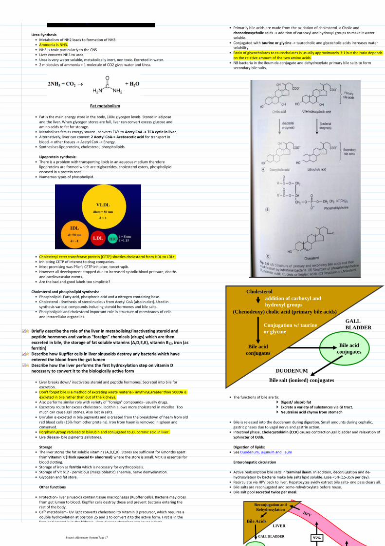

Learning Objectives
Define Liver Failure and its main types
Understand the important underlying pathophysiology
Name important causes of acute and chronic liver failure
Know the clinical features and complications of liver failure
Be aware of possible treatments for liver failure
Define Liver Failure and its main types
Rate of onset- manifestations and outcome
Cause
Clinical features
Characterised by bull
Rate of decline of function determines the way the syndrome manifests and the likely outcome
bull
Main features are the consequence of the accumulation of toxins resulting from the loss of the detoxifying function of the liver
bull
To understand liver failure one must remind oneself of the functions of the liver
Excretion bilirubin cholesterol hormones drugsbullEnzyme activationbullStorage glycogen fat vitamins (ADEK) and regulation of glucosebullSynthesis plasma proteins (eg albumin clotting factors) bilebullDetoxificationbullImmune regulation- Kupffer cells- antigens immune complexesbull
Main types are acute and chronic
Liver Failure- Insufficient hepatocyte function to maintain homeostasis
Understand the important underlying pathophysiology
Centrilobular necrosis of hepatocytesbullMononuclear cell infiltrate portal tract and lobulesbullFatty changebullActivation of macrophagesbullRelease of cytokines- TNF interleukin 16bull
Name important causes of acute and chronic liver failure
Hyperacute- le7 days between jaundice and encephalopathy
Acute- 1-4 weeks
Subacute- 5-12 weeks
Develops in a previously normal liver and is subdivided according to interval between the onset of jaundice and encephalopathy
bull
Acute liver failure- specific aetiologies
Aetiology= main influence on rate of deterioration and prognosis
Time from jaundice to encephalopathy Common aetiology
Hyperacute le7 days Paracetamol
Hepatitis A B
Acute 1 ndash 4 weeks Hepatitis A B E
Idiosyncratic drug toxicity
Subacute 5-28 weeks NANB hepatitis
Infection HSV (herpes simplex) EBV varicellabullDrugs Isoniazid ecstasy halothanebullMetabolic Wilsons ReyesbullVascular Budd-Chiari ischaemicbullOther Fatty liver of pregnancy lymphoma amanita phalloidesbull
Other causes
This is a mushroom that tastes pleasant but the fatal dose is only 30gbullRisk is not decreased by cooking and there is delayed presentation of liver and kidney failure and no known antidote
bull
Amanita phalloides
Khat- this is a leaf that is chewed in eastern African and the Arabian peninsula and contains cathionone- amphetamine like substance May cause chronic hepatitis
Rarer Causes
Budd-Chiari syndromeObstruction of hepatic veins at any site from lobule of IVC to right atriumbullPresent with abdominal pain hepatomegaly ascites and histologically shows sinusoidal distension
bull
Causes include thrombophilia webs veno-occlusive diseasebull
Wilsons DiseaseAutosomal recessive 130000bullCopper accumulation in liver basal ganglia and corneabull
Know the clinical features and complications of liver failure
Clinical features of acute liver failure include
This is a reversible neuropsychiatric state which impairs cognition and consciousness
It is caused by hepatocellular dysfunction and portal-systemic shunting Shunting leads to loss of metabolism and detox process
Brain is exposed to increasing levels of ammonia and other neurotransmitters
Grade 1 - Confused
Grade 2 - Drowsy
Grade 3- Sleeping but can be roused
Grade 4- Coma unrousable
There are four Grades of Hepatic Encephalopathy
Hepatic encephalopathy1
Cause of death in 30-50 with acute liver failure
Present in 80 with grade 4 encephalopathy
Blood supply in the brain is in a balance with carotid artery pressure vs intracerebral pressure
Disruption of blood brain barrier and increased osmosis into brain
Death actually results from brain stem herniation and cerebral hypoxia
Multifactorial- vasogenic leads to disruption of blood brain barrier cytotoxic mechanisms results in increased uptake of water into brain cells
Systolic hypertensioniIncreased muscle toneiiMyoclonus- (Muscle twitching)iiiDecerbrate posturingivDysconjugate eye movementsvLoss of pupillary reflexesviRespiratory arrestvii
Clinical features include
Cerebral oedema2
Liver synthesises all coagulation apart from Factor VIII It also synthesises inhibitors of coagulation and proteins involved on the fibrinolytic system so coagulopathy is very complex
Platelet count falls and platelets dysfunctional
Bleeding mucous membranes GIT brain
Measure the prothrombin time
Coagulopathy 3
Hypoglycaemia due to high insulin and low liver uptake
Decreased gluconeogenesis potassium levels due to increased urinary loss
Decreased sodium and metabolic acidosis
Metabolic acidosis especially with paracetamol toxicity also lactic acidosis due to reduced tissue perfusion by time of grade 3 encephalopathy Also hypo(phosphataemia calcaemia and magnesaemia)
Metabolic glucose potassium sodium and potential metabolic acidosis4
Common in both acute and chronic liver failure
Poor host defence can be caused by Kupffer cell failure and polymorphic dysfunction Which in turn causes a reduced conscious state
There may also possibly be increased access for infection such as with endotracheal tubes lines catheters and ascites
There may be both bacterial and fungal infections Bacteria are mainly gram positive especially Staphylococcus but it can be any Fungal infection is unrecognised in up to 13 of cases
Infection5
Know the clinical features and complications of liver failure
Portal hypertension1Clinical features of acute liver failure include
Liver Failure07 May 20121513
Stuarts Alimentary System Page 19
Wilsons DiseaseAutosomal recessive 130000bullCopper accumulation in liver basal ganglia and corneabullCan present with acute or chronic liver diseasebullTreatment is with penicillamine a copper chelatorbullIt is due to failure of copper excretion into bilebull
Acute Fatty Liver of PregnancyIncidence 11000 UKbullAetiology is inherited defects of fatty oxidationbullPresents in the 3rd trimester with RUQ pain vomiting then later with jaundice encephalopathy ascites and bleeding
bull
Treatment is urgent deliverybull
Chronic liver failure- can complicate virtually all forms of chronic liver disease
CausesAlcoholic liver diseasebullChronic viral hepatitis (CB)bullAutoimmune chronic active hepatitisbullPrimary biliary cirrhosisbullPrimary sclerosing cholangitisbullHaemochromatosisbull
Child-Pugh grade
Stage A B C
Bilirubin
umoll
lt17 17-34 gt34
Albumin
gl
gt35 30-35 lt30
Ascites none Easily controlled Poorly controlled
Encephalopathy None Grade 1 Grade 2-4
Clotting (PT) normal lt14 gt14
Non alcoholic fatty liver diseasebull
Be aware of possible treatments for liver failure
Remove toxic drug N-acetylcysteine used to combat paracetamol overdose
Underlying causes1
Preventioncontrol of infection and or bleeding
Nutrition
Early renal support
Recognitionmanagement of raised intracranial pressure
Supportive2
In acute liver failure - Grade 3 or 4 encephalopathy
Chronic liver failure- Child-Pugh grade BC
60 for acute
90 for chronic
Survival- 12 month survival
Transplantation3
Artificial support is an attractive option due to the scarcity of organs and delays associated with transplantations
There can be the potential for full recovery especially with paracetamol
Can also be used as a bridge to either a transplant or recovery
Replace necessary liver function synthetic eliminatory and metabolici
eg Albumin and clotting factors in addition to other plasma proteins
Metabolise fats proteins cholesterol and excrete drugs
Counter adverse effects of necrotic liverii
The system must
Human or animal
Biological- live hepatocytesi
There are 2 approaches
Artificial liver support4
Know the clinical features and complications of liver failure
Carries blood from abdominal alimentary tract (ie spleen gall bladder pancreas bowel) to the liver
Normal portal pressure is 7mm Hg
Portal venous system
Collaterals develop
Encephalopathy- lack of ammonia processing
Septicaemia
Impaired liver regeneration
Portal systemic shunting
Portal circulation can be blocked inside or outside of liver therefore collaterals develop to carry portal blood into the systemic circulation
Portal pressure correlates particularly with nodule formation
Portal hypertension block (liver nodule development)
Portal vein runs posterior to pancreas- formed by union of superior mesenteric and splenic vein
Inferior mesenteric brings blood from left colon and rectum
Portal hypertension1
Supplied by left gastric vein and drain into azygos system
Deviation of blood into these channels leads to varicosities in lower end of oesophagus
Bleeding leads to haematemesis melaena encephalopathy
Oesophageal varices
Sodium retention
Portal hypertension
Hypoalbuminaemia
Extracellular fluid within the peritoneal cavity
Spontaneous bacterial peritonitis
Renal failure encephalopathy
Complications
Ascites2
Sodium retention due to kidney response to perceived reduced circulating plasma volume (peripheral vasodilation AV shunting) sodium retention mediated by renin-angiotensin-aldosterone system
bull
Acute tubular necrosis-acute liver failure
Renal vasoconstriction
Decreased renal prostaglandins
Sepsis
Bleeding and hypotension
Hepatorenal syndrome (functional renal failure)- chronic liver disease
Renal failure3
Clinical features of acute liver failure include
Stuarts Alimentary System Page 20
Human or animal
Immortal cell line
Eg mars system- selectively eliminates toxins bound to albumin
Logistically easier and cheaper to target toxins that accumulate in liver
Non-biological- blood purification - adsorption and dialysis techniquesii
Or auxillary liver transplant- donor liver placed alongside native liver
Stuarts Alimentary System Page 21
Learning Objectives
Understand the production and excretion of bilirubinDescribe the features of pre-hepatic hepatic and post-hepatic jaundiceGive two examples of each of these types of jaundiceDescribe the pathogenesis of the symptoms and signs associated with jaundice
Bile Overview Features
Bile production is necessary for cholesterol homeostasis dietary lipid vitamin absorption and for the removal of xenobioticsdrugsendogenous waste products eg cholesterol metabolites adrenocortical and other steroid hormones
bull
Bile Production
Cholesterol and Alkaline Phosphatase (ALP) will be raised if there is an obstructionbull500-600ml of bile is produced daily It is golden yellow in colour resulting from the glucoronides of bile pigments
bull
60 bile secreted by hepatocytes (liver cells)bullUp to 40 secreted by cholangiocytes (biliary epithelial cells)bullBile drains from liver through bile ducts and into duodenum at duodenal papillabullBiliary tree called the bile ducts they enlarge as they become further away from their origin
bull
Role of biliary treeAlters pH fluidity and modifies bile as it flows throughbullH20 drawn into bile (osmosis through paracellular junctions)bullGlutathione split into constituent amino acids and reabsorbedbullLuminal glucose and some organic acids also reabsorbedbullHCO3
- and Cl- actively secreted into bile by CFTR mechanismbullCholangiocytes contribute IgA by exocytosisbull
8
Bile
flow
Lipids
Bile acids
Organic anions
Cl-
Cl-
HCO3-
H2O
Hepatocyte
Cholangiocyte
Bile Formation
Bile FlowBile flow closely related to concentration of bile acids and salt in bloodbullBiliary excretion of bile salts and toxins is performed by transporters expressed on apical surface of hepatocytes and cholangiocytes
bull
These biliary transporters also govern rate of bile flowbullDysfunction of the transporters is a major cause of cholestasisbull
Biliary salt excretory pump (BSEP) responsible for active transport of bile acids across hepatocyte canalicular membranes into bile and secretion of bile acids is a major determinant of bile flow
MDR related proteins (MRP1 and 3)
Products of the familial intrahepatic cholestasis gene (FIC1) and multidrug resistance genes (MDR1 and 3)
MDR1 mediates the canalicular excretion of xenobiotics and cytotoxins
MDR3 encodes a phospholipid transporter protein that translocates phosphatidylcholine from the inner to outer leaflet of the canalicular membrane
Main transporters include the bull
When people have high levels of bile salts they can get high levels of inflammation in the bile duct due to bile salt detergent properties
bull
Na and K salts of bile acids are conjugated in the liver to glycine and taurine (cysteine derivative)
bull
Bile acids synthesised from cholesterolbull4 acids in humans bullTwo primary ones are Cholic acid and Chenodeoxycholic acid which are converted by colonic bacteria to Deoxycholic aid and Lithocholic acid which are secondary acids
bull
Remember that bile salts form micelles and are amphipathic with hydrophilic domain facing out and hydrophobic domain facing in which means that free fatty acids and cholesterol are inside
bull
Detergent like actions make bile salts potentially cytotoxic in high concentrations but cell membranes are protected by other intraluminal lipids eg phosphatidylcholine in biliary tree and fatty acids in GIT and also by their own plasmalemma content of cholesterol and glycolipids
bull
Anatomy of the biliary system
Canal of Hering- Hepatocytes of extreme periportal zone make contact with bile duct lining cells and there is a short stretch where bile flows in channels lined by a mixture of bile duct lining cells and hepatocytes
bull
Sphincter of Oddi controls the release of bile when were fasting bile cant pass into the gut and go up into the gall bladder Cholecystekinin opens the sphincter causes contraction of the gall bladder and subsequent excretion of bile into the duodenum
bull
Bile salts
Understand the production and excretion of bilirubin
Bilirubin is a H20 insoluble yellow pigmentbull75 BR from Hb breakdownbull22 from catabolism of other haem proteinsbull3 from ineffective bone marrow erythropoiesisbull
BR is bound to albumin with most dissociating in the liverbullThere is more free than conjugatedbullFree BR enters hepatocytes binds cytoplasmic proteins and becomes conjugated to glucoronic acid (UDPGT from smooth ER) then to diglucoronide-BR which is more soluble and greater than free BR It is transported across concentration gradient into bile canaliculi into the GIT
bull
Urobilinogens - H2O soluble colourless derivatives of BR formed by action of GIT bacteria
bull
GIT mucosa is relatively impermeable to conjugated BR but is permeable to unconjugated BR and Urobilinogens
bull
Therefore some unconjugated BR enters enterohepatic circulation and some forms urobilinogens
bull
Some bilirubin goes to the kidney (urobilinogen)bull20 urobilinogens reabsorbed into general circulation and is excreted in urinebullSome urobilinogens passed in stool as Stercobilinogen bull
Oxidation of stercobilinogen to stercobilin causes brown colouration of faeces bull
Describe the features of pre-hepatic hepatic and post-hepatic jaundice
Cholestasis is a cause of jaundice and means the cessation of bile flowbullJaundice- Excess bilirubin in blood (gt34-50 microML)bullYellow tinge to skin sclera mucous membranesbull
Jaundice07 May 20122024
Stuarts Alimentary System Page 22
of bile duct lining cells and hepatocytesSphincter of Oddi controls the release of bile when were fasting bile cant pass into the gut and go up into the gall bladder Cholecystekinin opens the sphincter causes contraction of the gall bladder and subsequent excretion of bile into the duodenum
bull
Functions of Gall BladderStores bile (50ml) released after meal for fat digestionbullAcidifies bilebullConcentrates bile by H20 diffusion following net absorption of Na+ Cl- Ca2+ HCO3
-
(decreased intra-cystic pH)bull
Describe the features of pre-hepatic hepatic and post-hepatic jaundice
Cholestasis is a cause of jaundice and means the cessation of bile flowbullJaundice- Excess bilirubin in blood (gt34-50 microML)bullYellow tinge to skin sclera mucous membranesbullCholestasis normally results in jaundicebullHowever jaundice does not necessarily mean there is cholestasisbull
Haemolysis
Massive transfusion
Haematoma resorption
Ineffective erythropoiesis
Increased quantity of BR frombull
Look for Hb drop without overt bleeding where BRgtgtgt LFTsbullIx blood film haepatoglobins LDHbull
Pre-hepatic jaundice
Hepatocytes are malfunctioningbullDefective uptake and conjugationbullDefective BR excretionbull
Liver FailureAcute FulminantbullAcute or ChronicbullViral hepatitis EtOH AID PBC PSCbullIntraheptic cholestasis sepsis TPN drugsbull
Amount of BR being made is normal and blood flow is normal The problem is with the conjugation of the BR to make it bound and therefore inactive
HepaticHepatocellular
Defective transport of BR by biliary duct system eg common bile duct stones HepPancBil malignancy local Lnpathy
bull
Lookout for sepsis (cholangitis)bullCholangiocarcinoma is cancer of the bile duct themselvesbull
4141
Post hepatic Obstructive
Give two examples of each of these types of jaundiceDescribe the pathogenesis of the symptoms and signs associated with
jaundice
Pre-hepatic- rate of formation of bilirubingt removal
Damage to liver cells stop conjugation and or excretion Associated with severe hepatocellular damage
a
Failure to uptake bilebReflux of bile between hepatocytesc
Hepatic-
Damage to intrahepatic bile ductsaObstruction of intra or extrahepatic bile ductsb
Post hepatic-
Pre-Hepatic Hepatic Post-Hepatic
Inherited disorders of BR metabolism
Hepatitis due to Intrahep cholestasis 2ndary to drugs or virus
- Crigler-Najjar -Viral Primary sclerosing cholangitis
- Gilbertrsquos -Autoimmune Primary biliary cirrhosis
- Rotor -Drugs Gallstones in CBD
- Dubin-Johnson - Alcohol Cholangiocarcinoma
Carcinoma head of pancreas
Haemolysis Metabolic liver disease
Carcinoma ampulla
Obstructive jaundice is associated with a pale stool and dark urinebull
Stuarts Alimentary System Page 23
Learning Objectives
Outline the embryonic development of the pancreas
Review the anatomical regions and main anatomical relations of the pancreas
Distinguish between the exocrine and endocrine parts of the pancreas in structural and functional terms
Sketch the duct system of the pancreas
Define a pancreatic acinus
Describe the subcellular organisation of synthesis and secretion by the pancreatic acinar cells
List the most important components of the pancreatic (exocrine) secretions and define their roles in digestion
Explain the mechanism for bicarbonate secretion in terms of ion exchange pumps and membrane ion channels and the dependence on active transport
Understand that acinar cells synthesise enzymes for the digestion of carbohydrate lipids and proteins and store these in an inactive form in zymogen granules and explain how these enzymes are activated when they enter the duodenum
Explain how nervous stimulation and the hormones secretin and CCK regulate the release of pancreatic juice
Outline the embryonic development of the pancreas
The pancreas develops as a part of the ventral foregut mesentery arising at the foregut-midgut junction
bull
Composed of both a dorsal and ventral bud with the ventral bud being part of the hepatobiliary bud
bull
Ventral and dorsal pancreatic buds fuse with what was originally the ventral bud now being called the uncinate process
bull
The development also explains why there is an accessory pancreatic duct because the pancreas originally was two separate structures
bull
Ventral bud duct becomes main pancreatic ductbull
Review the anatomical regions and main anatomical relations of the pancreas
Uncinate- Hooklike in LatinbullEndocrine function is more concentrated in the tailbullPortal vein forms behind the head of the pancreas and gets trapped between uncinate process and neck region
bull
Portal vein formed by SMV + Splenic VeinbullAssociated with the 2nd part of duodenum at the ampulla of Vaterbull
Lies mainly on the posterior abdominal wall extending from C-shaped duodenum to hilum of spleen
bull
Pancreatic juice reaches duodenum via the main and accessory pancreatic glands
bull
Distinguish between the exocrine and endocrine parts of the pancreas in structural and functional terms
Endocrine- Secretion into the blood stream to have effect on distant target organ (AutocrineParacrine)- Ductless GlandsExocrine- Secretion into a duct to have direct local effect
Insulin- Anabolic hormone promotes glucose transport into cells and storage as glycogen reduces blood glucose promotes protein synthesis and lipogenesisGlucagon- Increases gluconeogenesis and glycogenolysisSomatostatin- Endocrine cyanide- BRUTAL
Islets of LangerhansbullSecretes hormones into blood- Insulin and Glucagon in addition to somatostatin and pancreatic polypeptide
bull
Endocrine- 2 of the gland
Secretes (Pancreatic Juice) into duodenum via pancreatic ductcommon bile duct in addition to having a digestive function
bullExocrine- 98 of the gland
Sketch the duct system of the pancreas
Exocrine Endocrine
Ducts Derived from the branching duct system
Acini are grape-like clusters of secretory units
Lose contact with ducts-become islets
Acinar cells secrete pro-enzymes into ducts
Differentiate into alpha and beta cells secreting into the blood
More endocrine function from the tail of the pancreas than the head
Pancreas structure and function11 May 20121357
Stuarts Alimentary System Page 24
List the most important components of the pancreatic (exocrine) secretions and define their roles in digestion
Duct and centroacinar cellbullJuice = rich in bicarbonate ~120mM where the plasmas is only 25mM pH 75-8bull
Prevents damage to duodenal mucosa
Raises pH to optimum range for pancreatic enzymes to work
Neutralises acid chyme from the stomachbull
Washes low volume enzyme secretion out of pancreas into duodenumbull
38
Effect of Duodenal pH on Bicarbonate
Secretion Rate
Duodenal pH lt3
= not much more
increase in
bicarbonate
secretion
Duodenal pH lt 5
= significant linear
increase in
pancreatic
bicarbonate
secretion
It may seem strange that bicarbonate secretion stops at pH which is still acid Howver bile also contains bicarbonate and helps neutralise the acid chyme
bull
Brunners glands secrete alkaline fluidbull
Bicarbonate-
H2O + CO2 lt=gt H2CO3 lt=gt H+ + HCO3-bull
Blood coming away from stomach is quite alkali- alkali tidebullBlood coming away from pancreas is relatively acidic- acid tidebull
Explain the mechanism for bicarbonate secretion in terms of ion exchange pumps and membrane ion channels and the dependence on active transport
Simplified
Main posterior relations are IVC abdominal aorta and the left kidney with the pancreas also having close relations with (and supply from) the coeliac and superior mesenteric arteries
bull
More endocrine function from the tail of the pancreas than the head
Define a pancreatic acinus
A small sac like cavity in the pancreas containing acinar cells that secrete low volume high viscous enzyme rich fluid there are also duct and centroacinar cells that secrete high volume watery HCO3
- into the lumen
Understand that acinar cells synthesise enzymes for the digestion of carbohydrate lipids and proteins and store these in an inactive form in zymogen granules and explain how these enzymes are activated when they enter the duodenum
Trypsinogen a precursor of a powerful protease which is converted to trypsin by enterokinase an enzyme present on the brush border of the small intestine
Chymotrypsinogen which can be activated by trypsin
Proelastase
Procarboxypeptidase
Prophospholipase A
Some of the main enzymes that the pancreas releases arebull
Zymogen means pro-enzymebullProteases are secreted as inactive pro-enzymes which protects acini against bull
CentroAcinar
Stuarts Alimentary System Page 25
What this does show well is the role of the CFTR chlorine channels in that it is important for chlorine to be able to be pass out of cell into the lumen so that it can then be exchanged later for the bicarbonate
bull
Detailed
Na+
CO2 + H2O H+ + HCO3-
CO2
HCO3-
Na+
H+
BLOOD DUCT CELL LUMEN
Catalysed by carbonic anhydrasebullSeparation of H+ and HCO3
-bullNa moves down concentration gradient via paracellular tight junctionsbullH20 followsbull
Na+
CO2 + H2O H+ + HCO3-
CO2
HCO3-
Na+
H+
BLOOD DUCT CELL LUMEN
Na+ Na+
Cl-
Cl-
At basolateral membrane sodium enters down concentration gradient resulting in H+ ion export into the blood- acidic tide
bull
Hydrogen carbonate is exchanged for Cl- which is in lumen due to CFTR channelsbullExchange is driven by electrochemical gradientsbullHigh ec (blood) Na compared to ic (duct cells)- ec= extracellularbullHigh Cl in lumen compared to ic (duct cell)bull
Na+
CO2 + H2O H+ + HCO3-
CO2
HCO3-
Na+
H+
BLOOD DUCT CELL LUMEN
Na+ Na+
Cl-
Cl-
K+K+
Na gradient into the cell maintained by sodium potassium ATPase Uses ATP therefore Primary Active Transport
bull
Na+
CO2 + H2O H+ + HCO3-
CO2
HCO3-
Na+
H+
BLOOD DUCT CELL LUMEN
Na+ Na+
Cl-
Cl-
K+K+
Potassium returns to blood via K channelsbull
Explain how nervous stimulation and the hormones secretin and CCK regulate the release of pancreatic juice
Gut is stimulated by the vagus nerve which is parasympatheticbull
Control of secretion
Cephalic phaseMediated by cholinergic vagus nerve The juice secreted during this phase is mainly enzyme rich containing very little HCO3
-bull
In response to vagal stimulation the acinar cells also secrete kallikreins which catalyse the production of bradykinin a vasodilator This results in increased blood flow to the pancreas and increased volume of secretion
bull
Gastric PhaseThe presence of food in the stomach stimulates the secretion of pancreatic juice via a hormonal mechanism
bull
Activation of chemoreceptors in the walls of the stomach by peptides and the activation of mechanoreceptors causes the release of the hormone gastrin from G cells and the local circulation Stimulation of cholinergic is also involved in this phase of control During the gastric phase the secretion of both the enzyme-rich and the alkaline components of pancreatic juice is increased
bull
Intestinal phase- 70-80 of pancreatic secretionFood material in the duodenum stimulates both the secretion of the alkaline and enzymatic components of the pancreatic juice
bull
Acid stimulates the release of secretin in the walls of the intestine and this hormone stimulates the duct cells to secrete the alkaline fluid Note this is a feedback control mechanism which helps control the pH of the contents in the duodenum
bull
The enzyme rich juice is released during the intestinal phase in response to fat and peptides in the food The fats and peptides cause release of CCK from the walls of the duodenum into the blood CCK also stimulates the acinar cells to secrete enzymes
bull
Trypsin in the duodenum inhibits the release of enzymes via inhibition of CCK releasebullSecretin exerts a permissive effect on the secretion of enzymes it does not stimulate enzyme secretion on its own but it enhances the effect of CCK
bull
Similarly CCK exerts a permissive effect on the secretion of the alkaline fluid by secretin
bull
Vagus stimulation causes release of mainly the enzyme rich secretion but if the vagi are sectioned the alkali secretion elicited in response to secretin is reduced by 50 indicating a functional overlap between the effects of vagal stimulation and secretin Thus the vagal mechanism may enhance the effect of secretin
bull
Extra informationBicarbonate rich component is controlled by release of a hormone Secretin (cAMP)bullEnzyme rich component secretion is controlled by vagal reflex and by a hormone Cholecystokinin (CCK) (Ca2+PLC)
bull
CCK also stimulates bile secretionbull
Switching off CCKCephalic phase ends when meal eatenbullAbsorption of fats and peptides removes local luminal stimulus for CCK release from mucosa
bull
Possibly other mechanismsbull
Stimulus Interaction
CCK alone has no effect on bicarbonate secretion
bull
CCK can markedly increase bicarbonate secretion that has been stimulated by secretin
bull
Vagus nerve has similar effect to CCK
bull
Secretin no effect on enzyme secretion
bull
Proelastase
Procarboxypeptidase
Prophospholipase A
Zymogen means pro-enzymebullProteases are secreted as inactive pro-enzymes which protects acini against autodigestion
bull
Pancreatic juice contains a trypsin inhibitor to prevent activationbullEnzymes only become active in the duodenumbullThe problem is that a blockage of the pancreatic duct may overload protection mechanisms and result in auto-digestion= acute pancreatitis
bull
Pancreatic secretions adapt to diet eg high protein low carbs increases proportion of proteases decreases proportion of amylases
bull
Pancreatic enzymes (+bile) are essential for normal digestion of a meal Lack of these can lead to malnutrition even if the dietary input is ok (Unlike salivary gastric enzymes)
bull
Increased faecal fat-occurs when pancreatic lipase secretion significantly reduced
Social anus cannot differentiate between oil and air
Orlistat-
Stuarts Alimentary System Page 26
Potassium returns to blood via K channelsbullCl- returns to lumen via CFTRbull
Stuarts Alimentary System Page 27
Learning Objectives
Define acute and chronic pancreatitis
List the symptoms and signs of acute pancreatitis
List four causes of acute pancreatitis
List blood tests and imaging modalities which are useful for patients with pancreatitis
List the complications of acute and chronic pancreatitis
List three causes of chronic pancreatitis
Define acute and chronic pancreatitis
Acute Pancreatitis- Is an acute inflammatory process of the pancreas with variable involvement of other regional tissues or remote organ systems
bull
150-420 per million populationbullSevere pancreatitis has mortality rate of 20bullAetiology of acute pancreatitis should be determined in 80 of patients (20 as idiopathic)
bull
Pancreas is retroperitoneal apart from tailbullLocated in C shape of duodenumbullSecretions from accessory duct enter duodenum at Ampulla of Vaterbull
List four causes of acute pancreatitis
The most common causes of pancreatitis are alcoholism and cholelithiasis Other causes are metabolic infections medications and vasculitis
List the symptoms and signs of acute pancreatitis
Abdominal pain radiating to backbullVomiting- near to stomach and duodenum inflammation can make you vomitbullOrgan failure (chest kidney)bullJaundice cholangitis- due to proximity to duct and gall stonesbull
Severe acute pancreatitisaMild acute pancreatitisbAcute fluid collectioncPancreatic necrosis and infected necrosisdAcute pseudocystePancreatic abscessf
The scope of clinical presentation also includesbull
Clinically (pain and vomiting) and elevated pancreatic enzymes (lipase lasts longer and has more specificity than amylase)
bull
CXR (to exclude perforated ulcer)bullUS (for gall stones)bullCT (in doubtful cases and to exclude other pathology)bull
Diagnosis of acute pancreatitis
Previous gall stonesbullAlcohol intakebullFamily historybullDrug intakebullExposure to viral causes or prodromalbull
History
List blood tests and imaging modalities which are useful for patients with pancreatitis
Pancreatic enzymesbullLiver function testsbullUS of gall bladderbull
Initial investigations
Fasting plasma lipidsbullFasting plasma calciumbullViral antibody titresbullRepeated biliary USbullMRCP- MRI scan of the liver and the pancreasbullCTbull
Subsequent investigations
Another USbullEUS- endoscopic ultrasoundbullERCP (bile for crystal bile pancreatic cytology sphincter of Oddi manometry) Itrsquos a type of endoscopy
bull
Pancreatic function testsbull
Dilatation of the pancreatic duct can occur if a bile stone obstructs the ductbull
Further investigations
Clinical impressions of severitybullBMIgt30 bullPleural effusion on CXR- usually on the left sidebullAPACHE II scoregt8bull
Determining severity of pancreatitis
Glasgow score 3 or morebullCRPgt150 (Normal range is about 5)bull
After 48 hours you may be able to detect organ failurebullBlack on CT = necroticbull
24H after admission
List the complications of acute and chronic pancreatitis
Multiple organ failurebullNecrotising pancreatitis abscessbullHaemorrhagebullBiliary complicationsbullPseudocyst- fluid collected in gland forms a cystbull
Management of pancreatitis
IV fluids respiratory active monitoring
Supportivebull
Severe attack due to gall stones
Jaundice cholangitis dilated CBD (cystic bile duct
ERCPbull
Conflicting evidence
Maximum 14 days
Prophylactic antibodiesbull
FNA (fine needle aspiration) for 30 people with necrosis bull
Embolisation for haemorrhage
Drainage of abscess
Interventional radiologybull
Cholecystectomy during the same admission
Necrescetomy
Drainage of pseuodcyst
Surgicalbull
Define acute and chronic pancreatitisList three causes of chronic pancreatitis
Chronic pancreatitis
Pancreatitis11 May 20121526
Stuarts Alimentary System Page 28
The most common causes of chronic pancreatitis are alcoholism microlithisasis and idiopathic
bull
Rare causes are hereditary pancreatitis hyperparathyroidism and obstruction of the main pancreatic duct caused by stenosis stones or cancer
bull
The picture above shows that in chronic pancreatitis episodes of acute pancreatitis are not cleared and they have repeated attacks of inflammation
bull
Symptoms may be very similar to acute pancreatitis but occasionally there is no pain or there may be severe epigastric pain that can last for many hours or multiple days
bull
When lipase and proteases secretions are reduced to lt10 of normal level the patient will develop steatorrhoea
bull
Management of chronic pancreatitis
Enzyme replacementbullAdequate nutritionbullInuslin supplementbullManagement of secondary complications eg jaundicebull
Chronic relapsing casesPain managementbullRehabilitation of the alcoholic patientbull
Intractable pain not responsive otherwise
Management of a secondary complication eg stricture CBD
Strechnic nerves- coeliac plexus- cut the nerves so pain cannot be felt
Surgical intervention (pancreatectomy)bull
Diffuse calcium stones= calcification of pancreas and thus chronic pancreatitisbull
Acute pancreatitis CT scan demonstrating a pancreatic abscess
Chronic pancreatitis An example of a focal area of compression of the main pancreatic duct with signs of distal obstruction 34- chronic obstructive pancreatitis
Stuarts Alimentary System Page 29
Learning Objectives
To describe principles of energy balance
To describe methods of assessment of energy stores and risks
To understand mechanisms of obesity
To describe complications of obesity
To understand the basics of obesity management
To describe principles of energy balance
In a steady state energy in must equal energy out1Overweightobese individual has increased total energy expenditure2
REE
thermogenesis
Physical activity
CHO
FAT
PROTEIN
ENERGY
INTAKEENERGY
EXPENDITURE
70kg Weight
56Kg FFM
14kg Fat Mass
20
REE= Resting energy expenditurebullSomebody who eats more just develops a new set point for their weight-they do not gain weight indefinitely
bull
Dietary Energy
REE
Exercise
Thermogenesis
Faclultative T
We are very efficient at exercise so that not much energy is wastedbullThermogenesis is the cost of storing foodbull
Lean body mass (FFM) is proportional to resting
energy expenditure
1000
1500
2000
2500
3000
30 50 70 90 110LBM( kg)
RE
E (
kc
ald
ay)
FFM (lean body mass) is proportional to our energy expenditurebullReally tells us how many mitochondria somebody hasbull
To describe methods of assessment of energy stores and risks
Weight and height1Regional adiposityenergy partitioning2
Weight- definition of morbidity and mortality Not defined as standard deviation from the norm Weight is defined as increased risk of early death
bull
BMI= Weight Height2bullWaist to hip ratio the metabolic syndrome Better predictor of ischaemic heart disease
bull
Metabolic syndromeAssociated with too much omental fatbull
Fasting glucose
gt60mmoll
Waist circumference
Mengt102
Womengt88
Microalbumin
Insulin resistance
Hypertension
BPgt13580
HDL
Menlt10
Womenlt13
Metabolic
Syndrome
BMI is not as good as a predictor of cardiac events as waist to hip ratiobullOmental fat is more metabolically active and drains directly through the liverbull
To understand mechanisms of obesity
Physiologically designed to protect against starvationbullHumans with opposable thumbs were able to break open bones and suck out the marrow inside
bull
1st half of century energy expenditure was driving energy intakebullGradually in 1960s food became readily available reduced in cost driving decreased energy consumption
bull
Causes of obesity
Energy intake per person of food sold fairly constant Increase in energy intake is not necessarily the main cause of obesity
Although we are eating the same amount of calories we are eating more fat in comparison to carbohydrate
Autoregulation following fat ingestion is difficult
Satiety involves complex endocrine regulation
Energy intakebull
Big reduction in energy expenditure over recent years
Energy Usagebull
To describe complications of obesity
MortalitybullMorbiditybullEconomicbull
Life span was reduced with a BMI of 35 or 30bull
Psychological morbidity
Diabetes and metabolic
syndrome
Cardiovascular disease
Gynaecologic disease Pregnancy
bullFetus
bullMother
bullPregnancy
bullObesity
Chest disease
Malignancy
Rheumatological
disease
PsychologicalbullCardiovascularbullGastrointestinalbullChest diseasebullEndocrinebullGynaecological- Poly cystic ovary syndrome
bull
Obstetricbull
Obesity and Malnutrition14 May 20120856
Stuarts Alimentary System Page 30
comparison to carbohydrateAutoregulation following fat ingestion is difficult
Satiety involves complex endocrine regulation
Big reduction in energy expenditure over recent years
Today we walk 3000 steps a day Amish community walk 18-20000 a day
Energy Usagebull
Most obesity is not monogenic
However obesity does have some major genetic factors Weight gain and storage is predicted well by genetics eg twin pairs
FTO gene - homozygous for this gene means you are 16 more likely to be obese
Genetics loads the gun the environment pulls the trigger- BRUTAL
Genesbull
White adipose tissue- Stores most fat and produces leptin
Leptin tells hypothalamus you have enough energy
White adipose tissue is an endocrine gland in its own right
Leptin isnt about regulating excess calories itrsquos the means by which low energy stores tell the body to stop menstruation and antibody production
Insulin may have an effect on satiety intake
Ghrelin from stomach tells us we are hungry
PYY (Polypeptide Y) produced by small intestine that tells hypothalamus you have eaten a big meal
Endocrinologybull
Energy (or fat) homeostasis
LEPTIN
White adipose tissue
Energy
expenditure
(Thermogenesis)
Satiety
(intake)
Hypothalamus
INSULIN
GhrelinPYYhunger
You have eaten big meal
Life span was reduced with a BMI of 35 or 30bull
Psychological morbidity
Diabetes and metabolic
syndrome
Cardiovascular disease
Gynaecologic disease Pregnancy
bullFetus
bullMother
bullPregnancy
bullObesity
Chest disease
Malignancy
Rheumatological
disease
High diabetes risk may be due to the fact that genes predispose to hyperglycaemia and omental fat levels
bull
To understand the basics of obesity management
WhybullFollow evidence basebullLifestyle diet and exercisebullPharmacologicalbullSurgicalbull
100kggtgt10kg weight lossPCOSbullOesophagitisbullCHDbullOsteoathritisbullLiver functionbullFunctionbull
Atkins associated with greatest weight loss over 12 months compared to Zone Ornish and LEARN diets
bull
n=16
n=21n=19
Role of diet and physical activity
in the management of obesity
0
ndash2
ndash4
ndash6
ndash8
Diet only
Change in b
odyw
eig
ht at w
eek 1
2 (
)
Adapted from Bond Brill et al Int J Obes 2002 26 1484ndash93
Diet +
30-minute walk
Diet +
60-minute walk
ndash46
ndash62ndash66
902 kg 936 kg 884 kg Baseline
p005
People are quite likely to put weight back onbull
Pharmacotherapy
Orlistat is a GI lipase inhibitorbullSibutramine- NA uptake had other effectsbullRimonabant CBI- Cannabinoid receptor can cause depressionbull
Surgery
Diets
Stomach is reduced to 20ml in sizebullBoth very safe and effective means of weight loss recommended for people with a bull
Every kg = 1mmHG less in BP
In diabetesbull
Mortality 20bull
09mmHg per kg bull
HbA1c 15 bull
Risk of DM 40 bull
LDL 15 bull
HDL 8bull
Stuarts Alimentary System Page 31
Both very safe and effective means of weight loss recommended for people with a BMI gt40
bull
Endobarrier- tube 30cm long and fits into the pylorus bypassing the duodenum and enters the small intestine Food contact with the duodenum is important in satiety
bull
Lifestyle change had best outcome for people who were going on to develop diabetes
bull
Denmark taxed saturated fat in foodbull
Stuarts Alimentary System Page 32
Learning Objectives
To demonstrate a basic understanding of the role of nutrition in health and disease
To demonstrate a basic understanding of the role of the gastrointestinal track in maintaining nutritional status
To have a basic understanding of the role of the macro-nutrients in health and disease
To demonstrate a basic understanding of the role of nutrition in health and disease
Although the gut has many functions its main purpose is to act as a barrier that allows the movement of nutrients across it and it is highly adapted for this function
bull
Most nutrients are efficiently absorbed in the small bowelbullStates of malabsorption have disastrous effects on functionbullCoeliac disease causes flattening of villibull
Below 1200kcal
A third of the global population live below recommended nutritional needsbull
Most of Type 2 diabetes
30-40 CVD
Largest risk factor for many cancers
Half of western society is overweight and 25 are obese Accounts forbull
MASLOWS HIERARCHY OF NEEDS- GCSE BUSINESS STUDIES
Physical needs- supply of water and foodbullDensity of malnutrition is directly related to low life expectancybullGambia- obesity and malnutrition side by sidebull
UnderweightMonitoring body weight can be complicated by fluid balancebullArm circumference- can be used to measure subcutaneous fatbullUndernourishment in children can be determined by using growth chartsbull
Reference Nutrient Intake- This is 25SD above the Estimated Average Intake (the amount actually needed)
bullDietary reference values
Nutrient Requirement= Metabolic demand Efficiency of utilisationbullAlso can be thought of as the intake associated with absence of disease needed to cure sign of deficiency or needed to maintain circulating levels or tissue concentration
bull
Amount which must be consumed by an individual to maintain optimal health and function and or avoid deficiency
bull
Observation of intakes
Balance studies
Physiological estimates
Clinical studies
Functional tests
Methods used to determine requirementsbull
EssentialbullRoles as an anti oxidant (Fe3+ to Fe2+)bullImportant in formation of collagenbullDeficiency is scurvybull
Vitamin C- Ascorbic Acid
There are different needs for each nutrient which also vary before individuals and life stages eg women of childbearing age need more iron than men
bull
Only absorb 10 of iron from diet
bull
If you dont supply the body with enough energy then nutrients will be misused eg deamination of amino acids to generate glucose and energy
bull
Obligatory energy expenditure= Basal Metabolic RatebullAdaptive thermogenesis- Amount of energy needed to keep warm and absorb nutrientsbullAs you get older you lose muscle mass which drives down energy intakebull
Energy Metabolism
Glycogen Trigylcerides Protein
Aceytl
CoA
Glucose Free Fatty Acids Amino Acids
Pyruvate
Glycogenolysis
Glycolysis
Lipolysis
-Oxidation
Proteinolysis
Deamination
and oxidation
Energy Metabolism
Energy
PyruvateAmino
Acid
Fatty
Acid
ACETYL CoA
TCAKetone
Bodies
Fatty
Acids
Fates of Acetyl CoA
To have a basic understanding of the role of the macro-nutrients in health and disease
Under nutrition14 May 20121006
Stuarts Alimentary System Page 33
Deficiency is scurvybull
PyruvateAmino
Acid
Fatty
Acid
ACETYL CoA
TCAKetone
Bodies
Fatty
Acids
Fates of Acetyl CoA
Glycogen- Stored in the liver and muscleAdipose tissue- Major storeMuscle- Prolonged starvation
The human body will increase in weight twenty fold from a baby to an adultbullAll material in weight gain enters the human body as food and drinkbullGrossly abnormal diets will cause changes in body weight configuration and composition
bull
Example height 100 years ago limit related to energy intake now we possibly reach our genetic maximum
bull
Growth
Average human eats 900000 kcals per yearbullA 3 error would result in an extra 27000 kcals per yearbullThis would result in an extra 10lb weight gain per yearbullLeptin levels are high in the morningbullPYY- tells you to stop eatingbullThere are far more signals that tell you to eat then to stop eating in the human bodybull
GHRELIN
PYY3-36
HYPOTHALAMUS
PERIPHERAL SIGNALS REGULATING APPETITE
LEPTIN
Changes in AgeingDecline in body size liver and kidney massbullIncrease in body fatbullDecline in muscle massbullDecline in total body waterbull
Causes of under nutrition in the developing worldPoliticsbullClimatebullPoor waterbullPoor agricultural policybullDemand of the developed worldbullFood security will become the biggest public health issue in the coming yearsbull
Regulation of feeding
To have a basic understanding of the role of the macro-nutrients in health and disease
Thiamine occurs in the human body as free thiamine and as various phosphorylated forms
bull
Beriberi is a nervous system ailment caused by a deficiency of thiamine in the dietbullThiamine is involved in the breakdown of energy molecules such as glucose and is also found on the membranes of neurons
bull
Symptoms of beriberi include severe lethargy and fatigue together with complications affecting the cardiovascular nervous muscular and gastrointestinal symptoms
bull
Beriberi can almost be man made because thiamine bearing husks of white rice have been removed in polished white rice
bull
Thiamine- Vitamin B1
Nicotinamide is the derivative of niacin and used by the body to form the coenzymes NAD and NADP
bull
Deficiency state is PellagrabullThought initially to be an infectious disease as it spread rapidly around far east and Europe It was noted this occurred as staple crops were replaced with maize apart from in Mexico where crops were softened with lime
bull
Niacin- Vitamin B3
Severe malnutrition in children can affect IQbull7 of people over age of 70 are in malnutritionbull
Signs of under nutritionWeight lossbullLoss of subcutaneous fatbullMuscle wastingbullPeripheral oedemabullGlossitis cracking edges of mouthbullHair lossbullChronic infectionsbullPoor wound healing chronic wounds pressure soresbullListless apatheticbullRecurrent pulmonary infectionsbull
Complications relative to loss of LBM
Lean Body Mass
( loss of total)
Complications
(related to lost LBM)
Associated mortality
()
10 Impaired immunity
increased infection
10
20 Decreased healing
weakness infection
30
30 Too weak to sit
pressure sores
pneumonia no healing
50
40 Death usually from
pneumonia
100
Energy restrictionstarvation
Weight loss andchange in body
composition
Decrease in metabolic
activity of the FFM
Reduction in RMR
activepassive
Response to negative
energy balance
Response to reduced
energy flux
T3
Hormonal events InsulinT4T3=
GlucagonGH
Substrate mobilisation
FFA and amino acids
SNS activity and
catecholamines
metabolic flux
energy expenditure
MECHANISMS
Metabolism Response to Starvation (Short Term)No injury or lsquoStressrsquo (Protective Adaptation Occurs)
Lower metabolic rate
20-25kcalkgd
ENERGY DEPOT
FAT FATTY ACID
ENERGY
PRODUCTION
LEAN MASS
Minimal catabolism to
meet glucose needs
EROSION MINIMAL
Hormone adaptation Protein synthesis
preserves protein
Intact skin
90 kcal Ketones
Energy for To tissues
protein synthesis
Gluconeogenesis (10 kcal)
Oxygen
Glucose
For obligate users (brain)
Micronutrients needs
Amino Acids
Alanine
UREA
Heat loss blocked
Stuarts Alimentary System Page 34
Normal Metabolismnonstressed normal nutrient intake
CHO 55-60kcal Pathway to energy
oxygen used
FAT 25-30kcal Micronutrients
PROTEIN 5-10kcal Pathway to protein synthesis
ENERGY DEPOT
FAT MASS
ENERGY
PRODUCTION
For Cell function
Muscle function
Tissue repair
Normal metabolic rate
25-30kcalkgd
LEAN MASS
COMPARTMENT
Protein synthesis
adequate to maintain
physical and metabolic
machinery
Hormones are
balanced
Anabolic and
catabolic stimuli
Intact skin
Prevents heat amp
water loss
Stuarts Alimentary System Page 35
Learning Objectives
To recognise the clinical presentation of malabsorption
To understand the disease mechanisms leading to malabsorption
To learn about the clinical importance presentation complications and treatment of coeliac disease
To recognise the clinical presentation of malabsorption
Change in consistency of stools wateryexplosivefloating
Increase volume weight of stool with an increased watery texture
Normal frequency is 3day - 3week
Normal weight = lt200ml per day
Rarely lasts longer than 2 weeks
Osmotic
Secretory
Inflammatory
Chronic diarrhoea has many causes
Diarrhoeabull
Pale poorly formed stools with an offensive smell which is difficult to flush away Faecal fat content should be less than 6g a day
Steatorrhoeabull
Weight lossbullSigns and symptoms of nutrient deficienciesbullAnd in children growth failurebull
BMI scorebullWeight loss scorebullAcute disease effect scorebullScores are assigned to each individual section and divides people into low medium and high risk categories
bull
Muscle wastingbullLoss of fatbullOedemaascites low albuminbullAnaemia skin lesions poor wound healingbullAnthropometrybull
Malnutrition Universal Screening Tool
FatsbullAmino acids and proteinsbullCarbohydratesbullElectrolytes and waterbull
Nutrition
MineralsbullTrace elementsbullVitaminsbull
Reduced gastric tissuesecretion
Loss of pancreatic tissue
Impaired bile secretion
Abnormal digestionbullMaldigestion and malabsorption
Loss of functioning enterocytes
Pre and post mucosal effects
Single gene disorders
Abnormal absorptionbull
Fats- faecal elastasebullCommon tests
To understand the disease mechanisms leading to malabsorption
Causes of malabsorption
Common Coeliac disease Gluten sensitive enteropathy
Small bowel bacterial overgrowth
Gastric surgery and achlorhydria Intestinal blind loops post-surgery Jejunal diverticulaIntestinal stricturesFistulae (as in Crohns disease)Impaired peristalsis (fibrosis)
Pancreatic insufficiency Chronic pancreatitisCystic fibrosis
Less common
Short bowel syndrome Intestinal resection for Crohns diseasemesenteric vascular disease or injury
Chronic infections Tropical sprue Giardiasis Other parasites (eg Strongyloides)Tuberculosis AIDS Whipplersquos
Lymphoma IPSID EATL refractory sprue amp ulcerative jejuno-ileitis
Radiation enteritis Fibrosis Atrophy Strictures Lymphangiectasia
Intestinal lymphangiectasia Congenital Infective Fibrotic Malignant Cardiac
Drugs Orlistat Laxatives Neomycin Cholestyramine (and certain others with specific interactions)
Allergic Eosinophilic enteritis Milk and soya enteropathyAutoimmune enteropathy
Immunodeficiency Congenital acquired
Usually after surgery for Crohns diseasebullLess than 200cm of small intestine causes malabsorption malnutrition electrolyte imbalance progressive weight loss
bull
Short bowel syndrome
Malabsorption of specific nutrients
Nutrient Condition
Lactose Lactase non-persistence
Vitamin B12 Pernicious anaemia (loss of intrinsic factor)
Bile salts Bile salt diarrhoea
Hypolactasia in the small intestinal brush border membrane is usual adult human phenotype (non-persistence of neonatal lactase)
bull
Malabsorption of lactose in small intestine gives symptoms from breakdown in colon (H2Co2SCFAlactate)
bull
Iron deficiency- Hyper or hypothyroidism can cause pretty much any GI conditionGI tumour could cause an intestinal bleeding but does not necessarily exit from the rectum
To learn about the clinical importance presentation complications and treatment of coeliac disease
Frequent Clinical featuresMalaisebull
Coeliac disease- Inflammatory disease of upper small intestine resulting from gluten ingestion from genetically susceptible individuals
Common Clinical Features
Malabsorption21 May 20120857
Stuarts Alimentary System Page 36
Loss of functioning enterocytes
Pre and post mucosal effects
Single gene disorders
Fats- faecal elastasebullProtein and nitrogen- urinary excretion albuminbull
Also xylose absorption test of intestinal integrityfunction
Pancreolauryl test of pancreatic esterase activity
Minerals- ions calciumbull
Common tests
Enzyme stable during passage through GI tract and thus a small sample of stool is tested
bull
High sensitivity and specificity for diagnosing pancreatic insufficiencybull
Faecal Pancreatic Elastase
Frequent Clinical featuresMalaisebullFatiguebullSteattorhoeabullDiarrhoeabullWeight lossbullAnaemiabull
Coeliac disease- Inflammatory disease of upper small intestine resulting from gluten ingestion from genetically susceptible individuals
Possible decline in severity of illness
Less reliance on wheat-based diet
Under-diagnosis in the community may be related to change in presentation in coeliac disease
bullChanging presentation of coeliac disease
IgA classbullTissue transglutaminase is the autoantigen recognised by endomysial antibodies and allows the production of anti-tTG antibodies
bull
Autoantibodies
Intense pruritus
Blisters rarely present
Vesicular rashbull
IgA
Skin biopsybull
Dermatitis herpetiformis
Fraction of cereals that bindsbullProteins found in wheat barley ryebullThere are HLA association- HLA-A1 and HLA-BBbullEnteropathy- Inflammatory damage with cell death Hyperplastic crypts results from stimulated regeneration
bull
Main method of treatment is to avoid gluten and may be combined with nutritional supplements
bull
Gluten
Nutrient malabsorption and impaired nutritional status- slow growth anaemia neurological disorders
bull
Lymphoma- enteropathy associated T-cell lymphoma
Adenocarcinoma
Osteoporosis osteopenia
Small bowel malignancybull
Complications
AnorexiabullAbdominal painbullOral ulcersbullOsteopeniabull
Common Clinical Features
TetanybullOedemabullRashesbull
Rare Clinical Features
Stuarts Alimentary System Page 37
Learning Objectives
Describe the major features of the enteric nervous system
Explain how the autonomic nervous system and the enteric nervous system interact
Describe how gut hormones control GI function
Describe the major features of the enteric nervous system
The wall of the GI tract contains many neurons- 2nd only to CNS (10-100 million)bullRich plexus (network) of ganglia (nerve calls + glial cells) interconnected by tract of fine unmyelinated nerve fibres Doesnt really matter if you start digestion a few seconds late which is why the lack of myelination is not an issue
bull
The enteric nervous system integrates the motor and secretory activities of the GI system
bull
Can function independently of central controlbullIf the sympathetic and parasympathetic nerves to the gut are cut many motor and secretory activities continue as they are controlled by the enteric nervous system
bull
Inflammation (ulcerative colitis Crohns disease)bullPost-operative injurybullIrritable bowel syndromebullAgeing (constipation)bull
Enteric neural dysfunctiondegeneration
MotilitybullBlood flowbullWater and electrolyte transportbullSecretionbullAbsorptionbull
Enteric nervous system regulates
Multipolar- one axon but with multiple dendritesbullInterneurons- neurons between neurones that integrate the sensory input and effector output this allows fine control
bull
Myenteric plexus(Auerbachs)- is to regulate the circular and longitudinal muscle layers- controls peristalsis
bull
Gut secretionsaGut epithelial and endocrine functionbBlood flowc
The submucosal plexus mainly sense luminal conditions and controlsbull
Neurons
Submucosa is closer to the surface than the muscle layersbullMyenteric and submucosal plexuses respond and co-ordinate each other Allows a specific type of response
bull
Migrating myoelectric complex
Waves of electrical impulses moving through the gut Trying to sweep the undigested food out Begins at stomach then gradually moves down the GI tract Explained well in Vanders page 542
bull
Myenteric Plexus
Submucosal Plexus
Circular Muscle
Longitudinal
Muscle
MucosaMuscularis Mucosa
mMyenteric plexus (Auerbachrsquos plexus)
located between the circular and longitudinal smooth
muscle layers Controls activity of muscularis externa
Motility
Submucosal plexus (Meissnerrsquos plexus)
Sensing environment within lumen
Blood flow epithelial and enocrine cell function
Minor plexuses
including deep muscular plexus (inside
circular muscle) and the ganglia supplying
biliary system and pancreas
Explain how the autonomic nervous system and the enteric nervous system interact
Regulates smooth muscle cardiac muscle and glandsbullHas both sympathetic and parasympathetic nervous systembull
ANS
Sympathetic
Cell bodies of preganglionic neurons in the thoracic and lumbar spinal cord Cell bodies of postganglionic neurons in the
pre vertebral and para vertebral ganglia
Cell bodies of Preganglionic parasympathetic neurons are in the brainstem and sacral spinal cord
bull
Preganglionic neurons interact directly with enteric nervous systembull
Majority of sympathetic fibres do not directly innervate structures in the GI tract instead they terminate on neurons in the intramural plexuses
bull
Vasoconstrictor sympathetic fibres do directly innervate the blood vessels of the GI tract- coeliac superior and inferior mesenteric
bull
Sympathetic innervation
Autonomic nervous system
Vagal nuclei
Sacral spinal
cord
Vagus nerve
Pelvic nerves
Parasympathetic division Sympathetic division
Pre ganglionic fibers
Sympathetic
ganglia
Postganglionic fibers
Enteric nervous system
Myenteric
plexus
Submucosal
plexus
Smooth muscle Secretory
cells
Endocrine
cells Blood vessels
Thoracic and Lumbar SC
Central nervous system
Sympathetic and parasympathetic efferents
Myenteric plexus
Submucosal plexus
Chemo and
mechanoreceptors in
wall of GI tract
Smooth muscle
Endocrine cells
Secretory cells
Blood vessels
Splanchnic
and vagal
afferents
Local
afferents
Describe how gut hormones control GI function
Pretty major endocrine glandbull
Gastrin
Ghrelin
Somatostatin
Histamine
Secretin
CCK
Somatostatin
PYY
GIP (gastric
inhibitory peptide)
GLP-1
GLP-2
Oxyntomodulin
Neurotensin
Somatostatin
PYY
GLP1
Oxyntomodulin
Neurotensin
Somatostatin
The Gut Hormones
Insulin
Glucagon
Somatostatin
Pancreatic Polypeptide
Enteric Nervous System21 May 20120957
Stuarts Alimentary System Page 38
Short afferents for when it can be handled within the gut itselfbullInterestingly the nausea associated with travel sickness is part of an adaptation to stop you being poisoned The brain thinks that you are hallucinating because you are going so fast which must have resulted from poisoning so as a result you vomit up your last meal to try and clear the toxin
bull
Central nervous system
Sympathetic and parasympathetic efferents
Myenteric plexus
Submucosal plexus
Chemo and
mechanoreceptors in
wall of GI tract
Smooth muscle
Endocrine cells
Secretory cells
Blood vessels
Splanchnic
and vagal
afferents
Local
afferents
Gastrin
Ghrelin
Somatostatin
Histamine
Secretin
CCK
Somatostatin
PYY
GIP (gastric
inhibitory peptide)
GLP-1
GLP-2
Oxyntomodulin
Neurotensin
Somatostatin
PYY
GLP1
Oxyntomodulin
Neurotensin
Somatostatin
The Gut Hormones
Insulin
Glucagon
Somatostatin
Pancreatic Polypeptide
These are produced predominantly in the mucosa layer but with some also made in the submucosa
bull
Act on secretory cells located in the wall of the GI tract pancreas and liver to alter the rate or the composition their secretions
bull
Histamine released from stomach wall cells is a key physiological stimulus to HCl secretion by gastric parietal cells
bull
Somatostatin from the stomach can inhibit acid secretion by paracrine mechanismsbull
Regulation of the mechanical processes of digestion
Regulation of the chemical and enzymatic processes of digestion
Control of post absorptive processes involved in the assimilation of digested food and CNS feedback regulating intake
Effects on the growth and development of the GI tract
Functions of the gastrointestinal endocrine system includebull
Paracrine
Synthesised in gastric antrum and upper small intestinebull
Amino acids and peptides in the lumen of the stomach
Gastric distension
Vagus nerve directly-acetylcholine
Release stimulated bybull
Gastrin stimulates gastric acid secretionbullRelease inhibited when pH of stomach falls below pH 3bull
Gastrin
Endocrine cyanidebullSynthesised in endocrine D cells of the gastric and duodenal mucosa pancreas (also hypothalamus)
bull
Somatostatin is a universal inhibitorbullRelease in response to a mixed mealbull
Gastric intestinal and pancreatic secretions
Growth and proliferation
Release of gut hormones
Intestinal nutrient and electrolyte transport
G thatrsquos GRIM
Motility
Inhibition ofbull
Analogues used to treat neuroendocrine tumours- Octreotide- has a much longer half life
bull
Gastric hormones have a very short half life less than 10 minutesbull
Somatostatin
Describe how gut hormones control GI function
Secreted by the S cells of the upper duodenum and jejunumbullMajor stimulus is the presence of acid in the duodenum (pH falls below 45)bullStimulates pancreatic bicarbonate secretion (effect potentiated by CCK)bullHigh concentrations inhibition of gastric acid and gastric emptyingbull
Secretin
Secreted by cells most densely located in the small intestinebullRelease stimulated by fat and peptides in upper small intestinebull
Stimulates pancreatic enzyme release
Delays gastric emptying
Stimulates gall bladder contraction
Decreases food intake and meal size
Actionsbull
Cholecystokinin
Secreted by mucosal K cells (predominant in the duodenum and jejunum)bullGIP released following ingestion of a mixed mealbullFat is more potent stimulus than carbohydratebullStimulates insulin secretionbullGIP receptor antagonists reduce postprandial insulin releasebull
Gastric inhibitory peptide
Cells found throughout the mucosa of the terminal ileum colon and rectumbullReleased from L cells post prandially (particularly fat)bull
PYY reduces intestinal motility
Reduces gall bladder contraction
Reduces pancreatic exocrine secretion
Inhibitor of intestinal fluid and electrolyte secretion
Actionsbull
Peptide YY (PYY)
Hormone Location Stimulated by Actions
Gastrin G cells of gastric antrum and upper duodenal mucosa
Mechanical distension of stomachDirect vagal stimulationPeptides and amino acids in stomach
Release of gastric acid from oxynticparietal cellsRelease inhibited when pH falls below 3 in the stomach
Somatostatin D cells of duodenal and jejunal mucosa pancreas and hypothalamus
Released in response to a mixed meal InhibitsGastric intestinal and pancreatic secretionsGrowth and proliferationRelease of GI hormonesIntestinal nutrient and electrolyte transportMotility
Secretin S cells of upper duodenum and jejunum
pH below 45 in the duodenum Stimulates the release of bicarbonateHigh levels can reduce gastric acid and gastric emptying
CCK Cells located mostly densely in small intestine
Stimulated by fat and peptides in the small intestine
Stimulates pancreatic enzymesDelays gastric emptyingStimulates gall bladder contractionDecreases food intake and size of meals
GIP K cells of upper duodenum and jejunum
Ingestion of a mixed meal- fat is a large stimulus
Stimulates Insulin secretion
PYY L cells present from terminal ileum to colon and rectum
DecreasesMotilityGall bladder contractionPancreatic exocrine secretionsFluid and electrolyte transport
Stuarts Alimentary System Page 39
Learning Objectives
Review the anatomical sections and main anatomical relations of the large intestine and related structures
Compare the structure and functions of the small and large intestines
Describe how the motility of the large intestine is regulated
Describe the control of defaecation
Describe the main functions of the large intestine
Review the anatomical sections and main anatomical relations of the large intestine and related structures
Consists of the colon cecum appendix rectum and anal canalbullThe cecum is a blind pouch just distal to the ileocaecal valve- larger in herbivore for the breakdown of food with bacteria
bull
The appendix is a thin finger-like extension of the cecum- not physiologically relevant in humans Escape pod for good bacteria- hide in their during a diarrhoeal infection and helps with repopulation
bull
ColonThe principal functions of the colon are the reabsorption of electrolytes and water and the elimination of undigested food and waste
bull
15m long 6cm in diameterbullThe ascending colon is on the right side of the abdomen runs from the cecum to the hepatic flexure (the turn of the colon by the liver)
bull
The transverse colon runs from the hepatic flexure to the splenic flexure (turn of the colon by the spleen)
bull
The descending colon runs from the splenic flexure to the sigmoid colonbullSigmoid (s-shaped) colon runs from descending colon to the rectumbull
Taeniea coli- Bands of LONGITUDINAL muscle that leads to a pouched appearancebullContains fatty tags- appendices epiploicae suggested to have a protective function against intra-abdominal infections but still relatively unsure about true function
bull
Taeniea coli shorter than small intestinebullThey cause the formation of pouched ovoid segments called haustra (singular haustrum)
bull
Compare the structure and functions of the small and large intestines
Like the small intestine enterocytes and goblet cells are abundantbullAbundant cryptsbullStem cells found in cryptsbull
Mucosa appears smooth at the gross level because it has no villi (smaller SA than small intestine)
bull
Enterocytes have short irregular microvilli and primarily concerned with resorption of salts
bull
Water is absorbed as it passively follows the electrolytes resulting in more solid gut contents
bull
Large intestine
Higher number of goblet cells than small intestinebullMore prevalent in the crypts than along the surface number increases distally toward the rectum
bull
The mucus facilitates the passage of the increasingly solid colonic contents and covers bacteria and particulate matter
bull
Acetylcholine (parasympathetic and enteric nervous system) stimulates Goblet Cell secretion
bull
Goblet Cells
No Paneth cells in the large intestine this is because we want to keep the commensal bacterial
bull
Reminder that the function of Paneth cells is to release acidophilic granules containing bacterial lysozyme glycoproteins and zinc and to engulf some bacteria
bull
Glycocalyx does not contain digestive enzymesbull
Paneth cells
~05-15um high and they make up the brush borderbullThere are several thousand microvilli per cellbullThe surface of the microvilli are covered with glycocalyxbull
Rich carbohydrate layer on apical membrane
Protection from digestional lumen on apical membrane yet allows for absorption
Traps a layer of water and mucous known as the unstirred layer which regulates rate of absorption from intestinal lumen
Glycocalyxbull
Microvilli
Describe how the motility of the large intestine is regulated
Like the small intestine muscularis externa consists of an inner circular and outer longitudinal layer
bull
Circular muscles are segmentally thickenedbullLongitudinal layer concentrated in three bands known as the taenia colibullBetween the taenia coli the longitudinal layer is thinbullBundles of muscle from the taenia coli penetrate the circular layer at irregular intervals
bull
Taenia coli- shorter than circular muscle layer ovoid segments called haustra can contract individually
bull
Apart from rectum and anal canal the muscle layers are substantial and continuousbull
Muscle layers
Large Intestine21 May 20121051
Stuarts Alimentary System Page 40
Dilated distal portion of the alimentary canalbullHistologically similar to the colon but distinguished by transverse rectal folds in its submucosa and the absence of taenia coli
bull
Terminal portion is the anal canal Surrounded by internal (circular muscle) and external (striated muscle) anal sphincters
bull
Rectum
Describe the main functions of the large intestine
Colon absorbs electrolytes and waterbullMore is absorbed in the proximal colonbullNa and Cl absorbed by exchange mechanisms and ion channelsbullWater follows by osmosisbullK+ moves passively into lumen- loses potassium into the gutbullLarge intestine can reabsorb approx 45 litres water (usually 15 litres) Above this threshold you have diarrhoea
bull
Reabsorption
All mammals have symbiotic relationships with their gut microbial communitybullStomach and small intestine have few bacteriabullLarge intestine contains many essential to normal functionbullAverage weight of live bacteria is 15kgbull
Large intestine Flora
Synthesis and excretion of Vitamin K-germ free animals can have clotting problems
bull
Prevent colonization by pathogens by competing for attachment sites or for essential nutrients
bull
Antagonise other bacteria through the production of substances which inhibit or kill non-indigenous species Stops bad bacteria getting a foothold
bull
Stimulate the production of cross-reactive antibodies Antibodies produced against components of the normal flora can cross react with certain related pathogens and thereby prevent infection or invasion
bull
Stimulate the development of certain tissues including cecum and lymphatic tissues
bull
Change in gut morphologybull
Roles
Bacteroides- Gram negative anaerobic non-sporeforming bacteriabullBifidobacteria are gram positive non-sporeforming lactic acid bacteria Thought tostop colonisation by pathogens
bull
Species
1) Drug metabolism
2) Insulin resistance
3) Bile acid metabolism
4) Lipid metabolism
5) Obesity
Yakult- Just executing a bottle full of bacteriabull
Links between gut bacteria and
Between the taenia coli the longitudinal layer is thinbullBundles of muscle from the taenia coli penetrate the circular layer at irregular intervals
bull
Taenia coli- shorter than circular muscle layer ovoid segments called haustra can contract individually
bull
Apart from rectum and anal canal the muscle layers are substantial and continuousbullMovements of large intestine more complicated then small intestinebull
5-10cm per hour moving along the large intestinebullKneading process designed to absorb salt and waterbullTry to keep it up in the top of the large intestine due to the top being able to absorb more
bull
Haustral contraction- thickened circular muscle causes back and forth mixingbullShort propulsive movements every 30 mins which increase in frequency following a meal
bull
Large Intestine Motility
1-3 times daily- mass movement which resembles a peristaltic wavebullCan propel contents 13-34 of length of large intestine in few secondsbullFood that contains fibre promotes rapid transport through the colonbullStimulated to some degree whilst eatingbull
Mass movement
Parasympathetic vagus nerve controls most of large intestinebullExternal anal sphincter controlled by somatic motor fibres in the pudendal nervesbullAfferent sensory neurons detect pressurebullHirschsprungs disease- No enteric intramural gangliabullMyenteric plexus ganglia concentrated below taenia colibullHormonalparacrine control eg aldosterone can affect sodium and water absorption
bull
Control
Rectum filled with faeces by mass movement in the sigmoid colonbullStores until is convenient to voidbullDefecation reflex controlled primarily by the sacral spinal cord-both reflex and voluntary actions
bull
Describe the control of defaecation
Reflex to sudden distension of walls of rectumbullPressure receptors send signals via myenteric plexus to initiate peristaltic waves in descending sigmoid colon and rectum Internal anal sphincter inhibited
bull
Weak intrinsic signal augmented by autonomic reflexbullExternal anal sphincter under voluntary controlbullUrge resisted sensation subsidesbull
Reflex
Social rectum- Can distinguish between solid liquid and gas Cannot tell the difference between gas and oil very well
150gday adultbullTwo thirds waterbullSolids cellulose bacteria cell debris bile pigments salts (K+)bullBile pigments give colour- oxidation of Stercobilinogen to stercobilinbullBacterial fermentation gives odourbull
Faeces
Stuarts Alimentary System Page 41
Learning Objectives
Briefly describe the processes of diffusion and osmosis
Describe the different protein-mediated transport systems that move substances across membranes
Explain the mechanisms involved in absorbing water Na+ K+ Ca++ Cl- HCO3-
Iron and vitamins in the alimentary tract
Briefly describe the processes of diffusion and osmosis
Occurs rapidly over microscopic distances but slowly over macroscopic distancesbullCell membrane acts as a diffusion barrier enabling cells to be selectivebull
Diffusion
Movement of water across a semi-permeable from a hypotonic to a hypertonic solution
bull
Primary active transport uses hydrolysis of ATPbullSecondary active transport is coupled to sodium importbull
Osmosis
Explain the mechanisms involved in absorbing water Na+ K+ Ca++ Cl- HCO3-
Iron and vitamins in the alimentary tract
99 of H2O presented to the GI tract is absorbedbullGreatest amount of water is absorbed in the jejunumbull
Water
Alimentary SystemIngest 2L Saliva 12L
Gastric
secretions
2L
Pancreas
12L
Bile 07L
Intestinal
24L
Small Intestine
absorbs 8L
Colon
absorbs 14L
Paracellular = between red tight junctionsbullTranscellular transport= through the cells themselvesbull
Driven by Na+bull
Counter transport in exchange for H+ (proximal bowel)
Co-transport with amino acids monosaccharaides (jejunum)
Co-transport with Cl- (ileum)
Restricted movement through ion channels (colon)
Transport of Na from lumen into enterocyte complex and varies between species Becomes more efficient as you travel down the intestine
bull
Standing Gradient Osmosis
Jejunum
IleumColon
Tight junctionsProximal
bowel
Intercellular space
Hypertonic
Explain the mechanisms involved in absorbing water Na+ K+ Ca++ Cl-
HCO3- Iron and vitamins in the alimentary tract
Think DIC
Duodenum and Ileum absorb Ca2+bull
Ca2+ deficient diet increases guts ability to absorbbullVit D and parathyroid hormone stimulate absorptionbullDiet 1-6gday secretion 06g Absorb 07g bull
Calcium is a secondary messenger lies on top of sodium channels etcbull
MethodCalcium carried across apical membrane by Intestinal calcium-bindingprotein (IMcal) facilitated diffusion
bull
Binds to calbindin in cytosolbullCalcium pumped across basolateral membrane by Plasma Membrane Calcium ATPase (PMCA) and NaCa exchanger
bull
PMCA has a high affinity for Ca2+ but low capacity Maintains the very low concentrations of calcium normally observed within a cell
bull
The NaCa exchanger has a low affinity for Ca but a high capacity Requires large concentrations of calcium to be effective
bull
Calcium
Essential for normal Ca absorptionbullDeficiency causes rickets osteoporosisbullIncreases transport of Ca across the brush border although the exact mechanism is unclear
bull
Enhances the transport of calcium ions across the cytosolbullIncreases the levels of calbindinbullIncreases rate of extrusion across basolateral membrane by increasing the level of Ca ATPase in the membrane
bull
Vitamin D
Essentially iron is toxic- if over absorbed there are serious pathophysiological effects
bull
But only absorbs 05-15 mgs
Adults ingest approx 15-20mgdaybull
Hydroxide
Phosphate
Bicarbonate ions
Iron forms insoluble salts withbull
Vitamin C increases iron absorption by converting Fe3+ to Fe2+ which we can absorb (It prevents formation of insoluble complexes)
bull
Inorganic iron (Fe3 = ferric and Fe2= ferrous)aAs a heme group (haemoglobin myoglobin and cytochromes)b
Iron is present asbull
Heme is smaller part of diet but more easily absorbed (20 of presented rather than 5)
bull
Cannot absorb Fe3+ only Fe2+ can be absorbedbull
HemeDietary heme is highly bioavailablebullHeme is absorbed intact into the enterocytebull
Process of absorption
Takes place in duodenum- Duodenum + Iron= DI= Die of too much ironbullHeme is internalised into the enterocyte via Heme Carrier Protein 1 (HCP-1) bull
Iron
IMcal= Intestinal calcium binding proteinPMCA= Plasma Membrane Calcium ATPase
Specific issues in alimentary absorption21 May 20121351
Stuarts Alimentary System Page 42
Active transport of Na+ into lateral intercellular spaces by Na+K+ATPase transport in the lateral plasma membrane
bull
Cl and HCO3 are transported into the intercellular spaces due to electrical potential created by the Na+ transport
bull
High conc of ions in the intercellular spaces causes the fluid there to be hypertonic
bull
Osmotic flow of water from the gut lumen via adjacent cells and tight junctions into the intercellular space
bull
Water distends the intercellular channels and causes increased hydrostatic pressure
bull
Ions and water move across the basement membrane of the epithelium and are carried away by the capillaries
bull
Cl- is co-transported with Na+ in the ileum and exchanged with HCO3- in the
colons Both are types of secondary active transportbull
K+ diffuses in via paracellular pathways in the small intestine leaks out of cells in colon Passive transport
bull
In secretory diarrhoea the secretion of Cl Na and water into the intestinal lumen by cells in the crypts of Lieberkuhn is elevated
bull
Cholera toxin permanently activates adenylyl cyclase elevating cAMP in the crypt cells and thus enhances the secretion of Cl (and therefore also of Na and water)
bull
Cholera patients may produce up to 20 litres per day of watery stool Such patients are likely to die unless they are promptly and adequately rehydrated
bull
Diarrhoea
Intercellular space
Hypertonic
Heme is absorbed intact into the enterocytebull
Process of absorption
Takes place in duodenum- Duodenum + Iron= DI= Die of too much ironbullHeme is internalised into the enterocyte via Heme Carrier Protein 1 (HCP-1) and via receptor mediated endocytosis
bull
Fe2+ liberated by Heme oxygenasebullDuodenal Cytochrome B (Dcytb) catalyses the reduction of Fe3+ to Fe2+ in the process of iron absorption in the duodenum of mammals
bull
Fe2+ is transported via Divalent Metal Transporter 1 (DMT-1) a H+ coupled cotransporter
bull
Fe2+ binds to mobileferrin and is carried to the basolateral membrane and moves via Ferroportin ion channel into blood
bull
Hepcidin the major iron regulating protein suppresses ferroportin function to decreases iron absorption
bull
Hephaestin is a transmembrane copper-dependent ferroxidase that converts Fe2+ to Fe3+
bull
Binds to apoferritin in cytosol to form ferritin micelle
Ferritin is globular protein complex Fe2+ is oxidised to Fe3+ which crystallises within protein shell
A single ferritin molecule can store up to 4000 iron ions
In excess dietary iron absorption you produce more ferritin
Fe3+ travels in blood bound to transferrin ORbull
Prevention of iron toxicityIrreversible binding of iron to ferritin in the epithelial cells stops too much absorption of iron
bull
Ironferritin is not available for transport into plasmabullIronferritin is lost in the intestinal lumen and excreted in the faecesbullIncrease in iron concentration in the cytosol increases ferritin synthesis and decreases its synthesis of transferrin receptors
bull
Organic compounds that cannot be manufactured by the body but vital to metabolism
bull
Passive diffusion predominant mechanismbullFat soluble (ADEK) transported to brush border in micelles K taken up by active transport
bull
Specific transport mechanisms for vitamin C (ascorbic acid) folic acid vitamin B1 (thiamine) vitamin B12
bull
Vitamins
Takes place in ileum- B12 + Ileum= BIbullLiver contains a large store (2-5mg)bullMost Vitamin B12 in food is bound to proteinsbullLow pH and the digestion of proteins by pepsin releases free vit B12bullBinds to R proteinsbullCan actually be destroyed in GI tract therefore bound to R proteins which are later digested in duodenum
bull
Vitamin B12
Intrinsic factor
Vitamin B12 binding glycoproteinbullSecreted by Gastric parietal cellsbullVitB12IF is resistant to digestionbullNo IF then no absorption of Vit B12bullVitB12IF complex binds to cubulin receptor taken up in distal ileum-mechanism unknown but thought to involve receptor mediated endocytosis
bull
Once in cell Vit B12IF complex broken down-possibly in mitochondriabullB12 binds to protein Transcobalamin II (TCII) crosses basolateral membrane by unknown mechanism
bull
Travels to liver bound to TCIIbullTCII receptors on cells allow them to uptake complexbullProteolysis then breaks down TCII inside of cellsbull
Mechanism
Impaired absorption of vit B12 retards the maturation of red blood cellsbullPernicious anaemiabullSchilling test used to be used to check for B12 reabsorptionbull
Vitamin B12 deficiency
Divalent Metal Transporter
Heme Carrier Protein 1
Summary- Water
Ingests about 2Ldaybull7L enters the gastrointestinal tract in various secretionbull99 of the H20 presented to the GI tract is absorbedbullThe absorption of water is powered by the absorption of ionsbullThe greatest amount of water is absorbed in the small intestine esp the jejunumbull
Absorbed in the small intestinebullCalbindin (a calcium binding protein) facilitates the transport of Ca++ through the cytosol of the intestinal epithelial
bull
Ca++ is transported across the basolateral membrane by Ca ATPase and the NaCA++ exchange protein
bull
Vitamin D stimulates the absorption of CA++ by enhancing the synthesis of calbindin and Ca-ATPase
bull
Summary- Calcium
About 5 of inorganic iron ingested is absorbed by the small intestinebullApproximately 20 of the heme iron is absorbedbullInorganic iron is transported across the brush border via DMT-1 (Divalent Metal Transporter-1)
bull
In the epithelial cells majority of iron is bound to ferritin and is unavailable for absorptionbullTransported into blood by ferroportin converted to Fe3+ by Hephaestin so it can bind to transferrin
bull
Summary- Iron
Receptors on the ileal brush border membrane bind the IF B12 complex and allow Vitamin B12 to be taken up into epithelial cells
bull
Vitamin 12 appear in the plasma bound to Transcobalamin IIbull
Summary- Vit B12
Hephaestin
Transcobalamin II
IntrinsicFactor
Ferroportin
Cubulin
Qu 4 Which of the following statements about transport across the gut wall is incorrect
Stuarts Alimentary System Page 43
All other things being equal net osmotic water movement will occur from a solution of low tonicity to a solution of high tonicity
X Potassium is actively transported into enterocytes across the lateral membrane
Paracellular transport involves movement through tight junctions
Absorption of galactose across the apical membrane takes place by secondary active transport
Ca++ crosses the basolateral membrane by facilitated diffusion
X Mark = -2 (conf=2 )
Best Option Ca++ crosses the basolateral membrane by facilitated diffusionCa++ cross the apical membrane via a facilitated diffusion mechanism involving intestinal calcium-binding protein and is then pumped across the basolateral membrane The sodiumpotassium pump on the lateral membrane both pumps Na+ ions out of the cell and K+ ions in Galactose (and glucose) enter the cell via a Nasugar co-transporter This depends on a sodium gradient developed as a result of active transport elsewhere (ie the basolateral membrane pump) and is therefore secondary active paracellular transport is between cells and therefore must pass though the tight junctions
Pasted from lthttpswwwuclacuklaptlaptlitesysrunhtmicl08_alimentaryf=cleari=icl1k=1u=_st1511i=Imperialgt
Stuarts Alimentary System Page 44
Learning Objectives
To understand the biochemistry and metabolism of ethanol
To understand the physiological effects and disease processes that alcohol can bring about in the various organ systems with particular reference to liver disease and cirrhosis
To appreciate the psychological and material impact of alcohol on both the individual and society as a whole
To develop the knowledge and skills to advise on how to enjoy alcohol responsibly and to identify and deal with problem drinking
To understand the biochemistry and metabolism of ethanol
One of the simplest organic chemicals- C2H5OHbullInsoluble in fats and oilsbullThere is no plasma protein binding for ethanolbull
Metabolism of Ethanol
ALDH
CATALASEADH
ETHANOL
ACETALDEHYDE
CO2 + H20
CYP2E1
Cytochrome
P450-2e1
Alcohol-dehydrogenasebullAldehyde is very toxic- increases blood pressure nausea headachebullAldehyde dehydrogenase (ALDH)- genetic polymorphisms for this genebullAsia- capacity to breakdown alcohol is significantly reduced- a lot more side effects and more prone to liver disease
bull
Cytochrome P452E1- it is an inducible enzyme so people who drink heavily can still metabolise effectively
bull
Reactive Oxygen Species are generated via the CYP2E1 pathway tissue damage inflammation and stimulation of the immune system which leads to fibrosis and cirrhosis
bull
To understand the physiological effects and disease processes that alcohol can bring about in the various organ systems with particular reference to liver disease and cirrhosis
Ethanol and break down products are able to damage hepatocytesbull
Normal liver-gt Fatty liver-gt Fibrosis-gt Cirrhosis-gt Hepatocellular carcinomabullProgression of ALD
Fat droplets deposited in the liverbullOccurs in up to 60 of heavy drinkersbullLeads to steatohepatitis and abnormal LFTsbullReversible if alcohol reducedbullFat sets up an inflammatory reaction which stimulates the immune system and cytokines causing damage
bull
Fatty Liver
Space of Diss- migrating macrophages chronic inflammation causes them to develop into fixed hepatic stellate which lay down collagen fibres
bullHepatic Fibrosis
AsymptomaticbullIrreversible scarring of liver with fibrous bands and regenerative nodulesbullEventually develops in 20 after 15 yearsbullMorbidity common associated with jaundice ascites bleeding cachexia infections and encephalopathy
bull
3-5 per annum risk of developing liver cancerbullDeath in most within 10 yearsbull
Cirrhosis
To appreciate the psychological and material impact of alcohol on both the individual and society as a whole
Addictive element that is associated with alcoholbullAlcohol misuse (Problem drinking)bullAlcoholic liver diseasebull
These three are arranged in a Venn diaphragm
Alcohol addiction (Alcoholism)bull
Addiction is dealt by psychiatrists and drug and alcohol workersbull
Failure to carry out major obligation at work home or school because of repeated alcohol use
bull
Repeated use of alcohol even when it is physically dangerous to do sobullContinued use of alcohol despite knowing that it has caused or worsened social or interpersonal problems
bull
Alcohol misuse
Evidence of tolerance andor withdrawalbullAmount of duration of use often greater than intendedbullRepeated failure to control or reduce alcohol usebullWithdrawal symptomsbull
Alcohol dependence
Nature vs nurture
Suggestion that there must be a genetic component to alcohol addictionbullAlcohol and heroin addicted mice get clever- sleep with dirty old men mice to get their fix
bull
Adoption studies- birth parents had a much bigger affect then adopted parents which would indicate that there is a genetic component
bull
To understand the physiological effects and disease processes that alcohol can bring about in the various organ systems with particular reference to liver disease and cirrhosis
Cancer
Reflux
Oesophagitis
Oesophagusbull
Gastritis
Ulcers
Cancer
Stomachbull
Evidence that no alcohol whatsoever leads to a slight increased risk of strokebull
Alcohol and the upper GI tract
Wernickes encephalopathybullKorsakoffs psychosisbullOptic toxicitybullAutonomic dysfunctionbullPeripheral neuropathybull
Neurological effects
Increased incidence of infectious disease
Bacterial pneumonia Septicaemia TB Hepatitis C
Alcohol leads to immunosuppressionbull
Contributes to liver disease and renal dysfunction
Alcoholic hepatitis liver cirrhosis renal disease associated with IgA deposition
Alcohol leads to an increased incidence of autoimmunitybull
Alcohol and Immunity
Fetal alcohol syndrome is among the most commonly known causes of mental retardation and is a major public health problem
bullFetal alcohol syndrome
Other interesting information
Significantly improves life expectancy if you detox alcoholicsbull
EXAMINABLE
Up to 45 due to alcoholbull
Steatorrhoea vitamin deficiencies hypocalcaemia
Exocrine insufficiencybull
Diabetes
Endocrine insufficiencybull
Chronic Pancreatitis
To develop the knowledge and skills to advise on how to enjoy alcohol responsibly and to identify and deal with problem drinking
A unit= 8g of alcoholbullUseful link to check you are doing the calculations rightbullhttpwwwdrinkawarecouktips-and-toolsdrink-diarybull
Calculating units by mass
alcohol is the number of g of alcohol in 100mlbullGs of alcohol = percentage mass of alcohol x volume drunk100bullAnnoyingly to make it more complicated most drinks dont give the percentage of alcohol by mass they instead do it by volume
bull
Calculating units by volumeGs of alcohol= (Percentage volume x 0789 x volume drunk)100bullThe problem is when calculating percentage by volume is that ethanols density is not the same as water Therefore 10g of alcohol does not have a volume of 10ml In order to be able to do the calculation we need to multiply by ethanols density which is 0789
bull
Worked exampleLast night I drank 1 pint of lager at 5 alcohol by volume Calculate how many units I consumed
bull
Gs of alcohol= (5x 0789x 568)100=224076 g of alcoholbull8g of alcohol = 1 unit -gt 2240768=2801 units bull
Clinical features of Acute Alcohol Poisoning
Ataxia and anaesthesia leading to accidental injurybull
Alcohol Lifestyle and you25 May 20121358
Stuarts Alimentary System Page 45
Worked exampleLast night I drank 1 pint of lager at 5 alcohol by volume Calculate how many units I consumed
bull
Gs of alcohol= (5x 0789x 568)100=224076 g of alcoholbull8g of alcohol = 1 unit -gt 2240768=2801 units bull
I wouldnt think that they would ask you to do it by volume as they didnt teach us about the density of ethanol causing problems however it may crop up and is useful to know
bull
Food in the stomach dramatically reduces the amount that gets absorbed into the blood
bull
You can have liver disease without getting drunkbullAlcohol is very rapidly absorbed into the duodenumbull
First Pass Metabolism of Ethanol
0
10
20
30
40
50
60
70
80
0
20
40
60
80
105
135
165
210
270
Minutes
Blo
od
Alc
oh
ol
Co
ncen
tra
tio
n
Oral Alcohol
IV Alcohol
Alcohol induced hypoglycaemia is one of the serious complications of acute alcohol poisoning
bull
Clinical features of Acute Alcohol Poisoning
Ataxia and anaesthesia leading to accidental injurybullDysarthria (slurring of words) and nystagmus- weird eye twitchingbullDrowsiness which may progress to comabullInhalation of vomit which can be fatal- maintain airway by keeping patient in the recovery position
bull
Alcohol is a strong Leydig cell toxin it can have an adverse effect on the synthesis and secretion of testosterone and is one of the commonest causes of male impotence
bullEffects of Alcohol on Male Reproductive System
Hangover- end up dehydrating yourself Altered osmolarity of endolymphatics
Stuarts Alimentary System Page 46
Learning Objectives
To understand that diarrhoea is a very significant contributor to global morbidity and mortality in children under 5 years You should be familiar with the size of the problem
To understand the critical differences between acute and chronic presentations of diarrhoea in children
To be aware of the major advances in prevention and management that have lead to reductions in mortality from diarrhoea in the last 25 years
To understand that diarrhoea is a very significant contributor to global morbidity and mortality in children under 5 years You should be familiar with the size of the problem
One billion people worldwide lack access to clean waterbull25 billion do without adequate sanitationbullMedian episodes per year of diarrhoea = 32bull
Diarrhoea- Increase in stool frequency with change to loose or watery stool
To be aware of the major advances in prevention and management that have lead to reductions in mortality from diarrhoea in the last 25 years
Do not use anti-diarrhoea drugsbull
Virtually every child infected by age of 5 years
Faecal-oral spread
Enterocyte destruction osmotic diarrhoea
Rehydration is key
Rotavirusbull
Norovirsues
Sapporo virus
Calicivirusesbull
AdenovirusbullEnterovirusbull
Viral causes of diarrhoea
Enterotoxigenic EColi (ETEC) travellers diarrhoeabullShigellae spp Invasive DysenterybullVibrio choleraebullSalmonellaebullCampylobacterbullYersiniabull
Bacterial causes
Always look for viruses bullFever look for salmonella shigella campylobacterbull
Investigations
Female educationbullImproved supplemental feedingnutritionbullPromotion of breast-feedingbullEffect of free soap in a community meant that there was a 53 reduction in incidence of diarrhoea and pneumonia
bull
Reasons for declining mortality
Diarrhoea accounts for 20 of mortality lt 5 yearsbullSummary
Chronic Diarrhoea
Use heightweight measurementbullCoeliac disease- destruction of enterocytes brush borderbullInfections and post infection syndromesbullCarbohydrate intolerancebull
Permanent (eg Coeliac disease)
Temporary (eg Cows milk protein intolerance)
Mother can pass on enough cows milk protein just by drinking it herself
Food sensitivitiesbull
ComplicationsMalnutritionbullPoor growth delayed pubertybullAnaemia ricketsbullCognitive impairmentbull
To understand the critical differences between acute and chronic presentations of diarrhoea in children
lt2 weeksbull
Viral
Bacterial
Protozoal
Infectionbull
Acute diarrhoea
Types of diarrhoea
Cholera and some Ecoli toxins switches on chloride pump
Secretory- active secretion or inhibited absorption without structural damagebull
Maldigestion (eg pancreatic disease)
Osmotic laxatives
Osmotic- water drawn to gut lumen passivelybull
Infections
Inflammatory disease eg Crohns
Inflammatory- damaged mucosa cannot absorbbull
Still eatingfeedingbullStill passing urinebullPoo-blood Mucus FreqbullFood eaten- New OffbulltravelbullSwimmingbullPetsbullImmunodeficiencybull
History
Clinical
features
Dark sunken eyes
No tearsLow turgor
Sunken
fontanelle
Dry mucous
membranes
Delayed capillary refill time
Moderate 5-10 Severe gt10
General Drowsy Limp sweaty cold cyanotic
Pulse Tachycardic Rapid weak
BP May be normal Low
Urine output Reduced Absent
Percent of body weight water lossbull
Oral rehydration needs glucose in order to help with sodium absorptionbullIntra-osseus- if you cannot get the vein you go for flat bit of the tibia as this will be rapidly become part of the circulation
bull
ABC (airway breathing circulation)bullAny other focus for infectionbullDegree of dehydrationbull
Examination
1g of faeces may contain 10000000 virus particlesbull
CryptosporidiumbullGiardiabullEnteamoeba histolyticabull
Protozoa
Diarrhoea in Children25 May 20121358
Stuarts Alimentary System Page 47
Stuarts Alimentary System Page 48
Learning Objectives
How blood osmolality and pressure regulate thirst
The major hypothalamic circuits regulating appetite
The role of leptin in the regulation of body weight
How gut hormones can influence short term appetite
How blood osmolality and pressure regulate thirst
Body fluid osmolality is increasedaBlood volume is reducedbBlood pressure is reducedc
A person feels thirsty whenbull
The increase of the plasma osmolality is the more potent stimulus as just a 2-3 change induces a strong desire to drink
bull
A decrease of 10-15 in blood volume or arterial pressure is required to produce the same response
bull
As covered in other sections antidiuretic hormone acts on the kidneys to regulate the volume and osmolality of urine High levels of ADH cause the kidneys to retain more water and a small volume of urine is excreted
bull
Osmoreceptors that measure the osmolality of the blood are located in the Organum vasculosum (of the lamina terminalis (OVLT) or supraoptic crest) and the Subfornical organ (SFO)
bull
These osmoreceptor cells shrink or swell in response in changes to osmolality They then send signals to the ADH producing cells of the hypothalamus to alter ADH release
bull
Sensation of thirst
The feeling of thirst is decreased by drinking even before sufficient water has been absorbed by the GI tract to correct plasma osmolality (PO)
bull
Receptors in the mouth pharynx oesophagus seem to be involvedbullRelief of thirst sensation via these receptors is short livedbullThirst is only completely satisfied once PO is decreased or blood volume or arterial pressure is corrected
bull
Once again the renin-angiotensin-aldosterone system Angiotensin II invokes the sensation of thirst this is important because one of its important functions is to raise blood pressure and therefore it is released when blood pressure falls Angiotensin II actives SFO neurons to contribute to the homeostatic response to maintain body fluids at the correct levels
bull
The major hypothalamic circuits regulating appetite
3V
PARAVENTRICULAR NUCLEUS
VENTROMEDIAL HYPO
ARCUATE NUCLEUS
THE HYPOTHALAMUS
LATERAL HYPO
Agouti-related peptide GABA and neuropeptide Y are important orexigenic agents
bull
Leptin and Melanin stimulating hormone are important anorexigenic agentsbull
Orexigenic action
Anorexigenic action
Agouti-related peptide (AgRP) -amino butyric acid (GABA) Galanin Growth hormone releasing hormone (GHRH) Melanin concentrating hormone (MCH) Neuropeptide Y (NPY) Opioid peptides ( endorphin)
-Melanocyte stimulating hormone (-MSH) Amylin (islet amyloid polypeptide) Ciliary neurotrophic factor (CNTF) Cocaine and amphetamine regulated transcript (CART) Corticotrophin releasing hormone (CRH) Cholecystokinin (CCK) Glucagon-like peptide-1 amp 2 (GLP-12) Insulin Leptin Neurotensin Proinflammatory cytokines (IL1- TNF) Serotonin Urocortin
Arcuate Nucleus
This is a key brain area involved in the regulation of food intake Here there is an incomplete blood brain barrier which allows access to peripheral hormones that regulate food intake
bull
Stimulatory (NPYAgrp neuron)
Inhibitory (POMC neuron)
There are two neural populationbull
My Drawing
The role of leptin in the regulation of body weight
Leptin is a circulating hormone produced by adipose cells bullThey hypothalamus sense the concentration of the hormone then alters neuropeptides to increase or decrease food intake
bull
Leptin is low when there is low body fatbullLeptin levels are high when there is high body fatbullHormone that decreases food intake and increases thermogenesisbull
Can leptin cure obesitybull 3 ways in which leptin regulatory loop could
lead to obesity
Hunger and thirst29 August 20120038
Stuarts Alimentary System Page 49
No NPY or Agrp mutations associated with appetite have been discovered in humans
bull
POMC deficiency and MC4-R mutations cause morbid obesitybull
Can leptin cure obesitybull 3 ways in which leptin regulatory loop could
lead to obesity
A drug company paid a lot of money to commercial rights to leptin hoping that mechanism A or B would be leptins involvement in obesity Instead obese people have high levels of leptin because they have leptin resistance
bull
There are a small number of cases of congenital leptin deficiency which results in severely hyperphagic and obese individuals
bull
How gut hormones can influence short term appetite
We feel less hungry after a meal not due to the bulk in the stomach or the nutrients in the circulation but because of the release of hormones from the gut
bull
36 amino acid peptide with a tyrosine at both ends that is released into the circulation after a meal
Inhibits NPY release
Stimulates POMC neurons
Directly modulates neurons in arcuate nucleus
Decreases appetite
Peptide YY
These hormones are bull
Ghrelin is a gastric hormone that is 28 amino acids long (peptide hormone) and has an octanyl at the 3rd position High when fasting and falls after eating
Stimulates NPYAgrp neurons
Inhibits POMC neurons
Increases appetite
Ghrelin
Infusion of
ghrelin in nine
volunteers ndash
hunger scores
0 120 240Time (min)
2
3
4
5
6
7
8ghrelin saline
breakfast
hunger
lunch
start
infusion
Visual
Analogue
Hunger
Score
Gut hormones may represent a novel treatment for obesitybull
12 Volunteers lunch
PYY3-36 reduced food intake 36
Depression
Bowel cancer
Osteoarthritis
Gout
Sleep apnoea
Obesity is Associated with
Comorbidities
Stroke
Diabetes
Peripheral
vascular disease
Hypertension
Myocardial Infarction
Thrifty Gene Hypothesis
James Neel 1962bullSpecific genes have been selected for to increase metabolic efficiency and fat storage In the context of plentiful food and little exercise these genes predispose their carriers to obesity and diabetes
bull
Evolutionary it is sensible to put on weight as thin humans didnt survive famines so didnt pass their genes on to modern humans
bull
Evidence Populations historically prone to starvation become most obese when exposed to Western diet and sedentary life-style (eg Pima Indians and Pacific islanders)
bull
Adaptive Drift Hypothesis
In most animal species the fatter slower animals are eaten by predators and the thin starve in hard times
bull
10-20k years ago humans learned to defend themselves against predatorsbullThus obesity is not selected againstbullPutting on body fat then is a neutral change (genetic drift) However unlikely because of circumstances to put on much weight
bull
Stuarts Alimentary System Page 50

Learning Objectives
Understand the production and excretion of bilirubinDescribe the features of pre-hepatic hepatic and post-hepatic jaundiceGive two examples of each of these types of jaundiceDescribe the pathogenesis of the symptoms and signs associated with jaundice
Bile Overview Features
Bile production is necessary for cholesterol homeostasis dietary lipid vitamin absorption and for the removal of xenobioticsdrugsendogenous waste products eg cholesterol metabolites adrenocortical and other steroid hormones
bull
Bile Production
Cholesterol and Alkaline Phosphatase (ALP) will be raised if there is an obstructionbull500-600ml of bile is produced daily It is golden yellow in colour resulting from the glucoronides of bile pigments
bull
60 bile secreted by hepatocytes (liver cells)bullUp to 40 secreted by cholangiocytes (biliary epithelial cells)bullBile drains from liver through bile ducts and into duodenum at duodenal papillabullBiliary tree called the bile ducts they enlarge as they become further away from their origin
bull
Role of biliary treeAlters pH fluidity and modifies bile as it flows throughbullH20 drawn into bile (osmosis through paracellular junctions)bullGlutathione split into constituent amino acids and reabsorbedbullLuminal glucose and some organic acids also reabsorbedbullHCO3
- and Cl- actively secreted into bile by CFTR mechanismbullCholangiocytes contribute IgA by exocytosisbull
8
Bile
flow
Lipids
Bile acids
Organic anions
Cl-
Cl-
HCO3-
H2O
Hepatocyte
Cholangiocyte
Bile Formation
Bile FlowBile flow closely related to concentration of bile acids and salt in bloodbullBiliary excretion of bile salts and toxins is performed by transporters expressed on apical surface of hepatocytes and cholangiocytes
bull
These biliary transporters also govern rate of bile flowbullDysfunction of the transporters is a major cause of cholestasisbull
Biliary salt excretory pump (BSEP) responsible for active transport of bile acids across hepatocyte canalicular membranes into bile and secretion of bile acids is a major determinant of bile flow
MDR related proteins (MRP1 and 3)
Products of the familial intrahepatic cholestasis gene (FIC1) and multidrug resistance genes (MDR1 and 3)
MDR1 mediates the canalicular excretion of xenobiotics and cytotoxins
MDR3 encodes a phospholipid transporter protein that translocates phosphatidylcholine from the inner to outer leaflet of the canalicular membrane
Main transporters include the bull
When people have high levels of bile salts they can get high levels of inflammation in the bile duct due to bile salt detergent properties
bull
Na and K salts of bile acids are conjugated in the liver to glycine and taurine (cysteine derivative)
bull
Bile acids synthesised from cholesterolbull4 acids in humans bullTwo primary ones are Cholic acid and Chenodeoxycholic acid which are converted by colonic bacteria to Deoxycholic aid and Lithocholic acid which are secondary acids
bull
Remember that bile salts form micelles and are amphipathic with hydrophilic domain facing out and hydrophobic domain facing in which means that free fatty acids and cholesterol are inside
bull
Detergent like actions make bile salts potentially cytotoxic in high concentrations but cell membranes are protected by other intraluminal lipids eg phosphatidylcholine in biliary tree and fatty acids in GIT and also by their own plasmalemma content of cholesterol and glycolipids
bull
Anatomy of the biliary system
Canal of Hering- Hepatocytes of extreme periportal zone make contact with bile duct lining cells and there is a short stretch where bile flows in channels lined by a mixture of bile duct lining cells and hepatocytes
bull
Sphincter of Oddi controls the release of bile when were fasting bile cant pass into the gut and go up into the gall bladder Cholecystekinin opens the sphincter causes contraction of the gall bladder and subsequent excretion of bile into the duodenum
bull
Bile salts
Understand the production and excretion of bilirubin
Bilirubin is a H20 insoluble yellow pigmentbull75 BR from Hb breakdownbull22 from catabolism of other haem proteinsbull3 from ineffective bone marrow erythropoiesisbull
BR is bound to albumin with most dissociating in the liverbullThere is more free than conjugatedbullFree BR enters hepatocytes binds cytoplasmic proteins and becomes conjugated to glucoronic acid (UDPGT from smooth ER) then to diglucoronide-BR which is more soluble and greater than free BR It is transported across concentration gradient into bile canaliculi into the GIT
bull
Urobilinogens - H2O soluble colourless derivatives of BR formed by action of GIT bacteria
bull
GIT mucosa is relatively impermeable to conjugated BR but is permeable to unconjugated BR and Urobilinogens
bull
Therefore some unconjugated BR enters enterohepatic circulation and some forms urobilinogens
bull
Some bilirubin goes to the kidney (urobilinogen)bull20 urobilinogens reabsorbed into general circulation and is excreted in urinebullSome urobilinogens passed in stool as Stercobilinogen bull
Oxidation of stercobilinogen to stercobilin causes brown colouration of faeces bull
Describe the features of pre-hepatic hepatic and post-hepatic jaundice
Cholestasis is a cause of jaundice and means the cessation of bile flowbullJaundice- Excess bilirubin in blood (gt34-50 microML)bullYellow tinge to skin sclera mucous membranesbull
Jaundice07 May 20122024
Stuarts Alimentary System Page 22
of bile duct lining cells and hepatocytesSphincter of Oddi controls the release of bile when were fasting bile cant pass into the gut and go up into the gall bladder Cholecystekinin opens the sphincter causes contraction of the gall bladder and subsequent excretion of bile into the duodenum
bull
Functions of Gall BladderStores bile (50ml) released after meal for fat digestionbullAcidifies bilebullConcentrates bile by H20 diffusion following net absorption of Na+ Cl- Ca2+ HCO3
-
(decreased intra-cystic pH)bull
Describe the features of pre-hepatic hepatic and post-hepatic jaundice
Cholestasis is a cause of jaundice and means the cessation of bile flowbullJaundice- Excess bilirubin in blood (gt34-50 microML)bullYellow tinge to skin sclera mucous membranesbullCholestasis normally results in jaundicebullHowever jaundice does not necessarily mean there is cholestasisbull
Haemolysis
Massive transfusion
Haematoma resorption
Ineffective erythropoiesis
Increased quantity of BR frombull
Look for Hb drop without overt bleeding where BRgtgtgt LFTsbullIx blood film haepatoglobins LDHbull
Pre-hepatic jaundice
Hepatocytes are malfunctioningbullDefective uptake and conjugationbullDefective BR excretionbull
Liver FailureAcute FulminantbullAcute or ChronicbullViral hepatitis EtOH AID PBC PSCbullIntraheptic cholestasis sepsis TPN drugsbull
Amount of BR being made is normal and blood flow is normal The problem is with the conjugation of the BR to make it bound and therefore inactive
HepaticHepatocellular
Defective transport of BR by biliary duct system eg common bile duct stones HepPancBil malignancy local Lnpathy
bull
Lookout for sepsis (cholangitis)bullCholangiocarcinoma is cancer of the bile duct themselvesbull
4141
Post hepatic Obstructive
Give two examples of each of these types of jaundiceDescribe the pathogenesis of the symptoms and signs associated with
jaundice
Pre-hepatic- rate of formation of bilirubingt removal
Damage to liver cells stop conjugation and or excretion Associated with severe hepatocellular damage
a
Failure to uptake bilebReflux of bile between hepatocytesc
Hepatic-
Damage to intrahepatic bile ductsaObstruction of intra or extrahepatic bile ductsb
Post hepatic-
Pre-Hepatic Hepatic Post-Hepatic
Inherited disorders of BR metabolism
Hepatitis due to Intrahep cholestasis 2ndary to drugs or virus
- Crigler-Najjar -Viral Primary sclerosing cholangitis
- Gilbertrsquos -Autoimmune Primary biliary cirrhosis
- Rotor -Drugs Gallstones in CBD
- Dubin-Johnson - Alcohol Cholangiocarcinoma
Carcinoma head of pancreas
Haemolysis Metabolic liver disease
Carcinoma ampulla
Obstructive jaundice is associated with a pale stool and dark urinebull
Stuarts Alimentary System Page 23
Learning Objectives
Outline the embryonic development of the pancreas
Review the anatomical regions and main anatomical relations of the pancreas
Distinguish between the exocrine and endocrine parts of the pancreas in structural and functional terms
Sketch the duct system of the pancreas
Define a pancreatic acinus
Describe the subcellular organisation of synthesis and secretion by the pancreatic acinar cells
List the most important components of the pancreatic (exocrine) secretions and define their roles in digestion
Explain the mechanism for bicarbonate secretion in terms of ion exchange pumps and membrane ion channels and the dependence on active transport
Understand that acinar cells synthesise enzymes for the digestion of carbohydrate lipids and proteins and store these in an inactive form in zymogen granules and explain how these enzymes are activated when they enter the duodenum
Explain how nervous stimulation and the hormones secretin and CCK regulate the release of pancreatic juice
Outline the embryonic development of the pancreas
The pancreas develops as a part of the ventral foregut mesentery arising at the foregut-midgut junction
bull
Composed of both a dorsal and ventral bud with the ventral bud being part of the hepatobiliary bud
bull
Ventral and dorsal pancreatic buds fuse with what was originally the ventral bud now being called the uncinate process
bull
The development also explains why there is an accessory pancreatic duct because the pancreas originally was two separate structures
bull
Ventral bud duct becomes main pancreatic ductbull
Review the anatomical regions and main anatomical relations of the pancreas
Uncinate- Hooklike in LatinbullEndocrine function is more concentrated in the tailbullPortal vein forms behind the head of the pancreas and gets trapped between uncinate process and neck region
bull
Portal vein formed by SMV + Splenic VeinbullAssociated with the 2nd part of duodenum at the ampulla of Vaterbull
Lies mainly on the posterior abdominal wall extending from C-shaped duodenum to hilum of spleen
bull
Pancreatic juice reaches duodenum via the main and accessory pancreatic glands
bull
Distinguish between the exocrine and endocrine parts of the pancreas in structural and functional terms
Endocrine- Secretion into the blood stream to have effect on distant target organ (AutocrineParacrine)- Ductless GlandsExocrine- Secretion into a duct to have direct local effect
Insulin- Anabolic hormone promotes glucose transport into cells and storage as glycogen reduces blood glucose promotes protein synthesis and lipogenesisGlucagon- Increases gluconeogenesis and glycogenolysisSomatostatin- Endocrine cyanide- BRUTAL
Islets of LangerhansbullSecretes hormones into blood- Insulin and Glucagon in addition to somatostatin and pancreatic polypeptide
bull
Endocrine- 2 of the gland
Secretes (Pancreatic Juice) into duodenum via pancreatic ductcommon bile duct in addition to having a digestive function
bullExocrine- 98 of the gland
Sketch the duct system of the pancreas
Exocrine Endocrine
Ducts Derived from the branching duct system
Acini are grape-like clusters of secretory units
Lose contact with ducts-become islets
Acinar cells secrete pro-enzymes into ducts
Differentiate into alpha and beta cells secreting into the blood
More endocrine function from the tail of the pancreas than the head
Pancreas structure and function11 May 20121357
Stuarts Alimentary System Page 24
List the most important components of the pancreatic (exocrine) secretions and define their roles in digestion
Duct and centroacinar cellbullJuice = rich in bicarbonate ~120mM where the plasmas is only 25mM pH 75-8bull
Prevents damage to duodenal mucosa
Raises pH to optimum range for pancreatic enzymes to work
Neutralises acid chyme from the stomachbull
Washes low volume enzyme secretion out of pancreas into duodenumbull
38
Effect of Duodenal pH on Bicarbonate
Secretion Rate
Duodenal pH lt3
= not much more
increase in
bicarbonate
secretion
Duodenal pH lt 5
= significant linear
increase in
pancreatic
bicarbonate
secretion
It may seem strange that bicarbonate secretion stops at pH which is still acid Howver bile also contains bicarbonate and helps neutralise the acid chyme
bull
Brunners glands secrete alkaline fluidbull
Bicarbonate-
H2O + CO2 lt=gt H2CO3 lt=gt H+ + HCO3-bull
Blood coming away from stomach is quite alkali- alkali tidebullBlood coming away from pancreas is relatively acidic- acid tidebull
Explain the mechanism for bicarbonate secretion in terms of ion exchange pumps and membrane ion channels and the dependence on active transport
Simplified
Main posterior relations are IVC abdominal aorta and the left kidney with the pancreas also having close relations with (and supply from) the coeliac and superior mesenteric arteries
bull
More endocrine function from the tail of the pancreas than the head
Define a pancreatic acinus
A small sac like cavity in the pancreas containing acinar cells that secrete low volume high viscous enzyme rich fluid there are also duct and centroacinar cells that secrete high volume watery HCO3
- into the lumen
Understand that acinar cells synthesise enzymes for the digestion of carbohydrate lipids and proteins and store these in an inactive form in zymogen granules and explain how these enzymes are activated when they enter the duodenum
Trypsinogen a precursor of a powerful protease which is converted to trypsin by enterokinase an enzyme present on the brush border of the small intestine
Chymotrypsinogen which can be activated by trypsin
Proelastase
Procarboxypeptidase
Prophospholipase A
Some of the main enzymes that the pancreas releases arebull
Zymogen means pro-enzymebullProteases are secreted as inactive pro-enzymes which protects acini against bull
CentroAcinar
Stuarts Alimentary System Page 25
What this does show well is the role of the CFTR chlorine channels in that it is important for chlorine to be able to be pass out of cell into the lumen so that it can then be exchanged later for the bicarbonate
bull
Detailed
Na+
CO2 + H2O H+ + HCO3-
CO2
HCO3-
Na+
H+
BLOOD DUCT CELL LUMEN
Catalysed by carbonic anhydrasebullSeparation of H+ and HCO3
-bullNa moves down concentration gradient via paracellular tight junctionsbullH20 followsbull
Na+
CO2 + H2O H+ + HCO3-
CO2
HCO3-
Na+
H+
BLOOD DUCT CELL LUMEN
Na+ Na+
Cl-
Cl-
At basolateral membrane sodium enters down concentration gradient resulting in H+ ion export into the blood- acidic tide
bull
Hydrogen carbonate is exchanged for Cl- which is in lumen due to CFTR channelsbullExchange is driven by electrochemical gradientsbullHigh ec (blood) Na compared to ic (duct cells)- ec= extracellularbullHigh Cl in lumen compared to ic (duct cell)bull
Na+
CO2 + H2O H+ + HCO3-
CO2
HCO3-
Na+
H+
BLOOD DUCT CELL LUMEN
Na+ Na+
Cl-
Cl-
K+K+
Na gradient into the cell maintained by sodium potassium ATPase Uses ATP therefore Primary Active Transport
bull
Na+
CO2 + H2O H+ + HCO3-
CO2
HCO3-
Na+
H+
BLOOD DUCT CELL LUMEN
Na+ Na+
Cl-
Cl-
K+K+
Potassium returns to blood via K channelsbull
Explain how nervous stimulation and the hormones secretin and CCK regulate the release of pancreatic juice
Gut is stimulated by the vagus nerve which is parasympatheticbull
Control of secretion
Cephalic phaseMediated by cholinergic vagus nerve The juice secreted during this phase is mainly enzyme rich containing very little HCO3
-bull
In response to vagal stimulation the acinar cells also secrete kallikreins which catalyse the production of bradykinin a vasodilator This results in increased blood flow to the pancreas and increased volume of secretion
bull
Gastric PhaseThe presence of food in the stomach stimulates the secretion of pancreatic juice via a hormonal mechanism
bull
Activation of chemoreceptors in the walls of the stomach by peptides and the activation of mechanoreceptors causes the release of the hormone gastrin from G cells and the local circulation Stimulation of cholinergic is also involved in this phase of control During the gastric phase the secretion of both the enzyme-rich and the alkaline components of pancreatic juice is increased
bull
Intestinal phase- 70-80 of pancreatic secretionFood material in the duodenum stimulates both the secretion of the alkaline and enzymatic components of the pancreatic juice
bull
Acid stimulates the release of secretin in the walls of the intestine and this hormone stimulates the duct cells to secrete the alkaline fluid Note this is a feedback control mechanism which helps control the pH of the contents in the duodenum
bull
The enzyme rich juice is released during the intestinal phase in response to fat and peptides in the food The fats and peptides cause release of CCK from the walls of the duodenum into the blood CCK also stimulates the acinar cells to secrete enzymes
bull
Trypsin in the duodenum inhibits the release of enzymes via inhibition of CCK releasebullSecretin exerts a permissive effect on the secretion of enzymes it does not stimulate enzyme secretion on its own but it enhances the effect of CCK
bull
Similarly CCK exerts a permissive effect on the secretion of the alkaline fluid by secretin
bull
Vagus stimulation causes release of mainly the enzyme rich secretion but if the vagi are sectioned the alkali secretion elicited in response to secretin is reduced by 50 indicating a functional overlap between the effects of vagal stimulation and secretin Thus the vagal mechanism may enhance the effect of secretin
bull
Extra informationBicarbonate rich component is controlled by release of a hormone Secretin (cAMP)bullEnzyme rich component secretion is controlled by vagal reflex and by a hormone Cholecystokinin (CCK) (Ca2+PLC)
bull
CCK also stimulates bile secretionbull
Switching off CCKCephalic phase ends when meal eatenbullAbsorption of fats and peptides removes local luminal stimulus for CCK release from mucosa
bull
Possibly other mechanismsbull
Stimulus Interaction
CCK alone has no effect on bicarbonate secretion
bull
CCK can markedly increase bicarbonate secretion that has been stimulated by secretin
bull
Vagus nerve has similar effect to CCK
bull
Secretin no effect on enzyme secretion
bull
Proelastase
Procarboxypeptidase
Prophospholipase A
Zymogen means pro-enzymebullProteases are secreted as inactive pro-enzymes which protects acini against autodigestion
bull
Pancreatic juice contains a trypsin inhibitor to prevent activationbullEnzymes only become active in the duodenumbullThe problem is that a blockage of the pancreatic duct may overload protection mechanisms and result in auto-digestion= acute pancreatitis
bull
Pancreatic secretions adapt to diet eg high protein low carbs increases proportion of proteases decreases proportion of amylases
bull
Pancreatic enzymes (+bile) are essential for normal digestion of a meal Lack of these can lead to malnutrition even if the dietary input is ok (Unlike salivary gastric enzymes)
bull
Increased faecal fat-occurs when pancreatic lipase secretion significantly reduced
Social anus cannot differentiate between oil and air
Orlistat-
Stuarts Alimentary System Page 26
Potassium returns to blood via K channelsbullCl- returns to lumen via CFTRbull
Stuarts Alimentary System Page 27
Learning Objectives
Define acute and chronic pancreatitis
List the symptoms and signs of acute pancreatitis
List four causes of acute pancreatitis
List blood tests and imaging modalities which are useful for patients with pancreatitis
List the complications of acute and chronic pancreatitis
List three causes of chronic pancreatitis
Define acute and chronic pancreatitis
Acute Pancreatitis- Is an acute inflammatory process of the pancreas with variable involvement of other regional tissues or remote organ systems
bull
150-420 per million populationbullSevere pancreatitis has mortality rate of 20bullAetiology of acute pancreatitis should be determined in 80 of patients (20 as idiopathic)
bull
Pancreas is retroperitoneal apart from tailbullLocated in C shape of duodenumbullSecretions from accessory duct enter duodenum at Ampulla of Vaterbull
List four causes of acute pancreatitis
The most common causes of pancreatitis are alcoholism and cholelithiasis Other causes are metabolic infections medications and vasculitis
List the symptoms and signs of acute pancreatitis
Abdominal pain radiating to backbullVomiting- near to stomach and duodenum inflammation can make you vomitbullOrgan failure (chest kidney)bullJaundice cholangitis- due to proximity to duct and gall stonesbull
Severe acute pancreatitisaMild acute pancreatitisbAcute fluid collectioncPancreatic necrosis and infected necrosisdAcute pseudocystePancreatic abscessf
The scope of clinical presentation also includesbull
Clinically (pain and vomiting) and elevated pancreatic enzymes (lipase lasts longer and has more specificity than amylase)
bull
CXR (to exclude perforated ulcer)bullUS (for gall stones)bullCT (in doubtful cases and to exclude other pathology)bull
Diagnosis of acute pancreatitis
Previous gall stonesbullAlcohol intakebullFamily historybullDrug intakebullExposure to viral causes or prodromalbull
History
List blood tests and imaging modalities which are useful for patients with pancreatitis
Pancreatic enzymesbullLiver function testsbullUS of gall bladderbull
Initial investigations
Fasting plasma lipidsbullFasting plasma calciumbullViral antibody titresbullRepeated biliary USbullMRCP- MRI scan of the liver and the pancreasbullCTbull
Subsequent investigations
Another USbullEUS- endoscopic ultrasoundbullERCP (bile for crystal bile pancreatic cytology sphincter of Oddi manometry) Itrsquos a type of endoscopy
bull
Pancreatic function testsbull
Dilatation of the pancreatic duct can occur if a bile stone obstructs the ductbull
Further investigations
Clinical impressions of severitybullBMIgt30 bullPleural effusion on CXR- usually on the left sidebullAPACHE II scoregt8bull
Determining severity of pancreatitis
Glasgow score 3 or morebullCRPgt150 (Normal range is about 5)bull
After 48 hours you may be able to detect organ failurebullBlack on CT = necroticbull
24H after admission
List the complications of acute and chronic pancreatitis
Multiple organ failurebullNecrotising pancreatitis abscessbullHaemorrhagebullBiliary complicationsbullPseudocyst- fluid collected in gland forms a cystbull
Management of pancreatitis
IV fluids respiratory active monitoring
Supportivebull
Severe attack due to gall stones
Jaundice cholangitis dilated CBD (cystic bile duct
ERCPbull
Conflicting evidence
Maximum 14 days
Prophylactic antibodiesbull
FNA (fine needle aspiration) for 30 people with necrosis bull
Embolisation for haemorrhage
Drainage of abscess
Interventional radiologybull
Cholecystectomy during the same admission
Necrescetomy
Drainage of pseuodcyst
Surgicalbull
Define acute and chronic pancreatitisList three causes of chronic pancreatitis
Chronic pancreatitis
Pancreatitis11 May 20121526
Stuarts Alimentary System Page 28
The most common causes of chronic pancreatitis are alcoholism microlithisasis and idiopathic
bull
Rare causes are hereditary pancreatitis hyperparathyroidism and obstruction of the main pancreatic duct caused by stenosis stones or cancer
bull
The picture above shows that in chronic pancreatitis episodes of acute pancreatitis are not cleared and they have repeated attacks of inflammation
bull
Symptoms may be very similar to acute pancreatitis but occasionally there is no pain or there may be severe epigastric pain that can last for many hours or multiple days
bull
When lipase and proteases secretions are reduced to lt10 of normal level the patient will develop steatorrhoea
bull
Management of chronic pancreatitis
Enzyme replacementbullAdequate nutritionbullInuslin supplementbullManagement of secondary complications eg jaundicebull
Chronic relapsing casesPain managementbullRehabilitation of the alcoholic patientbull
Intractable pain not responsive otherwise
Management of a secondary complication eg stricture CBD
Strechnic nerves- coeliac plexus- cut the nerves so pain cannot be felt
Surgical intervention (pancreatectomy)bull
Diffuse calcium stones= calcification of pancreas and thus chronic pancreatitisbull
Acute pancreatitis CT scan demonstrating a pancreatic abscess
Chronic pancreatitis An example of a focal area of compression of the main pancreatic duct with signs of distal obstruction 34- chronic obstructive pancreatitis
Stuarts Alimentary System Page 29
Learning Objectives
To describe principles of energy balance
To describe methods of assessment of energy stores and risks
To understand mechanisms of obesity
To describe complications of obesity
To understand the basics of obesity management
To describe principles of energy balance
In a steady state energy in must equal energy out1Overweightobese individual has increased total energy expenditure2
REE
thermogenesis
Physical activity
CHO
FAT
PROTEIN
ENERGY
INTAKEENERGY
EXPENDITURE
70kg Weight
56Kg FFM
14kg Fat Mass
20
REE= Resting energy expenditurebullSomebody who eats more just develops a new set point for their weight-they do not gain weight indefinitely
bull
Dietary Energy
REE
Exercise
Thermogenesis
Faclultative T
We are very efficient at exercise so that not much energy is wastedbullThermogenesis is the cost of storing foodbull
Lean body mass (FFM) is proportional to resting
energy expenditure
1000
1500
2000
2500
3000
30 50 70 90 110LBM( kg)
RE
E (
kc
ald
ay)
FFM (lean body mass) is proportional to our energy expenditurebullReally tells us how many mitochondria somebody hasbull
To describe methods of assessment of energy stores and risks
Weight and height1Regional adiposityenergy partitioning2
Weight- definition of morbidity and mortality Not defined as standard deviation from the norm Weight is defined as increased risk of early death
bull
BMI= Weight Height2bullWaist to hip ratio the metabolic syndrome Better predictor of ischaemic heart disease
bull
Metabolic syndromeAssociated with too much omental fatbull
Fasting glucose
gt60mmoll
Waist circumference
Mengt102
Womengt88
Microalbumin
Insulin resistance
Hypertension
BPgt13580
HDL
Menlt10
Womenlt13
Metabolic
Syndrome
BMI is not as good as a predictor of cardiac events as waist to hip ratiobullOmental fat is more metabolically active and drains directly through the liverbull
To understand mechanisms of obesity
Physiologically designed to protect against starvationbullHumans with opposable thumbs were able to break open bones and suck out the marrow inside
bull
1st half of century energy expenditure was driving energy intakebullGradually in 1960s food became readily available reduced in cost driving decreased energy consumption
bull
Causes of obesity
Energy intake per person of food sold fairly constant Increase in energy intake is not necessarily the main cause of obesity
Although we are eating the same amount of calories we are eating more fat in comparison to carbohydrate
Autoregulation following fat ingestion is difficult
Satiety involves complex endocrine regulation
Energy intakebull
Big reduction in energy expenditure over recent years
Energy Usagebull
To describe complications of obesity
MortalitybullMorbiditybullEconomicbull
Life span was reduced with a BMI of 35 or 30bull
Psychological morbidity
Diabetes and metabolic
syndrome
Cardiovascular disease
Gynaecologic disease Pregnancy
bullFetus
bullMother
bullPregnancy
bullObesity
Chest disease
Malignancy
Rheumatological
disease
PsychologicalbullCardiovascularbullGastrointestinalbullChest diseasebullEndocrinebullGynaecological- Poly cystic ovary syndrome
bull
Obstetricbull
Obesity and Malnutrition14 May 20120856
Stuarts Alimentary System Page 30
comparison to carbohydrateAutoregulation following fat ingestion is difficult
Satiety involves complex endocrine regulation
Big reduction in energy expenditure over recent years
Today we walk 3000 steps a day Amish community walk 18-20000 a day
Energy Usagebull
Most obesity is not monogenic
However obesity does have some major genetic factors Weight gain and storage is predicted well by genetics eg twin pairs
FTO gene - homozygous for this gene means you are 16 more likely to be obese
Genetics loads the gun the environment pulls the trigger- BRUTAL
Genesbull
White adipose tissue- Stores most fat and produces leptin
Leptin tells hypothalamus you have enough energy
White adipose tissue is an endocrine gland in its own right
Leptin isnt about regulating excess calories itrsquos the means by which low energy stores tell the body to stop menstruation and antibody production
Insulin may have an effect on satiety intake
Ghrelin from stomach tells us we are hungry
PYY (Polypeptide Y) produced by small intestine that tells hypothalamus you have eaten a big meal
Endocrinologybull
Energy (or fat) homeostasis
LEPTIN
White adipose tissue
Energy
expenditure
(Thermogenesis)
Satiety
(intake)
Hypothalamus
INSULIN
GhrelinPYYhunger
You have eaten big meal
Life span was reduced with a BMI of 35 or 30bull
Psychological morbidity
Diabetes and metabolic
syndrome
Cardiovascular disease
Gynaecologic disease Pregnancy
bullFetus
bullMother
bullPregnancy
bullObesity
Chest disease
Malignancy
Rheumatological
disease
High diabetes risk may be due to the fact that genes predispose to hyperglycaemia and omental fat levels
bull
To understand the basics of obesity management
WhybullFollow evidence basebullLifestyle diet and exercisebullPharmacologicalbullSurgicalbull
100kggtgt10kg weight lossPCOSbullOesophagitisbullCHDbullOsteoathritisbullLiver functionbullFunctionbull
Atkins associated with greatest weight loss over 12 months compared to Zone Ornish and LEARN diets
bull
n=16
n=21n=19
Role of diet and physical activity
in the management of obesity
0
ndash2
ndash4
ndash6
ndash8
Diet only
Change in b
odyw
eig
ht at w
eek 1
2 (
)
Adapted from Bond Brill et al Int J Obes 2002 26 1484ndash93
Diet +
30-minute walk
Diet +
60-minute walk
ndash46
ndash62ndash66
902 kg 936 kg 884 kg Baseline
p005
People are quite likely to put weight back onbull
Pharmacotherapy
Orlistat is a GI lipase inhibitorbullSibutramine- NA uptake had other effectsbullRimonabant CBI- Cannabinoid receptor can cause depressionbull
Surgery
Diets
Stomach is reduced to 20ml in sizebullBoth very safe and effective means of weight loss recommended for people with a bull
Every kg = 1mmHG less in BP
In diabetesbull
Mortality 20bull
09mmHg per kg bull
HbA1c 15 bull
Risk of DM 40 bull
LDL 15 bull
HDL 8bull
Stuarts Alimentary System Page 31
Both very safe and effective means of weight loss recommended for people with a BMI gt40
bull
Endobarrier- tube 30cm long and fits into the pylorus bypassing the duodenum and enters the small intestine Food contact with the duodenum is important in satiety
bull
Lifestyle change had best outcome for people who were going on to develop diabetes
bull
Denmark taxed saturated fat in foodbull
Stuarts Alimentary System Page 32
Learning Objectives
To demonstrate a basic understanding of the role of nutrition in health and disease
To demonstrate a basic understanding of the role of the gastrointestinal track in maintaining nutritional status
To have a basic understanding of the role of the macro-nutrients in health and disease
To demonstrate a basic understanding of the role of nutrition in health and disease
Although the gut has many functions its main purpose is to act as a barrier that allows the movement of nutrients across it and it is highly adapted for this function
bull
Most nutrients are efficiently absorbed in the small bowelbullStates of malabsorption have disastrous effects on functionbullCoeliac disease causes flattening of villibull
Below 1200kcal
A third of the global population live below recommended nutritional needsbull
Most of Type 2 diabetes
30-40 CVD
Largest risk factor for many cancers
Half of western society is overweight and 25 are obese Accounts forbull
MASLOWS HIERARCHY OF NEEDS- GCSE BUSINESS STUDIES
Physical needs- supply of water and foodbullDensity of malnutrition is directly related to low life expectancybullGambia- obesity and malnutrition side by sidebull
UnderweightMonitoring body weight can be complicated by fluid balancebullArm circumference- can be used to measure subcutaneous fatbullUndernourishment in children can be determined by using growth chartsbull
Reference Nutrient Intake- This is 25SD above the Estimated Average Intake (the amount actually needed)
bullDietary reference values
Nutrient Requirement= Metabolic demand Efficiency of utilisationbullAlso can be thought of as the intake associated with absence of disease needed to cure sign of deficiency or needed to maintain circulating levels or tissue concentration
bull
Amount which must be consumed by an individual to maintain optimal health and function and or avoid deficiency
bull
Observation of intakes
Balance studies
Physiological estimates
Clinical studies
Functional tests
Methods used to determine requirementsbull
EssentialbullRoles as an anti oxidant (Fe3+ to Fe2+)bullImportant in formation of collagenbullDeficiency is scurvybull
Vitamin C- Ascorbic Acid
There are different needs for each nutrient which also vary before individuals and life stages eg women of childbearing age need more iron than men
bull
Only absorb 10 of iron from diet
bull
If you dont supply the body with enough energy then nutrients will be misused eg deamination of amino acids to generate glucose and energy
bull
Obligatory energy expenditure= Basal Metabolic RatebullAdaptive thermogenesis- Amount of energy needed to keep warm and absorb nutrientsbullAs you get older you lose muscle mass which drives down energy intakebull
Energy Metabolism
Glycogen Trigylcerides Protein
Aceytl
CoA
Glucose Free Fatty Acids Amino Acids
Pyruvate
Glycogenolysis
Glycolysis
Lipolysis
-Oxidation
Proteinolysis
Deamination
and oxidation
Energy Metabolism
Energy
PyruvateAmino
Acid
Fatty
Acid
ACETYL CoA
TCAKetone
Bodies
Fatty
Acids
Fates of Acetyl CoA
To have a basic understanding of the role of the macro-nutrients in health and disease
Under nutrition14 May 20121006
Stuarts Alimentary System Page 33
Deficiency is scurvybull
PyruvateAmino
Acid
Fatty
Acid
ACETYL CoA
TCAKetone
Bodies
Fatty
Acids
Fates of Acetyl CoA
Glycogen- Stored in the liver and muscleAdipose tissue- Major storeMuscle- Prolonged starvation
The human body will increase in weight twenty fold from a baby to an adultbullAll material in weight gain enters the human body as food and drinkbullGrossly abnormal diets will cause changes in body weight configuration and composition
bull
Example height 100 years ago limit related to energy intake now we possibly reach our genetic maximum
bull
Growth
Average human eats 900000 kcals per yearbullA 3 error would result in an extra 27000 kcals per yearbullThis would result in an extra 10lb weight gain per yearbullLeptin levels are high in the morningbullPYY- tells you to stop eatingbullThere are far more signals that tell you to eat then to stop eating in the human bodybull
GHRELIN
PYY3-36
HYPOTHALAMUS
PERIPHERAL SIGNALS REGULATING APPETITE
LEPTIN
Changes in AgeingDecline in body size liver and kidney massbullIncrease in body fatbullDecline in muscle massbullDecline in total body waterbull
Causes of under nutrition in the developing worldPoliticsbullClimatebullPoor waterbullPoor agricultural policybullDemand of the developed worldbullFood security will become the biggest public health issue in the coming yearsbull
Regulation of feeding
To have a basic understanding of the role of the macro-nutrients in health and disease
Thiamine occurs in the human body as free thiamine and as various phosphorylated forms
bull
Beriberi is a nervous system ailment caused by a deficiency of thiamine in the dietbullThiamine is involved in the breakdown of energy molecules such as glucose and is also found on the membranes of neurons
bull
Symptoms of beriberi include severe lethargy and fatigue together with complications affecting the cardiovascular nervous muscular and gastrointestinal symptoms
bull
Beriberi can almost be man made because thiamine bearing husks of white rice have been removed in polished white rice
bull
Thiamine- Vitamin B1
Nicotinamide is the derivative of niacin and used by the body to form the coenzymes NAD and NADP
bull
Deficiency state is PellagrabullThought initially to be an infectious disease as it spread rapidly around far east and Europe It was noted this occurred as staple crops were replaced with maize apart from in Mexico where crops were softened with lime
bull
Niacin- Vitamin B3
Severe malnutrition in children can affect IQbull7 of people over age of 70 are in malnutritionbull
Signs of under nutritionWeight lossbullLoss of subcutaneous fatbullMuscle wastingbullPeripheral oedemabullGlossitis cracking edges of mouthbullHair lossbullChronic infectionsbullPoor wound healing chronic wounds pressure soresbullListless apatheticbullRecurrent pulmonary infectionsbull
Complications relative to loss of LBM
Lean Body Mass
( loss of total)
Complications
(related to lost LBM)
Associated mortality
()
10 Impaired immunity
increased infection
10
20 Decreased healing
weakness infection
30
30 Too weak to sit
pressure sores
pneumonia no healing
50
40 Death usually from
pneumonia
100
Energy restrictionstarvation
Weight loss andchange in body
composition
Decrease in metabolic
activity of the FFM
Reduction in RMR
activepassive
Response to negative
energy balance
Response to reduced
energy flux
T3
Hormonal events InsulinT4T3=
GlucagonGH
Substrate mobilisation
FFA and amino acids
SNS activity and
catecholamines
metabolic flux
energy expenditure
MECHANISMS
Metabolism Response to Starvation (Short Term)No injury or lsquoStressrsquo (Protective Adaptation Occurs)
Lower metabolic rate
20-25kcalkgd
ENERGY DEPOT
FAT FATTY ACID
ENERGY
PRODUCTION
LEAN MASS
Minimal catabolism to
meet glucose needs
EROSION MINIMAL
Hormone adaptation Protein synthesis
preserves protein
Intact skin
90 kcal Ketones
Energy for To tissues
protein synthesis
Gluconeogenesis (10 kcal)
Oxygen
Glucose
For obligate users (brain)
Micronutrients needs
Amino Acids
Alanine
UREA
Heat loss blocked
Stuarts Alimentary System Page 34
Normal Metabolismnonstressed normal nutrient intake
CHO 55-60kcal Pathway to energy
oxygen used
FAT 25-30kcal Micronutrients
PROTEIN 5-10kcal Pathway to protein synthesis
ENERGY DEPOT
FAT MASS
ENERGY
PRODUCTION
For Cell function
Muscle function
Tissue repair
Normal metabolic rate
25-30kcalkgd
LEAN MASS
COMPARTMENT
Protein synthesis
adequate to maintain
physical and metabolic
machinery
Hormones are
balanced
Anabolic and
catabolic stimuli
Intact skin
Prevents heat amp
water loss
Stuarts Alimentary System Page 35
Learning Objectives
To recognise the clinical presentation of malabsorption
To understand the disease mechanisms leading to malabsorption
To learn about the clinical importance presentation complications and treatment of coeliac disease
To recognise the clinical presentation of malabsorption
Change in consistency of stools wateryexplosivefloating
Increase volume weight of stool with an increased watery texture
Normal frequency is 3day - 3week
Normal weight = lt200ml per day
Rarely lasts longer than 2 weeks
Osmotic
Secretory
Inflammatory
Chronic diarrhoea has many causes
Diarrhoeabull
Pale poorly formed stools with an offensive smell which is difficult to flush away Faecal fat content should be less than 6g a day
Steatorrhoeabull
Weight lossbullSigns and symptoms of nutrient deficienciesbullAnd in children growth failurebull
BMI scorebullWeight loss scorebullAcute disease effect scorebullScores are assigned to each individual section and divides people into low medium and high risk categories
bull
Muscle wastingbullLoss of fatbullOedemaascites low albuminbullAnaemia skin lesions poor wound healingbullAnthropometrybull
Malnutrition Universal Screening Tool
FatsbullAmino acids and proteinsbullCarbohydratesbullElectrolytes and waterbull
Nutrition
MineralsbullTrace elementsbullVitaminsbull
Reduced gastric tissuesecretion
Loss of pancreatic tissue
Impaired bile secretion
Abnormal digestionbullMaldigestion and malabsorption
Loss of functioning enterocytes
Pre and post mucosal effects
Single gene disorders
Abnormal absorptionbull
Fats- faecal elastasebullCommon tests
To understand the disease mechanisms leading to malabsorption
Causes of malabsorption
Common Coeliac disease Gluten sensitive enteropathy
Small bowel bacterial overgrowth
Gastric surgery and achlorhydria Intestinal blind loops post-surgery Jejunal diverticulaIntestinal stricturesFistulae (as in Crohns disease)Impaired peristalsis (fibrosis)
Pancreatic insufficiency Chronic pancreatitisCystic fibrosis
Less common
Short bowel syndrome Intestinal resection for Crohns diseasemesenteric vascular disease or injury
Chronic infections Tropical sprue Giardiasis Other parasites (eg Strongyloides)Tuberculosis AIDS Whipplersquos
Lymphoma IPSID EATL refractory sprue amp ulcerative jejuno-ileitis
Radiation enteritis Fibrosis Atrophy Strictures Lymphangiectasia
Intestinal lymphangiectasia Congenital Infective Fibrotic Malignant Cardiac
Drugs Orlistat Laxatives Neomycin Cholestyramine (and certain others with specific interactions)
Allergic Eosinophilic enteritis Milk and soya enteropathyAutoimmune enteropathy
Immunodeficiency Congenital acquired
Usually after surgery for Crohns diseasebullLess than 200cm of small intestine causes malabsorption malnutrition electrolyte imbalance progressive weight loss
bull
Short bowel syndrome
Malabsorption of specific nutrients
Nutrient Condition
Lactose Lactase non-persistence
Vitamin B12 Pernicious anaemia (loss of intrinsic factor)
Bile salts Bile salt diarrhoea
Hypolactasia in the small intestinal brush border membrane is usual adult human phenotype (non-persistence of neonatal lactase)
bull
Malabsorption of lactose in small intestine gives symptoms from breakdown in colon (H2Co2SCFAlactate)
bull
Iron deficiency- Hyper or hypothyroidism can cause pretty much any GI conditionGI tumour could cause an intestinal bleeding but does not necessarily exit from the rectum
To learn about the clinical importance presentation complications and treatment of coeliac disease
Frequent Clinical featuresMalaisebull
Coeliac disease- Inflammatory disease of upper small intestine resulting from gluten ingestion from genetically susceptible individuals
Common Clinical Features
Malabsorption21 May 20120857
Stuarts Alimentary System Page 36
Loss of functioning enterocytes
Pre and post mucosal effects
Single gene disorders
Fats- faecal elastasebullProtein and nitrogen- urinary excretion albuminbull
Also xylose absorption test of intestinal integrityfunction
Pancreolauryl test of pancreatic esterase activity
Minerals- ions calciumbull
Common tests
Enzyme stable during passage through GI tract and thus a small sample of stool is tested
bull
High sensitivity and specificity for diagnosing pancreatic insufficiencybull
Faecal Pancreatic Elastase
Frequent Clinical featuresMalaisebullFatiguebullSteattorhoeabullDiarrhoeabullWeight lossbullAnaemiabull
Coeliac disease- Inflammatory disease of upper small intestine resulting from gluten ingestion from genetically susceptible individuals
Possible decline in severity of illness
Less reliance on wheat-based diet
Under-diagnosis in the community may be related to change in presentation in coeliac disease
bullChanging presentation of coeliac disease
IgA classbullTissue transglutaminase is the autoantigen recognised by endomysial antibodies and allows the production of anti-tTG antibodies
bull
Autoantibodies
Intense pruritus
Blisters rarely present
Vesicular rashbull
IgA
Skin biopsybull
Dermatitis herpetiformis
Fraction of cereals that bindsbullProteins found in wheat barley ryebullThere are HLA association- HLA-A1 and HLA-BBbullEnteropathy- Inflammatory damage with cell death Hyperplastic crypts results from stimulated regeneration
bull
Main method of treatment is to avoid gluten and may be combined with nutritional supplements
bull
Gluten
Nutrient malabsorption and impaired nutritional status- slow growth anaemia neurological disorders
bull
Lymphoma- enteropathy associated T-cell lymphoma
Adenocarcinoma
Osteoporosis osteopenia
Small bowel malignancybull
Complications
AnorexiabullAbdominal painbullOral ulcersbullOsteopeniabull
Common Clinical Features
TetanybullOedemabullRashesbull
Rare Clinical Features
Stuarts Alimentary System Page 37
Learning Objectives
Describe the major features of the enteric nervous system
Explain how the autonomic nervous system and the enteric nervous system interact
Describe how gut hormones control GI function
Describe the major features of the enteric nervous system
The wall of the GI tract contains many neurons- 2nd only to CNS (10-100 million)bullRich plexus (network) of ganglia (nerve calls + glial cells) interconnected by tract of fine unmyelinated nerve fibres Doesnt really matter if you start digestion a few seconds late which is why the lack of myelination is not an issue
bull
The enteric nervous system integrates the motor and secretory activities of the GI system
bull
Can function independently of central controlbullIf the sympathetic and parasympathetic nerves to the gut are cut many motor and secretory activities continue as they are controlled by the enteric nervous system
bull
Inflammation (ulcerative colitis Crohns disease)bullPost-operative injurybullIrritable bowel syndromebullAgeing (constipation)bull
Enteric neural dysfunctiondegeneration
MotilitybullBlood flowbullWater and electrolyte transportbullSecretionbullAbsorptionbull
Enteric nervous system regulates
Multipolar- one axon but with multiple dendritesbullInterneurons- neurons between neurones that integrate the sensory input and effector output this allows fine control
bull
Myenteric plexus(Auerbachs)- is to regulate the circular and longitudinal muscle layers- controls peristalsis
bull
Gut secretionsaGut epithelial and endocrine functionbBlood flowc
The submucosal plexus mainly sense luminal conditions and controlsbull
Neurons
Submucosa is closer to the surface than the muscle layersbullMyenteric and submucosal plexuses respond and co-ordinate each other Allows a specific type of response
bull
Migrating myoelectric complex
Waves of electrical impulses moving through the gut Trying to sweep the undigested food out Begins at stomach then gradually moves down the GI tract Explained well in Vanders page 542
bull
Myenteric Plexus
Submucosal Plexus
Circular Muscle
Longitudinal
Muscle
MucosaMuscularis Mucosa
mMyenteric plexus (Auerbachrsquos plexus)
located between the circular and longitudinal smooth
muscle layers Controls activity of muscularis externa
Motility
Submucosal plexus (Meissnerrsquos plexus)
Sensing environment within lumen
Blood flow epithelial and enocrine cell function
Minor plexuses
including deep muscular plexus (inside
circular muscle) and the ganglia supplying
biliary system and pancreas
Explain how the autonomic nervous system and the enteric nervous system interact
Regulates smooth muscle cardiac muscle and glandsbullHas both sympathetic and parasympathetic nervous systembull
ANS
Sympathetic
Cell bodies of preganglionic neurons in the thoracic and lumbar spinal cord Cell bodies of postganglionic neurons in the
pre vertebral and para vertebral ganglia
Cell bodies of Preganglionic parasympathetic neurons are in the brainstem and sacral spinal cord
bull
Preganglionic neurons interact directly with enteric nervous systembull
Majority of sympathetic fibres do not directly innervate structures in the GI tract instead they terminate on neurons in the intramural plexuses
bull
Vasoconstrictor sympathetic fibres do directly innervate the blood vessels of the GI tract- coeliac superior and inferior mesenteric
bull
Sympathetic innervation
Autonomic nervous system
Vagal nuclei
Sacral spinal
cord
Vagus nerve
Pelvic nerves
Parasympathetic division Sympathetic division
Pre ganglionic fibers
Sympathetic
ganglia
Postganglionic fibers
Enteric nervous system
Myenteric
plexus
Submucosal
plexus
Smooth muscle Secretory
cells
Endocrine
cells Blood vessels
Thoracic and Lumbar SC
Central nervous system
Sympathetic and parasympathetic efferents
Myenteric plexus
Submucosal plexus
Chemo and
mechanoreceptors in
wall of GI tract
Smooth muscle
Endocrine cells
Secretory cells
Blood vessels
Splanchnic
and vagal
afferents
Local
afferents
Describe how gut hormones control GI function
Pretty major endocrine glandbull
Gastrin
Ghrelin
Somatostatin
Histamine
Secretin
CCK
Somatostatin
PYY
GIP (gastric
inhibitory peptide)
GLP-1
GLP-2
Oxyntomodulin
Neurotensin
Somatostatin
PYY
GLP1
Oxyntomodulin
Neurotensin
Somatostatin
The Gut Hormones
Insulin
Glucagon
Somatostatin
Pancreatic Polypeptide
Enteric Nervous System21 May 20120957
Stuarts Alimentary System Page 38
Short afferents for when it can be handled within the gut itselfbullInterestingly the nausea associated with travel sickness is part of an adaptation to stop you being poisoned The brain thinks that you are hallucinating because you are going so fast which must have resulted from poisoning so as a result you vomit up your last meal to try and clear the toxin
bull
Central nervous system
Sympathetic and parasympathetic efferents
Myenteric plexus
Submucosal plexus
Chemo and
mechanoreceptors in
wall of GI tract
Smooth muscle
Endocrine cells
Secretory cells
Blood vessels
Splanchnic
and vagal
afferents
Local
afferents
Gastrin
Ghrelin
Somatostatin
Histamine
Secretin
CCK
Somatostatin
PYY
GIP (gastric
inhibitory peptide)
GLP-1
GLP-2
Oxyntomodulin
Neurotensin
Somatostatin
PYY
GLP1
Oxyntomodulin
Neurotensin
Somatostatin
The Gut Hormones
Insulin
Glucagon
Somatostatin
Pancreatic Polypeptide
These are produced predominantly in the mucosa layer but with some also made in the submucosa
bull
Act on secretory cells located in the wall of the GI tract pancreas and liver to alter the rate or the composition their secretions
bull
Histamine released from stomach wall cells is a key physiological stimulus to HCl secretion by gastric parietal cells
bull
Somatostatin from the stomach can inhibit acid secretion by paracrine mechanismsbull
Regulation of the mechanical processes of digestion
Regulation of the chemical and enzymatic processes of digestion
Control of post absorptive processes involved in the assimilation of digested food and CNS feedback regulating intake
Effects on the growth and development of the GI tract
Functions of the gastrointestinal endocrine system includebull
Paracrine
Synthesised in gastric antrum and upper small intestinebull
Amino acids and peptides in the lumen of the stomach
Gastric distension
Vagus nerve directly-acetylcholine
Release stimulated bybull
Gastrin stimulates gastric acid secretionbullRelease inhibited when pH of stomach falls below pH 3bull
Gastrin
Endocrine cyanidebullSynthesised in endocrine D cells of the gastric and duodenal mucosa pancreas (also hypothalamus)
bull
Somatostatin is a universal inhibitorbullRelease in response to a mixed mealbull
Gastric intestinal and pancreatic secretions
Growth and proliferation
Release of gut hormones
Intestinal nutrient and electrolyte transport
G thatrsquos GRIM
Motility
Inhibition ofbull
Analogues used to treat neuroendocrine tumours- Octreotide- has a much longer half life
bull
Gastric hormones have a very short half life less than 10 minutesbull
Somatostatin
Describe how gut hormones control GI function
Secreted by the S cells of the upper duodenum and jejunumbullMajor stimulus is the presence of acid in the duodenum (pH falls below 45)bullStimulates pancreatic bicarbonate secretion (effect potentiated by CCK)bullHigh concentrations inhibition of gastric acid and gastric emptyingbull
Secretin
Secreted by cells most densely located in the small intestinebullRelease stimulated by fat and peptides in upper small intestinebull
Stimulates pancreatic enzyme release
Delays gastric emptying
Stimulates gall bladder contraction
Decreases food intake and meal size
Actionsbull
Cholecystokinin
Secreted by mucosal K cells (predominant in the duodenum and jejunum)bullGIP released following ingestion of a mixed mealbullFat is more potent stimulus than carbohydratebullStimulates insulin secretionbullGIP receptor antagonists reduce postprandial insulin releasebull
Gastric inhibitory peptide
Cells found throughout the mucosa of the terminal ileum colon and rectumbullReleased from L cells post prandially (particularly fat)bull
PYY reduces intestinal motility
Reduces gall bladder contraction
Reduces pancreatic exocrine secretion
Inhibitor of intestinal fluid and electrolyte secretion
Actionsbull
Peptide YY (PYY)
Hormone Location Stimulated by Actions
Gastrin G cells of gastric antrum and upper duodenal mucosa
Mechanical distension of stomachDirect vagal stimulationPeptides and amino acids in stomach
Release of gastric acid from oxynticparietal cellsRelease inhibited when pH falls below 3 in the stomach
Somatostatin D cells of duodenal and jejunal mucosa pancreas and hypothalamus
Released in response to a mixed meal InhibitsGastric intestinal and pancreatic secretionsGrowth and proliferationRelease of GI hormonesIntestinal nutrient and electrolyte transportMotility
Secretin S cells of upper duodenum and jejunum
pH below 45 in the duodenum Stimulates the release of bicarbonateHigh levels can reduce gastric acid and gastric emptying
CCK Cells located mostly densely in small intestine
Stimulated by fat and peptides in the small intestine
Stimulates pancreatic enzymesDelays gastric emptyingStimulates gall bladder contractionDecreases food intake and size of meals
GIP K cells of upper duodenum and jejunum
Ingestion of a mixed meal- fat is a large stimulus
Stimulates Insulin secretion
PYY L cells present from terminal ileum to colon and rectum
DecreasesMotilityGall bladder contractionPancreatic exocrine secretionsFluid and electrolyte transport
Stuarts Alimentary System Page 39
Learning Objectives
Review the anatomical sections and main anatomical relations of the large intestine and related structures
Compare the structure and functions of the small and large intestines
Describe how the motility of the large intestine is regulated
Describe the control of defaecation
Describe the main functions of the large intestine
Review the anatomical sections and main anatomical relations of the large intestine and related structures
Consists of the colon cecum appendix rectum and anal canalbullThe cecum is a blind pouch just distal to the ileocaecal valve- larger in herbivore for the breakdown of food with bacteria
bull
The appendix is a thin finger-like extension of the cecum- not physiologically relevant in humans Escape pod for good bacteria- hide in their during a diarrhoeal infection and helps with repopulation
bull
ColonThe principal functions of the colon are the reabsorption of electrolytes and water and the elimination of undigested food and waste
bull
15m long 6cm in diameterbullThe ascending colon is on the right side of the abdomen runs from the cecum to the hepatic flexure (the turn of the colon by the liver)
bull
The transverse colon runs from the hepatic flexure to the splenic flexure (turn of the colon by the spleen)
bull
The descending colon runs from the splenic flexure to the sigmoid colonbullSigmoid (s-shaped) colon runs from descending colon to the rectumbull
Taeniea coli- Bands of LONGITUDINAL muscle that leads to a pouched appearancebullContains fatty tags- appendices epiploicae suggested to have a protective function against intra-abdominal infections but still relatively unsure about true function
bull
Taeniea coli shorter than small intestinebullThey cause the formation of pouched ovoid segments called haustra (singular haustrum)
bull
Compare the structure and functions of the small and large intestines
Like the small intestine enterocytes and goblet cells are abundantbullAbundant cryptsbullStem cells found in cryptsbull
Mucosa appears smooth at the gross level because it has no villi (smaller SA than small intestine)
bull
Enterocytes have short irregular microvilli and primarily concerned with resorption of salts
bull
Water is absorbed as it passively follows the electrolytes resulting in more solid gut contents
bull
Large intestine
Higher number of goblet cells than small intestinebullMore prevalent in the crypts than along the surface number increases distally toward the rectum
bull
The mucus facilitates the passage of the increasingly solid colonic contents and covers bacteria and particulate matter
bull
Acetylcholine (parasympathetic and enteric nervous system) stimulates Goblet Cell secretion
bull
Goblet Cells
No Paneth cells in the large intestine this is because we want to keep the commensal bacterial
bull
Reminder that the function of Paneth cells is to release acidophilic granules containing bacterial lysozyme glycoproteins and zinc and to engulf some bacteria
bull
Glycocalyx does not contain digestive enzymesbull
Paneth cells
~05-15um high and they make up the brush borderbullThere are several thousand microvilli per cellbullThe surface of the microvilli are covered with glycocalyxbull
Rich carbohydrate layer on apical membrane
Protection from digestional lumen on apical membrane yet allows for absorption
Traps a layer of water and mucous known as the unstirred layer which regulates rate of absorption from intestinal lumen
Glycocalyxbull
Microvilli
Describe how the motility of the large intestine is regulated
Like the small intestine muscularis externa consists of an inner circular and outer longitudinal layer
bull
Circular muscles are segmentally thickenedbullLongitudinal layer concentrated in three bands known as the taenia colibullBetween the taenia coli the longitudinal layer is thinbullBundles of muscle from the taenia coli penetrate the circular layer at irregular intervals
bull
Taenia coli- shorter than circular muscle layer ovoid segments called haustra can contract individually
bull
Apart from rectum and anal canal the muscle layers are substantial and continuousbull
Muscle layers
Large Intestine21 May 20121051
Stuarts Alimentary System Page 40
Dilated distal portion of the alimentary canalbullHistologically similar to the colon but distinguished by transverse rectal folds in its submucosa and the absence of taenia coli
bull
Terminal portion is the anal canal Surrounded by internal (circular muscle) and external (striated muscle) anal sphincters
bull
Rectum
Describe the main functions of the large intestine
Colon absorbs electrolytes and waterbullMore is absorbed in the proximal colonbullNa and Cl absorbed by exchange mechanisms and ion channelsbullWater follows by osmosisbullK+ moves passively into lumen- loses potassium into the gutbullLarge intestine can reabsorb approx 45 litres water (usually 15 litres) Above this threshold you have diarrhoea
bull
Reabsorption
All mammals have symbiotic relationships with their gut microbial communitybullStomach and small intestine have few bacteriabullLarge intestine contains many essential to normal functionbullAverage weight of live bacteria is 15kgbull
Large intestine Flora
Synthesis and excretion of Vitamin K-germ free animals can have clotting problems
bull
Prevent colonization by pathogens by competing for attachment sites or for essential nutrients
bull
Antagonise other bacteria through the production of substances which inhibit or kill non-indigenous species Stops bad bacteria getting a foothold
bull
Stimulate the production of cross-reactive antibodies Antibodies produced against components of the normal flora can cross react with certain related pathogens and thereby prevent infection or invasion
bull
Stimulate the development of certain tissues including cecum and lymphatic tissues
bull
Change in gut morphologybull
Roles
Bacteroides- Gram negative anaerobic non-sporeforming bacteriabullBifidobacteria are gram positive non-sporeforming lactic acid bacteria Thought tostop colonisation by pathogens
bull
Species
1) Drug metabolism
2) Insulin resistance
3) Bile acid metabolism
4) Lipid metabolism
5) Obesity
Yakult- Just executing a bottle full of bacteriabull
Links between gut bacteria and
Between the taenia coli the longitudinal layer is thinbullBundles of muscle from the taenia coli penetrate the circular layer at irregular intervals
bull
Taenia coli- shorter than circular muscle layer ovoid segments called haustra can contract individually
bull
Apart from rectum and anal canal the muscle layers are substantial and continuousbullMovements of large intestine more complicated then small intestinebull
5-10cm per hour moving along the large intestinebullKneading process designed to absorb salt and waterbullTry to keep it up in the top of the large intestine due to the top being able to absorb more
bull
Haustral contraction- thickened circular muscle causes back and forth mixingbullShort propulsive movements every 30 mins which increase in frequency following a meal
bull
Large Intestine Motility
1-3 times daily- mass movement which resembles a peristaltic wavebullCan propel contents 13-34 of length of large intestine in few secondsbullFood that contains fibre promotes rapid transport through the colonbullStimulated to some degree whilst eatingbull
Mass movement
Parasympathetic vagus nerve controls most of large intestinebullExternal anal sphincter controlled by somatic motor fibres in the pudendal nervesbullAfferent sensory neurons detect pressurebullHirschsprungs disease- No enteric intramural gangliabullMyenteric plexus ganglia concentrated below taenia colibullHormonalparacrine control eg aldosterone can affect sodium and water absorption
bull
Control
Rectum filled with faeces by mass movement in the sigmoid colonbullStores until is convenient to voidbullDefecation reflex controlled primarily by the sacral spinal cord-both reflex and voluntary actions
bull
Describe the control of defaecation
Reflex to sudden distension of walls of rectumbullPressure receptors send signals via myenteric plexus to initiate peristaltic waves in descending sigmoid colon and rectum Internal anal sphincter inhibited
bull
Weak intrinsic signal augmented by autonomic reflexbullExternal anal sphincter under voluntary controlbullUrge resisted sensation subsidesbull
Reflex
Social rectum- Can distinguish between solid liquid and gas Cannot tell the difference between gas and oil very well
150gday adultbullTwo thirds waterbullSolids cellulose bacteria cell debris bile pigments salts (K+)bullBile pigments give colour- oxidation of Stercobilinogen to stercobilinbullBacterial fermentation gives odourbull
Faeces
Stuarts Alimentary System Page 41
Learning Objectives
Briefly describe the processes of diffusion and osmosis
Describe the different protein-mediated transport systems that move substances across membranes
Explain the mechanisms involved in absorbing water Na+ K+ Ca++ Cl- HCO3-
Iron and vitamins in the alimentary tract
Briefly describe the processes of diffusion and osmosis
Occurs rapidly over microscopic distances but slowly over macroscopic distancesbullCell membrane acts as a diffusion barrier enabling cells to be selectivebull
Diffusion
Movement of water across a semi-permeable from a hypotonic to a hypertonic solution
bull
Primary active transport uses hydrolysis of ATPbullSecondary active transport is coupled to sodium importbull
Osmosis
Explain the mechanisms involved in absorbing water Na+ K+ Ca++ Cl- HCO3-
Iron and vitamins in the alimentary tract
99 of H2O presented to the GI tract is absorbedbullGreatest amount of water is absorbed in the jejunumbull
Water
Alimentary SystemIngest 2L Saliva 12L
Gastric
secretions
2L
Pancreas
12L
Bile 07L
Intestinal
24L
Small Intestine
absorbs 8L
Colon
absorbs 14L
Paracellular = between red tight junctionsbullTranscellular transport= through the cells themselvesbull
Driven by Na+bull
Counter transport in exchange for H+ (proximal bowel)
Co-transport with amino acids monosaccharaides (jejunum)
Co-transport with Cl- (ileum)
Restricted movement through ion channels (colon)
Transport of Na from lumen into enterocyte complex and varies between species Becomes more efficient as you travel down the intestine
bull
Standing Gradient Osmosis
Jejunum
IleumColon
Tight junctionsProximal
bowel
Intercellular space
Hypertonic
Explain the mechanisms involved in absorbing water Na+ K+ Ca++ Cl-
HCO3- Iron and vitamins in the alimentary tract
Think DIC
Duodenum and Ileum absorb Ca2+bull
Ca2+ deficient diet increases guts ability to absorbbullVit D and parathyroid hormone stimulate absorptionbullDiet 1-6gday secretion 06g Absorb 07g bull
Calcium is a secondary messenger lies on top of sodium channels etcbull
MethodCalcium carried across apical membrane by Intestinal calcium-bindingprotein (IMcal) facilitated diffusion
bull
Binds to calbindin in cytosolbullCalcium pumped across basolateral membrane by Plasma Membrane Calcium ATPase (PMCA) and NaCa exchanger
bull
PMCA has a high affinity for Ca2+ but low capacity Maintains the very low concentrations of calcium normally observed within a cell
bull
The NaCa exchanger has a low affinity for Ca but a high capacity Requires large concentrations of calcium to be effective
bull
Calcium
Essential for normal Ca absorptionbullDeficiency causes rickets osteoporosisbullIncreases transport of Ca across the brush border although the exact mechanism is unclear
bull
Enhances the transport of calcium ions across the cytosolbullIncreases the levels of calbindinbullIncreases rate of extrusion across basolateral membrane by increasing the level of Ca ATPase in the membrane
bull
Vitamin D
Essentially iron is toxic- if over absorbed there are serious pathophysiological effects
bull
But only absorbs 05-15 mgs
Adults ingest approx 15-20mgdaybull
Hydroxide
Phosphate
Bicarbonate ions
Iron forms insoluble salts withbull
Vitamin C increases iron absorption by converting Fe3+ to Fe2+ which we can absorb (It prevents formation of insoluble complexes)
bull
Inorganic iron (Fe3 = ferric and Fe2= ferrous)aAs a heme group (haemoglobin myoglobin and cytochromes)b
Iron is present asbull
Heme is smaller part of diet but more easily absorbed (20 of presented rather than 5)
bull
Cannot absorb Fe3+ only Fe2+ can be absorbedbull
HemeDietary heme is highly bioavailablebullHeme is absorbed intact into the enterocytebull
Process of absorption
Takes place in duodenum- Duodenum + Iron= DI= Die of too much ironbullHeme is internalised into the enterocyte via Heme Carrier Protein 1 (HCP-1) bull
Iron
IMcal= Intestinal calcium binding proteinPMCA= Plasma Membrane Calcium ATPase
Specific issues in alimentary absorption21 May 20121351
Stuarts Alimentary System Page 42
Active transport of Na+ into lateral intercellular spaces by Na+K+ATPase transport in the lateral plasma membrane
bull
Cl and HCO3 are transported into the intercellular spaces due to electrical potential created by the Na+ transport
bull
High conc of ions in the intercellular spaces causes the fluid there to be hypertonic
bull
Osmotic flow of water from the gut lumen via adjacent cells and tight junctions into the intercellular space
bull
Water distends the intercellular channels and causes increased hydrostatic pressure
bull
Ions and water move across the basement membrane of the epithelium and are carried away by the capillaries
bull
Cl- is co-transported with Na+ in the ileum and exchanged with HCO3- in the
colons Both are types of secondary active transportbull
K+ diffuses in via paracellular pathways in the small intestine leaks out of cells in colon Passive transport
bull
In secretory diarrhoea the secretion of Cl Na and water into the intestinal lumen by cells in the crypts of Lieberkuhn is elevated
bull
Cholera toxin permanently activates adenylyl cyclase elevating cAMP in the crypt cells and thus enhances the secretion of Cl (and therefore also of Na and water)
bull
Cholera patients may produce up to 20 litres per day of watery stool Such patients are likely to die unless they are promptly and adequately rehydrated
bull
Diarrhoea
Intercellular space
Hypertonic
Heme is absorbed intact into the enterocytebull
Process of absorption
Takes place in duodenum- Duodenum + Iron= DI= Die of too much ironbullHeme is internalised into the enterocyte via Heme Carrier Protein 1 (HCP-1) and via receptor mediated endocytosis
bull
Fe2+ liberated by Heme oxygenasebullDuodenal Cytochrome B (Dcytb) catalyses the reduction of Fe3+ to Fe2+ in the process of iron absorption in the duodenum of mammals
bull
Fe2+ is transported via Divalent Metal Transporter 1 (DMT-1) a H+ coupled cotransporter
bull
Fe2+ binds to mobileferrin and is carried to the basolateral membrane and moves via Ferroportin ion channel into blood
bull
Hepcidin the major iron regulating protein suppresses ferroportin function to decreases iron absorption
bull
Hephaestin is a transmembrane copper-dependent ferroxidase that converts Fe2+ to Fe3+
bull
Binds to apoferritin in cytosol to form ferritin micelle
Ferritin is globular protein complex Fe2+ is oxidised to Fe3+ which crystallises within protein shell
A single ferritin molecule can store up to 4000 iron ions
In excess dietary iron absorption you produce more ferritin
Fe3+ travels in blood bound to transferrin ORbull
Prevention of iron toxicityIrreversible binding of iron to ferritin in the epithelial cells stops too much absorption of iron
bull
Ironferritin is not available for transport into plasmabullIronferritin is lost in the intestinal lumen and excreted in the faecesbullIncrease in iron concentration in the cytosol increases ferritin synthesis and decreases its synthesis of transferrin receptors
bull
Organic compounds that cannot be manufactured by the body but vital to metabolism
bull
Passive diffusion predominant mechanismbullFat soluble (ADEK) transported to brush border in micelles K taken up by active transport
bull
Specific transport mechanisms for vitamin C (ascorbic acid) folic acid vitamin B1 (thiamine) vitamin B12
bull
Vitamins
Takes place in ileum- B12 + Ileum= BIbullLiver contains a large store (2-5mg)bullMost Vitamin B12 in food is bound to proteinsbullLow pH and the digestion of proteins by pepsin releases free vit B12bullBinds to R proteinsbullCan actually be destroyed in GI tract therefore bound to R proteins which are later digested in duodenum
bull
Vitamin B12
Intrinsic factor
Vitamin B12 binding glycoproteinbullSecreted by Gastric parietal cellsbullVitB12IF is resistant to digestionbullNo IF then no absorption of Vit B12bullVitB12IF complex binds to cubulin receptor taken up in distal ileum-mechanism unknown but thought to involve receptor mediated endocytosis
bull
Once in cell Vit B12IF complex broken down-possibly in mitochondriabullB12 binds to protein Transcobalamin II (TCII) crosses basolateral membrane by unknown mechanism
bull
Travels to liver bound to TCIIbullTCII receptors on cells allow them to uptake complexbullProteolysis then breaks down TCII inside of cellsbull
Mechanism
Impaired absorption of vit B12 retards the maturation of red blood cellsbullPernicious anaemiabullSchilling test used to be used to check for B12 reabsorptionbull
Vitamin B12 deficiency
Divalent Metal Transporter
Heme Carrier Protein 1
Summary- Water
Ingests about 2Ldaybull7L enters the gastrointestinal tract in various secretionbull99 of the H20 presented to the GI tract is absorbedbullThe absorption of water is powered by the absorption of ionsbullThe greatest amount of water is absorbed in the small intestine esp the jejunumbull
Absorbed in the small intestinebullCalbindin (a calcium binding protein) facilitates the transport of Ca++ through the cytosol of the intestinal epithelial
bull
Ca++ is transported across the basolateral membrane by Ca ATPase and the NaCA++ exchange protein
bull
Vitamin D stimulates the absorption of CA++ by enhancing the synthesis of calbindin and Ca-ATPase
bull
Summary- Calcium
About 5 of inorganic iron ingested is absorbed by the small intestinebullApproximately 20 of the heme iron is absorbedbullInorganic iron is transported across the brush border via DMT-1 (Divalent Metal Transporter-1)
bull
In the epithelial cells majority of iron is bound to ferritin and is unavailable for absorptionbullTransported into blood by ferroportin converted to Fe3+ by Hephaestin so it can bind to transferrin
bull
Summary- Iron
Receptors on the ileal brush border membrane bind the IF B12 complex and allow Vitamin B12 to be taken up into epithelial cells
bull
Vitamin 12 appear in the plasma bound to Transcobalamin IIbull
Summary- Vit B12
Hephaestin
Transcobalamin II
IntrinsicFactor
Ferroportin
Cubulin
Qu 4 Which of the following statements about transport across the gut wall is incorrect
Stuarts Alimentary System Page 43
All other things being equal net osmotic water movement will occur from a solution of low tonicity to a solution of high tonicity
X Potassium is actively transported into enterocytes across the lateral membrane
Paracellular transport involves movement through tight junctions
Absorption of galactose across the apical membrane takes place by secondary active transport
Ca++ crosses the basolateral membrane by facilitated diffusion
X Mark = -2 (conf=2 )
Best Option Ca++ crosses the basolateral membrane by facilitated diffusionCa++ cross the apical membrane via a facilitated diffusion mechanism involving intestinal calcium-binding protein and is then pumped across the basolateral membrane The sodiumpotassium pump on the lateral membrane both pumps Na+ ions out of the cell and K+ ions in Galactose (and glucose) enter the cell via a Nasugar co-transporter This depends on a sodium gradient developed as a result of active transport elsewhere (ie the basolateral membrane pump) and is therefore secondary active paracellular transport is between cells and therefore must pass though the tight junctions
Pasted from lthttpswwwuclacuklaptlaptlitesysrunhtmicl08_alimentaryf=cleari=icl1k=1u=_st1511i=Imperialgt
Stuarts Alimentary System Page 44
Learning Objectives
To understand the biochemistry and metabolism of ethanol
To understand the physiological effects and disease processes that alcohol can bring about in the various organ systems with particular reference to liver disease and cirrhosis
To appreciate the psychological and material impact of alcohol on both the individual and society as a whole
To develop the knowledge and skills to advise on how to enjoy alcohol responsibly and to identify and deal with problem drinking
To understand the biochemistry and metabolism of ethanol
One of the simplest organic chemicals- C2H5OHbullInsoluble in fats and oilsbullThere is no plasma protein binding for ethanolbull
Metabolism of Ethanol
ALDH
CATALASEADH
ETHANOL
ACETALDEHYDE
CO2 + H20
CYP2E1
Cytochrome
P450-2e1
Alcohol-dehydrogenasebullAldehyde is very toxic- increases blood pressure nausea headachebullAldehyde dehydrogenase (ALDH)- genetic polymorphisms for this genebullAsia- capacity to breakdown alcohol is significantly reduced- a lot more side effects and more prone to liver disease
bull
Cytochrome P452E1- it is an inducible enzyme so people who drink heavily can still metabolise effectively
bull
Reactive Oxygen Species are generated via the CYP2E1 pathway tissue damage inflammation and stimulation of the immune system which leads to fibrosis and cirrhosis
bull
To understand the physiological effects and disease processes that alcohol can bring about in the various organ systems with particular reference to liver disease and cirrhosis
Ethanol and break down products are able to damage hepatocytesbull
Normal liver-gt Fatty liver-gt Fibrosis-gt Cirrhosis-gt Hepatocellular carcinomabullProgression of ALD
Fat droplets deposited in the liverbullOccurs in up to 60 of heavy drinkersbullLeads to steatohepatitis and abnormal LFTsbullReversible if alcohol reducedbullFat sets up an inflammatory reaction which stimulates the immune system and cytokines causing damage
bull
Fatty Liver
Space of Diss- migrating macrophages chronic inflammation causes them to develop into fixed hepatic stellate which lay down collagen fibres
bullHepatic Fibrosis
AsymptomaticbullIrreversible scarring of liver with fibrous bands and regenerative nodulesbullEventually develops in 20 after 15 yearsbullMorbidity common associated with jaundice ascites bleeding cachexia infections and encephalopathy
bull
3-5 per annum risk of developing liver cancerbullDeath in most within 10 yearsbull
Cirrhosis
To appreciate the psychological and material impact of alcohol on both the individual and society as a whole
Addictive element that is associated with alcoholbullAlcohol misuse (Problem drinking)bullAlcoholic liver diseasebull
These three are arranged in a Venn diaphragm
Alcohol addiction (Alcoholism)bull
Addiction is dealt by psychiatrists and drug and alcohol workersbull
Failure to carry out major obligation at work home or school because of repeated alcohol use
bull
Repeated use of alcohol even when it is physically dangerous to do sobullContinued use of alcohol despite knowing that it has caused or worsened social or interpersonal problems
bull
Alcohol misuse
Evidence of tolerance andor withdrawalbullAmount of duration of use often greater than intendedbullRepeated failure to control or reduce alcohol usebullWithdrawal symptomsbull
Alcohol dependence
Nature vs nurture
Suggestion that there must be a genetic component to alcohol addictionbullAlcohol and heroin addicted mice get clever- sleep with dirty old men mice to get their fix
bull
Adoption studies- birth parents had a much bigger affect then adopted parents which would indicate that there is a genetic component
bull
To understand the physiological effects and disease processes that alcohol can bring about in the various organ systems with particular reference to liver disease and cirrhosis
Cancer
Reflux
Oesophagitis
Oesophagusbull
Gastritis
Ulcers
Cancer
Stomachbull
Evidence that no alcohol whatsoever leads to a slight increased risk of strokebull
Alcohol and the upper GI tract
Wernickes encephalopathybullKorsakoffs psychosisbullOptic toxicitybullAutonomic dysfunctionbullPeripheral neuropathybull
Neurological effects
Increased incidence of infectious disease
Bacterial pneumonia Septicaemia TB Hepatitis C
Alcohol leads to immunosuppressionbull
Contributes to liver disease and renal dysfunction
Alcoholic hepatitis liver cirrhosis renal disease associated with IgA deposition
Alcohol leads to an increased incidence of autoimmunitybull
Alcohol and Immunity
Fetal alcohol syndrome is among the most commonly known causes of mental retardation and is a major public health problem
bullFetal alcohol syndrome
Other interesting information
Significantly improves life expectancy if you detox alcoholicsbull
EXAMINABLE
Up to 45 due to alcoholbull
Steatorrhoea vitamin deficiencies hypocalcaemia
Exocrine insufficiencybull
Diabetes
Endocrine insufficiencybull
Chronic Pancreatitis
To develop the knowledge and skills to advise on how to enjoy alcohol responsibly and to identify and deal with problem drinking
A unit= 8g of alcoholbullUseful link to check you are doing the calculations rightbullhttpwwwdrinkawarecouktips-and-toolsdrink-diarybull
Calculating units by mass
alcohol is the number of g of alcohol in 100mlbullGs of alcohol = percentage mass of alcohol x volume drunk100bullAnnoyingly to make it more complicated most drinks dont give the percentage of alcohol by mass they instead do it by volume
bull
Calculating units by volumeGs of alcohol= (Percentage volume x 0789 x volume drunk)100bullThe problem is when calculating percentage by volume is that ethanols density is not the same as water Therefore 10g of alcohol does not have a volume of 10ml In order to be able to do the calculation we need to multiply by ethanols density which is 0789
bull
Worked exampleLast night I drank 1 pint of lager at 5 alcohol by volume Calculate how many units I consumed
bull
Gs of alcohol= (5x 0789x 568)100=224076 g of alcoholbull8g of alcohol = 1 unit -gt 2240768=2801 units bull
Clinical features of Acute Alcohol Poisoning
Ataxia and anaesthesia leading to accidental injurybull
Alcohol Lifestyle and you25 May 20121358
Stuarts Alimentary System Page 45
Worked exampleLast night I drank 1 pint of lager at 5 alcohol by volume Calculate how many units I consumed
bull
Gs of alcohol= (5x 0789x 568)100=224076 g of alcoholbull8g of alcohol = 1 unit -gt 2240768=2801 units bull
I wouldnt think that they would ask you to do it by volume as they didnt teach us about the density of ethanol causing problems however it may crop up and is useful to know
bull
Food in the stomach dramatically reduces the amount that gets absorbed into the blood
bull
You can have liver disease without getting drunkbullAlcohol is very rapidly absorbed into the duodenumbull
First Pass Metabolism of Ethanol
0
10
20
30
40
50
60
70
80
0
20
40
60
80
105
135
165
210
270
Minutes
Blo
od
Alc
oh
ol
Co
ncen
tra
tio
n
Oral Alcohol
IV Alcohol
Alcohol induced hypoglycaemia is one of the serious complications of acute alcohol poisoning
bull
Clinical features of Acute Alcohol Poisoning
Ataxia and anaesthesia leading to accidental injurybullDysarthria (slurring of words) and nystagmus- weird eye twitchingbullDrowsiness which may progress to comabullInhalation of vomit which can be fatal- maintain airway by keeping patient in the recovery position
bull
Alcohol is a strong Leydig cell toxin it can have an adverse effect on the synthesis and secretion of testosterone and is one of the commonest causes of male impotence
bullEffects of Alcohol on Male Reproductive System
Hangover- end up dehydrating yourself Altered osmolarity of endolymphatics
Stuarts Alimentary System Page 46
Learning Objectives
To understand that diarrhoea is a very significant contributor to global morbidity and mortality in children under 5 years You should be familiar with the size of the problem
To understand the critical differences between acute and chronic presentations of diarrhoea in children
To be aware of the major advances in prevention and management that have lead to reductions in mortality from diarrhoea in the last 25 years
To understand that diarrhoea is a very significant contributor to global morbidity and mortality in children under 5 years You should be familiar with the size of the problem
One billion people worldwide lack access to clean waterbull25 billion do without adequate sanitationbullMedian episodes per year of diarrhoea = 32bull
Diarrhoea- Increase in stool frequency with change to loose or watery stool
To be aware of the major advances in prevention and management that have lead to reductions in mortality from diarrhoea in the last 25 years
Do not use anti-diarrhoea drugsbull
Virtually every child infected by age of 5 years
Faecal-oral spread
Enterocyte destruction osmotic diarrhoea
Rehydration is key
Rotavirusbull
Norovirsues
Sapporo virus
Calicivirusesbull
AdenovirusbullEnterovirusbull
Viral causes of diarrhoea
Enterotoxigenic EColi (ETEC) travellers diarrhoeabullShigellae spp Invasive DysenterybullVibrio choleraebullSalmonellaebullCampylobacterbullYersiniabull
Bacterial causes
Always look for viruses bullFever look for salmonella shigella campylobacterbull
Investigations
Female educationbullImproved supplemental feedingnutritionbullPromotion of breast-feedingbullEffect of free soap in a community meant that there was a 53 reduction in incidence of diarrhoea and pneumonia
bull
Reasons for declining mortality
Diarrhoea accounts for 20 of mortality lt 5 yearsbullSummary
Chronic Diarrhoea
Use heightweight measurementbullCoeliac disease- destruction of enterocytes brush borderbullInfections and post infection syndromesbullCarbohydrate intolerancebull
Permanent (eg Coeliac disease)
Temporary (eg Cows milk protein intolerance)
Mother can pass on enough cows milk protein just by drinking it herself
Food sensitivitiesbull
ComplicationsMalnutritionbullPoor growth delayed pubertybullAnaemia ricketsbullCognitive impairmentbull
To understand the critical differences between acute and chronic presentations of diarrhoea in children
lt2 weeksbull
Viral
Bacterial
Protozoal
Infectionbull
Acute diarrhoea
Types of diarrhoea
Cholera and some Ecoli toxins switches on chloride pump
Secretory- active secretion or inhibited absorption without structural damagebull
Maldigestion (eg pancreatic disease)
Osmotic laxatives
Osmotic- water drawn to gut lumen passivelybull
Infections
Inflammatory disease eg Crohns
Inflammatory- damaged mucosa cannot absorbbull
Still eatingfeedingbullStill passing urinebullPoo-blood Mucus FreqbullFood eaten- New OffbulltravelbullSwimmingbullPetsbullImmunodeficiencybull
History
Clinical
features
Dark sunken eyes
No tearsLow turgor
Sunken
fontanelle
Dry mucous
membranes
Delayed capillary refill time
Moderate 5-10 Severe gt10
General Drowsy Limp sweaty cold cyanotic
Pulse Tachycardic Rapid weak
BP May be normal Low
Urine output Reduced Absent
Percent of body weight water lossbull
Oral rehydration needs glucose in order to help with sodium absorptionbullIntra-osseus- if you cannot get the vein you go for flat bit of the tibia as this will be rapidly become part of the circulation
bull
ABC (airway breathing circulation)bullAny other focus for infectionbullDegree of dehydrationbull
Examination
1g of faeces may contain 10000000 virus particlesbull
CryptosporidiumbullGiardiabullEnteamoeba histolyticabull
Protozoa
Diarrhoea in Children25 May 20121358
Stuarts Alimentary System Page 47
Stuarts Alimentary System Page 48
Learning Objectives
How blood osmolality and pressure regulate thirst
The major hypothalamic circuits regulating appetite
The role of leptin in the regulation of body weight
How gut hormones can influence short term appetite
How blood osmolality and pressure regulate thirst
Body fluid osmolality is increasedaBlood volume is reducedbBlood pressure is reducedc
A person feels thirsty whenbull
The increase of the plasma osmolality is the more potent stimulus as just a 2-3 change induces a strong desire to drink
bull
A decrease of 10-15 in blood volume or arterial pressure is required to produce the same response
bull
As covered in other sections antidiuretic hormone acts on the kidneys to regulate the volume and osmolality of urine High levels of ADH cause the kidneys to retain more water and a small volume of urine is excreted
bull
Osmoreceptors that measure the osmolality of the blood are located in the Organum vasculosum (of the lamina terminalis (OVLT) or supraoptic crest) and the Subfornical organ (SFO)
bull
These osmoreceptor cells shrink or swell in response in changes to osmolality They then send signals to the ADH producing cells of the hypothalamus to alter ADH release
bull
Sensation of thirst
The feeling of thirst is decreased by drinking even before sufficient water has been absorbed by the GI tract to correct plasma osmolality (PO)
bull
Receptors in the mouth pharynx oesophagus seem to be involvedbullRelief of thirst sensation via these receptors is short livedbullThirst is only completely satisfied once PO is decreased or blood volume or arterial pressure is corrected
bull
Once again the renin-angiotensin-aldosterone system Angiotensin II invokes the sensation of thirst this is important because one of its important functions is to raise blood pressure and therefore it is released when blood pressure falls Angiotensin II actives SFO neurons to contribute to the homeostatic response to maintain body fluids at the correct levels
bull
The major hypothalamic circuits regulating appetite
3V
PARAVENTRICULAR NUCLEUS
VENTROMEDIAL HYPO
ARCUATE NUCLEUS
THE HYPOTHALAMUS
LATERAL HYPO
Agouti-related peptide GABA and neuropeptide Y are important orexigenic agents
bull
Leptin and Melanin stimulating hormone are important anorexigenic agentsbull
Orexigenic action
Anorexigenic action
Agouti-related peptide (AgRP) -amino butyric acid (GABA) Galanin Growth hormone releasing hormone (GHRH) Melanin concentrating hormone (MCH) Neuropeptide Y (NPY) Opioid peptides ( endorphin)
-Melanocyte stimulating hormone (-MSH) Amylin (islet amyloid polypeptide) Ciliary neurotrophic factor (CNTF) Cocaine and amphetamine regulated transcript (CART) Corticotrophin releasing hormone (CRH) Cholecystokinin (CCK) Glucagon-like peptide-1 amp 2 (GLP-12) Insulin Leptin Neurotensin Proinflammatory cytokines (IL1- TNF) Serotonin Urocortin
Arcuate Nucleus
This is a key brain area involved in the regulation of food intake Here there is an incomplete blood brain barrier which allows access to peripheral hormones that regulate food intake
bull
Stimulatory (NPYAgrp neuron)
Inhibitory (POMC neuron)
There are two neural populationbull
My Drawing
The role of leptin in the regulation of body weight
Leptin is a circulating hormone produced by adipose cells bullThey hypothalamus sense the concentration of the hormone then alters neuropeptides to increase or decrease food intake
bull
Leptin is low when there is low body fatbullLeptin levels are high when there is high body fatbullHormone that decreases food intake and increases thermogenesisbull
Can leptin cure obesitybull 3 ways in which leptin regulatory loop could
lead to obesity
Hunger and thirst29 August 20120038
Stuarts Alimentary System Page 49
No NPY or Agrp mutations associated with appetite have been discovered in humans
bull
POMC deficiency and MC4-R mutations cause morbid obesitybull
Can leptin cure obesitybull 3 ways in which leptin regulatory loop could
lead to obesity
A drug company paid a lot of money to commercial rights to leptin hoping that mechanism A or B would be leptins involvement in obesity Instead obese people have high levels of leptin because they have leptin resistance
bull
There are a small number of cases of congenital leptin deficiency which results in severely hyperphagic and obese individuals
bull
How gut hormones can influence short term appetite
We feel less hungry after a meal not due to the bulk in the stomach or the nutrients in the circulation but because of the release of hormones from the gut
bull
36 amino acid peptide with a tyrosine at both ends that is released into the circulation after a meal
Inhibits NPY release
Stimulates POMC neurons
Directly modulates neurons in arcuate nucleus
Decreases appetite
Peptide YY
These hormones are bull
Ghrelin is a gastric hormone that is 28 amino acids long (peptide hormone) and has an octanyl at the 3rd position High when fasting and falls after eating
Stimulates NPYAgrp neurons
Inhibits POMC neurons
Increases appetite
Ghrelin
Infusion of
ghrelin in nine
volunteers ndash
hunger scores
0 120 240Time (min)
2
3
4
5
6
7
8ghrelin saline
breakfast
hunger
lunch
start
infusion
Visual
Analogue
Hunger
Score
Gut hormones may represent a novel treatment for obesitybull
12 Volunteers lunch
PYY3-36 reduced food intake 36
Depression
Bowel cancer
Osteoarthritis
Gout
Sleep apnoea
Obesity is Associated with
Comorbidities
Stroke
Diabetes
Peripheral
vascular disease
Hypertension
Myocardial Infarction
Thrifty Gene Hypothesis
James Neel 1962bullSpecific genes have been selected for to increase metabolic efficiency and fat storage In the context of plentiful food and little exercise these genes predispose their carriers to obesity and diabetes
bull
Evolutionary it is sensible to put on weight as thin humans didnt survive famines so didnt pass their genes on to modern humans
bull
Evidence Populations historically prone to starvation become most obese when exposed to Western diet and sedentary life-style (eg Pima Indians and Pacific islanders)
bull
Adaptive Drift Hypothesis
In most animal species the fatter slower animals are eaten by predators and the thin starve in hard times
bull
10-20k years ago humans learned to defend themselves against predatorsbullThus obesity is not selected againstbullPutting on body fat then is a neutral change (genetic drift) However unlikely because of circumstances to put on much weight
bull
Stuarts Alimentary System Page 50

Learning Objectives
Outline the embryonic development of the pancreas
Review the anatomical regions and main anatomical relations of the pancreas
Distinguish between the exocrine and endocrine parts of the pancreas in structural and functional terms
Sketch the duct system of the pancreas
Define a pancreatic acinus
Describe the subcellular organisation of synthesis and secretion by the pancreatic acinar cells
List the most important components of the pancreatic (exocrine) secretions and define their roles in digestion
Explain the mechanism for bicarbonate secretion in terms of ion exchange pumps and membrane ion channels and the dependence on active transport
Understand that acinar cells synthesise enzymes for the digestion of carbohydrate lipids and proteins and store these in an inactive form in zymogen granules and explain how these enzymes are activated when they enter the duodenum
Explain how nervous stimulation and the hormones secretin and CCK regulate the release of pancreatic juice
Outline the embryonic development of the pancreas
The pancreas develops as a part of the ventral foregut mesentery arising at the foregut-midgut junction
bull
Composed of both a dorsal and ventral bud with the ventral bud being part of the hepatobiliary bud
bull
Ventral and dorsal pancreatic buds fuse with what was originally the ventral bud now being called the uncinate process
bull
The development also explains why there is an accessory pancreatic duct because the pancreas originally was two separate structures
bull
Ventral bud duct becomes main pancreatic ductbull
Review the anatomical regions and main anatomical relations of the pancreas
Uncinate- Hooklike in LatinbullEndocrine function is more concentrated in the tailbullPortal vein forms behind the head of the pancreas and gets trapped between uncinate process and neck region
bull
Portal vein formed by SMV + Splenic VeinbullAssociated with the 2nd part of duodenum at the ampulla of Vaterbull
Lies mainly on the posterior abdominal wall extending from C-shaped duodenum to hilum of spleen
bull
Pancreatic juice reaches duodenum via the main and accessory pancreatic glands
bull
Distinguish between the exocrine and endocrine parts of the pancreas in structural and functional terms
Endocrine- Secretion into the blood stream to have effect on distant target organ (AutocrineParacrine)- Ductless GlandsExocrine- Secretion into a duct to have direct local effect
Insulin- Anabolic hormone promotes glucose transport into cells and storage as glycogen reduces blood glucose promotes protein synthesis and lipogenesisGlucagon- Increases gluconeogenesis and glycogenolysisSomatostatin- Endocrine cyanide- BRUTAL
Islets of LangerhansbullSecretes hormones into blood- Insulin and Glucagon in addition to somatostatin and pancreatic polypeptide
bull
Endocrine- 2 of the gland
Secretes (Pancreatic Juice) into duodenum via pancreatic ductcommon bile duct in addition to having a digestive function
bullExocrine- 98 of the gland
Sketch the duct system of the pancreas
Exocrine Endocrine
Ducts Derived from the branching duct system
Acini are grape-like clusters of secretory units
Lose contact with ducts-become islets
Acinar cells secrete pro-enzymes into ducts
Differentiate into alpha and beta cells secreting into the blood
More endocrine function from the tail of the pancreas than the head
Pancreas structure and function11 May 20121357
Stuarts Alimentary System Page 24
List the most important components of the pancreatic (exocrine) secretions and define their roles in digestion
Duct and centroacinar cellbullJuice = rich in bicarbonate ~120mM where the plasmas is only 25mM pH 75-8bull
Prevents damage to duodenal mucosa
Raises pH to optimum range for pancreatic enzymes to work
Neutralises acid chyme from the stomachbull
Washes low volume enzyme secretion out of pancreas into duodenumbull
38
Effect of Duodenal pH on Bicarbonate
Secretion Rate
Duodenal pH lt3
= not much more
increase in
bicarbonate
secretion
Duodenal pH lt 5
= significant linear
increase in
pancreatic
bicarbonate
secretion
It may seem strange that bicarbonate secretion stops at pH which is still acid Howver bile also contains bicarbonate and helps neutralise the acid chyme
bull
Brunners glands secrete alkaline fluidbull
Bicarbonate-
H2O + CO2 lt=gt H2CO3 lt=gt H+ + HCO3-bull
Blood coming away from stomach is quite alkali- alkali tidebullBlood coming away from pancreas is relatively acidic- acid tidebull
Explain the mechanism for bicarbonate secretion in terms of ion exchange pumps and membrane ion channels and the dependence on active transport
Simplified
Main posterior relations are IVC abdominal aorta and the left kidney with the pancreas also having close relations with (and supply from) the coeliac and superior mesenteric arteries
bull
More endocrine function from the tail of the pancreas than the head
Define a pancreatic acinus
A small sac like cavity in the pancreas containing acinar cells that secrete low volume high viscous enzyme rich fluid there are also duct and centroacinar cells that secrete high volume watery HCO3
- into the lumen
Understand that acinar cells synthesise enzymes for the digestion of carbohydrate lipids and proteins and store these in an inactive form in zymogen granules and explain how these enzymes are activated when they enter the duodenum
Trypsinogen a precursor of a powerful protease which is converted to trypsin by enterokinase an enzyme present on the brush border of the small intestine
Chymotrypsinogen which can be activated by trypsin
Proelastase
Procarboxypeptidase
Prophospholipase A
Some of the main enzymes that the pancreas releases arebull
Zymogen means pro-enzymebullProteases are secreted as inactive pro-enzymes which protects acini against bull
CentroAcinar
Stuarts Alimentary System Page 25
What this does show well is the role of the CFTR chlorine channels in that it is important for chlorine to be able to be pass out of cell into the lumen so that it can then be exchanged later for the bicarbonate
bull
Detailed
Na+
CO2 + H2O H+ + HCO3-
CO2
HCO3-
Na+
H+
BLOOD DUCT CELL LUMEN
Catalysed by carbonic anhydrasebullSeparation of H+ and HCO3
-bullNa moves down concentration gradient via paracellular tight junctionsbullH20 followsbull
Na+
CO2 + H2O H+ + HCO3-
CO2
HCO3-
Na+
H+
BLOOD DUCT CELL LUMEN
Na+ Na+
Cl-
Cl-
At basolateral membrane sodium enters down concentration gradient resulting in H+ ion export into the blood- acidic tide
bull
Hydrogen carbonate is exchanged for Cl- which is in lumen due to CFTR channelsbullExchange is driven by electrochemical gradientsbullHigh ec (blood) Na compared to ic (duct cells)- ec= extracellularbullHigh Cl in lumen compared to ic (duct cell)bull
Na+
CO2 + H2O H+ + HCO3-
CO2
HCO3-
Na+
H+
BLOOD DUCT CELL LUMEN
Na+ Na+
Cl-
Cl-
K+K+
Na gradient into the cell maintained by sodium potassium ATPase Uses ATP therefore Primary Active Transport
bull
Na+
CO2 + H2O H+ + HCO3-
CO2
HCO3-
Na+
H+
BLOOD DUCT CELL LUMEN
Na+ Na+
Cl-
Cl-
K+K+
Potassium returns to blood via K channelsbull
Explain how nervous stimulation and the hormones secretin and CCK regulate the release of pancreatic juice
Gut is stimulated by the vagus nerve which is parasympatheticbull
Control of secretion
Cephalic phaseMediated by cholinergic vagus nerve The juice secreted during this phase is mainly enzyme rich containing very little HCO3
-bull
In response to vagal stimulation the acinar cells also secrete kallikreins which catalyse the production of bradykinin a vasodilator This results in increased blood flow to the pancreas and increased volume of secretion
bull
Gastric PhaseThe presence of food in the stomach stimulates the secretion of pancreatic juice via a hormonal mechanism
bull
Activation of chemoreceptors in the walls of the stomach by peptides and the activation of mechanoreceptors causes the release of the hormone gastrin from G cells and the local circulation Stimulation of cholinergic is also involved in this phase of control During the gastric phase the secretion of both the enzyme-rich and the alkaline components of pancreatic juice is increased
bull
Intestinal phase- 70-80 of pancreatic secretionFood material in the duodenum stimulates both the secretion of the alkaline and enzymatic components of the pancreatic juice
bull
Acid stimulates the release of secretin in the walls of the intestine and this hormone stimulates the duct cells to secrete the alkaline fluid Note this is a feedback control mechanism which helps control the pH of the contents in the duodenum
bull
The enzyme rich juice is released during the intestinal phase in response to fat and peptides in the food The fats and peptides cause release of CCK from the walls of the duodenum into the blood CCK also stimulates the acinar cells to secrete enzymes
bull
Trypsin in the duodenum inhibits the release of enzymes via inhibition of CCK releasebullSecretin exerts a permissive effect on the secretion of enzymes it does not stimulate enzyme secretion on its own but it enhances the effect of CCK
bull
Similarly CCK exerts a permissive effect on the secretion of the alkaline fluid by secretin
bull
Vagus stimulation causes release of mainly the enzyme rich secretion but if the vagi are sectioned the alkali secretion elicited in response to secretin is reduced by 50 indicating a functional overlap between the effects of vagal stimulation and secretin Thus the vagal mechanism may enhance the effect of secretin
bull
Extra informationBicarbonate rich component is controlled by release of a hormone Secretin (cAMP)bullEnzyme rich component secretion is controlled by vagal reflex and by a hormone Cholecystokinin (CCK) (Ca2+PLC)
bull
CCK also stimulates bile secretionbull
Switching off CCKCephalic phase ends when meal eatenbullAbsorption of fats and peptides removes local luminal stimulus for CCK release from mucosa
bull
Possibly other mechanismsbull
Stimulus Interaction
CCK alone has no effect on bicarbonate secretion
bull
CCK can markedly increase bicarbonate secretion that has been stimulated by secretin
bull
Vagus nerve has similar effect to CCK
bull
Secretin no effect on enzyme secretion
bull
Proelastase
Procarboxypeptidase
Prophospholipase A
Zymogen means pro-enzymebullProteases are secreted as inactive pro-enzymes which protects acini against autodigestion
bull
Pancreatic juice contains a trypsin inhibitor to prevent activationbullEnzymes only become active in the duodenumbullThe problem is that a blockage of the pancreatic duct may overload protection mechanisms and result in auto-digestion= acute pancreatitis
bull
Pancreatic secretions adapt to diet eg high protein low carbs increases proportion of proteases decreases proportion of amylases
bull
Pancreatic enzymes (+bile) are essential for normal digestion of a meal Lack of these can lead to malnutrition even if the dietary input is ok (Unlike salivary gastric enzymes)
bull
Increased faecal fat-occurs when pancreatic lipase secretion significantly reduced
Social anus cannot differentiate between oil and air
Orlistat-
Stuarts Alimentary System Page 26
Potassium returns to blood via K channelsbullCl- returns to lumen via CFTRbull
Stuarts Alimentary System Page 27
Learning Objectives
Define acute and chronic pancreatitis
List the symptoms and signs of acute pancreatitis
List four causes of acute pancreatitis
List blood tests and imaging modalities which are useful for patients with pancreatitis
List the complications of acute and chronic pancreatitis
List three causes of chronic pancreatitis
Define acute and chronic pancreatitis
Acute Pancreatitis- Is an acute inflammatory process of the pancreas with variable involvement of other regional tissues or remote organ systems
bull
150-420 per million populationbullSevere pancreatitis has mortality rate of 20bullAetiology of acute pancreatitis should be determined in 80 of patients (20 as idiopathic)
bull
Pancreas is retroperitoneal apart from tailbullLocated in C shape of duodenumbullSecretions from accessory duct enter duodenum at Ampulla of Vaterbull
List four causes of acute pancreatitis
The most common causes of pancreatitis are alcoholism and cholelithiasis Other causes are metabolic infections medications and vasculitis
List the symptoms and signs of acute pancreatitis
Abdominal pain radiating to backbullVomiting- near to stomach and duodenum inflammation can make you vomitbullOrgan failure (chest kidney)bullJaundice cholangitis- due to proximity to duct and gall stonesbull
Severe acute pancreatitisaMild acute pancreatitisbAcute fluid collectioncPancreatic necrosis and infected necrosisdAcute pseudocystePancreatic abscessf
The scope of clinical presentation also includesbull
Clinically (pain and vomiting) and elevated pancreatic enzymes (lipase lasts longer and has more specificity than amylase)
bull
CXR (to exclude perforated ulcer)bullUS (for gall stones)bullCT (in doubtful cases and to exclude other pathology)bull
Diagnosis of acute pancreatitis
Previous gall stonesbullAlcohol intakebullFamily historybullDrug intakebullExposure to viral causes or prodromalbull
History
List blood tests and imaging modalities which are useful for patients with pancreatitis
Pancreatic enzymesbullLiver function testsbullUS of gall bladderbull
Initial investigations
Fasting plasma lipidsbullFasting plasma calciumbullViral antibody titresbullRepeated biliary USbullMRCP- MRI scan of the liver and the pancreasbullCTbull
Subsequent investigations
Another USbullEUS- endoscopic ultrasoundbullERCP (bile for crystal bile pancreatic cytology sphincter of Oddi manometry) Itrsquos a type of endoscopy
bull
Pancreatic function testsbull
Dilatation of the pancreatic duct can occur if a bile stone obstructs the ductbull
Further investigations
Clinical impressions of severitybullBMIgt30 bullPleural effusion on CXR- usually on the left sidebullAPACHE II scoregt8bull
Determining severity of pancreatitis
Glasgow score 3 or morebullCRPgt150 (Normal range is about 5)bull
After 48 hours you may be able to detect organ failurebullBlack on CT = necroticbull
24H after admission
List the complications of acute and chronic pancreatitis
Multiple organ failurebullNecrotising pancreatitis abscessbullHaemorrhagebullBiliary complicationsbullPseudocyst- fluid collected in gland forms a cystbull
Management of pancreatitis
IV fluids respiratory active monitoring
Supportivebull
Severe attack due to gall stones
Jaundice cholangitis dilated CBD (cystic bile duct
ERCPbull
Conflicting evidence
Maximum 14 days
Prophylactic antibodiesbull
FNA (fine needle aspiration) for 30 people with necrosis bull
Embolisation for haemorrhage
Drainage of abscess
Interventional radiologybull
Cholecystectomy during the same admission
Necrescetomy
Drainage of pseuodcyst
Surgicalbull
Define acute and chronic pancreatitisList three causes of chronic pancreatitis
Chronic pancreatitis
Pancreatitis11 May 20121526
Stuarts Alimentary System Page 28
The most common causes of chronic pancreatitis are alcoholism microlithisasis and idiopathic
bull
Rare causes are hereditary pancreatitis hyperparathyroidism and obstruction of the main pancreatic duct caused by stenosis stones or cancer
bull
The picture above shows that in chronic pancreatitis episodes of acute pancreatitis are not cleared and they have repeated attacks of inflammation
bull
Symptoms may be very similar to acute pancreatitis but occasionally there is no pain or there may be severe epigastric pain that can last for many hours or multiple days
bull
When lipase and proteases secretions are reduced to lt10 of normal level the patient will develop steatorrhoea
bull
Management of chronic pancreatitis
Enzyme replacementbullAdequate nutritionbullInuslin supplementbullManagement of secondary complications eg jaundicebull
Chronic relapsing casesPain managementbullRehabilitation of the alcoholic patientbull
Intractable pain not responsive otherwise
Management of a secondary complication eg stricture CBD
Strechnic nerves- coeliac plexus- cut the nerves so pain cannot be felt
Surgical intervention (pancreatectomy)bull
Diffuse calcium stones= calcification of pancreas and thus chronic pancreatitisbull
Acute pancreatitis CT scan demonstrating a pancreatic abscess
Chronic pancreatitis An example of a focal area of compression of the main pancreatic duct with signs of distal obstruction 34- chronic obstructive pancreatitis
Stuarts Alimentary System Page 29
Learning Objectives
To describe principles of energy balance
To describe methods of assessment of energy stores and risks
To understand mechanisms of obesity
To describe complications of obesity
To understand the basics of obesity management
To describe principles of energy balance
In a steady state energy in must equal energy out1Overweightobese individual has increased total energy expenditure2
REE
thermogenesis
Physical activity
CHO
FAT
PROTEIN
ENERGY
INTAKEENERGY
EXPENDITURE
70kg Weight
56Kg FFM
14kg Fat Mass
20
REE= Resting energy expenditurebullSomebody who eats more just develops a new set point for their weight-they do not gain weight indefinitely
bull
Dietary Energy
REE
Exercise
Thermogenesis
Faclultative T
We are very efficient at exercise so that not much energy is wastedbullThermogenesis is the cost of storing foodbull
Lean body mass (FFM) is proportional to resting
energy expenditure
1000
1500
2000
2500
3000
30 50 70 90 110LBM( kg)
RE
E (
kc
ald
ay)
FFM (lean body mass) is proportional to our energy expenditurebullReally tells us how many mitochondria somebody hasbull
To describe methods of assessment of energy stores and risks
Weight and height1Regional adiposityenergy partitioning2
Weight- definition of morbidity and mortality Not defined as standard deviation from the norm Weight is defined as increased risk of early death
bull
BMI= Weight Height2bullWaist to hip ratio the metabolic syndrome Better predictor of ischaemic heart disease
bull
Metabolic syndromeAssociated with too much omental fatbull
Fasting glucose
gt60mmoll
Waist circumference
Mengt102
Womengt88
Microalbumin
Insulin resistance
Hypertension
BPgt13580
HDL
Menlt10
Womenlt13
Metabolic
Syndrome
BMI is not as good as a predictor of cardiac events as waist to hip ratiobullOmental fat is more metabolically active and drains directly through the liverbull
To understand mechanisms of obesity
Physiologically designed to protect against starvationbullHumans with opposable thumbs were able to break open bones and suck out the marrow inside
bull
1st half of century energy expenditure was driving energy intakebullGradually in 1960s food became readily available reduced in cost driving decreased energy consumption
bull
Causes of obesity
Energy intake per person of food sold fairly constant Increase in energy intake is not necessarily the main cause of obesity
Although we are eating the same amount of calories we are eating more fat in comparison to carbohydrate
Autoregulation following fat ingestion is difficult
Satiety involves complex endocrine regulation
Energy intakebull
Big reduction in energy expenditure over recent years
Energy Usagebull
To describe complications of obesity
MortalitybullMorbiditybullEconomicbull
Life span was reduced with a BMI of 35 or 30bull
Psychological morbidity
Diabetes and metabolic
syndrome
Cardiovascular disease
Gynaecologic disease Pregnancy
bullFetus
bullMother
bullPregnancy
bullObesity
Chest disease
Malignancy
Rheumatological
disease
PsychologicalbullCardiovascularbullGastrointestinalbullChest diseasebullEndocrinebullGynaecological- Poly cystic ovary syndrome
bull
Obstetricbull
Obesity and Malnutrition14 May 20120856
Stuarts Alimentary System Page 30
comparison to carbohydrateAutoregulation following fat ingestion is difficult
Satiety involves complex endocrine regulation
Big reduction in energy expenditure over recent years
Today we walk 3000 steps a day Amish community walk 18-20000 a day
Energy Usagebull
Most obesity is not monogenic
However obesity does have some major genetic factors Weight gain and storage is predicted well by genetics eg twin pairs
FTO gene - homozygous for this gene means you are 16 more likely to be obese
Genetics loads the gun the environment pulls the trigger- BRUTAL
Genesbull
White adipose tissue- Stores most fat and produces leptin
Leptin tells hypothalamus you have enough energy
White adipose tissue is an endocrine gland in its own right
Leptin isnt about regulating excess calories itrsquos the means by which low energy stores tell the body to stop menstruation and antibody production
Insulin may have an effect on satiety intake
Ghrelin from stomach tells us we are hungry
PYY (Polypeptide Y) produced by small intestine that tells hypothalamus you have eaten a big meal
Endocrinologybull
Energy (or fat) homeostasis
LEPTIN
White adipose tissue
Energy
expenditure
(Thermogenesis)
Satiety
(intake)
Hypothalamus
INSULIN
GhrelinPYYhunger
You have eaten big meal
Life span was reduced with a BMI of 35 or 30bull
Psychological morbidity
Diabetes and metabolic
syndrome
Cardiovascular disease
Gynaecologic disease Pregnancy
bullFetus
bullMother
bullPregnancy
bullObesity
Chest disease
Malignancy
Rheumatological
disease
High diabetes risk may be due to the fact that genes predispose to hyperglycaemia and omental fat levels
bull
To understand the basics of obesity management
WhybullFollow evidence basebullLifestyle diet and exercisebullPharmacologicalbullSurgicalbull
100kggtgt10kg weight lossPCOSbullOesophagitisbullCHDbullOsteoathritisbullLiver functionbullFunctionbull
Atkins associated with greatest weight loss over 12 months compared to Zone Ornish and LEARN diets
bull
n=16
n=21n=19
Role of diet and physical activity
in the management of obesity
0
ndash2
ndash4
ndash6
ndash8
Diet only
Change in b
odyw
eig
ht at w
eek 1
2 (
)
Adapted from Bond Brill et al Int J Obes 2002 26 1484ndash93
Diet +
30-minute walk
Diet +
60-minute walk
ndash46
ndash62ndash66
902 kg 936 kg 884 kg Baseline
p005
People are quite likely to put weight back onbull
Pharmacotherapy
Orlistat is a GI lipase inhibitorbullSibutramine- NA uptake had other effectsbullRimonabant CBI- Cannabinoid receptor can cause depressionbull
Surgery
Diets
Stomach is reduced to 20ml in sizebullBoth very safe and effective means of weight loss recommended for people with a bull
Every kg = 1mmHG less in BP
In diabetesbull
Mortality 20bull
09mmHg per kg bull
HbA1c 15 bull
Risk of DM 40 bull
LDL 15 bull
HDL 8bull
Stuarts Alimentary System Page 31
Both very safe and effective means of weight loss recommended for people with a BMI gt40
bull
Endobarrier- tube 30cm long and fits into the pylorus bypassing the duodenum and enters the small intestine Food contact with the duodenum is important in satiety
bull
Lifestyle change had best outcome for people who were going on to develop diabetes
bull
Denmark taxed saturated fat in foodbull
Stuarts Alimentary System Page 32
Learning Objectives
To demonstrate a basic understanding of the role of nutrition in health and disease
To demonstrate a basic understanding of the role of the gastrointestinal track in maintaining nutritional status
To have a basic understanding of the role of the macro-nutrients in health and disease
To demonstrate a basic understanding of the role of nutrition in health and disease
Although the gut has many functions its main purpose is to act as a barrier that allows the movement of nutrients across it and it is highly adapted for this function
bull
Most nutrients are efficiently absorbed in the small bowelbullStates of malabsorption have disastrous effects on functionbullCoeliac disease causes flattening of villibull
Below 1200kcal
A third of the global population live below recommended nutritional needsbull
Most of Type 2 diabetes
30-40 CVD
Largest risk factor for many cancers
Half of western society is overweight and 25 are obese Accounts forbull
MASLOWS HIERARCHY OF NEEDS- GCSE BUSINESS STUDIES
Physical needs- supply of water and foodbullDensity of malnutrition is directly related to low life expectancybullGambia- obesity and malnutrition side by sidebull
UnderweightMonitoring body weight can be complicated by fluid balancebullArm circumference- can be used to measure subcutaneous fatbullUndernourishment in children can be determined by using growth chartsbull
Reference Nutrient Intake- This is 25SD above the Estimated Average Intake (the amount actually needed)
bullDietary reference values
Nutrient Requirement= Metabolic demand Efficiency of utilisationbullAlso can be thought of as the intake associated with absence of disease needed to cure sign of deficiency or needed to maintain circulating levels or tissue concentration
bull
Amount which must be consumed by an individual to maintain optimal health and function and or avoid deficiency
bull
Observation of intakes
Balance studies
Physiological estimates
Clinical studies
Functional tests
Methods used to determine requirementsbull
EssentialbullRoles as an anti oxidant (Fe3+ to Fe2+)bullImportant in formation of collagenbullDeficiency is scurvybull
Vitamin C- Ascorbic Acid
There are different needs for each nutrient which also vary before individuals and life stages eg women of childbearing age need more iron than men
bull
Only absorb 10 of iron from diet
bull
If you dont supply the body with enough energy then nutrients will be misused eg deamination of amino acids to generate glucose and energy
bull
Obligatory energy expenditure= Basal Metabolic RatebullAdaptive thermogenesis- Amount of energy needed to keep warm and absorb nutrientsbullAs you get older you lose muscle mass which drives down energy intakebull
Energy Metabolism
Glycogen Trigylcerides Protein
Aceytl
CoA
Glucose Free Fatty Acids Amino Acids
Pyruvate
Glycogenolysis
Glycolysis
Lipolysis
-Oxidation
Proteinolysis
Deamination
and oxidation
Energy Metabolism
Energy
PyruvateAmino
Acid
Fatty
Acid
ACETYL CoA
TCAKetone
Bodies
Fatty
Acids
Fates of Acetyl CoA
To have a basic understanding of the role of the macro-nutrients in health and disease
Under nutrition14 May 20121006
Stuarts Alimentary System Page 33
Deficiency is scurvybull
PyruvateAmino
Acid
Fatty
Acid
ACETYL CoA
TCAKetone
Bodies
Fatty
Acids
Fates of Acetyl CoA
Glycogen- Stored in the liver and muscleAdipose tissue- Major storeMuscle- Prolonged starvation
The human body will increase in weight twenty fold from a baby to an adultbullAll material in weight gain enters the human body as food and drinkbullGrossly abnormal diets will cause changes in body weight configuration and composition
bull
Example height 100 years ago limit related to energy intake now we possibly reach our genetic maximum
bull
Growth
Average human eats 900000 kcals per yearbullA 3 error would result in an extra 27000 kcals per yearbullThis would result in an extra 10lb weight gain per yearbullLeptin levels are high in the morningbullPYY- tells you to stop eatingbullThere are far more signals that tell you to eat then to stop eating in the human bodybull
GHRELIN
PYY3-36
HYPOTHALAMUS
PERIPHERAL SIGNALS REGULATING APPETITE
LEPTIN
Changes in AgeingDecline in body size liver and kidney massbullIncrease in body fatbullDecline in muscle massbullDecline in total body waterbull
Causes of under nutrition in the developing worldPoliticsbullClimatebullPoor waterbullPoor agricultural policybullDemand of the developed worldbullFood security will become the biggest public health issue in the coming yearsbull
Regulation of feeding
To have a basic understanding of the role of the macro-nutrients in health and disease
Thiamine occurs in the human body as free thiamine and as various phosphorylated forms
bull
Beriberi is a nervous system ailment caused by a deficiency of thiamine in the dietbullThiamine is involved in the breakdown of energy molecules such as glucose and is also found on the membranes of neurons
bull
Symptoms of beriberi include severe lethargy and fatigue together with complications affecting the cardiovascular nervous muscular and gastrointestinal symptoms
bull
Beriberi can almost be man made because thiamine bearing husks of white rice have been removed in polished white rice
bull
Thiamine- Vitamin B1
Nicotinamide is the derivative of niacin and used by the body to form the coenzymes NAD and NADP
bull
Deficiency state is PellagrabullThought initially to be an infectious disease as it spread rapidly around far east and Europe It was noted this occurred as staple crops were replaced with maize apart from in Mexico where crops were softened with lime
bull
Niacin- Vitamin B3
Severe malnutrition in children can affect IQbull7 of people over age of 70 are in malnutritionbull
Signs of under nutritionWeight lossbullLoss of subcutaneous fatbullMuscle wastingbullPeripheral oedemabullGlossitis cracking edges of mouthbullHair lossbullChronic infectionsbullPoor wound healing chronic wounds pressure soresbullListless apatheticbullRecurrent pulmonary infectionsbull
Complications relative to loss of LBM
Lean Body Mass
( loss of total)
Complications
(related to lost LBM)
Associated mortality
()
10 Impaired immunity
increased infection
10
20 Decreased healing
weakness infection
30
30 Too weak to sit
pressure sores
pneumonia no healing
50
40 Death usually from
pneumonia
100
Energy restrictionstarvation
Weight loss andchange in body
composition
Decrease in metabolic
activity of the FFM
Reduction in RMR
activepassive
Response to negative
energy balance
Response to reduced
energy flux
T3
Hormonal events InsulinT4T3=
GlucagonGH
Substrate mobilisation
FFA and amino acids
SNS activity and
catecholamines
metabolic flux
energy expenditure
MECHANISMS
Metabolism Response to Starvation (Short Term)No injury or lsquoStressrsquo (Protective Adaptation Occurs)
Lower metabolic rate
20-25kcalkgd
ENERGY DEPOT
FAT FATTY ACID
ENERGY
PRODUCTION
LEAN MASS
Minimal catabolism to
meet glucose needs
EROSION MINIMAL
Hormone adaptation Protein synthesis
preserves protein
Intact skin
90 kcal Ketones
Energy for To tissues
protein synthesis
Gluconeogenesis (10 kcal)
Oxygen
Glucose
For obligate users (brain)
Micronutrients needs
Amino Acids
Alanine
UREA
Heat loss blocked
Stuarts Alimentary System Page 34
Normal Metabolismnonstressed normal nutrient intake
CHO 55-60kcal Pathway to energy
oxygen used
FAT 25-30kcal Micronutrients
PROTEIN 5-10kcal Pathway to protein synthesis
ENERGY DEPOT
FAT MASS
ENERGY
PRODUCTION
For Cell function
Muscle function
Tissue repair
Normal metabolic rate
25-30kcalkgd
LEAN MASS
COMPARTMENT
Protein synthesis
adequate to maintain
physical and metabolic
machinery
Hormones are
balanced
Anabolic and
catabolic stimuli
Intact skin
Prevents heat amp
water loss
Stuarts Alimentary System Page 35
Learning Objectives
To recognise the clinical presentation of malabsorption
To understand the disease mechanisms leading to malabsorption
To learn about the clinical importance presentation complications and treatment of coeliac disease
To recognise the clinical presentation of malabsorption
Change in consistency of stools wateryexplosivefloating
Increase volume weight of stool with an increased watery texture
Normal frequency is 3day - 3week
Normal weight = lt200ml per day
Rarely lasts longer than 2 weeks
Osmotic
Secretory
Inflammatory
Chronic diarrhoea has many causes
Diarrhoeabull
Pale poorly formed stools with an offensive smell which is difficult to flush away Faecal fat content should be less than 6g a day
Steatorrhoeabull
Weight lossbullSigns and symptoms of nutrient deficienciesbullAnd in children growth failurebull
BMI scorebullWeight loss scorebullAcute disease effect scorebullScores are assigned to each individual section and divides people into low medium and high risk categories
bull
Muscle wastingbullLoss of fatbullOedemaascites low albuminbullAnaemia skin lesions poor wound healingbullAnthropometrybull
Malnutrition Universal Screening Tool
FatsbullAmino acids and proteinsbullCarbohydratesbullElectrolytes and waterbull
Nutrition
MineralsbullTrace elementsbullVitaminsbull
Reduced gastric tissuesecretion
Loss of pancreatic tissue
Impaired bile secretion
Abnormal digestionbullMaldigestion and malabsorption
Loss of functioning enterocytes
Pre and post mucosal effects
Single gene disorders
Abnormal absorptionbull
Fats- faecal elastasebullCommon tests
To understand the disease mechanisms leading to malabsorption
Causes of malabsorption
Common Coeliac disease Gluten sensitive enteropathy
Small bowel bacterial overgrowth
Gastric surgery and achlorhydria Intestinal blind loops post-surgery Jejunal diverticulaIntestinal stricturesFistulae (as in Crohns disease)Impaired peristalsis (fibrosis)
Pancreatic insufficiency Chronic pancreatitisCystic fibrosis
Less common
Short bowel syndrome Intestinal resection for Crohns diseasemesenteric vascular disease or injury
Chronic infections Tropical sprue Giardiasis Other parasites (eg Strongyloides)Tuberculosis AIDS Whipplersquos
Lymphoma IPSID EATL refractory sprue amp ulcerative jejuno-ileitis
Radiation enteritis Fibrosis Atrophy Strictures Lymphangiectasia
Intestinal lymphangiectasia Congenital Infective Fibrotic Malignant Cardiac
Drugs Orlistat Laxatives Neomycin Cholestyramine (and certain others with specific interactions)
Allergic Eosinophilic enteritis Milk and soya enteropathyAutoimmune enteropathy
Immunodeficiency Congenital acquired
Usually after surgery for Crohns diseasebullLess than 200cm of small intestine causes malabsorption malnutrition electrolyte imbalance progressive weight loss
bull
Short bowel syndrome
Malabsorption of specific nutrients
Nutrient Condition
Lactose Lactase non-persistence
Vitamin B12 Pernicious anaemia (loss of intrinsic factor)
Bile salts Bile salt diarrhoea
Hypolactasia in the small intestinal brush border membrane is usual adult human phenotype (non-persistence of neonatal lactase)
bull
Malabsorption of lactose in small intestine gives symptoms from breakdown in colon (H2Co2SCFAlactate)
bull
Iron deficiency- Hyper or hypothyroidism can cause pretty much any GI conditionGI tumour could cause an intestinal bleeding but does not necessarily exit from the rectum
To learn about the clinical importance presentation complications and treatment of coeliac disease
Frequent Clinical featuresMalaisebull
Coeliac disease- Inflammatory disease of upper small intestine resulting from gluten ingestion from genetically susceptible individuals
Common Clinical Features
Malabsorption21 May 20120857
Stuarts Alimentary System Page 36
Loss of functioning enterocytes
Pre and post mucosal effects
Single gene disorders
Fats- faecal elastasebullProtein and nitrogen- urinary excretion albuminbull
Also xylose absorption test of intestinal integrityfunction
Pancreolauryl test of pancreatic esterase activity
Minerals- ions calciumbull
Common tests
Enzyme stable during passage through GI tract and thus a small sample of stool is tested
bull
High sensitivity and specificity for diagnosing pancreatic insufficiencybull
Faecal Pancreatic Elastase
Frequent Clinical featuresMalaisebullFatiguebullSteattorhoeabullDiarrhoeabullWeight lossbullAnaemiabull
Coeliac disease- Inflammatory disease of upper small intestine resulting from gluten ingestion from genetically susceptible individuals
Possible decline in severity of illness
Less reliance on wheat-based diet
Under-diagnosis in the community may be related to change in presentation in coeliac disease
bullChanging presentation of coeliac disease
IgA classbullTissue transglutaminase is the autoantigen recognised by endomysial antibodies and allows the production of anti-tTG antibodies
bull
Autoantibodies
Intense pruritus
Blisters rarely present
Vesicular rashbull
IgA
Skin biopsybull
Dermatitis herpetiformis
Fraction of cereals that bindsbullProteins found in wheat barley ryebullThere are HLA association- HLA-A1 and HLA-BBbullEnteropathy- Inflammatory damage with cell death Hyperplastic crypts results from stimulated regeneration
bull
Main method of treatment is to avoid gluten and may be combined with nutritional supplements
bull
Gluten
Nutrient malabsorption and impaired nutritional status- slow growth anaemia neurological disorders
bull
Lymphoma- enteropathy associated T-cell lymphoma
Adenocarcinoma
Osteoporosis osteopenia
Small bowel malignancybull
Complications
AnorexiabullAbdominal painbullOral ulcersbullOsteopeniabull
Common Clinical Features
TetanybullOedemabullRashesbull
Rare Clinical Features
Stuarts Alimentary System Page 37
Learning Objectives
Describe the major features of the enteric nervous system
Explain how the autonomic nervous system and the enteric nervous system interact
Describe how gut hormones control GI function
Describe the major features of the enteric nervous system
The wall of the GI tract contains many neurons- 2nd only to CNS (10-100 million)bullRich plexus (network) of ganglia (nerve calls + glial cells) interconnected by tract of fine unmyelinated nerve fibres Doesnt really matter if you start digestion a few seconds late which is why the lack of myelination is not an issue
bull
The enteric nervous system integrates the motor and secretory activities of the GI system
bull
Can function independently of central controlbullIf the sympathetic and parasympathetic nerves to the gut are cut many motor and secretory activities continue as they are controlled by the enteric nervous system
bull
Inflammation (ulcerative colitis Crohns disease)bullPost-operative injurybullIrritable bowel syndromebullAgeing (constipation)bull
Enteric neural dysfunctiondegeneration
MotilitybullBlood flowbullWater and electrolyte transportbullSecretionbullAbsorptionbull
Enteric nervous system regulates
Multipolar- one axon but with multiple dendritesbullInterneurons- neurons between neurones that integrate the sensory input and effector output this allows fine control
bull
Myenteric plexus(Auerbachs)- is to regulate the circular and longitudinal muscle layers- controls peristalsis
bull
Gut secretionsaGut epithelial and endocrine functionbBlood flowc
The submucosal plexus mainly sense luminal conditions and controlsbull
Neurons
Submucosa is closer to the surface than the muscle layersbullMyenteric and submucosal plexuses respond and co-ordinate each other Allows a specific type of response
bull
Migrating myoelectric complex
Waves of electrical impulses moving through the gut Trying to sweep the undigested food out Begins at stomach then gradually moves down the GI tract Explained well in Vanders page 542
bull
Myenteric Plexus
Submucosal Plexus
Circular Muscle
Longitudinal
Muscle
MucosaMuscularis Mucosa
mMyenteric plexus (Auerbachrsquos plexus)
located between the circular and longitudinal smooth
muscle layers Controls activity of muscularis externa
Motility
Submucosal plexus (Meissnerrsquos plexus)
Sensing environment within lumen
Blood flow epithelial and enocrine cell function
Minor plexuses
including deep muscular plexus (inside
circular muscle) and the ganglia supplying
biliary system and pancreas
Explain how the autonomic nervous system and the enteric nervous system interact
Regulates smooth muscle cardiac muscle and glandsbullHas both sympathetic and parasympathetic nervous systembull
ANS
Sympathetic
Cell bodies of preganglionic neurons in the thoracic and lumbar spinal cord Cell bodies of postganglionic neurons in the
pre vertebral and para vertebral ganglia
Cell bodies of Preganglionic parasympathetic neurons are in the brainstem and sacral spinal cord
bull
Preganglionic neurons interact directly with enteric nervous systembull
Majority of sympathetic fibres do not directly innervate structures in the GI tract instead they terminate on neurons in the intramural plexuses
bull
Vasoconstrictor sympathetic fibres do directly innervate the blood vessels of the GI tract- coeliac superior and inferior mesenteric
bull
Sympathetic innervation
Autonomic nervous system
Vagal nuclei
Sacral spinal
cord
Vagus nerve
Pelvic nerves
Parasympathetic division Sympathetic division
Pre ganglionic fibers
Sympathetic
ganglia
Postganglionic fibers
Enteric nervous system
Myenteric
plexus
Submucosal
plexus
Smooth muscle Secretory
cells
Endocrine
cells Blood vessels
Thoracic and Lumbar SC
Central nervous system
Sympathetic and parasympathetic efferents
Myenteric plexus
Submucosal plexus
Chemo and
mechanoreceptors in
wall of GI tract
Smooth muscle
Endocrine cells
Secretory cells
Blood vessels
Splanchnic
and vagal
afferents
Local
afferents
Describe how gut hormones control GI function
Pretty major endocrine glandbull
Gastrin
Ghrelin
Somatostatin
Histamine
Secretin
CCK
Somatostatin
PYY
GIP (gastric
inhibitory peptide)
GLP-1
GLP-2
Oxyntomodulin
Neurotensin
Somatostatin
PYY
GLP1
Oxyntomodulin
Neurotensin
Somatostatin
The Gut Hormones
Insulin
Glucagon
Somatostatin
Pancreatic Polypeptide
Enteric Nervous System21 May 20120957
Stuarts Alimentary System Page 38
Short afferents for when it can be handled within the gut itselfbullInterestingly the nausea associated with travel sickness is part of an adaptation to stop you being poisoned The brain thinks that you are hallucinating because you are going so fast which must have resulted from poisoning so as a result you vomit up your last meal to try and clear the toxin
bull
Central nervous system
Sympathetic and parasympathetic efferents
Myenteric plexus
Submucosal plexus
Chemo and
mechanoreceptors in
wall of GI tract
Smooth muscle
Endocrine cells
Secretory cells
Blood vessels
Splanchnic
and vagal
afferents
Local
afferents
Gastrin
Ghrelin
Somatostatin
Histamine
Secretin
CCK
Somatostatin
PYY
GIP (gastric
inhibitory peptide)
GLP-1
GLP-2
Oxyntomodulin
Neurotensin
Somatostatin
PYY
GLP1
Oxyntomodulin
Neurotensin
Somatostatin
The Gut Hormones
Insulin
Glucagon
Somatostatin
Pancreatic Polypeptide
These are produced predominantly in the mucosa layer but with some also made in the submucosa
bull
Act on secretory cells located in the wall of the GI tract pancreas and liver to alter the rate or the composition their secretions
bull
Histamine released from stomach wall cells is a key physiological stimulus to HCl secretion by gastric parietal cells
bull
Somatostatin from the stomach can inhibit acid secretion by paracrine mechanismsbull
Regulation of the mechanical processes of digestion
Regulation of the chemical and enzymatic processes of digestion
Control of post absorptive processes involved in the assimilation of digested food and CNS feedback regulating intake
Effects on the growth and development of the GI tract
Functions of the gastrointestinal endocrine system includebull
Paracrine
Synthesised in gastric antrum and upper small intestinebull
Amino acids and peptides in the lumen of the stomach
Gastric distension
Vagus nerve directly-acetylcholine
Release stimulated bybull
Gastrin stimulates gastric acid secretionbullRelease inhibited when pH of stomach falls below pH 3bull
Gastrin
Endocrine cyanidebullSynthesised in endocrine D cells of the gastric and duodenal mucosa pancreas (also hypothalamus)
bull
Somatostatin is a universal inhibitorbullRelease in response to a mixed mealbull
Gastric intestinal and pancreatic secretions
Growth and proliferation
Release of gut hormones
Intestinal nutrient and electrolyte transport
G thatrsquos GRIM
Motility
Inhibition ofbull
Analogues used to treat neuroendocrine tumours- Octreotide- has a much longer half life
bull
Gastric hormones have a very short half life less than 10 minutesbull
Somatostatin
Describe how gut hormones control GI function
Secreted by the S cells of the upper duodenum and jejunumbullMajor stimulus is the presence of acid in the duodenum (pH falls below 45)bullStimulates pancreatic bicarbonate secretion (effect potentiated by CCK)bullHigh concentrations inhibition of gastric acid and gastric emptyingbull
Secretin
Secreted by cells most densely located in the small intestinebullRelease stimulated by fat and peptides in upper small intestinebull
Stimulates pancreatic enzyme release
Delays gastric emptying
Stimulates gall bladder contraction
Decreases food intake and meal size
Actionsbull
Cholecystokinin
Secreted by mucosal K cells (predominant in the duodenum and jejunum)bullGIP released following ingestion of a mixed mealbullFat is more potent stimulus than carbohydratebullStimulates insulin secretionbullGIP receptor antagonists reduce postprandial insulin releasebull
Gastric inhibitory peptide
Cells found throughout the mucosa of the terminal ileum colon and rectumbullReleased from L cells post prandially (particularly fat)bull
PYY reduces intestinal motility
Reduces gall bladder contraction
Reduces pancreatic exocrine secretion
Inhibitor of intestinal fluid and electrolyte secretion
Actionsbull
Peptide YY (PYY)
Hormone Location Stimulated by Actions
Gastrin G cells of gastric antrum and upper duodenal mucosa
Mechanical distension of stomachDirect vagal stimulationPeptides and amino acids in stomach
Release of gastric acid from oxynticparietal cellsRelease inhibited when pH falls below 3 in the stomach
Somatostatin D cells of duodenal and jejunal mucosa pancreas and hypothalamus
Released in response to a mixed meal InhibitsGastric intestinal and pancreatic secretionsGrowth and proliferationRelease of GI hormonesIntestinal nutrient and electrolyte transportMotility
Secretin S cells of upper duodenum and jejunum
pH below 45 in the duodenum Stimulates the release of bicarbonateHigh levels can reduce gastric acid and gastric emptying
CCK Cells located mostly densely in small intestine
Stimulated by fat and peptides in the small intestine
Stimulates pancreatic enzymesDelays gastric emptyingStimulates gall bladder contractionDecreases food intake and size of meals
GIP K cells of upper duodenum and jejunum
Ingestion of a mixed meal- fat is a large stimulus
Stimulates Insulin secretion
PYY L cells present from terminal ileum to colon and rectum
DecreasesMotilityGall bladder contractionPancreatic exocrine secretionsFluid and electrolyte transport
Stuarts Alimentary System Page 39
Learning Objectives
Review the anatomical sections and main anatomical relations of the large intestine and related structures
Compare the structure and functions of the small and large intestines
Describe how the motility of the large intestine is regulated
Describe the control of defaecation
Describe the main functions of the large intestine
Review the anatomical sections and main anatomical relations of the large intestine and related structures
Consists of the colon cecum appendix rectum and anal canalbullThe cecum is a blind pouch just distal to the ileocaecal valve- larger in herbivore for the breakdown of food with bacteria
bull
The appendix is a thin finger-like extension of the cecum- not physiologically relevant in humans Escape pod for good bacteria- hide in their during a diarrhoeal infection and helps with repopulation
bull
ColonThe principal functions of the colon are the reabsorption of electrolytes and water and the elimination of undigested food and waste
bull
15m long 6cm in diameterbullThe ascending colon is on the right side of the abdomen runs from the cecum to the hepatic flexure (the turn of the colon by the liver)
bull
The transverse colon runs from the hepatic flexure to the splenic flexure (turn of the colon by the spleen)
bull
The descending colon runs from the splenic flexure to the sigmoid colonbullSigmoid (s-shaped) colon runs from descending colon to the rectumbull
Taeniea coli- Bands of LONGITUDINAL muscle that leads to a pouched appearancebullContains fatty tags- appendices epiploicae suggested to have a protective function against intra-abdominal infections but still relatively unsure about true function
bull
Taeniea coli shorter than small intestinebullThey cause the formation of pouched ovoid segments called haustra (singular haustrum)
bull
Compare the structure and functions of the small and large intestines
Like the small intestine enterocytes and goblet cells are abundantbullAbundant cryptsbullStem cells found in cryptsbull
Mucosa appears smooth at the gross level because it has no villi (smaller SA than small intestine)
bull
Enterocytes have short irregular microvilli and primarily concerned with resorption of salts
bull
Water is absorbed as it passively follows the electrolytes resulting in more solid gut contents
bull
Large intestine
Higher number of goblet cells than small intestinebullMore prevalent in the crypts than along the surface number increases distally toward the rectum
bull
The mucus facilitates the passage of the increasingly solid colonic contents and covers bacteria and particulate matter
bull
Acetylcholine (parasympathetic and enteric nervous system) stimulates Goblet Cell secretion
bull
Goblet Cells
No Paneth cells in the large intestine this is because we want to keep the commensal bacterial
bull
Reminder that the function of Paneth cells is to release acidophilic granules containing bacterial lysozyme glycoproteins and zinc and to engulf some bacteria
bull
Glycocalyx does not contain digestive enzymesbull
Paneth cells
~05-15um high and they make up the brush borderbullThere are several thousand microvilli per cellbullThe surface of the microvilli are covered with glycocalyxbull
Rich carbohydrate layer on apical membrane
Protection from digestional lumen on apical membrane yet allows for absorption
Traps a layer of water and mucous known as the unstirred layer which regulates rate of absorption from intestinal lumen
Glycocalyxbull
Microvilli
Describe how the motility of the large intestine is regulated
Like the small intestine muscularis externa consists of an inner circular and outer longitudinal layer
bull
Circular muscles are segmentally thickenedbullLongitudinal layer concentrated in three bands known as the taenia colibullBetween the taenia coli the longitudinal layer is thinbullBundles of muscle from the taenia coli penetrate the circular layer at irregular intervals
bull
Taenia coli- shorter than circular muscle layer ovoid segments called haustra can contract individually
bull
Apart from rectum and anal canal the muscle layers are substantial and continuousbull
Muscle layers
Large Intestine21 May 20121051
Stuarts Alimentary System Page 40
Dilated distal portion of the alimentary canalbullHistologically similar to the colon but distinguished by transverse rectal folds in its submucosa and the absence of taenia coli
bull
Terminal portion is the anal canal Surrounded by internal (circular muscle) and external (striated muscle) anal sphincters
bull
Rectum
Describe the main functions of the large intestine
Colon absorbs electrolytes and waterbullMore is absorbed in the proximal colonbullNa and Cl absorbed by exchange mechanisms and ion channelsbullWater follows by osmosisbullK+ moves passively into lumen- loses potassium into the gutbullLarge intestine can reabsorb approx 45 litres water (usually 15 litres) Above this threshold you have diarrhoea
bull
Reabsorption
All mammals have symbiotic relationships with their gut microbial communitybullStomach and small intestine have few bacteriabullLarge intestine contains many essential to normal functionbullAverage weight of live bacteria is 15kgbull
Large intestine Flora
Synthesis and excretion of Vitamin K-germ free animals can have clotting problems
bull
Prevent colonization by pathogens by competing for attachment sites or for essential nutrients
bull
Antagonise other bacteria through the production of substances which inhibit or kill non-indigenous species Stops bad bacteria getting a foothold
bull
Stimulate the production of cross-reactive antibodies Antibodies produced against components of the normal flora can cross react with certain related pathogens and thereby prevent infection or invasion
bull
Stimulate the development of certain tissues including cecum and lymphatic tissues
bull
Change in gut morphologybull
Roles
Bacteroides- Gram negative anaerobic non-sporeforming bacteriabullBifidobacteria are gram positive non-sporeforming lactic acid bacteria Thought tostop colonisation by pathogens
bull
Species
1) Drug metabolism
2) Insulin resistance
3) Bile acid metabolism
4) Lipid metabolism
5) Obesity
Yakult- Just executing a bottle full of bacteriabull
Links between gut bacteria and
Between the taenia coli the longitudinal layer is thinbullBundles of muscle from the taenia coli penetrate the circular layer at irregular intervals
bull
Taenia coli- shorter than circular muscle layer ovoid segments called haustra can contract individually
bull
Apart from rectum and anal canal the muscle layers are substantial and continuousbullMovements of large intestine more complicated then small intestinebull
5-10cm per hour moving along the large intestinebullKneading process designed to absorb salt and waterbullTry to keep it up in the top of the large intestine due to the top being able to absorb more
bull
Haustral contraction- thickened circular muscle causes back and forth mixingbullShort propulsive movements every 30 mins which increase in frequency following a meal
bull
Large Intestine Motility
1-3 times daily- mass movement which resembles a peristaltic wavebullCan propel contents 13-34 of length of large intestine in few secondsbullFood that contains fibre promotes rapid transport through the colonbullStimulated to some degree whilst eatingbull
Mass movement
Parasympathetic vagus nerve controls most of large intestinebullExternal anal sphincter controlled by somatic motor fibres in the pudendal nervesbullAfferent sensory neurons detect pressurebullHirschsprungs disease- No enteric intramural gangliabullMyenteric plexus ganglia concentrated below taenia colibullHormonalparacrine control eg aldosterone can affect sodium and water absorption
bull
Control
Rectum filled with faeces by mass movement in the sigmoid colonbullStores until is convenient to voidbullDefecation reflex controlled primarily by the sacral spinal cord-both reflex and voluntary actions
bull
Describe the control of defaecation
Reflex to sudden distension of walls of rectumbullPressure receptors send signals via myenteric plexus to initiate peristaltic waves in descending sigmoid colon and rectum Internal anal sphincter inhibited
bull
Weak intrinsic signal augmented by autonomic reflexbullExternal anal sphincter under voluntary controlbullUrge resisted sensation subsidesbull
Reflex
Social rectum- Can distinguish between solid liquid and gas Cannot tell the difference between gas and oil very well
150gday adultbullTwo thirds waterbullSolids cellulose bacteria cell debris bile pigments salts (K+)bullBile pigments give colour- oxidation of Stercobilinogen to stercobilinbullBacterial fermentation gives odourbull
Faeces
Stuarts Alimentary System Page 41
Learning Objectives
Briefly describe the processes of diffusion and osmosis
Describe the different protein-mediated transport systems that move substances across membranes
Explain the mechanisms involved in absorbing water Na+ K+ Ca++ Cl- HCO3-
Iron and vitamins in the alimentary tract
Briefly describe the processes of diffusion and osmosis
Occurs rapidly over microscopic distances but slowly over macroscopic distancesbullCell membrane acts as a diffusion barrier enabling cells to be selectivebull
Diffusion
Movement of water across a semi-permeable from a hypotonic to a hypertonic solution
bull
Primary active transport uses hydrolysis of ATPbullSecondary active transport is coupled to sodium importbull
Osmosis
Explain the mechanisms involved in absorbing water Na+ K+ Ca++ Cl- HCO3-
Iron and vitamins in the alimentary tract
99 of H2O presented to the GI tract is absorbedbullGreatest amount of water is absorbed in the jejunumbull
Water
Alimentary SystemIngest 2L Saliva 12L
Gastric
secretions
2L
Pancreas
12L
Bile 07L
Intestinal
24L
Small Intestine
absorbs 8L
Colon
absorbs 14L
Paracellular = between red tight junctionsbullTranscellular transport= through the cells themselvesbull
Driven by Na+bull
Counter transport in exchange for H+ (proximal bowel)
Co-transport with amino acids monosaccharaides (jejunum)
Co-transport with Cl- (ileum)
Restricted movement through ion channels (colon)
Transport of Na from lumen into enterocyte complex and varies between species Becomes more efficient as you travel down the intestine
bull
Standing Gradient Osmosis
Jejunum
IleumColon
Tight junctionsProximal
bowel
Intercellular space
Hypertonic
Explain the mechanisms involved in absorbing water Na+ K+ Ca++ Cl-
HCO3- Iron and vitamins in the alimentary tract
Think DIC
Duodenum and Ileum absorb Ca2+bull
Ca2+ deficient diet increases guts ability to absorbbullVit D and parathyroid hormone stimulate absorptionbullDiet 1-6gday secretion 06g Absorb 07g bull
Calcium is a secondary messenger lies on top of sodium channels etcbull
MethodCalcium carried across apical membrane by Intestinal calcium-bindingprotein (IMcal) facilitated diffusion
bull
Binds to calbindin in cytosolbullCalcium pumped across basolateral membrane by Plasma Membrane Calcium ATPase (PMCA) and NaCa exchanger
bull
PMCA has a high affinity for Ca2+ but low capacity Maintains the very low concentrations of calcium normally observed within a cell
bull
The NaCa exchanger has a low affinity for Ca but a high capacity Requires large concentrations of calcium to be effective
bull
Calcium
Essential for normal Ca absorptionbullDeficiency causes rickets osteoporosisbullIncreases transport of Ca across the brush border although the exact mechanism is unclear
bull
Enhances the transport of calcium ions across the cytosolbullIncreases the levels of calbindinbullIncreases rate of extrusion across basolateral membrane by increasing the level of Ca ATPase in the membrane
bull
Vitamin D
Essentially iron is toxic- if over absorbed there are serious pathophysiological effects
bull
But only absorbs 05-15 mgs
Adults ingest approx 15-20mgdaybull
Hydroxide
Phosphate
Bicarbonate ions
Iron forms insoluble salts withbull
Vitamin C increases iron absorption by converting Fe3+ to Fe2+ which we can absorb (It prevents formation of insoluble complexes)
bull
Inorganic iron (Fe3 = ferric and Fe2= ferrous)aAs a heme group (haemoglobin myoglobin and cytochromes)b
Iron is present asbull
Heme is smaller part of diet but more easily absorbed (20 of presented rather than 5)
bull
Cannot absorb Fe3+ only Fe2+ can be absorbedbull
HemeDietary heme is highly bioavailablebullHeme is absorbed intact into the enterocytebull
Process of absorption
Takes place in duodenum- Duodenum + Iron= DI= Die of too much ironbullHeme is internalised into the enterocyte via Heme Carrier Protein 1 (HCP-1) bull
Iron
IMcal= Intestinal calcium binding proteinPMCA= Plasma Membrane Calcium ATPase
Specific issues in alimentary absorption21 May 20121351
Stuarts Alimentary System Page 42
Active transport of Na+ into lateral intercellular spaces by Na+K+ATPase transport in the lateral plasma membrane
bull
Cl and HCO3 are transported into the intercellular spaces due to electrical potential created by the Na+ transport
bull
High conc of ions in the intercellular spaces causes the fluid there to be hypertonic
bull
Osmotic flow of water from the gut lumen via adjacent cells and tight junctions into the intercellular space
bull
Water distends the intercellular channels and causes increased hydrostatic pressure
bull
Ions and water move across the basement membrane of the epithelium and are carried away by the capillaries
bull
Cl- is co-transported with Na+ in the ileum and exchanged with HCO3- in the
colons Both are types of secondary active transportbull
K+ diffuses in via paracellular pathways in the small intestine leaks out of cells in colon Passive transport
bull
In secretory diarrhoea the secretion of Cl Na and water into the intestinal lumen by cells in the crypts of Lieberkuhn is elevated
bull
Cholera toxin permanently activates adenylyl cyclase elevating cAMP in the crypt cells and thus enhances the secretion of Cl (and therefore also of Na and water)
bull
Cholera patients may produce up to 20 litres per day of watery stool Such patients are likely to die unless they are promptly and adequately rehydrated
bull
Diarrhoea
Intercellular space
Hypertonic
Heme is absorbed intact into the enterocytebull
Process of absorption
Takes place in duodenum- Duodenum + Iron= DI= Die of too much ironbullHeme is internalised into the enterocyte via Heme Carrier Protein 1 (HCP-1) and via receptor mediated endocytosis
bull
Fe2+ liberated by Heme oxygenasebullDuodenal Cytochrome B (Dcytb) catalyses the reduction of Fe3+ to Fe2+ in the process of iron absorption in the duodenum of mammals
bull
Fe2+ is transported via Divalent Metal Transporter 1 (DMT-1) a H+ coupled cotransporter
bull
Fe2+ binds to mobileferrin and is carried to the basolateral membrane and moves via Ferroportin ion channel into blood
bull
Hepcidin the major iron regulating protein suppresses ferroportin function to decreases iron absorption
bull
Hephaestin is a transmembrane copper-dependent ferroxidase that converts Fe2+ to Fe3+
bull
Binds to apoferritin in cytosol to form ferritin micelle
Ferritin is globular protein complex Fe2+ is oxidised to Fe3+ which crystallises within protein shell
A single ferritin molecule can store up to 4000 iron ions
In excess dietary iron absorption you produce more ferritin
Fe3+ travels in blood bound to transferrin ORbull
Prevention of iron toxicityIrreversible binding of iron to ferritin in the epithelial cells stops too much absorption of iron
bull
Ironferritin is not available for transport into plasmabullIronferritin is lost in the intestinal lumen and excreted in the faecesbullIncrease in iron concentration in the cytosol increases ferritin synthesis and decreases its synthesis of transferrin receptors
bull
Organic compounds that cannot be manufactured by the body but vital to metabolism
bull
Passive diffusion predominant mechanismbullFat soluble (ADEK) transported to brush border in micelles K taken up by active transport
bull
Specific transport mechanisms for vitamin C (ascorbic acid) folic acid vitamin B1 (thiamine) vitamin B12
bull
Vitamins
Takes place in ileum- B12 + Ileum= BIbullLiver contains a large store (2-5mg)bullMost Vitamin B12 in food is bound to proteinsbullLow pH and the digestion of proteins by pepsin releases free vit B12bullBinds to R proteinsbullCan actually be destroyed in GI tract therefore bound to R proteins which are later digested in duodenum
bull
Vitamin B12
Intrinsic factor
Vitamin B12 binding glycoproteinbullSecreted by Gastric parietal cellsbullVitB12IF is resistant to digestionbullNo IF then no absorption of Vit B12bullVitB12IF complex binds to cubulin receptor taken up in distal ileum-mechanism unknown but thought to involve receptor mediated endocytosis
bull
Once in cell Vit B12IF complex broken down-possibly in mitochondriabullB12 binds to protein Transcobalamin II (TCII) crosses basolateral membrane by unknown mechanism
bull
Travels to liver bound to TCIIbullTCII receptors on cells allow them to uptake complexbullProteolysis then breaks down TCII inside of cellsbull
Mechanism
Impaired absorption of vit B12 retards the maturation of red blood cellsbullPernicious anaemiabullSchilling test used to be used to check for B12 reabsorptionbull
Vitamin B12 deficiency
Divalent Metal Transporter
Heme Carrier Protein 1
Summary- Water
Ingests about 2Ldaybull7L enters the gastrointestinal tract in various secretionbull99 of the H20 presented to the GI tract is absorbedbullThe absorption of water is powered by the absorption of ionsbullThe greatest amount of water is absorbed in the small intestine esp the jejunumbull
Absorbed in the small intestinebullCalbindin (a calcium binding protein) facilitates the transport of Ca++ through the cytosol of the intestinal epithelial
bull
Ca++ is transported across the basolateral membrane by Ca ATPase and the NaCA++ exchange protein
bull
Vitamin D stimulates the absorption of CA++ by enhancing the synthesis of calbindin and Ca-ATPase
bull
Summary- Calcium
About 5 of inorganic iron ingested is absorbed by the small intestinebullApproximately 20 of the heme iron is absorbedbullInorganic iron is transported across the brush border via DMT-1 (Divalent Metal Transporter-1)
bull
In the epithelial cells majority of iron is bound to ferritin and is unavailable for absorptionbullTransported into blood by ferroportin converted to Fe3+ by Hephaestin so it can bind to transferrin
bull
Summary- Iron
Receptors on the ileal brush border membrane bind the IF B12 complex and allow Vitamin B12 to be taken up into epithelial cells
bull
Vitamin 12 appear in the plasma bound to Transcobalamin IIbull
Summary- Vit B12
Hephaestin
Transcobalamin II
IntrinsicFactor
Ferroportin
Cubulin
Qu 4 Which of the following statements about transport across the gut wall is incorrect
Stuarts Alimentary System Page 43
All other things being equal net osmotic water movement will occur from a solution of low tonicity to a solution of high tonicity
X Potassium is actively transported into enterocytes across the lateral membrane
Paracellular transport involves movement through tight junctions
Absorption of galactose across the apical membrane takes place by secondary active transport
Ca++ crosses the basolateral membrane by facilitated diffusion
X Mark = -2 (conf=2 )
Best Option Ca++ crosses the basolateral membrane by facilitated diffusionCa++ cross the apical membrane via a facilitated diffusion mechanism involving intestinal calcium-binding protein and is then pumped across the basolateral membrane The sodiumpotassium pump on the lateral membrane both pumps Na+ ions out of the cell and K+ ions in Galactose (and glucose) enter the cell via a Nasugar co-transporter This depends on a sodium gradient developed as a result of active transport elsewhere (ie the basolateral membrane pump) and is therefore secondary active paracellular transport is between cells and therefore must pass though the tight junctions
Pasted from lthttpswwwuclacuklaptlaptlitesysrunhtmicl08_alimentaryf=cleari=icl1k=1u=_st1511i=Imperialgt
Stuarts Alimentary System Page 44
Learning Objectives
To understand the biochemistry and metabolism of ethanol
To understand the physiological effects and disease processes that alcohol can bring about in the various organ systems with particular reference to liver disease and cirrhosis
To appreciate the psychological and material impact of alcohol on both the individual and society as a whole
To develop the knowledge and skills to advise on how to enjoy alcohol responsibly and to identify and deal with problem drinking
To understand the biochemistry and metabolism of ethanol
One of the simplest organic chemicals- C2H5OHbullInsoluble in fats and oilsbullThere is no plasma protein binding for ethanolbull
Metabolism of Ethanol
ALDH
CATALASEADH
ETHANOL
ACETALDEHYDE
CO2 + H20
CYP2E1
Cytochrome
P450-2e1
Alcohol-dehydrogenasebullAldehyde is very toxic- increases blood pressure nausea headachebullAldehyde dehydrogenase (ALDH)- genetic polymorphisms for this genebullAsia- capacity to breakdown alcohol is significantly reduced- a lot more side effects and more prone to liver disease
bull
Cytochrome P452E1- it is an inducible enzyme so people who drink heavily can still metabolise effectively
bull
Reactive Oxygen Species are generated via the CYP2E1 pathway tissue damage inflammation and stimulation of the immune system which leads to fibrosis and cirrhosis
bull
To understand the physiological effects and disease processes that alcohol can bring about in the various organ systems with particular reference to liver disease and cirrhosis
Ethanol and break down products are able to damage hepatocytesbull
Normal liver-gt Fatty liver-gt Fibrosis-gt Cirrhosis-gt Hepatocellular carcinomabullProgression of ALD
Fat droplets deposited in the liverbullOccurs in up to 60 of heavy drinkersbullLeads to steatohepatitis and abnormal LFTsbullReversible if alcohol reducedbullFat sets up an inflammatory reaction which stimulates the immune system and cytokines causing damage
bull
Fatty Liver
Space of Diss- migrating macrophages chronic inflammation causes them to develop into fixed hepatic stellate which lay down collagen fibres
bullHepatic Fibrosis
AsymptomaticbullIrreversible scarring of liver with fibrous bands and regenerative nodulesbullEventually develops in 20 after 15 yearsbullMorbidity common associated with jaundice ascites bleeding cachexia infections and encephalopathy
bull
3-5 per annum risk of developing liver cancerbullDeath in most within 10 yearsbull
Cirrhosis
To appreciate the psychological and material impact of alcohol on both the individual and society as a whole
Addictive element that is associated with alcoholbullAlcohol misuse (Problem drinking)bullAlcoholic liver diseasebull
These three are arranged in a Venn diaphragm
Alcohol addiction (Alcoholism)bull
Addiction is dealt by psychiatrists and drug and alcohol workersbull
Failure to carry out major obligation at work home or school because of repeated alcohol use
bull
Repeated use of alcohol even when it is physically dangerous to do sobullContinued use of alcohol despite knowing that it has caused or worsened social or interpersonal problems
bull
Alcohol misuse
Evidence of tolerance andor withdrawalbullAmount of duration of use often greater than intendedbullRepeated failure to control or reduce alcohol usebullWithdrawal symptomsbull
Alcohol dependence
Nature vs nurture
Suggestion that there must be a genetic component to alcohol addictionbullAlcohol and heroin addicted mice get clever- sleep with dirty old men mice to get their fix
bull
Adoption studies- birth parents had a much bigger affect then adopted parents which would indicate that there is a genetic component
bull
To understand the physiological effects and disease processes that alcohol can bring about in the various organ systems with particular reference to liver disease and cirrhosis
Cancer
Reflux
Oesophagitis
Oesophagusbull
Gastritis
Ulcers
Cancer
Stomachbull
Evidence that no alcohol whatsoever leads to a slight increased risk of strokebull
Alcohol and the upper GI tract
Wernickes encephalopathybullKorsakoffs psychosisbullOptic toxicitybullAutonomic dysfunctionbullPeripheral neuropathybull
Neurological effects
Increased incidence of infectious disease
Bacterial pneumonia Septicaemia TB Hepatitis C
Alcohol leads to immunosuppressionbull
Contributes to liver disease and renal dysfunction
Alcoholic hepatitis liver cirrhosis renal disease associated with IgA deposition
Alcohol leads to an increased incidence of autoimmunitybull
Alcohol and Immunity
Fetal alcohol syndrome is among the most commonly known causes of mental retardation and is a major public health problem
bullFetal alcohol syndrome
Other interesting information
Significantly improves life expectancy if you detox alcoholicsbull
EXAMINABLE
Up to 45 due to alcoholbull
Steatorrhoea vitamin deficiencies hypocalcaemia
Exocrine insufficiencybull
Diabetes
Endocrine insufficiencybull
Chronic Pancreatitis
To develop the knowledge and skills to advise on how to enjoy alcohol responsibly and to identify and deal with problem drinking
A unit= 8g of alcoholbullUseful link to check you are doing the calculations rightbullhttpwwwdrinkawarecouktips-and-toolsdrink-diarybull
Calculating units by mass
alcohol is the number of g of alcohol in 100mlbullGs of alcohol = percentage mass of alcohol x volume drunk100bullAnnoyingly to make it more complicated most drinks dont give the percentage of alcohol by mass they instead do it by volume
bull
Calculating units by volumeGs of alcohol= (Percentage volume x 0789 x volume drunk)100bullThe problem is when calculating percentage by volume is that ethanols density is not the same as water Therefore 10g of alcohol does not have a volume of 10ml In order to be able to do the calculation we need to multiply by ethanols density which is 0789
bull
Worked exampleLast night I drank 1 pint of lager at 5 alcohol by volume Calculate how many units I consumed
bull
Gs of alcohol= (5x 0789x 568)100=224076 g of alcoholbull8g of alcohol = 1 unit -gt 2240768=2801 units bull
Clinical features of Acute Alcohol Poisoning
Ataxia and anaesthesia leading to accidental injurybull
Alcohol Lifestyle and you25 May 20121358
Stuarts Alimentary System Page 45
Worked exampleLast night I drank 1 pint of lager at 5 alcohol by volume Calculate how many units I consumed
bull
Gs of alcohol= (5x 0789x 568)100=224076 g of alcoholbull8g of alcohol = 1 unit -gt 2240768=2801 units bull
I wouldnt think that they would ask you to do it by volume as they didnt teach us about the density of ethanol causing problems however it may crop up and is useful to know
bull
Food in the stomach dramatically reduces the amount that gets absorbed into the blood
bull
You can have liver disease without getting drunkbullAlcohol is very rapidly absorbed into the duodenumbull
First Pass Metabolism of Ethanol
0
10
20
30
40
50
60
70
80
0
20
40
60
80
105
135
165
210
270
Minutes
Blo
od
Alc
oh
ol
Co
ncen
tra
tio
n
Oral Alcohol
IV Alcohol
Alcohol induced hypoglycaemia is one of the serious complications of acute alcohol poisoning
bull
Clinical features of Acute Alcohol Poisoning
Ataxia and anaesthesia leading to accidental injurybullDysarthria (slurring of words) and nystagmus- weird eye twitchingbullDrowsiness which may progress to comabullInhalation of vomit which can be fatal- maintain airway by keeping patient in the recovery position
bull
Alcohol is a strong Leydig cell toxin it can have an adverse effect on the synthesis and secretion of testosterone and is one of the commonest causes of male impotence
bullEffects of Alcohol on Male Reproductive System
Hangover- end up dehydrating yourself Altered osmolarity of endolymphatics
Stuarts Alimentary System Page 46
Learning Objectives
To understand that diarrhoea is a very significant contributor to global morbidity and mortality in children under 5 years You should be familiar with the size of the problem
To understand the critical differences between acute and chronic presentations of diarrhoea in children
To be aware of the major advances in prevention and management that have lead to reductions in mortality from diarrhoea in the last 25 years
To understand that diarrhoea is a very significant contributor to global morbidity and mortality in children under 5 years You should be familiar with the size of the problem
One billion people worldwide lack access to clean waterbull25 billion do without adequate sanitationbullMedian episodes per year of diarrhoea = 32bull
Diarrhoea- Increase in stool frequency with change to loose or watery stool
To be aware of the major advances in prevention and management that have lead to reductions in mortality from diarrhoea in the last 25 years
Do not use anti-diarrhoea drugsbull
Virtually every child infected by age of 5 years
Faecal-oral spread
Enterocyte destruction osmotic diarrhoea
Rehydration is key
Rotavirusbull
Norovirsues
Sapporo virus
Calicivirusesbull
AdenovirusbullEnterovirusbull
Viral causes of diarrhoea
Enterotoxigenic EColi (ETEC) travellers diarrhoeabullShigellae spp Invasive DysenterybullVibrio choleraebullSalmonellaebullCampylobacterbullYersiniabull
Bacterial causes
Always look for viruses bullFever look for salmonella shigella campylobacterbull
Investigations
Female educationbullImproved supplemental feedingnutritionbullPromotion of breast-feedingbullEffect of free soap in a community meant that there was a 53 reduction in incidence of diarrhoea and pneumonia
bull
Reasons for declining mortality
Diarrhoea accounts for 20 of mortality lt 5 yearsbullSummary
Chronic Diarrhoea
Use heightweight measurementbullCoeliac disease- destruction of enterocytes brush borderbullInfections and post infection syndromesbullCarbohydrate intolerancebull
Permanent (eg Coeliac disease)
Temporary (eg Cows milk protein intolerance)
Mother can pass on enough cows milk protein just by drinking it herself
Food sensitivitiesbull
ComplicationsMalnutritionbullPoor growth delayed pubertybullAnaemia ricketsbullCognitive impairmentbull
To understand the critical differences between acute and chronic presentations of diarrhoea in children
lt2 weeksbull
Viral
Bacterial
Protozoal
Infectionbull
Acute diarrhoea
Types of diarrhoea
Cholera and some Ecoli toxins switches on chloride pump
Secretory- active secretion or inhibited absorption without structural damagebull
Maldigestion (eg pancreatic disease)
Osmotic laxatives
Osmotic- water drawn to gut lumen passivelybull
Infections
Inflammatory disease eg Crohns
Inflammatory- damaged mucosa cannot absorbbull
Still eatingfeedingbullStill passing urinebullPoo-blood Mucus FreqbullFood eaten- New OffbulltravelbullSwimmingbullPetsbullImmunodeficiencybull
History
Clinical
features
Dark sunken eyes
No tearsLow turgor
Sunken
fontanelle
Dry mucous
membranes
Delayed capillary refill time
Moderate 5-10 Severe gt10
General Drowsy Limp sweaty cold cyanotic
Pulse Tachycardic Rapid weak
BP May be normal Low
Urine output Reduced Absent
Percent of body weight water lossbull
Oral rehydration needs glucose in order to help with sodium absorptionbullIntra-osseus- if you cannot get the vein you go for flat bit of the tibia as this will be rapidly become part of the circulation
bull
ABC (airway breathing circulation)bullAny other focus for infectionbullDegree of dehydrationbull
Examination
1g of faeces may contain 10000000 virus particlesbull
CryptosporidiumbullGiardiabullEnteamoeba histolyticabull
Protozoa
Diarrhoea in Children25 May 20121358
Stuarts Alimentary System Page 47
Stuarts Alimentary System Page 48
Learning Objectives
How blood osmolality and pressure regulate thirst
The major hypothalamic circuits regulating appetite
The role of leptin in the regulation of body weight
How gut hormones can influence short term appetite
How blood osmolality and pressure regulate thirst
Body fluid osmolality is increasedaBlood volume is reducedbBlood pressure is reducedc
A person feels thirsty whenbull
The increase of the plasma osmolality is the more potent stimulus as just a 2-3 change induces a strong desire to drink
bull
A decrease of 10-15 in blood volume or arterial pressure is required to produce the same response
bull
As covered in other sections antidiuretic hormone acts on the kidneys to regulate the volume and osmolality of urine High levels of ADH cause the kidneys to retain more water and a small volume of urine is excreted
bull
Osmoreceptors that measure the osmolality of the blood are located in the Organum vasculosum (of the lamina terminalis (OVLT) or supraoptic crest) and the Subfornical organ (SFO)
bull
These osmoreceptor cells shrink or swell in response in changes to osmolality They then send signals to the ADH producing cells of the hypothalamus to alter ADH release
bull
Sensation of thirst
The feeling of thirst is decreased by drinking even before sufficient water has been absorbed by the GI tract to correct plasma osmolality (PO)
bull
Receptors in the mouth pharynx oesophagus seem to be involvedbullRelief of thirst sensation via these receptors is short livedbullThirst is only completely satisfied once PO is decreased or blood volume or arterial pressure is corrected
bull
Once again the renin-angiotensin-aldosterone system Angiotensin II invokes the sensation of thirst this is important because one of its important functions is to raise blood pressure and therefore it is released when blood pressure falls Angiotensin II actives SFO neurons to contribute to the homeostatic response to maintain body fluids at the correct levels
bull
The major hypothalamic circuits regulating appetite
3V
PARAVENTRICULAR NUCLEUS
VENTROMEDIAL HYPO
ARCUATE NUCLEUS
THE HYPOTHALAMUS
LATERAL HYPO
Agouti-related peptide GABA and neuropeptide Y are important orexigenic agents
bull
Leptin and Melanin stimulating hormone are important anorexigenic agentsbull
Orexigenic action
Anorexigenic action
Agouti-related peptide (AgRP) -amino butyric acid (GABA) Galanin Growth hormone releasing hormone (GHRH) Melanin concentrating hormone (MCH) Neuropeptide Y (NPY) Opioid peptides ( endorphin)
-Melanocyte stimulating hormone (-MSH) Amylin (islet amyloid polypeptide) Ciliary neurotrophic factor (CNTF) Cocaine and amphetamine regulated transcript (CART) Corticotrophin releasing hormone (CRH) Cholecystokinin (CCK) Glucagon-like peptide-1 amp 2 (GLP-12) Insulin Leptin Neurotensin Proinflammatory cytokines (IL1- TNF) Serotonin Urocortin
Arcuate Nucleus
This is a key brain area involved in the regulation of food intake Here there is an incomplete blood brain barrier which allows access to peripheral hormones that regulate food intake
bull
Stimulatory (NPYAgrp neuron)
Inhibitory (POMC neuron)
There are two neural populationbull
My Drawing
The role of leptin in the regulation of body weight
Leptin is a circulating hormone produced by adipose cells bullThey hypothalamus sense the concentration of the hormone then alters neuropeptides to increase or decrease food intake
bull
Leptin is low when there is low body fatbullLeptin levels are high when there is high body fatbullHormone that decreases food intake and increases thermogenesisbull
Can leptin cure obesitybull 3 ways in which leptin regulatory loop could
lead to obesity
Hunger and thirst29 August 20120038
Stuarts Alimentary System Page 49
No NPY or Agrp mutations associated with appetite have been discovered in humans
bull
POMC deficiency and MC4-R mutations cause morbid obesitybull
Can leptin cure obesitybull 3 ways in which leptin regulatory loop could
lead to obesity
A drug company paid a lot of money to commercial rights to leptin hoping that mechanism A or B would be leptins involvement in obesity Instead obese people have high levels of leptin because they have leptin resistance
bull
There are a small number of cases of congenital leptin deficiency which results in severely hyperphagic and obese individuals
bull
How gut hormones can influence short term appetite
We feel less hungry after a meal not due to the bulk in the stomach or the nutrients in the circulation but because of the release of hormones from the gut
bull
36 amino acid peptide with a tyrosine at both ends that is released into the circulation after a meal
Inhibits NPY release
Stimulates POMC neurons
Directly modulates neurons in arcuate nucleus
Decreases appetite
Peptide YY
These hormones are bull
Ghrelin is a gastric hormone that is 28 amino acids long (peptide hormone) and has an octanyl at the 3rd position High when fasting and falls after eating
Stimulates NPYAgrp neurons
Inhibits POMC neurons
Increases appetite
Ghrelin
Infusion of
ghrelin in nine
volunteers ndash
hunger scores
0 120 240Time (min)
2
3
4
5
6
7
8ghrelin saline
breakfast
hunger
lunch
start
infusion
Visual
Analogue
Hunger
Score
Gut hormones may represent a novel treatment for obesitybull
12 Volunteers lunch
PYY3-36 reduced food intake 36
Depression
Bowel cancer
Osteoarthritis
Gout
Sleep apnoea
Obesity is Associated with
Comorbidities
Stroke
Diabetes
Peripheral
vascular disease
Hypertension
Myocardial Infarction
Thrifty Gene Hypothesis
James Neel 1962bullSpecific genes have been selected for to increase metabolic efficiency and fat storage In the context of plentiful food and little exercise these genes predispose their carriers to obesity and diabetes
bull
Evolutionary it is sensible to put on weight as thin humans didnt survive famines so didnt pass their genes on to modern humans
bull
Evidence Populations historically prone to starvation become most obese when exposed to Western diet and sedentary life-style (eg Pima Indians and Pacific islanders)
bull
Adaptive Drift Hypothesis
In most animal species the fatter slower animals are eaten by predators and the thin starve in hard times
bull
10-20k years ago humans learned to defend themselves against predatorsbullThus obesity is not selected againstbullPutting on body fat then is a neutral change (genetic drift) However unlikely because of circumstances to put on much weight
bull
Stuarts Alimentary System Page 50

Learning Objectives
Define acute and chronic pancreatitis
List the symptoms and signs of acute pancreatitis
List four causes of acute pancreatitis
List blood tests and imaging modalities which are useful for patients with pancreatitis
List the complications of acute and chronic pancreatitis
List three causes of chronic pancreatitis
Define acute and chronic pancreatitis
Acute Pancreatitis- Is an acute inflammatory process of the pancreas with variable involvement of other regional tissues or remote organ systems
bull
150-420 per million populationbullSevere pancreatitis has mortality rate of 20bullAetiology of acute pancreatitis should be determined in 80 of patients (20 as idiopathic)
bull
Pancreas is retroperitoneal apart from tailbullLocated in C shape of duodenumbullSecretions from accessory duct enter duodenum at Ampulla of Vaterbull
List four causes of acute pancreatitis
The most common causes of pancreatitis are alcoholism and cholelithiasis Other causes are metabolic infections medications and vasculitis
List the symptoms and signs of acute pancreatitis
Abdominal pain radiating to backbullVomiting- near to stomach and duodenum inflammation can make you vomitbullOrgan failure (chest kidney)bullJaundice cholangitis- due to proximity to duct and gall stonesbull
Severe acute pancreatitisaMild acute pancreatitisbAcute fluid collectioncPancreatic necrosis and infected necrosisdAcute pseudocystePancreatic abscessf
The scope of clinical presentation also includesbull
Clinically (pain and vomiting) and elevated pancreatic enzymes (lipase lasts longer and has more specificity than amylase)
bull
CXR (to exclude perforated ulcer)bullUS (for gall stones)bullCT (in doubtful cases and to exclude other pathology)bull
Diagnosis of acute pancreatitis
Previous gall stonesbullAlcohol intakebullFamily historybullDrug intakebullExposure to viral causes or prodromalbull
History
List blood tests and imaging modalities which are useful for patients with pancreatitis
Pancreatic enzymesbullLiver function testsbullUS of gall bladderbull
Initial investigations
Fasting plasma lipidsbullFasting plasma calciumbullViral antibody titresbullRepeated biliary USbullMRCP- MRI scan of the liver and the pancreasbullCTbull
Subsequent investigations
Another USbullEUS- endoscopic ultrasoundbullERCP (bile for crystal bile pancreatic cytology sphincter of Oddi manometry) Itrsquos a type of endoscopy
bull
Pancreatic function testsbull
Dilatation of the pancreatic duct can occur if a bile stone obstructs the ductbull
Further investigations
Clinical impressions of severitybullBMIgt30 bullPleural effusion on CXR- usually on the left sidebullAPACHE II scoregt8bull
Determining severity of pancreatitis
Glasgow score 3 or morebullCRPgt150 (Normal range is about 5)bull
After 48 hours you may be able to detect organ failurebullBlack on CT = necroticbull
24H after admission
List the complications of acute and chronic pancreatitis
Multiple organ failurebullNecrotising pancreatitis abscessbullHaemorrhagebullBiliary complicationsbullPseudocyst- fluid collected in gland forms a cystbull
Management of pancreatitis
IV fluids respiratory active monitoring
Supportivebull
Severe attack due to gall stones
Jaundice cholangitis dilated CBD (cystic bile duct
ERCPbull
Conflicting evidence
Maximum 14 days
Prophylactic antibodiesbull
FNA (fine needle aspiration) for 30 people with necrosis bull
Embolisation for haemorrhage
Drainage of abscess
Interventional radiologybull
Cholecystectomy during the same admission
Necrescetomy
Drainage of pseuodcyst
Surgicalbull
Define acute and chronic pancreatitisList three causes of chronic pancreatitis
Chronic pancreatitis
Pancreatitis11 May 20121526
Stuarts Alimentary System Page 28
The most common causes of chronic pancreatitis are alcoholism microlithisasis and idiopathic
bull
Rare causes are hereditary pancreatitis hyperparathyroidism and obstruction of the main pancreatic duct caused by stenosis stones or cancer
bull
The picture above shows that in chronic pancreatitis episodes of acute pancreatitis are not cleared and they have repeated attacks of inflammation
bull
Symptoms may be very similar to acute pancreatitis but occasionally there is no pain or there may be severe epigastric pain that can last for many hours or multiple days
bull
When lipase and proteases secretions are reduced to lt10 of normal level the patient will develop steatorrhoea
bull
Management of chronic pancreatitis
Enzyme replacementbullAdequate nutritionbullInuslin supplementbullManagement of secondary complications eg jaundicebull
Chronic relapsing casesPain managementbullRehabilitation of the alcoholic patientbull
Intractable pain not responsive otherwise
Management of a secondary complication eg stricture CBD
Strechnic nerves- coeliac plexus- cut the nerves so pain cannot be felt
Surgical intervention (pancreatectomy)bull
Diffuse calcium stones= calcification of pancreas and thus chronic pancreatitisbull
Acute pancreatitis CT scan demonstrating a pancreatic abscess
Chronic pancreatitis An example of a focal area of compression of the main pancreatic duct with signs of distal obstruction 34- chronic obstructive pancreatitis
Stuarts Alimentary System Page 29
Learning Objectives
To describe principles of energy balance
To describe methods of assessment of energy stores and risks
To understand mechanisms of obesity
To describe complications of obesity
To understand the basics of obesity management
To describe principles of energy balance
In a steady state energy in must equal energy out1Overweightobese individual has increased total energy expenditure2
REE
thermogenesis
Physical activity
CHO
FAT
PROTEIN
ENERGY
INTAKEENERGY
EXPENDITURE
70kg Weight
56Kg FFM
14kg Fat Mass
20
REE= Resting energy expenditurebullSomebody who eats more just develops a new set point for their weight-they do not gain weight indefinitely
bull
Dietary Energy
REE
Exercise
Thermogenesis
Faclultative T
We are very efficient at exercise so that not much energy is wastedbullThermogenesis is the cost of storing foodbull
Lean body mass (FFM) is proportional to resting
energy expenditure
1000
1500
2000
2500
3000
30 50 70 90 110LBM( kg)
RE
E (
kc
ald
ay)
FFM (lean body mass) is proportional to our energy expenditurebullReally tells us how many mitochondria somebody hasbull
To describe methods of assessment of energy stores and risks
Weight and height1Regional adiposityenergy partitioning2
Weight- definition of morbidity and mortality Not defined as standard deviation from the norm Weight is defined as increased risk of early death
bull
BMI= Weight Height2bullWaist to hip ratio the metabolic syndrome Better predictor of ischaemic heart disease
bull
Metabolic syndromeAssociated with too much omental fatbull
Fasting glucose
gt60mmoll
Waist circumference
Mengt102
Womengt88
Microalbumin
Insulin resistance
Hypertension
BPgt13580
HDL
Menlt10
Womenlt13
Metabolic
Syndrome
BMI is not as good as a predictor of cardiac events as waist to hip ratiobullOmental fat is more metabolically active and drains directly through the liverbull
To understand mechanisms of obesity
Physiologically designed to protect against starvationbullHumans with opposable thumbs were able to break open bones and suck out the marrow inside
bull
1st half of century energy expenditure was driving energy intakebullGradually in 1960s food became readily available reduced in cost driving decreased energy consumption
bull
Causes of obesity
Energy intake per person of food sold fairly constant Increase in energy intake is not necessarily the main cause of obesity
Although we are eating the same amount of calories we are eating more fat in comparison to carbohydrate
Autoregulation following fat ingestion is difficult
Satiety involves complex endocrine regulation
Energy intakebull
Big reduction in energy expenditure over recent years
Energy Usagebull
To describe complications of obesity
MortalitybullMorbiditybullEconomicbull
Life span was reduced with a BMI of 35 or 30bull
Psychological morbidity
Diabetes and metabolic
syndrome
Cardiovascular disease
Gynaecologic disease Pregnancy
bullFetus
bullMother
bullPregnancy
bullObesity
Chest disease
Malignancy
Rheumatological
disease
PsychologicalbullCardiovascularbullGastrointestinalbullChest diseasebullEndocrinebullGynaecological- Poly cystic ovary syndrome
bull
Obstetricbull
Obesity and Malnutrition14 May 20120856
Stuarts Alimentary System Page 30
comparison to carbohydrateAutoregulation following fat ingestion is difficult
Satiety involves complex endocrine regulation
Big reduction in energy expenditure over recent years
Today we walk 3000 steps a day Amish community walk 18-20000 a day
Energy Usagebull
Most obesity is not monogenic
However obesity does have some major genetic factors Weight gain and storage is predicted well by genetics eg twin pairs
FTO gene - homozygous for this gene means you are 16 more likely to be obese
Genetics loads the gun the environment pulls the trigger- BRUTAL
Genesbull
White adipose tissue- Stores most fat and produces leptin
Leptin tells hypothalamus you have enough energy
White adipose tissue is an endocrine gland in its own right
Leptin isnt about regulating excess calories itrsquos the means by which low energy stores tell the body to stop menstruation and antibody production
Insulin may have an effect on satiety intake
Ghrelin from stomach tells us we are hungry
PYY (Polypeptide Y) produced by small intestine that tells hypothalamus you have eaten a big meal
Endocrinologybull
Energy (or fat) homeostasis
LEPTIN
White adipose tissue
Energy
expenditure
(Thermogenesis)
Satiety
(intake)
Hypothalamus
INSULIN
GhrelinPYYhunger
You have eaten big meal
Life span was reduced with a BMI of 35 or 30bull
Psychological morbidity
Diabetes and metabolic
syndrome
Cardiovascular disease
Gynaecologic disease Pregnancy
bullFetus
bullMother
bullPregnancy
bullObesity
Chest disease
Malignancy
Rheumatological
disease
High diabetes risk may be due to the fact that genes predispose to hyperglycaemia and omental fat levels
bull
To understand the basics of obesity management
WhybullFollow evidence basebullLifestyle diet and exercisebullPharmacologicalbullSurgicalbull
100kggtgt10kg weight lossPCOSbullOesophagitisbullCHDbullOsteoathritisbullLiver functionbullFunctionbull
Atkins associated with greatest weight loss over 12 months compared to Zone Ornish and LEARN diets
bull
n=16
n=21n=19
Role of diet and physical activity
in the management of obesity
0
ndash2
ndash4
ndash6
ndash8
Diet only
Change in b
odyw
eig
ht at w
eek 1
2 (
)
Adapted from Bond Brill et al Int J Obes 2002 26 1484ndash93
Diet +
30-minute walk
Diet +
60-minute walk
ndash46
ndash62ndash66
902 kg 936 kg 884 kg Baseline
p005
People are quite likely to put weight back onbull
Pharmacotherapy
Orlistat is a GI lipase inhibitorbullSibutramine- NA uptake had other effectsbullRimonabant CBI- Cannabinoid receptor can cause depressionbull
Surgery
Diets
Stomach is reduced to 20ml in sizebullBoth very safe and effective means of weight loss recommended for people with a bull
Every kg = 1mmHG less in BP
In diabetesbull
Mortality 20bull
09mmHg per kg bull
HbA1c 15 bull
Risk of DM 40 bull
LDL 15 bull
HDL 8bull
Stuarts Alimentary System Page 31
Both very safe and effective means of weight loss recommended for people with a BMI gt40
bull
Endobarrier- tube 30cm long and fits into the pylorus bypassing the duodenum and enters the small intestine Food contact with the duodenum is important in satiety
bull
Lifestyle change had best outcome for people who were going on to develop diabetes
bull
Denmark taxed saturated fat in foodbull
Stuarts Alimentary System Page 32
Learning Objectives
To demonstrate a basic understanding of the role of nutrition in health and disease
To demonstrate a basic understanding of the role of the gastrointestinal track in maintaining nutritional status
To have a basic understanding of the role of the macro-nutrients in health and disease
To demonstrate a basic understanding of the role of nutrition in health and disease
Although the gut has many functions its main purpose is to act as a barrier that allows the movement of nutrients across it and it is highly adapted for this function
bull
Most nutrients are efficiently absorbed in the small bowelbullStates of malabsorption have disastrous effects on functionbullCoeliac disease causes flattening of villibull
Below 1200kcal
A third of the global population live below recommended nutritional needsbull
Most of Type 2 diabetes
30-40 CVD
Largest risk factor for many cancers
Half of western society is overweight and 25 are obese Accounts forbull
MASLOWS HIERARCHY OF NEEDS- GCSE BUSINESS STUDIES
Physical needs- supply of water and foodbullDensity of malnutrition is directly related to low life expectancybullGambia- obesity and malnutrition side by sidebull
UnderweightMonitoring body weight can be complicated by fluid balancebullArm circumference- can be used to measure subcutaneous fatbullUndernourishment in children can be determined by using growth chartsbull
Reference Nutrient Intake- This is 25SD above the Estimated Average Intake (the amount actually needed)
bullDietary reference values
Nutrient Requirement= Metabolic demand Efficiency of utilisationbullAlso can be thought of as the intake associated with absence of disease needed to cure sign of deficiency or needed to maintain circulating levels or tissue concentration
bull
Amount which must be consumed by an individual to maintain optimal health and function and or avoid deficiency
bull
Observation of intakes
Balance studies
Physiological estimates
Clinical studies
Functional tests
Methods used to determine requirementsbull
EssentialbullRoles as an anti oxidant (Fe3+ to Fe2+)bullImportant in formation of collagenbullDeficiency is scurvybull
Vitamin C- Ascorbic Acid
There are different needs for each nutrient which also vary before individuals and life stages eg women of childbearing age need more iron than men
bull
Only absorb 10 of iron from diet
bull
If you dont supply the body with enough energy then nutrients will be misused eg deamination of amino acids to generate glucose and energy
bull
Obligatory energy expenditure= Basal Metabolic RatebullAdaptive thermogenesis- Amount of energy needed to keep warm and absorb nutrientsbullAs you get older you lose muscle mass which drives down energy intakebull
Energy Metabolism
Glycogen Trigylcerides Protein
Aceytl
CoA
Glucose Free Fatty Acids Amino Acids
Pyruvate
Glycogenolysis
Glycolysis
Lipolysis
-Oxidation
Proteinolysis
Deamination
and oxidation
Energy Metabolism
Energy
PyruvateAmino
Acid
Fatty
Acid
ACETYL CoA
TCAKetone
Bodies
Fatty
Acids
Fates of Acetyl CoA
To have a basic understanding of the role of the macro-nutrients in health and disease
Under nutrition14 May 20121006
Stuarts Alimentary System Page 33
Deficiency is scurvybull
PyruvateAmino
Acid
Fatty
Acid
ACETYL CoA
TCAKetone
Bodies
Fatty
Acids
Fates of Acetyl CoA
Glycogen- Stored in the liver and muscleAdipose tissue- Major storeMuscle- Prolonged starvation
The human body will increase in weight twenty fold from a baby to an adultbullAll material in weight gain enters the human body as food and drinkbullGrossly abnormal diets will cause changes in body weight configuration and composition
bull
Example height 100 years ago limit related to energy intake now we possibly reach our genetic maximum
bull
Growth
Average human eats 900000 kcals per yearbullA 3 error would result in an extra 27000 kcals per yearbullThis would result in an extra 10lb weight gain per yearbullLeptin levels are high in the morningbullPYY- tells you to stop eatingbullThere are far more signals that tell you to eat then to stop eating in the human bodybull
GHRELIN
PYY3-36
HYPOTHALAMUS
PERIPHERAL SIGNALS REGULATING APPETITE
LEPTIN
Changes in AgeingDecline in body size liver and kidney massbullIncrease in body fatbullDecline in muscle massbullDecline in total body waterbull
Causes of under nutrition in the developing worldPoliticsbullClimatebullPoor waterbullPoor agricultural policybullDemand of the developed worldbullFood security will become the biggest public health issue in the coming yearsbull
Regulation of feeding
To have a basic understanding of the role of the macro-nutrients in health and disease
Thiamine occurs in the human body as free thiamine and as various phosphorylated forms
bull
Beriberi is a nervous system ailment caused by a deficiency of thiamine in the dietbullThiamine is involved in the breakdown of energy molecules such as glucose and is also found on the membranes of neurons
bull
Symptoms of beriberi include severe lethargy and fatigue together with complications affecting the cardiovascular nervous muscular and gastrointestinal symptoms
bull
Beriberi can almost be man made because thiamine bearing husks of white rice have been removed in polished white rice
bull
Thiamine- Vitamin B1
Nicotinamide is the derivative of niacin and used by the body to form the coenzymes NAD and NADP
bull
Deficiency state is PellagrabullThought initially to be an infectious disease as it spread rapidly around far east and Europe It was noted this occurred as staple crops were replaced with maize apart from in Mexico where crops were softened with lime
bull
Niacin- Vitamin B3
Severe malnutrition in children can affect IQbull7 of people over age of 70 are in malnutritionbull
Signs of under nutritionWeight lossbullLoss of subcutaneous fatbullMuscle wastingbullPeripheral oedemabullGlossitis cracking edges of mouthbullHair lossbullChronic infectionsbullPoor wound healing chronic wounds pressure soresbullListless apatheticbullRecurrent pulmonary infectionsbull
Complications relative to loss of LBM
Lean Body Mass
( loss of total)
Complications
(related to lost LBM)
Associated mortality
()
10 Impaired immunity
increased infection
10
20 Decreased healing
weakness infection
30
30 Too weak to sit
pressure sores
pneumonia no healing
50
40 Death usually from
pneumonia
100
Energy restrictionstarvation
Weight loss andchange in body
composition
Decrease in metabolic
activity of the FFM
Reduction in RMR
activepassive
Response to negative
energy balance
Response to reduced
energy flux
T3
Hormonal events InsulinT4T3=
GlucagonGH
Substrate mobilisation
FFA and amino acids
SNS activity and
catecholamines
metabolic flux
energy expenditure
MECHANISMS
Metabolism Response to Starvation (Short Term)No injury or lsquoStressrsquo (Protective Adaptation Occurs)
Lower metabolic rate
20-25kcalkgd
ENERGY DEPOT
FAT FATTY ACID
ENERGY
PRODUCTION
LEAN MASS
Minimal catabolism to
meet glucose needs
EROSION MINIMAL
Hormone adaptation Protein synthesis
preserves protein
Intact skin
90 kcal Ketones
Energy for To tissues
protein synthesis
Gluconeogenesis (10 kcal)
Oxygen
Glucose
For obligate users (brain)
Micronutrients needs
Amino Acids
Alanine
UREA
Heat loss blocked
Stuarts Alimentary System Page 34
Normal Metabolismnonstressed normal nutrient intake
CHO 55-60kcal Pathway to energy
oxygen used
FAT 25-30kcal Micronutrients
PROTEIN 5-10kcal Pathway to protein synthesis
ENERGY DEPOT
FAT MASS
ENERGY
PRODUCTION
For Cell function
Muscle function
Tissue repair
Normal metabolic rate
25-30kcalkgd
LEAN MASS
COMPARTMENT
Protein synthesis
adequate to maintain
physical and metabolic
machinery
Hormones are
balanced
Anabolic and
catabolic stimuli
Intact skin
Prevents heat amp
water loss
Stuarts Alimentary System Page 35
Learning Objectives
To recognise the clinical presentation of malabsorption
To understand the disease mechanisms leading to malabsorption
To learn about the clinical importance presentation complications and treatment of coeliac disease
To recognise the clinical presentation of malabsorption
Change in consistency of stools wateryexplosivefloating
Increase volume weight of stool with an increased watery texture
Normal frequency is 3day - 3week
Normal weight = lt200ml per day
Rarely lasts longer than 2 weeks
Osmotic
Secretory
Inflammatory
Chronic diarrhoea has many causes
Diarrhoeabull
Pale poorly formed stools with an offensive smell which is difficult to flush away Faecal fat content should be less than 6g a day
Steatorrhoeabull
Weight lossbullSigns and symptoms of nutrient deficienciesbullAnd in children growth failurebull
BMI scorebullWeight loss scorebullAcute disease effect scorebullScores are assigned to each individual section and divides people into low medium and high risk categories
bull
Muscle wastingbullLoss of fatbullOedemaascites low albuminbullAnaemia skin lesions poor wound healingbullAnthropometrybull
Malnutrition Universal Screening Tool
FatsbullAmino acids and proteinsbullCarbohydratesbullElectrolytes and waterbull
Nutrition
MineralsbullTrace elementsbullVitaminsbull
Reduced gastric tissuesecretion
Loss of pancreatic tissue
Impaired bile secretion
Abnormal digestionbullMaldigestion and malabsorption
Loss of functioning enterocytes
Pre and post mucosal effects
Single gene disorders
Abnormal absorptionbull
Fats- faecal elastasebullCommon tests
To understand the disease mechanisms leading to malabsorption
Causes of malabsorption
Common Coeliac disease Gluten sensitive enteropathy
Small bowel bacterial overgrowth
Gastric surgery and achlorhydria Intestinal blind loops post-surgery Jejunal diverticulaIntestinal stricturesFistulae (as in Crohns disease)Impaired peristalsis (fibrosis)
Pancreatic insufficiency Chronic pancreatitisCystic fibrosis
Less common
Short bowel syndrome Intestinal resection for Crohns diseasemesenteric vascular disease or injury
Chronic infections Tropical sprue Giardiasis Other parasites (eg Strongyloides)Tuberculosis AIDS Whipplersquos
Lymphoma IPSID EATL refractory sprue amp ulcerative jejuno-ileitis
Radiation enteritis Fibrosis Atrophy Strictures Lymphangiectasia
Intestinal lymphangiectasia Congenital Infective Fibrotic Malignant Cardiac
Drugs Orlistat Laxatives Neomycin Cholestyramine (and certain others with specific interactions)
Allergic Eosinophilic enteritis Milk and soya enteropathyAutoimmune enteropathy
Immunodeficiency Congenital acquired
Usually after surgery for Crohns diseasebullLess than 200cm of small intestine causes malabsorption malnutrition electrolyte imbalance progressive weight loss
bull
Short bowel syndrome
Malabsorption of specific nutrients
Nutrient Condition
Lactose Lactase non-persistence
Vitamin B12 Pernicious anaemia (loss of intrinsic factor)
Bile salts Bile salt diarrhoea
Hypolactasia in the small intestinal brush border membrane is usual adult human phenotype (non-persistence of neonatal lactase)
bull
Malabsorption of lactose in small intestine gives symptoms from breakdown in colon (H2Co2SCFAlactate)
bull
Iron deficiency- Hyper or hypothyroidism can cause pretty much any GI conditionGI tumour could cause an intestinal bleeding but does not necessarily exit from the rectum
To learn about the clinical importance presentation complications and treatment of coeliac disease
Frequent Clinical featuresMalaisebull
Coeliac disease- Inflammatory disease of upper small intestine resulting from gluten ingestion from genetically susceptible individuals
Common Clinical Features
Malabsorption21 May 20120857
Stuarts Alimentary System Page 36
Loss of functioning enterocytes
Pre and post mucosal effects
Single gene disorders
Fats- faecal elastasebullProtein and nitrogen- urinary excretion albuminbull
Also xylose absorption test of intestinal integrityfunction
Pancreolauryl test of pancreatic esterase activity
Minerals- ions calciumbull
Common tests
Enzyme stable during passage through GI tract and thus a small sample of stool is tested
bull
High sensitivity and specificity for diagnosing pancreatic insufficiencybull
Faecal Pancreatic Elastase
Frequent Clinical featuresMalaisebullFatiguebullSteattorhoeabullDiarrhoeabullWeight lossbullAnaemiabull
Coeliac disease- Inflammatory disease of upper small intestine resulting from gluten ingestion from genetically susceptible individuals
Possible decline in severity of illness
Less reliance on wheat-based diet
Under-diagnosis in the community may be related to change in presentation in coeliac disease
bullChanging presentation of coeliac disease
IgA classbullTissue transglutaminase is the autoantigen recognised by endomysial antibodies and allows the production of anti-tTG antibodies
bull
Autoantibodies
Intense pruritus
Blisters rarely present
Vesicular rashbull
IgA
Skin biopsybull
Dermatitis herpetiformis
Fraction of cereals that bindsbullProteins found in wheat barley ryebullThere are HLA association- HLA-A1 and HLA-BBbullEnteropathy- Inflammatory damage with cell death Hyperplastic crypts results from stimulated regeneration
bull
Main method of treatment is to avoid gluten and may be combined with nutritional supplements
bull
Gluten
Nutrient malabsorption and impaired nutritional status- slow growth anaemia neurological disorders
bull
Lymphoma- enteropathy associated T-cell lymphoma
Adenocarcinoma
Osteoporosis osteopenia
Small bowel malignancybull
Complications
AnorexiabullAbdominal painbullOral ulcersbullOsteopeniabull
Common Clinical Features
TetanybullOedemabullRashesbull
Rare Clinical Features
Stuarts Alimentary System Page 37
Learning Objectives
Describe the major features of the enteric nervous system
Explain how the autonomic nervous system and the enteric nervous system interact
Describe how gut hormones control GI function
Describe the major features of the enteric nervous system
The wall of the GI tract contains many neurons- 2nd only to CNS (10-100 million)bullRich plexus (network) of ganglia (nerve calls + glial cells) interconnected by tract of fine unmyelinated nerve fibres Doesnt really matter if you start digestion a few seconds late which is why the lack of myelination is not an issue
bull
The enteric nervous system integrates the motor and secretory activities of the GI system
bull
Can function independently of central controlbullIf the sympathetic and parasympathetic nerves to the gut are cut many motor and secretory activities continue as they are controlled by the enteric nervous system
bull
Inflammation (ulcerative colitis Crohns disease)bullPost-operative injurybullIrritable bowel syndromebullAgeing (constipation)bull
Enteric neural dysfunctiondegeneration
MotilitybullBlood flowbullWater and electrolyte transportbullSecretionbullAbsorptionbull
Enteric nervous system regulates
Multipolar- one axon but with multiple dendritesbullInterneurons- neurons between neurones that integrate the sensory input and effector output this allows fine control
bull
Myenteric plexus(Auerbachs)- is to regulate the circular and longitudinal muscle layers- controls peristalsis
bull
Gut secretionsaGut epithelial and endocrine functionbBlood flowc
The submucosal plexus mainly sense luminal conditions and controlsbull
Neurons
Submucosa is closer to the surface than the muscle layersbullMyenteric and submucosal plexuses respond and co-ordinate each other Allows a specific type of response
bull
Migrating myoelectric complex
Waves of electrical impulses moving through the gut Trying to sweep the undigested food out Begins at stomach then gradually moves down the GI tract Explained well in Vanders page 542
bull
Myenteric Plexus
Submucosal Plexus
Circular Muscle
Longitudinal
Muscle
MucosaMuscularis Mucosa
mMyenteric plexus (Auerbachrsquos plexus)
located between the circular and longitudinal smooth
muscle layers Controls activity of muscularis externa
Motility
Submucosal plexus (Meissnerrsquos plexus)
Sensing environment within lumen
Blood flow epithelial and enocrine cell function
Minor plexuses
including deep muscular plexus (inside
circular muscle) and the ganglia supplying
biliary system and pancreas
Explain how the autonomic nervous system and the enteric nervous system interact
Regulates smooth muscle cardiac muscle and glandsbullHas both sympathetic and parasympathetic nervous systembull
ANS
Sympathetic
Cell bodies of preganglionic neurons in the thoracic and lumbar spinal cord Cell bodies of postganglionic neurons in the
pre vertebral and para vertebral ganglia
Cell bodies of Preganglionic parasympathetic neurons are in the brainstem and sacral spinal cord
bull
Preganglionic neurons interact directly with enteric nervous systembull
Majority of sympathetic fibres do not directly innervate structures in the GI tract instead they terminate on neurons in the intramural plexuses
bull
Vasoconstrictor sympathetic fibres do directly innervate the blood vessels of the GI tract- coeliac superior and inferior mesenteric
bull
Sympathetic innervation
Autonomic nervous system
Vagal nuclei
Sacral spinal
cord
Vagus nerve
Pelvic nerves
Parasympathetic division Sympathetic division
Pre ganglionic fibers
Sympathetic
ganglia
Postganglionic fibers
Enteric nervous system
Myenteric
plexus
Submucosal
plexus
Smooth muscle Secretory
cells
Endocrine
cells Blood vessels
Thoracic and Lumbar SC
Central nervous system
Sympathetic and parasympathetic efferents
Myenteric plexus
Submucosal plexus
Chemo and
mechanoreceptors in
wall of GI tract
Smooth muscle
Endocrine cells
Secretory cells
Blood vessels
Splanchnic
and vagal
afferents
Local
afferents
Describe how gut hormones control GI function
Pretty major endocrine glandbull
Gastrin
Ghrelin
Somatostatin
Histamine
Secretin
CCK
Somatostatin
PYY
GIP (gastric
inhibitory peptide)
GLP-1
GLP-2
Oxyntomodulin
Neurotensin
Somatostatin
PYY
GLP1
Oxyntomodulin
Neurotensin
Somatostatin
The Gut Hormones
Insulin
Glucagon
Somatostatin
Pancreatic Polypeptide
Enteric Nervous System21 May 20120957
Stuarts Alimentary System Page 38
Short afferents for when it can be handled within the gut itselfbullInterestingly the nausea associated with travel sickness is part of an adaptation to stop you being poisoned The brain thinks that you are hallucinating because you are going so fast which must have resulted from poisoning so as a result you vomit up your last meal to try and clear the toxin
bull
Central nervous system
Sympathetic and parasympathetic efferents
Myenteric plexus
Submucosal plexus
Chemo and
mechanoreceptors in
wall of GI tract
Smooth muscle
Endocrine cells
Secretory cells
Blood vessels
Splanchnic
and vagal
afferents
Local
afferents
Gastrin
Ghrelin
Somatostatin
Histamine
Secretin
CCK
Somatostatin
PYY
GIP (gastric
inhibitory peptide)
GLP-1
GLP-2
Oxyntomodulin
Neurotensin
Somatostatin
PYY
GLP1
Oxyntomodulin
Neurotensin
Somatostatin
The Gut Hormones
Insulin
Glucagon
Somatostatin
Pancreatic Polypeptide
These are produced predominantly in the mucosa layer but with some also made in the submucosa
bull
Act on secretory cells located in the wall of the GI tract pancreas and liver to alter the rate or the composition their secretions
bull
Histamine released from stomach wall cells is a key physiological stimulus to HCl secretion by gastric parietal cells
bull
Somatostatin from the stomach can inhibit acid secretion by paracrine mechanismsbull
Regulation of the mechanical processes of digestion
Regulation of the chemical and enzymatic processes of digestion
Control of post absorptive processes involved in the assimilation of digested food and CNS feedback regulating intake
Effects on the growth and development of the GI tract
Functions of the gastrointestinal endocrine system includebull
Paracrine
Synthesised in gastric antrum and upper small intestinebull
Amino acids and peptides in the lumen of the stomach
Gastric distension
Vagus nerve directly-acetylcholine
Release stimulated bybull
Gastrin stimulates gastric acid secretionbullRelease inhibited when pH of stomach falls below pH 3bull
Gastrin
Endocrine cyanidebullSynthesised in endocrine D cells of the gastric and duodenal mucosa pancreas (also hypothalamus)
bull
Somatostatin is a universal inhibitorbullRelease in response to a mixed mealbull
Gastric intestinal and pancreatic secretions
Growth and proliferation
Release of gut hormones
Intestinal nutrient and electrolyte transport
G thatrsquos GRIM
Motility
Inhibition ofbull
Analogues used to treat neuroendocrine tumours- Octreotide- has a much longer half life
bull
Gastric hormones have a very short half life less than 10 minutesbull
Somatostatin
Describe how gut hormones control GI function
Secreted by the S cells of the upper duodenum and jejunumbullMajor stimulus is the presence of acid in the duodenum (pH falls below 45)bullStimulates pancreatic bicarbonate secretion (effect potentiated by CCK)bullHigh concentrations inhibition of gastric acid and gastric emptyingbull
Secretin
Secreted by cells most densely located in the small intestinebullRelease stimulated by fat and peptides in upper small intestinebull
Stimulates pancreatic enzyme release
Delays gastric emptying
Stimulates gall bladder contraction
Decreases food intake and meal size
Actionsbull
Cholecystokinin
Secreted by mucosal K cells (predominant in the duodenum and jejunum)bullGIP released following ingestion of a mixed mealbullFat is more potent stimulus than carbohydratebullStimulates insulin secretionbullGIP receptor antagonists reduce postprandial insulin releasebull
Gastric inhibitory peptide
Cells found throughout the mucosa of the terminal ileum colon and rectumbullReleased from L cells post prandially (particularly fat)bull
PYY reduces intestinal motility
Reduces gall bladder contraction
Reduces pancreatic exocrine secretion
Inhibitor of intestinal fluid and electrolyte secretion
Actionsbull
Peptide YY (PYY)
Hormone Location Stimulated by Actions
Gastrin G cells of gastric antrum and upper duodenal mucosa
Mechanical distension of stomachDirect vagal stimulationPeptides and amino acids in stomach
Release of gastric acid from oxynticparietal cellsRelease inhibited when pH falls below 3 in the stomach
Somatostatin D cells of duodenal and jejunal mucosa pancreas and hypothalamus
Released in response to a mixed meal InhibitsGastric intestinal and pancreatic secretionsGrowth and proliferationRelease of GI hormonesIntestinal nutrient and electrolyte transportMotility
Secretin S cells of upper duodenum and jejunum
pH below 45 in the duodenum Stimulates the release of bicarbonateHigh levels can reduce gastric acid and gastric emptying
CCK Cells located mostly densely in small intestine
Stimulated by fat and peptides in the small intestine
Stimulates pancreatic enzymesDelays gastric emptyingStimulates gall bladder contractionDecreases food intake and size of meals
GIP K cells of upper duodenum and jejunum
Ingestion of a mixed meal- fat is a large stimulus
Stimulates Insulin secretion
PYY L cells present from terminal ileum to colon and rectum
DecreasesMotilityGall bladder contractionPancreatic exocrine secretionsFluid and electrolyte transport
Stuarts Alimentary System Page 39
Learning Objectives
Review the anatomical sections and main anatomical relations of the large intestine and related structures
Compare the structure and functions of the small and large intestines
Describe how the motility of the large intestine is regulated
Describe the control of defaecation
Describe the main functions of the large intestine
Review the anatomical sections and main anatomical relations of the large intestine and related structures
Consists of the colon cecum appendix rectum and anal canalbullThe cecum is a blind pouch just distal to the ileocaecal valve- larger in herbivore for the breakdown of food with bacteria
bull
The appendix is a thin finger-like extension of the cecum- not physiologically relevant in humans Escape pod for good bacteria- hide in their during a diarrhoeal infection and helps with repopulation
bull
ColonThe principal functions of the colon are the reabsorption of electrolytes and water and the elimination of undigested food and waste
bull
15m long 6cm in diameterbullThe ascending colon is on the right side of the abdomen runs from the cecum to the hepatic flexure (the turn of the colon by the liver)
bull
The transverse colon runs from the hepatic flexure to the splenic flexure (turn of the colon by the spleen)
bull
The descending colon runs from the splenic flexure to the sigmoid colonbullSigmoid (s-shaped) colon runs from descending colon to the rectumbull
Taeniea coli- Bands of LONGITUDINAL muscle that leads to a pouched appearancebullContains fatty tags- appendices epiploicae suggested to have a protective function against intra-abdominal infections but still relatively unsure about true function
bull
Taeniea coli shorter than small intestinebullThey cause the formation of pouched ovoid segments called haustra (singular haustrum)
bull
Compare the structure and functions of the small and large intestines
Like the small intestine enterocytes and goblet cells are abundantbullAbundant cryptsbullStem cells found in cryptsbull
Mucosa appears smooth at the gross level because it has no villi (smaller SA than small intestine)
bull
Enterocytes have short irregular microvilli and primarily concerned with resorption of salts
bull
Water is absorbed as it passively follows the electrolytes resulting in more solid gut contents
bull
Large intestine
Higher number of goblet cells than small intestinebullMore prevalent in the crypts than along the surface number increases distally toward the rectum
bull
The mucus facilitates the passage of the increasingly solid colonic contents and covers bacteria and particulate matter
bull
Acetylcholine (parasympathetic and enteric nervous system) stimulates Goblet Cell secretion
bull
Goblet Cells
No Paneth cells in the large intestine this is because we want to keep the commensal bacterial
bull
Reminder that the function of Paneth cells is to release acidophilic granules containing bacterial lysozyme glycoproteins and zinc and to engulf some bacteria
bull
Glycocalyx does not contain digestive enzymesbull
Paneth cells
~05-15um high and they make up the brush borderbullThere are several thousand microvilli per cellbullThe surface of the microvilli are covered with glycocalyxbull
Rich carbohydrate layer on apical membrane
Protection from digestional lumen on apical membrane yet allows for absorption
Traps a layer of water and mucous known as the unstirred layer which regulates rate of absorption from intestinal lumen
Glycocalyxbull
Microvilli
Describe how the motility of the large intestine is regulated
Like the small intestine muscularis externa consists of an inner circular and outer longitudinal layer
bull
Circular muscles are segmentally thickenedbullLongitudinal layer concentrated in three bands known as the taenia colibullBetween the taenia coli the longitudinal layer is thinbullBundles of muscle from the taenia coli penetrate the circular layer at irregular intervals
bull
Taenia coli- shorter than circular muscle layer ovoid segments called haustra can contract individually
bull
Apart from rectum and anal canal the muscle layers are substantial and continuousbull
Muscle layers
Large Intestine21 May 20121051
Stuarts Alimentary System Page 40
Dilated distal portion of the alimentary canalbullHistologically similar to the colon but distinguished by transverse rectal folds in its submucosa and the absence of taenia coli
bull
Terminal portion is the anal canal Surrounded by internal (circular muscle) and external (striated muscle) anal sphincters
bull
Rectum
Describe the main functions of the large intestine
Colon absorbs electrolytes and waterbullMore is absorbed in the proximal colonbullNa and Cl absorbed by exchange mechanisms and ion channelsbullWater follows by osmosisbullK+ moves passively into lumen- loses potassium into the gutbullLarge intestine can reabsorb approx 45 litres water (usually 15 litres) Above this threshold you have diarrhoea
bull
Reabsorption
All mammals have symbiotic relationships with their gut microbial communitybullStomach and small intestine have few bacteriabullLarge intestine contains many essential to normal functionbullAverage weight of live bacteria is 15kgbull
Large intestine Flora
Synthesis and excretion of Vitamin K-germ free animals can have clotting problems
bull
Prevent colonization by pathogens by competing for attachment sites or for essential nutrients
bull
Antagonise other bacteria through the production of substances which inhibit or kill non-indigenous species Stops bad bacteria getting a foothold
bull
Stimulate the production of cross-reactive antibodies Antibodies produced against components of the normal flora can cross react with certain related pathogens and thereby prevent infection or invasion
bull
Stimulate the development of certain tissues including cecum and lymphatic tissues
bull
Change in gut morphologybull
Roles
Bacteroides- Gram negative anaerobic non-sporeforming bacteriabullBifidobacteria are gram positive non-sporeforming lactic acid bacteria Thought tostop colonisation by pathogens
bull
Species
1) Drug metabolism
2) Insulin resistance
3) Bile acid metabolism
4) Lipid metabolism
5) Obesity
Yakult- Just executing a bottle full of bacteriabull
Links between gut bacteria and
Between the taenia coli the longitudinal layer is thinbullBundles of muscle from the taenia coli penetrate the circular layer at irregular intervals
bull
Taenia coli- shorter than circular muscle layer ovoid segments called haustra can contract individually
bull
Apart from rectum and anal canal the muscle layers are substantial and continuousbullMovements of large intestine more complicated then small intestinebull
5-10cm per hour moving along the large intestinebullKneading process designed to absorb salt and waterbullTry to keep it up in the top of the large intestine due to the top being able to absorb more
bull
Haustral contraction- thickened circular muscle causes back and forth mixingbullShort propulsive movements every 30 mins which increase in frequency following a meal
bull
Large Intestine Motility
1-3 times daily- mass movement which resembles a peristaltic wavebullCan propel contents 13-34 of length of large intestine in few secondsbullFood that contains fibre promotes rapid transport through the colonbullStimulated to some degree whilst eatingbull
Mass movement
Parasympathetic vagus nerve controls most of large intestinebullExternal anal sphincter controlled by somatic motor fibres in the pudendal nervesbullAfferent sensory neurons detect pressurebullHirschsprungs disease- No enteric intramural gangliabullMyenteric plexus ganglia concentrated below taenia colibullHormonalparacrine control eg aldosterone can affect sodium and water absorption
bull
Control
Rectum filled with faeces by mass movement in the sigmoid colonbullStores until is convenient to voidbullDefecation reflex controlled primarily by the sacral spinal cord-both reflex and voluntary actions
bull
Describe the control of defaecation
Reflex to sudden distension of walls of rectumbullPressure receptors send signals via myenteric plexus to initiate peristaltic waves in descending sigmoid colon and rectum Internal anal sphincter inhibited
bull
Weak intrinsic signal augmented by autonomic reflexbullExternal anal sphincter under voluntary controlbullUrge resisted sensation subsidesbull
Reflex
Social rectum- Can distinguish between solid liquid and gas Cannot tell the difference between gas and oil very well
150gday adultbullTwo thirds waterbullSolids cellulose bacteria cell debris bile pigments salts (K+)bullBile pigments give colour- oxidation of Stercobilinogen to stercobilinbullBacterial fermentation gives odourbull
Faeces
Stuarts Alimentary System Page 41
Learning Objectives
Briefly describe the processes of diffusion and osmosis
Describe the different protein-mediated transport systems that move substances across membranes
Explain the mechanisms involved in absorbing water Na+ K+ Ca++ Cl- HCO3-
Iron and vitamins in the alimentary tract
Briefly describe the processes of diffusion and osmosis
Occurs rapidly over microscopic distances but slowly over macroscopic distancesbullCell membrane acts as a diffusion barrier enabling cells to be selectivebull
Diffusion
Movement of water across a semi-permeable from a hypotonic to a hypertonic solution
bull
Primary active transport uses hydrolysis of ATPbullSecondary active transport is coupled to sodium importbull
Osmosis
Explain the mechanisms involved in absorbing water Na+ K+ Ca++ Cl- HCO3-
Iron and vitamins in the alimentary tract
99 of H2O presented to the GI tract is absorbedbullGreatest amount of water is absorbed in the jejunumbull
Water
Alimentary SystemIngest 2L Saliva 12L
Gastric
secretions
2L
Pancreas
12L
Bile 07L
Intestinal
24L
Small Intestine
absorbs 8L
Colon
absorbs 14L
Paracellular = between red tight junctionsbullTranscellular transport= through the cells themselvesbull
Driven by Na+bull
Counter transport in exchange for H+ (proximal bowel)
Co-transport with amino acids monosaccharaides (jejunum)
Co-transport with Cl- (ileum)
Restricted movement through ion channels (colon)
Transport of Na from lumen into enterocyte complex and varies between species Becomes more efficient as you travel down the intestine
bull
Standing Gradient Osmosis
Jejunum
IleumColon
Tight junctionsProximal
bowel
Intercellular space
Hypertonic
Explain the mechanisms involved in absorbing water Na+ K+ Ca++ Cl-
HCO3- Iron and vitamins in the alimentary tract
Think DIC
Duodenum and Ileum absorb Ca2+bull
Ca2+ deficient diet increases guts ability to absorbbullVit D and parathyroid hormone stimulate absorptionbullDiet 1-6gday secretion 06g Absorb 07g bull
Calcium is a secondary messenger lies on top of sodium channels etcbull
MethodCalcium carried across apical membrane by Intestinal calcium-bindingprotein (IMcal) facilitated diffusion
bull
Binds to calbindin in cytosolbullCalcium pumped across basolateral membrane by Plasma Membrane Calcium ATPase (PMCA) and NaCa exchanger
bull
PMCA has a high affinity for Ca2+ but low capacity Maintains the very low concentrations of calcium normally observed within a cell
bull
The NaCa exchanger has a low affinity for Ca but a high capacity Requires large concentrations of calcium to be effective
bull
Calcium
Essential for normal Ca absorptionbullDeficiency causes rickets osteoporosisbullIncreases transport of Ca across the brush border although the exact mechanism is unclear
bull
Enhances the transport of calcium ions across the cytosolbullIncreases the levels of calbindinbullIncreases rate of extrusion across basolateral membrane by increasing the level of Ca ATPase in the membrane
bull
Vitamin D
Essentially iron is toxic- if over absorbed there are serious pathophysiological effects
bull
But only absorbs 05-15 mgs
Adults ingest approx 15-20mgdaybull
Hydroxide
Phosphate
Bicarbonate ions
Iron forms insoluble salts withbull
Vitamin C increases iron absorption by converting Fe3+ to Fe2+ which we can absorb (It prevents formation of insoluble complexes)
bull
Inorganic iron (Fe3 = ferric and Fe2= ferrous)aAs a heme group (haemoglobin myoglobin and cytochromes)b
Iron is present asbull
Heme is smaller part of diet but more easily absorbed (20 of presented rather than 5)
bull
Cannot absorb Fe3+ only Fe2+ can be absorbedbull
HemeDietary heme is highly bioavailablebullHeme is absorbed intact into the enterocytebull
Process of absorption
Takes place in duodenum- Duodenum + Iron= DI= Die of too much ironbullHeme is internalised into the enterocyte via Heme Carrier Protein 1 (HCP-1) bull
Iron
IMcal= Intestinal calcium binding proteinPMCA= Plasma Membrane Calcium ATPase
Specific issues in alimentary absorption21 May 20121351
Stuarts Alimentary System Page 42
Active transport of Na+ into lateral intercellular spaces by Na+K+ATPase transport in the lateral plasma membrane
bull
Cl and HCO3 are transported into the intercellular spaces due to electrical potential created by the Na+ transport
bull
High conc of ions in the intercellular spaces causes the fluid there to be hypertonic
bull
Osmotic flow of water from the gut lumen via adjacent cells and tight junctions into the intercellular space
bull
Water distends the intercellular channels and causes increased hydrostatic pressure
bull
Ions and water move across the basement membrane of the epithelium and are carried away by the capillaries
bull
Cl- is co-transported with Na+ in the ileum and exchanged with HCO3- in the
colons Both are types of secondary active transportbull
K+ diffuses in via paracellular pathways in the small intestine leaks out of cells in colon Passive transport
bull
In secretory diarrhoea the secretion of Cl Na and water into the intestinal lumen by cells in the crypts of Lieberkuhn is elevated
bull
Cholera toxin permanently activates adenylyl cyclase elevating cAMP in the crypt cells and thus enhances the secretion of Cl (and therefore also of Na and water)
bull
Cholera patients may produce up to 20 litres per day of watery stool Such patients are likely to die unless they are promptly and adequately rehydrated
bull
Diarrhoea
Intercellular space
Hypertonic
Heme is absorbed intact into the enterocytebull
Process of absorption
Takes place in duodenum- Duodenum + Iron= DI= Die of too much ironbullHeme is internalised into the enterocyte via Heme Carrier Protein 1 (HCP-1) and via receptor mediated endocytosis
bull
Fe2+ liberated by Heme oxygenasebullDuodenal Cytochrome B (Dcytb) catalyses the reduction of Fe3+ to Fe2+ in the process of iron absorption in the duodenum of mammals
bull
Fe2+ is transported via Divalent Metal Transporter 1 (DMT-1) a H+ coupled cotransporter
bull
Fe2+ binds to mobileferrin and is carried to the basolateral membrane and moves via Ferroportin ion channel into blood
bull
Hepcidin the major iron regulating protein suppresses ferroportin function to decreases iron absorption
bull
Hephaestin is a transmembrane copper-dependent ferroxidase that converts Fe2+ to Fe3+
bull
Binds to apoferritin in cytosol to form ferritin micelle
Ferritin is globular protein complex Fe2+ is oxidised to Fe3+ which crystallises within protein shell
A single ferritin molecule can store up to 4000 iron ions
In excess dietary iron absorption you produce more ferritin
Fe3+ travels in blood bound to transferrin ORbull
Prevention of iron toxicityIrreversible binding of iron to ferritin in the epithelial cells stops too much absorption of iron
bull
Ironferritin is not available for transport into plasmabullIronferritin is lost in the intestinal lumen and excreted in the faecesbullIncrease in iron concentration in the cytosol increases ferritin synthesis and decreases its synthesis of transferrin receptors
bull
Organic compounds that cannot be manufactured by the body but vital to metabolism
bull
Passive diffusion predominant mechanismbullFat soluble (ADEK) transported to brush border in micelles K taken up by active transport
bull
Specific transport mechanisms for vitamin C (ascorbic acid) folic acid vitamin B1 (thiamine) vitamin B12
bull
Vitamins
Takes place in ileum- B12 + Ileum= BIbullLiver contains a large store (2-5mg)bullMost Vitamin B12 in food is bound to proteinsbullLow pH and the digestion of proteins by pepsin releases free vit B12bullBinds to R proteinsbullCan actually be destroyed in GI tract therefore bound to R proteins which are later digested in duodenum
bull
Vitamin B12
Intrinsic factor
Vitamin B12 binding glycoproteinbullSecreted by Gastric parietal cellsbullVitB12IF is resistant to digestionbullNo IF then no absorption of Vit B12bullVitB12IF complex binds to cubulin receptor taken up in distal ileum-mechanism unknown but thought to involve receptor mediated endocytosis
bull
Once in cell Vit B12IF complex broken down-possibly in mitochondriabullB12 binds to protein Transcobalamin II (TCII) crosses basolateral membrane by unknown mechanism
bull
Travels to liver bound to TCIIbullTCII receptors on cells allow them to uptake complexbullProteolysis then breaks down TCII inside of cellsbull
Mechanism
Impaired absorption of vit B12 retards the maturation of red blood cellsbullPernicious anaemiabullSchilling test used to be used to check for B12 reabsorptionbull
Vitamin B12 deficiency
Divalent Metal Transporter
Heme Carrier Protein 1
Summary- Water
Ingests about 2Ldaybull7L enters the gastrointestinal tract in various secretionbull99 of the H20 presented to the GI tract is absorbedbullThe absorption of water is powered by the absorption of ionsbullThe greatest amount of water is absorbed in the small intestine esp the jejunumbull
Absorbed in the small intestinebullCalbindin (a calcium binding protein) facilitates the transport of Ca++ through the cytosol of the intestinal epithelial
bull
Ca++ is transported across the basolateral membrane by Ca ATPase and the NaCA++ exchange protein
bull
Vitamin D stimulates the absorption of CA++ by enhancing the synthesis of calbindin and Ca-ATPase
bull
Summary- Calcium
About 5 of inorganic iron ingested is absorbed by the small intestinebullApproximately 20 of the heme iron is absorbedbullInorganic iron is transported across the brush border via DMT-1 (Divalent Metal Transporter-1)
bull
In the epithelial cells majority of iron is bound to ferritin and is unavailable for absorptionbullTransported into blood by ferroportin converted to Fe3+ by Hephaestin so it can bind to transferrin
bull
Summary- Iron
Receptors on the ileal brush border membrane bind the IF B12 complex and allow Vitamin B12 to be taken up into epithelial cells
bull
Vitamin 12 appear in the plasma bound to Transcobalamin IIbull
Summary- Vit B12
Hephaestin
Transcobalamin II
IntrinsicFactor
Ferroportin
Cubulin
Qu 4 Which of the following statements about transport across the gut wall is incorrect
Stuarts Alimentary System Page 43
All other things being equal net osmotic water movement will occur from a solution of low tonicity to a solution of high tonicity
X Potassium is actively transported into enterocytes across the lateral membrane
Paracellular transport involves movement through tight junctions
Absorption of galactose across the apical membrane takes place by secondary active transport
Ca++ crosses the basolateral membrane by facilitated diffusion
X Mark = -2 (conf=2 )
Best Option Ca++ crosses the basolateral membrane by facilitated diffusionCa++ cross the apical membrane via a facilitated diffusion mechanism involving intestinal calcium-binding protein and is then pumped across the basolateral membrane The sodiumpotassium pump on the lateral membrane both pumps Na+ ions out of the cell and K+ ions in Galactose (and glucose) enter the cell via a Nasugar co-transporter This depends on a sodium gradient developed as a result of active transport elsewhere (ie the basolateral membrane pump) and is therefore secondary active paracellular transport is between cells and therefore must pass though the tight junctions
Pasted from lthttpswwwuclacuklaptlaptlitesysrunhtmicl08_alimentaryf=cleari=icl1k=1u=_st1511i=Imperialgt
Stuarts Alimentary System Page 44
Learning Objectives
To understand the biochemistry and metabolism of ethanol
To understand the physiological effects and disease processes that alcohol can bring about in the various organ systems with particular reference to liver disease and cirrhosis
To appreciate the psychological and material impact of alcohol on both the individual and society as a whole
To develop the knowledge and skills to advise on how to enjoy alcohol responsibly and to identify and deal with problem drinking
To understand the biochemistry and metabolism of ethanol
One of the simplest organic chemicals- C2H5OHbullInsoluble in fats and oilsbullThere is no plasma protein binding for ethanolbull
Metabolism of Ethanol
ALDH
CATALASEADH
ETHANOL
ACETALDEHYDE
CO2 + H20
CYP2E1
Cytochrome
P450-2e1
Alcohol-dehydrogenasebullAldehyde is very toxic- increases blood pressure nausea headachebullAldehyde dehydrogenase (ALDH)- genetic polymorphisms for this genebullAsia- capacity to breakdown alcohol is significantly reduced- a lot more side effects and more prone to liver disease
bull
Cytochrome P452E1- it is an inducible enzyme so people who drink heavily can still metabolise effectively
bull
Reactive Oxygen Species are generated via the CYP2E1 pathway tissue damage inflammation and stimulation of the immune system which leads to fibrosis and cirrhosis
bull
To understand the physiological effects and disease processes that alcohol can bring about in the various organ systems with particular reference to liver disease and cirrhosis
Ethanol and break down products are able to damage hepatocytesbull
Normal liver-gt Fatty liver-gt Fibrosis-gt Cirrhosis-gt Hepatocellular carcinomabullProgression of ALD
Fat droplets deposited in the liverbullOccurs in up to 60 of heavy drinkersbullLeads to steatohepatitis and abnormal LFTsbullReversible if alcohol reducedbullFat sets up an inflammatory reaction which stimulates the immune system and cytokines causing damage
bull
Fatty Liver
Space of Diss- migrating macrophages chronic inflammation causes them to develop into fixed hepatic stellate which lay down collagen fibres
bullHepatic Fibrosis
AsymptomaticbullIrreversible scarring of liver with fibrous bands and regenerative nodulesbullEventually develops in 20 after 15 yearsbullMorbidity common associated with jaundice ascites bleeding cachexia infections and encephalopathy
bull
3-5 per annum risk of developing liver cancerbullDeath in most within 10 yearsbull
Cirrhosis
To appreciate the psychological and material impact of alcohol on both the individual and society as a whole
Addictive element that is associated with alcoholbullAlcohol misuse (Problem drinking)bullAlcoholic liver diseasebull
These three are arranged in a Venn diaphragm
Alcohol addiction (Alcoholism)bull
Addiction is dealt by psychiatrists and drug and alcohol workersbull
Failure to carry out major obligation at work home or school because of repeated alcohol use
bull
Repeated use of alcohol even when it is physically dangerous to do sobullContinued use of alcohol despite knowing that it has caused or worsened social or interpersonal problems
bull
Alcohol misuse
Evidence of tolerance andor withdrawalbullAmount of duration of use often greater than intendedbullRepeated failure to control or reduce alcohol usebullWithdrawal symptomsbull
Alcohol dependence
Nature vs nurture
Suggestion that there must be a genetic component to alcohol addictionbullAlcohol and heroin addicted mice get clever- sleep with dirty old men mice to get their fix
bull
Adoption studies- birth parents had a much bigger affect then adopted parents which would indicate that there is a genetic component
bull
To understand the physiological effects and disease processes that alcohol can bring about in the various organ systems with particular reference to liver disease and cirrhosis
Cancer
Reflux
Oesophagitis
Oesophagusbull
Gastritis
Ulcers
Cancer
Stomachbull
Evidence that no alcohol whatsoever leads to a slight increased risk of strokebull
Alcohol and the upper GI tract
Wernickes encephalopathybullKorsakoffs psychosisbullOptic toxicitybullAutonomic dysfunctionbullPeripheral neuropathybull
Neurological effects
Increased incidence of infectious disease
Bacterial pneumonia Septicaemia TB Hepatitis C
Alcohol leads to immunosuppressionbull
Contributes to liver disease and renal dysfunction
Alcoholic hepatitis liver cirrhosis renal disease associated with IgA deposition
Alcohol leads to an increased incidence of autoimmunitybull
Alcohol and Immunity
Fetal alcohol syndrome is among the most commonly known causes of mental retardation and is a major public health problem
bullFetal alcohol syndrome
Other interesting information
Significantly improves life expectancy if you detox alcoholicsbull
EXAMINABLE
Up to 45 due to alcoholbull
Steatorrhoea vitamin deficiencies hypocalcaemia
Exocrine insufficiencybull
Diabetes
Endocrine insufficiencybull
Chronic Pancreatitis
To develop the knowledge and skills to advise on how to enjoy alcohol responsibly and to identify and deal with problem drinking
A unit= 8g of alcoholbullUseful link to check you are doing the calculations rightbullhttpwwwdrinkawarecouktips-and-toolsdrink-diarybull
Calculating units by mass
alcohol is the number of g of alcohol in 100mlbullGs of alcohol = percentage mass of alcohol x volume drunk100bullAnnoyingly to make it more complicated most drinks dont give the percentage of alcohol by mass they instead do it by volume
bull
Calculating units by volumeGs of alcohol= (Percentage volume x 0789 x volume drunk)100bullThe problem is when calculating percentage by volume is that ethanols density is not the same as water Therefore 10g of alcohol does not have a volume of 10ml In order to be able to do the calculation we need to multiply by ethanols density which is 0789
bull
Worked exampleLast night I drank 1 pint of lager at 5 alcohol by volume Calculate how many units I consumed
bull
Gs of alcohol= (5x 0789x 568)100=224076 g of alcoholbull8g of alcohol = 1 unit -gt 2240768=2801 units bull
Clinical features of Acute Alcohol Poisoning
Ataxia and anaesthesia leading to accidental injurybull
Alcohol Lifestyle and you25 May 20121358
Stuarts Alimentary System Page 45
Worked exampleLast night I drank 1 pint of lager at 5 alcohol by volume Calculate how many units I consumed
bull
Gs of alcohol= (5x 0789x 568)100=224076 g of alcoholbull8g of alcohol = 1 unit -gt 2240768=2801 units bull
I wouldnt think that they would ask you to do it by volume as they didnt teach us about the density of ethanol causing problems however it may crop up and is useful to know
bull
Food in the stomach dramatically reduces the amount that gets absorbed into the blood
bull
You can have liver disease without getting drunkbullAlcohol is very rapidly absorbed into the duodenumbull
First Pass Metabolism of Ethanol
0
10
20
30
40
50
60
70
80
0
20
40
60
80
105
135
165
210
270
Minutes
Blo
od
Alc
oh
ol
Co
ncen
tra
tio
n
Oral Alcohol
IV Alcohol
Alcohol induced hypoglycaemia is one of the serious complications of acute alcohol poisoning
bull
Clinical features of Acute Alcohol Poisoning
Ataxia and anaesthesia leading to accidental injurybullDysarthria (slurring of words) and nystagmus- weird eye twitchingbullDrowsiness which may progress to comabullInhalation of vomit which can be fatal- maintain airway by keeping patient in the recovery position
bull
Alcohol is a strong Leydig cell toxin it can have an adverse effect on the synthesis and secretion of testosterone and is one of the commonest causes of male impotence
bullEffects of Alcohol on Male Reproductive System
Hangover- end up dehydrating yourself Altered osmolarity of endolymphatics
Stuarts Alimentary System Page 46
Learning Objectives
To understand that diarrhoea is a very significant contributor to global morbidity and mortality in children under 5 years You should be familiar with the size of the problem
To understand the critical differences between acute and chronic presentations of diarrhoea in children
To be aware of the major advances in prevention and management that have lead to reductions in mortality from diarrhoea in the last 25 years
To understand that diarrhoea is a very significant contributor to global morbidity and mortality in children under 5 years You should be familiar with the size of the problem
One billion people worldwide lack access to clean waterbull25 billion do without adequate sanitationbullMedian episodes per year of diarrhoea = 32bull
Diarrhoea- Increase in stool frequency with change to loose or watery stool
To be aware of the major advances in prevention and management that have lead to reductions in mortality from diarrhoea in the last 25 years
Do not use anti-diarrhoea drugsbull
Virtually every child infected by age of 5 years
Faecal-oral spread
Enterocyte destruction osmotic diarrhoea
Rehydration is key
Rotavirusbull
Norovirsues
Sapporo virus
Calicivirusesbull
AdenovirusbullEnterovirusbull
Viral causes of diarrhoea
Enterotoxigenic EColi (ETEC) travellers diarrhoeabullShigellae spp Invasive DysenterybullVibrio choleraebullSalmonellaebullCampylobacterbullYersiniabull
Bacterial causes
Always look for viruses bullFever look for salmonella shigella campylobacterbull
Investigations
Female educationbullImproved supplemental feedingnutritionbullPromotion of breast-feedingbullEffect of free soap in a community meant that there was a 53 reduction in incidence of diarrhoea and pneumonia
bull
Reasons for declining mortality
Diarrhoea accounts for 20 of mortality lt 5 yearsbullSummary
Chronic Diarrhoea
Use heightweight measurementbullCoeliac disease- destruction of enterocytes brush borderbullInfections and post infection syndromesbullCarbohydrate intolerancebull
Permanent (eg Coeliac disease)
Temporary (eg Cows milk protein intolerance)
Mother can pass on enough cows milk protein just by drinking it herself
Food sensitivitiesbull
ComplicationsMalnutritionbullPoor growth delayed pubertybullAnaemia ricketsbullCognitive impairmentbull
To understand the critical differences between acute and chronic presentations of diarrhoea in children
lt2 weeksbull
Viral
Bacterial
Protozoal
Infectionbull
Acute diarrhoea
Types of diarrhoea
Cholera and some Ecoli toxins switches on chloride pump
Secretory- active secretion or inhibited absorption without structural damagebull
Maldigestion (eg pancreatic disease)
Osmotic laxatives
Osmotic- water drawn to gut lumen passivelybull
Infections
Inflammatory disease eg Crohns
Inflammatory- damaged mucosa cannot absorbbull
Still eatingfeedingbullStill passing urinebullPoo-blood Mucus FreqbullFood eaten- New OffbulltravelbullSwimmingbullPetsbullImmunodeficiencybull
History
Clinical
features
Dark sunken eyes
No tearsLow turgor
Sunken
fontanelle
Dry mucous
membranes
Delayed capillary refill time
Moderate 5-10 Severe gt10
General Drowsy Limp sweaty cold cyanotic
Pulse Tachycardic Rapid weak
BP May be normal Low
Urine output Reduced Absent
Percent of body weight water lossbull
Oral rehydration needs glucose in order to help with sodium absorptionbullIntra-osseus- if you cannot get the vein you go for flat bit of the tibia as this will be rapidly become part of the circulation
bull
ABC (airway breathing circulation)bullAny other focus for infectionbullDegree of dehydrationbull
Examination
1g of faeces may contain 10000000 virus particlesbull
CryptosporidiumbullGiardiabullEnteamoeba histolyticabull
Protozoa
Diarrhoea in Children25 May 20121358
Stuarts Alimentary System Page 47
Stuarts Alimentary System Page 48
Learning Objectives
How blood osmolality and pressure regulate thirst
The major hypothalamic circuits regulating appetite
The role of leptin in the regulation of body weight
How gut hormones can influence short term appetite
How blood osmolality and pressure regulate thirst
Body fluid osmolality is increasedaBlood volume is reducedbBlood pressure is reducedc
A person feels thirsty whenbull
The increase of the plasma osmolality is the more potent stimulus as just a 2-3 change induces a strong desire to drink
bull
A decrease of 10-15 in blood volume or arterial pressure is required to produce the same response
bull
As covered in other sections antidiuretic hormone acts on the kidneys to regulate the volume and osmolality of urine High levels of ADH cause the kidneys to retain more water and a small volume of urine is excreted
bull
Osmoreceptors that measure the osmolality of the blood are located in the Organum vasculosum (of the lamina terminalis (OVLT) or supraoptic crest) and the Subfornical organ (SFO)
bull
These osmoreceptor cells shrink or swell in response in changes to osmolality They then send signals to the ADH producing cells of the hypothalamus to alter ADH release
bull
Sensation of thirst
The feeling of thirst is decreased by drinking even before sufficient water has been absorbed by the GI tract to correct plasma osmolality (PO)
bull
Receptors in the mouth pharynx oesophagus seem to be involvedbullRelief of thirst sensation via these receptors is short livedbullThirst is only completely satisfied once PO is decreased or blood volume or arterial pressure is corrected
bull
Once again the renin-angiotensin-aldosterone system Angiotensin II invokes the sensation of thirst this is important because one of its important functions is to raise blood pressure and therefore it is released when blood pressure falls Angiotensin II actives SFO neurons to contribute to the homeostatic response to maintain body fluids at the correct levels
bull
The major hypothalamic circuits regulating appetite
3V
PARAVENTRICULAR NUCLEUS
VENTROMEDIAL HYPO
ARCUATE NUCLEUS
THE HYPOTHALAMUS
LATERAL HYPO
Agouti-related peptide GABA and neuropeptide Y are important orexigenic agents
bull
Leptin and Melanin stimulating hormone are important anorexigenic agentsbull
Orexigenic action
Anorexigenic action
Agouti-related peptide (AgRP) -amino butyric acid (GABA) Galanin Growth hormone releasing hormone (GHRH) Melanin concentrating hormone (MCH) Neuropeptide Y (NPY) Opioid peptides ( endorphin)
-Melanocyte stimulating hormone (-MSH) Amylin (islet amyloid polypeptide) Ciliary neurotrophic factor (CNTF) Cocaine and amphetamine regulated transcript (CART) Corticotrophin releasing hormone (CRH) Cholecystokinin (CCK) Glucagon-like peptide-1 amp 2 (GLP-12) Insulin Leptin Neurotensin Proinflammatory cytokines (IL1- TNF) Serotonin Urocortin
Arcuate Nucleus
This is a key brain area involved in the regulation of food intake Here there is an incomplete blood brain barrier which allows access to peripheral hormones that regulate food intake
bull
Stimulatory (NPYAgrp neuron)
Inhibitory (POMC neuron)
There are two neural populationbull
My Drawing
The role of leptin in the regulation of body weight
Leptin is a circulating hormone produced by adipose cells bullThey hypothalamus sense the concentration of the hormone then alters neuropeptides to increase or decrease food intake
bull
Leptin is low when there is low body fatbullLeptin levels are high when there is high body fatbullHormone that decreases food intake and increases thermogenesisbull
Can leptin cure obesitybull 3 ways in which leptin regulatory loop could
lead to obesity
Hunger and thirst29 August 20120038
Stuarts Alimentary System Page 49
No NPY or Agrp mutations associated with appetite have been discovered in humans
bull
POMC deficiency and MC4-R mutations cause morbid obesitybull
Can leptin cure obesitybull 3 ways in which leptin regulatory loop could
lead to obesity
A drug company paid a lot of money to commercial rights to leptin hoping that mechanism A or B would be leptins involvement in obesity Instead obese people have high levels of leptin because they have leptin resistance
bull
There are a small number of cases of congenital leptin deficiency which results in severely hyperphagic and obese individuals
bull
How gut hormones can influence short term appetite
We feel less hungry after a meal not due to the bulk in the stomach or the nutrients in the circulation but because of the release of hormones from the gut
bull
36 amino acid peptide with a tyrosine at both ends that is released into the circulation after a meal
Inhibits NPY release
Stimulates POMC neurons
Directly modulates neurons in arcuate nucleus
Decreases appetite
Peptide YY
These hormones are bull
Ghrelin is a gastric hormone that is 28 amino acids long (peptide hormone) and has an octanyl at the 3rd position High when fasting and falls after eating
Stimulates NPYAgrp neurons
Inhibits POMC neurons
Increases appetite
Ghrelin
Infusion of
ghrelin in nine
volunteers ndash
hunger scores
0 120 240Time (min)
2
3
4
5
6
7
8ghrelin saline
breakfast
hunger
lunch
start
infusion
Visual
Analogue
Hunger
Score
Gut hormones may represent a novel treatment for obesitybull
12 Volunteers lunch
PYY3-36 reduced food intake 36
Depression
Bowel cancer
Osteoarthritis
Gout
Sleep apnoea
Obesity is Associated with
Comorbidities
Stroke
Diabetes
Peripheral
vascular disease
Hypertension
Myocardial Infarction
Thrifty Gene Hypothesis
James Neel 1962bullSpecific genes have been selected for to increase metabolic efficiency and fat storage In the context of plentiful food and little exercise these genes predispose their carriers to obesity and diabetes
bull
Evolutionary it is sensible to put on weight as thin humans didnt survive famines so didnt pass their genes on to modern humans
bull
Evidence Populations historically prone to starvation become most obese when exposed to Western diet and sedentary life-style (eg Pima Indians and Pacific islanders)
bull
Adaptive Drift Hypothesis
In most animal species the fatter slower animals are eaten by predators and the thin starve in hard times
bull
10-20k years ago humans learned to defend themselves against predatorsbullThus obesity is not selected againstbullPutting on body fat then is a neutral change (genetic drift) However unlikely because of circumstances to put on much weight
bull
Stuarts Alimentary System Page 50

Learning Objectives
To describe principles of energy balance
To describe methods of assessment of energy stores and risks
To understand mechanisms of obesity
To describe complications of obesity
To understand the basics of obesity management
To describe principles of energy balance
In a steady state energy in must equal energy out1Overweightobese individual has increased total energy expenditure2
REE
thermogenesis
Physical activity
CHO
FAT
PROTEIN
ENERGY
INTAKEENERGY
EXPENDITURE
70kg Weight
56Kg FFM
14kg Fat Mass
20
REE= Resting energy expenditurebullSomebody who eats more just develops a new set point for their weight-they do not gain weight indefinitely
bull
Dietary Energy
REE
Exercise
Thermogenesis
Faclultative T
We are very efficient at exercise so that not much energy is wastedbullThermogenesis is the cost of storing foodbull
Lean body mass (FFM) is proportional to resting
energy expenditure
1000
1500
2000
2500
3000
30 50 70 90 110LBM( kg)
RE
E (
kc
ald
ay)
FFM (lean body mass) is proportional to our energy expenditurebullReally tells us how many mitochondria somebody hasbull
To describe methods of assessment of energy stores and risks
Weight and height1Regional adiposityenergy partitioning2
Weight- definition of morbidity and mortality Not defined as standard deviation from the norm Weight is defined as increased risk of early death
bull
BMI= Weight Height2bullWaist to hip ratio the metabolic syndrome Better predictor of ischaemic heart disease
bull
Metabolic syndromeAssociated with too much omental fatbull
Fasting glucose
gt60mmoll
Waist circumference
Mengt102
Womengt88
Microalbumin
Insulin resistance
Hypertension
BPgt13580
HDL
Menlt10
Womenlt13
Metabolic
Syndrome
BMI is not as good as a predictor of cardiac events as waist to hip ratiobullOmental fat is more metabolically active and drains directly through the liverbull
To understand mechanisms of obesity
Physiologically designed to protect against starvationbullHumans with opposable thumbs were able to break open bones and suck out the marrow inside
bull
1st half of century energy expenditure was driving energy intakebullGradually in 1960s food became readily available reduced in cost driving decreased energy consumption
bull
Causes of obesity
Energy intake per person of food sold fairly constant Increase in energy intake is not necessarily the main cause of obesity
Although we are eating the same amount of calories we are eating more fat in comparison to carbohydrate
Autoregulation following fat ingestion is difficult
Satiety involves complex endocrine regulation
Energy intakebull
Big reduction in energy expenditure over recent years
Energy Usagebull
To describe complications of obesity
MortalitybullMorbiditybullEconomicbull
Life span was reduced with a BMI of 35 or 30bull
Psychological morbidity
Diabetes and metabolic
syndrome
Cardiovascular disease
Gynaecologic disease Pregnancy
bullFetus
bullMother
bullPregnancy
bullObesity
Chest disease
Malignancy
Rheumatological
disease
PsychologicalbullCardiovascularbullGastrointestinalbullChest diseasebullEndocrinebullGynaecological- Poly cystic ovary syndrome
bull
Obstetricbull
Obesity and Malnutrition14 May 20120856
Stuarts Alimentary System Page 30
comparison to carbohydrateAutoregulation following fat ingestion is difficult
Satiety involves complex endocrine regulation
Big reduction in energy expenditure over recent years
Today we walk 3000 steps a day Amish community walk 18-20000 a day
Energy Usagebull
Most obesity is not monogenic
However obesity does have some major genetic factors Weight gain and storage is predicted well by genetics eg twin pairs
FTO gene - homozygous for this gene means you are 16 more likely to be obese
Genetics loads the gun the environment pulls the trigger- BRUTAL
Genesbull
White adipose tissue- Stores most fat and produces leptin
Leptin tells hypothalamus you have enough energy
White adipose tissue is an endocrine gland in its own right
Leptin isnt about regulating excess calories itrsquos the means by which low energy stores tell the body to stop menstruation and antibody production
Insulin may have an effect on satiety intake
Ghrelin from stomach tells us we are hungry
PYY (Polypeptide Y) produced by small intestine that tells hypothalamus you have eaten a big meal
Endocrinologybull
Energy (or fat) homeostasis
LEPTIN
White adipose tissue
Energy
expenditure
(Thermogenesis)
Satiety
(intake)
Hypothalamus
INSULIN
GhrelinPYYhunger
You have eaten big meal
Life span was reduced with a BMI of 35 or 30bull
Psychological morbidity
Diabetes and metabolic
syndrome
Cardiovascular disease
Gynaecologic disease Pregnancy
bullFetus
bullMother
bullPregnancy
bullObesity
Chest disease
Malignancy
Rheumatological
disease
High diabetes risk may be due to the fact that genes predispose to hyperglycaemia and omental fat levels
bull
To understand the basics of obesity management
WhybullFollow evidence basebullLifestyle diet and exercisebullPharmacologicalbullSurgicalbull
100kggtgt10kg weight lossPCOSbullOesophagitisbullCHDbullOsteoathritisbullLiver functionbullFunctionbull
Atkins associated with greatest weight loss over 12 months compared to Zone Ornish and LEARN diets
bull
n=16
n=21n=19
Role of diet and physical activity
in the management of obesity
0
ndash2
ndash4
ndash6
ndash8
Diet only
Change in b
odyw
eig
ht at w
eek 1
2 (
)
Adapted from Bond Brill et al Int J Obes 2002 26 1484ndash93
Diet +
30-minute walk
Diet +
60-minute walk
ndash46
ndash62ndash66
902 kg 936 kg 884 kg Baseline
p005
People are quite likely to put weight back onbull
Pharmacotherapy
Orlistat is a GI lipase inhibitorbullSibutramine- NA uptake had other effectsbullRimonabant CBI- Cannabinoid receptor can cause depressionbull
Surgery
Diets
Stomach is reduced to 20ml in sizebullBoth very safe and effective means of weight loss recommended for people with a bull
Every kg = 1mmHG less in BP
In diabetesbull
Mortality 20bull
09mmHg per kg bull
HbA1c 15 bull
Risk of DM 40 bull
LDL 15 bull
HDL 8bull
Stuarts Alimentary System Page 31
Both very safe and effective means of weight loss recommended for people with a BMI gt40
bull
Endobarrier- tube 30cm long and fits into the pylorus bypassing the duodenum and enters the small intestine Food contact with the duodenum is important in satiety
bull
Lifestyle change had best outcome for people who were going on to develop diabetes
bull
Denmark taxed saturated fat in foodbull
Stuarts Alimentary System Page 32
Learning Objectives
To demonstrate a basic understanding of the role of nutrition in health and disease
To demonstrate a basic understanding of the role of the gastrointestinal track in maintaining nutritional status
To have a basic understanding of the role of the macro-nutrients in health and disease
To demonstrate a basic understanding of the role of nutrition in health and disease
Although the gut has many functions its main purpose is to act as a barrier that allows the movement of nutrients across it and it is highly adapted for this function
bull
Most nutrients are efficiently absorbed in the small bowelbullStates of malabsorption have disastrous effects on functionbullCoeliac disease causes flattening of villibull
Below 1200kcal
A third of the global population live below recommended nutritional needsbull
Most of Type 2 diabetes
30-40 CVD
Largest risk factor for many cancers
Half of western society is overweight and 25 are obese Accounts forbull
MASLOWS HIERARCHY OF NEEDS- GCSE BUSINESS STUDIES
Physical needs- supply of water and foodbullDensity of malnutrition is directly related to low life expectancybullGambia- obesity and malnutrition side by sidebull
UnderweightMonitoring body weight can be complicated by fluid balancebullArm circumference- can be used to measure subcutaneous fatbullUndernourishment in children can be determined by using growth chartsbull
Reference Nutrient Intake- This is 25SD above the Estimated Average Intake (the amount actually needed)
bullDietary reference values
Nutrient Requirement= Metabolic demand Efficiency of utilisationbullAlso can be thought of as the intake associated with absence of disease needed to cure sign of deficiency or needed to maintain circulating levels or tissue concentration
bull
Amount which must be consumed by an individual to maintain optimal health and function and or avoid deficiency
bull
Observation of intakes
Balance studies
Physiological estimates
Clinical studies
Functional tests
Methods used to determine requirementsbull
EssentialbullRoles as an anti oxidant (Fe3+ to Fe2+)bullImportant in formation of collagenbullDeficiency is scurvybull
Vitamin C- Ascorbic Acid
There are different needs for each nutrient which also vary before individuals and life stages eg women of childbearing age need more iron than men
bull
Only absorb 10 of iron from diet
bull
If you dont supply the body with enough energy then nutrients will be misused eg deamination of amino acids to generate glucose and energy
bull
Obligatory energy expenditure= Basal Metabolic RatebullAdaptive thermogenesis- Amount of energy needed to keep warm and absorb nutrientsbullAs you get older you lose muscle mass which drives down energy intakebull
Energy Metabolism
Glycogen Trigylcerides Protein
Aceytl
CoA
Glucose Free Fatty Acids Amino Acids
Pyruvate
Glycogenolysis
Glycolysis
Lipolysis
-Oxidation
Proteinolysis
Deamination
and oxidation
Energy Metabolism
Energy
PyruvateAmino
Acid
Fatty
Acid
ACETYL CoA
TCAKetone
Bodies
Fatty
Acids
Fates of Acetyl CoA
To have a basic understanding of the role of the macro-nutrients in health and disease
Under nutrition14 May 20121006
Stuarts Alimentary System Page 33
Deficiency is scurvybull
PyruvateAmino
Acid
Fatty
Acid
ACETYL CoA
TCAKetone
Bodies
Fatty
Acids
Fates of Acetyl CoA
Glycogen- Stored in the liver and muscleAdipose tissue- Major storeMuscle- Prolonged starvation
The human body will increase in weight twenty fold from a baby to an adultbullAll material in weight gain enters the human body as food and drinkbullGrossly abnormal diets will cause changes in body weight configuration and composition
bull
Example height 100 years ago limit related to energy intake now we possibly reach our genetic maximum
bull
Growth
Average human eats 900000 kcals per yearbullA 3 error would result in an extra 27000 kcals per yearbullThis would result in an extra 10lb weight gain per yearbullLeptin levels are high in the morningbullPYY- tells you to stop eatingbullThere are far more signals that tell you to eat then to stop eating in the human bodybull
GHRELIN
PYY3-36
HYPOTHALAMUS
PERIPHERAL SIGNALS REGULATING APPETITE
LEPTIN
Changes in AgeingDecline in body size liver and kidney massbullIncrease in body fatbullDecline in muscle massbullDecline in total body waterbull
Causes of under nutrition in the developing worldPoliticsbullClimatebullPoor waterbullPoor agricultural policybullDemand of the developed worldbullFood security will become the biggest public health issue in the coming yearsbull
Regulation of feeding
To have a basic understanding of the role of the macro-nutrients in health and disease
Thiamine occurs in the human body as free thiamine and as various phosphorylated forms
bull
Beriberi is a nervous system ailment caused by a deficiency of thiamine in the dietbullThiamine is involved in the breakdown of energy molecules such as glucose and is also found on the membranes of neurons
bull
Symptoms of beriberi include severe lethargy and fatigue together with complications affecting the cardiovascular nervous muscular and gastrointestinal symptoms
bull
Beriberi can almost be man made because thiamine bearing husks of white rice have been removed in polished white rice
bull
Thiamine- Vitamin B1
Nicotinamide is the derivative of niacin and used by the body to form the coenzymes NAD and NADP
bull
Deficiency state is PellagrabullThought initially to be an infectious disease as it spread rapidly around far east and Europe It was noted this occurred as staple crops were replaced with maize apart from in Mexico where crops were softened with lime
bull
Niacin- Vitamin B3
Severe malnutrition in children can affect IQbull7 of people over age of 70 are in malnutritionbull
Signs of under nutritionWeight lossbullLoss of subcutaneous fatbullMuscle wastingbullPeripheral oedemabullGlossitis cracking edges of mouthbullHair lossbullChronic infectionsbullPoor wound healing chronic wounds pressure soresbullListless apatheticbullRecurrent pulmonary infectionsbull
Complications relative to loss of LBM
Lean Body Mass
( loss of total)
Complications
(related to lost LBM)
Associated mortality
()
10 Impaired immunity
increased infection
10
20 Decreased healing
weakness infection
30
30 Too weak to sit
pressure sores
pneumonia no healing
50
40 Death usually from
pneumonia
100
Energy restrictionstarvation
Weight loss andchange in body
composition
Decrease in metabolic
activity of the FFM
Reduction in RMR
activepassive
Response to negative
energy balance
Response to reduced
energy flux
T3
Hormonal events InsulinT4T3=
GlucagonGH
Substrate mobilisation
FFA and amino acids
SNS activity and
catecholamines
metabolic flux
energy expenditure
MECHANISMS
Metabolism Response to Starvation (Short Term)No injury or lsquoStressrsquo (Protective Adaptation Occurs)
Lower metabolic rate
20-25kcalkgd
ENERGY DEPOT
FAT FATTY ACID
ENERGY
PRODUCTION
LEAN MASS
Minimal catabolism to
meet glucose needs
EROSION MINIMAL
Hormone adaptation Protein synthesis
preserves protein
Intact skin
90 kcal Ketones
Energy for To tissues
protein synthesis
Gluconeogenesis (10 kcal)
Oxygen
Glucose
For obligate users (brain)
Micronutrients needs
Amino Acids
Alanine
UREA
Heat loss blocked
Stuarts Alimentary System Page 34
Normal Metabolismnonstressed normal nutrient intake
CHO 55-60kcal Pathway to energy
oxygen used
FAT 25-30kcal Micronutrients
PROTEIN 5-10kcal Pathway to protein synthesis
ENERGY DEPOT
FAT MASS
ENERGY
PRODUCTION
For Cell function
Muscle function
Tissue repair
Normal metabolic rate
25-30kcalkgd
LEAN MASS
COMPARTMENT
Protein synthesis
adequate to maintain
physical and metabolic
machinery
Hormones are
balanced
Anabolic and
catabolic stimuli
Intact skin
Prevents heat amp
water loss
Stuarts Alimentary System Page 35
Learning Objectives
To recognise the clinical presentation of malabsorption
To understand the disease mechanisms leading to malabsorption
To learn about the clinical importance presentation complications and treatment of coeliac disease
To recognise the clinical presentation of malabsorption
Change in consistency of stools wateryexplosivefloating
Increase volume weight of stool with an increased watery texture
Normal frequency is 3day - 3week
Normal weight = lt200ml per day
Rarely lasts longer than 2 weeks
Osmotic
Secretory
Inflammatory
Chronic diarrhoea has many causes
Diarrhoeabull
Pale poorly formed stools with an offensive smell which is difficult to flush away Faecal fat content should be less than 6g a day
Steatorrhoeabull
Weight lossbullSigns and symptoms of nutrient deficienciesbullAnd in children growth failurebull
BMI scorebullWeight loss scorebullAcute disease effect scorebullScores are assigned to each individual section and divides people into low medium and high risk categories
bull
Muscle wastingbullLoss of fatbullOedemaascites low albuminbullAnaemia skin lesions poor wound healingbullAnthropometrybull
Malnutrition Universal Screening Tool
FatsbullAmino acids and proteinsbullCarbohydratesbullElectrolytes and waterbull
Nutrition
MineralsbullTrace elementsbullVitaminsbull
Reduced gastric tissuesecretion
Loss of pancreatic tissue
Impaired bile secretion
Abnormal digestionbullMaldigestion and malabsorption
Loss of functioning enterocytes
Pre and post mucosal effects
Single gene disorders
Abnormal absorptionbull
Fats- faecal elastasebullCommon tests
To understand the disease mechanisms leading to malabsorption
Causes of malabsorption
Common Coeliac disease Gluten sensitive enteropathy
Small bowel bacterial overgrowth
Gastric surgery and achlorhydria Intestinal blind loops post-surgery Jejunal diverticulaIntestinal stricturesFistulae (as in Crohns disease)Impaired peristalsis (fibrosis)
Pancreatic insufficiency Chronic pancreatitisCystic fibrosis
Less common
Short bowel syndrome Intestinal resection for Crohns diseasemesenteric vascular disease or injury
Chronic infections Tropical sprue Giardiasis Other parasites (eg Strongyloides)Tuberculosis AIDS Whipplersquos
Lymphoma IPSID EATL refractory sprue amp ulcerative jejuno-ileitis
Radiation enteritis Fibrosis Atrophy Strictures Lymphangiectasia
Intestinal lymphangiectasia Congenital Infective Fibrotic Malignant Cardiac
Drugs Orlistat Laxatives Neomycin Cholestyramine (and certain others with specific interactions)
Allergic Eosinophilic enteritis Milk and soya enteropathyAutoimmune enteropathy
Immunodeficiency Congenital acquired
Usually after surgery for Crohns diseasebullLess than 200cm of small intestine causes malabsorption malnutrition electrolyte imbalance progressive weight loss
bull
Short bowel syndrome
Malabsorption of specific nutrients
Nutrient Condition
Lactose Lactase non-persistence
Vitamin B12 Pernicious anaemia (loss of intrinsic factor)
Bile salts Bile salt diarrhoea
Hypolactasia in the small intestinal brush border membrane is usual adult human phenotype (non-persistence of neonatal lactase)
bull
Malabsorption of lactose in small intestine gives symptoms from breakdown in colon (H2Co2SCFAlactate)
bull
Iron deficiency- Hyper or hypothyroidism can cause pretty much any GI conditionGI tumour could cause an intestinal bleeding but does not necessarily exit from the rectum
To learn about the clinical importance presentation complications and treatment of coeliac disease
Frequent Clinical featuresMalaisebull
Coeliac disease- Inflammatory disease of upper small intestine resulting from gluten ingestion from genetically susceptible individuals
Common Clinical Features
Malabsorption21 May 20120857
Stuarts Alimentary System Page 36
Loss of functioning enterocytes
Pre and post mucosal effects
Single gene disorders
Fats- faecal elastasebullProtein and nitrogen- urinary excretion albuminbull
Also xylose absorption test of intestinal integrityfunction
Pancreolauryl test of pancreatic esterase activity
Minerals- ions calciumbull
Common tests
Enzyme stable during passage through GI tract and thus a small sample of stool is tested
bull
High sensitivity and specificity for diagnosing pancreatic insufficiencybull
Faecal Pancreatic Elastase
Frequent Clinical featuresMalaisebullFatiguebullSteattorhoeabullDiarrhoeabullWeight lossbullAnaemiabull
Coeliac disease- Inflammatory disease of upper small intestine resulting from gluten ingestion from genetically susceptible individuals
Possible decline in severity of illness
Less reliance on wheat-based diet
Under-diagnosis in the community may be related to change in presentation in coeliac disease
bullChanging presentation of coeliac disease
IgA classbullTissue transglutaminase is the autoantigen recognised by endomysial antibodies and allows the production of anti-tTG antibodies
bull
Autoantibodies
Intense pruritus
Blisters rarely present
Vesicular rashbull
IgA
Skin biopsybull
Dermatitis herpetiformis
Fraction of cereals that bindsbullProteins found in wheat barley ryebullThere are HLA association- HLA-A1 and HLA-BBbullEnteropathy- Inflammatory damage with cell death Hyperplastic crypts results from stimulated regeneration
bull
Main method of treatment is to avoid gluten and may be combined with nutritional supplements
bull
Gluten
Nutrient malabsorption and impaired nutritional status- slow growth anaemia neurological disorders
bull
Lymphoma- enteropathy associated T-cell lymphoma
Adenocarcinoma
Osteoporosis osteopenia
Small bowel malignancybull
Complications
AnorexiabullAbdominal painbullOral ulcersbullOsteopeniabull
Common Clinical Features
TetanybullOedemabullRashesbull
Rare Clinical Features
Stuarts Alimentary System Page 37
Learning Objectives
Describe the major features of the enteric nervous system
Explain how the autonomic nervous system and the enteric nervous system interact
Describe how gut hormones control GI function
Describe the major features of the enteric nervous system
The wall of the GI tract contains many neurons- 2nd only to CNS (10-100 million)bullRich plexus (network) of ganglia (nerve calls + glial cells) interconnected by tract of fine unmyelinated nerve fibres Doesnt really matter if you start digestion a few seconds late which is why the lack of myelination is not an issue
bull
The enteric nervous system integrates the motor and secretory activities of the GI system
bull
Can function independently of central controlbullIf the sympathetic and parasympathetic nerves to the gut are cut many motor and secretory activities continue as they are controlled by the enteric nervous system
bull
Inflammation (ulcerative colitis Crohns disease)bullPost-operative injurybullIrritable bowel syndromebullAgeing (constipation)bull
Enteric neural dysfunctiondegeneration
MotilitybullBlood flowbullWater and electrolyte transportbullSecretionbullAbsorptionbull
Enteric nervous system regulates
Multipolar- one axon but with multiple dendritesbullInterneurons- neurons between neurones that integrate the sensory input and effector output this allows fine control
bull
Myenteric plexus(Auerbachs)- is to regulate the circular and longitudinal muscle layers- controls peristalsis
bull
Gut secretionsaGut epithelial and endocrine functionbBlood flowc
The submucosal plexus mainly sense luminal conditions and controlsbull
Neurons
Submucosa is closer to the surface than the muscle layersbullMyenteric and submucosal plexuses respond and co-ordinate each other Allows a specific type of response
bull
Migrating myoelectric complex
Waves of electrical impulses moving through the gut Trying to sweep the undigested food out Begins at stomach then gradually moves down the GI tract Explained well in Vanders page 542
bull
Myenteric Plexus
Submucosal Plexus
Circular Muscle
Longitudinal
Muscle
MucosaMuscularis Mucosa
mMyenteric plexus (Auerbachrsquos plexus)
located between the circular and longitudinal smooth
muscle layers Controls activity of muscularis externa
Motility
Submucosal plexus (Meissnerrsquos plexus)
Sensing environment within lumen
Blood flow epithelial and enocrine cell function
Minor plexuses
including deep muscular plexus (inside
circular muscle) and the ganglia supplying
biliary system and pancreas
Explain how the autonomic nervous system and the enteric nervous system interact
Regulates smooth muscle cardiac muscle and glandsbullHas both sympathetic and parasympathetic nervous systembull
ANS
Sympathetic
Cell bodies of preganglionic neurons in the thoracic and lumbar spinal cord Cell bodies of postganglionic neurons in the
pre vertebral and para vertebral ganglia
Cell bodies of Preganglionic parasympathetic neurons are in the brainstem and sacral spinal cord
bull
Preganglionic neurons interact directly with enteric nervous systembull
Majority of sympathetic fibres do not directly innervate structures in the GI tract instead they terminate on neurons in the intramural plexuses
bull
Vasoconstrictor sympathetic fibres do directly innervate the blood vessels of the GI tract- coeliac superior and inferior mesenteric
bull
Sympathetic innervation
Autonomic nervous system
Vagal nuclei
Sacral spinal
cord
Vagus nerve
Pelvic nerves
Parasympathetic division Sympathetic division
Pre ganglionic fibers
Sympathetic
ganglia
Postganglionic fibers
Enteric nervous system
Myenteric
plexus
Submucosal
plexus
Smooth muscle Secretory
cells
Endocrine
cells Blood vessels
Thoracic and Lumbar SC
Central nervous system
Sympathetic and parasympathetic efferents
Myenteric plexus
Submucosal plexus
Chemo and
mechanoreceptors in
wall of GI tract
Smooth muscle
Endocrine cells
Secretory cells
Blood vessels
Splanchnic
and vagal
afferents
Local
afferents
Describe how gut hormones control GI function
Pretty major endocrine glandbull
Gastrin
Ghrelin
Somatostatin
Histamine
Secretin
CCK
Somatostatin
PYY
GIP (gastric
inhibitory peptide)
GLP-1
GLP-2
Oxyntomodulin
Neurotensin
Somatostatin
PYY
GLP1
Oxyntomodulin
Neurotensin
Somatostatin
The Gut Hormones
Insulin
Glucagon
Somatostatin
Pancreatic Polypeptide
Enteric Nervous System21 May 20120957
Stuarts Alimentary System Page 38
Short afferents for when it can be handled within the gut itselfbullInterestingly the nausea associated with travel sickness is part of an adaptation to stop you being poisoned The brain thinks that you are hallucinating because you are going so fast which must have resulted from poisoning so as a result you vomit up your last meal to try and clear the toxin
bull
Central nervous system
Sympathetic and parasympathetic efferents
Myenteric plexus
Submucosal plexus
Chemo and
mechanoreceptors in
wall of GI tract
Smooth muscle
Endocrine cells
Secretory cells
Blood vessels
Splanchnic
and vagal
afferents
Local
afferents
Gastrin
Ghrelin
Somatostatin
Histamine
Secretin
CCK
Somatostatin
PYY
GIP (gastric
inhibitory peptide)
GLP-1
GLP-2
Oxyntomodulin
Neurotensin
Somatostatin
PYY
GLP1
Oxyntomodulin
Neurotensin
Somatostatin
The Gut Hormones
Insulin
Glucagon
Somatostatin
Pancreatic Polypeptide
These are produced predominantly in the mucosa layer but with some also made in the submucosa
bull
Act on secretory cells located in the wall of the GI tract pancreas and liver to alter the rate or the composition their secretions
bull
Histamine released from stomach wall cells is a key physiological stimulus to HCl secretion by gastric parietal cells
bull
Somatostatin from the stomach can inhibit acid secretion by paracrine mechanismsbull
Regulation of the mechanical processes of digestion
Regulation of the chemical and enzymatic processes of digestion
Control of post absorptive processes involved in the assimilation of digested food and CNS feedback regulating intake
Effects on the growth and development of the GI tract
Functions of the gastrointestinal endocrine system includebull
Paracrine
Synthesised in gastric antrum and upper small intestinebull
Amino acids and peptides in the lumen of the stomach
Gastric distension
Vagus nerve directly-acetylcholine
Release stimulated bybull
Gastrin stimulates gastric acid secretionbullRelease inhibited when pH of stomach falls below pH 3bull
Gastrin
Endocrine cyanidebullSynthesised in endocrine D cells of the gastric and duodenal mucosa pancreas (also hypothalamus)
bull
Somatostatin is a universal inhibitorbullRelease in response to a mixed mealbull
Gastric intestinal and pancreatic secretions
Growth and proliferation
Release of gut hormones
Intestinal nutrient and electrolyte transport
G thatrsquos GRIM
Motility
Inhibition ofbull
Analogues used to treat neuroendocrine tumours- Octreotide- has a much longer half life
bull
Gastric hormones have a very short half life less than 10 minutesbull
Somatostatin
Describe how gut hormones control GI function
Secreted by the S cells of the upper duodenum and jejunumbullMajor stimulus is the presence of acid in the duodenum (pH falls below 45)bullStimulates pancreatic bicarbonate secretion (effect potentiated by CCK)bullHigh concentrations inhibition of gastric acid and gastric emptyingbull
Secretin
Secreted by cells most densely located in the small intestinebullRelease stimulated by fat and peptides in upper small intestinebull
Stimulates pancreatic enzyme release
Delays gastric emptying
Stimulates gall bladder contraction
Decreases food intake and meal size
Actionsbull
Cholecystokinin
Secreted by mucosal K cells (predominant in the duodenum and jejunum)bullGIP released following ingestion of a mixed mealbullFat is more potent stimulus than carbohydratebullStimulates insulin secretionbullGIP receptor antagonists reduce postprandial insulin releasebull
Gastric inhibitory peptide
Cells found throughout the mucosa of the terminal ileum colon and rectumbullReleased from L cells post prandially (particularly fat)bull
PYY reduces intestinal motility
Reduces gall bladder contraction
Reduces pancreatic exocrine secretion
Inhibitor of intestinal fluid and electrolyte secretion
Actionsbull
Peptide YY (PYY)
Hormone Location Stimulated by Actions
Gastrin G cells of gastric antrum and upper duodenal mucosa
Mechanical distension of stomachDirect vagal stimulationPeptides and amino acids in stomach
Release of gastric acid from oxynticparietal cellsRelease inhibited when pH falls below 3 in the stomach
Somatostatin D cells of duodenal and jejunal mucosa pancreas and hypothalamus
Released in response to a mixed meal InhibitsGastric intestinal and pancreatic secretionsGrowth and proliferationRelease of GI hormonesIntestinal nutrient and electrolyte transportMotility
Secretin S cells of upper duodenum and jejunum
pH below 45 in the duodenum Stimulates the release of bicarbonateHigh levels can reduce gastric acid and gastric emptying
CCK Cells located mostly densely in small intestine
Stimulated by fat and peptides in the small intestine
Stimulates pancreatic enzymesDelays gastric emptyingStimulates gall bladder contractionDecreases food intake and size of meals
GIP K cells of upper duodenum and jejunum
Ingestion of a mixed meal- fat is a large stimulus
Stimulates Insulin secretion
PYY L cells present from terminal ileum to colon and rectum
DecreasesMotilityGall bladder contractionPancreatic exocrine secretionsFluid and electrolyte transport
Stuarts Alimentary System Page 39
Learning Objectives
Review the anatomical sections and main anatomical relations of the large intestine and related structures
Compare the structure and functions of the small and large intestines
Describe how the motility of the large intestine is regulated
Describe the control of defaecation
Describe the main functions of the large intestine
Review the anatomical sections and main anatomical relations of the large intestine and related structures
Consists of the colon cecum appendix rectum and anal canalbullThe cecum is a blind pouch just distal to the ileocaecal valve- larger in herbivore for the breakdown of food with bacteria
bull
The appendix is a thin finger-like extension of the cecum- not physiologically relevant in humans Escape pod for good bacteria- hide in their during a diarrhoeal infection and helps with repopulation
bull
ColonThe principal functions of the colon are the reabsorption of electrolytes and water and the elimination of undigested food and waste
bull
15m long 6cm in diameterbullThe ascending colon is on the right side of the abdomen runs from the cecum to the hepatic flexure (the turn of the colon by the liver)
bull
The transverse colon runs from the hepatic flexure to the splenic flexure (turn of the colon by the spleen)
bull
The descending colon runs from the splenic flexure to the sigmoid colonbullSigmoid (s-shaped) colon runs from descending colon to the rectumbull
Taeniea coli- Bands of LONGITUDINAL muscle that leads to a pouched appearancebullContains fatty tags- appendices epiploicae suggested to have a protective function against intra-abdominal infections but still relatively unsure about true function
bull
Taeniea coli shorter than small intestinebullThey cause the formation of pouched ovoid segments called haustra (singular haustrum)
bull
Compare the structure and functions of the small and large intestines
Like the small intestine enterocytes and goblet cells are abundantbullAbundant cryptsbullStem cells found in cryptsbull
Mucosa appears smooth at the gross level because it has no villi (smaller SA than small intestine)
bull
Enterocytes have short irregular microvilli and primarily concerned with resorption of salts
bull
Water is absorbed as it passively follows the electrolytes resulting in more solid gut contents
bull
Large intestine
Higher number of goblet cells than small intestinebullMore prevalent in the crypts than along the surface number increases distally toward the rectum
bull
The mucus facilitates the passage of the increasingly solid colonic contents and covers bacteria and particulate matter
bull
Acetylcholine (parasympathetic and enteric nervous system) stimulates Goblet Cell secretion
bull
Goblet Cells
No Paneth cells in the large intestine this is because we want to keep the commensal bacterial
bull
Reminder that the function of Paneth cells is to release acidophilic granules containing bacterial lysozyme glycoproteins and zinc and to engulf some bacteria
bull
Glycocalyx does not contain digestive enzymesbull
Paneth cells
~05-15um high and they make up the brush borderbullThere are several thousand microvilli per cellbullThe surface of the microvilli are covered with glycocalyxbull
Rich carbohydrate layer on apical membrane
Protection from digestional lumen on apical membrane yet allows for absorption
Traps a layer of water and mucous known as the unstirred layer which regulates rate of absorption from intestinal lumen
Glycocalyxbull
Microvilli
Describe how the motility of the large intestine is regulated
Like the small intestine muscularis externa consists of an inner circular and outer longitudinal layer
bull
Circular muscles are segmentally thickenedbullLongitudinal layer concentrated in three bands known as the taenia colibullBetween the taenia coli the longitudinal layer is thinbullBundles of muscle from the taenia coli penetrate the circular layer at irregular intervals
bull
Taenia coli- shorter than circular muscle layer ovoid segments called haustra can contract individually
bull
Apart from rectum and anal canal the muscle layers are substantial and continuousbull
Muscle layers
Large Intestine21 May 20121051
Stuarts Alimentary System Page 40
Dilated distal portion of the alimentary canalbullHistologically similar to the colon but distinguished by transverse rectal folds in its submucosa and the absence of taenia coli
bull
Terminal portion is the anal canal Surrounded by internal (circular muscle) and external (striated muscle) anal sphincters
bull
Rectum
Describe the main functions of the large intestine
Colon absorbs electrolytes and waterbullMore is absorbed in the proximal colonbullNa and Cl absorbed by exchange mechanisms and ion channelsbullWater follows by osmosisbullK+ moves passively into lumen- loses potassium into the gutbullLarge intestine can reabsorb approx 45 litres water (usually 15 litres) Above this threshold you have diarrhoea
bull
Reabsorption
All mammals have symbiotic relationships with their gut microbial communitybullStomach and small intestine have few bacteriabullLarge intestine contains many essential to normal functionbullAverage weight of live bacteria is 15kgbull
Large intestine Flora
Synthesis and excretion of Vitamin K-germ free animals can have clotting problems
bull
Prevent colonization by pathogens by competing for attachment sites or for essential nutrients
bull
Antagonise other bacteria through the production of substances which inhibit or kill non-indigenous species Stops bad bacteria getting a foothold
bull
Stimulate the production of cross-reactive antibodies Antibodies produced against components of the normal flora can cross react with certain related pathogens and thereby prevent infection or invasion
bull
Stimulate the development of certain tissues including cecum and lymphatic tissues
bull
Change in gut morphologybull
Roles
Bacteroides- Gram negative anaerobic non-sporeforming bacteriabullBifidobacteria are gram positive non-sporeforming lactic acid bacteria Thought tostop colonisation by pathogens
bull
Species
1) Drug metabolism
2) Insulin resistance
3) Bile acid metabolism
4) Lipid metabolism
5) Obesity
Yakult- Just executing a bottle full of bacteriabull
Links between gut bacteria and
Between the taenia coli the longitudinal layer is thinbullBundles of muscle from the taenia coli penetrate the circular layer at irregular intervals
bull
Taenia coli- shorter than circular muscle layer ovoid segments called haustra can contract individually
bull
Apart from rectum and anal canal the muscle layers are substantial and continuousbullMovements of large intestine more complicated then small intestinebull
5-10cm per hour moving along the large intestinebullKneading process designed to absorb salt and waterbullTry to keep it up in the top of the large intestine due to the top being able to absorb more
bull
Haustral contraction- thickened circular muscle causes back and forth mixingbullShort propulsive movements every 30 mins which increase in frequency following a meal
bull
Large Intestine Motility
1-3 times daily- mass movement which resembles a peristaltic wavebullCan propel contents 13-34 of length of large intestine in few secondsbullFood that contains fibre promotes rapid transport through the colonbullStimulated to some degree whilst eatingbull
Mass movement
Parasympathetic vagus nerve controls most of large intestinebullExternal anal sphincter controlled by somatic motor fibres in the pudendal nervesbullAfferent sensory neurons detect pressurebullHirschsprungs disease- No enteric intramural gangliabullMyenteric plexus ganglia concentrated below taenia colibullHormonalparacrine control eg aldosterone can affect sodium and water absorption
bull
Control
Rectum filled with faeces by mass movement in the sigmoid colonbullStores until is convenient to voidbullDefecation reflex controlled primarily by the sacral spinal cord-both reflex and voluntary actions
bull
Describe the control of defaecation
Reflex to sudden distension of walls of rectumbullPressure receptors send signals via myenteric plexus to initiate peristaltic waves in descending sigmoid colon and rectum Internal anal sphincter inhibited
bull
Weak intrinsic signal augmented by autonomic reflexbullExternal anal sphincter under voluntary controlbullUrge resisted sensation subsidesbull
Reflex
Social rectum- Can distinguish between solid liquid and gas Cannot tell the difference between gas and oil very well
150gday adultbullTwo thirds waterbullSolids cellulose bacteria cell debris bile pigments salts (K+)bullBile pigments give colour- oxidation of Stercobilinogen to stercobilinbullBacterial fermentation gives odourbull
Faeces
Stuarts Alimentary System Page 41
Learning Objectives
Briefly describe the processes of diffusion and osmosis
Describe the different protein-mediated transport systems that move substances across membranes
Explain the mechanisms involved in absorbing water Na+ K+ Ca++ Cl- HCO3-
Iron and vitamins in the alimentary tract
Briefly describe the processes of diffusion and osmosis
Occurs rapidly over microscopic distances but slowly over macroscopic distancesbullCell membrane acts as a diffusion barrier enabling cells to be selectivebull
Diffusion
Movement of water across a semi-permeable from a hypotonic to a hypertonic solution
bull
Primary active transport uses hydrolysis of ATPbullSecondary active transport is coupled to sodium importbull
Osmosis
Explain the mechanisms involved in absorbing water Na+ K+ Ca++ Cl- HCO3-
Iron and vitamins in the alimentary tract
99 of H2O presented to the GI tract is absorbedbullGreatest amount of water is absorbed in the jejunumbull
Water
Alimentary SystemIngest 2L Saliva 12L
Gastric
secretions
2L
Pancreas
12L
Bile 07L
Intestinal
24L
Small Intestine
absorbs 8L
Colon
absorbs 14L
Paracellular = between red tight junctionsbullTranscellular transport= through the cells themselvesbull
Driven by Na+bull
Counter transport in exchange for H+ (proximal bowel)
Co-transport with amino acids monosaccharaides (jejunum)
Co-transport with Cl- (ileum)
Restricted movement through ion channels (colon)
Transport of Na from lumen into enterocyte complex and varies between species Becomes more efficient as you travel down the intestine
bull
Standing Gradient Osmosis
Jejunum
IleumColon
Tight junctionsProximal
bowel
Intercellular space
Hypertonic
Explain the mechanisms involved in absorbing water Na+ K+ Ca++ Cl-
HCO3- Iron and vitamins in the alimentary tract
Think DIC
Duodenum and Ileum absorb Ca2+bull
Ca2+ deficient diet increases guts ability to absorbbullVit D and parathyroid hormone stimulate absorptionbullDiet 1-6gday secretion 06g Absorb 07g bull
Calcium is a secondary messenger lies on top of sodium channels etcbull
MethodCalcium carried across apical membrane by Intestinal calcium-bindingprotein (IMcal) facilitated diffusion
bull
Binds to calbindin in cytosolbullCalcium pumped across basolateral membrane by Plasma Membrane Calcium ATPase (PMCA) and NaCa exchanger
bull
PMCA has a high affinity for Ca2+ but low capacity Maintains the very low concentrations of calcium normally observed within a cell
bull
The NaCa exchanger has a low affinity for Ca but a high capacity Requires large concentrations of calcium to be effective
bull
Calcium
Essential for normal Ca absorptionbullDeficiency causes rickets osteoporosisbullIncreases transport of Ca across the brush border although the exact mechanism is unclear
bull
Enhances the transport of calcium ions across the cytosolbullIncreases the levels of calbindinbullIncreases rate of extrusion across basolateral membrane by increasing the level of Ca ATPase in the membrane
bull
Vitamin D
Essentially iron is toxic- if over absorbed there are serious pathophysiological effects
bull
But only absorbs 05-15 mgs
Adults ingest approx 15-20mgdaybull
Hydroxide
Phosphate
Bicarbonate ions
Iron forms insoluble salts withbull
Vitamin C increases iron absorption by converting Fe3+ to Fe2+ which we can absorb (It prevents formation of insoluble complexes)
bull
Inorganic iron (Fe3 = ferric and Fe2= ferrous)aAs a heme group (haemoglobin myoglobin and cytochromes)b
Iron is present asbull
Heme is smaller part of diet but more easily absorbed (20 of presented rather than 5)
bull
Cannot absorb Fe3+ only Fe2+ can be absorbedbull
HemeDietary heme is highly bioavailablebullHeme is absorbed intact into the enterocytebull
Process of absorption
Takes place in duodenum- Duodenum + Iron= DI= Die of too much ironbullHeme is internalised into the enterocyte via Heme Carrier Protein 1 (HCP-1) bull
Iron
IMcal= Intestinal calcium binding proteinPMCA= Plasma Membrane Calcium ATPase
Specific issues in alimentary absorption21 May 20121351
Stuarts Alimentary System Page 42
Active transport of Na+ into lateral intercellular spaces by Na+K+ATPase transport in the lateral plasma membrane
bull
Cl and HCO3 are transported into the intercellular spaces due to electrical potential created by the Na+ transport
bull
High conc of ions in the intercellular spaces causes the fluid there to be hypertonic
bull
Osmotic flow of water from the gut lumen via adjacent cells and tight junctions into the intercellular space
bull
Water distends the intercellular channels and causes increased hydrostatic pressure
bull
Ions and water move across the basement membrane of the epithelium and are carried away by the capillaries
bull
Cl- is co-transported with Na+ in the ileum and exchanged with HCO3- in the
colons Both are types of secondary active transportbull
K+ diffuses in via paracellular pathways in the small intestine leaks out of cells in colon Passive transport
bull
In secretory diarrhoea the secretion of Cl Na and water into the intestinal lumen by cells in the crypts of Lieberkuhn is elevated
bull
Cholera toxin permanently activates adenylyl cyclase elevating cAMP in the crypt cells and thus enhances the secretion of Cl (and therefore also of Na and water)
bull
Cholera patients may produce up to 20 litres per day of watery stool Such patients are likely to die unless they are promptly and adequately rehydrated
bull
Diarrhoea
Intercellular space
Hypertonic
Heme is absorbed intact into the enterocytebull
Process of absorption
Takes place in duodenum- Duodenum + Iron= DI= Die of too much ironbullHeme is internalised into the enterocyte via Heme Carrier Protein 1 (HCP-1) and via receptor mediated endocytosis
bull
Fe2+ liberated by Heme oxygenasebullDuodenal Cytochrome B (Dcytb) catalyses the reduction of Fe3+ to Fe2+ in the process of iron absorption in the duodenum of mammals
bull
Fe2+ is transported via Divalent Metal Transporter 1 (DMT-1) a H+ coupled cotransporter
bull
Fe2+ binds to mobileferrin and is carried to the basolateral membrane and moves via Ferroportin ion channel into blood
bull
Hepcidin the major iron regulating protein suppresses ferroportin function to decreases iron absorption
bull
Hephaestin is a transmembrane copper-dependent ferroxidase that converts Fe2+ to Fe3+
bull
Binds to apoferritin in cytosol to form ferritin micelle
Ferritin is globular protein complex Fe2+ is oxidised to Fe3+ which crystallises within protein shell
A single ferritin molecule can store up to 4000 iron ions
In excess dietary iron absorption you produce more ferritin
Fe3+ travels in blood bound to transferrin ORbull
Prevention of iron toxicityIrreversible binding of iron to ferritin in the epithelial cells stops too much absorption of iron
bull
Ironferritin is not available for transport into plasmabullIronferritin is lost in the intestinal lumen and excreted in the faecesbullIncrease in iron concentration in the cytosol increases ferritin synthesis and decreases its synthesis of transferrin receptors
bull
Organic compounds that cannot be manufactured by the body but vital to metabolism
bull
Passive diffusion predominant mechanismbullFat soluble (ADEK) transported to brush border in micelles K taken up by active transport
bull
Specific transport mechanisms for vitamin C (ascorbic acid) folic acid vitamin B1 (thiamine) vitamin B12
bull
Vitamins
Takes place in ileum- B12 + Ileum= BIbullLiver contains a large store (2-5mg)bullMost Vitamin B12 in food is bound to proteinsbullLow pH and the digestion of proteins by pepsin releases free vit B12bullBinds to R proteinsbullCan actually be destroyed in GI tract therefore bound to R proteins which are later digested in duodenum
bull
Vitamin B12
Intrinsic factor
Vitamin B12 binding glycoproteinbullSecreted by Gastric parietal cellsbullVitB12IF is resistant to digestionbullNo IF then no absorption of Vit B12bullVitB12IF complex binds to cubulin receptor taken up in distal ileum-mechanism unknown but thought to involve receptor mediated endocytosis
bull
Once in cell Vit B12IF complex broken down-possibly in mitochondriabullB12 binds to protein Transcobalamin II (TCII) crosses basolateral membrane by unknown mechanism
bull
Travels to liver bound to TCIIbullTCII receptors on cells allow them to uptake complexbullProteolysis then breaks down TCII inside of cellsbull
Mechanism
Impaired absorption of vit B12 retards the maturation of red blood cellsbullPernicious anaemiabullSchilling test used to be used to check for B12 reabsorptionbull
Vitamin B12 deficiency
Divalent Metal Transporter
Heme Carrier Protein 1
Summary- Water
Ingests about 2Ldaybull7L enters the gastrointestinal tract in various secretionbull99 of the H20 presented to the GI tract is absorbedbullThe absorption of water is powered by the absorption of ionsbullThe greatest amount of water is absorbed in the small intestine esp the jejunumbull
Absorbed in the small intestinebullCalbindin (a calcium binding protein) facilitates the transport of Ca++ through the cytosol of the intestinal epithelial
bull
Ca++ is transported across the basolateral membrane by Ca ATPase and the NaCA++ exchange protein
bull
Vitamin D stimulates the absorption of CA++ by enhancing the synthesis of calbindin and Ca-ATPase
bull
Summary- Calcium
About 5 of inorganic iron ingested is absorbed by the small intestinebullApproximately 20 of the heme iron is absorbedbullInorganic iron is transported across the brush border via DMT-1 (Divalent Metal Transporter-1)
bull
In the epithelial cells majority of iron is bound to ferritin and is unavailable for absorptionbullTransported into blood by ferroportin converted to Fe3+ by Hephaestin so it can bind to transferrin
bull
Summary- Iron
Receptors on the ileal brush border membrane bind the IF B12 complex and allow Vitamin B12 to be taken up into epithelial cells
bull
Vitamin 12 appear in the plasma bound to Transcobalamin IIbull
Summary- Vit B12
Hephaestin
Transcobalamin II
IntrinsicFactor
Ferroportin
Cubulin
Qu 4 Which of the following statements about transport across the gut wall is incorrect
Stuarts Alimentary System Page 43
All other things being equal net osmotic water movement will occur from a solution of low tonicity to a solution of high tonicity
X Potassium is actively transported into enterocytes across the lateral membrane
Paracellular transport involves movement through tight junctions
Absorption of galactose across the apical membrane takes place by secondary active transport
Ca++ crosses the basolateral membrane by facilitated diffusion
X Mark = -2 (conf=2 )
Best Option Ca++ crosses the basolateral membrane by facilitated diffusionCa++ cross the apical membrane via a facilitated diffusion mechanism involving intestinal calcium-binding protein and is then pumped across the basolateral membrane The sodiumpotassium pump on the lateral membrane both pumps Na+ ions out of the cell and K+ ions in Galactose (and glucose) enter the cell via a Nasugar co-transporter This depends on a sodium gradient developed as a result of active transport elsewhere (ie the basolateral membrane pump) and is therefore secondary active paracellular transport is between cells and therefore must pass though the tight junctions
Pasted from lthttpswwwuclacuklaptlaptlitesysrunhtmicl08_alimentaryf=cleari=icl1k=1u=_st1511i=Imperialgt
Stuarts Alimentary System Page 44
Learning Objectives
To understand the biochemistry and metabolism of ethanol
To understand the physiological effects and disease processes that alcohol can bring about in the various organ systems with particular reference to liver disease and cirrhosis
To appreciate the psychological and material impact of alcohol on both the individual and society as a whole
To develop the knowledge and skills to advise on how to enjoy alcohol responsibly and to identify and deal with problem drinking
To understand the biochemistry and metabolism of ethanol
One of the simplest organic chemicals- C2H5OHbullInsoluble in fats and oilsbullThere is no plasma protein binding for ethanolbull
Metabolism of Ethanol
ALDH
CATALASEADH
ETHANOL
ACETALDEHYDE
CO2 + H20
CYP2E1
Cytochrome
P450-2e1
Alcohol-dehydrogenasebullAldehyde is very toxic- increases blood pressure nausea headachebullAldehyde dehydrogenase (ALDH)- genetic polymorphisms for this genebullAsia- capacity to breakdown alcohol is significantly reduced- a lot more side effects and more prone to liver disease
bull
Cytochrome P452E1- it is an inducible enzyme so people who drink heavily can still metabolise effectively
bull
Reactive Oxygen Species are generated via the CYP2E1 pathway tissue damage inflammation and stimulation of the immune system which leads to fibrosis and cirrhosis
bull
To understand the physiological effects and disease processes that alcohol can bring about in the various organ systems with particular reference to liver disease and cirrhosis
Ethanol and break down products are able to damage hepatocytesbull
Normal liver-gt Fatty liver-gt Fibrosis-gt Cirrhosis-gt Hepatocellular carcinomabullProgression of ALD
Fat droplets deposited in the liverbullOccurs in up to 60 of heavy drinkersbullLeads to steatohepatitis and abnormal LFTsbullReversible if alcohol reducedbullFat sets up an inflammatory reaction which stimulates the immune system and cytokines causing damage
bull
Fatty Liver
Space of Diss- migrating macrophages chronic inflammation causes them to develop into fixed hepatic stellate which lay down collagen fibres
bullHepatic Fibrosis
AsymptomaticbullIrreversible scarring of liver with fibrous bands and regenerative nodulesbullEventually develops in 20 after 15 yearsbullMorbidity common associated with jaundice ascites bleeding cachexia infections and encephalopathy
bull
3-5 per annum risk of developing liver cancerbullDeath in most within 10 yearsbull
Cirrhosis
To appreciate the psychological and material impact of alcohol on both the individual and society as a whole
Addictive element that is associated with alcoholbullAlcohol misuse (Problem drinking)bullAlcoholic liver diseasebull
These three are arranged in a Venn diaphragm
Alcohol addiction (Alcoholism)bull
Addiction is dealt by psychiatrists and drug and alcohol workersbull
Failure to carry out major obligation at work home or school because of repeated alcohol use
bull
Repeated use of alcohol even when it is physically dangerous to do sobullContinued use of alcohol despite knowing that it has caused or worsened social or interpersonal problems
bull
Alcohol misuse
Evidence of tolerance andor withdrawalbullAmount of duration of use often greater than intendedbullRepeated failure to control or reduce alcohol usebullWithdrawal symptomsbull
Alcohol dependence
Nature vs nurture
Suggestion that there must be a genetic component to alcohol addictionbullAlcohol and heroin addicted mice get clever- sleep with dirty old men mice to get their fix
bull
Adoption studies- birth parents had a much bigger affect then adopted parents which would indicate that there is a genetic component
bull
To understand the physiological effects and disease processes that alcohol can bring about in the various organ systems with particular reference to liver disease and cirrhosis
Cancer
Reflux
Oesophagitis
Oesophagusbull
Gastritis
Ulcers
Cancer
Stomachbull
Evidence that no alcohol whatsoever leads to a slight increased risk of strokebull
Alcohol and the upper GI tract
Wernickes encephalopathybullKorsakoffs psychosisbullOptic toxicitybullAutonomic dysfunctionbullPeripheral neuropathybull
Neurological effects
Increased incidence of infectious disease
Bacterial pneumonia Septicaemia TB Hepatitis C
Alcohol leads to immunosuppressionbull
Contributes to liver disease and renal dysfunction
Alcoholic hepatitis liver cirrhosis renal disease associated with IgA deposition
Alcohol leads to an increased incidence of autoimmunitybull
Alcohol and Immunity
Fetal alcohol syndrome is among the most commonly known causes of mental retardation and is a major public health problem
bullFetal alcohol syndrome
Other interesting information
Significantly improves life expectancy if you detox alcoholicsbull
EXAMINABLE
Up to 45 due to alcoholbull
Steatorrhoea vitamin deficiencies hypocalcaemia
Exocrine insufficiencybull
Diabetes
Endocrine insufficiencybull
Chronic Pancreatitis
To develop the knowledge and skills to advise on how to enjoy alcohol responsibly and to identify and deal with problem drinking
A unit= 8g of alcoholbullUseful link to check you are doing the calculations rightbullhttpwwwdrinkawarecouktips-and-toolsdrink-diarybull
Calculating units by mass
alcohol is the number of g of alcohol in 100mlbullGs of alcohol = percentage mass of alcohol x volume drunk100bullAnnoyingly to make it more complicated most drinks dont give the percentage of alcohol by mass they instead do it by volume
bull
Calculating units by volumeGs of alcohol= (Percentage volume x 0789 x volume drunk)100bullThe problem is when calculating percentage by volume is that ethanols density is not the same as water Therefore 10g of alcohol does not have a volume of 10ml In order to be able to do the calculation we need to multiply by ethanols density which is 0789
bull
Worked exampleLast night I drank 1 pint of lager at 5 alcohol by volume Calculate how many units I consumed
bull
Gs of alcohol= (5x 0789x 568)100=224076 g of alcoholbull8g of alcohol = 1 unit -gt 2240768=2801 units bull
Clinical features of Acute Alcohol Poisoning
Ataxia and anaesthesia leading to accidental injurybull
Alcohol Lifestyle and you25 May 20121358
Stuarts Alimentary System Page 45
Worked exampleLast night I drank 1 pint of lager at 5 alcohol by volume Calculate how many units I consumed
bull
Gs of alcohol= (5x 0789x 568)100=224076 g of alcoholbull8g of alcohol = 1 unit -gt 2240768=2801 units bull
I wouldnt think that they would ask you to do it by volume as they didnt teach us about the density of ethanol causing problems however it may crop up and is useful to know
bull
Food in the stomach dramatically reduces the amount that gets absorbed into the blood
bull
You can have liver disease without getting drunkbullAlcohol is very rapidly absorbed into the duodenumbull
First Pass Metabolism of Ethanol
0
10
20
30
40
50
60
70
80
0
20
40
60
80
105
135
165
210
270
Minutes
Blo
od
Alc
oh
ol
Co
ncen
tra
tio
n
Oral Alcohol
IV Alcohol
Alcohol induced hypoglycaemia is one of the serious complications of acute alcohol poisoning
bull
Clinical features of Acute Alcohol Poisoning
Ataxia and anaesthesia leading to accidental injurybullDysarthria (slurring of words) and nystagmus- weird eye twitchingbullDrowsiness which may progress to comabullInhalation of vomit which can be fatal- maintain airway by keeping patient in the recovery position
bull
Alcohol is a strong Leydig cell toxin it can have an adverse effect on the synthesis and secretion of testosterone and is one of the commonest causes of male impotence
bullEffects of Alcohol on Male Reproductive System
Hangover- end up dehydrating yourself Altered osmolarity of endolymphatics
Stuarts Alimentary System Page 46
Learning Objectives
To understand that diarrhoea is a very significant contributor to global morbidity and mortality in children under 5 years You should be familiar with the size of the problem
To understand the critical differences between acute and chronic presentations of diarrhoea in children
To be aware of the major advances in prevention and management that have lead to reductions in mortality from diarrhoea in the last 25 years
To understand that diarrhoea is a very significant contributor to global morbidity and mortality in children under 5 years You should be familiar with the size of the problem
One billion people worldwide lack access to clean waterbull25 billion do without adequate sanitationbullMedian episodes per year of diarrhoea = 32bull
Diarrhoea- Increase in stool frequency with change to loose or watery stool
To be aware of the major advances in prevention and management that have lead to reductions in mortality from diarrhoea in the last 25 years
Do not use anti-diarrhoea drugsbull
Virtually every child infected by age of 5 years
Faecal-oral spread
Enterocyte destruction osmotic diarrhoea
Rehydration is key
Rotavirusbull
Norovirsues
Sapporo virus
Calicivirusesbull
AdenovirusbullEnterovirusbull
Viral causes of diarrhoea
Enterotoxigenic EColi (ETEC) travellers diarrhoeabullShigellae spp Invasive DysenterybullVibrio choleraebullSalmonellaebullCampylobacterbullYersiniabull
Bacterial causes
Always look for viruses bullFever look for salmonella shigella campylobacterbull
Investigations
Female educationbullImproved supplemental feedingnutritionbullPromotion of breast-feedingbullEffect of free soap in a community meant that there was a 53 reduction in incidence of diarrhoea and pneumonia
bull
Reasons for declining mortality
Diarrhoea accounts for 20 of mortality lt 5 yearsbullSummary
Chronic Diarrhoea
Use heightweight measurementbullCoeliac disease- destruction of enterocytes brush borderbullInfections and post infection syndromesbullCarbohydrate intolerancebull
Permanent (eg Coeliac disease)
Temporary (eg Cows milk protein intolerance)
Mother can pass on enough cows milk protein just by drinking it herself
Food sensitivitiesbull
ComplicationsMalnutritionbullPoor growth delayed pubertybullAnaemia ricketsbullCognitive impairmentbull
To understand the critical differences between acute and chronic presentations of diarrhoea in children
lt2 weeksbull
Viral
Bacterial
Protozoal
Infectionbull
Acute diarrhoea
Types of diarrhoea
Cholera and some Ecoli toxins switches on chloride pump
Secretory- active secretion or inhibited absorption without structural damagebull
Maldigestion (eg pancreatic disease)
Osmotic laxatives
Osmotic- water drawn to gut lumen passivelybull
Infections
Inflammatory disease eg Crohns
Inflammatory- damaged mucosa cannot absorbbull
Still eatingfeedingbullStill passing urinebullPoo-blood Mucus FreqbullFood eaten- New OffbulltravelbullSwimmingbullPetsbullImmunodeficiencybull
History
Clinical
features
Dark sunken eyes
No tearsLow turgor
Sunken
fontanelle
Dry mucous
membranes
Delayed capillary refill time
Moderate 5-10 Severe gt10
General Drowsy Limp sweaty cold cyanotic
Pulse Tachycardic Rapid weak
BP May be normal Low
Urine output Reduced Absent
Percent of body weight water lossbull
Oral rehydration needs glucose in order to help with sodium absorptionbullIntra-osseus- if you cannot get the vein you go for flat bit of the tibia as this will be rapidly become part of the circulation
bull
ABC (airway breathing circulation)bullAny other focus for infectionbullDegree of dehydrationbull
Examination
1g of faeces may contain 10000000 virus particlesbull
CryptosporidiumbullGiardiabullEnteamoeba histolyticabull
Protozoa
Diarrhoea in Children25 May 20121358
Stuarts Alimentary System Page 47
Stuarts Alimentary System Page 48
Learning Objectives
How blood osmolality and pressure regulate thirst
The major hypothalamic circuits regulating appetite
The role of leptin in the regulation of body weight
How gut hormones can influence short term appetite
How blood osmolality and pressure regulate thirst
Body fluid osmolality is increasedaBlood volume is reducedbBlood pressure is reducedc
A person feels thirsty whenbull
The increase of the plasma osmolality is the more potent stimulus as just a 2-3 change induces a strong desire to drink
bull
A decrease of 10-15 in blood volume or arterial pressure is required to produce the same response
bull
As covered in other sections antidiuretic hormone acts on the kidneys to regulate the volume and osmolality of urine High levels of ADH cause the kidneys to retain more water and a small volume of urine is excreted
bull
Osmoreceptors that measure the osmolality of the blood are located in the Organum vasculosum (of the lamina terminalis (OVLT) or supraoptic crest) and the Subfornical organ (SFO)
bull
These osmoreceptor cells shrink or swell in response in changes to osmolality They then send signals to the ADH producing cells of the hypothalamus to alter ADH release
bull
Sensation of thirst
The feeling of thirst is decreased by drinking even before sufficient water has been absorbed by the GI tract to correct plasma osmolality (PO)
bull
Receptors in the mouth pharynx oesophagus seem to be involvedbullRelief of thirst sensation via these receptors is short livedbullThirst is only completely satisfied once PO is decreased or blood volume or arterial pressure is corrected
bull
Once again the renin-angiotensin-aldosterone system Angiotensin II invokes the sensation of thirst this is important because one of its important functions is to raise blood pressure and therefore it is released when blood pressure falls Angiotensin II actives SFO neurons to contribute to the homeostatic response to maintain body fluids at the correct levels
bull
The major hypothalamic circuits regulating appetite
3V
PARAVENTRICULAR NUCLEUS
VENTROMEDIAL HYPO
ARCUATE NUCLEUS
THE HYPOTHALAMUS
LATERAL HYPO
Agouti-related peptide GABA and neuropeptide Y are important orexigenic agents
bull
Leptin and Melanin stimulating hormone are important anorexigenic agentsbull
Orexigenic action
Anorexigenic action
Agouti-related peptide (AgRP) -amino butyric acid (GABA) Galanin Growth hormone releasing hormone (GHRH) Melanin concentrating hormone (MCH) Neuropeptide Y (NPY) Opioid peptides ( endorphin)
-Melanocyte stimulating hormone (-MSH) Amylin (islet amyloid polypeptide) Ciliary neurotrophic factor (CNTF) Cocaine and amphetamine regulated transcript (CART) Corticotrophin releasing hormone (CRH) Cholecystokinin (CCK) Glucagon-like peptide-1 amp 2 (GLP-12) Insulin Leptin Neurotensin Proinflammatory cytokines (IL1- TNF) Serotonin Urocortin
Arcuate Nucleus
This is a key brain area involved in the regulation of food intake Here there is an incomplete blood brain barrier which allows access to peripheral hormones that regulate food intake
bull
Stimulatory (NPYAgrp neuron)
Inhibitory (POMC neuron)
There are two neural populationbull
My Drawing
The role of leptin in the regulation of body weight
Leptin is a circulating hormone produced by adipose cells bullThey hypothalamus sense the concentration of the hormone then alters neuropeptides to increase or decrease food intake
bull
Leptin is low when there is low body fatbullLeptin levels are high when there is high body fatbullHormone that decreases food intake and increases thermogenesisbull
Can leptin cure obesitybull 3 ways in which leptin regulatory loop could
lead to obesity
Hunger and thirst29 August 20120038
Stuarts Alimentary System Page 49
No NPY or Agrp mutations associated with appetite have been discovered in humans
bull
POMC deficiency and MC4-R mutations cause morbid obesitybull
Can leptin cure obesitybull 3 ways in which leptin regulatory loop could
lead to obesity
A drug company paid a lot of money to commercial rights to leptin hoping that mechanism A or B would be leptins involvement in obesity Instead obese people have high levels of leptin because they have leptin resistance
bull
There are a small number of cases of congenital leptin deficiency which results in severely hyperphagic and obese individuals
bull
How gut hormones can influence short term appetite
We feel less hungry after a meal not due to the bulk in the stomach or the nutrients in the circulation but because of the release of hormones from the gut
bull
36 amino acid peptide with a tyrosine at both ends that is released into the circulation after a meal
Inhibits NPY release
Stimulates POMC neurons
Directly modulates neurons in arcuate nucleus
Decreases appetite
Peptide YY
These hormones are bull
Ghrelin is a gastric hormone that is 28 amino acids long (peptide hormone) and has an octanyl at the 3rd position High when fasting and falls after eating
Stimulates NPYAgrp neurons
Inhibits POMC neurons
Increases appetite
Ghrelin
Infusion of
ghrelin in nine
volunteers ndash
hunger scores
0 120 240Time (min)
2
3
4
5
6
7
8ghrelin saline
breakfast
hunger
lunch
start
infusion
Visual
Analogue
Hunger
Score
Gut hormones may represent a novel treatment for obesitybull
12 Volunteers lunch
PYY3-36 reduced food intake 36
Depression
Bowel cancer
Osteoarthritis
Gout
Sleep apnoea
Obesity is Associated with
Comorbidities
Stroke
Diabetes
Peripheral
vascular disease
Hypertension
Myocardial Infarction
Thrifty Gene Hypothesis
James Neel 1962bullSpecific genes have been selected for to increase metabolic efficiency and fat storage In the context of plentiful food and little exercise these genes predispose their carriers to obesity and diabetes
bull
Evolutionary it is sensible to put on weight as thin humans didnt survive famines so didnt pass their genes on to modern humans
bull
Evidence Populations historically prone to starvation become most obese when exposed to Western diet and sedentary life-style (eg Pima Indians and Pacific islanders)
bull
Adaptive Drift Hypothesis
In most animal species the fatter slower animals are eaten by predators and the thin starve in hard times
bull
10-20k years ago humans learned to defend themselves against predatorsbullThus obesity is not selected againstbullPutting on body fat then is a neutral change (genetic drift) However unlikely because of circumstances to put on much weight
bull
Stuarts Alimentary System Page 50

Learning Objectives
To demonstrate a basic understanding of the role of nutrition in health and disease
To demonstrate a basic understanding of the role of the gastrointestinal track in maintaining nutritional status
To have a basic understanding of the role of the macro-nutrients in health and disease
To demonstrate a basic understanding of the role of nutrition in health and disease
Although the gut has many functions its main purpose is to act as a barrier that allows the movement of nutrients across it and it is highly adapted for this function
bull
Most nutrients are efficiently absorbed in the small bowelbullStates of malabsorption have disastrous effects on functionbullCoeliac disease causes flattening of villibull
Below 1200kcal
A third of the global population live below recommended nutritional needsbull
Most of Type 2 diabetes
30-40 CVD
Largest risk factor for many cancers
Half of western society is overweight and 25 are obese Accounts forbull
MASLOWS HIERARCHY OF NEEDS- GCSE BUSINESS STUDIES
Physical needs- supply of water and foodbullDensity of malnutrition is directly related to low life expectancybullGambia- obesity and malnutrition side by sidebull
UnderweightMonitoring body weight can be complicated by fluid balancebullArm circumference- can be used to measure subcutaneous fatbullUndernourishment in children can be determined by using growth chartsbull
Reference Nutrient Intake- This is 25SD above the Estimated Average Intake (the amount actually needed)
bullDietary reference values
Nutrient Requirement= Metabolic demand Efficiency of utilisationbullAlso can be thought of as the intake associated with absence of disease needed to cure sign of deficiency or needed to maintain circulating levels or tissue concentration
bull
Amount which must be consumed by an individual to maintain optimal health and function and or avoid deficiency
bull
Observation of intakes
Balance studies
Physiological estimates
Clinical studies
Functional tests
Methods used to determine requirementsbull
EssentialbullRoles as an anti oxidant (Fe3+ to Fe2+)bullImportant in formation of collagenbullDeficiency is scurvybull
Vitamin C- Ascorbic Acid
There are different needs for each nutrient which also vary before individuals and life stages eg women of childbearing age need more iron than men
bull
Only absorb 10 of iron from diet
bull
If you dont supply the body with enough energy then nutrients will be misused eg deamination of amino acids to generate glucose and energy
bull
Obligatory energy expenditure= Basal Metabolic RatebullAdaptive thermogenesis- Amount of energy needed to keep warm and absorb nutrientsbullAs you get older you lose muscle mass which drives down energy intakebull
Energy Metabolism
Glycogen Trigylcerides Protein
Aceytl
CoA
Glucose Free Fatty Acids Amino Acids
Pyruvate
Glycogenolysis
Glycolysis
Lipolysis
-Oxidation
Proteinolysis
Deamination
and oxidation
Energy Metabolism
Energy
PyruvateAmino
Acid
Fatty
Acid
ACETYL CoA
TCAKetone
Bodies
Fatty
Acids
Fates of Acetyl CoA
To have a basic understanding of the role of the macro-nutrients in health and disease
Under nutrition14 May 20121006
Stuarts Alimentary System Page 33
Deficiency is scurvybull
PyruvateAmino
Acid
Fatty
Acid
ACETYL CoA
TCAKetone
Bodies
Fatty
Acids
Fates of Acetyl CoA
Glycogen- Stored in the liver and muscleAdipose tissue- Major storeMuscle- Prolonged starvation
The human body will increase in weight twenty fold from a baby to an adultbullAll material in weight gain enters the human body as food and drinkbullGrossly abnormal diets will cause changes in body weight configuration and composition
bull
Example height 100 years ago limit related to energy intake now we possibly reach our genetic maximum
bull
Growth
Average human eats 900000 kcals per yearbullA 3 error would result in an extra 27000 kcals per yearbullThis would result in an extra 10lb weight gain per yearbullLeptin levels are high in the morningbullPYY- tells you to stop eatingbullThere are far more signals that tell you to eat then to stop eating in the human bodybull
GHRELIN
PYY3-36
HYPOTHALAMUS
PERIPHERAL SIGNALS REGULATING APPETITE
LEPTIN
Changes in AgeingDecline in body size liver and kidney massbullIncrease in body fatbullDecline in muscle massbullDecline in total body waterbull
Causes of under nutrition in the developing worldPoliticsbullClimatebullPoor waterbullPoor agricultural policybullDemand of the developed worldbullFood security will become the biggest public health issue in the coming yearsbull
Regulation of feeding
To have a basic understanding of the role of the macro-nutrients in health and disease
Thiamine occurs in the human body as free thiamine and as various phosphorylated forms
bull
Beriberi is a nervous system ailment caused by a deficiency of thiamine in the dietbullThiamine is involved in the breakdown of energy molecules such as glucose and is also found on the membranes of neurons
bull
Symptoms of beriberi include severe lethargy and fatigue together with complications affecting the cardiovascular nervous muscular and gastrointestinal symptoms
bull
Beriberi can almost be man made because thiamine bearing husks of white rice have been removed in polished white rice
bull
Thiamine- Vitamin B1
Nicotinamide is the derivative of niacin and used by the body to form the coenzymes NAD and NADP
bull
Deficiency state is PellagrabullThought initially to be an infectious disease as it spread rapidly around far east and Europe It was noted this occurred as staple crops were replaced with maize apart from in Mexico where crops were softened with lime
bull
Niacin- Vitamin B3
Severe malnutrition in children can affect IQbull7 of people over age of 70 are in malnutritionbull
Signs of under nutritionWeight lossbullLoss of subcutaneous fatbullMuscle wastingbullPeripheral oedemabullGlossitis cracking edges of mouthbullHair lossbullChronic infectionsbullPoor wound healing chronic wounds pressure soresbullListless apatheticbullRecurrent pulmonary infectionsbull
Complications relative to loss of LBM
Lean Body Mass
( loss of total)
Complications
(related to lost LBM)
Associated mortality
()
10 Impaired immunity
increased infection
10
20 Decreased healing
weakness infection
30
30 Too weak to sit
pressure sores
pneumonia no healing
50
40 Death usually from
pneumonia
100
Energy restrictionstarvation
Weight loss andchange in body
composition
Decrease in metabolic
activity of the FFM
Reduction in RMR
activepassive
Response to negative
energy balance
Response to reduced
energy flux
T3
Hormonal events InsulinT4T3=
GlucagonGH
Substrate mobilisation
FFA and amino acids
SNS activity and
catecholamines
metabolic flux
energy expenditure
MECHANISMS
Metabolism Response to Starvation (Short Term)No injury or lsquoStressrsquo (Protective Adaptation Occurs)
Lower metabolic rate
20-25kcalkgd
ENERGY DEPOT
FAT FATTY ACID
ENERGY
PRODUCTION
LEAN MASS
Minimal catabolism to
meet glucose needs
EROSION MINIMAL
Hormone adaptation Protein synthesis
preserves protein
Intact skin
90 kcal Ketones
Energy for To tissues
protein synthesis
Gluconeogenesis (10 kcal)
Oxygen
Glucose
For obligate users (brain)
Micronutrients needs
Amino Acids
Alanine
UREA
Heat loss blocked
Stuarts Alimentary System Page 34
Normal Metabolismnonstressed normal nutrient intake
CHO 55-60kcal Pathway to energy
oxygen used
FAT 25-30kcal Micronutrients
PROTEIN 5-10kcal Pathway to protein synthesis
ENERGY DEPOT
FAT MASS
ENERGY
PRODUCTION
For Cell function
Muscle function
Tissue repair
Normal metabolic rate
25-30kcalkgd
LEAN MASS
COMPARTMENT
Protein synthesis
adequate to maintain
physical and metabolic
machinery
Hormones are
balanced
Anabolic and
catabolic stimuli
Intact skin
Prevents heat amp
water loss
Stuarts Alimentary System Page 35
Learning Objectives
To recognise the clinical presentation of malabsorption
To understand the disease mechanisms leading to malabsorption
To learn about the clinical importance presentation complications and treatment of coeliac disease
To recognise the clinical presentation of malabsorption
Change in consistency of stools wateryexplosivefloating
Increase volume weight of stool with an increased watery texture
Normal frequency is 3day - 3week
Normal weight = lt200ml per day
Rarely lasts longer than 2 weeks
Osmotic
Secretory
Inflammatory
Chronic diarrhoea has many causes
Diarrhoeabull
Pale poorly formed stools with an offensive smell which is difficult to flush away Faecal fat content should be less than 6g a day
Steatorrhoeabull
Weight lossbullSigns and symptoms of nutrient deficienciesbullAnd in children growth failurebull
BMI scorebullWeight loss scorebullAcute disease effect scorebullScores are assigned to each individual section and divides people into low medium and high risk categories
bull
Muscle wastingbullLoss of fatbullOedemaascites low albuminbullAnaemia skin lesions poor wound healingbullAnthropometrybull
Malnutrition Universal Screening Tool
FatsbullAmino acids and proteinsbullCarbohydratesbullElectrolytes and waterbull
Nutrition
MineralsbullTrace elementsbullVitaminsbull
Reduced gastric tissuesecretion
Loss of pancreatic tissue
Impaired bile secretion
Abnormal digestionbullMaldigestion and malabsorption
Loss of functioning enterocytes
Pre and post mucosal effects
Single gene disorders
Abnormal absorptionbull
Fats- faecal elastasebullCommon tests
To understand the disease mechanisms leading to malabsorption
Causes of malabsorption
Common Coeliac disease Gluten sensitive enteropathy
Small bowel bacterial overgrowth
Gastric surgery and achlorhydria Intestinal blind loops post-surgery Jejunal diverticulaIntestinal stricturesFistulae (as in Crohns disease)Impaired peristalsis (fibrosis)
Pancreatic insufficiency Chronic pancreatitisCystic fibrosis
Less common
Short bowel syndrome Intestinal resection for Crohns diseasemesenteric vascular disease or injury
Chronic infections Tropical sprue Giardiasis Other parasites (eg Strongyloides)Tuberculosis AIDS Whipplersquos
Lymphoma IPSID EATL refractory sprue amp ulcerative jejuno-ileitis
Radiation enteritis Fibrosis Atrophy Strictures Lymphangiectasia
Intestinal lymphangiectasia Congenital Infective Fibrotic Malignant Cardiac
Drugs Orlistat Laxatives Neomycin Cholestyramine (and certain others with specific interactions)
Allergic Eosinophilic enteritis Milk and soya enteropathyAutoimmune enteropathy
Immunodeficiency Congenital acquired
Usually after surgery for Crohns diseasebullLess than 200cm of small intestine causes malabsorption malnutrition electrolyte imbalance progressive weight loss
bull
Short bowel syndrome
Malabsorption of specific nutrients
Nutrient Condition
Lactose Lactase non-persistence
Vitamin B12 Pernicious anaemia (loss of intrinsic factor)
Bile salts Bile salt diarrhoea
Hypolactasia in the small intestinal brush border membrane is usual adult human phenotype (non-persistence of neonatal lactase)
bull
Malabsorption of lactose in small intestine gives symptoms from breakdown in colon (H2Co2SCFAlactate)
bull
Iron deficiency- Hyper or hypothyroidism can cause pretty much any GI conditionGI tumour could cause an intestinal bleeding but does not necessarily exit from the rectum
To learn about the clinical importance presentation complications and treatment of coeliac disease
Frequent Clinical featuresMalaisebull
Coeliac disease- Inflammatory disease of upper small intestine resulting from gluten ingestion from genetically susceptible individuals
Common Clinical Features
Malabsorption21 May 20120857
Stuarts Alimentary System Page 36
Loss of functioning enterocytes
Pre and post mucosal effects
Single gene disorders
Fats- faecal elastasebullProtein and nitrogen- urinary excretion albuminbull
Also xylose absorption test of intestinal integrityfunction
Pancreolauryl test of pancreatic esterase activity
Minerals- ions calciumbull
Common tests
Enzyme stable during passage through GI tract and thus a small sample of stool is tested
bull
High sensitivity and specificity for diagnosing pancreatic insufficiencybull
Faecal Pancreatic Elastase
Frequent Clinical featuresMalaisebullFatiguebullSteattorhoeabullDiarrhoeabullWeight lossbullAnaemiabull
Coeliac disease- Inflammatory disease of upper small intestine resulting from gluten ingestion from genetically susceptible individuals
Possible decline in severity of illness
Less reliance on wheat-based diet
Under-diagnosis in the community may be related to change in presentation in coeliac disease
bullChanging presentation of coeliac disease
IgA classbullTissue transglutaminase is the autoantigen recognised by endomysial antibodies and allows the production of anti-tTG antibodies
bull
Autoantibodies
Intense pruritus
Blisters rarely present
Vesicular rashbull
IgA
Skin biopsybull
Dermatitis herpetiformis
Fraction of cereals that bindsbullProteins found in wheat barley ryebullThere are HLA association- HLA-A1 and HLA-BBbullEnteropathy- Inflammatory damage with cell death Hyperplastic crypts results from stimulated regeneration
bull
Main method of treatment is to avoid gluten and may be combined with nutritional supplements
bull
Gluten
Nutrient malabsorption and impaired nutritional status- slow growth anaemia neurological disorders
bull
Lymphoma- enteropathy associated T-cell lymphoma
Adenocarcinoma
Osteoporosis osteopenia
Small bowel malignancybull
Complications
AnorexiabullAbdominal painbullOral ulcersbullOsteopeniabull
Common Clinical Features
TetanybullOedemabullRashesbull
Rare Clinical Features
Stuarts Alimentary System Page 37
Learning Objectives
Describe the major features of the enteric nervous system
Explain how the autonomic nervous system and the enteric nervous system interact
Describe how gut hormones control GI function
Describe the major features of the enteric nervous system
The wall of the GI tract contains many neurons- 2nd only to CNS (10-100 million)bullRich plexus (network) of ganglia (nerve calls + glial cells) interconnected by tract of fine unmyelinated nerve fibres Doesnt really matter if you start digestion a few seconds late which is why the lack of myelination is not an issue
bull
The enteric nervous system integrates the motor and secretory activities of the GI system
bull
Can function independently of central controlbullIf the sympathetic and parasympathetic nerves to the gut are cut many motor and secretory activities continue as they are controlled by the enteric nervous system
bull
Inflammation (ulcerative colitis Crohns disease)bullPost-operative injurybullIrritable bowel syndromebullAgeing (constipation)bull
Enteric neural dysfunctiondegeneration
MotilitybullBlood flowbullWater and electrolyte transportbullSecretionbullAbsorptionbull
Enteric nervous system regulates
Multipolar- one axon but with multiple dendritesbullInterneurons- neurons between neurones that integrate the sensory input and effector output this allows fine control
bull
Myenteric plexus(Auerbachs)- is to regulate the circular and longitudinal muscle layers- controls peristalsis
bull
Gut secretionsaGut epithelial and endocrine functionbBlood flowc
The submucosal plexus mainly sense luminal conditions and controlsbull
Neurons
Submucosa is closer to the surface than the muscle layersbullMyenteric and submucosal plexuses respond and co-ordinate each other Allows a specific type of response
bull
Migrating myoelectric complex
Waves of electrical impulses moving through the gut Trying to sweep the undigested food out Begins at stomach then gradually moves down the GI tract Explained well in Vanders page 542
bull
Myenteric Plexus
Submucosal Plexus
Circular Muscle
Longitudinal
Muscle
MucosaMuscularis Mucosa
mMyenteric plexus (Auerbachrsquos plexus)
located between the circular and longitudinal smooth
muscle layers Controls activity of muscularis externa
Motility
Submucosal plexus (Meissnerrsquos plexus)
Sensing environment within lumen
Blood flow epithelial and enocrine cell function
Minor plexuses
including deep muscular plexus (inside
circular muscle) and the ganglia supplying
biliary system and pancreas
Explain how the autonomic nervous system and the enteric nervous system interact
Regulates smooth muscle cardiac muscle and glandsbullHas both sympathetic and parasympathetic nervous systembull
ANS
Sympathetic
Cell bodies of preganglionic neurons in the thoracic and lumbar spinal cord Cell bodies of postganglionic neurons in the
pre vertebral and para vertebral ganglia
Cell bodies of Preganglionic parasympathetic neurons are in the brainstem and sacral spinal cord
bull
Preganglionic neurons interact directly with enteric nervous systembull
Majority of sympathetic fibres do not directly innervate structures in the GI tract instead they terminate on neurons in the intramural plexuses
bull
Vasoconstrictor sympathetic fibres do directly innervate the blood vessels of the GI tract- coeliac superior and inferior mesenteric
bull
Sympathetic innervation
Autonomic nervous system
Vagal nuclei
Sacral spinal
cord
Vagus nerve
Pelvic nerves
Parasympathetic division Sympathetic division
Pre ganglionic fibers
Sympathetic
ganglia
Postganglionic fibers
Enteric nervous system
Myenteric
plexus
Submucosal
plexus
Smooth muscle Secretory
cells
Endocrine
cells Blood vessels
Thoracic and Lumbar SC
Central nervous system
Sympathetic and parasympathetic efferents
Myenteric plexus
Submucosal plexus
Chemo and
mechanoreceptors in
wall of GI tract
Smooth muscle
Endocrine cells
Secretory cells
Blood vessels
Splanchnic
and vagal
afferents
Local
afferents
Describe how gut hormones control GI function
Pretty major endocrine glandbull
Gastrin
Ghrelin
Somatostatin
Histamine
Secretin
CCK
Somatostatin
PYY
GIP (gastric
inhibitory peptide)
GLP-1
GLP-2
Oxyntomodulin
Neurotensin
Somatostatin
PYY
GLP1
Oxyntomodulin
Neurotensin
Somatostatin
The Gut Hormones
Insulin
Glucagon
Somatostatin
Pancreatic Polypeptide
Enteric Nervous System21 May 20120957
Stuarts Alimentary System Page 38
Short afferents for when it can be handled within the gut itselfbullInterestingly the nausea associated with travel sickness is part of an adaptation to stop you being poisoned The brain thinks that you are hallucinating because you are going so fast which must have resulted from poisoning so as a result you vomit up your last meal to try and clear the toxin
bull
Central nervous system
Sympathetic and parasympathetic efferents
Myenteric plexus
Submucosal plexus
Chemo and
mechanoreceptors in
wall of GI tract
Smooth muscle
Endocrine cells
Secretory cells
Blood vessels
Splanchnic
and vagal
afferents
Local
afferents
Gastrin
Ghrelin
Somatostatin
Histamine
Secretin
CCK
Somatostatin
PYY
GIP (gastric
inhibitory peptide)
GLP-1
GLP-2
Oxyntomodulin
Neurotensin
Somatostatin
PYY
GLP1
Oxyntomodulin
Neurotensin
Somatostatin
The Gut Hormones
Insulin
Glucagon
Somatostatin
Pancreatic Polypeptide
These are produced predominantly in the mucosa layer but with some also made in the submucosa
bull
Act on secretory cells located in the wall of the GI tract pancreas and liver to alter the rate or the composition their secretions
bull
Histamine released from stomach wall cells is a key physiological stimulus to HCl secretion by gastric parietal cells
bull
Somatostatin from the stomach can inhibit acid secretion by paracrine mechanismsbull
Regulation of the mechanical processes of digestion
Regulation of the chemical and enzymatic processes of digestion
Control of post absorptive processes involved in the assimilation of digested food and CNS feedback regulating intake
Effects on the growth and development of the GI tract
Functions of the gastrointestinal endocrine system includebull
Paracrine
Synthesised in gastric antrum and upper small intestinebull
Amino acids and peptides in the lumen of the stomach
Gastric distension
Vagus nerve directly-acetylcholine
Release stimulated bybull
Gastrin stimulates gastric acid secretionbullRelease inhibited when pH of stomach falls below pH 3bull
Gastrin
Endocrine cyanidebullSynthesised in endocrine D cells of the gastric and duodenal mucosa pancreas (also hypothalamus)
bull
Somatostatin is a universal inhibitorbullRelease in response to a mixed mealbull
Gastric intestinal and pancreatic secretions
Growth and proliferation
Release of gut hormones
Intestinal nutrient and electrolyte transport
G thatrsquos GRIM
Motility
Inhibition ofbull
Analogues used to treat neuroendocrine tumours- Octreotide- has a much longer half life
bull
Gastric hormones have a very short half life less than 10 minutesbull
Somatostatin
Describe how gut hormones control GI function
Secreted by the S cells of the upper duodenum and jejunumbullMajor stimulus is the presence of acid in the duodenum (pH falls below 45)bullStimulates pancreatic bicarbonate secretion (effect potentiated by CCK)bullHigh concentrations inhibition of gastric acid and gastric emptyingbull
Secretin
Secreted by cells most densely located in the small intestinebullRelease stimulated by fat and peptides in upper small intestinebull
Stimulates pancreatic enzyme release
Delays gastric emptying
Stimulates gall bladder contraction
Decreases food intake and meal size
Actionsbull
Cholecystokinin
Secreted by mucosal K cells (predominant in the duodenum and jejunum)bullGIP released following ingestion of a mixed mealbullFat is more potent stimulus than carbohydratebullStimulates insulin secretionbullGIP receptor antagonists reduce postprandial insulin releasebull
Gastric inhibitory peptide
Cells found throughout the mucosa of the terminal ileum colon and rectumbullReleased from L cells post prandially (particularly fat)bull
PYY reduces intestinal motility
Reduces gall bladder contraction
Reduces pancreatic exocrine secretion
Inhibitor of intestinal fluid and electrolyte secretion
Actionsbull
Peptide YY (PYY)
Hormone Location Stimulated by Actions
Gastrin G cells of gastric antrum and upper duodenal mucosa
Mechanical distension of stomachDirect vagal stimulationPeptides and amino acids in stomach
Release of gastric acid from oxynticparietal cellsRelease inhibited when pH falls below 3 in the stomach
Somatostatin D cells of duodenal and jejunal mucosa pancreas and hypothalamus
Released in response to a mixed meal InhibitsGastric intestinal and pancreatic secretionsGrowth and proliferationRelease of GI hormonesIntestinal nutrient and electrolyte transportMotility
Secretin S cells of upper duodenum and jejunum
pH below 45 in the duodenum Stimulates the release of bicarbonateHigh levels can reduce gastric acid and gastric emptying
CCK Cells located mostly densely in small intestine
Stimulated by fat and peptides in the small intestine
Stimulates pancreatic enzymesDelays gastric emptyingStimulates gall bladder contractionDecreases food intake and size of meals
GIP K cells of upper duodenum and jejunum
Ingestion of a mixed meal- fat is a large stimulus
Stimulates Insulin secretion
PYY L cells present from terminal ileum to colon and rectum
DecreasesMotilityGall bladder contractionPancreatic exocrine secretionsFluid and electrolyte transport
Stuarts Alimentary System Page 39
Learning Objectives
Review the anatomical sections and main anatomical relations of the large intestine and related structures
Compare the structure and functions of the small and large intestines
Describe how the motility of the large intestine is regulated
Describe the control of defaecation
Describe the main functions of the large intestine
Review the anatomical sections and main anatomical relations of the large intestine and related structures
Consists of the colon cecum appendix rectum and anal canalbullThe cecum is a blind pouch just distal to the ileocaecal valve- larger in herbivore for the breakdown of food with bacteria
bull
The appendix is a thin finger-like extension of the cecum- not physiologically relevant in humans Escape pod for good bacteria- hide in their during a diarrhoeal infection and helps with repopulation
bull
ColonThe principal functions of the colon are the reabsorption of electrolytes and water and the elimination of undigested food and waste
bull
15m long 6cm in diameterbullThe ascending colon is on the right side of the abdomen runs from the cecum to the hepatic flexure (the turn of the colon by the liver)
bull
The transverse colon runs from the hepatic flexure to the splenic flexure (turn of the colon by the spleen)
bull
The descending colon runs from the splenic flexure to the sigmoid colonbullSigmoid (s-shaped) colon runs from descending colon to the rectumbull
Taeniea coli- Bands of LONGITUDINAL muscle that leads to a pouched appearancebullContains fatty tags- appendices epiploicae suggested to have a protective function against intra-abdominal infections but still relatively unsure about true function
bull
Taeniea coli shorter than small intestinebullThey cause the formation of pouched ovoid segments called haustra (singular haustrum)
bull
Compare the structure and functions of the small and large intestines
Like the small intestine enterocytes and goblet cells are abundantbullAbundant cryptsbullStem cells found in cryptsbull
Mucosa appears smooth at the gross level because it has no villi (smaller SA than small intestine)
bull
Enterocytes have short irregular microvilli and primarily concerned with resorption of salts
bull
Water is absorbed as it passively follows the electrolytes resulting in more solid gut contents
bull
Large intestine
Higher number of goblet cells than small intestinebullMore prevalent in the crypts than along the surface number increases distally toward the rectum
bull
The mucus facilitates the passage of the increasingly solid colonic contents and covers bacteria and particulate matter
bull
Acetylcholine (parasympathetic and enteric nervous system) stimulates Goblet Cell secretion
bull
Goblet Cells
No Paneth cells in the large intestine this is because we want to keep the commensal bacterial
bull
Reminder that the function of Paneth cells is to release acidophilic granules containing bacterial lysozyme glycoproteins and zinc and to engulf some bacteria
bull
Glycocalyx does not contain digestive enzymesbull
Paneth cells
~05-15um high and they make up the brush borderbullThere are several thousand microvilli per cellbullThe surface of the microvilli are covered with glycocalyxbull
Rich carbohydrate layer on apical membrane
Protection from digestional lumen on apical membrane yet allows for absorption
Traps a layer of water and mucous known as the unstirred layer which regulates rate of absorption from intestinal lumen
Glycocalyxbull
Microvilli
Describe how the motility of the large intestine is regulated
Like the small intestine muscularis externa consists of an inner circular and outer longitudinal layer
bull
Circular muscles are segmentally thickenedbullLongitudinal layer concentrated in three bands known as the taenia colibullBetween the taenia coli the longitudinal layer is thinbullBundles of muscle from the taenia coli penetrate the circular layer at irregular intervals
bull
Taenia coli- shorter than circular muscle layer ovoid segments called haustra can contract individually
bull
Apart from rectum and anal canal the muscle layers are substantial and continuousbull
Muscle layers
Large Intestine21 May 20121051
Stuarts Alimentary System Page 40
Dilated distal portion of the alimentary canalbullHistologically similar to the colon but distinguished by transverse rectal folds in its submucosa and the absence of taenia coli
bull
Terminal portion is the anal canal Surrounded by internal (circular muscle) and external (striated muscle) anal sphincters
bull
Rectum
Describe the main functions of the large intestine
Colon absorbs electrolytes and waterbullMore is absorbed in the proximal colonbullNa and Cl absorbed by exchange mechanisms and ion channelsbullWater follows by osmosisbullK+ moves passively into lumen- loses potassium into the gutbullLarge intestine can reabsorb approx 45 litres water (usually 15 litres) Above this threshold you have diarrhoea
bull
Reabsorption
All mammals have symbiotic relationships with their gut microbial communitybullStomach and small intestine have few bacteriabullLarge intestine contains many essential to normal functionbullAverage weight of live bacteria is 15kgbull
Large intestine Flora
Synthesis and excretion of Vitamin K-germ free animals can have clotting problems
bull
Prevent colonization by pathogens by competing for attachment sites or for essential nutrients
bull
Antagonise other bacteria through the production of substances which inhibit or kill non-indigenous species Stops bad bacteria getting a foothold
bull
Stimulate the production of cross-reactive antibodies Antibodies produced against components of the normal flora can cross react with certain related pathogens and thereby prevent infection or invasion
bull
Stimulate the development of certain tissues including cecum and lymphatic tissues
bull
Change in gut morphologybull
Roles
Bacteroides- Gram negative anaerobic non-sporeforming bacteriabullBifidobacteria are gram positive non-sporeforming lactic acid bacteria Thought tostop colonisation by pathogens
bull
Species
1) Drug metabolism
2) Insulin resistance
3) Bile acid metabolism
4) Lipid metabolism
5) Obesity
Yakult- Just executing a bottle full of bacteriabull
Links between gut bacteria and
Between the taenia coli the longitudinal layer is thinbullBundles of muscle from the taenia coli penetrate the circular layer at irregular intervals
bull
Taenia coli- shorter than circular muscle layer ovoid segments called haustra can contract individually
bull
Apart from rectum and anal canal the muscle layers are substantial and continuousbullMovements of large intestine more complicated then small intestinebull
5-10cm per hour moving along the large intestinebullKneading process designed to absorb salt and waterbullTry to keep it up in the top of the large intestine due to the top being able to absorb more
bull
Haustral contraction- thickened circular muscle causes back and forth mixingbullShort propulsive movements every 30 mins which increase in frequency following a meal
bull
Large Intestine Motility
1-3 times daily- mass movement which resembles a peristaltic wavebullCan propel contents 13-34 of length of large intestine in few secondsbullFood that contains fibre promotes rapid transport through the colonbullStimulated to some degree whilst eatingbull
Mass movement
Parasympathetic vagus nerve controls most of large intestinebullExternal anal sphincter controlled by somatic motor fibres in the pudendal nervesbullAfferent sensory neurons detect pressurebullHirschsprungs disease- No enteric intramural gangliabullMyenteric plexus ganglia concentrated below taenia colibullHormonalparacrine control eg aldosterone can affect sodium and water absorption
bull
Control
Rectum filled with faeces by mass movement in the sigmoid colonbullStores until is convenient to voidbullDefecation reflex controlled primarily by the sacral spinal cord-both reflex and voluntary actions
bull
Describe the control of defaecation
Reflex to sudden distension of walls of rectumbullPressure receptors send signals via myenteric plexus to initiate peristaltic waves in descending sigmoid colon and rectum Internal anal sphincter inhibited
bull
Weak intrinsic signal augmented by autonomic reflexbullExternal anal sphincter under voluntary controlbullUrge resisted sensation subsidesbull
Reflex
Social rectum- Can distinguish between solid liquid and gas Cannot tell the difference between gas and oil very well
150gday adultbullTwo thirds waterbullSolids cellulose bacteria cell debris bile pigments salts (K+)bullBile pigments give colour- oxidation of Stercobilinogen to stercobilinbullBacterial fermentation gives odourbull
Faeces
Stuarts Alimentary System Page 41
Learning Objectives
Briefly describe the processes of diffusion and osmosis
Describe the different protein-mediated transport systems that move substances across membranes
Explain the mechanisms involved in absorbing water Na+ K+ Ca++ Cl- HCO3-
Iron and vitamins in the alimentary tract
Briefly describe the processes of diffusion and osmosis
Occurs rapidly over microscopic distances but slowly over macroscopic distancesbullCell membrane acts as a diffusion barrier enabling cells to be selectivebull
Diffusion
Movement of water across a semi-permeable from a hypotonic to a hypertonic solution
bull
Primary active transport uses hydrolysis of ATPbullSecondary active transport is coupled to sodium importbull
Osmosis
Explain the mechanisms involved in absorbing water Na+ K+ Ca++ Cl- HCO3-
Iron and vitamins in the alimentary tract
99 of H2O presented to the GI tract is absorbedbullGreatest amount of water is absorbed in the jejunumbull
Water
Alimentary SystemIngest 2L Saliva 12L
Gastric
secretions
2L
Pancreas
12L
Bile 07L
Intestinal
24L
Small Intestine
absorbs 8L
Colon
absorbs 14L
Paracellular = between red tight junctionsbullTranscellular transport= through the cells themselvesbull
Driven by Na+bull
Counter transport in exchange for H+ (proximal bowel)
Co-transport with amino acids monosaccharaides (jejunum)
Co-transport with Cl- (ileum)
Restricted movement through ion channels (colon)
Transport of Na from lumen into enterocyte complex and varies between species Becomes more efficient as you travel down the intestine
bull
Standing Gradient Osmosis
Jejunum
IleumColon
Tight junctionsProximal
bowel
Intercellular space
Hypertonic
Explain the mechanisms involved in absorbing water Na+ K+ Ca++ Cl-
HCO3- Iron and vitamins in the alimentary tract
Think DIC
Duodenum and Ileum absorb Ca2+bull
Ca2+ deficient diet increases guts ability to absorbbullVit D and parathyroid hormone stimulate absorptionbullDiet 1-6gday secretion 06g Absorb 07g bull
Calcium is a secondary messenger lies on top of sodium channels etcbull
MethodCalcium carried across apical membrane by Intestinal calcium-bindingprotein (IMcal) facilitated diffusion
bull
Binds to calbindin in cytosolbullCalcium pumped across basolateral membrane by Plasma Membrane Calcium ATPase (PMCA) and NaCa exchanger
bull
PMCA has a high affinity for Ca2+ but low capacity Maintains the very low concentrations of calcium normally observed within a cell
bull
The NaCa exchanger has a low affinity for Ca but a high capacity Requires large concentrations of calcium to be effective
bull
Calcium
Essential for normal Ca absorptionbullDeficiency causes rickets osteoporosisbullIncreases transport of Ca across the brush border although the exact mechanism is unclear
bull
Enhances the transport of calcium ions across the cytosolbullIncreases the levels of calbindinbullIncreases rate of extrusion across basolateral membrane by increasing the level of Ca ATPase in the membrane
bull
Vitamin D
Essentially iron is toxic- if over absorbed there are serious pathophysiological effects
bull
But only absorbs 05-15 mgs
Adults ingest approx 15-20mgdaybull
Hydroxide
Phosphate
Bicarbonate ions
Iron forms insoluble salts withbull
Vitamin C increases iron absorption by converting Fe3+ to Fe2+ which we can absorb (It prevents formation of insoluble complexes)
bull
Inorganic iron (Fe3 = ferric and Fe2= ferrous)aAs a heme group (haemoglobin myoglobin and cytochromes)b
Iron is present asbull
Heme is smaller part of diet but more easily absorbed (20 of presented rather than 5)
bull
Cannot absorb Fe3+ only Fe2+ can be absorbedbull
HemeDietary heme is highly bioavailablebullHeme is absorbed intact into the enterocytebull
Process of absorption
Takes place in duodenum- Duodenum + Iron= DI= Die of too much ironbullHeme is internalised into the enterocyte via Heme Carrier Protein 1 (HCP-1) bull
Iron
IMcal= Intestinal calcium binding proteinPMCA= Plasma Membrane Calcium ATPase
Specific issues in alimentary absorption21 May 20121351
Stuarts Alimentary System Page 42
Active transport of Na+ into lateral intercellular spaces by Na+K+ATPase transport in the lateral plasma membrane
bull
Cl and HCO3 are transported into the intercellular spaces due to electrical potential created by the Na+ transport
bull
High conc of ions in the intercellular spaces causes the fluid there to be hypertonic
bull
Osmotic flow of water from the gut lumen via adjacent cells and tight junctions into the intercellular space
bull
Water distends the intercellular channels and causes increased hydrostatic pressure
bull
Ions and water move across the basement membrane of the epithelium and are carried away by the capillaries
bull
Cl- is co-transported with Na+ in the ileum and exchanged with HCO3- in the
colons Both are types of secondary active transportbull
K+ diffuses in via paracellular pathways in the small intestine leaks out of cells in colon Passive transport
bull
In secretory diarrhoea the secretion of Cl Na and water into the intestinal lumen by cells in the crypts of Lieberkuhn is elevated
bull
Cholera toxin permanently activates adenylyl cyclase elevating cAMP in the crypt cells and thus enhances the secretion of Cl (and therefore also of Na and water)
bull
Cholera patients may produce up to 20 litres per day of watery stool Such patients are likely to die unless they are promptly and adequately rehydrated
bull
Diarrhoea
Intercellular space
Hypertonic
Heme is absorbed intact into the enterocytebull
Process of absorption
Takes place in duodenum- Duodenum + Iron= DI= Die of too much ironbullHeme is internalised into the enterocyte via Heme Carrier Protein 1 (HCP-1) and via receptor mediated endocytosis
bull
Fe2+ liberated by Heme oxygenasebullDuodenal Cytochrome B (Dcytb) catalyses the reduction of Fe3+ to Fe2+ in the process of iron absorption in the duodenum of mammals
bull
Fe2+ is transported via Divalent Metal Transporter 1 (DMT-1) a H+ coupled cotransporter
bull
Fe2+ binds to mobileferrin and is carried to the basolateral membrane and moves via Ferroportin ion channel into blood
bull
Hepcidin the major iron regulating protein suppresses ferroportin function to decreases iron absorption
bull
Hephaestin is a transmembrane copper-dependent ferroxidase that converts Fe2+ to Fe3+
bull
Binds to apoferritin in cytosol to form ferritin micelle
Ferritin is globular protein complex Fe2+ is oxidised to Fe3+ which crystallises within protein shell
A single ferritin molecule can store up to 4000 iron ions
In excess dietary iron absorption you produce more ferritin
Fe3+ travels in blood bound to transferrin ORbull
Prevention of iron toxicityIrreversible binding of iron to ferritin in the epithelial cells stops too much absorption of iron
bull
Ironferritin is not available for transport into plasmabullIronferritin is lost in the intestinal lumen and excreted in the faecesbullIncrease in iron concentration in the cytosol increases ferritin synthesis and decreases its synthesis of transferrin receptors
bull
Organic compounds that cannot be manufactured by the body but vital to metabolism
bull
Passive diffusion predominant mechanismbullFat soluble (ADEK) transported to brush border in micelles K taken up by active transport
bull
Specific transport mechanisms for vitamin C (ascorbic acid) folic acid vitamin B1 (thiamine) vitamin B12
bull
Vitamins
Takes place in ileum- B12 + Ileum= BIbullLiver contains a large store (2-5mg)bullMost Vitamin B12 in food is bound to proteinsbullLow pH and the digestion of proteins by pepsin releases free vit B12bullBinds to R proteinsbullCan actually be destroyed in GI tract therefore bound to R proteins which are later digested in duodenum
bull
Vitamin B12
Intrinsic factor
Vitamin B12 binding glycoproteinbullSecreted by Gastric parietal cellsbullVitB12IF is resistant to digestionbullNo IF then no absorption of Vit B12bullVitB12IF complex binds to cubulin receptor taken up in distal ileum-mechanism unknown but thought to involve receptor mediated endocytosis
bull
Once in cell Vit B12IF complex broken down-possibly in mitochondriabullB12 binds to protein Transcobalamin II (TCII) crosses basolateral membrane by unknown mechanism
bull
Travels to liver bound to TCIIbullTCII receptors on cells allow them to uptake complexbullProteolysis then breaks down TCII inside of cellsbull
Mechanism
Impaired absorption of vit B12 retards the maturation of red blood cellsbullPernicious anaemiabullSchilling test used to be used to check for B12 reabsorptionbull
Vitamin B12 deficiency
Divalent Metal Transporter
Heme Carrier Protein 1
Summary- Water
Ingests about 2Ldaybull7L enters the gastrointestinal tract in various secretionbull99 of the H20 presented to the GI tract is absorbedbullThe absorption of water is powered by the absorption of ionsbullThe greatest amount of water is absorbed in the small intestine esp the jejunumbull
Absorbed in the small intestinebullCalbindin (a calcium binding protein) facilitates the transport of Ca++ through the cytosol of the intestinal epithelial
bull
Ca++ is transported across the basolateral membrane by Ca ATPase and the NaCA++ exchange protein
bull
Vitamin D stimulates the absorption of CA++ by enhancing the synthesis of calbindin and Ca-ATPase
bull
Summary- Calcium
About 5 of inorganic iron ingested is absorbed by the small intestinebullApproximately 20 of the heme iron is absorbedbullInorganic iron is transported across the brush border via DMT-1 (Divalent Metal Transporter-1)
bull
In the epithelial cells majority of iron is bound to ferritin and is unavailable for absorptionbullTransported into blood by ferroportin converted to Fe3+ by Hephaestin so it can bind to transferrin
bull
Summary- Iron
Receptors on the ileal brush border membrane bind the IF B12 complex and allow Vitamin B12 to be taken up into epithelial cells
bull
Vitamin 12 appear in the plasma bound to Transcobalamin IIbull
Summary- Vit B12
Hephaestin
Transcobalamin II
IntrinsicFactor
Ferroportin
Cubulin
Qu 4 Which of the following statements about transport across the gut wall is incorrect
Stuarts Alimentary System Page 43
All other things being equal net osmotic water movement will occur from a solution of low tonicity to a solution of high tonicity
X Potassium is actively transported into enterocytes across the lateral membrane
Paracellular transport involves movement through tight junctions
Absorption of galactose across the apical membrane takes place by secondary active transport
Ca++ crosses the basolateral membrane by facilitated diffusion
X Mark = -2 (conf=2 )
Best Option Ca++ crosses the basolateral membrane by facilitated diffusionCa++ cross the apical membrane via a facilitated diffusion mechanism involving intestinal calcium-binding protein and is then pumped across the basolateral membrane The sodiumpotassium pump on the lateral membrane both pumps Na+ ions out of the cell and K+ ions in Galactose (and glucose) enter the cell via a Nasugar co-transporter This depends on a sodium gradient developed as a result of active transport elsewhere (ie the basolateral membrane pump) and is therefore secondary active paracellular transport is between cells and therefore must pass though the tight junctions
Pasted from lthttpswwwuclacuklaptlaptlitesysrunhtmicl08_alimentaryf=cleari=icl1k=1u=_st1511i=Imperialgt
Stuarts Alimentary System Page 44
Learning Objectives
To understand the biochemistry and metabolism of ethanol
To understand the physiological effects and disease processes that alcohol can bring about in the various organ systems with particular reference to liver disease and cirrhosis
To appreciate the psychological and material impact of alcohol on both the individual and society as a whole
To develop the knowledge and skills to advise on how to enjoy alcohol responsibly and to identify and deal with problem drinking
To understand the biochemistry and metabolism of ethanol
One of the simplest organic chemicals- C2H5OHbullInsoluble in fats and oilsbullThere is no plasma protein binding for ethanolbull
Metabolism of Ethanol
ALDH
CATALASEADH
ETHANOL
ACETALDEHYDE
CO2 + H20
CYP2E1
Cytochrome
P450-2e1
Alcohol-dehydrogenasebullAldehyde is very toxic- increases blood pressure nausea headachebullAldehyde dehydrogenase (ALDH)- genetic polymorphisms for this genebullAsia- capacity to breakdown alcohol is significantly reduced- a lot more side effects and more prone to liver disease
bull
Cytochrome P452E1- it is an inducible enzyme so people who drink heavily can still metabolise effectively
bull
Reactive Oxygen Species are generated via the CYP2E1 pathway tissue damage inflammation and stimulation of the immune system which leads to fibrosis and cirrhosis
bull
To understand the physiological effects and disease processes that alcohol can bring about in the various organ systems with particular reference to liver disease and cirrhosis
Ethanol and break down products are able to damage hepatocytesbull
Normal liver-gt Fatty liver-gt Fibrosis-gt Cirrhosis-gt Hepatocellular carcinomabullProgression of ALD
Fat droplets deposited in the liverbullOccurs in up to 60 of heavy drinkersbullLeads to steatohepatitis and abnormal LFTsbullReversible if alcohol reducedbullFat sets up an inflammatory reaction which stimulates the immune system and cytokines causing damage
bull
Fatty Liver
Space of Diss- migrating macrophages chronic inflammation causes them to develop into fixed hepatic stellate which lay down collagen fibres
bullHepatic Fibrosis
AsymptomaticbullIrreversible scarring of liver with fibrous bands and regenerative nodulesbullEventually develops in 20 after 15 yearsbullMorbidity common associated with jaundice ascites bleeding cachexia infections and encephalopathy
bull
3-5 per annum risk of developing liver cancerbullDeath in most within 10 yearsbull
Cirrhosis
To appreciate the psychological and material impact of alcohol on both the individual and society as a whole
Addictive element that is associated with alcoholbullAlcohol misuse (Problem drinking)bullAlcoholic liver diseasebull
These three are arranged in a Venn diaphragm
Alcohol addiction (Alcoholism)bull
Addiction is dealt by psychiatrists and drug and alcohol workersbull
Failure to carry out major obligation at work home or school because of repeated alcohol use
bull
Repeated use of alcohol even when it is physically dangerous to do sobullContinued use of alcohol despite knowing that it has caused or worsened social or interpersonal problems
bull
Alcohol misuse
Evidence of tolerance andor withdrawalbullAmount of duration of use often greater than intendedbullRepeated failure to control or reduce alcohol usebullWithdrawal symptomsbull
Alcohol dependence
Nature vs nurture
Suggestion that there must be a genetic component to alcohol addictionbullAlcohol and heroin addicted mice get clever- sleep with dirty old men mice to get their fix
bull
Adoption studies- birth parents had a much bigger affect then adopted parents which would indicate that there is a genetic component
bull
To understand the physiological effects and disease processes that alcohol can bring about in the various organ systems with particular reference to liver disease and cirrhosis
Cancer
Reflux
Oesophagitis
Oesophagusbull
Gastritis
Ulcers
Cancer
Stomachbull
Evidence that no alcohol whatsoever leads to a slight increased risk of strokebull
Alcohol and the upper GI tract
Wernickes encephalopathybullKorsakoffs psychosisbullOptic toxicitybullAutonomic dysfunctionbullPeripheral neuropathybull
Neurological effects
Increased incidence of infectious disease
Bacterial pneumonia Septicaemia TB Hepatitis C
Alcohol leads to immunosuppressionbull
Contributes to liver disease and renal dysfunction
Alcoholic hepatitis liver cirrhosis renal disease associated with IgA deposition
Alcohol leads to an increased incidence of autoimmunitybull
Alcohol and Immunity
Fetal alcohol syndrome is among the most commonly known causes of mental retardation and is a major public health problem
bullFetal alcohol syndrome
Other interesting information
Significantly improves life expectancy if you detox alcoholicsbull
EXAMINABLE
Up to 45 due to alcoholbull
Steatorrhoea vitamin deficiencies hypocalcaemia
Exocrine insufficiencybull
Diabetes
Endocrine insufficiencybull
Chronic Pancreatitis
To develop the knowledge and skills to advise on how to enjoy alcohol responsibly and to identify and deal with problem drinking
A unit= 8g of alcoholbullUseful link to check you are doing the calculations rightbullhttpwwwdrinkawarecouktips-and-toolsdrink-diarybull
Calculating units by mass
alcohol is the number of g of alcohol in 100mlbullGs of alcohol = percentage mass of alcohol x volume drunk100bullAnnoyingly to make it more complicated most drinks dont give the percentage of alcohol by mass they instead do it by volume
bull
Calculating units by volumeGs of alcohol= (Percentage volume x 0789 x volume drunk)100bullThe problem is when calculating percentage by volume is that ethanols density is not the same as water Therefore 10g of alcohol does not have a volume of 10ml In order to be able to do the calculation we need to multiply by ethanols density which is 0789
bull
Worked exampleLast night I drank 1 pint of lager at 5 alcohol by volume Calculate how many units I consumed
bull
Gs of alcohol= (5x 0789x 568)100=224076 g of alcoholbull8g of alcohol = 1 unit -gt 2240768=2801 units bull
Clinical features of Acute Alcohol Poisoning
Ataxia and anaesthesia leading to accidental injurybull
Alcohol Lifestyle and you25 May 20121358
Stuarts Alimentary System Page 45
Worked exampleLast night I drank 1 pint of lager at 5 alcohol by volume Calculate how many units I consumed
bull
Gs of alcohol= (5x 0789x 568)100=224076 g of alcoholbull8g of alcohol = 1 unit -gt 2240768=2801 units bull
I wouldnt think that they would ask you to do it by volume as they didnt teach us about the density of ethanol causing problems however it may crop up and is useful to know
bull
Food in the stomach dramatically reduces the amount that gets absorbed into the blood
bull
You can have liver disease without getting drunkbullAlcohol is very rapidly absorbed into the duodenumbull
First Pass Metabolism of Ethanol
0
10
20
30
40
50
60
70
80
0
20
40
60
80
105
135
165
210
270
Minutes
Blo
od
Alc
oh
ol
Co
ncen
tra
tio
n
Oral Alcohol
IV Alcohol
Alcohol induced hypoglycaemia is one of the serious complications of acute alcohol poisoning
bull
Clinical features of Acute Alcohol Poisoning
Ataxia and anaesthesia leading to accidental injurybullDysarthria (slurring of words) and nystagmus- weird eye twitchingbullDrowsiness which may progress to comabullInhalation of vomit which can be fatal- maintain airway by keeping patient in the recovery position
bull
Alcohol is a strong Leydig cell toxin it can have an adverse effect on the synthesis and secretion of testosterone and is one of the commonest causes of male impotence
bullEffects of Alcohol on Male Reproductive System
Hangover- end up dehydrating yourself Altered osmolarity of endolymphatics
Stuarts Alimentary System Page 46
Learning Objectives
To understand that diarrhoea is a very significant contributor to global morbidity and mortality in children under 5 years You should be familiar with the size of the problem
To understand the critical differences between acute and chronic presentations of diarrhoea in children
To be aware of the major advances in prevention and management that have lead to reductions in mortality from diarrhoea in the last 25 years
To understand that diarrhoea is a very significant contributor to global morbidity and mortality in children under 5 years You should be familiar with the size of the problem
One billion people worldwide lack access to clean waterbull25 billion do without adequate sanitationbullMedian episodes per year of diarrhoea = 32bull
Diarrhoea- Increase in stool frequency with change to loose or watery stool
To be aware of the major advances in prevention and management that have lead to reductions in mortality from diarrhoea in the last 25 years
Do not use anti-diarrhoea drugsbull
Virtually every child infected by age of 5 years
Faecal-oral spread
Enterocyte destruction osmotic diarrhoea
Rehydration is key
Rotavirusbull
Norovirsues
Sapporo virus
Calicivirusesbull
AdenovirusbullEnterovirusbull
Viral causes of diarrhoea
Enterotoxigenic EColi (ETEC) travellers diarrhoeabullShigellae spp Invasive DysenterybullVibrio choleraebullSalmonellaebullCampylobacterbullYersiniabull
Bacterial causes
Always look for viruses bullFever look for salmonella shigella campylobacterbull
Investigations
Female educationbullImproved supplemental feedingnutritionbullPromotion of breast-feedingbullEffect of free soap in a community meant that there was a 53 reduction in incidence of diarrhoea and pneumonia
bull
Reasons for declining mortality
Diarrhoea accounts for 20 of mortality lt 5 yearsbullSummary
Chronic Diarrhoea
Use heightweight measurementbullCoeliac disease- destruction of enterocytes brush borderbullInfections and post infection syndromesbullCarbohydrate intolerancebull
Permanent (eg Coeliac disease)
Temporary (eg Cows milk protein intolerance)
Mother can pass on enough cows milk protein just by drinking it herself
Food sensitivitiesbull
ComplicationsMalnutritionbullPoor growth delayed pubertybullAnaemia ricketsbullCognitive impairmentbull
To understand the critical differences between acute and chronic presentations of diarrhoea in children
lt2 weeksbull
Viral
Bacterial
Protozoal
Infectionbull
Acute diarrhoea
Types of diarrhoea
Cholera and some Ecoli toxins switches on chloride pump
Secretory- active secretion or inhibited absorption without structural damagebull
Maldigestion (eg pancreatic disease)
Osmotic laxatives
Osmotic- water drawn to gut lumen passivelybull
Infections
Inflammatory disease eg Crohns
Inflammatory- damaged mucosa cannot absorbbull
Still eatingfeedingbullStill passing urinebullPoo-blood Mucus FreqbullFood eaten- New OffbulltravelbullSwimmingbullPetsbullImmunodeficiencybull
History
Clinical
features
Dark sunken eyes
No tearsLow turgor
Sunken
fontanelle
Dry mucous
membranes
Delayed capillary refill time
Moderate 5-10 Severe gt10
General Drowsy Limp sweaty cold cyanotic
Pulse Tachycardic Rapid weak
BP May be normal Low
Urine output Reduced Absent
Percent of body weight water lossbull
Oral rehydration needs glucose in order to help with sodium absorptionbullIntra-osseus- if you cannot get the vein you go for flat bit of the tibia as this will be rapidly become part of the circulation
bull
ABC (airway breathing circulation)bullAny other focus for infectionbullDegree of dehydrationbull
Examination
1g of faeces may contain 10000000 virus particlesbull
CryptosporidiumbullGiardiabullEnteamoeba histolyticabull
Protozoa
Diarrhoea in Children25 May 20121358
Stuarts Alimentary System Page 47
Stuarts Alimentary System Page 48
Learning Objectives
How blood osmolality and pressure regulate thirst
The major hypothalamic circuits regulating appetite
The role of leptin in the regulation of body weight
How gut hormones can influence short term appetite
How blood osmolality and pressure regulate thirst
Body fluid osmolality is increasedaBlood volume is reducedbBlood pressure is reducedc
A person feels thirsty whenbull
The increase of the plasma osmolality is the more potent stimulus as just a 2-3 change induces a strong desire to drink
bull
A decrease of 10-15 in blood volume or arterial pressure is required to produce the same response
bull
As covered in other sections antidiuretic hormone acts on the kidneys to regulate the volume and osmolality of urine High levels of ADH cause the kidneys to retain more water and a small volume of urine is excreted
bull
Osmoreceptors that measure the osmolality of the blood are located in the Organum vasculosum (of the lamina terminalis (OVLT) or supraoptic crest) and the Subfornical organ (SFO)
bull
These osmoreceptor cells shrink or swell in response in changes to osmolality They then send signals to the ADH producing cells of the hypothalamus to alter ADH release
bull
Sensation of thirst
The feeling of thirst is decreased by drinking even before sufficient water has been absorbed by the GI tract to correct plasma osmolality (PO)
bull
Receptors in the mouth pharynx oesophagus seem to be involvedbullRelief of thirst sensation via these receptors is short livedbullThirst is only completely satisfied once PO is decreased or blood volume or arterial pressure is corrected
bull
Once again the renin-angiotensin-aldosterone system Angiotensin II invokes the sensation of thirst this is important because one of its important functions is to raise blood pressure and therefore it is released when blood pressure falls Angiotensin II actives SFO neurons to contribute to the homeostatic response to maintain body fluids at the correct levels
bull
The major hypothalamic circuits regulating appetite
3V
PARAVENTRICULAR NUCLEUS
VENTROMEDIAL HYPO
ARCUATE NUCLEUS
THE HYPOTHALAMUS
LATERAL HYPO
Agouti-related peptide GABA and neuropeptide Y are important orexigenic agents
bull
Leptin and Melanin stimulating hormone are important anorexigenic agentsbull
Orexigenic action
Anorexigenic action
Agouti-related peptide (AgRP) -amino butyric acid (GABA) Galanin Growth hormone releasing hormone (GHRH) Melanin concentrating hormone (MCH) Neuropeptide Y (NPY) Opioid peptides ( endorphin)
-Melanocyte stimulating hormone (-MSH) Amylin (islet amyloid polypeptide) Ciliary neurotrophic factor (CNTF) Cocaine and amphetamine regulated transcript (CART) Corticotrophin releasing hormone (CRH) Cholecystokinin (CCK) Glucagon-like peptide-1 amp 2 (GLP-12) Insulin Leptin Neurotensin Proinflammatory cytokines (IL1- TNF) Serotonin Urocortin
Arcuate Nucleus
This is a key brain area involved in the regulation of food intake Here there is an incomplete blood brain barrier which allows access to peripheral hormones that regulate food intake
bull
Stimulatory (NPYAgrp neuron)
Inhibitory (POMC neuron)
There are two neural populationbull
My Drawing
The role of leptin in the regulation of body weight
Leptin is a circulating hormone produced by adipose cells bullThey hypothalamus sense the concentration of the hormone then alters neuropeptides to increase or decrease food intake
bull
Leptin is low when there is low body fatbullLeptin levels are high when there is high body fatbullHormone that decreases food intake and increases thermogenesisbull
Can leptin cure obesitybull 3 ways in which leptin regulatory loop could
lead to obesity
Hunger and thirst29 August 20120038
Stuarts Alimentary System Page 49
No NPY or Agrp mutations associated with appetite have been discovered in humans
bull
POMC deficiency and MC4-R mutations cause morbid obesitybull
Can leptin cure obesitybull 3 ways in which leptin regulatory loop could
lead to obesity
A drug company paid a lot of money to commercial rights to leptin hoping that mechanism A or B would be leptins involvement in obesity Instead obese people have high levels of leptin because they have leptin resistance
bull
There are a small number of cases of congenital leptin deficiency which results in severely hyperphagic and obese individuals
bull
How gut hormones can influence short term appetite
We feel less hungry after a meal not due to the bulk in the stomach or the nutrients in the circulation but because of the release of hormones from the gut
bull
36 amino acid peptide with a tyrosine at both ends that is released into the circulation after a meal
Inhibits NPY release
Stimulates POMC neurons
Directly modulates neurons in arcuate nucleus
Decreases appetite
Peptide YY
These hormones are bull
Ghrelin is a gastric hormone that is 28 amino acids long (peptide hormone) and has an octanyl at the 3rd position High when fasting and falls after eating
Stimulates NPYAgrp neurons
Inhibits POMC neurons
Increases appetite
Ghrelin
Infusion of
ghrelin in nine
volunteers ndash
hunger scores
0 120 240Time (min)
2
3
4
5
6
7
8ghrelin saline
breakfast
hunger
lunch
start
infusion
Visual
Analogue
Hunger
Score
Gut hormones may represent a novel treatment for obesitybull
12 Volunteers lunch
PYY3-36 reduced food intake 36
Depression
Bowel cancer
Osteoarthritis
Gout
Sleep apnoea
Obesity is Associated with
Comorbidities
Stroke
Diabetes
Peripheral
vascular disease
Hypertension
Myocardial Infarction
Thrifty Gene Hypothesis
James Neel 1962bullSpecific genes have been selected for to increase metabolic efficiency and fat storage In the context of plentiful food and little exercise these genes predispose their carriers to obesity and diabetes
bull
Evolutionary it is sensible to put on weight as thin humans didnt survive famines so didnt pass their genes on to modern humans
bull
Evidence Populations historically prone to starvation become most obese when exposed to Western diet and sedentary life-style (eg Pima Indians and Pacific islanders)
bull
Adaptive Drift Hypothesis
In most animal species the fatter slower animals are eaten by predators and the thin starve in hard times
bull
10-20k years ago humans learned to defend themselves against predatorsbullThus obesity is not selected againstbullPutting on body fat then is a neutral change (genetic drift) However unlikely because of circumstances to put on much weight
bull
Stuarts Alimentary System Page 50

Learning Objectives
To recognise the clinical presentation of malabsorption
To understand the disease mechanisms leading to malabsorption
To learn about the clinical importance presentation complications and treatment of coeliac disease
To recognise the clinical presentation of malabsorption
Change in consistency of stools wateryexplosivefloating
Increase volume weight of stool with an increased watery texture
Normal frequency is 3day - 3week
Normal weight = lt200ml per day
Rarely lasts longer than 2 weeks
Osmotic
Secretory
Inflammatory
Chronic diarrhoea has many causes
Diarrhoeabull
Pale poorly formed stools with an offensive smell which is difficult to flush away Faecal fat content should be less than 6g a day
Steatorrhoeabull
Weight lossbullSigns and symptoms of nutrient deficienciesbullAnd in children growth failurebull
BMI scorebullWeight loss scorebullAcute disease effect scorebullScores are assigned to each individual section and divides people into low medium and high risk categories
bull
Muscle wastingbullLoss of fatbullOedemaascites low albuminbullAnaemia skin lesions poor wound healingbullAnthropometrybull
Malnutrition Universal Screening Tool
FatsbullAmino acids and proteinsbullCarbohydratesbullElectrolytes and waterbull
Nutrition
MineralsbullTrace elementsbullVitaminsbull
Reduced gastric tissuesecretion
Loss of pancreatic tissue
Impaired bile secretion
Abnormal digestionbullMaldigestion and malabsorption
Loss of functioning enterocytes
Pre and post mucosal effects
Single gene disorders
Abnormal absorptionbull
Fats- faecal elastasebullCommon tests
To understand the disease mechanisms leading to malabsorption
Causes of malabsorption
Common Coeliac disease Gluten sensitive enteropathy
Small bowel bacterial overgrowth
Gastric surgery and achlorhydria Intestinal blind loops post-surgery Jejunal diverticulaIntestinal stricturesFistulae (as in Crohns disease)Impaired peristalsis (fibrosis)
Pancreatic insufficiency Chronic pancreatitisCystic fibrosis
Less common
Short bowel syndrome Intestinal resection for Crohns diseasemesenteric vascular disease or injury
Chronic infections Tropical sprue Giardiasis Other parasites (eg Strongyloides)Tuberculosis AIDS Whipplersquos
Lymphoma IPSID EATL refractory sprue amp ulcerative jejuno-ileitis
Radiation enteritis Fibrosis Atrophy Strictures Lymphangiectasia
Intestinal lymphangiectasia Congenital Infective Fibrotic Malignant Cardiac
Drugs Orlistat Laxatives Neomycin Cholestyramine (and certain others with specific interactions)
Allergic Eosinophilic enteritis Milk and soya enteropathyAutoimmune enteropathy
Immunodeficiency Congenital acquired
Usually after surgery for Crohns diseasebullLess than 200cm of small intestine causes malabsorption malnutrition electrolyte imbalance progressive weight loss
bull
Short bowel syndrome
Malabsorption of specific nutrients
Nutrient Condition
Lactose Lactase non-persistence
Vitamin B12 Pernicious anaemia (loss of intrinsic factor)
Bile salts Bile salt diarrhoea
Hypolactasia in the small intestinal brush border membrane is usual adult human phenotype (non-persistence of neonatal lactase)
bull
Malabsorption of lactose in small intestine gives symptoms from breakdown in colon (H2Co2SCFAlactate)
bull
Iron deficiency- Hyper or hypothyroidism can cause pretty much any GI conditionGI tumour could cause an intestinal bleeding but does not necessarily exit from the rectum
To learn about the clinical importance presentation complications and treatment of coeliac disease
Frequent Clinical featuresMalaisebull
Coeliac disease- Inflammatory disease of upper small intestine resulting from gluten ingestion from genetically susceptible individuals
Common Clinical Features
Malabsorption21 May 20120857
Stuarts Alimentary System Page 36
Loss of functioning enterocytes
Pre and post mucosal effects
Single gene disorders
Fats- faecal elastasebullProtein and nitrogen- urinary excretion albuminbull
Also xylose absorption test of intestinal integrityfunction
Pancreolauryl test of pancreatic esterase activity
Minerals- ions calciumbull
Common tests
Enzyme stable during passage through GI tract and thus a small sample of stool is tested
bull
High sensitivity and specificity for diagnosing pancreatic insufficiencybull
Faecal Pancreatic Elastase
Frequent Clinical featuresMalaisebullFatiguebullSteattorhoeabullDiarrhoeabullWeight lossbullAnaemiabull
Coeliac disease- Inflammatory disease of upper small intestine resulting from gluten ingestion from genetically susceptible individuals
Possible decline in severity of illness
Less reliance on wheat-based diet
Under-diagnosis in the community may be related to change in presentation in coeliac disease
bullChanging presentation of coeliac disease
IgA classbullTissue transglutaminase is the autoantigen recognised by endomysial antibodies and allows the production of anti-tTG antibodies
bull
Autoantibodies
Intense pruritus
Blisters rarely present
Vesicular rashbull
IgA
Skin biopsybull
Dermatitis herpetiformis
Fraction of cereals that bindsbullProteins found in wheat barley ryebullThere are HLA association- HLA-A1 and HLA-BBbullEnteropathy- Inflammatory damage with cell death Hyperplastic crypts results from stimulated regeneration
bull
Main method of treatment is to avoid gluten and may be combined with nutritional supplements
bull
Gluten
Nutrient malabsorption and impaired nutritional status- slow growth anaemia neurological disorders
bull
Lymphoma- enteropathy associated T-cell lymphoma
Adenocarcinoma
Osteoporosis osteopenia
Small bowel malignancybull
Complications
AnorexiabullAbdominal painbullOral ulcersbullOsteopeniabull
Common Clinical Features
TetanybullOedemabullRashesbull
Rare Clinical Features
Stuarts Alimentary System Page 37
Learning Objectives
Describe the major features of the enteric nervous system
Explain how the autonomic nervous system and the enteric nervous system interact
Describe how gut hormones control GI function
Describe the major features of the enteric nervous system
The wall of the GI tract contains many neurons- 2nd only to CNS (10-100 million)bullRich plexus (network) of ganglia (nerve calls + glial cells) interconnected by tract of fine unmyelinated nerve fibres Doesnt really matter if you start digestion a few seconds late which is why the lack of myelination is not an issue
bull
The enteric nervous system integrates the motor and secretory activities of the GI system
bull
Can function independently of central controlbullIf the sympathetic and parasympathetic nerves to the gut are cut many motor and secretory activities continue as they are controlled by the enteric nervous system
bull
Inflammation (ulcerative colitis Crohns disease)bullPost-operative injurybullIrritable bowel syndromebullAgeing (constipation)bull
Enteric neural dysfunctiondegeneration
MotilitybullBlood flowbullWater and electrolyte transportbullSecretionbullAbsorptionbull
Enteric nervous system regulates
Multipolar- one axon but with multiple dendritesbullInterneurons- neurons between neurones that integrate the sensory input and effector output this allows fine control
bull
Myenteric plexus(Auerbachs)- is to regulate the circular and longitudinal muscle layers- controls peristalsis
bull
Gut secretionsaGut epithelial and endocrine functionbBlood flowc
The submucosal plexus mainly sense luminal conditions and controlsbull
Neurons
Submucosa is closer to the surface than the muscle layersbullMyenteric and submucosal plexuses respond and co-ordinate each other Allows a specific type of response
bull
Migrating myoelectric complex
Waves of electrical impulses moving through the gut Trying to sweep the undigested food out Begins at stomach then gradually moves down the GI tract Explained well in Vanders page 542
bull
Myenteric Plexus
Submucosal Plexus
Circular Muscle
Longitudinal
Muscle
MucosaMuscularis Mucosa
mMyenteric plexus (Auerbachrsquos plexus)
located between the circular and longitudinal smooth
muscle layers Controls activity of muscularis externa
Motility
Submucosal plexus (Meissnerrsquos plexus)
Sensing environment within lumen
Blood flow epithelial and enocrine cell function
Minor plexuses
including deep muscular plexus (inside
circular muscle) and the ganglia supplying
biliary system and pancreas
Explain how the autonomic nervous system and the enteric nervous system interact
Regulates smooth muscle cardiac muscle and glandsbullHas both sympathetic and parasympathetic nervous systembull
ANS
Sympathetic
Cell bodies of preganglionic neurons in the thoracic and lumbar spinal cord Cell bodies of postganglionic neurons in the
pre vertebral and para vertebral ganglia
Cell bodies of Preganglionic parasympathetic neurons are in the brainstem and sacral spinal cord
bull
Preganglionic neurons interact directly with enteric nervous systembull
Majority of sympathetic fibres do not directly innervate structures in the GI tract instead they terminate on neurons in the intramural plexuses
bull
Vasoconstrictor sympathetic fibres do directly innervate the blood vessels of the GI tract- coeliac superior and inferior mesenteric
bull
Sympathetic innervation
Autonomic nervous system
Vagal nuclei
Sacral spinal
cord
Vagus nerve
Pelvic nerves
Parasympathetic division Sympathetic division
Pre ganglionic fibers
Sympathetic
ganglia
Postganglionic fibers
Enteric nervous system
Myenteric
plexus
Submucosal
plexus
Smooth muscle Secretory
cells
Endocrine
cells Blood vessels
Thoracic and Lumbar SC
Central nervous system
Sympathetic and parasympathetic efferents
Myenteric plexus
Submucosal plexus
Chemo and
mechanoreceptors in
wall of GI tract
Smooth muscle
Endocrine cells
Secretory cells
Blood vessels
Splanchnic
and vagal
afferents
Local
afferents
Describe how gut hormones control GI function
Pretty major endocrine glandbull
Gastrin
Ghrelin
Somatostatin
Histamine
Secretin
CCK
Somatostatin
PYY
GIP (gastric
inhibitory peptide)
GLP-1
GLP-2
Oxyntomodulin
Neurotensin
Somatostatin
PYY
GLP1
Oxyntomodulin
Neurotensin
Somatostatin
The Gut Hormones
Insulin
Glucagon
Somatostatin
Pancreatic Polypeptide
Enteric Nervous System21 May 20120957
Stuarts Alimentary System Page 38
Short afferents for when it can be handled within the gut itselfbullInterestingly the nausea associated with travel sickness is part of an adaptation to stop you being poisoned The brain thinks that you are hallucinating because you are going so fast which must have resulted from poisoning so as a result you vomit up your last meal to try and clear the toxin
bull
Central nervous system
Sympathetic and parasympathetic efferents
Myenteric plexus
Submucosal plexus
Chemo and
mechanoreceptors in
wall of GI tract
Smooth muscle
Endocrine cells
Secretory cells
Blood vessels
Splanchnic
and vagal
afferents
Local
afferents
Gastrin
Ghrelin
Somatostatin
Histamine
Secretin
CCK
Somatostatin
PYY
GIP (gastric
inhibitory peptide)
GLP-1
GLP-2
Oxyntomodulin
Neurotensin
Somatostatin
PYY
GLP1
Oxyntomodulin
Neurotensin
Somatostatin
The Gut Hormones
Insulin
Glucagon
Somatostatin
Pancreatic Polypeptide
These are produced predominantly in the mucosa layer but with some also made in the submucosa
bull
Act on secretory cells located in the wall of the GI tract pancreas and liver to alter the rate or the composition their secretions
bull
Histamine released from stomach wall cells is a key physiological stimulus to HCl secretion by gastric parietal cells
bull
Somatostatin from the stomach can inhibit acid secretion by paracrine mechanismsbull
Regulation of the mechanical processes of digestion
Regulation of the chemical and enzymatic processes of digestion
Control of post absorptive processes involved in the assimilation of digested food and CNS feedback regulating intake
Effects on the growth and development of the GI tract
Functions of the gastrointestinal endocrine system includebull
Paracrine
Synthesised in gastric antrum and upper small intestinebull
Amino acids and peptides in the lumen of the stomach
Gastric distension
Vagus nerve directly-acetylcholine
Release stimulated bybull
Gastrin stimulates gastric acid secretionbullRelease inhibited when pH of stomach falls below pH 3bull
Gastrin
Endocrine cyanidebullSynthesised in endocrine D cells of the gastric and duodenal mucosa pancreas (also hypothalamus)
bull
Somatostatin is a universal inhibitorbullRelease in response to a mixed mealbull
Gastric intestinal and pancreatic secretions
Growth and proliferation
Release of gut hormones
Intestinal nutrient and electrolyte transport
G thatrsquos GRIM
Motility
Inhibition ofbull
Analogues used to treat neuroendocrine tumours- Octreotide- has a much longer half life
bull
Gastric hormones have a very short half life less than 10 minutesbull
Somatostatin
Describe how gut hormones control GI function
Secreted by the S cells of the upper duodenum and jejunumbullMajor stimulus is the presence of acid in the duodenum (pH falls below 45)bullStimulates pancreatic bicarbonate secretion (effect potentiated by CCK)bullHigh concentrations inhibition of gastric acid and gastric emptyingbull
Secretin
Secreted by cells most densely located in the small intestinebullRelease stimulated by fat and peptides in upper small intestinebull
Stimulates pancreatic enzyme release
Delays gastric emptying
Stimulates gall bladder contraction
Decreases food intake and meal size
Actionsbull
Cholecystokinin
Secreted by mucosal K cells (predominant in the duodenum and jejunum)bullGIP released following ingestion of a mixed mealbullFat is more potent stimulus than carbohydratebullStimulates insulin secretionbullGIP receptor antagonists reduce postprandial insulin releasebull
Gastric inhibitory peptide
Cells found throughout the mucosa of the terminal ileum colon and rectumbullReleased from L cells post prandially (particularly fat)bull
PYY reduces intestinal motility
Reduces gall bladder contraction
Reduces pancreatic exocrine secretion
Inhibitor of intestinal fluid and electrolyte secretion
Actionsbull
Peptide YY (PYY)
Hormone Location Stimulated by Actions
Gastrin G cells of gastric antrum and upper duodenal mucosa
Mechanical distension of stomachDirect vagal stimulationPeptides and amino acids in stomach
Release of gastric acid from oxynticparietal cellsRelease inhibited when pH falls below 3 in the stomach
Somatostatin D cells of duodenal and jejunal mucosa pancreas and hypothalamus
Released in response to a mixed meal InhibitsGastric intestinal and pancreatic secretionsGrowth and proliferationRelease of GI hormonesIntestinal nutrient and electrolyte transportMotility
Secretin S cells of upper duodenum and jejunum
pH below 45 in the duodenum Stimulates the release of bicarbonateHigh levels can reduce gastric acid and gastric emptying
CCK Cells located mostly densely in small intestine
Stimulated by fat and peptides in the small intestine
Stimulates pancreatic enzymesDelays gastric emptyingStimulates gall bladder contractionDecreases food intake and size of meals
GIP K cells of upper duodenum and jejunum
Ingestion of a mixed meal- fat is a large stimulus
Stimulates Insulin secretion
PYY L cells present from terminal ileum to colon and rectum
DecreasesMotilityGall bladder contractionPancreatic exocrine secretionsFluid and electrolyte transport
Stuarts Alimentary System Page 39
Learning Objectives
Review the anatomical sections and main anatomical relations of the large intestine and related structures
Compare the structure and functions of the small and large intestines
Describe how the motility of the large intestine is regulated
Describe the control of defaecation
Describe the main functions of the large intestine
Review the anatomical sections and main anatomical relations of the large intestine and related structures
Consists of the colon cecum appendix rectum and anal canalbullThe cecum is a blind pouch just distal to the ileocaecal valve- larger in herbivore for the breakdown of food with bacteria
bull
The appendix is a thin finger-like extension of the cecum- not physiologically relevant in humans Escape pod for good bacteria- hide in their during a diarrhoeal infection and helps with repopulation
bull
ColonThe principal functions of the colon are the reabsorption of electrolytes and water and the elimination of undigested food and waste
bull
15m long 6cm in diameterbullThe ascending colon is on the right side of the abdomen runs from the cecum to the hepatic flexure (the turn of the colon by the liver)
bull
The transverse colon runs from the hepatic flexure to the splenic flexure (turn of the colon by the spleen)
bull
The descending colon runs from the splenic flexure to the sigmoid colonbullSigmoid (s-shaped) colon runs from descending colon to the rectumbull
Taeniea coli- Bands of LONGITUDINAL muscle that leads to a pouched appearancebullContains fatty tags- appendices epiploicae suggested to have a protective function against intra-abdominal infections but still relatively unsure about true function
bull
Taeniea coli shorter than small intestinebullThey cause the formation of pouched ovoid segments called haustra (singular haustrum)
bull
Compare the structure and functions of the small and large intestines
Like the small intestine enterocytes and goblet cells are abundantbullAbundant cryptsbullStem cells found in cryptsbull
Mucosa appears smooth at the gross level because it has no villi (smaller SA than small intestine)
bull
Enterocytes have short irregular microvilli and primarily concerned with resorption of salts
bull
Water is absorbed as it passively follows the electrolytes resulting in more solid gut contents
bull
Large intestine
Higher number of goblet cells than small intestinebullMore prevalent in the crypts than along the surface number increases distally toward the rectum
bull
The mucus facilitates the passage of the increasingly solid colonic contents and covers bacteria and particulate matter
bull
Acetylcholine (parasympathetic and enteric nervous system) stimulates Goblet Cell secretion
bull
Goblet Cells
No Paneth cells in the large intestine this is because we want to keep the commensal bacterial
bull
Reminder that the function of Paneth cells is to release acidophilic granules containing bacterial lysozyme glycoproteins and zinc and to engulf some bacteria
bull
Glycocalyx does not contain digestive enzymesbull
Paneth cells
~05-15um high and they make up the brush borderbullThere are several thousand microvilli per cellbullThe surface of the microvilli are covered with glycocalyxbull
Rich carbohydrate layer on apical membrane
Protection from digestional lumen on apical membrane yet allows for absorption
Traps a layer of water and mucous known as the unstirred layer which regulates rate of absorption from intestinal lumen
Glycocalyxbull
Microvilli
Describe how the motility of the large intestine is regulated
Like the small intestine muscularis externa consists of an inner circular and outer longitudinal layer
bull
Circular muscles are segmentally thickenedbullLongitudinal layer concentrated in three bands known as the taenia colibullBetween the taenia coli the longitudinal layer is thinbullBundles of muscle from the taenia coli penetrate the circular layer at irregular intervals
bull
Taenia coli- shorter than circular muscle layer ovoid segments called haustra can contract individually
bull
Apart from rectum and anal canal the muscle layers are substantial and continuousbull
Muscle layers
Large Intestine21 May 20121051
Stuarts Alimentary System Page 40
Dilated distal portion of the alimentary canalbullHistologically similar to the colon but distinguished by transverse rectal folds in its submucosa and the absence of taenia coli
bull
Terminal portion is the anal canal Surrounded by internal (circular muscle) and external (striated muscle) anal sphincters
bull
Rectum
Describe the main functions of the large intestine
Colon absorbs electrolytes and waterbullMore is absorbed in the proximal colonbullNa and Cl absorbed by exchange mechanisms and ion channelsbullWater follows by osmosisbullK+ moves passively into lumen- loses potassium into the gutbullLarge intestine can reabsorb approx 45 litres water (usually 15 litres) Above this threshold you have diarrhoea
bull
Reabsorption
All mammals have symbiotic relationships with their gut microbial communitybullStomach and small intestine have few bacteriabullLarge intestine contains many essential to normal functionbullAverage weight of live bacteria is 15kgbull
Large intestine Flora
Synthesis and excretion of Vitamin K-germ free animals can have clotting problems
bull
Prevent colonization by pathogens by competing for attachment sites or for essential nutrients
bull
Antagonise other bacteria through the production of substances which inhibit or kill non-indigenous species Stops bad bacteria getting a foothold
bull
Stimulate the production of cross-reactive antibodies Antibodies produced against components of the normal flora can cross react with certain related pathogens and thereby prevent infection or invasion
bull
Stimulate the development of certain tissues including cecum and lymphatic tissues
bull
Change in gut morphologybull
Roles
Bacteroides- Gram negative anaerobic non-sporeforming bacteriabullBifidobacteria are gram positive non-sporeforming lactic acid bacteria Thought tostop colonisation by pathogens
bull
Species
1) Drug metabolism
2) Insulin resistance
3) Bile acid metabolism
4) Lipid metabolism
5) Obesity
Yakult- Just executing a bottle full of bacteriabull
Links between gut bacteria and
Between the taenia coli the longitudinal layer is thinbullBundles of muscle from the taenia coli penetrate the circular layer at irregular intervals
bull
Taenia coli- shorter than circular muscle layer ovoid segments called haustra can contract individually
bull
Apart from rectum and anal canal the muscle layers are substantial and continuousbullMovements of large intestine more complicated then small intestinebull
5-10cm per hour moving along the large intestinebullKneading process designed to absorb salt and waterbullTry to keep it up in the top of the large intestine due to the top being able to absorb more
bull
Haustral contraction- thickened circular muscle causes back and forth mixingbullShort propulsive movements every 30 mins which increase in frequency following a meal
bull
Large Intestine Motility
1-3 times daily- mass movement which resembles a peristaltic wavebullCan propel contents 13-34 of length of large intestine in few secondsbullFood that contains fibre promotes rapid transport through the colonbullStimulated to some degree whilst eatingbull
Mass movement
Parasympathetic vagus nerve controls most of large intestinebullExternal anal sphincter controlled by somatic motor fibres in the pudendal nervesbullAfferent sensory neurons detect pressurebullHirschsprungs disease- No enteric intramural gangliabullMyenteric plexus ganglia concentrated below taenia colibullHormonalparacrine control eg aldosterone can affect sodium and water absorption
bull
Control
Rectum filled with faeces by mass movement in the sigmoid colonbullStores until is convenient to voidbullDefecation reflex controlled primarily by the sacral spinal cord-both reflex and voluntary actions
bull
Describe the control of defaecation
Reflex to sudden distension of walls of rectumbullPressure receptors send signals via myenteric plexus to initiate peristaltic waves in descending sigmoid colon and rectum Internal anal sphincter inhibited
bull
Weak intrinsic signal augmented by autonomic reflexbullExternal anal sphincter under voluntary controlbullUrge resisted sensation subsidesbull
Reflex
Social rectum- Can distinguish between solid liquid and gas Cannot tell the difference between gas and oil very well
150gday adultbullTwo thirds waterbullSolids cellulose bacteria cell debris bile pigments salts (K+)bullBile pigments give colour- oxidation of Stercobilinogen to stercobilinbullBacterial fermentation gives odourbull
Faeces
Stuarts Alimentary System Page 41
Learning Objectives
Briefly describe the processes of diffusion and osmosis
Describe the different protein-mediated transport systems that move substances across membranes
Explain the mechanisms involved in absorbing water Na+ K+ Ca++ Cl- HCO3-
Iron and vitamins in the alimentary tract
Briefly describe the processes of diffusion and osmosis
Occurs rapidly over microscopic distances but slowly over macroscopic distancesbullCell membrane acts as a diffusion barrier enabling cells to be selectivebull
Diffusion
Movement of water across a semi-permeable from a hypotonic to a hypertonic solution
bull
Primary active transport uses hydrolysis of ATPbullSecondary active transport is coupled to sodium importbull
Osmosis
Explain the mechanisms involved in absorbing water Na+ K+ Ca++ Cl- HCO3-
Iron and vitamins in the alimentary tract
99 of H2O presented to the GI tract is absorbedbullGreatest amount of water is absorbed in the jejunumbull
Water
Alimentary SystemIngest 2L Saliva 12L
Gastric
secretions
2L
Pancreas
12L
Bile 07L
Intestinal
24L
Small Intestine
absorbs 8L
Colon
absorbs 14L
Paracellular = between red tight junctionsbullTranscellular transport= through the cells themselvesbull
Driven by Na+bull
Counter transport in exchange for H+ (proximal bowel)
Co-transport with amino acids monosaccharaides (jejunum)
Co-transport with Cl- (ileum)
Restricted movement through ion channels (colon)
Transport of Na from lumen into enterocyte complex and varies between species Becomes more efficient as you travel down the intestine
bull
Standing Gradient Osmosis
Jejunum
IleumColon
Tight junctionsProximal
bowel
Intercellular space
Hypertonic
Explain the mechanisms involved in absorbing water Na+ K+ Ca++ Cl-
HCO3- Iron and vitamins in the alimentary tract
Think DIC
Duodenum and Ileum absorb Ca2+bull
Ca2+ deficient diet increases guts ability to absorbbullVit D and parathyroid hormone stimulate absorptionbullDiet 1-6gday secretion 06g Absorb 07g bull
Calcium is a secondary messenger lies on top of sodium channels etcbull
MethodCalcium carried across apical membrane by Intestinal calcium-bindingprotein (IMcal) facilitated diffusion
bull
Binds to calbindin in cytosolbullCalcium pumped across basolateral membrane by Plasma Membrane Calcium ATPase (PMCA) and NaCa exchanger
bull
PMCA has a high affinity for Ca2+ but low capacity Maintains the very low concentrations of calcium normally observed within a cell
bull
The NaCa exchanger has a low affinity for Ca but a high capacity Requires large concentrations of calcium to be effective
bull
Calcium
Essential for normal Ca absorptionbullDeficiency causes rickets osteoporosisbullIncreases transport of Ca across the brush border although the exact mechanism is unclear
bull
Enhances the transport of calcium ions across the cytosolbullIncreases the levels of calbindinbullIncreases rate of extrusion across basolateral membrane by increasing the level of Ca ATPase in the membrane
bull
Vitamin D
Essentially iron is toxic- if over absorbed there are serious pathophysiological effects
bull
But only absorbs 05-15 mgs
Adults ingest approx 15-20mgdaybull
Hydroxide
Phosphate
Bicarbonate ions
Iron forms insoluble salts withbull
Vitamin C increases iron absorption by converting Fe3+ to Fe2+ which we can absorb (It prevents formation of insoluble complexes)
bull
Inorganic iron (Fe3 = ferric and Fe2= ferrous)aAs a heme group (haemoglobin myoglobin and cytochromes)b
Iron is present asbull
Heme is smaller part of diet but more easily absorbed (20 of presented rather than 5)
bull
Cannot absorb Fe3+ only Fe2+ can be absorbedbull
HemeDietary heme is highly bioavailablebullHeme is absorbed intact into the enterocytebull
Process of absorption
Takes place in duodenum- Duodenum + Iron= DI= Die of too much ironbullHeme is internalised into the enterocyte via Heme Carrier Protein 1 (HCP-1) bull
Iron
IMcal= Intestinal calcium binding proteinPMCA= Plasma Membrane Calcium ATPase
Specific issues in alimentary absorption21 May 20121351
Stuarts Alimentary System Page 42
Active transport of Na+ into lateral intercellular spaces by Na+K+ATPase transport in the lateral plasma membrane
bull
Cl and HCO3 are transported into the intercellular spaces due to electrical potential created by the Na+ transport
bull
High conc of ions in the intercellular spaces causes the fluid there to be hypertonic
bull
Osmotic flow of water from the gut lumen via adjacent cells and tight junctions into the intercellular space
bull
Water distends the intercellular channels and causes increased hydrostatic pressure
bull
Ions and water move across the basement membrane of the epithelium and are carried away by the capillaries
bull
Cl- is co-transported with Na+ in the ileum and exchanged with HCO3- in the
colons Both are types of secondary active transportbull
K+ diffuses in via paracellular pathways in the small intestine leaks out of cells in colon Passive transport
bull
In secretory diarrhoea the secretion of Cl Na and water into the intestinal lumen by cells in the crypts of Lieberkuhn is elevated
bull
Cholera toxin permanently activates adenylyl cyclase elevating cAMP in the crypt cells and thus enhances the secretion of Cl (and therefore also of Na and water)
bull
Cholera patients may produce up to 20 litres per day of watery stool Such patients are likely to die unless they are promptly and adequately rehydrated
bull
Diarrhoea
Intercellular space
Hypertonic
Heme is absorbed intact into the enterocytebull
Process of absorption
Takes place in duodenum- Duodenum + Iron= DI= Die of too much ironbullHeme is internalised into the enterocyte via Heme Carrier Protein 1 (HCP-1) and via receptor mediated endocytosis
bull
Fe2+ liberated by Heme oxygenasebullDuodenal Cytochrome B (Dcytb) catalyses the reduction of Fe3+ to Fe2+ in the process of iron absorption in the duodenum of mammals
bull
Fe2+ is transported via Divalent Metal Transporter 1 (DMT-1) a H+ coupled cotransporter
bull
Fe2+ binds to mobileferrin and is carried to the basolateral membrane and moves via Ferroportin ion channel into blood
bull
Hepcidin the major iron regulating protein suppresses ferroportin function to decreases iron absorption
bull
Hephaestin is a transmembrane copper-dependent ferroxidase that converts Fe2+ to Fe3+
bull
Binds to apoferritin in cytosol to form ferritin micelle
Ferritin is globular protein complex Fe2+ is oxidised to Fe3+ which crystallises within protein shell
A single ferritin molecule can store up to 4000 iron ions
In excess dietary iron absorption you produce more ferritin
Fe3+ travels in blood bound to transferrin ORbull
Prevention of iron toxicityIrreversible binding of iron to ferritin in the epithelial cells stops too much absorption of iron
bull
Ironferritin is not available for transport into plasmabullIronferritin is lost in the intestinal lumen and excreted in the faecesbullIncrease in iron concentration in the cytosol increases ferritin synthesis and decreases its synthesis of transferrin receptors
bull
Organic compounds that cannot be manufactured by the body but vital to metabolism
bull
Passive diffusion predominant mechanismbullFat soluble (ADEK) transported to brush border in micelles K taken up by active transport
bull
Specific transport mechanisms for vitamin C (ascorbic acid) folic acid vitamin B1 (thiamine) vitamin B12
bull
Vitamins
Takes place in ileum- B12 + Ileum= BIbullLiver contains a large store (2-5mg)bullMost Vitamin B12 in food is bound to proteinsbullLow pH and the digestion of proteins by pepsin releases free vit B12bullBinds to R proteinsbullCan actually be destroyed in GI tract therefore bound to R proteins which are later digested in duodenum
bull
Vitamin B12
Intrinsic factor
Vitamin B12 binding glycoproteinbullSecreted by Gastric parietal cellsbullVitB12IF is resistant to digestionbullNo IF then no absorption of Vit B12bullVitB12IF complex binds to cubulin receptor taken up in distal ileum-mechanism unknown but thought to involve receptor mediated endocytosis
bull
Once in cell Vit B12IF complex broken down-possibly in mitochondriabullB12 binds to protein Transcobalamin II (TCII) crosses basolateral membrane by unknown mechanism
bull
Travels to liver bound to TCIIbullTCII receptors on cells allow them to uptake complexbullProteolysis then breaks down TCII inside of cellsbull
Mechanism
Impaired absorption of vit B12 retards the maturation of red blood cellsbullPernicious anaemiabullSchilling test used to be used to check for B12 reabsorptionbull
Vitamin B12 deficiency
Divalent Metal Transporter
Heme Carrier Protein 1
Summary- Water
Ingests about 2Ldaybull7L enters the gastrointestinal tract in various secretionbull99 of the H20 presented to the GI tract is absorbedbullThe absorption of water is powered by the absorption of ionsbullThe greatest amount of water is absorbed in the small intestine esp the jejunumbull
Absorbed in the small intestinebullCalbindin (a calcium binding protein) facilitates the transport of Ca++ through the cytosol of the intestinal epithelial
bull
Ca++ is transported across the basolateral membrane by Ca ATPase and the NaCA++ exchange protein
bull
Vitamin D stimulates the absorption of CA++ by enhancing the synthesis of calbindin and Ca-ATPase
bull
Summary- Calcium
About 5 of inorganic iron ingested is absorbed by the small intestinebullApproximately 20 of the heme iron is absorbedbullInorganic iron is transported across the brush border via DMT-1 (Divalent Metal Transporter-1)
bull
In the epithelial cells majority of iron is bound to ferritin and is unavailable for absorptionbullTransported into blood by ferroportin converted to Fe3+ by Hephaestin so it can bind to transferrin
bull
Summary- Iron
Receptors on the ileal brush border membrane bind the IF B12 complex and allow Vitamin B12 to be taken up into epithelial cells
bull
Vitamin 12 appear in the plasma bound to Transcobalamin IIbull
Summary- Vit B12
Hephaestin
Transcobalamin II
IntrinsicFactor
Ferroportin
Cubulin
Qu 4 Which of the following statements about transport across the gut wall is incorrect
Stuarts Alimentary System Page 43
All other things being equal net osmotic water movement will occur from a solution of low tonicity to a solution of high tonicity
X Potassium is actively transported into enterocytes across the lateral membrane
Paracellular transport involves movement through tight junctions
Absorption of galactose across the apical membrane takes place by secondary active transport
Ca++ crosses the basolateral membrane by facilitated diffusion
X Mark = -2 (conf=2 )
Best Option Ca++ crosses the basolateral membrane by facilitated diffusionCa++ cross the apical membrane via a facilitated diffusion mechanism involving intestinal calcium-binding protein and is then pumped across the basolateral membrane The sodiumpotassium pump on the lateral membrane both pumps Na+ ions out of the cell and K+ ions in Galactose (and glucose) enter the cell via a Nasugar co-transporter This depends on a sodium gradient developed as a result of active transport elsewhere (ie the basolateral membrane pump) and is therefore secondary active paracellular transport is between cells and therefore must pass though the tight junctions
Pasted from lthttpswwwuclacuklaptlaptlitesysrunhtmicl08_alimentaryf=cleari=icl1k=1u=_st1511i=Imperialgt
Stuarts Alimentary System Page 44
Learning Objectives
To understand the biochemistry and metabolism of ethanol
To understand the physiological effects and disease processes that alcohol can bring about in the various organ systems with particular reference to liver disease and cirrhosis
To appreciate the psychological and material impact of alcohol on both the individual and society as a whole
To develop the knowledge and skills to advise on how to enjoy alcohol responsibly and to identify and deal with problem drinking
To understand the biochemistry and metabolism of ethanol
One of the simplest organic chemicals- C2H5OHbullInsoluble in fats and oilsbullThere is no plasma protein binding for ethanolbull
Metabolism of Ethanol
ALDH
CATALASEADH
ETHANOL
ACETALDEHYDE
CO2 + H20
CYP2E1
Cytochrome
P450-2e1
Alcohol-dehydrogenasebullAldehyde is very toxic- increases blood pressure nausea headachebullAldehyde dehydrogenase (ALDH)- genetic polymorphisms for this genebullAsia- capacity to breakdown alcohol is significantly reduced- a lot more side effects and more prone to liver disease
bull
Cytochrome P452E1- it is an inducible enzyme so people who drink heavily can still metabolise effectively
bull
Reactive Oxygen Species are generated via the CYP2E1 pathway tissue damage inflammation and stimulation of the immune system which leads to fibrosis and cirrhosis
bull
To understand the physiological effects and disease processes that alcohol can bring about in the various organ systems with particular reference to liver disease and cirrhosis
Ethanol and break down products are able to damage hepatocytesbull
Normal liver-gt Fatty liver-gt Fibrosis-gt Cirrhosis-gt Hepatocellular carcinomabullProgression of ALD
Fat droplets deposited in the liverbullOccurs in up to 60 of heavy drinkersbullLeads to steatohepatitis and abnormal LFTsbullReversible if alcohol reducedbullFat sets up an inflammatory reaction which stimulates the immune system and cytokines causing damage
bull
Fatty Liver
Space of Diss- migrating macrophages chronic inflammation causes them to develop into fixed hepatic stellate which lay down collagen fibres
bullHepatic Fibrosis
AsymptomaticbullIrreversible scarring of liver with fibrous bands and regenerative nodulesbullEventually develops in 20 after 15 yearsbullMorbidity common associated with jaundice ascites bleeding cachexia infections and encephalopathy
bull
3-5 per annum risk of developing liver cancerbullDeath in most within 10 yearsbull
Cirrhosis
To appreciate the psychological and material impact of alcohol on both the individual and society as a whole
Addictive element that is associated with alcoholbullAlcohol misuse (Problem drinking)bullAlcoholic liver diseasebull
These three are arranged in a Venn diaphragm
Alcohol addiction (Alcoholism)bull
Addiction is dealt by psychiatrists and drug and alcohol workersbull
Failure to carry out major obligation at work home or school because of repeated alcohol use
bull
Repeated use of alcohol even when it is physically dangerous to do sobullContinued use of alcohol despite knowing that it has caused or worsened social or interpersonal problems
bull
Alcohol misuse
Evidence of tolerance andor withdrawalbullAmount of duration of use often greater than intendedbullRepeated failure to control or reduce alcohol usebullWithdrawal symptomsbull
Alcohol dependence
Nature vs nurture
Suggestion that there must be a genetic component to alcohol addictionbullAlcohol and heroin addicted mice get clever- sleep with dirty old men mice to get their fix
bull
Adoption studies- birth parents had a much bigger affect then adopted parents which would indicate that there is a genetic component
bull
To understand the physiological effects and disease processes that alcohol can bring about in the various organ systems with particular reference to liver disease and cirrhosis
Cancer
Reflux
Oesophagitis
Oesophagusbull
Gastritis
Ulcers
Cancer
Stomachbull
Evidence that no alcohol whatsoever leads to a slight increased risk of strokebull
Alcohol and the upper GI tract
Wernickes encephalopathybullKorsakoffs psychosisbullOptic toxicitybullAutonomic dysfunctionbullPeripheral neuropathybull
Neurological effects
Increased incidence of infectious disease
Bacterial pneumonia Septicaemia TB Hepatitis C
Alcohol leads to immunosuppressionbull
Contributes to liver disease and renal dysfunction
Alcoholic hepatitis liver cirrhosis renal disease associated with IgA deposition
Alcohol leads to an increased incidence of autoimmunitybull
Alcohol and Immunity
Fetal alcohol syndrome is among the most commonly known causes of mental retardation and is a major public health problem
bullFetal alcohol syndrome
Other interesting information
Significantly improves life expectancy if you detox alcoholicsbull
EXAMINABLE
Up to 45 due to alcoholbull
Steatorrhoea vitamin deficiencies hypocalcaemia
Exocrine insufficiencybull
Diabetes
Endocrine insufficiencybull
Chronic Pancreatitis
To develop the knowledge and skills to advise on how to enjoy alcohol responsibly and to identify and deal with problem drinking
A unit= 8g of alcoholbullUseful link to check you are doing the calculations rightbullhttpwwwdrinkawarecouktips-and-toolsdrink-diarybull
Calculating units by mass
alcohol is the number of g of alcohol in 100mlbullGs of alcohol = percentage mass of alcohol x volume drunk100bullAnnoyingly to make it more complicated most drinks dont give the percentage of alcohol by mass they instead do it by volume
bull
Calculating units by volumeGs of alcohol= (Percentage volume x 0789 x volume drunk)100bullThe problem is when calculating percentage by volume is that ethanols density is not the same as water Therefore 10g of alcohol does not have a volume of 10ml In order to be able to do the calculation we need to multiply by ethanols density which is 0789
bull
Worked exampleLast night I drank 1 pint of lager at 5 alcohol by volume Calculate how many units I consumed
bull
Gs of alcohol= (5x 0789x 568)100=224076 g of alcoholbull8g of alcohol = 1 unit -gt 2240768=2801 units bull
Clinical features of Acute Alcohol Poisoning
Ataxia and anaesthesia leading to accidental injurybull
Alcohol Lifestyle and you25 May 20121358
Stuarts Alimentary System Page 45
Worked exampleLast night I drank 1 pint of lager at 5 alcohol by volume Calculate how many units I consumed
bull
Gs of alcohol= (5x 0789x 568)100=224076 g of alcoholbull8g of alcohol = 1 unit -gt 2240768=2801 units bull
I wouldnt think that they would ask you to do it by volume as they didnt teach us about the density of ethanol causing problems however it may crop up and is useful to know
bull
Food in the stomach dramatically reduces the amount that gets absorbed into the blood
bull
You can have liver disease without getting drunkbullAlcohol is very rapidly absorbed into the duodenumbull
First Pass Metabolism of Ethanol
0
10
20
30
40
50
60
70
80
0
20
40
60
80
105
135
165
210
270
Minutes
Blo
od
Alc
oh
ol
Co
ncen
tra
tio
n
Oral Alcohol
IV Alcohol
Alcohol induced hypoglycaemia is one of the serious complications of acute alcohol poisoning
bull
Clinical features of Acute Alcohol Poisoning
Ataxia and anaesthesia leading to accidental injurybullDysarthria (slurring of words) and nystagmus- weird eye twitchingbullDrowsiness which may progress to comabullInhalation of vomit which can be fatal- maintain airway by keeping patient in the recovery position
bull
Alcohol is a strong Leydig cell toxin it can have an adverse effect on the synthesis and secretion of testosterone and is one of the commonest causes of male impotence
bullEffects of Alcohol on Male Reproductive System
Hangover- end up dehydrating yourself Altered osmolarity of endolymphatics
Stuarts Alimentary System Page 46
Learning Objectives
To understand that diarrhoea is a very significant contributor to global morbidity and mortality in children under 5 years You should be familiar with the size of the problem
To understand the critical differences between acute and chronic presentations of diarrhoea in children
To be aware of the major advances in prevention and management that have lead to reductions in mortality from diarrhoea in the last 25 years
To understand that diarrhoea is a very significant contributor to global morbidity and mortality in children under 5 years You should be familiar with the size of the problem
One billion people worldwide lack access to clean waterbull25 billion do without adequate sanitationbullMedian episodes per year of diarrhoea = 32bull
Diarrhoea- Increase in stool frequency with change to loose or watery stool
To be aware of the major advances in prevention and management that have lead to reductions in mortality from diarrhoea in the last 25 years
Do not use anti-diarrhoea drugsbull
Virtually every child infected by age of 5 years
Faecal-oral spread
Enterocyte destruction osmotic diarrhoea
Rehydration is key
Rotavirusbull
Norovirsues
Sapporo virus
Calicivirusesbull
AdenovirusbullEnterovirusbull
Viral causes of diarrhoea
Enterotoxigenic EColi (ETEC) travellers diarrhoeabullShigellae spp Invasive DysenterybullVibrio choleraebullSalmonellaebullCampylobacterbullYersiniabull
Bacterial causes
Always look for viruses bullFever look for salmonella shigella campylobacterbull
Investigations
Female educationbullImproved supplemental feedingnutritionbullPromotion of breast-feedingbullEffect of free soap in a community meant that there was a 53 reduction in incidence of diarrhoea and pneumonia
bull
Reasons for declining mortality
Diarrhoea accounts for 20 of mortality lt 5 yearsbullSummary
Chronic Diarrhoea
Use heightweight measurementbullCoeliac disease- destruction of enterocytes brush borderbullInfections and post infection syndromesbullCarbohydrate intolerancebull
Permanent (eg Coeliac disease)
Temporary (eg Cows milk protein intolerance)
Mother can pass on enough cows milk protein just by drinking it herself
Food sensitivitiesbull
ComplicationsMalnutritionbullPoor growth delayed pubertybullAnaemia ricketsbullCognitive impairmentbull
To understand the critical differences between acute and chronic presentations of diarrhoea in children
lt2 weeksbull
Viral
Bacterial
Protozoal
Infectionbull
Acute diarrhoea
Types of diarrhoea
Cholera and some Ecoli toxins switches on chloride pump
Secretory- active secretion or inhibited absorption without structural damagebull
Maldigestion (eg pancreatic disease)
Osmotic laxatives
Osmotic- water drawn to gut lumen passivelybull
Infections
Inflammatory disease eg Crohns
Inflammatory- damaged mucosa cannot absorbbull
Still eatingfeedingbullStill passing urinebullPoo-blood Mucus FreqbullFood eaten- New OffbulltravelbullSwimmingbullPetsbullImmunodeficiencybull
History
Clinical
features
Dark sunken eyes
No tearsLow turgor
Sunken
fontanelle
Dry mucous
membranes
Delayed capillary refill time
Moderate 5-10 Severe gt10
General Drowsy Limp sweaty cold cyanotic
Pulse Tachycardic Rapid weak
BP May be normal Low
Urine output Reduced Absent
Percent of body weight water lossbull
Oral rehydration needs glucose in order to help with sodium absorptionbullIntra-osseus- if you cannot get the vein you go for flat bit of the tibia as this will be rapidly become part of the circulation
bull
ABC (airway breathing circulation)bullAny other focus for infectionbullDegree of dehydrationbull
Examination
1g of faeces may contain 10000000 virus particlesbull
CryptosporidiumbullGiardiabullEnteamoeba histolyticabull
Protozoa
Diarrhoea in Children25 May 20121358
Stuarts Alimentary System Page 47
Stuarts Alimentary System Page 48
Learning Objectives
How blood osmolality and pressure regulate thirst
The major hypothalamic circuits regulating appetite
The role of leptin in the regulation of body weight
How gut hormones can influence short term appetite
How blood osmolality and pressure regulate thirst
Body fluid osmolality is increasedaBlood volume is reducedbBlood pressure is reducedc
A person feels thirsty whenbull
The increase of the plasma osmolality is the more potent stimulus as just a 2-3 change induces a strong desire to drink
bull
A decrease of 10-15 in blood volume or arterial pressure is required to produce the same response
bull
As covered in other sections antidiuretic hormone acts on the kidneys to regulate the volume and osmolality of urine High levels of ADH cause the kidneys to retain more water and a small volume of urine is excreted
bull
Osmoreceptors that measure the osmolality of the blood are located in the Organum vasculosum (of the lamina terminalis (OVLT) or supraoptic crest) and the Subfornical organ (SFO)
bull
These osmoreceptor cells shrink or swell in response in changes to osmolality They then send signals to the ADH producing cells of the hypothalamus to alter ADH release
bull
Sensation of thirst
The feeling of thirst is decreased by drinking even before sufficient water has been absorbed by the GI tract to correct plasma osmolality (PO)
bull
Receptors in the mouth pharynx oesophagus seem to be involvedbullRelief of thirst sensation via these receptors is short livedbullThirst is only completely satisfied once PO is decreased or blood volume or arterial pressure is corrected
bull
Once again the renin-angiotensin-aldosterone system Angiotensin II invokes the sensation of thirst this is important because one of its important functions is to raise blood pressure and therefore it is released when blood pressure falls Angiotensin II actives SFO neurons to contribute to the homeostatic response to maintain body fluids at the correct levels
bull
The major hypothalamic circuits regulating appetite
3V
PARAVENTRICULAR NUCLEUS
VENTROMEDIAL HYPO
ARCUATE NUCLEUS
THE HYPOTHALAMUS
LATERAL HYPO
Agouti-related peptide GABA and neuropeptide Y are important orexigenic agents
bull
Leptin and Melanin stimulating hormone are important anorexigenic agentsbull
Orexigenic action
Anorexigenic action
Agouti-related peptide (AgRP) -amino butyric acid (GABA) Galanin Growth hormone releasing hormone (GHRH) Melanin concentrating hormone (MCH) Neuropeptide Y (NPY) Opioid peptides ( endorphin)
-Melanocyte stimulating hormone (-MSH) Amylin (islet amyloid polypeptide) Ciliary neurotrophic factor (CNTF) Cocaine and amphetamine regulated transcript (CART) Corticotrophin releasing hormone (CRH) Cholecystokinin (CCK) Glucagon-like peptide-1 amp 2 (GLP-12) Insulin Leptin Neurotensin Proinflammatory cytokines (IL1- TNF) Serotonin Urocortin
Arcuate Nucleus
This is a key brain area involved in the regulation of food intake Here there is an incomplete blood brain barrier which allows access to peripheral hormones that regulate food intake
bull
Stimulatory (NPYAgrp neuron)
Inhibitory (POMC neuron)
There are two neural populationbull
My Drawing
The role of leptin in the regulation of body weight
Leptin is a circulating hormone produced by adipose cells bullThey hypothalamus sense the concentration of the hormone then alters neuropeptides to increase or decrease food intake
bull
Leptin is low when there is low body fatbullLeptin levels are high when there is high body fatbullHormone that decreases food intake and increases thermogenesisbull
Can leptin cure obesitybull 3 ways in which leptin regulatory loop could
lead to obesity
Hunger and thirst29 August 20120038
Stuarts Alimentary System Page 49
No NPY or Agrp mutations associated with appetite have been discovered in humans
bull
POMC deficiency and MC4-R mutations cause morbid obesitybull
Can leptin cure obesitybull 3 ways in which leptin regulatory loop could
lead to obesity
A drug company paid a lot of money to commercial rights to leptin hoping that mechanism A or B would be leptins involvement in obesity Instead obese people have high levels of leptin because they have leptin resistance
bull
There are a small number of cases of congenital leptin deficiency which results in severely hyperphagic and obese individuals
bull
How gut hormones can influence short term appetite
We feel less hungry after a meal not due to the bulk in the stomach or the nutrients in the circulation but because of the release of hormones from the gut
bull
36 amino acid peptide with a tyrosine at both ends that is released into the circulation after a meal
Inhibits NPY release
Stimulates POMC neurons
Directly modulates neurons in arcuate nucleus
Decreases appetite
Peptide YY
These hormones are bull
Ghrelin is a gastric hormone that is 28 amino acids long (peptide hormone) and has an octanyl at the 3rd position High when fasting and falls after eating
Stimulates NPYAgrp neurons
Inhibits POMC neurons
Increases appetite
Ghrelin
Infusion of
ghrelin in nine
volunteers ndash
hunger scores
0 120 240Time (min)
2
3
4
5
6
7
8ghrelin saline
breakfast
hunger
lunch
start
infusion
Visual
Analogue
Hunger
Score
Gut hormones may represent a novel treatment for obesitybull
12 Volunteers lunch
PYY3-36 reduced food intake 36
Depression
Bowel cancer
Osteoarthritis
Gout
Sleep apnoea
Obesity is Associated with
Comorbidities
Stroke
Diabetes
Peripheral
vascular disease
Hypertension
Myocardial Infarction
Thrifty Gene Hypothesis
James Neel 1962bullSpecific genes have been selected for to increase metabolic efficiency and fat storage In the context of plentiful food and little exercise these genes predispose their carriers to obesity and diabetes
bull
Evolutionary it is sensible to put on weight as thin humans didnt survive famines so didnt pass their genes on to modern humans
bull
Evidence Populations historically prone to starvation become most obese when exposed to Western diet and sedentary life-style (eg Pima Indians and Pacific islanders)
bull
Adaptive Drift Hypothesis
In most animal species the fatter slower animals are eaten by predators and the thin starve in hard times
bull
10-20k years ago humans learned to defend themselves against predatorsbullThus obesity is not selected againstbullPutting on body fat then is a neutral change (genetic drift) However unlikely because of circumstances to put on much weight
bull
Stuarts Alimentary System Page 50

Learning Objectives
Describe the major features of the enteric nervous system
Explain how the autonomic nervous system and the enteric nervous system interact
Describe how gut hormones control GI function
Describe the major features of the enteric nervous system
The wall of the GI tract contains many neurons- 2nd only to CNS (10-100 million)bullRich plexus (network) of ganglia (nerve calls + glial cells) interconnected by tract of fine unmyelinated nerve fibres Doesnt really matter if you start digestion a few seconds late which is why the lack of myelination is not an issue
bull
The enteric nervous system integrates the motor and secretory activities of the GI system
bull
Can function independently of central controlbullIf the sympathetic and parasympathetic nerves to the gut are cut many motor and secretory activities continue as they are controlled by the enteric nervous system
bull
Inflammation (ulcerative colitis Crohns disease)bullPost-operative injurybullIrritable bowel syndromebullAgeing (constipation)bull
Enteric neural dysfunctiondegeneration
MotilitybullBlood flowbullWater and electrolyte transportbullSecretionbullAbsorptionbull
Enteric nervous system regulates
Multipolar- one axon but with multiple dendritesbullInterneurons- neurons between neurones that integrate the sensory input and effector output this allows fine control
bull
Myenteric plexus(Auerbachs)- is to regulate the circular and longitudinal muscle layers- controls peristalsis
bull
Gut secretionsaGut epithelial and endocrine functionbBlood flowc
The submucosal plexus mainly sense luminal conditions and controlsbull
Neurons
Submucosa is closer to the surface than the muscle layersbullMyenteric and submucosal plexuses respond and co-ordinate each other Allows a specific type of response
bull
Migrating myoelectric complex
Waves of electrical impulses moving through the gut Trying to sweep the undigested food out Begins at stomach then gradually moves down the GI tract Explained well in Vanders page 542
bull
Myenteric Plexus
Submucosal Plexus
Circular Muscle
Longitudinal
Muscle
MucosaMuscularis Mucosa
mMyenteric plexus (Auerbachrsquos plexus)
located between the circular and longitudinal smooth
muscle layers Controls activity of muscularis externa
Motility
Submucosal plexus (Meissnerrsquos plexus)
Sensing environment within lumen
Blood flow epithelial and enocrine cell function
Minor plexuses
including deep muscular plexus (inside
circular muscle) and the ganglia supplying
biliary system and pancreas
Explain how the autonomic nervous system and the enteric nervous system interact
Regulates smooth muscle cardiac muscle and glandsbullHas both sympathetic and parasympathetic nervous systembull
ANS
Sympathetic
Cell bodies of preganglionic neurons in the thoracic and lumbar spinal cord Cell bodies of postganglionic neurons in the
pre vertebral and para vertebral ganglia
Cell bodies of Preganglionic parasympathetic neurons are in the brainstem and sacral spinal cord
bull
Preganglionic neurons interact directly with enteric nervous systembull
Majority of sympathetic fibres do not directly innervate structures in the GI tract instead they terminate on neurons in the intramural plexuses
bull
Vasoconstrictor sympathetic fibres do directly innervate the blood vessels of the GI tract- coeliac superior and inferior mesenteric
bull
Sympathetic innervation
Autonomic nervous system
Vagal nuclei
Sacral spinal
cord
Vagus nerve
Pelvic nerves
Parasympathetic division Sympathetic division
Pre ganglionic fibers
Sympathetic
ganglia
Postganglionic fibers
Enteric nervous system
Myenteric
plexus
Submucosal
plexus
Smooth muscle Secretory
cells
Endocrine
cells Blood vessels
Thoracic and Lumbar SC
Central nervous system
Sympathetic and parasympathetic efferents
Myenteric plexus
Submucosal plexus
Chemo and
mechanoreceptors in
wall of GI tract
Smooth muscle
Endocrine cells
Secretory cells
Blood vessels
Splanchnic
and vagal
afferents
Local
afferents
Describe how gut hormones control GI function
Pretty major endocrine glandbull
Gastrin
Ghrelin
Somatostatin
Histamine
Secretin
CCK
Somatostatin
PYY
GIP (gastric
inhibitory peptide)
GLP-1
GLP-2
Oxyntomodulin
Neurotensin
Somatostatin
PYY
GLP1
Oxyntomodulin
Neurotensin
Somatostatin
The Gut Hormones
Insulin
Glucagon
Somatostatin
Pancreatic Polypeptide
Enteric Nervous System21 May 20120957
Stuarts Alimentary System Page 38
Short afferents for when it can be handled within the gut itselfbullInterestingly the nausea associated with travel sickness is part of an adaptation to stop you being poisoned The brain thinks that you are hallucinating because you are going so fast which must have resulted from poisoning so as a result you vomit up your last meal to try and clear the toxin
bull
Central nervous system
Sympathetic and parasympathetic efferents
Myenteric plexus
Submucosal plexus
Chemo and
mechanoreceptors in
wall of GI tract
Smooth muscle
Endocrine cells
Secretory cells
Blood vessels
Splanchnic
and vagal
afferents
Local
afferents
Gastrin
Ghrelin
Somatostatin
Histamine
Secretin
CCK
Somatostatin
PYY
GIP (gastric
inhibitory peptide)
GLP-1
GLP-2
Oxyntomodulin
Neurotensin
Somatostatin
PYY
GLP1
Oxyntomodulin
Neurotensin
Somatostatin
The Gut Hormones
Insulin
Glucagon
Somatostatin
Pancreatic Polypeptide
These are produced predominantly in the mucosa layer but with some also made in the submucosa
bull
Act on secretory cells located in the wall of the GI tract pancreas and liver to alter the rate or the composition their secretions
bull
Histamine released from stomach wall cells is a key physiological stimulus to HCl secretion by gastric parietal cells
bull
Somatostatin from the stomach can inhibit acid secretion by paracrine mechanismsbull
Regulation of the mechanical processes of digestion
Regulation of the chemical and enzymatic processes of digestion
Control of post absorptive processes involved in the assimilation of digested food and CNS feedback regulating intake
Effects on the growth and development of the GI tract
Functions of the gastrointestinal endocrine system includebull
Paracrine
Synthesised in gastric antrum and upper small intestinebull
Amino acids and peptides in the lumen of the stomach
Gastric distension
Vagus nerve directly-acetylcholine
Release stimulated bybull
Gastrin stimulates gastric acid secretionbullRelease inhibited when pH of stomach falls below pH 3bull
Gastrin
Endocrine cyanidebullSynthesised in endocrine D cells of the gastric and duodenal mucosa pancreas (also hypothalamus)
bull
Somatostatin is a universal inhibitorbullRelease in response to a mixed mealbull
Gastric intestinal and pancreatic secretions
Growth and proliferation
Release of gut hormones
Intestinal nutrient and electrolyte transport
G thatrsquos GRIM
Motility
Inhibition ofbull
Analogues used to treat neuroendocrine tumours- Octreotide- has a much longer half life
bull
Gastric hormones have a very short half life less than 10 minutesbull
Somatostatin
Describe how gut hormones control GI function
Secreted by the S cells of the upper duodenum and jejunumbullMajor stimulus is the presence of acid in the duodenum (pH falls below 45)bullStimulates pancreatic bicarbonate secretion (effect potentiated by CCK)bullHigh concentrations inhibition of gastric acid and gastric emptyingbull
Secretin
Secreted by cells most densely located in the small intestinebullRelease stimulated by fat and peptides in upper small intestinebull
Stimulates pancreatic enzyme release
Delays gastric emptying
Stimulates gall bladder contraction
Decreases food intake and meal size
Actionsbull
Cholecystokinin
Secreted by mucosal K cells (predominant in the duodenum and jejunum)bullGIP released following ingestion of a mixed mealbullFat is more potent stimulus than carbohydratebullStimulates insulin secretionbullGIP receptor antagonists reduce postprandial insulin releasebull
Gastric inhibitory peptide
Cells found throughout the mucosa of the terminal ileum colon and rectumbullReleased from L cells post prandially (particularly fat)bull
PYY reduces intestinal motility
Reduces gall bladder contraction
Reduces pancreatic exocrine secretion
Inhibitor of intestinal fluid and electrolyte secretion
Actionsbull
Peptide YY (PYY)
Hormone Location Stimulated by Actions
Gastrin G cells of gastric antrum and upper duodenal mucosa
Mechanical distension of stomachDirect vagal stimulationPeptides and amino acids in stomach
Release of gastric acid from oxynticparietal cellsRelease inhibited when pH falls below 3 in the stomach
Somatostatin D cells of duodenal and jejunal mucosa pancreas and hypothalamus
Released in response to a mixed meal InhibitsGastric intestinal and pancreatic secretionsGrowth and proliferationRelease of GI hormonesIntestinal nutrient and electrolyte transportMotility
Secretin S cells of upper duodenum and jejunum
pH below 45 in the duodenum Stimulates the release of bicarbonateHigh levels can reduce gastric acid and gastric emptying
CCK Cells located mostly densely in small intestine
Stimulated by fat and peptides in the small intestine
Stimulates pancreatic enzymesDelays gastric emptyingStimulates gall bladder contractionDecreases food intake and size of meals
GIP K cells of upper duodenum and jejunum
Ingestion of a mixed meal- fat is a large stimulus
Stimulates Insulin secretion
PYY L cells present from terminal ileum to colon and rectum
DecreasesMotilityGall bladder contractionPancreatic exocrine secretionsFluid and electrolyte transport
Stuarts Alimentary System Page 39
Learning Objectives
Review the anatomical sections and main anatomical relations of the large intestine and related structures
Compare the structure and functions of the small and large intestines
Describe how the motility of the large intestine is regulated
Describe the control of defaecation
Describe the main functions of the large intestine
Review the anatomical sections and main anatomical relations of the large intestine and related structures
Consists of the colon cecum appendix rectum and anal canalbullThe cecum is a blind pouch just distal to the ileocaecal valve- larger in herbivore for the breakdown of food with bacteria
bull
The appendix is a thin finger-like extension of the cecum- not physiologically relevant in humans Escape pod for good bacteria- hide in their during a diarrhoeal infection and helps with repopulation
bull
ColonThe principal functions of the colon are the reabsorption of electrolytes and water and the elimination of undigested food and waste
bull
15m long 6cm in diameterbullThe ascending colon is on the right side of the abdomen runs from the cecum to the hepatic flexure (the turn of the colon by the liver)
bull
The transverse colon runs from the hepatic flexure to the splenic flexure (turn of the colon by the spleen)
bull
The descending colon runs from the splenic flexure to the sigmoid colonbullSigmoid (s-shaped) colon runs from descending colon to the rectumbull
Taeniea coli- Bands of LONGITUDINAL muscle that leads to a pouched appearancebullContains fatty tags- appendices epiploicae suggested to have a protective function against intra-abdominal infections but still relatively unsure about true function
bull
Taeniea coli shorter than small intestinebullThey cause the formation of pouched ovoid segments called haustra (singular haustrum)
bull
Compare the structure and functions of the small and large intestines
Like the small intestine enterocytes and goblet cells are abundantbullAbundant cryptsbullStem cells found in cryptsbull
Mucosa appears smooth at the gross level because it has no villi (smaller SA than small intestine)
bull
Enterocytes have short irregular microvilli and primarily concerned with resorption of salts
bull
Water is absorbed as it passively follows the electrolytes resulting in more solid gut contents
bull
Large intestine
Higher number of goblet cells than small intestinebullMore prevalent in the crypts than along the surface number increases distally toward the rectum
bull
The mucus facilitates the passage of the increasingly solid colonic contents and covers bacteria and particulate matter
bull
Acetylcholine (parasympathetic and enteric nervous system) stimulates Goblet Cell secretion
bull
Goblet Cells
No Paneth cells in the large intestine this is because we want to keep the commensal bacterial
bull
Reminder that the function of Paneth cells is to release acidophilic granules containing bacterial lysozyme glycoproteins and zinc and to engulf some bacteria
bull
Glycocalyx does not contain digestive enzymesbull
Paneth cells
~05-15um high and they make up the brush borderbullThere are several thousand microvilli per cellbullThe surface of the microvilli are covered with glycocalyxbull
Rich carbohydrate layer on apical membrane
Protection from digestional lumen on apical membrane yet allows for absorption
Traps a layer of water and mucous known as the unstirred layer which regulates rate of absorption from intestinal lumen
Glycocalyxbull
Microvilli
Describe how the motility of the large intestine is regulated
Like the small intestine muscularis externa consists of an inner circular and outer longitudinal layer
bull
Circular muscles are segmentally thickenedbullLongitudinal layer concentrated in three bands known as the taenia colibullBetween the taenia coli the longitudinal layer is thinbullBundles of muscle from the taenia coli penetrate the circular layer at irregular intervals
bull
Taenia coli- shorter than circular muscle layer ovoid segments called haustra can contract individually
bull
Apart from rectum and anal canal the muscle layers are substantial and continuousbull
Muscle layers
Large Intestine21 May 20121051
Stuarts Alimentary System Page 40
Dilated distal portion of the alimentary canalbullHistologically similar to the colon but distinguished by transverse rectal folds in its submucosa and the absence of taenia coli
bull
Terminal portion is the anal canal Surrounded by internal (circular muscle) and external (striated muscle) anal sphincters
bull
Rectum
Describe the main functions of the large intestine
Colon absorbs electrolytes and waterbullMore is absorbed in the proximal colonbullNa and Cl absorbed by exchange mechanisms and ion channelsbullWater follows by osmosisbullK+ moves passively into lumen- loses potassium into the gutbullLarge intestine can reabsorb approx 45 litres water (usually 15 litres) Above this threshold you have diarrhoea
bull
Reabsorption
All mammals have symbiotic relationships with their gut microbial communitybullStomach and small intestine have few bacteriabullLarge intestine contains many essential to normal functionbullAverage weight of live bacteria is 15kgbull
Large intestine Flora
Synthesis and excretion of Vitamin K-germ free animals can have clotting problems
bull
Prevent colonization by pathogens by competing for attachment sites or for essential nutrients
bull
Antagonise other bacteria through the production of substances which inhibit or kill non-indigenous species Stops bad bacteria getting a foothold
bull
Stimulate the production of cross-reactive antibodies Antibodies produced against components of the normal flora can cross react with certain related pathogens and thereby prevent infection or invasion
bull
Stimulate the development of certain tissues including cecum and lymphatic tissues
bull
Change in gut morphologybull
Roles
Bacteroides- Gram negative anaerobic non-sporeforming bacteriabullBifidobacteria are gram positive non-sporeforming lactic acid bacteria Thought tostop colonisation by pathogens
bull
Species
1) Drug metabolism
2) Insulin resistance
3) Bile acid metabolism
4) Lipid metabolism
5) Obesity
Yakult- Just executing a bottle full of bacteriabull
Links between gut bacteria and
Between the taenia coli the longitudinal layer is thinbullBundles of muscle from the taenia coli penetrate the circular layer at irregular intervals
bull
Taenia coli- shorter than circular muscle layer ovoid segments called haustra can contract individually
bull
Apart from rectum and anal canal the muscle layers are substantial and continuousbullMovements of large intestine more complicated then small intestinebull
5-10cm per hour moving along the large intestinebullKneading process designed to absorb salt and waterbullTry to keep it up in the top of the large intestine due to the top being able to absorb more
bull
Haustral contraction- thickened circular muscle causes back and forth mixingbullShort propulsive movements every 30 mins which increase in frequency following a meal
bull
Large Intestine Motility
1-3 times daily- mass movement which resembles a peristaltic wavebullCan propel contents 13-34 of length of large intestine in few secondsbullFood that contains fibre promotes rapid transport through the colonbullStimulated to some degree whilst eatingbull
Mass movement
Parasympathetic vagus nerve controls most of large intestinebullExternal anal sphincter controlled by somatic motor fibres in the pudendal nervesbullAfferent sensory neurons detect pressurebullHirschsprungs disease- No enteric intramural gangliabullMyenteric plexus ganglia concentrated below taenia colibullHormonalparacrine control eg aldosterone can affect sodium and water absorption
bull
Control
Rectum filled with faeces by mass movement in the sigmoid colonbullStores until is convenient to voidbullDefecation reflex controlled primarily by the sacral spinal cord-both reflex and voluntary actions
bull
Describe the control of defaecation
Reflex to sudden distension of walls of rectumbullPressure receptors send signals via myenteric plexus to initiate peristaltic waves in descending sigmoid colon and rectum Internal anal sphincter inhibited
bull
Weak intrinsic signal augmented by autonomic reflexbullExternal anal sphincter under voluntary controlbullUrge resisted sensation subsidesbull
Reflex
Social rectum- Can distinguish between solid liquid and gas Cannot tell the difference between gas and oil very well
150gday adultbullTwo thirds waterbullSolids cellulose bacteria cell debris bile pigments salts (K+)bullBile pigments give colour- oxidation of Stercobilinogen to stercobilinbullBacterial fermentation gives odourbull
Faeces
Stuarts Alimentary System Page 41
Learning Objectives
Briefly describe the processes of diffusion and osmosis
Describe the different protein-mediated transport systems that move substances across membranes
Explain the mechanisms involved in absorbing water Na+ K+ Ca++ Cl- HCO3-
Iron and vitamins in the alimentary tract
Briefly describe the processes of diffusion and osmosis
Occurs rapidly over microscopic distances but slowly over macroscopic distancesbullCell membrane acts as a diffusion barrier enabling cells to be selectivebull
Diffusion
Movement of water across a semi-permeable from a hypotonic to a hypertonic solution
bull
Primary active transport uses hydrolysis of ATPbullSecondary active transport is coupled to sodium importbull
Osmosis
Explain the mechanisms involved in absorbing water Na+ K+ Ca++ Cl- HCO3-
Iron and vitamins in the alimentary tract
99 of H2O presented to the GI tract is absorbedbullGreatest amount of water is absorbed in the jejunumbull
Water
Alimentary SystemIngest 2L Saliva 12L
Gastric
secretions
2L
Pancreas
12L
Bile 07L
Intestinal
24L
Small Intestine
absorbs 8L
Colon
absorbs 14L
Paracellular = between red tight junctionsbullTranscellular transport= through the cells themselvesbull
Driven by Na+bull
Counter transport in exchange for H+ (proximal bowel)
Co-transport with amino acids monosaccharaides (jejunum)
Co-transport with Cl- (ileum)
Restricted movement through ion channels (colon)
Transport of Na from lumen into enterocyte complex and varies between species Becomes more efficient as you travel down the intestine
bull
Standing Gradient Osmosis
Jejunum
IleumColon
Tight junctionsProximal
bowel
Intercellular space
Hypertonic
Explain the mechanisms involved in absorbing water Na+ K+ Ca++ Cl-
HCO3- Iron and vitamins in the alimentary tract
Think DIC
Duodenum and Ileum absorb Ca2+bull
Ca2+ deficient diet increases guts ability to absorbbullVit D and parathyroid hormone stimulate absorptionbullDiet 1-6gday secretion 06g Absorb 07g bull
Calcium is a secondary messenger lies on top of sodium channels etcbull
MethodCalcium carried across apical membrane by Intestinal calcium-bindingprotein (IMcal) facilitated diffusion
bull
Binds to calbindin in cytosolbullCalcium pumped across basolateral membrane by Plasma Membrane Calcium ATPase (PMCA) and NaCa exchanger
bull
PMCA has a high affinity for Ca2+ but low capacity Maintains the very low concentrations of calcium normally observed within a cell
bull
The NaCa exchanger has a low affinity for Ca but a high capacity Requires large concentrations of calcium to be effective
bull
Calcium
Essential for normal Ca absorptionbullDeficiency causes rickets osteoporosisbullIncreases transport of Ca across the brush border although the exact mechanism is unclear
bull
Enhances the transport of calcium ions across the cytosolbullIncreases the levels of calbindinbullIncreases rate of extrusion across basolateral membrane by increasing the level of Ca ATPase in the membrane
bull
Vitamin D
Essentially iron is toxic- if over absorbed there are serious pathophysiological effects
bull
But only absorbs 05-15 mgs
Adults ingest approx 15-20mgdaybull
Hydroxide
Phosphate
Bicarbonate ions
Iron forms insoluble salts withbull
Vitamin C increases iron absorption by converting Fe3+ to Fe2+ which we can absorb (It prevents formation of insoluble complexes)
bull
Inorganic iron (Fe3 = ferric and Fe2= ferrous)aAs a heme group (haemoglobin myoglobin and cytochromes)b
Iron is present asbull
Heme is smaller part of diet but more easily absorbed (20 of presented rather than 5)
bull
Cannot absorb Fe3+ only Fe2+ can be absorbedbull
HemeDietary heme is highly bioavailablebullHeme is absorbed intact into the enterocytebull
Process of absorption
Takes place in duodenum- Duodenum + Iron= DI= Die of too much ironbullHeme is internalised into the enterocyte via Heme Carrier Protein 1 (HCP-1) bull
Iron
IMcal= Intestinal calcium binding proteinPMCA= Plasma Membrane Calcium ATPase
Specific issues in alimentary absorption21 May 20121351
Stuarts Alimentary System Page 42
Active transport of Na+ into lateral intercellular spaces by Na+K+ATPase transport in the lateral plasma membrane
bull
Cl and HCO3 are transported into the intercellular spaces due to electrical potential created by the Na+ transport
bull
High conc of ions in the intercellular spaces causes the fluid there to be hypertonic
bull
Osmotic flow of water from the gut lumen via adjacent cells and tight junctions into the intercellular space
bull
Water distends the intercellular channels and causes increased hydrostatic pressure
bull
Ions and water move across the basement membrane of the epithelium and are carried away by the capillaries
bull
Cl- is co-transported with Na+ in the ileum and exchanged with HCO3- in the
colons Both are types of secondary active transportbull
K+ diffuses in via paracellular pathways in the small intestine leaks out of cells in colon Passive transport
bull
In secretory diarrhoea the secretion of Cl Na and water into the intestinal lumen by cells in the crypts of Lieberkuhn is elevated
bull
Cholera toxin permanently activates adenylyl cyclase elevating cAMP in the crypt cells and thus enhances the secretion of Cl (and therefore also of Na and water)
bull
Cholera patients may produce up to 20 litres per day of watery stool Such patients are likely to die unless they are promptly and adequately rehydrated
bull
Diarrhoea
Intercellular space
Hypertonic
Heme is absorbed intact into the enterocytebull
Process of absorption
Takes place in duodenum- Duodenum + Iron= DI= Die of too much ironbullHeme is internalised into the enterocyte via Heme Carrier Protein 1 (HCP-1) and via receptor mediated endocytosis
bull
Fe2+ liberated by Heme oxygenasebullDuodenal Cytochrome B (Dcytb) catalyses the reduction of Fe3+ to Fe2+ in the process of iron absorption in the duodenum of mammals
bull
Fe2+ is transported via Divalent Metal Transporter 1 (DMT-1) a H+ coupled cotransporter
bull
Fe2+ binds to mobileferrin and is carried to the basolateral membrane and moves via Ferroportin ion channel into blood
bull
Hepcidin the major iron regulating protein suppresses ferroportin function to decreases iron absorption
bull
Hephaestin is a transmembrane copper-dependent ferroxidase that converts Fe2+ to Fe3+
bull
Binds to apoferritin in cytosol to form ferritin micelle
Ferritin is globular protein complex Fe2+ is oxidised to Fe3+ which crystallises within protein shell
A single ferritin molecule can store up to 4000 iron ions
In excess dietary iron absorption you produce more ferritin
Fe3+ travels in blood bound to transferrin ORbull
Prevention of iron toxicityIrreversible binding of iron to ferritin in the epithelial cells stops too much absorption of iron
bull
Ironferritin is not available for transport into plasmabullIronferritin is lost in the intestinal lumen and excreted in the faecesbullIncrease in iron concentration in the cytosol increases ferritin synthesis and decreases its synthesis of transferrin receptors
bull
Organic compounds that cannot be manufactured by the body but vital to metabolism
bull
Passive diffusion predominant mechanismbullFat soluble (ADEK) transported to brush border in micelles K taken up by active transport
bull
Specific transport mechanisms for vitamin C (ascorbic acid) folic acid vitamin B1 (thiamine) vitamin B12
bull
Vitamins
Takes place in ileum- B12 + Ileum= BIbullLiver contains a large store (2-5mg)bullMost Vitamin B12 in food is bound to proteinsbullLow pH and the digestion of proteins by pepsin releases free vit B12bullBinds to R proteinsbullCan actually be destroyed in GI tract therefore bound to R proteins which are later digested in duodenum
bull
Vitamin B12
Intrinsic factor
Vitamin B12 binding glycoproteinbullSecreted by Gastric parietal cellsbullVitB12IF is resistant to digestionbullNo IF then no absorption of Vit B12bullVitB12IF complex binds to cubulin receptor taken up in distal ileum-mechanism unknown but thought to involve receptor mediated endocytosis
bull
Once in cell Vit B12IF complex broken down-possibly in mitochondriabullB12 binds to protein Transcobalamin II (TCII) crosses basolateral membrane by unknown mechanism
bull
Travels to liver bound to TCIIbullTCII receptors on cells allow them to uptake complexbullProteolysis then breaks down TCII inside of cellsbull
Mechanism
Impaired absorption of vit B12 retards the maturation of red blood cellsbullPernicious anaemiabullSchilling test used to be used to check for B12 reabsorptionbull
Vitamin B12 deficiency
Divalent Metal Transporter
Heme Carrier Protein 1
Summary- Water
Ingests about 2Ldaybull7L enters the gastrointestinal tract in various secretionbull99 of the H20 presented to the GI tract is absorbedbullThe absorption of water is powered by the absorption of ionsbullThe greatest amount of water is absorbed in the small intestine esp the jejunumbull
Absorbed in the small intestinebullCalbindin (a calcium binding protein) facilitates the transport of Ca++ through the cytosol of the intestinal epithelial
bull
Ca++ is transported across the basolateral membrane by Ca ATPase and the NaCA++ exchange protein
bull
Vitamin D stimulates the absorption of CA++ by enhancing the synthesis of calbindin and Ca-ATPase
bull
Summary- Calcium
About 5 of inorganic iron ingested is absorbed by the small intestinebullApproximately 20 of the heme iron is absorbedbullInorganic iron is transported across the brush border via DMT-1 (Divalent Metal Transporter-1)
bull
In the epithelial cells majority of iron is bound to ferritin and is unavailable for absorptionbullTransported into blood by ferroportin converted to Fe3+ by Hephaestin so it can bind to transferrin
bull
Summary- Iron
Receptors on the ileal brush border membrane bind the IF B12 complex and allow Vitamin B12 to be taken up into epithelial cells
bull
Vitamin 12 appear in the plasma bound to Transcobalamin IIbull
Summary- Vit B12
Hephaestin
Transcobalamin II
IntrinsicFactor
Ferroportin
Cubulin
Qu 4 Which of the following statements about transport across the gut wall is incorrect
Stuarts Alimentary System Page 43
All other things being equal net osmotic water movement will occur from a solution of low tonicity to a solution of high tonicity
X Potassium is actively transported into enterocytes across the lateral membrane
Paracellular transport involves movement through tight junctions
Absorption of galactose across the apical membrane takes place by secondary active transport
Ca++ crosses the basolateral membrane by facilitated diffusion
X Mark = -2 (conf=2 )
Best Option Ca++ crosses the basolateral membrane by facilitated diffusionCa++ cross the apical membrane via a facilitated diffusion mechanism involving intestinal calcium-binding protein and is then pumped across the basolateral membrane The sodiumpotassium pump on the lateral membrane both pumps Na+ ions out of the cell and K+ ions in Galactose (and glucose) enter the cell via a Nasugar co-transporter This depends on a sodium gradient developed as a result of active transport elsewhere (ie the basolateral membrane pump) and is therefore secondary active paracellular transport is between cells and therefore must pass though the tight junctions
Pasted from lthttpswwwuclacuklaptlaptlitesysrunhtmicl08_alimentaryf=cleari=icl1k=1u=_st1511i=Imperialgt
Stuarts Alimentary System Page 44
Learning Objectives
To understand the biochemistry and metabolism of ethanol
To understand the physiological effects and disease processes that alcohol can bring about in the various organ systems with particular reference to liver disease and cirrhosis
To appreciate the psychological and material impact of alcohol on both the individual and society as a whole
To develop the knowledge and skills to advise on how to enjoy alcohol responsibly and to identify and deal with problem drinking
To understand the biochemistry and metabolism of ethanol
One of the simplest organic chemicals- C2H5OHbullInsoluble in fats and oilsbullThere is no plasma protein binding for ethanolbull
Metabolism of Ethanol
ALDH
CATALASEADH
ETHANOL
ACETALDEHYDE
CO2 + H20
CYP2E1
Cytochrome
P450-2e1
Alcohol-dehydrogenasebullAldehyde is very toxic- increases blood pressure nausea headachebullAldehyde dehydrogenase (ALDH)- genetic polymorphisms for this genebullAsia- capacity to breakdown alcohol is significantly reduced- a lot more side effects and more prone to liver disease
bull
Cytochrome P452E1- it is an inducible enzyme so people who drink heavily can still metabolise effectively
bull
Reactive Oxygen Species are generated via the CYP2E1 pathway tissue damage inflammation and stimulation of the immune system which leads to fibrosis and cirrhosis
bull
To understand the physiological effects and disease processes that alcohol can bring about in the various organ systems with particular reference to liver disease and cirrhosis
Ethanol and break down products are able to damage hepatocytesbull
Normal liver-gt Fatty liver-gt Fibrosis-gt Cirrhosis-gt Hepatocellular carcinomabullProgression of ALD
Fat droplets deposited in the liverbullOccurs in up to 60 of heavy drinkersbullLeads to steatohepatitis and abnormal LFTsbullReversible if alcohol reducedbullFat sets up an inflammatory reaction which stimulates the immune system and cytokines causing damage
bull
Fatty Liver
Space of Diss- migrating macrophages chronic inflammation causes them to develop into fixed hepatic stellate which lay down collagen fibres
bullHepatic Fibrosis
AsymptomaticbullIrreversible scarring of liver with fibrous bands and regenerative nodulesbullEventually develops in 20 after 15 yearsbullMorbidity common associated with jaundice ascites bleeding cachexia infections and encephalopathy
bull
3-5 per annum risk of developing liver cancerbullDeath in most within 10 yearsbull
Cirrhosis
To appreciate the psychological and material impact of alcohol on both the individual and society as a whole
Addictive element that is associated with alcoholbullAlcohol misuse (Problem drinking)bullAlcoholic liver diseasebull
These three are arranged in a Venn diaphragm
Alcohol addiction (Alcoholism)bull
Addiction is dealt by psychiatrists and drug and alcohol workersbull
Failure to carry out major obligation at work home or school because of repeated alcohol use
bull
Repeated use of alcohol even when it is physically dangerous to do sobullContinued use of alcohol despite knowing that it has caused or worsened social or interpersonal problems
bull
Alcohol misuse
Evidence of tolerance andor withdrawalbullAmount of duration of use often greater than intendedbullRepeated failure to control or reduce alcohol usebullWithdrawal symptomsbull
Alcohol dependence
Nature vs nurture
Suggestion that there must be a genetic component to alcohol addictionbullAlcohol and heroin addicted mice get clever- sleep with dirty old men mice to get their fix
bull
Adoption studies- birth parents had a much bigger affect then adopted parents which would indicate that there is a genetic component
bull
To understand the physiological effects and disease processes that alcohol can bring about in the various organ systems with particular reference to liver disease and cirrhosis
Cancer
Reflux
Oesophagitis
Oesophagusbull
Gastritis
Ulcers
Cancer
Stomachbull
Evidence that no alcohol whatsoever leads to a slight increased risk of strokebull
Alcohol and the upper GI tract
Wernickes encephalopathybullKorsakoffs psychosisbullOptic toxicitybullAutonomic dysfunctionbullPeripheral neuropathybull
Neurological effects
Increased incidence of infectious disease
Bacterial pneumonia Septicaemia TB Hepatitis C
Alcohol leads to immunosuppressionbull
Contributes to liver disease and renal dysfunction
Alcoholic hepatitis liver cirrhosis renal disease associated with IgA deposition
Alcohol leads to an increased incidence of autoimmunitybull
Alcohol and Immunity
Fetal alcohol syndrome is among the most commonly known causes of mental retardation and is a major public health problem
bullFetal alcohol syndrome
Other interesting information
Significantly improves life expectancy if you detox alcoholicsbull
EXAMINABLE
Up to 45 due to alcoholbull
Steatorrhoea vitamin deficiencies hypocalcaemia
Exocrine insufficiencybull
Diabetes
Endocrine insufficiencybull
Chronic Pancreatitis
To develop the knowledge and skills to advise on how to enjoy alcohol responsibly and to identify and deal with problem drinking
A unit= 8g of alcoholbullUseful link to check you are doing the calculations rightbullhttpwwwdrinkawarecouktips-and-toolsdrink-diarybull
Calculating units by mass
alcohol is the number of g of alcohol in 100mlbullGs of alcohol = percentage mass of alcohol x volume drunk100bullAnnoyingly to make it more complicated most drinks dont give the percentage of alcohol by mass they instead do it by volume
bull
Calculating units by volumeGs of alcohol= (Percentage volume x 0789 x volume drunk)100bullThe problem is when calculating percentage by volume is that ethanols density is not the same as water Therefore 10g of alcohol does not have a volume of 10ml In order to be able to do the calculation we need to multiply by ethanols density which is 0789
bull
Worked exampleLast night I drank 1 pint of lager at 5 alcohol by volume Calculate how many units I consumed
bull
Gs of alcohol= (5x 0789x 568)100=224076 g of alcoholbull8g of alcohol = 1 unit -gt 2240768=2801 units bull
Clinical features of Acute Alcohol Poisoning
Ataxia and anaesthesia leading to accidental injurybull
Alcohol Lifestyle and you25 May 20121358
Stuarts Alimentary System Page 45
Worked exampleLast night I drank 1 pint of lager at 5 alcohol by volume Calculate how many units I consumed
bull
Gs of alcohol= (5x 0789x 568)100=224076 g of alcoholbull8g of alcohol = 1 unit -gt 2240768=2801 units bull
I wouldnt think that they would ask you to do it by volume as they didnt teach us about the density of ethanol causing problems however it may crop up and is useful to know
bull
Food in the stomach dramatically reduces the amount that gets absorbed into the blood
bull
You can have liver disease without getting drunkbullAlcohol is very rapidly absorbed into the duodenumbull
First Pass Metabolism of Ethanol
0
10
20
30
40
50
60
70
80
0
20
40
60
80
105
135
165
210
270
Minutes
Blo
od
Alc
oh
ol
Co
ncen
tra
tio
n
Oral Alcohol
IV Alcohol
Alcohol induced hypoglycaemia is one of the serious complications of acute alcohol poisoning
bull
Clinical features of Acute Alcohol Poisoning
Ataxia and anaesthesia leading to accidental injurybullDysarthria (slurring of words) and nystagmus- weird eye twitchingbullDrowsiness which may progress to comabullInhalation of vomit which can be fatal- maintain airway by keeping patient in the recovery position
bull
Alcohol is a strong Leydig cell toxin it can have an adverse effect on the synthesis and secretion of testosterone and is one of the commonest causes of male impotence
bullEffects of Alcohol on Male Reproductive System
Hangover- end up dehydrating yourself Altered osmolarity of endolymphatics
Stuarts Alimentary System Page 46
Learning Objectives
To understand that diarrhoea is a very significant contributor to global morbidity and mortality in children under 5 years You should be familiar with the size of the problem
To understand the critical differences between acute and chronic presentations of diarrhoea in children
To be aware of the major advances in prevention and management that have lead to reductions in mortality from diarrhoea in the last 25 years
To understand that diarrhoea is a very significant contributor to global morbidity and mortality in children under 5 years You should be familiar with the size of the problem
One billion people worldwide lack access to clean waterbull25 billion do without adequate sanitationbullMedian episodes per year of diarrhoea = 32bull
Diarrhoea- Increase in stool frequency with change to loose or watery stool
To be aware of the major advances in prevention and management that have lead to reductions in mortality from diarrhoea in the last 25 years
Do not use anti-diarrhoea drugsbull
Virtually every child infected by age of 5 years
Faecal-oral spread
Enterocyte destruction osmotic diarrhoea
Rehydration is key
Rotavirusbull
Norovirsues
Sapporo virus
Calicivirusesbull
AdenovirusbullEnterovirusbull
Viral causes of diarrhoea
Enterotoxigenic EColi (ETEC) travellers diarrhoeabullShigellae spp Invasive DysenterybullVibrio choleraebullSalmonellaebullCampylobacterbullYersiniabull
Bacterial causes
Always look for viruses bullFever look for salmonella shigella campylobacterbull
Investigations
Female educationbullImproved supplemental feedingnutritionbullPromotion of breast-feedingbullEffect of free soap in a community meant that there was a 53 reduction in incidence of diarrhoea and pneumonia
bull
Reasons for declining mortality
Diarrhoea accounts for 20 of mortality lt 5 yearsbullSummary
Chronic Diarrhoea
Use heightweight measurementbullCoeliac disease- destruction of enterocytes brush borderbullInfections and post infection syndromesbullCarbohydrate intolerancebull
Permanent (eg Coeliac disease)
Temporary (eg Cows milk protein intolerance)
Mother can pass on enough cows milk protein just by drinking it herself
Food sensitivitiesbull
ComplicationsMalnutritionbullPoor growth delayed pubertybullAnaemia ricketsbullCognitive impairmentbull
To understand the critical differences between acute and chronic presentations of diarrhoea in children
lt2 weeksbull
Viral
Bacterial
Protozoal
Infectionbull
Acute diarrhoea
Types of diarrhoea
Cholera and some Ecoli toxins switches on chloride pump
Secretory- active secretion or inhibited absorption without structural damagebull
Maldigestion (eg pancreatic disease)
Osmotic laxatives
Osmotic- water drawn to gut lumen passivelybull
Infections
Inflammatory disease eg Crohns
Inflammatory- damaged mucosa cannot absorbbull
Still eatingfeedingbullStill passing urinebullPoo-blood Mucus FreqbullFood eaten- New OffbulltravelbullSwimmingbullPetsbullImmunodeficiencybull
History
Clinical
features
Dark sunken eyes
No tearsLow turgor
Sunken
fontanelle
Dry mucous
membranes
Delayed capillary refill time
Moderate 5-10 Severe gt10
General Drowsy Limp sweaty cold cyanotic
Pulse Tachycardic Rapid weak
BP May be normal Low
Urine output Reduced Absent
Percent of body weight water lossbull
Oral rehydration needs glucose in order to help with sodium absorptionbullIntra-osseus- if you cannot get the vein you go for flat bit of the tibia as this will be rapidly become part of the circulation
bull
ABC (airway breathing circulation)bullAny other focus for infectionbullDegree of dehydrationbull
Examination
1g of faeces may contain 10000000 virus particlesbull
CryptosporidiumbullGiardiabullEnteamoeba histolyticabull
Protozoa
Diarrhoea in Children25 May 20121358
Stuarts Alimentary System Page 47
Stuarts Alimentary System Page 48
Learning Objectives
How blood osmolality and pressure regulate thirst
The major hypothalamic circuits regulating appetite
The role of leptin in the regulation of body weight
How gut hormones can influence short term appetite
How blood osmolality and pressure regulate thirst
Body fluid osmolality is increasedaBlood volume is reducedbBlood pressure is reducedc
A person feels thirsty whenbull
The increase of the plasma osmolality is the more potent stimulus as just a 2-3 change induces a strong desire to drink
bull
A decrease of 10-15 in blood volume or arterial pressure is required to produce the same response
bull
As covered in other sections antidiuretic hormone acts on the kidneys to regulate the volume and osmolality of urine High levels of ADH cause the kidneys to retain more water and a small volume of urine is excreted
bull
Osmoreceptors that measure the osmolality of the blood are located in the Organum vasculosum (of the lamina terminalis (OVLT) or supraoptic crest) and the Subfornical organ (SFO)
bull
These osmoreceptor cells shrink or swell in response in changes to osmolality They then send signals to the ADH producing cells of the hypothalamus to alter ADH release
bull
Sensation of thirst
The feeling of thirst is decreased by drinking even before sufficient water has been absorbed by the GI tract to correct plasma osmolality (PO)
bull
Receptors in the mouth pharynx oesophagus seem to be involvedbullRelief of thirst sensation via these receptors is short livedbullThirst is only completely satisfied once PO is decreased or blood volume or arterial pressure is corrected
bull
Once again the renin-angiotensin-aldosterone system Angiotensin II invokes the sensation of thirst this is important because one of its important functions is to raise blood pressure and therefore it is released when blood pressure falls Angiotensin II actives SFO neurons to contribute to the homeostatic response to maintain body fluids at the correct levels
bull
The major hypothalamic circuits regulating appetite
3V
PARAVENTRICULAR NUCLEUS
VENTROMEDIAL HYPO
ARCUATE NUCLEUS
THE HYPOTHALAMUS
LATERAL HYPO
Agouti-related peptide GABA and neuropeptide Y are important orexigenic agents
bull
Leptin and Melanin stimulating hormone are important anorexigenic agentsbull
Orexigenic action
Anorexigenic action
Agouti-related peptide (AgRP) -amino butyric acid (GABA) Galanin Growth hormone releasing hormone (GHRH) Melanin concentrating hormone (MCH) Neuropeptide Y (NPY) Opioid peptides ( endorphin)
-Melanocyte stimulating hormone (-MSH) Amylin (islet amyloid polypeptide) Ciliary neurotrophic factor (CNTF) Cocaine and amphetamine regulated transcript (CART) Corticotrophin releasing hormone (CRH) Cholecystokinin (CCK) Glucagon-like peptide-1 amp 2 (GLP-12) Insulin Leptin Neurotensin Proinflammatory cytokines (IL1- TNF) Serotonin Urocortin
Arcuate Nucleus
This is a key brain area involved in the regulation of food intake Here there is an incomplete blood brain barrier which allows access to peripheral hormones that regulate food intake
bull
Stimulatory (NPYAgrp neuron)
Inhibitory (POMC neuron)
There are two neural populationbull
My Drawing
The role of leptin in the regulation of body weight
Leptin is a circulating hormone produced by adipose cells bullThey hypothalamus sense the concentration of the hormone then alters neuropeptides to increase or decrease food intake
bull
Leptin is low when there is low body fatbullLeptin levels are high when there is high body fatbullHormone that decreases food intake and increases thermogenesisbull
Can leptin cure obesitybull 3 ways in which leptin regulatory loop could
lead to obesity
Hunger and thirst29 August 20120038
Stuarts Alimentary System Page 49
No NPY or Agrp mutations associated with appetite have been discovered in humans
bull
POMC deficiency and MC4-R mutations cause morbid obesitybull
Can leptin cure obesitybull 3 ways in which leptin regulatory loop could
lead to obesity
A drug company paid a lot of money to commercial rights to leptin hoping that mechanism A or B would be leptins involvement in obesity Instead obese people have high levels of leptin because they have leptin resistance
bull
There are a small number of cases of congenital leptin deficiency which results in severely hyperphagic and obese individuals
bull
How gut hormones can influence short term appetite
We feel less hungry after a meal not due to the bulk in the stomach or the nutrients in the circulation but because of the release of hormones from the gut
bull
36 amino acid peptide with a tyrosine at both ends that is released into the circulation after a meal
Inhibits NPY release
Stimulates POMC neurons
Directly modulates neurons in arcuate nucleus
Decreases appetite
Peptide YY
These hormones are bull
Ghrelin is a gastric hormone that is 28 amino acids long (peptide hormone) and has an octanyl at the 3rd position High when fasting and falls after eating
Stimulates NPYAgrp neurons
Inhibits POMC neurons
Increases appetite
Ghrelin
Infusion of
ghrelin in nine
volunteers ndash
hunger scores
0 120 240Time (min)
2
3
4
5
6
7
8ghrelin saline
breakfast
hunger
lunch
start
infusion
Visual
Analogue
Hunger
Score
Gut hormones may represent a novel treatment for obesitybull
12 Volunteers lunch
PYY3-36 reduced food intake 36
Depression
Bowel cancer
Osteoarthritis
Gout
Sleep apnoea
Obesity is Associated with
Comorbidities
Stroke
Diabetes
Peripheral
vascular disease
Hypertension
Myocardial Infarction
Thrifty Gene Hypothesis
James Neel 1962bullSpecific genes have been selected for to increase metabolic efficiency and fat storage In the context of plentiful food and little exercise these genes predispose their carriers to obesity and diabetes
bull
Evolutionary it is sensible to put on weight as thin humans didnt survive famines so didnt pass their genes on to modern humans
bull
Evidence Populations historically prone to starvation become most obese when exposed to Western diet and sedentary life-style (eg Pima Indians and Pacific islanders)
bull
Adaptive Drift Hypothesis
In most animal species the fatter slower animals are eaten by predators and the thin starve in hard times
bull
10-20k years ago humans learned to defend themselves against predatorsbullThus obesity is not selected againstbullPutting on body fat then is a neutral change (genetic drift) However unlikely because of circumstances to put on much weight
bull
Stuarts Alimentary System Page 50

Learning Objectives
Review the anatomical sections and main anatomical relations of the large intestine and related structures
Compare the structure and functions of the small and large intestines
Describe how the motility of the large intestine is regulated
Describe the control of defaecation
Describe the main functions of the large intestine
Review the anatomical sections and main anatomical relations of the large intestine and related structures
Consists of the colon cecum appendix rectum and anal canalbullThe cecum is a blind pouch just distal to the ileocaecal valve- larger in herbivore for the breakdown of food with bacteria
bull
The appendix is a thin finger-like extension of the cecum- not physiologically relevant in humans Escape pod for good bacteria- hide in their during a diarrhoeal infection and helps with repopulation
bull
ColonThe principal functions of the colon are the reabsorption of electrolytes and water and the elimination of undigested food and waste
bull
15m long 6cm in diameterbullThe ascending colon is on the right side of the abdomen runs from the cecum to the hepatic flexure (the turn of the colon by the liver)
bull
The transverse colon runs from the hepatic flexure to the splenic flexure (turn of the colon by the spleen)
bull
The descending colon runs from the splenic flexure to the sigmoid colonbullSigmoid (s-shaped) colon runs from descending colon to the rectumbull
Taeniea coli- Bands of LONGITUDINAL muscle that leads to a pouched appearancebullContains fatty tags- appendices epiploicae suggested to have a protective function against intra-abdominal infections but still relatively unsure about true function
bull
Taeniea coli shorter than small intestinebullThey cause the formation of pouched ovoid segments called haustra (singular haustrum)
bull
Compare the structure and functions of the small and large intestines
Like the small intestine enterocytes and goblet cells are abundantbullAbundant cryptsbullStem cells found in cryptsbull
Mucosa appears smooth at the gross level because it has no villi (smaller SA than small intestine)
bull
Enterocytes have short irregular microvilli and primarily concerned with resorption of salts
bull
Water is absorbed as it passively follows the electrolytes resulting in more solid gut contents
bull
Large intestine
Higher number of goblet cells than small intestinebullMore prevalent in the crypts than along the surface number increases distally toward the rectum
bull
The mucus facilitates the passage of the increasingly solid colonic contents and covers bacteria and particulate matter
bull
Acetylcholine (parasympathetic and enteric nervous system) stimulates Goblet Cell secretion
bull
Goblet Cells
No Paneth cells in the large intestine this is because we want to keep the commensal bacterial
bull
Reminder that the function of Paneth cells is to release acidophilic granules containing bacterial lysozyme glycoproteins and zinc and to engulf some bacteria
bull
Glycocalyx does not contain digestive enzymesbull
Paneth cells
~05-15um high and they make up the brush borderbullThere are several thousand microvilli per cellbullThe surface of the microvilli are covered with glycocalyxbull
Rich carbohydrate layer on apical membrane
Protection from digestional lumen on apical membrane yet allows for absorption
Traps a layer of water and mucous known as the unstirred layer which regulates rate of absorption from intestinal lumen
Glycocalyxbull
Microvilli
Describe how the motility of the large intestine is regulated
Like the small intestine muscularis externa consists of an inner circular and outer longitudinal layer
bull
Circular muscles are segmentally thickenedbullLongitudinal layer concentrated in three bands known as the taenia colibullBetween the taenia coli the longitudinal layer is thinbullBundles of muscle from the taenia coli penetrate the circular layer at irregular intervals
bull
Taenia coli- shorter than circular muscle layer ovoid segments called haustra can contract individually
bull
Apart from rectum and anal canal the muscle layers are substantial and continuousbull
Muscle layers
Large Intestine21 May 20121051
Stuarts Alimentary System Page 40
Dilated distal portion of the alimentary canalbullHistologically similar to the colon but distinguished by transverse rectal folds in its submucosa and the absence of taenia coli
bull
Terminal portion is the anal canal Surrounded by internal (circular muscle) and external (striated muscle) anal sphincters
bull
Rectum
Describe the main functions of the large intestine
Colon absorbs electrolytes and waterbullMore is absorbed in the proximal colonbullNa and Cl absorbed by exchange mechanisms and ion channelsbullWater follows by osmosisbullK+ moves passively into lumen- loses potassium into the gutbullLarge intestine can reabsorb approx 45 litres water (usually 15 litres) Above this threshold you have diarrhoea
bull
Reabsorption
All mammals have symbiotic relationships with their gut microbial communitybullStomach and small intestine have few bacteriabullLarge intestine contains many essential to normal functionbullAverage weight of live bacteria is 15kgbull
Large intestine Flora
Synthesis and excretion of Vitamin K-germ free animals can have clotting problems
bull
Prevent colonization by pathogens by competing for attachment sites or for essential nutrients
bull
Antagonise other bacteria through the production of substances which inhibit or kill non-indigenous species Stops bad bacteria getting a foothold
bull
Stimulate the production of cross-reactive antibodies Antibodies produced against components of the normal flora can cross react with certain related pathogens and thereby prevent infection or invasion
bull
Stimulate the development of certain tissues including cecum and lymphatic tissues
bull
Change in gut morphologybull
Roles
Bacteroides- Gram negative anaerobic non-sporeforming bacteriabullBifidobacteria are gram positive non-sporeforming lactic acid bacteria Thought tostop colonisation by pathogens
bull
Species
1) Drug metabolism
2) Insulin resistance
3) Bile acid metabolism
4) Lipid metabolism
5) Obesity
Yakult- Just executing a bottle full of bacteriabull
Links between gut bacteria and
Between the taenia coli the longitudinal layer is thinbullBundles of muscle from the taenia coli penetrate the circular layer at irregular intervals
bull
Taenia coli- shorter than circular muscle layer ovoid segments called haustra can contract individually
bull
Apart from rectum and anal canal the muscle layers are substantial and continuousbullMovements of large intestine more complicated then small intestinebull
5-10cm per hour moving along the large intestinebullKneading process designed to absorb salt and waterbullTry to keep it up in the top of the large intestine due to the top being able to absorb more
bull
Haustral contraction- thickened circular muscle causes back and forth mixingbullShort propulsive movements every 30 mins which increase in frequency following a meal
bull
Large Intestine Motility
1-3 times daily- mass movement which resembles a peristaltic wavebullCan propel contents 13-34 of length of large intestine in few secondsbullFood that contains fibre promotes rapid transport through the colonbullStimulated to some degree whilst eatingbull
Mass movement
Parasympathetic vagus nerve controls most of large intestinebullExternal anal sphincter controlled by somatic motor fibres in the pudendal nervesbullAfferent sensory neurons detect pressurebullHirschsprungs disease- No enteric intramural gangliabullMyenteric plexus ganglia concentrated below taenia colibullHormonalparacrine control eg aldosterone can affect sodium and water absorption
bull
Control
Rectum filled with faeces by mass movement in the sigmoid colonbullStores until is convenient to voidbullDefecation reflex controlled primarily by the sacral spinal cord-both reflex and voluntary actions
bull
Describe the control of defaecation
Reflex to sudden distension of walls of rectumbullPressure receptors send signals via myenteric plexus to initiate peristaltic waves in descending sigmoid colon and rectum Internal anal sphincter inhibited
bull
Weak intrinsic signal augmented by autonomic reflexbullExternal anal sphincter under voluntary controlbullUrge resisted sensation subsidesbull
Reflex
Social rectum- Can distinguish between solid liquid and gas Cannot tell the difference between gas and oil very well
150gday adultbullTwo thirds waterbullSolids cellulose bacteria cell debris bile pigments salts (K+)bullBile pigments give colour- oxidation of Stercobilinogen to stercobilinbullBacterial fermentation gives odourbull
Faeces
Stuarts Alimentary System Page 41
Learning Objectives
Briefly describe the processes of diffusion and osmosis
Describe the different protein-mediated transport systems that move substances across membranes
Explain the mechanisms involved in absorbing water Na+ K+ Ca++ Cl- HCO3-
Iron and vitamins in the alimentary tract
Briefly describe the processes of diffusion and osmosis
Occurs rapidly over microscopic distances but slowly over macroscopic distancesbullCell membrane acts as a diffusion barrier enabling cells to be selectivebull
Diffusion
Movement of water across a semi-permeable from a hypotonic to a hypertonic solution
bull
Primary active transport uses hydrolysis of ATPbullSecondary active transport is coupled to sodium importbull
Osmosis
Explain the mechanisms involved in absorbing water Na+ K+ Ca++ Cl- HCO3-
Iron and vitamins in the alimentary tract
99 of H2O presented to the GI tract is absorbedbullGreatest amount of water is absorbed in the jejunumbull
Water
Alimentary SystemIngest 2L Saliva 12L
Gastric
secretions
2L
Pancreas
12L
Bile 07L
Intestinal
24L
Small Intestine
absorbs 8L
Colon
absorbs 14L
Paracellular = between red tight junctionsbullTranscellular transport= through the cells themselvesbull
Driven by Na+bull
Counter transport in exchange for H+ (proximal bowel)
Co-transport with amino acids monosaccharaides (jejunum)
Co-transport with Cl- (ileum)
Restricted movement through ion channels (colon)
Transport of Na from lumen into enterocyte complex and varies between species Becomes more efficient as you travel down the intestine
bull
Standing Gradient Osmosis
Jejunum
IleumColon
Tight junctionsProximal
bowel
Intercellular space
Hypertonic
Explain the mechanisms involved in absorbing water Na+ K+ Ca++ Cl-
HCO3- Iron and vitamins in the alimentary tract
Think DIC
Duodenum and Ileum absorb Ca2+bull
Ca2+ deficient diet increases guts ability to absorbbullVit D and parathyroid hormone stimulate absorptionbullDiet 1-6gday secretion 06g Absorb 07g bull
Calcium is a secondary messenger lies on top of sodium channels etcbull
MethodCalcium carried across apical membrane by Intestinal calcium-bindingprotein (IMcal) facilitated diffusion
bull
Binds to calbindin in cytosolbullCalcium pumped across basolateral membrane by Plasma Membrane Calcium ATPase (PMCA) and NaCa exchanger
bull
PMCA has a high affinity for Ca2+ but low capacity Maintains the very low concentrations of calcium normally observed within a cell
bull
The NaCa exchanger has a low affinity for Ca but a high capacity Requires large concentrations of calcium to be effective
bull
Calcium
Essential for normal Ca absorptionbullDeficiency causes rickets osteoporosisbullIncreases transport of Ca across the brush border although the exact mechanism is unclear
bull
Enhances the transport of calcium ions across the cytosolbullIncreases the levels of calbindinbullIncreases rate of extrusion across basolateral membrane by increasing the level of Ca ATPase in the membrane
bull
Vitamin D
Essentially iron is toxic- if over absorbed there are serious pathophysiological effects
bull
But only absorbs 05-15 mgs
Adults ingest approx 15-20mgdaybull
Hydroxide
Phosphate
Bicarbonate ions
Iron forms insoluble salts withbull
Vitamin C increases iron absorption by converting Fe3+ to Fe2+ which we can absorb (It prevents formation of insoluble complexes)
bull
Inorganic iron (Fe3 = ferric and Fe2= ferrous)aAs a heme group (haemoglobin myoglobin and cytochromes)b
Iron is present asbull
Heme is smaller part of diet but more easily absorbed (20 of presented rather than 5)
bull
Cannot absorb Fe3+ only Fe2+ can be absorbedbull
HemeDietary heme is highly bioavailablebullHeme is absorbed intact into the enterocytebull
Process of absorption
Takes place in duodenum- Duodenum + Iron= DI= Die of too much ironbullHeme is internalised into the enterocyte via Heme Carrier Protein 1 (HCP-1) bull
Iron
IMcal= Intestinal calcium binding proteinPMCA= Plasma Membrane Calcium ATPase
Specific issues in alimentary absorption21 May 20121351
Stuarts Alimentary System Page 42
Active transport of Na+ into lateral intercellular spaces by Na+K+ATPase transport in the lateral plasma membrane
bull
Cl and HCO3 are transported into the intercellular spaces due to electrical potential created by the Na+ transport
bull
High conc of ions in the intercellular spaces causes the fluid there to be hypertonic
bull
Osmotic flow of water from the gut lumen via adjacent cells and tight junctions into the intercellular space
bull
Water distends the intercellular channels and causes increased hydrostatic pressure
bull
Ions and water move across the basement membrane of the epithelium and are carried away by the capillaries
bull
Cl- is co-transported with Na+ in the ileum and exchanged with HCO3- in the
colons Both are types of secondary active transportbull
K+ diffuses in via paracellular pathways in the small intestine leaks out of cells in colon Passive transport
bull
In secretory diarrhoea the secretion of Cl Na and water into the intestinal lumen by cells in the crypts of Lieberkuhn is elevated
bull
Cholera toxin permanently activates adenylyl cyclase elevating cAMP in the crypt cells and thus enhances the secretion of Cl (and therefore also of Na and water)
bull
Cholera patients may produce up to 20 litres per day of watery stool Such patients are likely to die unless they are promptly and adequately rehydrated
bull
Diarrhoea
Intercellular space
Hypertonic
Heme is absorbed intact into the enterocytebull
Process of absorption
Takes place in duodenum- Duodenum + Iron= DI= Die of too much ironbullHeme is internalised into the enterocyte via Heme Carrier Protein 1 (HCP-1) and via receptor mediated endocytosis
bull
Fe2+ liberated by Heme oxygenasebullDuodenal Cytochrome B (Dcytb) catalyses the reduction of Fe3+ to Fe2+ in the process of iron absorption in the duodenum of mammals
bull
Fe2+ is transported via Divalent Metal Transporter 1 (DMT-1) a H+ coupled cotransporter
bull
Fe2+ binds to mobileferrin and is carried to the basolateral membrane and moves via Ferroportin ion channel into blood
bull
Hepcidin the major iron regulating protein suppresses ferroportin function to decreases iron absorption
bull
Hephaestin is a transmembrane copper-dependent ferroxidase that converts Fe2+ to Fe3+
bull
Binds to apoferritin in cytosol to form ferritin micelle
Ferritin is globular protein complex Fe2+ is oxidised to Fe3+ which crystallises within protein shell
A single ferritin molecule can store up to 4000 iron ions
In excess dietary iron absorption you produce more ferritin
Fe3+ travels in blood bound to transferrin ORbull
Prevention of iron toxicityIrreversible binding of iron to ferritin in the epithelial cells stops too much absorption of iron
bull
Ironferritin is not available for transport into plasmabullIronferritin is lost in the intestinal lumen and excreted in the faecesbullIncrease in iron concentration in the cytosol increases ferritin synthesis and decreases its synthesis of transferrin receptors
bull
Organic compounds that cannot be manufactured by the body but vital to metabolism
bull
Passive diffusion predominant mechanismbullFat soluble (ADEK) transported to brush border in micelles K taken up by active transport
bull
Specific transport mechanisms for vitamin C (ascorbic acid) folic acid vitamin B1 (thiamine) vitamin B12
bull
Vitamins
Takes place in ileum- B12 + Ileum= BIbullLiver contains a large store (2-5mg)bullMost Vitamin B12 in food is bound to proteinsbullLow pH and the digestion of proteins by pepsin releases free vit B12bullBinds to R proteinsbullCan actually be destroyed in GI tract therefore bound to R proteins which are later digested in duodenum
bull
Vitamin B12
Intrinsic factor
Vitamin B12 binding glycoproteinbullSecreted by Gastric parietal cellsbullVitB12IF is resistant to digestionbullNo IF then no absorption of Vit B12bullVitB12IF complex binds to cubulin receptor taken up in distal ileum-mechanism unknown but thought to involve receptor mediated endocytosis
bull
Once in cell Vit B12IF complex broken down-possibly in mitochondriabullB12 binds to protein Transcobalamin II (TCII) crosses basolateral membrane by unknown mechanism
bull
Travels to liver bound to TCIIbullTCII receptors on cells allow them to uptake complexbullProteolysis then breaks down TCII inside of cellsbull
Mechanism
Impaired absorption of vit B12 retards the maturation of red blood cellsbullPernicious anaemiabullSchilling test used to be used to check for B12 reabsorptionbull
Vitamin B12 deficiency
Divalent Metal Transporter
Heme Carrier Protein 1
Summary- Water
Ingests about 2Ldaybull7L enters the gastrointestinal tract in various secretionbull99 of the H20 presented to the GI tract is absorbedbullThe absorption of water is powered by the absorption of ionsbullThe greatest amount of water is absorbed in the small intestine esp the jejunumbull
Absorbed in the small intestinebullCalbindin (a calcium binding protein) facilitates the transport of Ca++ through the cytosol of the intestinal epithelial
bull
Ca++ is transported across the basolateral membrane by Ca ATPase and the NaCA++ exchange protein
bull
Vitamin D stimulates the absorption of CA++ by enhancing the synthesis of calbindin and Ca-ATPase
bull
Summary- Calcium
About 5 of inorganic iron ingested is absorbed by the small intestinebullApproximately 20 of the heme iron is absorbedbullInorganic iron is transported across the brush border via DMT-1 (Divalent Metal Transporter-1)
bull
In the epithelial cells majority of iron is bound to ferritin and is unavailable for absorptionbullTransported into blood by ferroportin converted to Fe3+ by Hephaestin so it can bind to transferrin
bull
Summary- Iron
Receptors on the ileal brush border membrane bind the IF B12 complex and allow Vitamin B12 to be taken up into epithelial cells
bull
Vitamin 12 appear in the plasma bound to Transcobalamin IIbull
Summary- Vit B12
Hephaestin
Transcobalamin II
IntrinsicFactor
Ferroportin
Cubulin
Qu 4 Which of the following statements about transport across the gut wall is incorrect
Stuarts Alimentary System Page 43
All other things being equal net osmotic water movement will occur from a solution of low tonicity to a solution of high tonicity
X Potassium is actively transported into enterocytes across the lateral membrane
Paracellular transport involves movement through tight junctions
Absorption of galactose across the apical membrane takes place by secondary active transport
Ca++ crosses the basolateral membrane by facilitated diffusion
X Mark = -2 (conf=2 )
Best Option Ca++ crosses the basolateral membrane by facilitated diffusionCa++ cross the apical membrane via a facilitated diffusion mechanism involving intestinal calcium-binding protein and is then pumped across the basolateral membrane The sodiumpotassium pump on the lateral membrane both pumps Na+ ions out of the cell and K+ ions in Galactose (and glucose) enter the cell via a Nasugar co-transporter This depends on a sodium gradient developed as a result of active transport elsewhere (ie the basolateral membrane pump) and is therefore secondary active paracellular transport is between cells and therefore must pass though the tight junctions
Pasted from lthttpswwwuclacuklaptlaptlitesysrunhtmicl08_alimentaryf=cleari=icl1k=1u=_st1511i=Imperialgt
Stuarts Alimentary System Page 44
Learning Objectives
To understand the biochemistry and metabolism of ethanol
To understand the physiological effects and disease processes that alcohol can bring about in the various organ systems with particular reference to liver disease and cirrhosis
To appreciate the psychological and material impact of alcohol on both the individual and society as a whole
To develop the knowledge and skills to advise on how to enjoy alcohol responsibly and to identify and deal with problem drinking
To understand the biochemistry and metabolism of ethanol
One of the simplest organic chemicals- C2H5OHbullInsoluble in fats and oilsbullThere is no plasma protein binding for ethanolbull
Metabolism of Ethanol
ALDH
CATALASEADH
ETHANOL
ACETALDEHYDE
CO2 + H20
CYP2E1
Cytochrome
P450-2e1
Alcohol-dehydrogenasebullAldehyde is very toxic- increases blood pressure nausea headachebullAldehyde dehydrogenase (ALDH)- genetic polymorphisms for this genebullAsia- capacity to breakdown alcohol is significantly reduced- a lot more side effects and more prone to liver disease
bull
Cytochrome P452E1- it is an inducible enzyme so people who drink heavily can still metabolise effectively
bull
Reactive Oxygen Species are generated via the CYP2E1 pathway tissue damage inflammation and stimulation of the immune system which leads to fibrosis and cirrhosis
bull
To understand the physiological effects and disease processes that alcohol can bring about in the various organ systems with particular reference to liver disease and cirrhosis
Ethanol and break down products are able to damage hepatocytesbull
Normal liver-gt Fatty liver-gt Fibrosis-gt Cirrhosis-gt Hepatocellular carcinomabullProgression of ALD
Fat droplets deposited in the liverbullOccurs in up to 60 of heavy drinkersbullLeads to steatohepatitis and abnormal LFTsbullReversible if alcohol reducedbullFat sets up an inflammatory reaction which stimulates the immune system and cytokines causing damage
bull
Fatty Liver
Space of Diss- migrating macrophages chronic inflammation causes them to develop into fixed hepatic stellate which lay down collagen fibres
bullHepatic Fibrosis
AsymptomaticbullIrreversible scarring of liver with fibrous bands and regenerative nodulesbullEventually develops in 20 after 15 yearsbullMorbidity common associated with jaundice ascites bleeding cachexia infections and encephalopathy
bull
3-5 per annum risk of developing liver cancerbullDeath in most within 10 yearsbull
Cirrhosis
To appreciate the psychological and material impact of alcohol on both the individual and society as a whole
Addictive element that is associated with alcoholbullAlcohol misuse (Problem drinking)bullAlcoholic liver diseasebull
These three are arranged in a Venn diaphragm
Alcohol addiction (Alcoholism)bull
Addiction is dealt by psychiatrists and drug and alcohol workersbull
Failure to carry out major obligation at work home or school because of repeated alcohol use
bull
Repeated use of alcohol even when it is physically dangerous to do sobullContinued use of alcohol despite knowing that it has caused or worsened social or interpersonal problems
bull
Alcohol misuse
Evidence of tolerance andor withdrawalbullAmount of duration of use often greater than intendedbullRepeated failure to control or reduce alcohol usebullWithdrawal symptomsbull
Alcohol dependence
Nature vs nurture
Suggestion that there must be a genetic component to alcohol addictionbullAlcohol and heroin addicted mice get clever- sleep with dirty old men mice to get their fix
bull
Adoption studies- birth parents had a much bigger affect then adopted parents which would indicate that there is a genetic component
bull
To understand the physiological effects and disease processes that alcohol can bring about in the various organ systems with particular reference to liver disease and cirrhosis
Cancer
Reflux
Oesophagitis
Oesophagusbull
Gastritis
Ulcers
Cancer
Stomachbull
Evidence that no alcohol whatsoever leads to a slight increased risk of strokebull
Alcohol and the upper GI tract
Wernickes encephalopathybullKorsakoffs psychosisbullOptic toxicitybullAutonomic dysfunctionbullPeripheral neuropathybull
Neurological effects
Increased incidence of infectious disease
Bacterial pneumonia Septicaemia TB Hepatitis C
Alcohol leads to immunosuppressionbull
Contributes to liver disease and renal dysfunction
Alcoholic hepatitis liver cirrhosis renal disease associated with IgA deposition
Alcohol leads to an increased incidence of autoimmunitybull
Alcohol and Immunity
Fetal alcohol syndrome is among the most commonly known causes of mental retardation and is a major public health problem
bullFetal alcohol syndrome
Other interesting information
Significantly improves life expectancy if you detox alcoholicsbull
EXAMINABLE
Up to 45 due to alcoholbull
Steatorrhoea vitamin deficiencies hypocalcaemia
Exocrine insufficiencybull
Diabetes
Endocrine insufficiencybull
Chronic Pancreatitis
To develop the knowledge and skills to advise on how to enjoy alcohol responsibly and to identify and deal with problem drinking
A unit= 8g of alcoholbullUseful link to check you are doing the calculations rightbullhttpwwwdrinkawarecouktips-and-toolsdrink-diarybull
Calculating units by mass
alcohol is the number of g of alcohol in 100mlbullGs of alcohol = percentage mass of alcohol x volume drunk100bullAnnoyingly to make it more complicated most drinks dont give the percentage of alcohol by mass they instead do it by volume
bull
Calculating units by volumeGs of alcohol= (Percentage volume x 0789 x volume drunk)100bullThe problem is when calculating percentage by volume is that ethanols density is not the same as water Therefore 10g of alcohol does not have a volume of 10ml In order to be able to do the calculation we need to multiply by ethanols density which is 0789
bull
Worked exampleLast night I drank 1 pint of lager at 5 alcohol by volume Calculate how many units I consumed
bull
Gs of alcohol= (5x 0789x 568)100=224076 g of alcoholbull8g of alcohol = 1 unit -gt 2240768=2801 units bull
Clinical features of Acute Alcohol Poisoning
Ataxia and anaesthesia leading to accidental injurybull
Alcohol Lifestyle and you25 May 20121358
Stuarts Alimentary System Page 45
Worked exampleLast night I drank 1 pint of lager at 5 alcohol by volume Calculate how many units I consumed
bull
Gs of alcohol= (5x 0789x 568)100=224076 g of alcoholbull8g of alcohol = 1 unit -gt 2240768=2801 units bull
I wouldnt think that they would ask you to do it by volume as they didnt teach us about the density of ethanol causing problems however it may crop up and is useful to know
bull
Food in the stomach dramatically reduces the amount that gets absorbed into the blood
bull
You can have liver disease without getting drunkbullAlcohol is very rapidly absorbed into the duodenumbull
First Pass Metabolism of Ethanol
0
10
20
30
40
50
60
70
80
0
20
40
60
80
105
135
165
210
270
Minutes
Blo
od
Alc
oh
ol
Co
ncen
tra
tio
n
Oral Alcohol
IV Alcohol
Alcohol induced hypoglycaemia is one of the serious complications of acute alcohol poisoning
bull
Clinical features of Acute Alcohol Poisoning
Ataxia and anaesthesia leading to accidental injurybullDysarthria (slurring of words) and nystagmus- weird eye twitchingbullDrowsiness which may progress to comabullInhalation of vomit which can be fatal- maintain airway by keeping patient in the recovery position
bull
Alcohol is a strong Leydig cell toxin it can have an adverse effect on the synthesis and secretion of testosterone and is one of the commonest causes of male impotence
bullEffects of Alcohol on Male Reproductive System
Hangover- end up dehydrating yourself Altered osmolarity of endolymphatics
Stuarts Alimentary System Page 46
Learning Objectives
To understand that diarrhoea is a very significant contributor to global morbidity and mortality in children under 5 years You should be familiar with the size of the problem
To understand the critical differences between acute and chronic presentations of diarrhoea in children
To be aware of the major advances in prevention and management that have lead to reductions in mortality from diarrhoea in the last 25 years
To understand that diarrhoea is a very significant contributor to global morbidity and mortality in children under 5 years You should be familiar with the size of the problem
One billion people worldwide lack access to clean waterbull25 billion do without adequate sanitationbullMedian episodes per year of diarrhoea = 32bull
Diarrhoea- Increase in stool frequency with change to loose or watery stool
To be aware of the major advances in prevention and management that have lead to reductions in mortality from diarrhoea in the last 25 years
Do not use anti-diarrhoea drugsbull
Virtually every child infected by age of 5 years
Faecal-oral spread
Enterocyte destruction osmotic diarrhoea
Rehydration is key
Rotavirusbull
Norovirsues
Sapporo virus
Calicivirusesbull
AdenovirusbullEnterovirusbull
Viral causes of diarrhoea
Enterotoxigenic EColi (ETEC) travellers diarrhoeabullShigellae spp Invasive DysenterybullVibrio choleraebullSalmonellaebullCampylobacterbullYersiniabull
Bacterial causes
Always look for viruses bullFever look for salmonella shigella campylobacterbull
Investigations
Female educationbullImproved supplemental feedingnutritionbullPromotion of breast-feedingbullEffect of free soap in a community meant that there was a 53 reduction in incidence of diarrhoea and pneumonia
bull
Reasons for declining mortality
Diarrhoea accounts for 20 of mortality lt 5 yearsbullSummary
Chronic Diarrhoea
Use heightweight measurementbullCoeliac disease- destruction of enterocytes brush borderbullInfections and post infection syndromesbullCarbohydrate intolerancebull
Permanent (eg Coeliac disease)
Temporary (eg Cows milk protein intolerance)
Mother can pass on enough cows milk protein just by drinking it herself
Food sensitivitiesbull
ComplicationsMalnutritionbullPoor growth delayed pubertybullAnaemia ricketsbullCognitive impairmentbull
To understand the critical differences between acute and chronic presentations of diarrhoea in children
lt2 weeksbull
Viral
Bacterial
Protozoal
Infectionbull
Acute diarrhoea
Types of diarrhoea
Cholera and some Ecoli toxins switches on chloride pump
Secretory- active secretion or inhibited absorption without structural damagebull
Maldigestion (eg pancreatic disease)
Osmotic laxatives
Osmotic- water drawn to gut lumen passivelybull
Infections
Inflammatory disease eg Crohns
Inflammatory- damaged mucosa cannot absorbbull
Still eatingfeedingbullStill passing urinebullPoo-blood Mucus FreqbullFood eaten- New OffbulltravelbullSwimmingbullPetsbullImmunodeficiencybull
History
Clinical
features
Dark sunken eyes
No tearsLow turgor
Sunken
fontanelle
Dry mucous
membranes
Delayed capillary refill time
Moderate 5-10 Severe gt10
General Drowsy Limp sweaty cold cyanotic
Pulse Tachycardic Rapid weak
BP May be normal Low
Urine output Reduced Absent
Percent of body weight water lossbull
Oral rehydration needs glucose in order to help with sodium absorptionbullIntra-osseus- if you cannot get the vein you go for flat bit of the tibia as this will be rapidly become part of the circulation
bull
ABC (airway breathing circulation)bullAny other focus for infectionbullDegree of dehydrationbull
Examination
1g of faeces may contain 10000000 virus particlesbull
CryptosporidiumbullGiardiabullEnteamoeba histolyticabull
Protozoa
Diarrhoea in Children25 May 20121358
Stuarts Alimentary System Page 47
Stuarts Alimentary System Page 48
Learning Objectives
How blood osmolality and pressure regulate thirst
The major hypothalamic circuits regulating appetite
The role of leptin in the regulation of body weight
How gut hormones can influence short term appetite
How blood osmolality and pressure regulate thirst
Body fluid osmolality is increasedaBlood volume is reducedbBlood pressure is reducedc
A person feels thirsty whenbull
The increase of the plasma osmolality is the more potent stimulus as just a 2-3 change induces a strong desire to drink
bull
A decrease of 10-15 in blood volume or arterial pressure is required to produce the same response
bull
As covered in other sections antidiuretic hormone acts on the kidneys to regulate the volume and osmolality of urine High levels of ADH cause the kidneys to retain more water and a small volume of urine is excreted
bull
Osmoreceptors that measure the osmolality of the blood are located in the Organum vasculosum (of the lamina terminalis (OVLT) or supraoptic crest) and the Subfornical organ (SFO)
bull
These osmoreceptor cells shrink or swell in response in changes to osmolality They then send signals to the ADH producing cells of the hypothalamus to alter ADH release
bull
Sensation of thirst
The feeling of thirst is decreased by drinking even before sufficient water has been absorbed by the GI tract to correct plasma osmolality (PO)
bull
Receptors in the mouth pharynx oesophagus seem to be involvedbullRelief of thirst sensation via these receptors is short livedbullThirst is only completely satisfied once PO is decreased or blood volume or arterial pressure is corrected
bull
Once again the renin-angiotensin-aldosterone system Angiotensin II invokes the sensation of thirst this is important because one of its important functions is to raise blood pressure and therefore it is released when blood pressure falls Angiotensin II actives SFO neurons to contribute to the homeostatic response to maintain body fluids at the correct levels
bull
The major hypothalamic circuits regulating appetite
3V
PARAVENTRICULAR NUCLEUS
VENTROMEDIAL HYPO
ARCUATE NUCLEUS
THE HYPOTHALAMUS
LATERAL HYPO
Agouti-related peptide GABA and neuropeptide Y are important orexigenic agents
bull
Leptin and Melanin stimulating hormone are important anorexigenic agentsbull
Orexigenic action
Anorexigenic action
Agouti-related peptide (AgRP) -amino butyric acid (GABA) Galanin Growth hormone releasing hormone (GHRH) Melanin concentrating hormone (MCH) Neuropeptide Y (NPY) Opioid peptides ( endorphin)
-Melanocyte stimulating hormone (-MSH) Amylin (islet amyloid polypeptide) Ciliary neurotrophic factor (CNTF) Cocaine and amphetamine regulated transcript (CART) Corticotrophin releasing hormone (CRH) Cholecystokinin (CCK) Glucagon-like peptide-1 amp 2 (GLP-12) Insulin Leptin Neurotensin Proinflammatory cytokines (IL1- TNF) Serotonin Urocortin
Arcuate Nucleus
This is a key brain area involved in the regulation of food intake Here there is an incomplete blood brain barrier which allows access to peripheral hormones that regulate food intake
bull
Stimulatory (NPYAgrp neuron)
Inhibitory (POMC neuron)
There are two neural populationbull
My Drawing
The role of leptin in the regulation of body weight
Leptin is a circulating hormone produced by adipose cells bullThey hypothalamus sense the concentration of the hormone then alters neuropeptides to increase or decrease food intake
bull
Leptin is low when there is low body fatbullLeptin levels are high when there is high body fatbullHormone that decreases food intake and increases thermogenesisbull
Can leptin cure obesitybull 3 ways in which leptin regulatory loop could
lead to obesity
Hunger and thirst29 August 20120038
Stuarts Alimentary System Page 49
No NPY or Agrp mutations associated with appetite have been discovered in humans
bull
POMC deficiency and MC4-R mutations cause morbid obesitybull
Can leptin cure obesitybull 3 ways in which leptin regulatory loop could
lead to obesity
A drug company paid a lot of money to commercial rights to leptin hoping that mechanism A or B would be leptins involvement in obesity Instead obese people have high levels of leptin because they have leptin resistance
bull
There are a small number of cases of congenital leptin deficiency which results in severely hyperphagic and obese individuals
bull
How gut hormones can influence short term appetite
We feel less hungry after a meal not due to the bulk in the stomach or the nutrients in the circulation but because of the release of hormones from the gut
bull
36 amino acid peptide with a tyrosine at both ends that is released into the circulation after a meal
Inhibits NPY release
Stimulates POMC neurons
Directly modulates neurons in arcuate nucleus
Decreases appetite
Peptide YY
These hormones are bull
Ghrelin is a gastric hormone that is 28 amino acids long (peptide hormone) and has an octanyl at the 3rd position High when fasting and falls after eating
Stimulates NPYAgrp neurons
Inhibits POMC neurons
Increases appetite
Ghrelin
Infusion of
ghrelin in nine
volunteers ndash
hunger scores
0 120 240Time (min)
2
3
4
5
6
7
8ghrelin saline
breakfast
hunger
lunch
start
infusion
Visual
Analogue
Hunger
Score
Gut hormones may represent a novel treatment for obesitybull
12 Volunteers lunch
PYY3-36 reduced food intake 36
Depression
Bowel cancer
Osteoarthritis
Gout
Sleep apnoea
Obesity is Associated with
Comorbidities
Stroke
Diabetes
Peripheral
vascular disease
Hypertension
Myocardial Infarction
Thrifty Gene Hypothesis
James Neel 1962bullSpecific genes have been selected for to increase metabolic efficiency and fat storage In the context of plentiful food and little exercise these genes predispose their carriers to obesity and diabetes
bull
Evolutionary it is sensible to put on weight as thin humans didnt survive famines so didnt pass their genes on to modern humans
bull
Evidence Populations historically prone to starvation become most obese when exposed to Western diet and sedentary life-style (eg Pima Indians and Pacific islanders)
bull
Adaptive Drift Hypothesis
In most animal species the fatter slower animals are eaten by predators and the thin starve in hard times
bull
10-20k years ago humans learned to defend themselves against predatorsbullThus obesity is not selected againstbullPutting on body fat then is a neutral change (genetic drift) However unlikely because of circumstances to put on much weight
bull
Stuarts Alimentary System Page 50

Learning Objectives
Briefly describe the processes of diffusion and osmosis
Describe the different protein-mediated transport systems that move substances across membranes
Explain the mechanisms involved in absorbing water Na+ K+ Ca++ Cl- HCO3-
Iron and vitamins in the alimentary tract
Briefly describe the processes of diffusion and osmosis
Occurs rapidly over microscopic distances but slowly over macroscopic distancesbullCell membrane acts as a diffusion barrier enabling cells to be selectivebull
Diffusion
Movement of water across a semi-permeable from a hypotonic to a hypertonic solution
bull
Primary active transport uses hydrolysis of ATPbullSecondary active transport is coupled to sodium importbull
Osmosis
Explain the mechanisms involved in absorbing water Na+ K+ Ca++ Cl- HCO3-
Iron and vitamins in the alimentary tract
99 of H2O presented to the GI tract is absorbedbullGreatest amount of water is absorbed in the jejunumbull
Water
Alimentary SystemIngest 2L Saliva 12L
Gastric
secretions
2L
Pancreas
12L
Bile 07L
Intestinal
24L
Small Intestine
absorbs 8L
Colon
absorbs 14L
Paracellular = between red tight junctionsbullTranscellular transport= through the cells themselvesbull
Driven by Na+bull
Counter transport in exchange for H+ (proximal bowel)
Co-transport with amino acids monosaccharaides (jejunum)
Co-transport with Cl- (ileum)
Restricted movement through ion channels (colon)
Transport of Na from lumen into enterocyte complex and varies between species Becomes more efficient as you travel down the intestine
bull
Standing Gradient Osmosis
Jejunum
IleumColon
Tight junctionsProximal
bowel
Intercellular space
Hypertonic
Explain the mechanisms involved in absorbing water Na+ K+ Ca++ Cl-
HCO3- Iron and vitamins in the alimentary tract
Think DIC
Duodenum and Ileum absorb Ca2+bull
Ca2+ deficient diet increases guts ability to absorbbullVit D and parathyroid hormone stimulate absorptionbullDiet 1-6gday secretion 06g Absorb 07g bull
Calcium is a secondary messenger lies on top of sodium channels etcbull
MethodCalcium carried across apical membrane by Intestinal calcium-bindingprotein (IMcal) facilitated diffusion
bull
Binds to calbindin in cytosolbullCalcium pumped across basolateral membrane by Plasma Membrane Calcium ATPase (PMCA) and NaCa exchanger
bull
PMCA has a high affinity for Ca2+ but low capacity Maintains the very low concentrations of calcium normally observed within a cell
bull
The NaCa exchanger has a low affinity for Ca but a high capacity Requires large concentrations of calcium to be effective
bull
Calcium
Essential for normal Ca absorptionbullDeficiency causes rickets osteoporosisbullIncreases transport of Ca across the brush border although the exact mechanism is unclear
bull
Enhances the transport of calcium ions across the cytosolbullIncreases the levels of calbindinbullIncreases rate of extrusion across basolateral membrane by increasing the level of Ca ATPase in the membrane
bull
Vitamin D
Essentially iron is toxic- if over absorbed there are serious pathophysiological effects
bull
But only absorbs 05-15 mgs
Adults ingest approx 15-20mgdaybull
Hydroxide
Phosphate
Bicarbonate ions
Iron forms insoluble salts withbull
Vitamin C increases iron absorption by converting Fe3+ to Fe2+ which we can absorb (It prevents formation of insoluble complexes)
bull
Inorganic iron (Fe3 = ferric and Fe2= ferrous)aAs a heme group (haemoglobin myoglobin and cytochromes)b
Iron is present asbull
Heme is smaller part of diet but more easily absorbed (20 of presented rather than 5)
bull
Cannot absorb Fe3+ only Fe2+ can be absorbedbull
HemeDietary heme is highly bioavailablebullHeme is absorbed intact into the enterocytebull
Process of absorption
Takes place in duodenum- Duodenum + Iron= DI= Die of too much ironbullHeme is internalised into the enterocyte via Heme Carrier Protein 1 (HCP-1) bull
Iron
IMcal= Intestinal calcium binding proteinPMCA= Plasma Membrane Calcium ATPase
Specific issues in alimentary absorption21 May 20121351
Stuarts Alimentary System Page 42
Active transport of Na+ into lateral intercellular spaces by Na+K+ATPase transport in the lateral plasma membrane
bull
Cl and HCO3 are transported into the intercellular spaces due to electrical potential created by the Na+ transport
bull
High conc of ions in the intercellular spaces causes the fluid there to be hypertonic
bull
Osmotic flow of water from the gut lumen via adjacent cells and tight junctions into the intercellular space
bull
Water distends the intercellular channels and causes increased hydrostatic pressure
bull
Ions and water move across the basement membrane of the epithelium and are carried away by the capillaries
bull
Cl- is co-transported with Na+ in the ileum and exchanged with HCO3- in the
colons Both are types of secondary active transportbull
K+ diffuses in via paracellular pathways in the small intestine leaks out of cells in colon Passive transport
bull
In secretory diarrhoea the secretion of Cl Na and water into the intestinal lumen by cells in the crypts of Lieberkuhn is elevated
bull
Cholera toxin permanently activates adenylyl cyclase elevating cAMP in the crypt cells and thus enhances the secretion of Cl (and therefore also of Na and water)
bull
Cholera patients may produce up to 20 litres per day of watery stool Such patients are likely to die unless they are promptly and adequately rehydrated
bull
Diarrhoea
Intercellular space
Hypertonic
Heme is absorbed intact into the enterocytebull
Process of absorption
Takes place in duodenum- Duodenum + Iron= DI= Die of too much ironbullHeme is internalised into the enterocyte via Heme Carrier Protein 1 (HCP-1) and via receptor mediated endocytosis
bull
Fe2+ liberated by Heme oxygenasebullDuodenal Cytochrome B (Dcytb) catalyses the reduction of Fe3+ to Fe2+ in the process of iron absorption in the duodenum of mammals
bull
Fe2+ is transported via Divalent Metal Transporter 1 (DMT-1) a H+ coupled cotransporter
bull
Fe2+ binds to mobileferrin and is carried to the basolateral membrane and moves via Ferroportin ion channel into blood
bull
Hepcidin the major iron regulating protein suppresses ferroportin function to decreases iron absorption
bull
Hephaestin is a transmembrane copper-dependent ferroxidase that converts Fe2+ to Fe3+
bull
Binds to apoferritin in cytosol to form ferritin micelle
Ferritin is globular protein complex Fe2+ is oxidised to Fe3+ which crystallises within protein shell
A single ferritin molecule can store up to 4000 iron ions
In excess dietary iron absorption you produce more ferritin
Fe3+ travels in blood bound to transferrin ORbull
Prevention of iron toxicityIrreversible binding of iron to ferritin in the epithelial cells stops too much absorption of iron
bull
Ironferritin is not available for transport into plasmabullIronferritin is lost in the intestinal lumen and excreted in the faecesbullIncrease in iron concentration in the cytosol increases ferritin synthesis and decreases its synthesis of transferrin receptors
bull
Organic compounds that cannot be manufactured by the body but vital to metabolism
bull
Passive diffusion predominant mechanismbullFat soluble (ADEK) transported to brush border in micelles K taken up by active transport
bull
Specific transport mechanisms for vitamin C (ascorbic acid) folic acid vitamin B1 (thiamine) vitamin B12
bull
Vitamins
Takes place in ileum- B12 + Ileum= BIbullLiver contains a large store (2-5mg)bullMost Vitamin B12 in food is bound to proteinsbullLow pH and the digestion of proteins by pepsin releases free vit B12bullBinds to R proteinsbullCan actually be destroyed in GI tract therefore bound to R proteins which are later digested in duodenum
bull
Vitamin B12
Intrinsic factor
Vitamin B12 binding glycoproteinbullSecreted by Gastric parietal cellsbullVitB12IF is resistant to digestionbullNo IF then no absorption of Vit B12bullVitB12IF complex binds to cubulin receptor taken up in distal ileum-mechanism unknown but thought to involve receptor mediated endocytosis
bull
Once in cell Vit B12IF complex broken down-possibly in mitochondriabullB12 binds to protein Transcobalamin II (TCII) crosses basolateral membrane by unknown mechanism
bull
Travels to liver bound to TCIIbullTCII receptors on cells allow them to uptake complexbullProteolysis then breaks down TCII inside of cellsbull
Mechanism
Impaired absorption of vit B12 retards the maturation of red blood cellsbullPernicious anaemiabullSchilling test used to be used to check for B12 reabsorptionbull
Vitamin B12 deficiency
Divalent Metal Transporter
Heme Carrier Protein 1
Summary- Water
Ingests about 2Ldaybull7L enters the gastrointestinal tract in various secretionbull99 of the H20 presented to the GI tract is absorbedbullThe absorption of water is powered by the absorption of ionsbullThe greatest amount of water is absorbed in the small intestine esp the jejunumbull
Absorbed in the small intestinebullCalbindin (a calcium binding protein) facilitates the transport of Ca++ through the cytosol of the intestinal epithelial
bull
Ca++ is transported across the basolateral membrane by Ca ATPase and the NaCA++ exchange protein
bull
Vitamin D stimulates the absorption of CA++ by enhancing the synthesis of calbindin and Ca-ATPase
bull
Summary- Calcium
About 5 of inorganic iron ingested is absorbed by the small intestinebullApproximately 20 of the heme iron is absorbedbullInorganic iron is transported across the brush border via DMT-1 (Divalent Metal Transporter-1)
bull
In the epithelial cells majority of iron is bound to ferritin and is unavailable for absorptionbullTransported into blood by ferroportin converted to Fe3+ by Hephaestin so it can bind to transferrin
bull
Summary- Iron
Receptors on the ileal brush border membrane bind the IF B12 complex and allow Vitamin B12 to be taken up into epithelial cells
bull
Vitamin 12 appear in the plasma bound to Transcobalamin IIbull
Summary- Vit B12
Hephaestin
Transcobalamin II
IntrinsicFactor
Ferroportin
Cubulin
Qu 4 Which of the following statements about transport across the gut wall is incorrect
Stuarts Alimentary System Page 43
All other things being equal net osmotic water movement will occur from a solution of low tonicity to a solution of high tonicity
X Potassium is actively transported into enterocytes across the lateral membrane
Paracellular transport involves movement through tight junctions
Absorption of galactose across the apical membrane takes place by secondary active transport
Ca++ crosses the basolateral membrane by facilitated diffusion
X Mark = -2 (conf=2 )
Best Option Ca++ crosses the basolateral membrane by facilitated diffusionCa++ cross the apical membrane via a facilitated diffusion mechanism involving intestinal calcium-binding protein and is then pumped across the basolateral membrane The sodiumpotassium pump on the lateral membrane both pumps Na+ ions out of the cell and K+ ions in Galactose (and glucose) enter the cell via a Nasugar co-transporter This depends on a sodium gradient developed as a result of active transport elsewhere (ie the basolateral membrane pump) and is therefore secondary active paracellular transport is between cells and therefore must pass though the tight junctions
Pasted from lthttpswwwuclacuklaptlaptlitesysrunhtmicl08_alimentaryf=cleari=icl1k=1u=_st1511i=Imperialgt
Stuarts Alimentary System Page 44
Learning Objectives
To understand the biochemistry and metabolism of ethanol
To understand the physiological effects and disease processes that alcohol can bring about in the various organ systems with particular reference to liver disease and cirrhosis
To appreciate the psychological and material impact of alcohol on both the individual and society as a whole
To develop the knowledge and skills to advise on how to enjoy alcohol responsibly and to identify and deal with problem drinking
To understand the biochemistry and metabolism of ethanol
One of the simplest organic chemicals- C2H5OHbullInsoluble in fats and oilsbullThere is no plasma protein binding for ethanolbull
Metabolism of Ethanol
ALDH
CATALASEADH
ETHANOL
ACETALDEHYDE
CO2 + H20
CYP2E1
Cytochrome
P450-2e1
Alcohol-dehydrogenasebullAldehyde is very toxic- increases blood pressure nausea headachebullAldehyde dehydrogenase (ALDH)- genetic polymorphisms for this genebullAsia- capacity to breakdown alcohol is significantly reduced- a lot more side effects and more prone to liver disease
bull
Cytochrome P452E1- it is an inducible enzyme so people who drink heavily can still metabolise effectively
bull
Reactive Oxygen Species are generated via the CYP2E1 pathway tissue damage inflammation and stimulation of the immune system which leads to fibrosis and cirrhosis
bull
To understand the physiological effects and disease processes that alcohol can bring about in the various organ systems with particular reference to liver disease and cirrhosis
Ethanol and break down products are able to damage hepatocytesbull
Normal liver-gt Fatty liver-gt Fibrosis-gt Cirrhosis-gt Hepatocellular carcinomabullProgression of ALD
Fat droplets deposited in the liverbullOccurs in up to 60 of heavy drinkersbullLeads to steatohepatitis and abnormal LFTsbullReversible if alcohol reducedbullFat sets up an inflammatory reaction which stimulates the immune system and cytokines causing damage
bull
Fatty Liver
Space of Diss- migrating macrophages chronic inflammation causes them to develop into fixed hepatic stellate which lay down collagen fibres
bullHepatic Fibrosis
AsymptomaticbullIrreversible scarring of liver with fibrous bands and regenerative nodulesbullEventually develops in 20 after 15 yearsbullMorbidity common associated with jaundice ascites bleeding cachexia infections and encephalopathy
bull
3-5 per annum risk of developing liver cancerbullDeath in most within 10 yearsbull
Cirrhosis
To appreciate the psychological and material impact of alcohol on both the individual and society as a whole
Addictive element that is associated with alcoholbullAlcohol misuse (Problem drinking)bullAlcoholic liver diseasebull
These three are arranged in a Venn diaphragm
Alcohol addiction (Alcoholism)bull
Addiction is dealt by psychiatrists and drug and alcohol workersbull
Failure to carry out major obligation at work home or school because of repeated alcohol use
bull
Repeated use of alcohol even when it is physically dangerous to do sobullContinued use of alcohol despite knowing that it has caused or worsened social or interpersonal problems
bull
Alcohol misuse
Evidence of tolerance andor withdrawalbullAmount of duration of use often greater than intendedbullRepeated failure to control or reduce alcohol usebullWithdrawal symptomsbull
Alcohol dependence
Nature vs nurture
Suggestion that there must be a genetic component to alcohol addictionbullAlcohol and heroin addicted mice get clever- sleep with dirty old men mice to get their fix
bull
Adoption studies- birth parents had a much bigger affect then adopted parents which would indicate that there is a genetic component
bull
To understand the physiological effects and disease processes that alcohol can bring about in the various organ systems with particular reference to liver disease and cirrhosis
Cancer
Reflux
Oesophagitis
Oesophagusbull
Gastritis
Ulcers
Cancer
Stomachbull
Evidence that no alcohol whatsoever leads to a slight increased risk of strokebull
Alcohol and the upper GI tract
Wernickes encephalopathybullKorsakoffs psychosisbullOptic toxicitybullAutonomic dysfunctionbullPeripheral neuropathybull
Neurological effects
Increased incidence of infectious disease
Bacterial pneumonia Septicaemia TB Hepatitis C
Alcohol leads to immunosuppressionbull
Contributes to liver disease and renal dysfunction
Alcoholic hepatitis liver cirrhosis renal disease associated with IgA deposition
Alcohol leads to an increased incidence of autoimmunitybull
Alcohol and Immunity
Fetal alcohol syndrome is among the most commonly known causes of mental retardation and is a major public health problem
bullFetal alcohol syndrome
Other interesting information
Significantly improves life expectancy if you detox alcoholicsbull
EXAMINABLE
Up to 45 due to alcoholbull
Steatorrhoea vitamin deficiencies hypocalcaemia
Exocrine insufficiencybull
Diabetes
Endocrine insufficiencybull
Chronic Pancreatitis
To develop the knowledge and skills to advise on how to enjoy alcohol responsibly and to identify and deal with problem drinking
A unit= 8g of alcoholbullUseful link to check you are doing the calculations rightbullhttpwwwdrinkawarecouktips-and-toolsdrink-diarybull
Calculating units by mass
alcohol is the number of g of alcohol in 100mlbullGs of alcohol = percentage mass of alcohol x volume drunk100bullAnnoyingly to make it more complicated most drinks dont give the percentage of alcohol by mass they instead do it by volume
bull
Calculating units by volumeGs of alcohol= (Percentage volume x 0789 x volume drunk)100bullThe problem is when calculating percentage by volume is that ethanols density is not the same as water Therefore 10g of alcohol does not have a volume of 10ml In order to be able to do the calculation we need to multiply by ethanols density which is 0789
bull
Worked exampleLast night I drank 1 pint of lager at 5 alcohol by volume Calculate how many units I consumed
bull
Gs of alcohol= (5x 0789x 568)100=224076 g of alcoholbull8g of alcohol = 1 unit -gt 2240768=2801 units bull
Clinical features of Acute Alcohol Poisoning
Ataxia and anaesthesia leading to accidental injurybull
Alcohol Lifestyle and you25 May 20121358
Stuarts Alimentary System Page 45
Worked exampleLast night I drank 1 pint of lager at 5 alcohol by volume Calculate how many units I consumed
bull
Gs of alcohol= (5x 0789x 568)100=224076 g of alcoholbull8g of alcohol = 1 unit -gt 2240768=2801 units bull
I wouldnt think that they would ask you to do it by volume as they didnt teach us about the density of ethanol causing problems however it may crop up and is useful to know
bull
Food in the stomach dramatically reduces the amount that gets absorbed into the blood
bull
You can have liver disease without getting drunkbullAlcohol is very rapidly absorbed into the duodenumbull
First Pass Metabolism of Ethanol
0
10
20
30
40
50
60
70
80
0
20
40
60
80
105
135
165
210
270
Minutes
Blo
od
Alc
oh
ol
Co
ncen
tra
tio
n
Oral Alcohol
IV Alcohol
Alcohol induced hypoglycaemia is one of the serious complications of acute alcohol poisoning
bull
Clinical features of Acute Alcohol Poisoning
Ataxia and anaesthesia leading to accidental injurybullDysarthria (slurring of words) and nystagmus- weird eye twitchingbullDrowsiness which may progress to comabullInhalation of vomit which can be fatal- maintain airway by keeping patient in the recovery position
bull
Alcohol is a strong Leydig cell toxin it can have an adverse effect on the synthesis and secretion of testosterone and is one of the commonest causes of male impotence
bullEffects of Alcohol on Male Reproductive System
Hangover- end up dehydrating yourself Altered osmolarity of endolymphatics
Stuarts Alimentary System Page 46
Learning Objectives
To understand that diarrhoea is a very significant contributor to global morbidity and mortality in children under 5 years You should be familiar with the size of the problem
To understand the critical differences between acute and chronic presentations of diarrhoea in children
To be aware of the major advances in prevention and management that have lead to reductions in mortality from diarrhoea in the last 25 years
To understand that diarrhoea is a very significant contributor to global morbidity and mortality in children under 5 years You should be familiar with the size of the problem
One billion people worldwide lack access to clean waterbull25 billion do without adequate sanitationbullMedian episodes per year of diarrhoea = 32bull
Diarrhoea- Increase in stool frequency with change to loose or watery stool
To be aware of the major advances in prevention and management that have lead to reductions in mortality from diarrhoea in the last 25 years
Do not use anti-diarrhoea drugsbull
Virtually every child infected by age of 5 years
Faecal-oral spread
Enterocyte destruction osmotic diarrhoea
Rehydration is key
Rotavirusbull
Norovirsues
Sapporo virus
Calicivirusesbull
AdenovirusbullEnterovirusbull
Viral causes of diarrhoea
Enterotoxigenic EColi (ETEC) travellers diarrhoeabullShigellae spp Invasive DysenterybullVibrio choleraebullSalmonellaebullCampylobacterbullYersiniabull
Bacterial causes
Always look for viruses bullFever look for salmonella shigella campylobacterbull
Investigations
Female educationbullImproved supplemental feedingnutritionbullPromotion of breast-feedingbullEffect of free soap in a community meant that there was a 53 reduction in incidence of diarrhoea and pneumonia
bull
Reasons for declining mortality
Diarrhoea accounts for 20 of mortality lt 5 yearsbullSummary
Chronic Diarrhoea
Use heightweight measurementbullCoeliac disease- destruction of enterocytes brush borderbullInfections and post infection syndromesbullCarbohydrate intolerancebull
Permanent (eg Coeliac disease)
Temporary (eg Cows milk protein intolerance)
Mother can pass on enough cows milk protein just by drinking it herself
Food sensitivitiesbull
ComplicationsMalnutritionbullPoor growth delayed pubertybullAnaemia ricketsbullCognitive impairmentbull
To understand the critical differences between acute and chronic presentations of diarrhoea in children
lt2 weeksbull
Viral
Bacterial
Protozoal
Infectionbull
Acute diarrhoea
Types of diarrhoea
Cholera and some Ecoli toxins switches on chloride pump
Secretory- active secretion or inhibited absorption without structural damagebull
Maldigestion (eg pancreatic disease)
Osmotic laxatives
Osmotic- water drawn to gut lumen passivelybull
Infections
Inflammatory disease eg Crohns
Inflammatory- damaged mucosa cannot absorbbull
Still eatingfeedingbullStill passing urinebullPoo-blood Mucus FreqbullFood eaten- New OffbulltravelbullSwimmingbullPetsbullImmunodeficiencybull
History
Clinical
features
Dark sunken eyes
No tearsLow turgor
Sunken
fontanelle
Dry mucous
membranes
Delayed capillary refill time
Moderate 5-10 Severe gt10
General Drowsy Limp sweaty cold cyanotic
Pulse Tachycardic Rapid weak
BP May be normal Low
Urine output Reduced Absent
Percent of body weight water lossbull
Oral rehydration needs glucose in order to help with sodium absorptionbullIntra-osseus- if you cannot get the vein you go for flat bit of the tibia as this will be rapidly become part of the circulation
bull
ABC (airway breathing circulation)bullAny other focus for infectionbullDegree of dehydrationbull
Examination
1g of faeces may contain 10000000 virus particlesbull
CryptosporidiumbullGiardiabullEnteamoeba histolyticabull
Protozoa
Diarrhoea in Children25 May 20121358
Stuarts Alimentary System Page 47
Stuarts Alimentary System Page 48
Learning Objectives
How blood osmolality and pressure regulate thirst
The major hypothalamic circuits regulating appetite
The role of leptin in the regulation of body weight
How gut hormones can influence short term appetite
How blood osmolality and pressure regulate thirst
Body fluid osmolality is increasedaBlood volume is reducedbBlood pressure is reducedc
A person feels thirsty whenbull
The increase of the plasma osmolality is the more potent stimulus as just a 2-3 change induces a strong desire to drink
bull
A decrease of 10-15 in blood volume or arterial pressure is required to produce the same response
bull
As covered in other sections antidiuretic hormone acts on the kidneys to regulate the volume and osmolality of urine High levels of ADH cause the kidneys to retain more water and a small volume of urine is excreted
bull
Osmoreceptors that measure the osmolality of the blood are located in the Organum vasculosum (of the lamina terminalis (OVLT) or supraoptic crest) and the Subfornical organ (SFO)
bull
These osmoreceptor cells shrink or swell in response in changes to osmolality They then send signals to the ADH producing cells of the hypothalamus to alter ADH release
bull
Sensation of thirst
The feeling of thirst is decreased by drinking even before sufficient water has been absorbed by the GI tract to correct plasma osmolality (PO)
bull
Receptors in the mouth pharynx oesophagus seem to be involvedbullRelief of thirst sensation via these receptors is short livedbullThirst is only completely satisfied once PO is decreased or blood volume or arterial pressure is corrected
bull
Once again the renin-angiotensin-aldosterone system Angiotensin II invokes the sensation of thirst this is important because one of its important functions is to raise blood pressure and therefore it is released when blood pressure falls Angiotensin II actives SFO neurons to contribute to the homeostatic response to maintain body fluids at the correct levels
bull
The major hypothalamic circuits regulating appetite
3V
PARAVENTRICULAR NUCLEUS
VENTROMEDIAL HYPO
ARCUATE NUCLEUS
THE HYPOTHALAMUS
LATERAL HYPO
Agouti-related peptide GABA and neuropeptide Y are important orexigenic agents
bull
Leptin and Melanin stimulating hormone are important anorexigenic agentsbull
Orexigenic action
Anorexigenic action
Agouti-related peptide (AgRP) -amino butyric acid (GABA) Galanin Growth hormone releasing hormone (GHRH) Melanin concentrating hormone (MCH) Neuropeptide Y (NPY) Opioid peptides ( endorphin)
-Melanocyte stimulating hormone (-MSH) Amylin (islet amyloid polypeptide) Ciliary neurotrophic factor (CNTF) Cocaine and amphetamine regulated transcript (CART) Corticotrophin releasing hormone (CRH) Cholecystokinin (CCK) Glucagon-like peptide-1 amp 2 (GLP-12) Insulin Leptin Neurotensin Proinflammatory cytokines (IL1- TNF) Serotonin Urocortin
Arcuate Nucleus
This is a key brain area involved in the regulation of food intake Here there is an incomplete blood brain barrier which allows access to peripheral hormones that regulate food intake
bull
Stimulatory (NPYAgrp neuron)
Inhibitory (POMC neuron)
There are two neural populationbull
My Drawing
The role of leptin in the regulation of body weight
Leptin is a circulating hormone produced by adipose cells bullThey hypothalamus sense the concentration of the hormone then alters neuropeptides to increase or decrease food intake
bull
Leptin is low when there is low body fatbullLeptin levels are high when there is high body fatbullHormone that decreases food intake and increases thermogenesisbull
Can leptin cure obesitybull 3 ways in which leptin regulatory loop could
lead to obesity
Hunger and thirst29 August 20120038
Stuarts Alimentary System Page 49
No NPY or Agrp mutations associated with appetite have been discovered in humans
bull
POMC deficiency and MC4-R mutations cause morbid obesitybull
Can leptin cure obesitybull 3 ways in which leptin regulatory loop could
lead to obesity
A drug company paid a lot of money to commercial rights to leptin hoping that mechanism A or B would be leptins involvement in obesity Instead obese people have high levels of leptin because they have leptin resistance
bull
There are a small number of cases of congenital leptin deficiency which results in severely hyperphagic and obese individuals
bull
How gut hormones can influence short term appetite
We feel less hungry after a meal not due to the bulk in the stomach or the nutrients in the circulation but because of the release of hormones from the gut
bull
36 amino acid peptide with a tyrosine at both ends that is released into the circulation after a meal
Inhibits NPY release
Stimulates POMC neurons
Directly modulates neurons in arcuate nucleus
Decreases appetite
Peptide YY
These hormones are bull
Ghrelin is a gastric hormone that is 28 amino acids long (peptide hormone) and has an octanyl at the 3rd position High when fasting and falls after eating
Stimulates NPYAgrp neurons
Inhibits POMC neurons
Increases appetite
Ghrelin
Infusion of
ghrelin in nine
volunteers ndash
hunger scores
0 120 240Time (min)
2
3
4
5
6
7
8ghrelin saline
breakfast
hunger
lunch
start
infusion
Visual
Analogue
Hunger
Score
Gut hormones may represent a novel treatment for obesitybull
12 Volunteers lunch
PYY3-36 reduced food intake 36
Depression
Bowel cancer
Osteoarthritis
Gout
Sleep apnoea
Obesity is Associated with
Comorbidities
Stroke
Diabetes
Peripheral
vascular disease
Hypertension
Myocardial Infarction
Thrifty Gene Hypothesis
James Neel 1962bullSpecific genes have been selected for to increase metabolic efficiency and fat storage In the context of plentiful food and little exercise these genes predispose their carriers to obesity and diabetes
bull
Evolutionary it is sensible to put on weight as thin humans didnt survive famines so didnt pass their genes on to modern humans
bull
Evidence Populations historically prone to starvation become most obese when exposed to Western diet and sedentary life-style (eg Pima Indians and Pacific islanders)
bull
Adaptive Drift Hypothesis
In most animal species the fatter slower animals are eaten by predators and the thin starve in hard times
bull
10-20k years ago humans learned to defend themselves against predatorsbullThus obesity is not selected againstbullPutting on body fat then is a neutral change (genetic drift) However unlikely because of circumstances to put on much weight
bull
Stuarts Alimentary System Page 50

Learning Objectives
To understand the biochemistry and metabolism of ethanol
To understand the physiological effects and disease processes that alcohol can bring about in the various organ systems with particular reference to liver disease and cirrhosis
To appreciate the psychological and material impact of alcohol on both the individual and society as a whole
To develop the knowledge and skills to advise on how to enjoy alcohol responsibly and to identify and deal with problem drinking
To understand the biochemistry and metabolism of ethanol
One of the simplest organic chemicals- C2H5OHbullInsoluble in fats and oilsbullThere is no plasma protein binding for ethanolbull
Metabolism of Ethanol
ALDH
CATALASEADH
ETHANOL
ACETALDEHYDE
CO2 + H20
CYP2E1
Cytochrome
P450-2e1
Alcohol-dehydrogenasebullAldehyde is very toxic- increases blood pressure nausea headachebullAldehyde dehydrogenase (ALDH)- genetic polymorphisms for this genebullAsia- capacity to breakdown alcohol is significantly reduced- a lot more side effects and more prone to liver disease
bull
Cytochrome P452E1- it is an inducible enzyme so people who drink heavily can still metabolise effectively
bull
Reactive Oxygen Species are generated via the CYP2E1 pathway tissue damage inflammation and stimulation of the immune system which leads to fibrosis and cirrhosis
bull
To understand the physiological effects and disease processes that alcohol can bring about in the various organ systems with particular reference to liver disease and cirrhosis
Ethanol and break down products are able to damage hepatocytesbull
Normal liver-gt Fatty liver-gt Fibrosis-gt Cirrhosis-gt Hepatocellular carcinomabullProgression of ALD
Fat droplets deposited in the liverbullOccurs in up to 60 of heavy drinkersbullLeads to steatohepatitis and abnormal LFTsbullReversible if alcohol reducedbullFat sets up an inflammatory reaction which stimulates the immune system and cytokines causing damage
bull
Fatty Liver
Space of Diss- migrating macrophages chronic inflammation causes them to develop into fixed hepatic stellate which lay down collagen fibres
bullHepatic Fibrosis
AsymptomaticbullIrreversible scarring of liver with fibrous bands and regenerative nodulesbullEventually develops in 20 after 15 yearsbullMorbidity common associated with jaundice ascites bleeding cachexia infections and encephalopathy
bull
3-5 per annum risk of developing liver cancerbullDeath in most within 10 yearsbull
Cirrhosis
To appreciate the psychological and material impact of alcohol on both the individual and society as a whole
Addictive element that is associated with alcoholbullAlcohol misuse (Problem drinking)bullAlcoholic liver diseasebull
These three are arranged in a Venn diaphragm
Alcohol addiction (Alcoholism)bull
Addiction is dealt by psychiatrists and drug and alcohol workersbull
Failure to carry out major obligation at work home or school because of repeated alcohol use
bull
Repeated use of alcohol even when it is physically dangerous to do sobullContinued use of alcohol despite knowing that it has caused or worsened social or interpersonal problems
bull
Alcohol misuse
Evidence of tolerance andor withdrawalbullAmount of duration of use often greater than intendedbullRepeated failure to control or reduce alcohol usebullWithdrawal symptomsbull
Alcohol dependence
Nature vs nurture
Suggestion that there must be a genetic component to alcohol addictionbullAlcohol and heroin addicted mice get clever- sleep with dirty old men mice to get their fix
bull
Adoption studies- birth parents had a much bigger affect then adopted parents which would indicate that there is a genetic component
bull
To understand the physiological effects and disease processes that alcohol can bring about in the various organ systems with particular reference to liver disease and cirrhosis
Cancer
Reflux
Oesophagitis
Oesophagusbull
Gastritis
Ulcers
Cancer
Stomachbull
Evidence that no alcohol whatsoever leads to a slight increased risk of strokebull
Alcohol and the upper GI tract
Wernickes encephalopathybullKorsakoffs psychosisbullOptic toxicitybullAutonomic dysfunctionbullPeripheral neuropathybull
Neurological effects
Increased incidence of infectious disease
Bacterial pneumonia Septicaemia TB Hepatitis C
Alcohol leads to immunosuppressionbull
Contributes to liver disease and renal dysfunction
Alcoholic hepatitis liver cirrhosis renal disease associated with IgA deposition
Alcohol leads to an increased incidence of autoimmunitybull
Alcohol and Immunity
Fetal alcohol syndrome is among the most commonly known causes of mental retardation and is a major public health problem
bullFetal alcohol syndrome
Other interesting information
Significantly improves life expectancy if you detox alcoholicsbull
EXAMINABLE
Up to 45 due to alcoholbull
Steatorrhoea vitamin deficiencies hypocalcaemia
Exocrine insufficiencybull
Diabetes
Endocrine insufficiencybull
Chronic Pancreatitis
To develop the knowledge and skills to advise on how to enjoy alcohol responsibly and to identify and deal with problem drinking
A unit= 8g of alcoholbullUseful link to check you are doing the calculations rightbullhttpwwwdrinkawarecouktips-and-toolsdrink-diarybull
Calculating units by mass
alcohol is the number of g of alcohol in 100mlbullGs of alcohol = percentage mass of alcohol x volume drunk100bullAnnoyingly to make it more complicated most drinks dont give the percentage of alcohol by mass they instead do it by volume
bull
Calculating units by volumeGs of alcohol= (Percentage volume x 0789 x volume drunk)100bullThe problem is when calculating percentage by volume is that ethanols density is not the same as water Therefore 10g of alcohol does not have a volume of 10ml In order to be able to do the calculation we need to multiply by ethanols density which is 0789
bull
Worked exampleLast night I drank 1 pint of lager at 5 alcohol by volume Calculate how many units I consumed
bull
Gs of alcohol= (5x 0789x 568)100=224076 g of alcoholbull8g of alcohol = 1 unit -gt 2240768=2801 units bull
Clinical features of Acute Alcohol Poisoning
Ataxia and anaesthesia leading to accidental injurybull
Alcohol Lifestyle and you25 May 20121358
Stuarts Alimentary System Page 45
Worked exampleLast night I drank 1 pint of lager at 5 alcohol by volume Calculate how many units I consumed
bull
Gs of alcohol= (5x 0789x 568)100=224076 g of alcoholbull8g of alcohol = 1 unit -gt 2240768=2801 units bull
I wouldnt think that they would ask you to do it by volume as they didnt teach us about the density of ethanol causing problems however it may crop up and is useful to know
bull
Food in the stomach dramatically reduces the amount that gets absorbed into the blood
bull
You can have liver disease without getting drunkbullAlcohol is very rapidly absorbed into the duodenumbull
First Pass Metabolism of Ethanol
0
10
20
30
40
50
60
70
80
0
20
40
60
80
105
135
165
210
270
Minutes
Blo
od
Alc
oh
ol
Co
ncen
tra
tio
n
Oral Alcohol
IV Alcohol
Alcohol induced hypoglycaemia is one of the serious complications of acute alcohol poisoning
bull
Clinical features of Acute Alcohol Poisoning
Ataxia and anaesthesia leading to accidental injurybullDysarthria (slurring of words) and nystagmus- weird eye twitchingbullDrowsiness which may progress to comabullInhalation of vomit which can be fatal- maintain airway by keeping patient in the recovery position
bull
Alcohol is a strong Leydig cell toxin it can have an adverse effect on the synthesis and secretion of testosterone and is one of the commonest causes of male impotence
bullEffects of Alcohol on Male Reproductive System
Hangover- end up dehydrating yourself Altered osmolarity of endolymphatics
Stuarts Alimentary System Page 46
Learning Objectives
To understand that diarrhoea is a very significant contributor to global morbidity and mortality in children under 5 years You should be familiar with the size of the problem
To understand the critical differences between acute and chronic presentations of diarrhoea in children
To be aware of the major advances in prevention and management that have lead to reductions in mortality from diarrhoea in the last 25 years
To understand that diarrhoea is a very significant contributor to global morbidity and mortality in children under 5 years You should be familiar with the size of the problem
One billion people worldwide lack access to clean waterbull25 billion do without adequate sanitationbullMedian episodes per year of diarrhoea = 32bull
Diarrhoea- Increase in stool frequency with change to loose or watery stool
To be aware of the major advances in prevention and management that have lead to reductions in mortality from diarrhoea in the last 25 years
Do not use anti-diarrhoea drugsbull
Virtually every child infected by age of 5 years
Faecal-oral spread
Enterocyte destruction osmotic diarrhoea
Rehydration is key
Rotavirusbull
Norovirsues
Sapporo virus
Calicivirusesbull
AdenovirusbullEnterovirusbull
Viral causes of diarrhoea
Enterotoxigenic EColi (ETEC) travellers diarrhoeabullShigellae spp Invasive DysenterybullVibrio choleraebullSalmonellaebullCampylobacterbullYersiniabull
Bacterial causes
Always look for viruses bullFever look for salmonella shigella campylobacterbull
Investigations
Female educationbullImproved supplemental feedingnutritionbullPromotion of breast-feedingbullEffect of free soap in a community meant that there was a 53 reduction in incidence of diarrhoea and pneumonia
bull
Reasons for declining mortality
Diarrhoea accounts for 20 of mortality lt 5 yearsbullSummary
Chronic Diarrhoea
Use heightweight measurementbullCoeliac disease- destruction of enterocytes brush borderbullInfections and post infection syndromesbullCarbohydrate intolerancebull
Permanent (eg Coeliac disease)
Temporary (eg Cows milk protein intolerance)
Mother can pass on enough cows milk protein just by drinking it herself
Food sensitivitiesbull
ComplicationsMalnutritionbullPoor growth delayed pubertybullAnaemia ricketsbullCognitive impairmentbull
To understand the critical differences between acute and chronic presentations of diarrhoea in children
lt2 weeksbull
Viral
Bacterial
Protozoal
Infectionbull
Acute diarrhoea
Types of diarrhoea
Cholera and some Ecoli toxins switches on chloride pump
Secretory- active secretion or inhibited absorption without structural damagebull
Maldigestion (eg pancreatic disease)
Osmotic laxatives
Osmotic- water drawn to gut lumen passivelybull
Infections
Inflammatory disease eg Crohns
Inflammatory- damaged mucosa cannot absorbbull
Still eatingfeedingbullStill passing urinebullPoo-blood Mucus FreqbullFood eaten- New OffbulltravelbullSwimmingbullPetsbullImmunodeficiencybull
History
Clinical
features
Dark sunken eyes
No tearsLow turgor
Sunken
fontanelle
Dry mucous
membranes
Delayed capillary refill time
Moderate 5-10 Severe gt10
General Drowsy Limp sweaty cold cyanotic
Pulse Tachycardic Rapid weak
BP May be normal Low
Urine output Reduced Absent
Percent of body weight water lossbull
Oral rehydration needs glucose in order to help with sodium absorptionbullIntra-osseus- if you cannot get the vein you go for flat bit of the tibia as this will be rapidly become part of the circulation
bull
ABC (airway breathing circulation)bullAny other focus for infectionbullDegree of dehydrationbull
Examination
1g of faeces may contain 10000000 virus particlesbull
CryptosporidiumbullGiardiabullEnteamoeba histolyticabull
Protozoa
Diarrhoea in Children25 May 20121358
Stuarts Alimentary System Page 47
Stuarts Alimentary System Page 48
Learning Objectives
How blood osmolality and pressure regulate thirst
The major hypothalamic circuits regulating appetite
The role of leptin in the regulation of body weight
How gut hormones can influence short term appetite
How blood osmolality and pressure regulate thirst
Body fluid osmolality is increasedaBlood volume is reducedbBlood pressure is reducedc
A person feels thirsty whenbull
The increase of the plasma osmolality is the more potent stimulus as just a 2-3 change induces a strong desire to drink
bull
A decrease of 10-15 in blood volume or arterial pressure is required to produce the same response
bull
As covered in other sections antidiuretic hormone acts on the kidneys to regulate the volume and osmolality of urine High levels of ADH cause the kidneys to retain more water and a small volume of urine is excreted
bull
Osmoreceptors that measure the osmolality of the blood are located in the Organum vasculosum (of the lamina terminalis (OVLT) or supraoptic crest) and the Subfornical organ (SFO)
bull
These osmoreceptor cells shrink or swell in response in changes to osmolality They then send signals to the ADH producing cells of the hypothalamus to alter ADH release
bull
Sensation of thirst
The feeling of thirst is decreased by drinking even before sufficient water has been absorbed by the GI tract to correct plasma osmolality (PO)
bull
Receptors in the mouth pharynx oesophagus seem to be involvedbullRelief of thirst sensation via these receptors is short livedbullThirst is only completely satisfied once PO is decreased or blood volume or arterial pressure is corrected
bull
Once again the renin-angiotensin-aldosterone system Angiotensin II invokes the sensation of thirst this is important because one of its important functions is to raise blood pressure and therefore it is released when blood pressure falls Angiotensin II actives SFO neurons to contribute to the homeostatic response to maintain body fluids at the correct levels
bull
The major hypothalamic circuits regulating appetite
3V
PARAVENTRICULAR NUCLEUS
VENTROMEDIAL HYPO
ARCUATE NUCLEUS
THE HYPOTHALAMUS
LATERAL HYPO
Agouti-related peptide GABA and neuropeptide Y are important orexigenic agents
bull
Leptin and Melanin stimulating hormone are important anorexigenic agentsbull
Orexigenic action
Anorexigenic action
Agouti-related peptide (AgRP) -amino butyric acid (GABA) Galanin Growth hormone releasing hormone (GHRH) Melanin concentrating hormone (MCH) Neuropeptide Y (NPY) Opioid peptides ( endorphin)
-Melanocyte stimulating hormone (-MSH) Amylin (islet amyloid polypeptide) Ciliary neurotrophic factor (CNTF) Cocaine and amphetamine regulated transcript (CART) Corticotrophin releasing hormone (CRH) Cholecystokinin (CCK) Glucagon-like peptide-1 amp 2 (GLP-12) Insulin Leptin Neurotensin Proinflammatory cytokines (IL1- TNF) Serotonin Urocortin
Arcuate Nucleus
This is a key brain area involved in the regulation of food intake Here there is an incomplete blood brain barrier which allows access to peripheral hormones that regulate food intake
bull
Stimulatory (NPYAgrp neuron)
Inhibitory (POMC neuron)
There are two neural populationbull
My Drawing
The role of leptin in the regulation of body weight
Leptin is a circulating hormone produced by adipose cells bullThey hypothalamus sense the concentration of the hormone then alters neuropeptides to increase or decrease food intake
bull
Leptin is low when there is low body fatbullLeptin levels are high when there is high body fatbullHormone that decreases food intake and increases thermogenesisbull
Can leptin cure obesitybull 3 ways in which leptin regulatory loop could
lead to obesity
Hunger and thirst29 August 20120038
Stuarts Alimentary System Page 49
No NPY or Agrp mutations associated with appetite have been discovered in humans
bull
POMC deficiency and MC4-R mutations cause morbid obesitybull
Can leptin cure obesitybull 3 ways in which leptin regulatory loop could
lead to obesity
A drug company paid a lot of money to commercial rights to leptin hoping that mechanism A or B would be leptins involvement in obesity Instead obese people have high levels of leptin because they have leptin resistance
bull
There are a small number of cases of congenital leptin deficiency which results in severely hyperphagic and obese individuals
bull
How gut hormones can influence short term appetite
We feel less hungry after a meal not due to the bulk in the stomach or the nutrients in the circulation but because of the release of hormones from the gut
bull
36 amino acid peptide with a tyrosine at both ends that is released into the circulation after a meal
Inhibits NPY release
Stimulates POMC neurons
Directly modulates neurons in arcuate nucleus
Decreases appetite
Peptide YY
These hormones are bull
Ghrelin is a gastric hormone that is 28 amino acids long (peptide hormone) and has an octanyl at the 3rd position High when fasting and falls after eating
Stimulates NPYAgrp neurons
Inhibits POMC neurons
Increases appetite
Ghrelin
Infusion of
ghrelin in nine
volunteers ndash
hunger scores
0 120 240Time (min)
2
3
4
5
6
7
8ghrelin saline
breakfast
hunger
lunch
start
infusion
Visual
Analogue
Hunger
Score
Gut hormones may represent a novel treatment for obesitybull
12 Volunteers lunch
PYY3-36 reduced food intake 36
Depression
Bowel cancer
Osteoarthritis
Gout
Sleep apnoea
Obesity is Associated with
Comorbidities
Stroke
Diabetes
Peripheral
vascular disease
Hypertension
Myocardial Infarction
Thrifty Gene Hypothesis
James Neel 1962bullSpecific genes have been selected for to increase metabolic efficiency and fat storage In the context of plentiful food and little exercise these genes predispose their carriers to obesity and diabetes
bull
Evolutionary it is sensible to put on weight as thin humans didnt survive famines so didnt pass their genes on to modern humans
bull
Evidence Populations historically prone to starvation become most obese when exposed to Western diet and sedentary life-style (eg Pima Indians and Pacific islanders)
bull
Adaptive Drift Hypothesis
In most animal species the fatter slower animals are eaten by predators and the thin starve in hard times
bull
10-20k years ago humans learned to defend themselves against predatorsbullThus obesity is not selected againstbullPutting on body fat then is a neutral change (genetic drift) However unlikely because of circumstances to put on much weight
bull
Stuarts Alimentary System Page 50

Learning Objectives
To understand that diarrhoea is a very significant contributor to global morbidity and mortality in children under 5 years You should be familiar with the size of the problem
To understand the critical differences between acute and chronic presentations of diarrhoea in children
To be aware of the major advances in prevention and management that have lead to reductions in mortality from diarrhoea in the last 25 years
To understand that diarrhoea is a very significant contributor to global morbidity and mortality in children under 5 years You should be familiar with the size of the problem
One billion people worldwide lack access to clean waterbull25 billion do without adequate sanitationbullMedian episodes per year of diarrhoea = 32bull
Diarrhoea- Increase in stool frequency with change to loose or watery stool
To be aware of the major advances in prevention and management that have lead to reductions in mortality from diarrhoea in the last 25 years
Do not use anti-diarrhoea drugsbull
Virtually every child infected by age of 5 years
Faecal-oral spread
Enterocyte destruction osmotic diarrhoea
Rehydration is key
Rotavirusbull
Norovirsues
Sapporo virus
Calicivirusesbull
AdenovirusbullEnterovirusbull
Viral causes of diarrhoea
Enterotoxigenic EColi (ETEC) travellers diarrhoeabullShigellae spp Invasive DysenterybullVibrio choleraebullSalmonellaebullCampylobacterbullYersiniabull
Bacterial causes
Always look for viruses bullFever look for salmonella shigella campylobacterbull
Investigations
Female educationbullImproved supplemental feedingnutritionbullPromotion of breast-feedingbullEffect of free soap in a community meant that there was a 53 reduction in incidence of diarrhoea and pneumonia
bull
Reasons for declining mortality
Diarrhoea accounts for 20 of mortality lt 5 yearsbullSummary
Chronic Diarrhoea
Use heightweight measurementbullCoeliac disease- destruction of enterocytes brush borderbullInfections and post infection syndromesbullCarbohydrate intolerancebull
Permanent (eg Coeliac disease)
Temporary (eg Cows milk protein intolerance)
Mother can pass on enough cows milk protein just by drinking it herself
Food sensitivitiesbull
ComplicationsMalnutritionbullPoor growth delayed pubertybullAnaemia ricketsbullCognitive impairmentbull
To understand the critical differences between acute and chronic presentations of diarrhoea in children
lt2 weeksbull
Viral
Bacterial
Protozoal
Infectionbull
Acute diarrhoea
Types of diarrhoea
Cholera and some Ecoli toxins switches on chloride pump
Secretory- active secretion or inhibited absorption without structural damagebull
Maldigestion (eg pancreatic disease)
Osmotic laxatives
Osmotic- water drawn to gut lumen passivelybull
Infections
Inflammatory disease eg Crohns
Inflammatory- damaged mucosa cannot absorbbull
Still eatingfeedingbullStill passing urinebullPoo-blood Mucus FreqbullFood eaten- New OffbulltravelbullSwimmingbullPetsbullImmunodeficiencybull
History
Clinical
features
Dark sunken eyes
No tearsLow turgor
Sunken
fontanelle
Dry mucous
membranes
Delayed capillary refill time
Moderate 5-10 Severe gt10
General Drowsy Limp sweaty cold cyanotic
Pulse Tachycardic Rapid weak
BP May be normal Low
Urine output Reduced Absent
Percent of body weight water lossbull
Oral rehydration needs glucose in order to help with sodium absorptionbullIntra-osseus- if you cannot get the vein you go for flat bit of the tibia as this will be rapidly become part of the circulation
bull
ABC (airway breathing circulation)bullAny other focus for infectionbullDegree of dehydrationbull
Examination
1g of faeces may contain 10000000 virus particlesbull
CryptosporidiumbullGiardiabullEnteamoeba histolyticabull
Protozoa
Diarrhoea in Children25 May 20121358
Stuarts Alimentary System Page 47
Stuarts Alimentary System Page 48
Learning Objectives
How blood osmolality and pressure regulate thirst
The major hypothalamic circuits regulating appetite
The role of leptin in the regulation of body weight
How gut hormones can influence short term appetite
How blood osmolality and pressure regulate thirst
Body fluid osmolality is increasedaBlood volume is reducedbBlood pressure is reducedc
A person feels thirsty whenbull
The increase of the plasma osmolality is the more potent stimulus as just a 2-3 change induces a strong desire to drink
bull
A decrease of 10-15 in blood volume or arterial pressure is required to produce the same response
bull
As covered in other sections antidiuretic hormone acts on the kidneys to regulate the volume and osmolality of urine High levels of ADH cause the kidneys to retain more water and a small volume of urine is excreted
bull
Osmoreceptors that measure the osmolality of the blood are located in the Organum vasculosum (of the lamina terminalis (OVLT) or supraoptic crest) and the Subfornical organ (SFO)
bull
These osmoreceptor cells shrink or swell in response in changes to osmolality They then send signals to the ADH producing cells of the hypothalamus to alter ADH release
bull
Sensation of thirst
The feeling of thirst is decreased by drinking even before sufficient water has been absorbed by the GI tract to correct plasma osmolality (PO)
bull
Receptors in the mouth pharynx oesophagus seem to be involvedbullRelief of thirst sensation via these receptors is short livedbullThirst is only completely satisfied once PO is decreased or blood volume or arterial pressure is corrected
bull
Once again the renin-angiotensin-aldosterone system Angiotensin II invokes the sensation of thirst this is important because one of its important functions is to raise blood pressure and therefore it is released when blood pressure falls Angiotensin II actives SFO neurons to contribute to the homeostatic response to maintain body fluids at the correct levels
bull
The major hypothalamic circuits regulating appetite
3V
PARAVENTRICULAR NUCLEUS
VENTROMEDIAL HYPO
ARCUATE NUCLEUS
THE HYPOTHALAMUS
LATERAL HYPO
Agouti-related peptide GABA and neuropeptide Y are important orexigenic agents
bull
Leptin and Melanin stimulating hormone are important anorexigenic agentsbull
Orexigenic action
Anorexigenic action
Agouti-related peptide (AgRP) -amino butyric acid (GABA) Galanin Growth hormone releasing hormone (GHRH) Melanin concentrating hormone (MCH) Neuropeptide Y (NPY) Opioid peptides ( endorphin)
-Melanocyte stimulating hormone (-MSH) Amylin (islet amyloid polypeptide) Ciliary neurotrophic factor (CNTF) Cocaine and amphetamine regulated transcript (CART) Corticotrophin releasing hormone (CRH) Cholecystokinin (CCK) Glucagon-like peptide-1 amp 2 (GLP-12) Insulin Leptin Neurotensin Proinflammatory cytokines (IL1- TNF) Serotonin Urocortin
Arcuate Nucleus
This is a key brain area involved in the regulation of food intake Here there is an incomplete blood brain barrier which allows access to peripheral hormones that regulate food intake
bull
Stimulatory (NPYAgrp neuron)
Inhibitory (POMC neuron)
There are two neural populationbull
My Drawing
The role of leptin in the regulation of body weight
Leptin is a circulating hormone produced by adipose cells bullThey hypothalamus sense the concentration of the hormone then alters neuropeptides to increase or decrease food intake
bull
Leptin is low when there is low body fatbullLeptin levels are high when there is high body fatbullHormone that decreases food intake and increases thermogenesisbull
Can leptin cure obesitybull 3 ways in which leptin regulatory loop could
lead to obesity
Hunger and thirst29 August 20120038
Stuarts Alimentary System Page 49
No NPY or Agrp mutations associated with appetite have been discovered in humans
bull
POMC deficiency and MC4-R mutations cause morbid obesitybull
Can leptin cure obesitybull 3 ways in which leptin regulatory loop could
lead to obesity
A drug company paid a lot of money to commercial rights to leptin hoping that mechanism A or B would be leptins involvement in obesity Instead obese people have high levels of leptin because they have leptin resistance
bull
There are a small number of cases of congenital leptin deficiency which results in severely hyperphagic and obese individuals
bull
How gut hormones can influence short term appetite
We feel less hungry after a meal not due to the bulk in the stomach or the nutrients in the circulation but because of the release of hormones from the gut
bull
36 amino acid peptide with a tyrosine at both ends that is released into the circulation after a meal
Inhibits NPY release
Stimulates POMC neurons
Directly modulates neurons in arcuate nucleus
Decreases appetite
Peptide YY
These hormones are bull
Ghrelin is a gastric hormone that is 28 amino acids long (peptide hormone) and has an octanyl at the 3rd position High when fasting and falls after eating
Stimulates NPYAgrp neurons
Inhibits POMC neurons
Increases appetite
Ghrelin
Infusion of
ghrelin in nine
volunteers ndash
hunger scores
0 120 240Time (min)
2
3
4
5
6
7
8ghrelin saline
breakfast
hunger
lunch
start
infusion
Visual
Analogue
Hunger
Score
Gut hormones may represent a novel treatment for obesitybull
12 Volunteers lunch
PYY3-36 reduced food intake 36
Depression
Bowel cancer
Osteoarthritis
Gout
Sleep apnoea
Obesity is Associated with
Comorbidities
Stroke
Diabetes
Peripheral
vascular disease
Hypertension
Myocardial Infarction
Thrifty Gene Hypothesis
James Neel 1962bullSpecific genes have been selected for to increase metabolic efficiency and fat storage In the context of plentiful food and little exercise these genes predispose their carriers to obesity and diabetes
bull
Evolutionary it is sensible to put on weight as thin humans didnt survive famines so didnt pass their genes on to modern humans
bull
Evidence Populations historically prone to starvation become most obese when exposed to Western diet and sedentary life-style (eg Pima Indians and Pacific islanders)
bull
Adaptive Drift Hypothesis
In most animal species the fatter slower animals are eaten by predators and the thin starve in hard times
bull
10-20k years ago humans learned to defend themselves against predatorsbullThus obesity is not selected againstbullPutting on body fat then is a neutral change (genetic drift) However unlikely because of circumstances to put on much weight
bull
Stuarts Alimentary System Page 50