Al · Skeletal Muscles in Chick Embryo ... 72%ofthemoleculesof a-toxinwereactive. ... muscle group...
5
Proc. Nat. Acad. Sci. USA Vol. 70, No. 6, pp. 1708-1712, June 1973 Effects of a Snake a-Neurotoxin on the Development of Innervated Skeletal Muscles in Chick Embryo (cholinergic receptor/choline acetyltransferase/acetylcholinesterase/ embryonic development of neuromuscular junction) G. GIACOBINI, G. FILOGAMO, M. WEBER, P. BOQUET, AND J. P. CHANGEUX Department of Human Anatomy, University of Turin, Turin, Italy; and Department of Molecular Biology, Institut Pasteur, Paris, France Communicated by Frankois Jacob, March 15, 1973 ABSTRACT The evolution of the cholinergic (nicotinic) receptor in chick muscles is monitored during embryonic development with a tritiated a-neurotoxin from Naja nigri- collis and compared with the appearance of acetylcholines- terase. The specific activity of these two proteins reaches a maximum around the 12th day of incubation. By contrast, choline acetyltransferase reaches an early maximum of specific activity around the 7th day of development, and later continuously increases until hatching. Injection of a-toxin in the yolk sac at early stages of development causes an atrophy of skeletal and extrinsic ocular-muscles and of their innervation. In 16-day embryos treated by the a-toxin, the endplates revealed by the Koelle reaction are almost completely absent. The total content and specific activities of acetylcholinesterase and choline acetyltrans- ferase in atrophic muscles are markedly reduced. Snake venom a-neurotoxins bind with a high affinity and a low reversibility to the cholinergic (nicotinic) receptor site (1-3). They have therefore been widely used to assay, characterize, and identify the protein that carries this site (see ref. 4). These neurotoxins can also be used to create a chronic block of neuromuscular transmission at the postsynaptic level and to analyze the consequences of this block in the differentiation, morphogenesis, and stability of a neuromuscular synapse. In this paper, we report on the cholinergic receptor sites in chick-embryonic muscles and on their evolution during de- velopment, in comparison to the appearance of choline acetyl- transferase and acetylcholinesterase. Injection of the a- toxin from Naja nigricollis at early stages of development causes a marked atrophy of skeletal muscles and of their in- nervation, accompanied by characteristic changes in their con- tent of choline acetyltransferase and acqtylcholinesterase. MATERIALS AND METHODS [3H]a-Toxin. The al-isotoxin was purified from the venom of Naja nigricollis by the method of Boquet et al. (5) and Karlsson et al. (6) and tritiated according to Menez et al. (7). The stock solution contained 24 MM a-toxin (10.2 Ci/mmol); 72% of the molecules of a-toxin were active. Embryos. Chick embryos were obtained from fertilized eggs of a local strain and incubated at 380 at a relative humidity of 70-80%; 3-, 4-, 5-, 6-, 8-, 11-, 12-, 14-, 16-, 18-, and 21-day- old embryos were used. Muscles were dissected under a stereo- microscope and quickly frozen; most samples were lyophilized and stored at -45°. With 3- to 6-day-old embryos, assays were made on posterior limb buds; from the 8th day of in- cubation and after, the posterior muscles of the leg were used. The embryos were treated with a-toxin by three successive injections (the 3rd, 8th, and 12th day of incubation) in the yolk sac with a Hamilton syringe, through a small window in the shell. Each time, 20-100 Al of a 1 mg/ml solution in sterile Ringer's solution was injected. Embryos were examined the 16th day of incubation. About 30% of the injected embryos died during development. Homogenization. The tissue was added to ice-cold Ringer's solution (for [3H ]a-toxin binding), to 0.5%O Triton X-100 in 0.2 M Na-phosphate (pH 7.4) (for choline acetyltransferase assay) or to 0.05% Triton X-100 in Ringer's solution (for acetylcholinesterase assay) and homogenized in a PT-10-OD Polytron Homogenizer (Luzern, Switzerland). The concentra- tion of tissue in the homogenates ranged from 1 to 10 mg of dry weight per ml. Enzymatic Assays. Choline acetyltransferase was measured by the radiometric method of McCaman and Hunt (8), as modified by Fonnum (9) (microassay). Acetylcholinesterase was estimated (2) by the method of Ellman et al. (10), with acetylthiocholine as the substrate. Proteins were measured by the method of Lowry et al. (11), using bovine-serum albumin as standard. Binding of [3H]a-Toxin. To 0-3 mg (dry weight) of ho- mogenized tissue was added 40 ,Al of 48 nM [3HIa-toxin; the final volume was adjusted to 540 Ml with Ringer's solu- tion. The mixture was incubated 4-6 hr at room temperature and filtered on a Millipore filter to remove the unbound [HH]a- toxin. The filter was then washed with 15 ml of Ringer's solution, dried, added to 10 ml of Toluene-POPOP, and counted in an Intertechnique liquid scintillation spectrometer (12). Histochemistry and Electron Microscopy. Acetylcholin- esterase was revealed by the histochemical method of Koelle and Friedenwald (13). For ultrastructural studies, muscles were fixed in 3.5% glutaraldehyde and postfixed in 2% osmic hydroxide; specimens were embedded in Araldite (Ciba) and observed with a Siemens 1 A electron microscope. RESULTS Binding of (3H)a-toxin to embryonic muscles during development It has been reported that a-toxins from snake venoms bind to vertebrate skeletal muscle (14-18), including chicken muscles (19). Fig. 1 confirms that the tritiated a-toxin from N. 1708
Transcript of Al · Skeletal Muscles in Chick Embryo ... 72%ofthemoleculesof a-toxinwereactive. ... muscle group...

Proc. Nat. Acad. Sci. USAVol. 70, No. 6, pp. 1708-1712, June 1973
Effects of a Snake a-Neurotoxin on the Development of InnervatedSkeletal Muscles in Chick Embryo
(cholinergic receptor/choline acetyltransferase/acetylcholinesterase/embryonic development of neuromuscular junction)
G. GIACOBINI, G. FILOGAMO, M. WEBER, P. BOQUET, AND J. P. CHANGEUX
Department of Human Anatomy, University of Turin, Turin, Italy; and Department of Molecular Biology, Institut Pasteur, Paris, France
Communicated by Frankois Jacob, March 15, 1973
ABSTRACT The evolution ofthe cholinergic (nicotinic)receptor in chick muscles is monitored during embryonicdevelopment with a tritiated a-neurotoxin from Naja nigri-collis and compared with the appearance of acetylcholines-terase. The specific activity of these two proteins reaches amaximum around the 12th day of incubation. By contrast,choline acetyltransferase reaches an early maximum ofspecific activity around the 7th day of development, andlater continuously increases until hatching. Injection ofa-toxin in the yolk sac at early stages ofdevelopment causesan atrophy of skeletal and extrinsic ocular-musclesand of their innervation. In 16-day embryos treated by thea-toxin, the endplates revealed by the Koelle reaction arealmost completely absent. The total content and specificactivities of acetylcholinesterase and choline acetyltrans-ferase in atrophic muscles are markedly reduced.
Snake venom a-neurotoxins bind with a high affinity and a lowreversibility to the cholinergic (nicotinic) receptor site (1-3).They have therefore been widely used to assay, characterize,and identify the protein that carries this site (see ref. 4).These neurotoxins can also be used to create a chronic blockof neuromuscular transmission at the postsynaptic level andto analyze the consequences of this block in the differentiation,morphogenesis, and stability of a neuromuscular synapse.
In this paper, we report on the cholinergic receptor sites inchick-embryonic muscles and on their evolution during de-velopment, in comparison to the appearance of choline acetyl-transferase and acetylcholinesterase. Injection of the a-toxin from Naja nigricollis at early stages of developmentcauses a marked atrophy of skeletal muscles and of their in-nervation, accompanied by characteristic changes in their con-tent of choline acetyltransferase and acqtylcholinesterase.
MATERIALS AND METHODS
[3H]a-Toxin. The al-isotoxin was purified from the venomof Naja nigricollis by the method of Boquet et al. (5) andKarlsson et al. (6) and tritiated according to Menez et al. (7).The stock solution contained 24 MM a-toxin (10.2 Ci/mmol);72% of the molecules of a-toxin were active.
Embryos. Chick embryos were obtained from fertilized eggsof a local strain and incubated at 380 at a relative humidity of70-80%; 3-, 4-, 5-, 6-, 8-, 11-, 12-, 14-, 16-, 18-, and 21-day-old embryos were used. Muscles were dissected under a stereo-microscope and quickly frozen; most samples were lyophilizedand stored at -45°. With 3- to 6-day-old embryos, assayswere made on posterior limb buds; from the 8th day of in-cubation and after, the posterior muscles of the leg were used.
The embryos were treated with a-toxin by three successiveinjections (the 3rd, 8th, and 12th day of incubation) in the yolksac with a Hamilton syringe, through a small window in theshell. Each time, 20-100 Al of a 1 mg/ml solution in sterileRinger's solution was injected. Embryos were examined the16th day of incubation. About 30% of the injected embryosdied during development.
Homogenization. The tissue was added to ice-cold Ringer'ssolution (for [3H ]a-toxin binding), to 0.5%O Triton X-100 in0.2 M Na-phosphate (pH 7.4) (for choline acetyltransferaseassay) or to 0.05% Triton X-100 in Ringer's solution (foracetylcholinesterase assay) and homogenized in a PT-10-ODPolytron Homogenizer (Luzern, Switzerland). The concentra-tion of tissue in the homogenates ranged from 1 to 10 mg ofdry weight per ml.
Enzymatic Assays. Choline acetyltransferase was measuredby the radiometric method of McCaman and Hunt (8), asmodified by Fonnum (9) (microassay). Acetylcholinesterasewas estimated (2) by the method of Ellman et al. (10), withacetylthiocholine as the substrate. Proteins were measured bythe method of Lowry et al. (11), using bovine-serum albuminas standard.Binding of [3H]a-Toxin. To 0-3 mg (dry weight) of ho-
mogenized tissue was added 40 ,Al of 48 nM [3HIa-toxin;the final volume was adjusted to 540 Ml with Ringer's solu-tion. The mixture was incubated 4-6 hr at room temperatureand filtered on a Millipore filter to remove the unbound [HH]a-toxin. The filter was then washed with 15 ml of Ringer'ssolution, dried, added to 10 ml of Toluene-POPOP, andcounted in an Intertechnique liquid scintillation spectrometer(12).
Histochemistry and Electron Microscopy. Acetylcholin-esterase was revealed by the histochemical method of Koelleand Friedenwald (13). For ultrastructural studies, muscleswere fixed in 3.5% glutaraldehyde and postfixed in 2% osmichydroxide; specimens were embedded in Araldite (Ciba) andobserved with a Siemens 1 A electron microscope.
RESULTSBinding of (3H)a-toxin to embryonic musclesduring developmentIt has been reported that a-toxins from snake venoms bindto vertebrate skeletal muscle (14-18), including chickenmuscles (19). Fig. 1 confirms that the tritiated a-toxin from N.
1708

a-Neurotoxin Effects on Chick Embryo 1709
ntgricollis binds to membrane fragments in homogenates ofchick-embryonic muscles. Under our experimental conditions10 mM decamethonium, a known cholinergic agonist, reducesby 97% the total amount of [3H]a-toxin bound.* In the pres-ence of 1 mM d-tubocurarine, a typical nicotinic antagonist, asimilar decrease occurs. These observations were repeated withhomogenates of 5-, 12-, 18-, and 21-day embryos. As alreadyextensively discussed (see refs. 1-4, 7, 14-18), the protectiveeffect of cholinergic ligands against a-toxin binding con-stitutes an excellent criterion for the selective binding of thea-toxin to the cholinergic receptor site. Therefore, the bindingof [3H Ia-toxin is assumed to reveal the presence of cholinergicreceptor sites at all these stages.
Titration curves of the type shown in Fig. 1 give estimatesof the number of a-toxin binding sites, or of cholinergic re-ceptor sites, present per mass of tissue. In 12-day embryos,there are 0.49 nmol of binding sites per g of muscle protein, avalue close to that reported with other muscle preparations(14-18), but smaller than that found with membrane frag-ments purified from Electrophorus electric organ (5-15 nmol/gof protein) (7, 12).
In Fig. 2 are presented the changes during development ofthe total number of toxin-binding sites in the posterior musclesof the leg and of the specific activity of these sites in the samemuscles (expressed as nmol/g of protein). In 4-day embryos,the amount of [3H la-toxin eventually bound to posterior limbbuds remains below the limits of resolution of the method.After the 4th day, sites are detected and their total numberincreases monotonically but much faster from the 4th to the12th day than from the 12th day to the 18th. In contrast, thespecific activity rises rapidly until the 12th day, where itreaches a maximum and subsequently decreases until the 18thday of incubation.
In Fig. 2 are also compared the changes in the levels ofacetylcholinesterase and choline aceyltransferase with thoseof the cholinergic receptor sites in the same group of muscles.It should be recalled that in embryonic-chick muscle, thecholinesterase activity arises primarily from acetylcholin-esterase, rather than from pseudocholinesterase (20, 21).The time-course of the variation in the level of acetylcholines-terase parallels that observed with toxin-binding sites, except,perhaps, that the initial, fast, increase lasts until the 16thinstead of the 12th day of incubation. The specific activity ofboth proteins reaches a peak on the 12th day. With cholineacetyltransferase the picture is different. The total transferaseactivity increases continuously until hatching without aplateau. The specific activity rises rapidly until the 6th day,falls from the 7th to the 11th day, but then increases againafter the 11th day of incubation until hatching. During muscledifferentiation, then, the content of the protein responsible foracetylcholine synthesis evolves in a manner different from thatof the proteins involved in acetylcholine recognition and deg-radation.Effect of a-toxin on neuromuscular developmentA 3-day-old hatched chick given 10 ,ug of a-toxin intra-muscularly dies in 20-30 min from respiration block. Ad-ministration of the total of 60-300 /.tg of a-toxin in threeinjections does not kill the embryo, however, which survivesuntil hatching, but it causes a reduction in its size and weight*A similar reduction occurs in the presence of 1 gA] decane-thonium and 100 ,uM carbamylcholine, flaxedil, or d-tubo-curarine.
F0.4 O.2000
EOQ2 VE
10 mM DECAMETHONIUM
mg PROTEIN
FIG. 1. Binding of [3H] a-toxin to a muscle homogenate from12-day embryos. The homogenate was incubated in 3.5 nM a-toxinwith or without prior incubation in 10mM decamethonium.
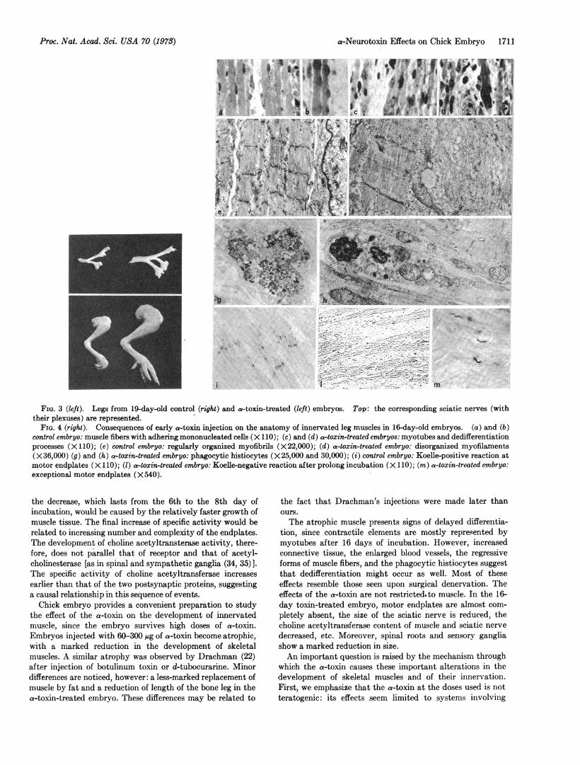
without evident malformation. The magnitude of the atrophyvaries from one injected embryo to the other, but a significantreduction in size was already noticed in 12-day embryos. In16-day embryos, the dry weight of leg muscles in treatedembryos was about half that of untreated embryos (Table 1).At that age the atrophy was evident in all skeletal musclesand also in extrinsic ocular muscles. Interestingly, the in-nervation of all these muscles was affected as well. Fig. 3shows the reduction of size of the sciatic nerve, of its spinalroots, and of corresponding sensory ganglia caused by toxininjections. Preliminary observations indicate that the mo-tility of these embryos is considerably reduced.
Histologically (Fig. 4), atrophic muscles from 16-daytreated embryos markedly differ from those of normal em-bryos. At that age, normal muscles contain typical adultfibers, with nuclei at the periphery and regularly organizedcross-striated myofibrils. Some mononucleated cells are closelyapplied to fibers; myotubes are rare (Fig. 4a and b). By con-trast, in toxin-treated embryos, the contractile elements aremostly myotubes (4c); in their cytoplasm myofilaments aredisorganized (4f) and they no longer make parallel bundlesexcept near the nuclei. A few myoblasts are still present andthe connective tissue is more abundant. Few, if any, musclefibers are present; at both ends these fibers show nucleiarranged in columns (4d). Similar figures commonly seen inmuscle cultures or after denervation are signs of dedifferen-tiation. Phagocytic histiocytes, with dense bodies, lysosomes,vacuoles, and masses of granular or filamentous materialare frequently observed (Fig. 4g and h). Preliminary obser-vations at the 12th day show that at this stage atrophic anddegenerative changes are much less marked than at the 16thday. The same observations were made by Drachman (22)with botulinum toxin.The distribution of acetylcholinesterase on muscle fibers
was revealed by the method of Koelle. In 16-day normalembryos only the endplate region reacts (4i). In toxin-treatedones, the Koelle reaction is almost completely negative(41). The few positive spots seen correspond to endplatesestablished with the rare mature muscle fibers present in theatrophic muscle (4m).
Finally, the content of choline acetyltransferase and acetyl-cholinesterase was estimated in leg muscles and sciatic nerveof toxin-treated and control embryos (Table 1). The total ac-tivities of transferase and esterase in leg muscles are, respec-tively, 5.9- and 8.4-times lower after toxin treatment. The
Proc. Nat. Acad. Sci. USA 70 (1973)

1710 Biochemistry: Giacobini et al.
decrease in the total activities is not simply due to the reduc-tion of muscle size and weight, since the specific activitiesof the two enzymes drop, respectively, 2.4 and 3.5 times. Insciatic nerve, the decrease of choline aceyltransferase specificactivity parallels that seen with muscle. Therefore, toxintreatment affects both the development of the muscle andthat of its innervation.
DISCUSSION
The development of motor innervation in chick embryo com-
prises three major steps (for review, see refs. 23, and 24): (i)from the 4th to the 6th day of incubation, root motor fibers
3 45 6 8 n 14lDAYS
FIG. 2. Developmental changes of choline acetyltransferase(CIAc), acetylcholinesterase (AChE), cholinergic receptor(ACh-R), dry weight (D.W.), and protein in embryonic leg mus-cles. (A) Total activities and protein (ChAc and AChE: ,uM permuscle group and hour; ACh-R:pM per muscle group; protein:mg). (B) Specific activities and dry weight (ChAc: ,.M per g ofprotein per hour; AChE: ,M per mg of protein per hour; ACh-R:nM per g of protein; D.W.: mg). Bars represent SEM; ACh-Rdeterminations were made in most cases in duplicate (at eachstage, single measures are indicated).
TABLE 1. Consequences of an early a-toxin injection onacetylcholinesterase and choline acetyltransferase content of
innervated leg muscles in 16-day chick embryos
Choline Acetylcholin-
Dry acetyltransferase esteraseDryweight Specific Total Specific Total(mg) activity activity activity activity
MuscleControl (2)* 27.5 7.58 0.11 3.13 47Treated (3) 11.7 2.98 0.02 0.87 5.7Ratio Control
Treated 2.35 2.5 5.8 3.6 8.4
NerveControl (2) 57.2Treated (3) 26.7Ratio Control shillTreated 2.1
* Same units as for Fig. 2. The embryos received 300 jug ofa-toxin in three injections at the 3rd, 8th, and 12th days of incu-bation, and were killed the 16th day when leg muscles were dis-sected out. In the controls the same opening in the shell was madeas in the experimental animals. A slight increase in choline acetyl-transferase results from this operation and explains the differenceseen between the numbers given here and in Fig. 2.
gradually invade limb-bud muscles; the first myotubes appearwith a diffuse localization of acetylcholinesterase; embryonicmovements of neurogenic origin (25-27) are present; (ii) fromthe 6th to the 12th day, the contractile elements grow anddifferentiate; a massive formation of myotubes occurs;acetylcholinesterase is gradually restricted to the myo-tendonjunctions (23); (iii) from the 12th day until hatching, musclefibers replace the myotubes, and the nerve endings differ-entiate into motor endplates with acetylcholinesterase local-ized at the subneural apparatus (23). The small irregularpotentials of the electromyogram evolve in typical actionpotentials (28) and acetylcholine sensitivity, diffuse in theearly stages, becomes restricted to the endplate region.The present data indicate that toxin-binding sites, and
therefore cholinergic receptor sites, as well as acetylcholin-esterase, are already present in embryonic muscles at step (i).The total and specific activities of these two proteins increasemarkedly during step (ii), which chronologically correspondsto the fusion of myoblasts into myotubes (see ref. 18). Thedecrease of specific activities observed during step (iii) re-sults from the slower increase of the content of cholinergicreceptor and acetylcholinesterase, and from the faster increasein muscle weight due largely to an enrichment in myofibrillarproteins (29, 30). During this last period, a redistribution ofthe sensitivity to acetylcholine and of acetylcholinesterase onmuscle surface takes place.The evolution of the choline acetyltransferase content dur-
ing embryonic development follows a different program. Firstof all, it is clear from Hebb's and Israel's work (31, 32) thatthis enzyme is almost exclusively present in the neural moietyof the innervated muscle and, therefore, characterizes nerveterminals. The observed time course is similar to that alreadydescribed by Giacobini (33) for axial and thigh muscles. Thefast increase in specific activity during the first step of de-velopment likely corresponds to the arrival of motor fibers;
Proc. Nat. Acad. Sci. USA 70 (1978)
a-Neurotoxin Effects on Chick Embryo 1711
Wi ?1.., . .
I
FIG. 3 (left). Legs from 19-day-old control (right) and a-toxin-treated (left) embryos. Top: the corresponding sciatic nerves (withtheir plexuses) are represented.
FIG. 4 (right). Consequences of early a-toxin injection on the anatomy of innervated leg muscles in 16-day-old embryos. (a) and (b)control embryo: muscle fibers with adhering mononucleated cells (X 110); (c) and (d) a-toxin-treated embryos: myotubes and dedifferentiationprocesses (Xl 10); (e) control embryo: regularly organized myofibrils (X22,000); (d) a-toxin-treated embryo: disorganized myofilaments(X36,000) (g) and (h) a-toxin-treated embryo: phagocytic histiocytes (X25,000 and 30,000); (i) control embryo: Koelle-positive reaction atmotor endplates (XI 10); (1) a-toxin-treated embryo: Koelle-negative reaction after prolong incubation (X 110); (m) a-toxin-treated embryo:exceptional motor endplates (X540).
the decrease, which lasts from the 6th to the 8th day ofincubation, would be caused by the relatively faster growth ofmuscle tissue. The final increase of specific activity would berelated to increasing number and complexity of the endplates.The development of choline acetyltransterase activity, there-fore, does not parallel that of receptor and that of acetyl-cholinesterase [as in spinal and sympathetic ganglia (34, 35) ].The specific activity of choline acetyltransferase increasesearlier than that of the two postsynaptic proteins, suggestinga causal relationship in this sequence of events.
Chick embryo provides a convenient preparation to studythe effect of the a-toxin on the development of innervatedmuscle, since the embryo survives high doses of a-toxin.Embryos injected with 60-300 1Ag of a-toxin become atrophic,with a marked reduction in the development of skeletalmuscles. A similar atrophy was observed by Drachman (22)after injection of botulinum toxin or d-tubocurarine. Minordifferences are noticed, however: a less-marked replacement ofmuscle by fat and a reduction of length of the bone leg in thea-toxin-treated embryo. These differences may be related to
the fact that Drachman's injections were made later thanours.The atrophic muscle presents signs of delayed differentia-
tion, since contractile elements are mostly represented bymyotubes after 16 days of incubation. However, increasedconnective tissue, the enlarged blood vessels, the regressiveforms of muscle fibers, and the phagocytic histiocytes suggestthat dedifferentiation might occur as well. Most of theseeffects resemble those seen upon surgical denervation. Theeffects of the a-toxin are not restrictedto muscle. In the 16-day toxin-treated embryo, motor endplates are almost com-pletely absent, the size of the sciatic nerve is reduced, thecholine acetyltransferase content of muscle and sciatic nervedecreased, etc. Moreover, spinal roots and sensory gangliashow a marked reduction in size.An important question is raised by the mechanism through
which the a-toxin causes these important alterations in thedevelopment of skeletal muscles and of their innervation.First, we emphasize that the a-toxin at the doses used is notteratogenic: its effects seem limited to systems involving
Proc. Nat. Acad. Sci. USA 70 (1973)
.,. Y,
.4
i
1712 Biochemistry: Giacobini et al.
typical nicotinic receptors. Second, most present evidenceindicates that a-toxin affects neither central nervous activitynor peripheral propagation of impulses (1). Although this lastalternative cannot be completely ruled out, the most likelyinterpretation is that the observed effects arise from blockadeby the a-toxin of the cholinergic receptor site of embryonicmuscle. All the postsynaptic effects of delayed differentiationand dedifferentiation would then be simply caused by thelack of postsynaptic activation and, therefore, of musclecontraction.The existence of presynaptic effects raises a question. How
does a postsynaptic blockade influence the motor nerve
terminal? A first possibility is that the cholinergic receptorsurface contributes to the recognition between motor nerve
fibers and muscle surface in the early stage of development:the a-toxin would then interfere with this recognition step (36).However, as early as the 4th day of incubation, when the firstinjection of a-toxin is made, Alconero (26) and Ripley andProvine (27) have observed neurogenic movements; at thisearly stage functional contacts are already made between thefree nerve endings and the contractile elements. Moreover, atday 16, in injected embryos, some abortive endplates are seen.
These two observations suggest that the a-toxin inpairs thedevelopment of the endplate rather than the establishment of theneuromuscular contact.Another possibility is that, during development, the muscle
cell informs the nerve terminal of its state of activity. Onechannel that might transmit this information could be thesensory pathway from the muscle to the motoneuron. Theextrinsic ocular muscles offer a situation particularly rele-vant to this point, since they receive essentially only motorinnervation (37). In these muscles as well, the a-toxin causes
a marked reduction in size, both of the muscle and of the cor-
responding motor nerves. Here, at least the sensory pathwaycould not be involved. We are, therefore, led to postulatethat a retrograde signal passes directly from the postsynapticto the presynaptic cell in a direction opposite to that of thetransmission of impulses. The chemical nature of this signalis completely unknown.Whatever the mechanism involved, it is clear that develop-
ment of neuromuscular synapses depends upon an essentialcorrelation between center and periphery (38-40). In addition,as in other systems (41-43), their differentiation and stabilityappears highly coupled with their functioning. The eventualimplication of this property in learning processes has beendiscussed recently (44, 45).
We are indebted to Dr. M. G. Robecchi-Giacobini and toMiss M. Penna for help with electron microscopy and to Drs.Menez and Fromageot for the tritiation of the a-toxin. ThisThis work was supported by the Italian National Research Coun-cil Grant 720075404 (G. G.) and 710019304 (G. F.), the NationalInstitutes of Health, and by funds from the Centre National dela Recherche Scientifique, the Del6gation Generale a la RechercheScientifique et Technique, the College de France, and thr Com-missariat a l'Energie Atomique.
1. Lee, C. Y. & Chang, C. C. (1966) Mem. Inst. Butantan Simp.Int. 33, 555-572.
2. Changeux, J. P., Kasai, M. & Lee. C. Y. (1970) Proc. Nat.Acad. Sci. USA 67, 1241-1247.
3. Miledi, R., Molinoff, P. & Potter, L. (1971) Nature 229, 554-557.
4. Hall, Z. W. (1972) Annu. Rev. Biochem. 41, 925-952.5. Boquet, P., Izard, Y., Jouannet, M. & Meaume, J. (1966)
C.R. Acad. Sci. Ser. D, 262, 1134-1137.6. Karlsson, E., Eaker, D. & Porath, J. (1965) Biochim. Bio-
phys. Acta 127, 505-520.7. Menez, A., Morgat, J. L., Fromageot, P., Ronseray, A. M.,
Boquet, P. & Changeux, J. P. (1971) FEBS Lett. 17, 333-335.
8. McCaman, R. E. & Hunt, J. M. (1965) J. Neurochem. 12,253-258.
9. Fonnum, F. (1969) Biochem. J. 115, 465-472.10. Ellman, G. L., Courtney, K. D., Andres, V. & Featherstone,
R. (1961) Biochem. Pharmacol. 7, 88-95.11. Lowry, 0. H., Rosebrough, N. J., Farr, A. L. & Randall,
R. J. (1951) J. Biol. Chem. 193, 265-275.12. Weber, M., Menez, A., Fromageot, P., Boquet, P. & Chang-
eux, J. P. (1972) C.R. Acad. Sci. Ser. D, 274, 1575-1578.13. Koelle, G. B. & Friedenwald, J. S. L. (1949) Proc. Soc. Exp.
Biol. Med. 70, 617-622.14. Miledi, R. & Potter, L. (1971) Nature 233, 599-603.15. Barnard, E., Wieckowski, J. & Chiu, T. H. (1971) Nature
234, 207-209.16. Fambrough, D. M. & Hartzell, H. C. (1972) Science 176,
189-191.17. Berg, D., Kelly, R. B., Sargent, P. B., Williamson, P. &
Hall, Z. (1972) Proc. Nat. Acad. Sci. USA 69, 147-151.18. Patrick, J., Heinemann, S., Lindstr0m, J., Schubert, D. &
Steinbach, J. H. (1972) Proc. Nat. Acad. Sci. USA 69, 2762-2766.
19. Vogel, Z., Sytkowski, A. J. & Nirenberg, M. W. (1972) Proc.Nat. Acad. Sci. USA 69, 3180-3184.
20. Millo, A. (1961) Riv. Biol. 54, 251-261.21. Goodwin, B. C. & Sizer, I. W. (1965) Develop. Biol. 11, 136-
153.22. Drachman, D. B. (1967) Arch. Neurol. (Chicago) 17, 206-
218.23. Filogamo, G. & Gabella, G. (1967) Arch. Biol. 78, 9-60.24. Couteaux, R. (1963) Proc. Roy. Soc. Ser. B 158, 457-480.25. Visintini, F. & Levi-Montalcini, R. (1939) Schweiz. Arch.
Neurol. Psychiat. 4, 119-150.26. Alconero, B. B. (1965) J. Embryol. Exp. Morphol. 13, 225-
266.27. Ripley, K. & Provine, R. (1972) Brain Res. 45, 127-134.28. Boethius, J. (1967) J. Exp. Zool. 165, 419-424.29. Dickerson, G. W. (1960) Biochem. J. 95, 33-40.30. Baril, E. F. & Herrman, H. (1967) Develop. Biol. 15, 318-
330.31. Hebb, C. O., Krnjevic', K. & Silver, A. (1964) J. Physiol.
Lond. 171, 504-513.32. Israel, M. (1970) Arch. Anat. Microsc. Morphol. Exp. 59,
67-98.33. Giacobini, G. (1972) J. Neurochem. 19, 1401-1403.34. Marchisio, P. C. & Consolo, S. (1968) J. Neurochem. 15, 759-
764.35. Giacobini, G., Marchisio, P. C., Giacobini, E. & Koslow,
S. H. (1970) J. Neurochem. 17, 1177-1185.36. Sytkowski, A. J., Vogel, Z. & Nirenberg, M. (1973) Proc.
Nat. Acad. Sci. USA 70, 270-274.37. Cowan, W. M. & Wenger, E. (1967) J. Exp. Zool. 164, 267-
280.38. Shorey, M. L. (1909) J. Exp. Zool. 7, 25-64.39. Hamburger, V. (1934) J. Exp. Zool. 68, 449-494.40. Jacobson, M. (1970) in Developmental Neurobiology, ed. Holt,
(Rinehart and Winston, New York), p. 228.41. Thoenen, H., Mueller, R. A. & Axelrod, J. (1969) J. Phar-
macol. Exp. Ther. 169, 249-264.42. Black, I. B., Hendry, I. A. & Iversen, L. L. (1971) Brain
Res. 34, 229-240.43. Thoenen, H. (1972) Pharmacol. Rev. 24, 255-267.44. Changeux, J. P. (1972) Communications 19, 37-47.45. Changeux, J. P., Courrfge, P. & Danchin, A. (1973) Proc.
Nat. Acad. Sci. USA, submitted.
Proc. Nat. Acad. Sci. USA 70 (1978)