Airway Management &Ventilation · Airway Management &Ventilation . ... anesthesia induces a number...
13
1 Airway Management &Ventilation Lyon Lee DVM PhD DACVA Introduction • Animals under anesthesia increase the risk of developing airway complications as anesthesia induces a number of abnormal physiologic conditions. • Most anesthetics obtund or abolish the swallowing reflex, a protective mechanism to prevent accidental inhalation of foreign materials that may be present in the mouth. • Maintaining a secure airway during perianesthetic period comes as a primary requirement for safe anesthesia and is a fundamental responsibility of the anesthesia provider. • Failure to do so can result in brain damage and death. • In some situations, maintaining patent airway can be performed by a simple maneuver such as extending the neck of a patient. • However, placing a tracheal tube into the trachea, without question, ensures a more secure airway. • This lecture covers some basic anatomy and physiology of the respiratory system, the necessary devices required to perform endotracheal intubation and techniques of airway management, basic principles of the ventilator, and ways how best to apply such knowledge into clinical practice. Definitions • ‘Airway’ is the passage way into or out of the lungs. Proper airway ensures adequate delivery of oxygen (O 2 ) and elimination of carbon dioxide (CO 2 • ‘Respiration’ is the total process of oxygen supply and carbon dioxide elimination. ) to and out of the body. • ‘Ventilation’ is the movement of gases in and out of the alveoli. Anatomic and Physiologic considerations Airways • Upper airways o nares, nasal cavity, pharynx, larynx • Trachea • Lower airways o bronchi, bronchioles, alveoli Upper airway obstruction • Anesthetics relax nasal alar and pharyngeal musculature and abolishes cough reflex, increasing the risk of causing upper airway obstruction. • Brachycephalic dogs are predisposed to the obstruction because of higher incidence of elongated soft palate, stenotic nares, everted laryngeal ventricles, and hypoplastic trachea.
Transcript of Airway Management &Ventilation · Airway Management &Ventilation . ... anesthesia induces a number...

1
Airway Management &Ventilation
Lyon Lee DVM PhD DACVA
Introduction • Animals under anesthesia increase the risk of developing airway complications as
anesthesia induces a number of abnormal physiologic conditions. • Most anesthetics obtund or abolish the swallowing reflex, a protective mechanism to
prevent accidental inhalation of foreign materials that may be present in the mouth. • Maintaining a secure airway during perianesthetic period comes as a primary requirement
for safe anesthesia and is a fundamental responsibility of the anesthesia provider. • Failure to do so can result in brain damage and death. • In some situations, maintaining patent airway can be performed by a simple maneuver
such as extending the neck of a patient. • However, placing a tracheal tube into the trachea, without question, ensures a more
secure airway. • This lecture covers some basic anatomy and physiology of the respiratory system, the
necessary devices required to perform endotracheal intubation and techniques of airway management, basic principles of the ventilator, and ways how best to apply such knowledge into clinical practice.
Definitions • ‘Airway’ is the passage way into or out of the lungs. Proper airway ensures adequate
delivery of oxygen (O2) and elimination of carbon dioxide (CO2• ‘Respiration’ is the total process of oxygen supply and carbon dioxide elimination.
) to and out of the body.
• ‘Ventilation’ is the movement of gases in and out of the alveoli.
Anatomic and Physiologic considerations Airways
• Upper airways o nares, nasal cavity, pharynx, larynx
• Trachea • Lower airways
o bronchi, bronchioles, alveoli
Upper airway obstruction
• Anesthetics relax nasal alar and pharyngeal musculature and abolishes cough reflex, increasing the risk of causing upper airway obstruction.
• Brachycephalic dogs are predisposed to the obstruction because of higher incidence of elongated soft palate, stenotic nares, everted laryngeal ventricles, and hypoplastic trachea.

2
• Treatment o Remove the source of obstruction (physical objects, regurgitated materials etc.) o Extend the head and pull the tongue out o Perform endotracheal intubation, or when intubation is not feasible perform
tracheostomy (but this is rarely needed)
Lower airway obstruction
• May develop due to accidental aspiration of regurgitants, saliva, or due to pulmonary edema, microbes induced infection, and presence of underlying small airway obstruction.
• Increased delivery of tidal volume, increased inspiratory pressure all have shown to improve ventilation in this patient, but in general harder to treat than upper airway complications.
• Depending on the magnitude of the complications (e.g., asthma or COPD) treatment with bronchodilators may be indicated.
Ventilation and gas exchange
• VE• V
; total minute ventilation T
• f: respiratory rate : tidal volume
VE= VT
x f
• Suppose the volume exhaled with each breath is 500 ml and there are 15 breaths/min, then the total volume leaving the lung each minute is 500 x 15 = 7500 ml/min (total minute ventilation, VE
• To increase ventilation, V)
T
Poiseuille’s law and airway resistance
or f, or both need to be increased.
• nLrPQ
8
4π=
• Q: Gas flow, P: pressure difference, r: tube radius, n: viscosity of the gas, L: tube length
• Ohm’s law states V = IR, or Pressure = Flow x Resistance • From above as defined by Poiseuille’s equation and Ohm’s law, Resistance changes
relative to several factors:
• Resistance = 4
8rnL
o Therefore, any reduction in the radius of the airway is INVERSELY related to the
resistance, since R = 41r
o A 50% reduction in the radius results not in a doubling of the resistance but rather a 16-fold increase!!
o However, doubling the length (L) through which the patient is breathing will simply double the resistance.

3
Changes of flow resistance in response to changes of radius
Face masks
• The photo above shows different sizes of masks. • Clear plastic masks offer advantages of being able to see inside if a patient regurgitates. • Use the smallest mask that will fit your patient - minimize dead space. • They provide a means of administering oxygen and inhalant anesthetic, but important to
recognize limitations: o They do NOT protect the airway from aspiration o They do NOT provide a patent airway o Limited ability to support ventilation if apnea or hypoventilation occurs
2r 161
≈Q ; R ≈16 r
Q= flow; R= resistance

4
Endotracheal intubation Endotracheal tubes
• Murphy (most commonly used) o Cuffed or uncuffed, hole at the tip (Murphy eye) o Murphy eye allows bypass of the air when the patient end is accidentally blocked
• Magill o Cuffed or uncuffed, no hole at the tip
• Cole o Tapered end provide tight seal, no cuff, no hole
The photo below shows a cuffed Murphy endotracheal tube
Murphy eye Inflated cuff

5
Ideal tube size
• Select the largest possible diameter without a danger to damaging larynx or trachea • Length should be from the tip of the nose to the point of the shoulder (see photo below) • Too long tube can result in endobronchial intubation and unintentional one lung
ventilation • Excessive tube length extending out of the oral cavity results in increased dead space
ventilation (cut to the length if necessary)
Point of shoulder
Intubation techniques
• Direct visualization; dogs, cats, small ruminants, swine, etc. (laryngoscopy useful) • Digital palpation; cattle (best performed with guide stomach tube) • Blind; horses (easier than in most species)
Proper cuff inflation
• Overinflation of the endotracheal tube cuff may cause o Pressure necrosis of the tracheal epithelium o Collapse of the endotracheal tube and airway occlusion (more common with softer
silicone type endotracheal tubes) • To properly inflate the endotracheal tube cuff, the ‘minimal leak method’ should be
employed o Intubate the patient o Administer a positive pressure breath, reaching a peak airway pressure of about 20
cmH2O (or watch for a moderate amount of chest rise if a pressure manometer is not available)

6
o While administering the positive pressure breath, listen for an airway leak or sniff for trace of anesthetic gas at the level of your patient’s mouth
o If you do not hear any air leaking around the endotracheal tube, do not inflate the cuff o If you do hear an air leak around the endotracheal tube, inflate the cuff with a small
amount of air (volume is relative to tube size) o Repeat the above process just until you no longer can hear a leak or smell anesthetic
gas at a peak airway pressure of just under 20 cmH2• You may need to recheck cuff inflation
O pressure
o After 15 - 20 minutes of anesthesia - as your patient’s anesthetic depth deepens, the laryngeal and tracheal muscles may relax, and an airway leak may develop
Special techniques for endotracheal intubation (difficult airways)
• Guide tube (stylet) technique • Retrograde intubation technique • Endotracheal intubation through nasal passage (nasotracheal intubation) • Endotracheal intubation through lateral pharyngostomy • Endotracheal intubation using a fiber-optic endoscope • Endotracheal intubation by tracheostomy • For full technical details of these techniques see pp528-531 of Veterinary Anesthesia
(Thurmon et al. 1996).
Laryngoscopy
• Laryngoscopes are composed of two parts; laryngeal blade and handle (see photo below). • The laryngoscope’s light source is the main benefit of laryngoscopy, but the blade can be
used to manipulate the tongue, soft palate, and epiglottis to view the glottis. NB. Use the laryngoscope to depress the base of the tongue, not the epiglottis. Depressing the epiglottis directly with the laryngeal blade increases the risk of traumatizing the epiglottis inducing airway complications.
• Blades come in different sizes and types, size ranging from 0 (small) to 5 (large) adequate for most small animals.
• Customized blades with extra length are available for use in some species including pigs, llamas, and big cats.
• Two most common types available in the market are the Miller (straight) and the McIntosh (curved), and there are numerous modifications based on these two.

7
Laryngeal blade
Laryngoscope handle
(Type) Miller
McIntosh
Miller
McIntosh McIntosh
(Size) 0 1 2 3 4
8
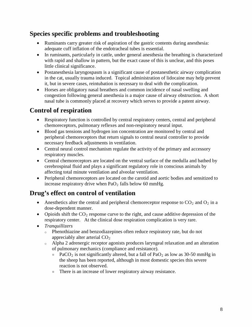
Species specific problems and troubleshooting • Ruminants carry greater risk of aspiration of the gastric contents during anesthesia:
adequate cuff inflation of the endotracheal tubes is essential. • In ruminants, particularly in cattle, under general anesthesia the breathing is characterized
with rapid and shallow in pattern, but the exact cause of this is unclear, and this poses little clinical significance.
• Postanesthesia laryngospasm is a significant cause of postanesthetic airway complication in the cat, usually trauma induced. Topical administration of lidocaine may help prevent it, but in severe cases, reintubation is necessary to deal with the complication.
• Horses are obligatory nasal breathers and common incidence of nasal swelling and congestion following general anesthesia is a major cause of airway obstruction. A short nasal tube is commonly placed at recovery which serves to provide a patent airway.
Control of respiration • Respiratory function is controlled by central respiratory centers, central and peripheral
chemoreceptors, pulmonary reflexes and non-respiratory neural input. • Blood gas tensions and hydrogen ion concentration are monitored by central and
peripheral chemoreceptors that return signals to central neural controller to provide necessary feedback adjustments in ventilation.
• Central neural control mechanism regulate the activity of the primary and accessory respiratory muscles.
• Central chemoreceptors are located on the ventral surface of the medulla and bathed by cerebrospinal fluid and plays a significant regulatory role in conscious animals by affecting total minute ventilation and alveolar ventilation.
• Peripheral chemoreceptors are located on the carotid and aortic bodies and sensitized to increase respiratory drive when PaO2
Drug’s effect on control of ventilation falls below 60 mmHg.
• Anesthetics alter the central and peripheral chemoreceptor response to CO2 and O2
• Opioids shift the CO
in a dose-dependent manner.
2
• Tranquillizers
response curve to the right, and cause additive depression of the respiratory center. At the clinical dose respiration complication is very rare.
o Phenothiazine and benzodiazepines often reduce respiratory rate, but do not appreciably alter arterial CO2
o Alpha 2 adrenergic receptor agonists produces laryngeal relaxation and an alteration of pulmonary mechanics (compliance and resistance).
¤ PaCO2 is not significantly altered, but a fall of PaO2
¤ There is an increase of lower respiratory airway resistance.
as low as 30-50 mmHg in the sheep has been reported, although in most domestic species this severe reaction is not observed.

9
Changes of ventilation-perfusion (V/Q) relationship during anesthesia
• PaO2
• Hypoxic pulmonary vasoconstriction (a protective mechanism) re-directs blood flow to better ventilated area in the lung. Anesthetics cause marked reduction in this protective response, resulting in further V/Q mismatch.
decreases more dramatically during general anesthesia in large animals than smaller animals. The primary attributor is believed to be increased V/Q mismatches.
Ventilator • Anesthesia ventilators provide controlled ventilation to patients under general anesthesia • Simply, anesthesia ventilator is a reservoir bag (a bellow or concertina bag) in a closed
container (bellows housing) that can substitute for the reservoir bag of an anesthesia breathing system.
• The anesthesia ventilator performs the same job as the anesthetist who periodically squeezes the breathing system’s reservoir bag to ventilate the patient, lessening laborious workload of the anesthetist and thereby minimizing the potential for distraction.
• Classification is through power source, drive mechanism and cycling mechanism. o Power source; electrical, or compressed gas or both o Drive mechanism; commonly compressed gas even when electrical source is used. o Cycling mechanism is typically time cycled. o Other mechanisms include pressure cycled, volume cycled, but timing mechanism
plays a major role in ventilatory function, even in these two. • For additional details about anesthesia ventilators see pages 535-556 of Veterinary
Anesthesia (Thurmon et al. 1996). • A variety of ventilators are manufactured by different vendors, and may come in a stand-
alone unit or built into the anesthetic machine. • The decision to choose which type will depend on what suits better for the clinical need a
practitioner has, based on the animal species and economic consideration. • The cost for most stand-alone ventilator adequate for small animal use should fall into the
range of $ 2000 to $ 5000.

10
Ascending bellows (see photo below) • Most new ventilators are of this type • Safer - if an airway leak is present, bellows no longer ascends to the top, letting you
know that a leak exists
Descending Bellows – Metomatic (see photos below)
• Older design, but still very functional. The ventilators that we routinely use in our hospital

11
Metomatic controls
• Power: on/off • Expiratory flow rate: used to adjust character of ventilation • Inspiratory flow rate: used to adjust character of ventilation • Expiratory time: controls respiratory rate • Inspiratory hold: used to maintain inspiratory pressure to reinflate atelectatic lungs • Inspiratory pressure: used to adjust maximal inspiratory pressure generated • Inspiratory trigger effort: can be adjusted to make ventilator function in ‘assist’ mode -
patient initiates breath, ventilator just assists to increase tidal volume (rarely used) • Tidal volume (knob on back of bellows): adjusts tidal volume

12
• Note drive gas is oxygen (see above photo on the left) – an important safety feature! • If a leak develops in the bellows, oxygen will enter the bellow and be delivered to the
patient; this prevents delivering a hypoxic gas mixture • Beware! Running a ventilator off of an E cylinder will deplete the cylinder fairly rapidly • A ventilator is nothing more than a mechanical rebreathing bag • Therefore, the ventilator always attaches where the rebreathing bag attaches to a
breathing circuit! (see above photo on the right)
13
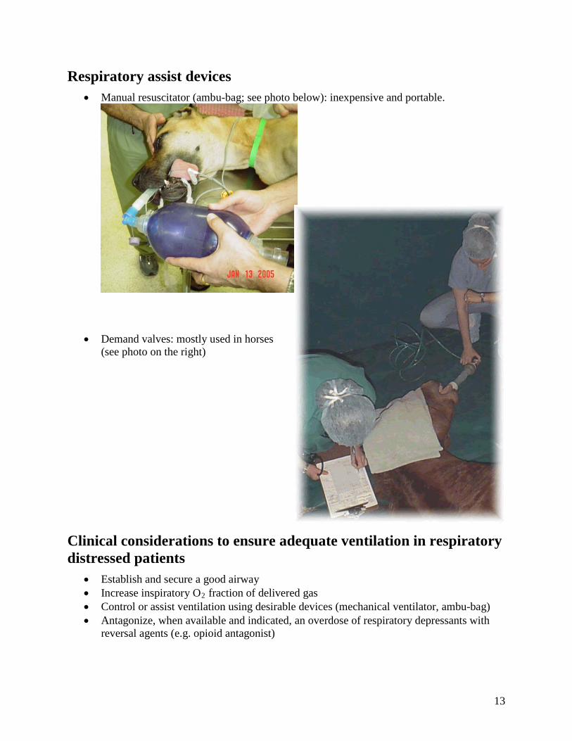
Respiratory assist devices • Manual resuscitator (ambu-bag; see photo below): inexpensive and portable.
• Demand valves: mostly used in horses (see photo on the right)
Clinical considerations to ensure adequate ventilation in respiratory distressed patients
• Establish and secure a good airway • Increase inspiratory O2• Control or assist ventilation using desirable devices (mechanical ventilator, ambu-bag)
fraction of delivered gas
• Antagonize, when available and indicated, an overdose of respiratory depressants with reversal agents (e.g. opioid antagonist)