Aim - HUMSC
13
Transcript of Aim - HUMSC


Page 1 of 11
بسم الله الرحمن الرحيم
2لاب فسيو
بالاعتماد على سلايدات الدكتور و كلامه و مصادر خارجية
السلايدات رح تكون داخل صندوق
Differential WBC Count
اته و صفاته الاساسية ن ن اعداد خلايا الدم البيضاء ... احنا اخذنا بالتفصيل الممل كل نوع و ايش ممي الاختلاف بي
ن المعدل الطبيعي لكل نوع الهدف من هذا الفحص نعرف اعدا د خلايا الدم البيضاء بانواعها المختلفة ولازم نكون عارفي
ي جسم الانسان ني اشارة لوجود خلل ف
... طبعا اي زيادة او نقصان بتعطينن
ي اشارة ل neutrocyteمثال اخذناه بالباثو لو زاد acute bacteria infection and necrose tissueفهذا بعطينن
الاساس الي رح نعتمد عليه بالفحص نوخذ عينة دم و نصبغ بصبغة خاصة سريعة التحضي و يدوية و كل نوع بعطي
ه ه عن غي ن استجابة معينة بمي
clean slide 2بتشبه الجهاز الي منستخدمه بالسكري مشان يطلع دم ..... lancet 1المكونات الي رح نحتاجهم
وري جدا تكون نظيفة حنى يطلع الفحص صحيح .... نوع من انواع microscope ......diff quick stain 3ضن
الصبغات
Aim:
To determine the percentage of each type of leukocytes in the peripheral blood.
Certain percentages can change during certain diseases so this test is valuable for
the diagnosis of these diseases.
Principle:
The test depends on making a blood film from the peripheral blood and staining it with Diff
Quick stain using manual method. Different types of WBCs are differentiated by their
morphological and staining characteristics.
Material:1 Lancets 2 Clean slides 3 Microscope 4 Diff Quick Stain

Page 2 of 11
I. Preparation of blood smear Is made on an ordinary slide
وه ح نفس الخطوات و كثي مرتب بفضل تحضن شفت فيديو بشر
رابط الفيديو
https://www.youtube.com/watch?v=nbRUiWl2Qrs
ح الارب ع خطواتركزوا عالصورة الي تح ت لانه بتشر
1. Slides must be spotlessly clean.
Hold them always by the edges.
Never put your fingers on the surface
of the slide. Prepare two clean slides.
2. Obtain a drop of blood from the tip
of the middle finger and place it at the
end of one slide. Put the slide on the
surface of your bench. Use the second
slide as a spreader by placing its
smooth edge at an angle of 45º to the
first slide.
3. Move the spreading slide backward
until it touches the drop of the blood
as in the diagram. The drop of the
blood will immediately spread across
the edge of the spreading slide
4. With the spreading slide at the angle
of 45º, push it slowly forward and
steadily across the horizontally placed
first slide

Page 3 of 11
سمك العينة بعتمد عالزاوية الي مسحت فيها الدم
كان السمك اكير 45كل ما كانت الزاية اكير من
ما بحتاج نعرضه للحراة نهائيا
Dلازم تكون لون العينة زي
بكون جهزت عينة الدم مشان اصبغها 8بنهاية خطوة
5. The thickness of the film may be
regulated by increasing or
decreasing the angle between the
slides or by varying the speed of
spreading. A quick spreading gives
a thick film; the wider the angle
the thicker the film.
6. Allow the blood smear to dry in
air at room temperature; it must
not be heated
7. A satisfactory film is very evenly
distributed and yellowish orange
in colour ; the red cells should be
close together but not overlapping
each other. Check that by looking
at the fill under the microscope
8. The “zone of morphology”
should be at least 2 cm in length.
The smear should occupy the
central area of the slide.

Page 4 of 11
تشوفوا الفيديو بالاول بعدين رح تصي الخطوات سهلة و ما رح تتغلبوا بالحفظقبل ما نحكي بالمرحلة الثانية بفضل
رابط الفيديو
https://youtu.be/Y27TvaJMKRE
II. Staining of blood smear
Do not stain a film until you have made a satisfactory one.
The stain used is the Diff Quick stain. This stain contains;
1 Diff Quick fixative reagent ( Triarylmethane dye, Methanol)
2 Diff Quick solution 1 (Xanthene dye (Eosin Y) in phosphate buffer)
3 Diff Quick solution 2 (Thiazine dye in methylene blue and azure A
phosphate buffer
1. Air dry the smear
2. Dip slide for 30 seconds into Fixative. Allow excess to drain after each
dip.
3. Dip slide for 30 seconds into Stain 1. Allow excess to drain after each dip.
4. Dip slide for 30 seconds into Stain 2. Allow excess to drain after each dip.
5. Rinse slide in distilled water or Weise's buffer, pH 7.2 to remove excess
stain.
6. Allow to dry in air or rapidly dehydrate in absolute alcohol.
7. Examine at low power to identify structures and then under oil
immersion
Page 5 of 11
ي بنصح تشوفوا ة و نفس الاشر ن الفيديو هذولة المرحلة الثالثة و الاخي هي
https://www.youtube.com/watch?v=ihSnTKLUazQ&t=132s
https://youtu.be/DFzndODp86s
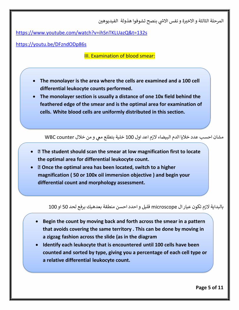
III. Examination of blood smear:
WBC counterخلية بتطلع معي و من خلال 100مشان احسب عدد خلايا الدم البيضاء لازم اعد اول
100او 50قليل و احدد احسن منطقة بعدهيك برفع لحد microscopeبالبداية لازم تكون عيار ال
• The monolayer is the area where the cells are examined and a 100 cell
differential leukocyte counts performed.
• The monolayer section is usually a distance of one 10x field behind the
feathered edge of the smear and is the optimal area for examination of
cells. White blood cells are uniformly distributed in this section.
• The student should scan the smear at low magnification first to locate
the optimal area for differential leukocyte count.
• Once the optimal area has been located, switch to a higher
magnification ( 50 or 100x oil immersion objective ) and begin your
differential count and morphology assessment.
• Begin the count by moving back and forth across the smear in a pattern
that avoids covering the same territory . This can be done by moving in
a zigzag fashion across the slide (as in the diagram
• Identify each leukocyte that is encountered until 100 cells have been
counted and sorted by type, giving you a percentage of each cell type or
a relative differential leukocyte count.

Page 6 of 11
زي بالصورة zig-zagببلش العد عن طريق
)6.300/cu mm-e Granulocytes (70%Th
Basophils Eosinophils Neutrophils
(0%-1%) (1%-4%) (50%-70)% Percentage
liberate histamine and heparin during allergic
reactions.
phagocytize and destroy the antigen-antibody complex
highly phagocytic (often called macrophages).
function
7μ 13μ 10-12μ size
-Scanty or abundant cytoplasm.
-Large deep blue cytoplasmic granules,
blue-black nucleus. -Lobulated nucleus (2 or
3 lobules). -They liberate heparin
into the blood, a substance that can
prevent blood coagulation.
-Large sized cells with abundant cytoplasm
-Large red-orange cytoplasmic granules, blue-purple nucleus Mutilobulatednucleus (2 or 3
lobules)
*abundant cytoplasm *Small pink granules in the
cytoplasm. *Multilobulatedpurple nucleus (three lobes or
more).
Character
after allergic reaction (such as asthma, parasitic infestation,
and Hay fever .)
acute pyogenic infection. Increase in NO

Page 7 of 11
2.700/cu mm)-The Agranulocytes (30%
Monocytes Lymphocytes
(2%-8%) (20%-40%) percentage phagocytic and help in tissue
repair after injury. -They are the scavenger cells and they are transferred into
tissue macrophages to engulf the dead tissues and
bacteria.
* specialized in production of immunity such as the synthesis of antibodies
function
-The Largest WBC with irregular blue or purple nucleus (Kidney shaped
nucleus).
Variable -7μ in diameter (small) to 10μ in diameter
(large)
Size(shape)
-Abundant blue-gray cytoplasm (nongranular).
with no cytoplasmic granules, light blue cytoplasm
-The scanty cytoplasm is pushed to the periphery of the cell by the large deep blue
or purple nucleus -Small lymphocytes have very large spherical nucleus, large lymphocytes have large oval
indented nucleus
Character
*innate immune * chronic inflammation.
-They perform a major role in immune system and increase in number during
chronic and viral infections
Increase NO

Page 8 of 11
Bleeding Time Test
(Duke Method)
يف ...هاي مسؤول ن ي اشارة لفاعلية الصفائح الفحص هذا اخذنا قبل هيك و بقيس الوقت اللازم حنى يتوقف الينية الصفائح و بالتالي بعطينن
ي طريقة ثانية باستخدام جهاز الضغط بس بتوخذ الدموية ... بحتاج اعمل جرح بالجلد ن دقايق و مش عملية ( 10... )ف
قبل ما نحكي عن الخطوات بفضل تشوفوا الفيديو
https://youtu.be/3UNK7w-Upss
Definition:
A bleeding time test determines how quickly the bleeding stopes. The test involves making small
punctures in the skin. The test is a basic assessment of the platelets function (i.e. platelet plug
formation).
Indication:
The bleeding time test is a common test to screen patients having bleeding tendency (i.e.
prolonged bleeding times). The bleeding time is used to measure the primary phase of
hemostasis.
Procedure:
•Clean the puncture site with an spirit to minimize the risk of infection.
•Make small cuts with the lancet on the tip of the middle finger (or the earlobe). The puncture
should be 3-4 mm deep to cause slight bleeding.
•Start the stopwatch immediately, swab the cut with filter paper every 30 secondsuntil no
more blood is absorbed (i.e. no more blood spots on the filter paper).
•Record the time it takes for the bleeding to stop.

Page 9 of 11
Clotting Time Test
(Capillary Tube Method or Wright’s Method)
extrinsicو هذا بحفز ال tissue thromboplastin (factor III)بفرز damageهسا الدم اول ما يتعرض
pathway ة حنى تتكون الخير
platelet aldheranceبدون intrinsicلانه انا بدي احسب فقط aPTTبستخدم intrinsicمشان اقيس ال
Interpretation:
Normal bleeding time is between 2-5 minutes. Prolonged bleeding time could indicate low
platelet count or the platelets are dysfunctional, and further testing will be required.
Abnormal results could indicate one of the following conditions
•Thrombocytopenia انخفاض عدد الصفائح الدموية
•Medications (Example aspirin and other cyclooxygenase inhibitors, & antibiotics such as
penicillin and cephalosporins)
Anti plateletزي
•Uremia (uremic platelets synthesizeless thromboxane A2, and the blood vessels in patients
with uremia produce greater quantities of platelet-inhibitory prostaglandin)
•Liver failure
•Leukemias
•Von Willebrand’sdisease
Indication:
•Clotting time is used as a screening test to measure all stages in the intrinsic coagulation system
and to monitor heparin therapy.
•This test is sensitive only in extreme factor deficiencies(such as in hemophilia). Therefore, it is of
limited use in today’s medical practice and its use is confined to demonstration.

Page 10 of 11
بناء عالفيديو
Definition:
•Is the time required for a fresh sample of blood to form a clot in vitro. The basis for the test is that
whole blood will form a solid clot when exposed to a foreign surface such as a glass tube.
•Clotting time test is a rough measure of all intrinsic clotting factors in the absence of tissue factors.
•Variations are wide and the test sensitivity is limited. The more recent sensitive test for the intrinsic
pathway is the partial thromboplastin time (PTT). Normal value of clotting time is 3-8 minutes.
Material Required:
•Sterile disposable pricking
needle or lancet
•Stop-watch
•Dry glass capillary tube
(narrow diameter top 2 mm,
minimum 10 cm long)
•Cotton Swab of absorbent
cotton
•Spirit wetted, cotton swab
•70% v/v ethyl alcohol
Procedure:
1.Apply alcohol 70% v/v to clean the fingertip with
cotton swab. Allow it to dry naturally.
2.Prick the finger. Remove the first drop.
3.Dip one end of the capillary into blood drop gently
without pressure (better to use more than one capillary
tube).
4.The timer is started when the first blood start to enter
the first capillary tube.
5.Allow to fill the capillary with blood by lowering the
end of fitted capillary. (Do not suck the blood).
6.After every 30 seconds, using stop-watch, break a
small piece of capillary.
7.Repeat breaking at regular time intervals, till fibrin
thread appearsat the broken end of capillary tube. Do
not pull away the cut pieces long apart and bristly.
8.Record time interval between pricking finger and first
appearance of fibrin thread between the broken ends
of capillary tube. That is clotting time of blood.
9.Don't forget to dispose of the broken capillary tube in
the SHARPS CONTAINER

Page 11 of 11
ي حال كان الوقت اكير ندقايق بكون السبب : 8ف
THE END
• Factors V, VII, VIII, IX, XI, XII Deficiencies
• Hemorrhagic disease of Newborn
• Vitamin K deficiency
• Heparin Therapy
• Presence of Circulating antibodies
(inhibitors)
• Anemia and leukemia
• Afibrinogenemiaand Pneumonia