Ahuva Grubstein Radiology Rabin Medical Center - ipus.org.il · PDF fileinternal...
57
Ahuva Grubstein Ahuva Grubstein Radiology Radiology Rabin Medical Center Rabin Medical Center
Transcript of Ahuva Grubstein Radiology Rabin Medical Center - ipus.org.il · PDF fileinternal...

Ahuva GrubsteinAhuva GrubsteinRadiologyRadiology
Rabin Medical CenterRabin Medical Center

Introduction Sub solid pulmonary nodules
••
MultidetectorMultidetector
CT , lowCT , low‐‐dose CT lung cancer dose CT lung cancer screening programsscreening programs
••
Varieties of small peripheral lung nodulesVarieties of small peripheral lung nodules
••
The importance and prevalence of The importance and prevalence of subsolidsubsolid pulmonary nodules pulmonary nodules

ReferencesReferences
Radiology: Volume 253: Number 3—December 2009Subsolid
Pulmonary Nodules and the Spectrum of Peripheral
Adenocarcinomas
of the Lung:Recommended Interim Guidelines for Assessment and
ManagementMyrna C. B. Godoy,MD
; David P. Naidich,MD
Radiology: Volume 245: Number 1—October 2007Persistent Pulmonary Nodular Ground‐Glass Opacity at Thin‐
Section CT: Histopathologic
ComparisonsHa Young Kim,MD, Young Mog
Shim,MD
et al

•
Bartjan
de Hoop, et al, Pulmonary Ground‐ Glass Nodules: Increase in Mass as an Early
Indicator of Growth ; Radiology April 2010 255:199‐206
•
Sang Min Lee, et al, Transient Part‐Solid Nodules Detected at Screening Thin‐Section
CT for Lung Cancer: Comparison with Persistent Part‐Solid Nodules; Radiology April
2010 255:242‐251

SubsolidSubsolid nodules nodules ‐‐ definitiondefinition
Subsolid
nodules are defined as focal nodular areas of increased lung attenuation through
which normal parenchymal
structures such as airways, vessels, and interlobular septa can be defined
•
part‐solid•
nonsolid = completely GGO
•
Solid nodules are defined as those that completely obscure the lung parenchyma
SubsolidSubsolid nodules nodules ‐‐ differentialdifferential
•
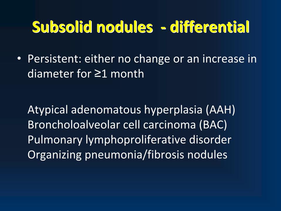
Persistent: either no change or an increase in diameter for ≥1 month
Atypical adenomatous
hyperplasia (AAH) Broncholoalveolar
cell carcinoma (BAC)
Pulmonary lymphoproliferative
disorder Organizing pneumonia/fibrosis nodules

•
new interim management guidelines.

BACBAC
•
Presents a unique growth pattern along alveolar septa without stromal
invasion and
has an indolent course
•
high incidence of multifocality
(25% vs
5%)

NoguchiNoguchi’’s s histologichistologic
classification for small , classification for small ,
peripheral peripheral adenocarcinomasadenocarcinomas, 1995, 2004, 1995, 2004
six subtypes, on the basis of the patterns of tumor growth•
A localized BAC;
•
B, localized BAC with foci of collapsed alveolar structures;•
C, localized BAC with foci of active fibroblastic proliferation;
•
D, poorly differentiated adenocarcinoma; •
E, tubular adenocarcinoma; and
•
F, papillary adenocarcinoma
with evidence of compressive and destructive growth.
Types A, B, and C represent a distinct grouping, as they show in common a predominant growth pattern involving “replacement”
of
alveolar lining cells.

1999 , 20041999 , 2004
•
The addition of AAH as a premalignant
lesion
•
Documented at histologic
evaluation of resected
lung cancers, ‐
accompanying
adenocarcinomas
in up to 60% of cases

Godoy
M C B , Naidich
D P Radiology 2009;253:606-622
©2009 by Radiological Society of North America
BAC, mucinous
typeAAH
mixed subtype adenocarcinoma
מציג
הערות מצגת
Photomicrograph demonstrates AAH. Alveolar wall is slightly thickened and lined by a single layer of cuboidal cells with mild to moderate nuclear atypia. It is apparent why these lesions present as subtle, often difficult to identify, small GGOs. (Hematoxylin-eosin stain; original magnification, ×400.)

AAHAAH<<‐‐‐‐>BAC>BAC
•
Foci AAH of appear as GGO lesions typically measure less than 5 mm (may be larger)
•
Noguchi type A and B lesions, which demonstrate purely lepidic
growth pattern, typically manifest
as pure GGO nodules greater than 5 mm
•
Noguchi type C lesions showing fibroblastic proliferation with stromal
invasion correlate with
lesions with mixed solid component and GGO

CT < CT < ‐‐‐‐ > Pathology> Pathology
Progression of lesions from those with pure GGO to those with mixed solid component
and GGO has been shown to occur in select cases, correlating to stepwise progression of Noguchi replacement‐type adenocarcinomas

BACBACMultifocalMultifocal? ?

BACBAC

ADENO CAADENO CA

Illustration of the relationship between the Noguchi histologic
classification of adenocarcinoma
of the lung (Noguchi types A though F) and corresponding CT appearances of these lesions.
Godoy
M C B , Naidich
D P Radiology 2009;253:606-622
©2009 by Radiological Society of North America
מציג
הערות מצגת
Illustration of the relationship between the Noguchi histologic classification of adenocarcinoma of the lung (Noguchi types A though F) and corresponding CT appearances of these lesions. As denoted by the large arrow on the right, there is also good correlation between CT appearances and worsening prognosis progressing through the Noguchi classification A to F.

CT scans (1-mm section) of BAC (Noguchi type B lesion), heterogeneous GGO, show a nodule with GGO with (a) superimposed reticulation and (b) air bronchiolograms.
Godoy
M C B , Naidich
D P Radiology 2009;253:606-622
©2009 by Radiological Society of North America
מציג
הערות מצגת
CT scans (1-mm section) of BAC (Noguchi type B lesion), heterogeneous GGO, show a nodule with GGO with (a) superimposed reticulation and (b) air bronchiolograms.

CT scans (1-mm section) of BAC (Noguchi type B lesion), heterogeneous GGO, show a nodule with GGO with (a) superimposed reticulation and (b) air bronchiolograms.
Godoy
M C B , Naidich
D P Radiology 2009;253:606-622
©2009 by Radiological Society of North America
מציג
הערות מצגת
CT scans (1-mm section) of BAC (Noguchi type B lesion), heterogeneous GGO, show a nodule with GGO with (a) superimposed reticulation and (b) air bronchiolograms.

CT scan of mixed subtype adenocarcinoma
with BAC component.
Godoy
M C B , Naidich
D P Radiology 2009;253:606-622
©2009 by Radiological Society of North America
מציג
הערות מצגת
CT scan of mixed subtype adenocarcinoma with BAC component. Retrospective target reconstruction, 1-mm section, shows two part-solid nodules in the middle lobe with a central solid component and a ground-glass halo, which typically corresponds to Noguchi type C adenocarcinoma. (Reprinted, with permission, from reference 23.)

•
Benign conditions, including Organizing pneumonia
Focal fibrosis
Focal inflammation
•
may also present as subsolid
nodules

Focal inflammation mimicking adenocarcinoma.
Godoy
M C B , Naidich
D P Radiology 2009;253:606-622
©2009 by Radiological Society of North America
מציג
הערות מצגת
Focal inflammation mimicking adenocarcinoma. (a) Magnified 1-mm CT section through the right upper lobe shows nodules with GGO initially diagnosed as probable BAC. (b) Follow-up CT scan obtained 3 months later shows near complete resolution of the lesion (arrow), now presumed to represent focal nonspecific inflammation. (Reprinted, with permission, from reference 23.)

HenschkeHenschke et al et al
•
High‐risk patients screened with low‐dose CT
•
Malignancy rate was greater for part‐solid (63%) than nonsolid (pure GGO) (18%)
nodules
•
34%
of subsolid
nodules proved to be malignant compared with 7%
for solid nodules

Kim et al Kim et al •
Persistent nonsolid nodules in a non screened
population
•
81%
of proved to be either AAH, BAC, or adenocarcinoma
with BAC features
••
19%
proved histologically
to represent either
organizing pneumonia or nonspecific fibrosis

The morphologic characteristics of nodules in terms of shape, marginal characteristics,
internal characteristics, and presence of pleural tag were not significantly different
between BAC or adenocarcinoma
with predominant BAC component, AAH, and
nonspecific fibrosis or organizing pneumonia groups
Retrospective review of surgical biopsy of GGO nodules, Retrospective review of surgical biopsy of GGO nodules, CT characteristicsCT characteristics
49 patients, 127 nodules49 patients, 127 nodules

••
75%75%
of pure persistent pulmonary GGO nodules turn out to be BAC or
adenocarcinoma
with predominant BAC component
The riskThe risk

Differences between benign and Differences between benign and
malignantmalignant
No differences between benign and malignant lesions when assessed by shape, marginal
characteristics, or the presence of pleural tags
MetastasesMetastases
They rarely represent metastases, even in patients with documented extrathoracic
tumors

Current Concepts in the Diagnosis and Current Concepts in the Diagnosis and
Management of Management of SubsolidSubsolid
NodulesNodules
•
Alternate approaches to diagnosis and management
•
Follow‐up surveillance CT??•
PET/CT??
•
Biopsy??

Growth RateGrowth RateVolume doubling time :Volume doubling time :
AAH AAH 988 days 988 days ±±
470 470
BAC BAC 567 days 567 days ±±
168168
AdenoAdeno
CACA
384 days 384 days ±±
212212squamoussquamous
cell CAcell CA
122 days 122 days ±±
6868

Growth RateGrowth Rate
Previous concept that lack of growth over
a 2‐year follow‐up indicates a benign etiology does not apply for subsolid
nodules

Two Two
discrete discrete
GGOsGGOs

2 2 ndnd
lesion: lesion: adenocarcinomaadenocarcinoma

Change in the attenuation Change in the attenuation becoming part solid becoming part solid
A change in the attenuation of lesions in itself is indicative of substantial interval change
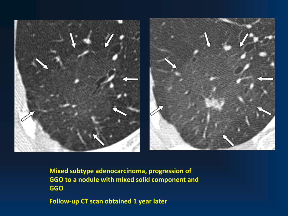
Mixed subtype adenocarcinoma, progression of
GGO to a nodule with mixed solid component and
GGO
Follow‐up CT scan obtained 1 year later

Role of PETRole of PET •
For lesions as small as 8–10 mm in size
•
FDG PET is accurate in differentiating benign from malignant lesions, with an overall sensitivity, specificity, and accuracy of 96%, 88%, and 94%, respectively
•
FDG PET, has a lower sensitivity for small (<10 mm) or slow‐growing lesions, such as
carcinoid
tumors and BAC

TsunezukaTsunezuka et alet alCorrelated the effectiveness of FDG PETFDG PET
to
characterize adenocarcinomas
(≤2 cm) with Noguchi classification:
falsefalse‐‐negative ratenegative rate
for type A lesions was 100%,for type B lesions was 80%, for type C lesions was 47%,
true‐positive rate for types D, E, and F lesions was 67%, 100%, and 86%, respectively .
Similar results have been reported by Yap and colleagues

Shimizu et al Role of Transthoracic
Needle Biopsy
• The diagnostic yield of CT‐guided FNA biopsy for
GGO nodules was 51.2%51.2%
• Lesions smaller than 10 mm with predominantly
ground‐glass appearance the diagnostic yield was as low as 35.2%35.2%

Kim et al Kim et al accuracy of CTaccuracy of CT‐‐guided core biopsy guided core biopsy
Overall concordance rate between core and surgical biopsies in malignant and
premalignant
lesions was 73%73%
transthoracic
FNA should only be performed in patients with subsolid
nodules at
CT who are either nonsurgical
candidates, surgical candidates for whom proof of
malignancy is still considered necessary, or who present with multifocal
disease

Patients with Multiple Patients with Multiple SubsolidSubsolid NodulesNodules
Variable‐sized subsolid
nodules. (a)
Magnified 1‐mm CT section through the right
upper lobe shows multiple small lesions with GGO and one dominant larger nodule
with GGO (arrow). (b)
CT scan at 4‐year follow‐up shows no substanital
interval
change (arrow) and the lesions were presumed to represent AAH and BAC
(dominant lesion).

Is there over diagnosis of Lung Cancer in Is there over diagnosis of Lung Cancer in Patients with Patients with SubsolidSubsolid
NodulesNodules ? ?
•
The potential of low‐dose CT lung cancer screening to decrease lung cancer mortality
•
Preliminary reports do seem to indicatedo seem to indicate that in select cases identification of small
subsolid
lesions at CT may in fact lead to over diagnosis and unnecessary treatment

Multiple subsolid
nodules with variable size and appearance.
In such cases, selective limited resection of the dominant lesions may be
acceptable, contrary to standard treatment of patients with multiple foci of BAC
and/or adenocarcinoma.
Suggested Guidelines in the Management of Suggested Guidelines in the Management of
SubsolidSubsolid
NodulesNodules
•
These do not differentiate between low‐
and high‐risk group as per Fleischner
criteria due
to the increased incidence of adenocarcinomas
in younger and nonsmoking
patients
••
In the light of individual clinical history.In the light of individual clinical history.

Smaller than 5 mmSmaller than 5 mm
Incidentally identified, isolated pure GGOs
smaller than 5 mm
foci of AAH sufficiently often to obviate routine follow‐up CT
studies
•
some of these lesions may prove to be BAC, the extreme rarity of
invasive adenocarcinomas
coupled with their extremely prolonged doubling times
suggest that there is no reason to undergo either the added expense or radiation exposure necessary to follow these lesions presumably over
prolonged time intervals measured in years

SubsolidSubsolid nodulenodule
>5 mmAAH
No additional
imaging or
follow up

Smaller than 10 mm , Pure GGOSmaller than 10 mm , Pure GGO ••
Conservative managementConservative management
requires an initial follow‐up examination in 3–6
months to document that lesions have not resolved spontaneously (or following antibiotic therapy).
•
For most of these lesions, continued long‐term follow‐up is likely preferable to surgical resection
•
Annual follow‐up should extend for more than 2 years
•
5 years?, the risk of subsequent development of cancer needs to be balanced against the risks of unnecessary radiation exposure and
surgical
intervention
•
Thin‐section, Low dose CT, with as low as 80 mAs
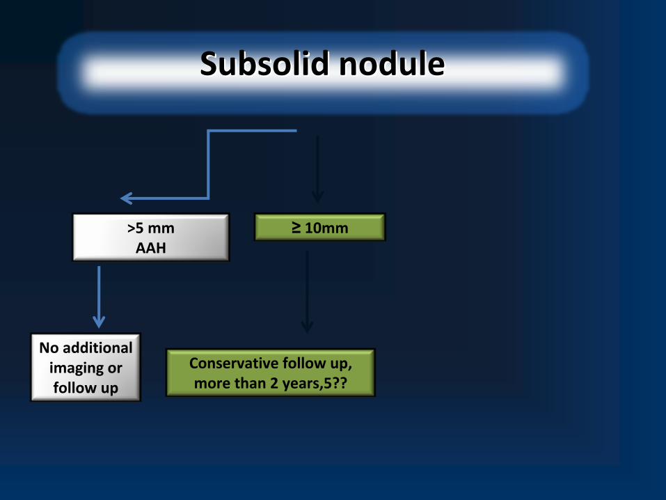
SubsolidSubsolid nodulenodule
>5 mmAAH
No additional
imaging or
follow up
≥
10mm
Conservative follow up,
more than 2 years,5??

Solitary Lesions 10 mm or Larger,Solitary Lesions 10 mm or Larger, pure GGOpure GGO
•
Nodules with pure GGO that are larger than 1 cm in size should be assumed as BAC or
invasive adenocarcinoma
provided if there is an increase in attenuation or development of a solid component or persistence
•
Although 20%‐25% will prove to be benign at resection

Solitary Lesions 10 mm or Larger,Solitary Lesions 10 mm or Larger, pure GGOpure GGO
••
Solitary lesions 10 mm or largerSolitary lesions 10 mm or larger
with pure GGOGGO
should be resectedresected, provided that persistence or growthpersistence or growth
of the lesion is
again established over at least a 3–6‐month period
•
Percutaneous
needle biopsy are limited given substantial sampling error.
•
PET or PET/CT remain doubtful as PET‐negative studies do not exclude the possibility of invasive adenocarcinoma, while
these lesions are also still unlikely to be associated with distant metastases

Lesions with Mixed Solid Component and GGOLesions with Mixed Solid Component and GGO
Should be presumed malignant and surgical resection should be considered, provided lack
of interval change over at least 3 months

Lesions with Mixed Solid Component and GGOLesions with Mixed Solid Component and GGO •
Any lesion with mixed solid component and GGO, regardless of sizeregardless of size,
represents malignancy with sufficient likelihood to warrant further
evaluationevaluation.
The evaluation should include :
•
PET/CT
greater likelihood for invasive tumors for which preoperative staging and assessment of prognosis is warranted.
•
Transthoracic
biopsy???
limited value of accurate differentiation between BAC and invasive adenocarcinomas
and the likelihood that these lesions will be resected,
regardless.
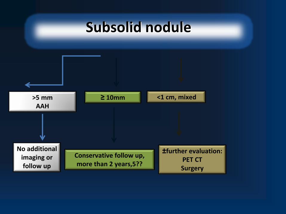
SubsolidSubsolid nodulenodule
>5 mmAAH
No additional
imaging or
follow up
≥
10mm
Conservative follow up,
more than 2 years,5??
<1 cm, mixed
±further evaluation: PET CT
Surgery

Multiple Multiple SubsolidSubsolid NodulesNodules
smaller than 5 mm with pure GGOsmaller than 5 mm with pure GGO
•
At least 1‐year follow‐up CT study
•
May be at greater risk than the general population for developing cancer.
•
However, continued long‐term follow‐up should not be considered necessary.
•
In general, follow‐up CT surveillance is to be preferred in cases in which multiple small (5–10‐mm in size) lesions are identified, as these most
likely represent either multifocal
AAH or in smokers, respiratory bronchiolitis
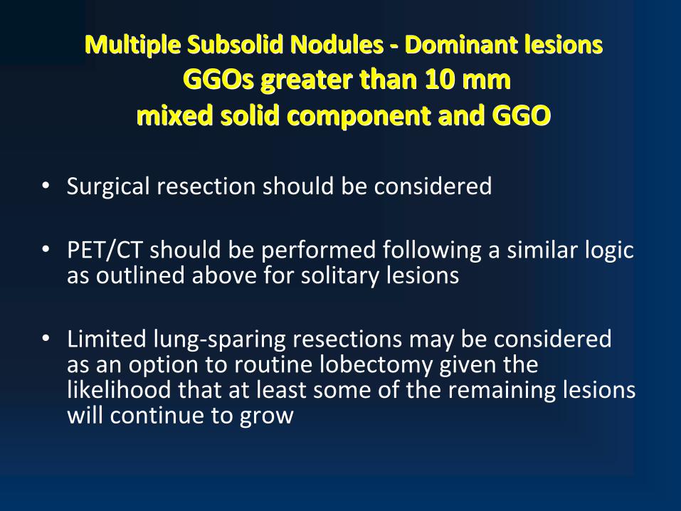
Multiple Multiple SubsolidSubsolid
Nodules Nodules ‐‐
Dominant lesionsDominant lesions GGOsGGOs
greater than 10 mm greater than 10 mm
mixed solid component and GGOmixed solid component and GGO
•
Surgical resection should be considered
•
PET/CT should be performed following a similar logic as outlined above for solitary lesions
•
Limited lung‐sparing resections may be considered as an option to routine lobectomy
given the
likelihood that at least some of the remaining lesions will continue to grow

SubsolidSubsolid nodulenodule
>5 mmAAH
No additional
imaging or
follow up
≥
10mm
Conservative follow up,
more than 2 years,5??
<1 cm, mixed
±further evaluation: PET CT
Surgery
Multiple
Without a
dominant lesion
1 year
follow up
With a
dominant lesion