Agrobacterium-Plant Cell DNA Transport: Have · PDF fileAgrobacterium-Plant Cell DNA...
13
The Plant Cell, Vol. 8, 1699-1710, October 1996 O 1996 American Society of Plant Physiologists Agrobacterium-Plant Cell DNA Transport: Have Virulence Proteins, Will Trave1 Jinsong Sheng and Vitaly Citovsky’ Department of Biochemistry and Cell Biology, lnstitute of Cell and Developmental Biology, State University of New York, Stony Brook, New York 11794-5215 INTRODUCTION Agrobacterium fumefaciens (Agrobacterium) is a soil phy- topathogen that genetically transforms host cells, causing crown gall tumors, an agronomically important disease that affects most dicotyledonous plants. In nature, these tumors are formed at the soil-air junction, the so-called crown of the plant. The Agrobacterium-plant cell interaction is the only known natural example of DNA transport between kingdoms. In this process, DNA is transported from wild-type Agrobac- terium into the plant cell nucleus. Expressionof this transferred DNA (T-DNA)results in neoplastic growths (tumors) on the host plant. The wild-type T-DNA carries genes involved in the synthesis of plant growth hormones and the production of opines, tumor-specific compounds formed by the condensa- tion of an amino acid with a keto acid or a sugar. It is the production of growth hormones in the transformed host cells that induces the formation of tumors. These tumors then syn- thesize opines, a major carbon and nitrogen source for Agrobacterium. Agrobacteria are usually classified based on the type of opines specified by the bacterial T-DNA, the most common strains being octopine or nopaline specific (Hooykaas and Beijersbergen, 1994). Opine import into and the subse- quent catabolism within the bacterial cell require specialized enzymes. Because these enzymes are encoded by the Agrobacterium tumor-inducing (Ti) plasmid, practically no other soil microorganismscan metabolizeopines, creating a favor- able biological niche for Agrobacterium. Three genetic components of Agrobacterium are required for plant cell transformation (Figure 1). The first component is the T-DNA, which is actually transported from the bacte- rium to the plant cell. The T-DNA is a discrete segment of DNA located on the 200-kb Ti plasmid of Agrobacterium; it is delineated by two 25-bp imperfect direct repeats known as the T-DNA borders. The second component is the 35-kb virulence (vir) region, also located on the Ti plasmid,which is composed of Seven major loci (virA, virB, virC, virD, vir€, viG, and vir/+). The protein products of these genes, termed virulence (Vir) proteins, respond to the specific compounds secreted by the wounded plant (see below) to generate a copy of the T-DNA To whom correspondence should be addressed. and mediate its transfer into the host cell. The third compo- nent is the suite of chmmosomalvirulence (chv) genes, located on the Agrobacterium chromosome. chv genes are involved in bacterial chemotaxistoward and attachment to the wounded plant cell (reviewed in Citovsky et al., 1992a; Zambryski, 1992). Because the T-DNA element is defined by its borders, the coding region of the wild-type T-DNA can be replaced by any DNA sequencewithout any effect on its transfer from Agrobac- terium to the plant, Thus, Agrobacterium is often used to produce transgenic plants expressing genes of interest. In ad- dition to this technical application, Agrobacterium represents a fascinatingmodel system to study the wide variety of biolog- ical processes that ultimately result in genetic transformation of the host plant cell. This review describes how Agrobacterium is used as such an experimental system. Specifically, we divide Agrobac- terium-plant cell interaction into seven steps, each of which represents afundamental aspect of prokaryotic as well as eu- karyotic cellular biology (Figure 1 and Table 1). Steps including Agrobacterium-host cell recognition, transduction of plant sig- nals, activation of vir genes, and production of a transferable copy of the T-DNA are characteristic of bacterial cunjugation. Transport of the T-DNA from the bacterial into the host plant cell may combine the hallmarks of both bacterial conjugation and vira1 infection, whereas T-DNA nuclear transport and in- tegration probably occur by typical eukaryotic mechanisms. Our detailed discussion of these steps concludes with an overview of future perspectives and emerging experimental approaches. CELL-CELL RECOGNITION Cell-cell contact is required for the onset of many intercellu- lar interactions, including host-pathogen recognition (see Alfano and Collmer, 1996, and Bent, 1996, in this issue). For example, most animal viruses initiate infection by binding to specific receptors on the host cell surface. Similarly, attach- ment of Agrobacterium to host plant cells is a prerequisitefor DNA transfer. Agrobacterium-host cell recognition is a two-
Transcript of Agrobacterium-Plant Cell DNA Transport: Have · PDF fileAgrobacterium-Plant Cell DNA...
The Plant Cell, Vol. 8, 1699-1710, October 1996 O 1996 American Society of Plant Physiologists
Agrobacterium-Plant Cell DNA Transport: Have Virulence Proteins, Will Trave1
Jinsong Sheng and Vitaly Citovsky’ Department of Biochemistry and Cell Biology, lnstitute of Cell and Developmental Biology, State University of New York, Stony Brook, New York 11794-5215
INTRODUCTION
Agrobacterium fumefaciens (Agrobacterium) is a soil phy- topathogen that genetically transforms host cells, causing crown gall tumors, an agronomically important disease that affects most dicotyledonous plants. In nature, these tumors are formed at the soil-air junction, the so-called crown of the plant. The Agrobacterium-plant cell interaction is the only known natural example of DNA transport between kingdoms. In this process, DNA is transported from wild-type Agrobac- terium into the plant cell nucleus. Expression of this transferred DNA (T-DNA) results in neoplastic growths (tumors) on the host plant. The wild-type T-DNA carries genes involved in the synthesis of plant growth hormones and the production of opines, tumor-specific compounds formed by the condensa- tion of an amino acid with a keto acid or a sugar. It is the production of growth hormones in the transformed host cells that induces the formation of tumors. These tumors then syn- thesize opines, a major carbon and nitrogen source for Agrobacterium. Agrobacteria are usually classified based on the type of opines specified by the bacterial T-DNA, the most common strains being octopine or nopaline specific (Hooykaas and Beijersbergen, 1994). Opine import into and the subse- quent catabolism within the bacterial cell require specialized enzymes. Because these enzymes are encoded by the Agrobacterium tumor-inducing (Ti) plasmid, practically no other soil microorganisms can metabolize opines, creating a favor- able biological niche for Agrobacterium.
Three genetic components of Agrobacterium are required for plant cell transformation (Figure 1). The first component is the T-DNA, which is actually transported from the bacte- rium to the plant cell. The T-DNA is a discrete segment of DNA located on the 200-kb Ti plasmid of Agrobacterium; it is delineated by two 25-bp imperfect direct repeats known as the T-DNA borders. The second component is the 35-kb virulence (vir) region, also located on the Ti plasmid, which is composed of Seven major loci (virA, virB, virC, virD, vir€, viG, and vir/+). The protein products of these genes, termed virulence (Vir) proteins, respond to the specific compounds secreted by the wounded plant (see below) to generate a copy of the T-DNA
To whom correspondence should be addressed.
and mediate its transfer into the host cell. The third compo- nent is the suite of chmmosomal virulence (chv) genes, located on the Agrobacterium chromosome. chv genes are involved in bacterial chemotaxis toward and attachment to the wounded plant cell (reviewed in Citovsky et al., 1992a; Zambryski, 1992).
Because the T-DNA element is defined by its borders, the coding region of the wild-type T-DNA can be replaced by any DNA sequence without any effect on its transfer from Agrobac- terium to the plant, Thus, Agrobacterium is often used to produce transgenic plants expressing genes of interest. In ad- dition to this technical application, Agrobacterium represents a fascinating model system to study the wide variety of biolog- ical processes that ultimately result in genetic transformation of the host plant cell.
This review describes how Agrobacterium is used as such an experimental system. Specifically, we divide Agrobac- terium-plant cell interaction into seven steps, each of which represents afundamental aspect of prokaryotic as well as eu- karyotic cellular biology (Figure 1 and Table 1). Steps including Agrobacterium-host cell recognition, transduction of plant sig- nals, activation of vir genes, and production of a transferable copy of the T-DNA are characteristic of bacterial cunjugation. Transport of the T-DNA from the bacterial into the host plant cell may combine the hallmarks of both bacterial conjugation and vira1 infection, whereas T-DNA nuclear transport and in- tegration probably occur by typical eukaryotic mechanisms. Our detailed discussion of these steps concludes with an overview of future perspectives and emerging experimental approaches.
CELL-CELL RECOGNITION
Cell-cell contact is required for the onset of many intercellu- lar interactions, including host-pathogen recognition (see Alfano and Collmer, 1996, and Bent, 1996, in this issue). For example, most animal viruses initiate infection by binding to specific receptors on the host cell surface. Similarly, attach- ment of Agrobacterium to host plant cells is a prerequisite for DNA transfer. Agrobacterium-host cell recognition is a two-

1700 The Plant Cell
plant phenolic signals
vitronectin-likeplant cellreceptors
AGROBACTERIUM
Figure 1. Agrobacterium-Plant Cell Interactions.
This diagram summarizes all major cellular reactions involved in T-DNA transport. Steps 1 through 7 indicate sequential processes that occurduring Agrobacterium infection. Step 1, binding of Agrobacterium to the host cell surface receptors; step 2, recognition of plant signal moleculesby the bacterial VirA/VirG sensor-transducer system; step 3, activation of the bacterial vir genes; step 4, production of the transferable T-strand;step 5, formation of the T-complex and its transport into the host plant cell; step 6, nuclear import of the T-complex; and step 7, T-DNA integration.IM, bacterial inner membrane; NPC, nuclear pore complex; OM, bacterial outer membrane; PP, bacterial periplasm.
step process. First, the bacteria loosely bind to the host cellsurface, and second, the bound bacteria synthesize cellulosefilaments that stabilize the initial binding, resulting in a tightassociation between Agrobacterium and the host cell (Matthysse,1986).
Because each plant cell binds a finite number of bacteria,saturable surface receptors are thought to be involved(reviewed in Citovsky et al., 1992a). In animals, vitronectin, animportant component of the extracellular matrix, is reportedto function as a receptor for several bacterial strains (Paulssonand Wadstrom, 1990). Vitronectin-like molecules, which havebeen found on the cell surface of many plant species, maymediate Agrobacterium-plant cell binding. Human vitronec-tin as well as anti-vitronectin antibodies block attachment of
Agrobacterium to cultured plant cells. Further, Agrobacteriumstrains that are unable to bind plant cells due to mutations intheir chromosomal chvB, pscA, or aft loci also show reducedbinding to vitronectin (Wagner and Matthysse, 1992). Poten-tially, a plant vitronectin-like protein (PVN) may represent oneof the receptors responsible for the specific interaction betweenAgrobacterium and plant cells (Figure 1, step 1). Interestingly,recent data indicate that the PVNs are related to animalvitronectin only immunologically and that no amino acid se-quence similarity exists between these proteins (Zhu et al.,1994). In addition to PVNs, other plant cell surface proteinsand carbohydrates are likely to be involved in the interactionwith Agrobacterium. However, the identity of these moleculesremains to be determined.

Agrobacterium-Plant Cell DNA Transport 1701
SIGNAL TRANSDUCTION AND TRANSCRIPTIONAL ACTlVATlON OF VIR GENES
Plant-induced T-DNA transport has been used as an ex- perimental system to study the general question of how bacterial cells monitor their surroundings to make appropri- ate adaptive responses. In prokaryotes, signal-response coupling often involves two families of signal-transducing pro- teins (socalled two-component regulatory systems). One family includes membrane sensor proteins, and the other family comprises cytoplasmic proteins that transduce information from the membrane sensor. A single bacterial cell has been suggested to contain as many as 50 pairs of these sensor- transducer proteins, regulating such fundamental and diverse biological processes as sporulation, transformation compe- tente, membrane transport, motility, intermediary metabolism, and pathogenicity (reviewed in Stocket al., 1990). To regulate infection of plants, Agrobacterium has evolved a two- component signal transduction system composed of the viru- lence proteins VirA and VirG. Together, these proteins sense signal molecules secreted by wounded plant cells and acti- vate the expression of other vir genes, thereby initiating the process of T-DNA transport (Winans et al., 1994).
Plant Signals
Wounded plants secrete sap with a characteristic acidic pH (5.0 to 5.8) and a high content of various phenolic compounds, such as lignin and flavonoid precursors. These conditions spe- cifically stimulate Agrobacterium vir gene expression. The best characterized and most effective vir gene inducers are monocy- clic phenolics such as acetosyringone (AS; Stachel et al., 1985). These molecules are not detected, or are detected at low lev- els, in uninjured plants, but their amounts significantly increase in wounded plant cells. The specific composition of phenolic compounds in plant exudates is thought to underlie the host specificity shown by many Agrobacterium strains (Citovsky et al., 1992b). Interestingly, many other plant-microbe interac- tions are initiated by specific phenolic compounds in host plant exudates. For example, flavonoids such as luteolin and chal- cone induce the Rhizobium nodulation genes that are required for symbiotic association of the bacterium with legumes. The chemical structure of these inducers is host-symbiont spe- cific (reviewed in Peters and Verma, 1990).
When only small amounts of phenolics are secreted from the damaged plant cell, the release of sugars may help to ac- tivate the major phenolic-mediated wound signaling pathway
Table 1. Summary of Cellular Processes lnvolved in Agrobacterium-Plant lnteractions
Specific Step in Agrobacterium-Plant Cell lnteraction in the Process Cellular Process
Cell-cell recognition Binding of Agrobacterium to the host , ChvA, ChvB, PscA, Att
Agrobacterium Proteins lnvolved
cell surface receptors
Signal transduction
Transcriptional activation
Recognition of plant signal molecules ChvE, VirA, VirG, IP21, PlOIa and activation of the T-DNA transport pathway
Expression of vir genes after phosphorylation of the transcriptional activator
Conjugal DNA metabolism Nicking at the T-DNA borders and mobilization of the transferable single- stranded copy of the T-DNA (T-strand)
VirG
VirDl, VirD2, VirCl, [VirD3]
lntercellular transportb Formation of protein-DNA T-complex; VirE2, VirEl, VirD2, VirD4, formation of a transmembrane channel; export of the T-complex into the cytoplasm of the host plant cell
VirB4, VirB7, VirB9, VirBl O, VirBll, [VirBl, other Vir6 proteins]
Nuclear import lnteraction with the host cell NLS receptors and transport of the T-complex through the nuclear pore
VirD2, VirE2
T-DNA integration lntegration into the plant cell genome; IVirD2, VirEP] synthesis of the second strand of the T-DNA
a The proteins with a proposed but not proven function are shown in brackets.
involved in the formation of the T-complex and probably its transport through the Vir6 channel. Vir6 proteins are involved in the formation of a transmembrane channel and possibly in export of the T-complex. VirE and VirD proteins are

1702 The Plant Cell
(Cangelosi et ai., 1990). Monosaccharides such as glucose and galactose significantly increase vir gene expression only when AS is limited or absent. This enhancement does not re- sult from nutritional benefits to Agrobacterium, because nonmetabolizable sugars also increase vir gene induction (reviewed in Citovsky et al., 1992b). Conversely, low opine levels further enhance vir gene expression in the presence of AS (Veluthambi et al., 1989). '
As a general rule, conditions that promote vir gene induc- tion are poor for bacterial growth, whereas conditions that support vegetative bacterial growth are unsuitable for vir gene induction. Therefore, it has been proposed that in the plant wound environment containing low opine concentrations, Agrobacterium stays in an optimal state for vir gene induction and T-DNA transfer (Veluthambi et al., 1989). The high opine concentrations produced in mature tumors restore Agrobac- terium to vegetative growth and allow the opines to be used as a carbon and nitrogen source (reviewed in Citovsky et ai., 1992b).
Although phenolic inducer molecules are required for the initiation of T-DNA transport, most of these compounds are bacteriostatic at high concentrations. A plant-inducible locus in the vir region, termed virH or pinE may be involved in the detoxification of the harmful phenolics secreted by the wounded plant. Specifically, two virH products share sequence homol- ogy with cytochrome P450-like enzymes (Kanemoto et al., 1989). These enzymes catalyze the NADH-dependent oxida- tion of different aromatic substrates. It is possible that the VirH proteins inactivate toxic plant phenolics by a similar mechanism.
vir Gene lnduction
Signal molecules released by the wounded plant cell are rec- ognized by the VirANirG two-component regulatory system. It is not clear whether the phenolic signals are sensed directly by the sensor component, VirA, or by another receptor pro- tein that then interacts with VirA (Figure l, step 2). Some data suggest that plant phenolics initially interact with the chro- mosomally encoded proteins P10 and P21 (Lee et al., 1992). However, more recent genetic data indicate that VirA senses plant signals directly; this hypothesis is consistent with the puz- zling inability of researchers to isolate non-VirA mutants that are unable to bind phenolic compounds. In other words, the mutations in all Agrobacterium mutants that do not bind phenolics have been localized to the virA locus (Lee et al., 1995). Direct interaction between VirA and AS, however, re- quires that AS diffuse through the outer membrane of Agrobacterium to reach VirA, which is associated with the bac- teria1 inner membrane and lacks extracellular domains (winans et al., 1994). Unlike phenolics, sugar enhancers have been shown conclusively to interact with a chromosomally encoded glucose/galactose binding protein, ChvE, which in turn inter- acts with VirA (Shimoda et al., 1993).
VirA is composed of four structural and functional domains: an N-terminal periplasmic domain (flanked by hydrophobic,
potentially transmembrane regions) and C-terminal linker, pro- tein kinase, and phosphoryl receiver domains. VirA shows amino acid sequence homology in its C terminus to bacterial sensor proteins, such as EnvZ, PhoR, NtrB, and CpxA. Like these sensor proteins, VirA functions as a protein kinase and phosphotransferase (reviewed in Winans et al., 1994). In the presence of plant signals, VirA autophosphorylates at its His- 474 residue. The phosphohistidine high energy phosphate bond is then transferred to the Asp-52 or possibly the Asp-8 resi- due in VirG, which shares amino acid homology with OmpR, NtrC, and many other transducer proteins. Unlike transducer components in other systems, however, phosphorylated VirG is very stable. The stability of VirG is thought to facilitate maxi- mal levels of vir gene induction (reviewed in Citovsky et al., 1992b).
To activate the expression of the other vir genes, VirG interacts with the vir box, a conserved 12-bp sequence (TNC- AATTGAAAPy) in the promoter regions of highly inducible vir genes. The incomplete dyad symmetry of the vir box suggests that VirG may bind as a dimer (reviewed in Citovsky et al., 1992b). In vivo, VirG phosphorylation is required for the acti- vation of vir gene expression, although the actual role of phosphorylation has not yet been determined (Figure 1, step 3). After VirG binds to a vir box-containing promoter, phos- phorylation may allow interactions with other proteins, such as RNA polymerase. In addition, although unphosphorylated VirG can specifically bind vir promoters, phosphorylation of VirG may increase its DNA binding affinity, as has been reported for the binding of OmpR and NtrC to Escherichia coli promoters (reviewed in Citovsky et al., 1992b). It is still unclear whether VirG phosphorylation occurs before or after its bind- ing to the vir box.
CONJUGAL DNA METABOLISM
The T-DNA Element
lnduction of vir gene expression ultimately results in the produc- tion of aT-DNA copy that is capable of genetically transforming plant cells. Different types of Ti plasmids carry T-DNA elements of different composition. For example, the T-DNA in the nopa- line Ti plasmid is a contiguous stretch of ~ 2 2 kb (Figure 1, step 4), whereas the octopine-specific T-DNA is composed of three independently transported adjacent T-DNAs: left (13 kb), central (1.5 kb), and right (7.8 kb). The borders of a T-DNA ele- ment are defined as conserved 25-bp sequences that delimit the transferred segments. Genetic studies using deletion mu- tants show that the right border is absolutely required for Agrobacterium pathogenicity, whereas the left border is not. Furthermore, inversion of the right border leads to reduced virulence and transfer of nearly the entire Ti plasmid instead of the T-DNA region (reviewed in Citovsky et al., 1992a; Zambryski, 1992). These results suggest that transfer of the

Agrobacterium-Plant Cell DNA Transport 1703
T-DNA is polar from right to left, as determined by the orienta- tion of the T-DNA border repeats.
Interestingly, the T-DNA itself &e., the DNA sequence be- tween the 25-bp repeats) has no effect on the efficiency of transfer. Consequently, nononcogenic (“disarmed”) Ti plasmids, with most of the interna1 sequences of the T-DNA replaced by the DNA of interest, are widely used as vectors for genetic transformation of plants.
Production of the Transferable T-Strand
vir-induced Agrobacterium cells generate a linear single- stranded copy of the T-DNA region, designated the T-strand (Stachel et al., 1986). The T-strand is found at approximately one copy per induced Agrobacterium cell and is derived from the coding strand of the T-DNA element (Stachel et al., 1986). T-strand production is thought to occur in a 5’ to 3’ direction, initiating at the right T-DNA border and terminating at the left border (reviewed in Citovsky et al., 1992a; Figure 1, step 4). VirDl and VirD2 proteins are thought to function together as an endonuclease that carries out site- and strand-specific nicks between the third and fourth base pair of the bottom strand of the T-DNA borders (Figure 1, step 4; see also Wang et al., 1987). In vitro studies demonstrate that purified VirD1 and VirD2 indeed act as a site-specific endonuclease on a supercoiled plasmid containing a 25-bp border repeat (Scheiffele et al., 1995). Following cleavage, VirD2 covalently attaches to the 5’ end of the T-strand at the right border nick and to the 5’ end of the remaining bottom strand of the Ti plasmid at the left border nick (Herrera-Estrella et al:, 1988; Figure 1, step 4). The excised T-strand is removed, and the resulting single-stranded gap is repaired, most likely by replacement DNA strand syn- thesis. The replacement reaction presumably removes the VirD2 molecule attached to the 5’end of the left border, restor- ing the circular DNA molecule of the Ti plasmid. Recent data indicate that VirD2 also may participate in ligating the left bor- der nick (Pansegrau et al., 1993). Finally, another virulence protein, VirC1, can enhance T-strand production from the oc- topine Ti plasmid when VirD1 and VirD2 are limiting (De Vos and Zambryski, 1989). Because so few plant-induced Agrobac- terium proteins are necessary for T-strand production, bacterial housekeeping enzymes of DNA repair and metabolism, such as helicases, also may be involved in this process.
There are strong parallels between T-DNA metabolism and the evolutionarily related process of bacterial conjugation that occurs in most bacteria, including Agrobacterium itself. For comprehensive evaluations of these parallels, the reader is referred to recent reviews that specifically discuss this point (Lessl and Lanka, 1994; Zupan and Zambryski, 1995). Briefly, the virulence system of Agrobacterium appears related to the well-studied conjugative transport system of the broad-range lncP plasmid RP4, initially identified in a pathogenic fseudo- monas strain (reviewed in Lessl and Lanka, 1994). The following functional analogy has been proposed: VirD1, VirD2, and VirD3 are Agrobacterium counterparts of the RP4-encoded TraJ, Tral,
and TraH proteins, respectively. Together, these proteins form a “relaxosome” complex that is required for nicking at the respective DNA sites (T-DNA borders and the origin of trans- fer, ofi7). Tral is thought to cleave the DNA, whereas TraJ facilitates Tral binding to oriT, and TraH stabilizes the entire relaxosome. Although TraH does not bind DNA, it interacts spe- cifically with both TraJ and Tral (reviewed in Lessl and Lanka, 1994). Additional studies are required to test whether VirDl and VirD3 indeed possess the TraJ and TraH activities. One of the major functional differences between Tral and VirD2 is that the latter also has a eukaryotic activity of guiding the T-strand into the host plant cell nucleus (see below).
Although the specific accumulation of T-strands in vir- induced Agrobacterium strongly suggests that these molecules are destined for transfer into the recipient cell (Stachel et al., 1986), direct evidence for this idea has been obtained only recently. First, a sensitive polymerase chain reaction assay showed that single-stranded, but not double-stranded, T-DNA is present in the host plant cells swn after the onset of Agrobac- terium infection (Yusibov et al., 1994). Second, an independent assay based on the different extrachromosomal recombina- tion properties of double-stranded and single-stranded DNA (ssDNA) indicated that the T-DNA derivatives enter the plant cell nucleus in a single-stranded form (Tinland et al., 1994).
Structural Model of the T-DNA Transfer Intermediate, the T-Complex
It is thought that the T-strand is transferred out of the bacte- rium and into the plant cell as a protein-nucleic acid complex. This T-DNA transport intermediate, designated the T-complex (Howard and Citovsky, 1990), is composed of at least three components (Figure 1, step 5). The T-strand DNA molecule, which carries the genetic information, and its cognate VirD2 and VirE2 proteins, which protect the T-strand, shape it into a transferable (thin and unfolded) form and supply specific tar- geting signals (see below). As mentioned previously, one molecule of VirD2 is covalently attached to each T-strand (Herrera-Estrella et al., 1988). Although both VirD1 and VirD2 are involved in border-specific nicking (De Vos and Zambryski, 1989), there is no experimental evidence that VirDl remains bound to the generated T-strand. Also associated with the T-strand is VirE2, an ssDNA binding protein (SSB; Citovsky et al., 1988,1989). Binding of VirE2 to ssDNA in vitro is strong and cooperative, leading to the formation of very stable un- folded VirE2-ssDNA complexes that are largely inaccessible to externa1 nucleolytic activity (Citovsky et al., 1989). Based on electron microscopy data and in vitro VirE2-ssDNA bind- ing kinetics (Citovsky et al., 1989), the nopaline-specific T-complex is proposed to be 3600 nm long and 2 nm wide. As such, it would contain ~ 6 0 0 molecules of VirE2 and one molecule of VirD2 and have a predicted molecular mass of 50,000 kD.
This structural and functional model of the T-complex im- plies that both VirD2 and VirE2 proteins are transported into

1704 The Plant Cell
the recipient plant cell together with the T-strand (Figure 1, step 5). This idea makes biological sense. It is likely that an SSB with as high an affinity for ssDNA as VirE2 (Citovsky et ai., 1989) will form a complex with the T-strand already inside Agrobacterium, especially if both VirE2 and the T-strand are transported into the plant cell through the same channel (as proposed by Binns et ai., 1995). Formation of T-complexes is alSO supported by the observation that in extracts from vir-induced Agrobacterium, T-strands and VirE2 are coim- munoprecipitated by anti-VirE2 antibodies (Christie et al., 1988). However, coinoculation of the same plant with Agrobacterium carrying a T-DNA but lacking VirE2 and a strain producing VirE2 but lacking T-DNA resulted in a productive infection by these separately nonpathogenic bacteria (Otten et al., 1984). There are two interpretations of this intercellular complemen- tation. First, VirE2 and T-strands can be transported independently from the Agrobacterium into the plant cell, and second, VirE2 functions primarily inside the plant cell. The latter conclusion is supported by the observation that virE2 expression in transgenic plants restores infectivity of a VirE2- deficient Agrobacterium (Citovsky et ai., 1992b; also see below).
Recent data indicate that another product of the vir€ locus, the VirEl protein, may be involved in transport of VirE2 from Agrobacterium into the host plant cell. A VirEl-deficient mu- tant having normal amounts of VirE2 and T-strands was not infectious, presumably because VirEl may be required for export of VirE2coated T-strands. However, when this Agrobac- terium mutant was coinoculated with a strain producing both VirEl and VirE2 but lacking T-DNA, the VirEl mutant became infectious, indicating functional export of T-strands from this strain (Sundberg et al., 1996). These results led to the sug- gestion that within Agrobacterium, T-strands may not form complexes with VirE2 (although they would still carry a cova- lently attached VirD2 molecule), and that VirEl assists export of VirE2 but not of T-strands (Sundberg et ai., 1996). However, alternative explanations cannot be excluded. For example, VirEl may mediate its own export (and possibly that of VirE2) into the coinoculated VirEl-deficient bacteria rather than into the host plant cell. In this case, the presence of the exported VirEl in the same cell with VirE2 and T-strands would allow the export of entire T-complexes. To determine conclusively whether T-strands and VirE2 exit Agrobacterium separately or as a complex requires the development of a more direct and quantitative export assay.
INTERCELLULAR TRANSPORT
lntercellular DNA transport requires a direct passageway be- tween donor and recipient cells. Thus, Agrobacterium is predicted to form a channel through which 1-complexes are transferred into the cytoplasm of the host plant cell. The mo- lecular mechanism by which this passageway is formed and functions is still a biological black box (Figure 1, step 5). How- ever, it seems likely that the Agrobacterium-plant cell channel
is encoded by the virB Iocus, most of which is required for bac- teria1 virulence but not for T-strand production (reviewed in Citovsky et ai., 1992a).
The virB operon contains 11 open reading frames, nine of which encode proteins shown to associate with bacterial mem- branes. Recent molecular studies have focused on severa1 individual VirB proteins. Specifically, Vir69 and VirBlO were each shown to form separate membrane-associated high mo- lecular weight complexes. Although not in the same complex with VirB10, Vir69 was absolutely required for VirBlO complex formation, possibly serving to stabilize this protein or to facili- tate its insertion into the bacterial membrane (Beaupre et ai., 1996). VirB9, in turn, was stabilized by VirB7 after the forma- tion of disulfide cross-linked Vir89-Vir87 heterodimers (Fernandez et ai., 1996; Spudich et ai., 1996). In addition to its dependence on Vir87, Vir69 accumulation at least partly required the production of VirB8 (Berger and Christie, 1994). Thus, the accumulation of some virB gene products depends on the presence of other Vir6 proteins. These data suggest that coordinate protein synthesis stabilizes individual VirB poly- peptides, perhaps allowing them to form a multiprotein channel structure (Figure 1, step 5). Similar protein stabilization through protein-protein ínteractions has been described during the for- mation of virulence-associated P pili in Gram-negative bacteria (Kuehn et al., 1993).
Agrobacterium-plant cell transport of T-complexes through the Vir8 channel is most likely an energy-dependent process. Two Vir6 proteins, Vir84 and VirB11, are the best candidates to provide energy for this translocation. VirB4 has a nucleo- tide binding site, whereas Vir611 is both an ATPase and a protein kinase, and both proteins localize to the inner bacterial membrane (Christie et al., 1989). Further, a recent study indi- cates that in AS-induced Agrobacterium, Vir61 is processed to a lower molecular weight form that is partially exported into the surrounding medium. It is possible that the secreted form of Vir81 interacts with the recipient plant cell, where it may mediate the docking of the Vir6 channel at the infection site on the cell surface (C. Baron and i? Zambryski, personal communication).
Like the generation of T-strands, the formation of the Vir6 channel appears to be evolutionarily related to bacterial con- jugation. Specifically, the virB operon is analogous to the RP4 tra2 region that encodes 11 proteins involved in DNA trans- port during conjugation. Six of the Tra2 proteins (TrbB, TrbC, TrbD, TrbE, TrbF, and Trbl) show significant similarities in amino acid composition, gene organization, and physical properties to six of the virB products (VirB11, VirB2, VirB3, VirB4, VirB5, and VirB10, respectively; reviewed in Lessl and Lanka, 1994). Severa1 VirB proteins also show homology with bacterial pro- tein export apparati, such as the predicted gene products of the Bordetella pertussis Ptloperon, which are required for ex- port of pertussis toxin protein (Weiss et al., 1993).
The parallels between T-DNA transport and RP4-mediated conjugation also suggest the assignment of a critical function to the VirD4 protein. VirD4, an essential virulence protein, as- sociates with the inner bacterial membrane and has significant
Agrobacterium-Plant Cell DNA Transport 1705
homology with the RW-encoded TraG protein. TraG is thought to link the mobilized RP4 plasmid DNA to the pilus channel (Lessl and Lanka, 1994). Similarly, VirD4 may form a link be- tween the transported T-complex and the VirB channel (Figure 1, step 5). Functional evidence in support of this hypothesis comes from recent experiments showing that VirD4, together with the Vir6 proteins, can substitute for TraG and Tra2 gene products, respectively, during conjugative transport of another plasmid, the lncQ RSF1010, between Agrobacterium cells (reviewed in Lessl and Lanka, 1994).
NUCLEAR IMPORT
VirD2 and VirE2 Probably Mediate Nuclear lmport of the T-Complex
Unlike bacterial conjugation, the recipient cell in Agrobacterium T-DNA transport is a eukaryote. Therefore, although the gener- ation of a transferable T-strand molecule may have parallels with bacterial conjugation, the delivery of the T-complex into the target cell does not. Instead, the later steps of T-DNA trans- fer, penetration into the host cell nucleus (Figure 1, step 6) and integration into the nuclear genome (Figure 1, step 7), are more related to virus infection. The T-complex thus resembles aviral (or subviral) particle that is capable of plant cell transforma- tion (Citovsky et al., 1992a).
Because pathogenic microorganisms often use existing cel- lular machinery for their own needs, Agrobacterium probably employs an endogenous cellular pathway for transport of the invading T-complex into the plant cell nucleus. Consequently, severa1 recent studies have used the Agrobacterium T-complex as a model system to examine the general process of nuclear import of nucleic acids in plant cells. The estimated size of the T-complex (50,000 kD) far exceeds the size-exclusion limit of the nuclear pore (60 kD), suggesting a requirement for ac- tive transport processes. Because the T-strand presumably does not itself carry targeting signals, T-complex nuclear im- port is most likely mediated by the VirD2 and VirE2 proteins (Figure 1, step 6). Indeed, VirD2 was shown to accumulate in the plant cell nucleus by use of both a VirD2 translational fusion to the P-glucuronidase reporter enzyme (GUS; Howard et al., 1992) and direct immunolocalization of VirD2 (Tinland et al., 1992).
As a rule, active nuclear import of proteins requires a spe- cific nuclear localization signal (NLS). The most common type of NLS is the bipartite signal originally described in the Xeno- pus protein nucleoplasmin (Robbins et al., 1991). The first active domain of a bipartite NLS consists of two adjacent basic residues and is followed by a variable-length linker and the second active domain, which contains at least three out of five basic amino acids (reviewed in Dingwall and Laskey, 1991). The functional VirD2 NLS was found to reside in the C termi- nus of the protein and to conform to the bipartite consensus motif (Howard et al., 1992). The biological relevance of this
NLS was confirmed by the observations that Agrobacterium T-DNA expression and tumorigenicity are reduced in NLS de- letion mutants of VirD2 (Shurvinton et al., 1992; Narasimhulu et al., 1996). Collectively, these results suggest that the VirD2 protein, which is attached to the 5’ end of the T-strand, acts to direct the T-complex to the host cell nucleus.
The very large predicted size of the T-complex and the residual tumorigenicity of Agrobacterium mutants with a deleted VirD2 NLS suggested that VirD2 is not the sole medi- ator of the T-complex nuclear uptake. Subsequently, nuclear localization of VirE2, the major structural component of the T-complex, was demonstrated using VirE2-GUS fusions. These experiments identified two functional NLSs within the central region of the VirE2 molecule. Although each VirE2 NLS was independently active, maximal VirE2 nuclear import re- quired the presence of both signals (Citovsky et al., 1992b). That VirE2 is involved in nuclear uptake of the T-complex is further supported by the observation that plants transgenic for VirE2 complement the virulence of an Agrobacterium strain with an inactivated vir€ locus (Citovsky et al., 1992b). In con- trol experiments, plants expressing an unrelated SSB did not complement tumor induction, indicating that VirE2 does not simply bind and protect the transported T-strand from cellular nucleases. In a second set of experiments, transgenic plants expressing a mutant VirE2 that is unable to bind ssDNA but retains both NLSs developed a significant resistance to infec- tion by wild-type Agrobacterium, suggesting that the mutant VirE2 may compete with the incoming T-complex for the host cell nuclear import machinery (Citovsky et al., 1994).
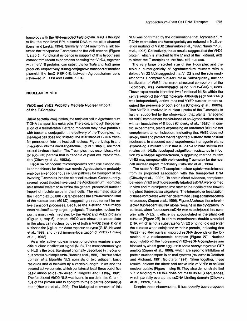
The role of VirE2 in T-complex nuclear uptake was inferred from its proposed association with the transported DNA (Citovsky et al., 1992b). To obtain direct evidence, complexes between VirE2 and fluorescently labeled ssDNA were formed in vitro and microinjected into stamen hair cells of the flower- ing plant Tidescantia virginiana. The intracellular localization of these complexes was then determined using epifluorescence microscopy (Zupan et al., 1996). Figure 2A shows that microin- jected fluorescent ssDNA alone remains in the cytoplasm. In contrast, when fluorescent ssDNA was microinjected in a com- plex with VirE2, it efficiently accumulated in the plant cell nucleus (Figure 28). In control experiments, double-stranded DNA, which is not a substrate for VirE2 binding, did not enter the nucleus when coinjected with this protein, indicating that VirE2-mediated nuclear import of ssDNA depends on the for- mation of a nucleoprotein complex (Figure 2C). Nuclear accumulation of the fluorescent VirE2-ssDNA complexes was blocked by wheat germ agglutinin anda nonhydrolyzable GTP analog (Zupan et al., 1996), which are specific inhibitors of protein nuclear import in animal systems (reviewed in Goldfarb and Michaud, 1991; Goldfarb, 1994). Taken together, these results indicate the direct and active role of VirE2 in ssDNA nuclear uptake (Figure 1, step 6). They also demonstrate that VirE2 binding to ssDNA does not mask its NLS sequences, which partially overlap the ssDNA binding domain (Citovsky et al., 19924 1994).
Despite these observations, it has recently been proposed

1706 The Plant Cell
Figure 2. VirE2-Mediated Nuclear Import of ssDNA.(A) to (C) Tradescantia stamen hairs were microinjected with fluores-cently labeled ssDNA alone (A), VirE2-ssDNA complexes (B), or amixture of VirE2 and double-stranded DMA (C), and examined byepifluorescence microscopy. For specific experimental conditions, seeZupan et al. (1996). This figure is reproduced from Zupan et al. (1996),with permission. N, position of the stamen hair cell nucleus, as deter-mined by using bright-field optics.(D) to (F) Xenopus oocytes were microinjected with fluorescently la-beled ssDNA alone (D), VirE2s20-ssDNA complexes (E), orVirE2s20-ssDN A complexes previously mixed with a 30-fold molar ex-cess of synthetic VirD2 NLS peptide competitor (F). Microinjectedoocytes were viewed by confocal microscopy. For specific experimen-tal conditions, see Guralnick et al. (1996). This figure is reproducedfrom Guralnick et al. (1996), with permission. N, position of the oocylenucleus, as determined by chromatin-specific 4',6-diamidino-2-phenylindole staining.
that VirE2 function may be limited to protection of the T-strand.This conclusion was based on the observation that, in tobacco,deletion of the entire virE2 gene did not further reducetumorigenicity of the VirD2 NLS-deficient strain below the al-ready low basal level of 4 to 7%, as compared with the wild-typeAgrobacterium (Rossi et al., 1996). Unlike microinjection offluorescent VirE2-ssDNA complexes, tumorigenicity measure-ments do not directly assay nuclear import and are thereforedifficult to interpret in these terms. Furthermore, these resultscontradict other reports that tobacco infection by Agrobacteriumwith deleted VirD2 NLS still results in 20 to 30% tumor forma-tion (Shurvinton et al., 1992) as well as in T-DNA transcription(Narasimhulu et al., 1996). Infection of other plant hosts, suchas Arabidopsis and potato, by this mutant showed an evensmaller dependency on VirD2 NLS (tumor formation andT-DNA transcription of 60% or greater), indicating that the VirD2NLS is not absolutely required for nuclear uptake of T-strands(Shurvinton et al., 1992; Narasimhulu et al., 1996).
VirD2 and VirE2 NLSs Represent Two Functional Typesof Plant Nuclear Targeting Signals
In Agrobacterium harboring the nopaline Ti plasmid, the 22-kb T-DNA region is predicted to produce a T-strand associatedwith one molecule of VirD2 and 600 molecules of VirE2, as-suming the average VirE2 binding capacity of 36 nucleotidesper protein monomer (Gietl et al., 1987; Citovsky et al., 1989;Sen et al., 1989). Because each VirE2 molecule carries twoNLSs (Citovsky et al., 1992b), whereas VirD2 contains one func-tional NLS (Howard et al., 1992), the nopaline-specificT-complex is predicted to have a VirD2 NLS:VirE2 NLS ratioof 1:1200. Why then would Agrobacterium need an NLS onVirD2 at all? One possibility is that VirD2 and VirE2 are im-ported into the plant cell nucleus by functionally differenttargeting sequences and that both signals are required for op-timal nuclear import of the T-complex. To test this hypothesis,the roles of the VirD2 and VirE2 NLSs were assayed in heter-ologous animal systems. These systems were chosen becausethey may lack one of the potential plant nuclear import mech-anisms, thereby allowing discrimination between the VirD2 andVirE2 NLS activities (Guralnick et al., 1996).
In these experiments, VirD2 and VirE2 were fluorescentlylabeled and microinjected into Xenopus oocytes and Drosophilaembryos. In both cases, microinjected VirD2 accumulated incell nuclei, as determined by confocal microscopy. The nu-clear uptake of VirD2 was specifically inhibited by an excessof free VirD2 NLS peptide, probably due to competition for thenuclear import machinery (Guilizia et al., 1994). VirD2 nuclearimport was also inhibited by coinjection of GTPyS, a non-hydrolyzable GTP analog known to block the GTPase Ran,which is absolutely essential for the transport of proteinsthrough the nuclear pore (reviewed in Goldfarb, 1994). Theseresults indicate that VirD2 is actively imported into the nucleiof animal cells and that this import is mediated by the VirD2

Agrobacterium-Plant Cell DNA Transport 1707
NLS, a bipartite NLS of the type that is evolutionarily conserved between plants and animals.
In contrast to VirD2, microinjected fluorescently labeled VirE2 remained in the cytoplasm in both Drosophila and Xenopus, suggesting that the VirE2 NLSs are not recognized in an ani- mal system and therefore are plant specific. Amino acid sequence analysis revealed that in both VirE2 NLSs, one un- charged amino acid residue interrupted the normally adjacent basic residues of the first domain of a bipartite NLS (see above). When this intervening residue was removed, the resulting mu- tant proteins accumulated in the animal cell nuclei. Furthermore, one VirE2 NLS mutant, designated VirE2s20, that retained the ssDNA binding activity of the wild-type VirE2 was shown to mediate import of fluorescently labeled ssDNA into the nuclei of Xenopus oocytes (Figures 2D to 2F; Guralnick et al., 1996).
That VirE2 NLSs function in plant but not animal cells sug- gests differences between plant and animal nuclear import machinery. This idea is consistent with observations that the yeast Mata2 NLS functions in plant (Hicks and Raikhel, 1995) but not mammalian cells (Chekky et al., 1989; Lanford et al., 1990). What is the molecular basis for this difference? NLS recognition most likely occurs through an interaction of the NLS with cellular receptors, usually belonging to the karyophe- rin a protein family (reviewed in Powers and Forbes, 1994; Gorlich and Mattaj, 1996). Thus, it is possible that plant cells have a subset of NLS receptors that recognize the VirE2 NLSs and that are absent in animal cells. Other plant cell NLS recep- tors may recognize the conserved bipartite-type NLS of VirD2, sharing this recognition with animal NLS binding proteins (Fig- ure 1, step 6). Alternatively, the same plant NLS receptors may recognize both the VirD2 and VirE2 NLSs but with different affinities, whereas animal receptors are more stringent, inter- acting only with the consensus NLS sequences.
lmplications for Nuclear lmport of T-DNA and Nucleic Acids in General
The functional variations in the NLS sequence may reflect cellular regulation of nuclear import of proteins and/or pro- tein-nucleic acid complexes. For example, the nuclear import of the Agrobacterium Tcomplex may occur in a polar and linear fashion potentially important for the subsequent integra- tion of the T-strand into the plant cell genome (reviewed in Zambryski, 1992; Citovsky and Zambryski, 1993, 1995). The T-complex model (Howard et al., 1990; Citovsky and Zambryski, 1993, 1995) proposes that the 5' end of the T-strand is as- sociated with the VirD2 molecule, whereas the 3'end probably has a VirE2 molecule attached in its proximity; the functional variation between the NLS signals of these proteins may specify the ends of the T-strand and determine the polarity of its trans- port and integration (Figure 1, step 6).
As suggested by experiments with VirE2, formation of com- plexes with a specialized transport protein(s) may be necessary
for DNA nuclear import in many eukaryotic organisms. This model for protein-mediated nuclear import of nucleic acids is supported by the recent observation that influenza virus nucleoprotein transports the vira1 genomic RNA into the cell nucleus in an in vitro system (ONeill et al., 1995). Polarity may represent another common feature of nucleic acid transport through the nuclear pore (Citovsky and Zambryski, 1993). For example, nuclear export of a 75s premessenger ribonucleo- protein particle in Chironomus tentans initiates exclusively at the 5' end of the RNA (Mehlin et al., 1992).
T-DNA INTEGRATION
Nuclear import of the Agrobacterium T-complex culminates with the integration of the transported T-strand into the host plant cell chromosome (Figure 1, step 7). The molecular mech- anisms by which this is achieved are largely unknown. Unlike other mobile DNA elements such as transposons and retroviruses, T-DNA does not encode enzymatic activities en- abling integration. Thus, T-DNA insertion into the plant genome must be mediated by proteins transported from the infecting bacterium and/or by host cell factors.
Recently, both T-strand-associated proteins, VirD2 and VirE2, have been implicated in the integration process (Fig- ure 1, step 7). An Agrobacterium mutant carrying an Arg to Gly substitution at position 129 in VirD2 produced T-DNA in- sertion junctions in which the normally conserved 5' end of the integrated DNA was truncated or significantly rearranged. These data suggest that integration may precede second- strand synthesis, which would be performed by the plant cell DNA repair machinery following T-strand integration (Tinland et al., 1995). However, a second study of T-DNA integration patterns led to the suggestion that T-strands are converted into a double-stranded form before integration (De Neve et al., 1996). 60th models propose that T-DNA integration initiates at the left border region and that VirD2 ligates the right border end of the T-DNA to the genomic plant DNA, thus completing the integration process.
In a detailed study of the early transcription of T-DNA in plant nuclei, a short amino acid sequence located downstream of VirD2 NLS and designated the o domain (Shurvinton et al., 1992) was found to be required for T-DNA integration (Narasimhulu et al., 1996). In addition, analysis of the T-DNA integration junctions suggested that VirE2 is also required for integration fidelity at the 3' but not the 5' end of the T-strand molecule (Rossi et al., 1996). This hypothesis is consistent with the T-complex model in which VirE2 and VirD2 specify the 3' and 5' ends of the T-strand, respectively (see above). Fi- nally, in an intriguing new approach, VirEP was shown to complement in part a RecA deletion mutant of E. coli, sug- gesting the direct involvement of VirE2 in the recombination process (W. Ream, personal communication).
Interestingly, a block in T-DNA integration was suggested
1708 The Plant Cell
to underlie the recalcitrance of maize, and possibly other monocots, to Agrobacterium infection (Narasimhulu et al., 1996). The same hypothesis was proposed earlier on the ba- sis of the lack of cell division during the monocot wound response (Binns and Thomashow, 1988). Consistent with this hypothesis is the observation that transiene expressed VirD2 and VirE2 accumulated in the nuclei of maize leaf epidermal cells (Citovsky et al., 1994), suggesting that nuclear import is not the limiting step in Agrobacterium interaction with monocotyledonous plants.
ACKNOWLEDGMENTS
We thank Christian Baron, Andrew Binns, Peter Christie, Stan Gelvin, Barbara Hohn, Ann Matthysse, Marc Van Montagu, Walt Ream, and Patricia Zambryski for the communication of recent results. We also thank Gail McLean and John Zupan for critical reading of the manu- script. In consideration of space constraints, we have included only representative references. The work in our laboratory is supported by grants from the National lnstitutes of Health (Grant No. ROI-GM50224), the U.S. Department of Agriculture (Grant No. 94-02564), and the United States-Israel Binational Research and Development Fund (BARD) (Grant No. US-2247-93) to V.C.
WHAT'S NEXT?
REFERENCES
Cellular reactions that occur within Agrobacterium before the T-DNA transfer process have been characterized in some de- tail. One event that still needs to be dissected at the molecular leve1 is the formation of the VirB channel and the subsequent targeting of the T-complex to and through this membrane pore. On the plant side, practically a11 of the molecular steps that transport the T-DNA to the plant cell nucleus, culminating with the integration into the plant cell genome, are still obscure. How does the VirB channel link to the host cell surface and mediate transport of the T-complex into the cell cytoplasm? What are the plant proteins that recognize VirD2 and VirE2 NLSs to mediate nuclear import of the T-complex? How is the T-complex uncoated? What is the underlying mechanism of T-DNA integration?
To address these questions, sensitive assays for nuclear im- port and integration need to be developed. Techniques using in vitro assays or genetically characterized cellular systems should provide many details that would be obscured during the complex process of Agrobacterium infection. For exam- ple, nuclear import of T-complexes may be studied by using isolated nuclei or permeabilized plant cells, whereas the in- tegration process can be assayed in a bacteriophage l-based cell-free system developed for retroviral integration or by genetic complementation of recombination-deficient recA mutants of E. coli. In addition to significantly advancing our understand- ing of basic biology, these studies may help to develop new techniques for the efficient genetic manipulation of plant spe- cies, including the agronomically important cereals that are normally recalcitrant to Agrobacterium infection.
Another exciting approach to examining the molecular path- way of the T-complex nuclear import and integration is the isolation of Arabidopsis mutants that are resistant to Agrobac- terium virulence, potentially due to specific blockage in one of these processes. Severa1 such mutants have been indepen- dently isolated in two laboratories, and their characterization is under way (C. Baron and P. Zambryski, personal communi- cation; S. Gelvin, personal communication).
Alfano, J.R., and Collmer, A. (1996). Bacterial pathogens in plants: Life up against the wall. Plant Cell 8, 1683-1698.
Eeaupre, C.E., Dale, E M , and Einns, A.N. (1996). lnteractions be- tween Vir6 membrane proteins involved in the movement of DNA from Agrobacterium tumefaciens into plant cells. J. Bacteriol., in press.
Bent, A.F. (1996). Plant disease resistance genes: Function meets struc- ture. Plant Cell 8, 1757-1771.
Berger, B.R., and Christie, P.J. (1994). Genetic complementation anal- ysis of the Agrobacterium tumefaciens vir6 operon: vir62 through vir811 are essential virulence genes. J. Bacteriol. 176, 3646-3660.
Einns, A.N., and Thomashow, Y.F. (1988). Cell biology of Agrobac- ferium infection and transformation of plants. Annu. Rev. Microbiol.
Einns, A.N., Beaupre, C.E., and Dale, E.M. (1995). lnhibition of VirB- mediated transfer of diverse substrates from Agrobacterium tumefa- ciens by the InQ plasmid RSF1010. J. Bacteriol. 177, 4890-4899.
Cangelosi, G.A., Ankenbauer, R.G., and Nester, E.W. (1990). Sug- ars induce the Agrobacterium virulence genes through a periplasmic binding protein and a transmembrane signal protein. Proc. Natl. Acad. Sci. USA 87, 6708-6712.
Chelsky, D., Ralph, R., and Jonak, G. (1989). Ssquence requirements for synthetic peptide-mediated translocation to the nucleus. MOI. Cell. Biol. 9, 2487-2492.
Christie, P.J., Ward, J.E., Winans, S.C., and Nester, E.W. (1988). The Agrobacterium tumefaciens virE2 gene product is a single-stranded- DNA-binding protein that associates with T-DNA. J. Bacteriol. 170,
Christie, P.J., Ward, J.E., Gordon, M.P., and Nester, E.W. (1989). A gene required for transter of T-DNA to plants encodes an ATPase with autophosphorylating activity. Proc. Natl. Acad. Sci. USA 86,
Citovsky, V., and Zambryski, P. (1993). Transport of nucleic acids through membrane channels: Snaking through small holes. Annu. Rev. Microbiol. 47, 167-197.
Citovsky, V., and Zambryski, P. (1995). Transport of protein-nucleic acid complexes within and between plant cells. Membr. Protein Trans- port 1, 39-57.
42, 575-606.
2659-2667.
9677-9681.

Agrobacterium-Plant Cell DNA Transport 1709
Citovsky, V., De Vos, G., and Zambryski, P. (1988). Single-stranded DNA binding protein encoded by the vir€ locus of Agrobacterium tumefaciens. Science 240, 501-504.
Citovsky, V., Wong, M.L., and Zambryski, P. (1989). Cooperative in- teraction of Agrobacterium VirE2 protein with single stranded DNA: lmplications for the T-DNA transfer process. Proc. Natl. Acad. Sci.
Citovsky, V., McLean, G., Greene, E., Howard, E., Kuldau, G., Thorstenson, Y., Zupan, J., and Zambryski, P. (1992a). Agrobac- terium-plant cell interaction: lnduction of vir genes and T-DNA transfer. In Molecular Signals in Plant-Microbe Communications, D.P.S. Verma, ed (Boca Raton, FL: CRC Press), pp. 169-198.
Citovsky, V., Zupan, J., Warnick, D., and Zambryski, P. (1992b). Nu- clear localization of Agrobacterium VirE2 protein in plant cells. Science 256, 1803-1805.
Citovsky, V., Warnick, D., and Zambryski, P. (1994). Nuclear import of Agrobacterium VirD2 and VirE2 proteins in maize and tobacco. Proc. Natl. Acad. Sci. USA 91, 3210-3214.
De Neve, M., De Buck, S., Jacobs, A., Van Montagu, M., and Depicker, A. (1996). T-DNA integration patterns in co-transformed cells suggest that T-DNA repeats originate from ligation of separate T-DNAs. Plant J., in press.
De Vos, G., and Zambryski, P. (1989). Expression of Agrobacterium nopaline specific VirD1, VirD2, and VirCl proteins and their require- ment for T-strand production of E. coli. MOI. Plant-Microbe Interact.
Dingwall, C., and Laskey, R.A. (1991). Nuclear targeting sequences-A consensus? Trends Biochem. Sci. 16, 478-481.
Fernandez, D., Spudich, G.M., Zhou, X.-R., and Christie, P.J. (1996). The Agrobacterium tumefaciens VirB7 lipoprotein is required for stabilization of VirB proteins during assembly of the Tcomplex trans- port apparatus. J. Bacteriol. 178, 3168-3176.
Gietl, C., Koukolikova-Nicola, Z., and Hohn, B. (1987). Mobilization of T-DNA from Agrobacterium to plant cells involves a protein that binds single-stranded DNA. Proc. Natl. Acad. Sci. USA 84,9006-9010.
Goldfarb, D. (1994). GTPase cycle for nuclear transport. Curr. Biol.
Goldfarb, D., and Michaud, N. (1991). Pathways for the nuclear trans- port of proteins and RNAs. Trends Cell Biol. 1, 20-24.
Gorlich, D., and Mattaj, I.W. (1996). Nucleocytoplasmic transport. Science 271, 1513-1518.
Guilizia, J., Dempsey, M., Sharova, N., Burkinsky, M., Goldfarb, D.S., and Stevenson, M. (1994). Reduced nuclear import of HIV-1 preintegration complexes in the presence of a prototypic nuclear targeting signal. J. Virol. 68, 2021-2025.
Guralnick, B., Thomsen, O., and Citovsky, V. (1996). Transport of DNA into the nuclei of Xenopus oocytes by a modified VirE2 protein of Agrobacterium. Plant Cell 8, 363-373.
Herrera-Estrella, A., Chen, Z., Van Montagu, M., and Wang, K. (1988). VirD proteins of Agrobacterium tumefaciens are required for the formation of a covalent DNA protein complex at the 5’terminus of T-strand molecules. EMBO J. 7, 4055-4062.
Hicks, G.R., and Raikhel, N.R. (1995). Nuclear localization signal bind- ing proteins in higher plant nuclei. Proc. Natl. Acad. Sci. USA 92, 734-738.
USA 86, 1193-1197.
2, 43-52.
4, 57-60.
Hooykaas, P.J.J., and Beijersbergen, A.G.M. (1994). The virulence system of Agrobacterium tumefaciens. Annu. Rev. Phytopathol. 32,
Howard, E.A., and Citovsky, V. (1990). The emerging structure of the Agrobacterium T-DNA transfer complex. Bioessays 12, 103-108.
Howard, E.A., Citovsky, V., and Zambryski, P. (1990). TheT-complex of Agrobacterium tumefaciens. UCLA Symp. MOI. Cell Biol. News Ser. 129, 1-11.
Howard, E., Zupan, J., Citovsky, V., and Zambryski, P. (1992). The VirD2 protein of A. tumefaciens contains a C-terminal bipartite nu- clear localization signal: lmplications for nuclear uptake of DNA in plant cells. Cell 68, 109-118.
Kanemoto, R.H., Powell, A.T., Akyoshi, D.E., Relger, D.A., Kerstetter, R.A., Nester, E.W., Hawes, M.C., and Gordon, M.P. (1989). Nucleotide sequence and analysis of the plant-inducible lo- cus pinF from Agrobacterium tumefaciens. J. Bacteriol. 17l,
Kuehn, M.J., Ogg, D.J., Kihlberg, J., Slonim, L.N., Flemmer, K., Bergfors, T., and Hultgern, S.J. (1993). Structural basis of pilus subunit recognition by the PapD chaperone. Science 262,1234-1241.
Lanford, R.E., Feldherr, C.M., White, R.G., Dunham, R.G., and Kanda, P. (1990). Comparison of diverse transport signals in syn- thetic peptide-induced nuclear transport. Exp. Cell Res. 186,32-38.
Lee, K., Dudley, M.W., Hess, K.M., Lynn, D.G., Joerger, R.D., and Binns, A.N. (1992). Mechanism of activation of Agrobacterium viru- lence genes: ldentification of phenol-binding proteins. Proc. Natl. Acad. Sci. USA 89, 8666-8670.
Lee, Y.-W., Jin, S., Sim, W.-S., and Nester, E.W. (1995). Genetic evi- dente for direct sensing of phenolic compounds by the VirA protein of Agrobacterium tumefaciens. Proc. Natl. Acad. Sci. USA 92,
Lessl, M., and Lanka, E. (1994). Common mechanisms in bacterial conjugation and Ti-mediated transfer to plant cells. Cell77,321-324.
Matthysse, A.G. (1986). lnitial interactions of Agrobacterium tumefa- ciens with plant host cells. Crit. Rev. Microbiol. 13, 281-307.
Mehlin, H., Daneholt, E., and Skoglund, U. (1992). Translocation of a specific premessenger ribonucleoprotein particle through the nu- clear pore studied with electron microscope tomography. Cell 69, 605-613.
Narasimhulu, S.B., Deng, X.-b., Sarria, R., and Gelvin, S.B. (1996). Early transcription of Agrobacterium T-DNA genes in tobacco and maize. Plant Cell 8, 873-886.
ONeill, R.E., Jaskunas, R., Blobel, G., Palese, P., and Moroianu, J. (1995). Nuclear import of influenza virus RNA can be mediated by vira1 nucleoprotein and transport factors required for protein im- port. J. Biol. Chem. 270, 22701-22704.
Otten, L., DeGreve, H., Leemans, J., Hain, R., Hooykass, P., and Schell, J. (1984). Restoration of virulence of vir region mutants of A. tumefaciens strain B6S3 by coinfection with normal and mutant Agrobacterium strains. MOI. Gen. Genet. 195, 159-163.
Pansegrau, W., Schoumacher, F., Hohn, B., and Lanka, E. (1993). Site-specific cleavage and joining of single-stranded DNA by VirD2 protein of Agrobacterium tumefaciens Ti plasmids: Analogy to bac- teria1 conjugation. Proc. Natl. Acad. Sci. USA 90, 11538-11542.
157-179.
2506-2512.
’
12245-12249.
1710 The Plant Cell
Paulsson, M., and Wadstrom, T. (1990). Vitronectin and type-l colla- gen binding by Staphyloccus aureus and coagulase-negative streptococci. FEMS Microbiol. Immunol. 65, 55-62.
Peters, N.K., and Verma, D.P.S. (1990). Phenolic compounds as regu- lators of gene expression in plant-microbe interactions. MOI. Plant-Microbe Interact. 3, 4-8.
Powers, M.A., and Forbes, D.J. (1994). Cytosolic factors in nuclear import: What's importin? Cell 79, 931-934.
Robbins, J., Dilworth, S.M., Laskey, R.A., and Dlngwall, C. (1991). Two interdependent basic domains in nucleoplasmin nuclear tar- geting sequence: ldentification of a class of bipartite nuclear targeting sequence. Cell 64, 615-623.
Rossi, L., Hohn, B., and Tinland, B. (1996). lntegration of complete transferred DNA units is dependent on the activity of virulence E2 protein of Agrobacterium tumefaciens. Proc. Natl. Acad. Sci. USA
Scheiffele, P., Pansegrau, W., and Lanka, E. (1995). lnitiation of Agrobacterium fumefaciens T-DNA processing. J. Biol. Chem. 270,
Sen, P., Pazour, G.J., Anderson, D., and Das, A. (1989). Coopera- tive binding of Agrobacterium tumefaciens VirE2 protein to single-stranded DNA. J. Bacteriol. 171, 2573-2580.
Shimoda, N., ToyodaYamamoto, A., Aoki, S., and Machida, Y. (1993). Genetic evidence for an interaction between the VirA sensor pro- tein and the ChvE sugar-binding protein of Agrobacterium. J. Biol. Chem. 268, 26552-26558.
Shurvinton, C.E., Hodges, L., and Ream, W. (1992). A nuclear lo- calization signal and the C-terminal omega sequence in the Agrobacterium tumefaciens VirD2 endonuclease are important for tumor formation. Proc. Natl. Acad. Sci. USA 89, 11837-11841.
Spudich, G.M., Fernandez, D., Zhou, X.-R., and Christie, P.J. (1996). lntermolecular disulfide bonds stabilize VirB7 homodimers and VirB7NirB9 heterodimers during biogenesis of Agrobacterium tumefa- ciens T-complex transport apparatus. Proc. Natl. Acad. Sci. USA 93, in press.
Stachel, S.E., Messens, E., Van Montagu, M., and Zambryski, P. (1985). ldentification of the signal molecules produced by wounded plant cells that activate T-DNA transfer in &robacterium fumefaciens. Nature 318, 624-629.
Stachel, SE., Timmerman, B., and Zambryski, I? (1986). Genera- tion of single-stranded T-DNA molecules during the initial stages of T-DNA transfer for Agrobacterium tumefaciens to plant cells. Na- ture 322, 706-712.
Stock, J.B., Stock, A.M., and Mottonen, J.M. (1990). Signal trans- duction in bacteria. Nature 344, 395-400.
Sundberg, C., Meek, L., Carrol, K., Das, A., and Ream, W. (1996). VirEl protein mediates export of single-stranded DNA binding pro-'
93, 126-130.
1269-1 276.
tein VirE2 from Agrobacterium tumefaciens into plant cells. J. Bacteriol. 178, 1207-1212.
Tinland, B., Koukolikova-Nicola, Z., Hall, M.N., and Hohn, 6. (1992). The T-DNA-linked VirD2 protein contains two distinct nuclear lo- calization signals. Proc. Natl. Acad. Sci. USA 89, 7442-7446.
Tinland, B., Hohn, B., and Puchta, H. (1994). Agrobacterium tumefa- ciens transfers single-stranded transferred DNA (T-DNA) into the plant cell nucleus. Proc. Natl. Acad. Sci. USA 91, 8000-8004.
Tinland, E., Schoumacher, F., Gloeckler, V., Bravo-Angel, A.M., and Hohn, B. (1995). The Agrobacterium tumefaciens virulence D2 protein is responsible for precise integration of T-DNA into the plant genome. EMBO J. 14, 3585-3595.
Veluthambi, K., Krishnan, M., Gould, J.H., Smith, R.H., and Gelvin, S.B. (1989). Opines stimulate induction of the vir genes of Agrobac- terium tumefaciens Ti plasmid. J. Bacteriol. 171, 3696-3703.
Wagner, V.T., and Matthysse, A.G. (1992). lnvolvement of vitronectin- like protein in attachment of Agrobacterium tumefaciens to carrot suspension culture cells. J. Bacteriol. 174, 5999-6003.
Wang, K., Stachel, S.E., Timmerman, E., Van Montagu, M., and Zambryski, P. (1987). Site-specific nick occurs within the 25 bp trans- fer promoting border sequence following induction of vir gene expression in Agrobacterium tumefaciens. Science 235, 587-591.
Weiss, A.A., Johnson, F.D., and Burns, D.L. (1993). Molecular char- acterization of an operon required for pertussis toxin secretion. Proc. Natl. Acad. Sci. USA 90, 2970-2974.
Winans, S.C., Mantis, N.J., Chen, C.Y., Chang, C.H., andHan, D.C. (1994). Host recognition by the VirA, VirG two-component regula- tory proteins of Agrobacterium tumefaciens. Res. Microbiol. 145,
Yusibov, V.M., Steck, T.R., Gupta, V., and Gelvin, S.B. (1994). As- sociation of single-stranded transferred DNA from Agrobacterium tumefaciens with tobacco cells. Proc. Natl. Acad. Sci. USA 91,
Zambryski, P. (1992). Chronicles from the Agrobacterium-plant cell DNA transfer story. Annu. Rev. Plant Physiol. Plant MOI. Biol. 43,
Zhu, J.-K., Damsz, B., Kononowicz, A.K., Bressan, R.A., and Hasegawa, P.M. (1994). A higher plant extracellularvitronectin-like adhesion protein is related to the translational elongation factor-1 a. Plant Cell 6, 393-404.
Zupan, J., and Zambryski, P. (1995). Transfer of T-DNAfrom Agrobac- terium to the plant cell. Plant Physiol. 107, 1041-1047.
Zupan, J., Citovsky, V., and Zambryski, P. (1996). Agrobacterium VirE2 protein mediates nuclear uptake of ssDNA in plant cells. Proc. Natl. Acad. Sci. USA 93, 2392-2397.
461-473.
2994-2998.
465-490.
DOI 10.1105/tpc.8.10.1699 1996;8;1699-1710Plant Cell
J Sheng and V CitovskyAgrobacterium-plant cell DNA transport: have virulence proteins, will travel.
This information is current as of May 23, 2018
Permissions https://www.copyright.com/ccc/openurl.do?sid=pd_hw1532298X&issn=1532298X&WT.mc_id=pd_hw1532298X
eTOCs http://www.plantcell.org/cgi/alerts/ctmain
Sign up for eTOCs at:
CiteTrack Alerts http://www.plantcell.org/cgi/alerts/ctmain
Sign up for CiteTrack Alerts at:
Subscription Information http://www.aspb.org/publications/subscriptions.cfm
is available at:Plant Physiology and The Plant CellSubscription Information for
ADVANCING THE SCIENCE OF PLANT BIOLOGY © American Society of Plant Biologists