PDF, Agrobacterium-Mediated Transformation of Plants - Cambia

RESEARCH COMMUNICATIONS
CURRENT SCIENCE, VOL. 78, NO. 9, 10 MAY 20001130
33. Smith, D. W. and Hanawalt, P. C. (eds), Molecular Photobio-logy: Inactivation and Recovery, Academic Press, New York,1969.
34. Kalia, V. K. and Jain, V., Indian J. Exp. Biol., 1993, 31, 224–230.
35. Jayasree, P. R., Ph D. thesis, Calicut University, Kerala, 1992. 36. Mittermayer, C., Braun, R. and Rusch, H. P., Biochim. Biophys.
Acta, 1964, 91, 399–405.
ACKNOWLEDGEMENTS. We thank Dr M. V. Joseph, Head andCo-ordinator, Department of Biotechnology, Prof. U. V. K. Moha-mmed, Head of the Department of Zoology, University of Calicut,and Prof. P. A. Wahid, Radiotracer Laboratory, Horticultural Col-lege, Thrissur for permission to use their departmental facilities forthis work. Financial assistance to P.R.J. by the Council of Scientificand Industrial Research, Govt. of India is gratefully acknowledged.
Received 2 August 1999; revised accepted 13 January 2000
Agrobacterium-mediated genetictransformation and regenerationof transgenic plants from cotyledonexplants of groundnut (Arachis hypo-gaea L.) via somatic embryogenesis
P. Venkatachalam†, N. Geetha, Abha Khandelwal,M. S. Shaila and G. Lakshmi Sita*Department of Microbiology and Cell Biology, Indian Institute ofScience, Bangalore 560 012, India†Rubber Research Institute of India, Kottayam 686 009, India
An efficient transformation protocol was developed forgroundnut (Arachis hypogaea L.) plants. Precultured coty-ledons were co-cultured with Agrobacterium tumefaciensstrain LBA 4404 harbouring the binary vector pBI121containing the uidA (GUS) and nptII genes for 2 days andcultured on an embryo induc-tion medium containing 0.5 mg/l NAA, 5.0 mg/l BAP,75 µµg/ml kanamycin and 300 µµg/ml cefotaxime. The puta-tively transformed embryos were transferred to the me-dium with reduced kanamycin (50 µµg/ml) for furtherdevelopment. Prolific shoots developed from these embryoson a MS medium containing 0.5 mg/l BAP and 50 µµg/mlkanamycin with a transformation efficiency of 47%. Theelongated kanamycin-resistant shoots were subsequentlyrooted on the MS medium supplemented with 1.0 mg/l IBA.The transgenic plants were later established in plasticcups. A strong GUS activity was detected in the putativelytransformed plants by histochemical assay. Transformationwas confirmed by PCR analyses. Integration of T-DNA intonuclear genome of transgenic plants was further con-firmed by Southern hybridization with nptII gene probe. Alarge number of transgenic plants were obtained in thisstudy. This protocol allows effective transformation andquick regeneration via embryogenesis.
GROUNDNUT or peanut (Arachis hypogaea L.) is one of theprincipal economic oilseed legumes and is largely cultivatedin many tropical and subtropical regions of the world. Theseeds are mostly used to supply vegetable oil, carbohy-drates and proteins for human as well as animal consump-tion. Crop improvement by conventional breeding in thisimportant oilseed crop is not as rapid as en-visaged to meet the demands of increasing population, es-pecially in seed quality improvement and developing virus-and insect-resistant varieties. There is an urgent need toimprove several commercially grown varieties in India andelsewhere. Tools of genetic engineering can be exploited asan additional method for introduction of agronomically use-ful traits into established cultivars. Major seed proteins ofgroundnut as well as of other leguminous crop species, aredeficient in the essential sulphur containing amino acid
*For correspondence. (e-mail: [email protected])

RESEARCH COMMUNICATIONS
CURRENT SCIENCE, VOL. 78, NO. 9, 10 MAY 2000 1131
methionine. Efforts are already made in this direction1. Thereare a few reports on transformation in some varieties ofgroundnut with marker genes2–4 as well as one or two desir-able genes like 2S albumin gene1 and Bt gene5. However, toaccomplish this and othergenetic engineering objectives, an efficient gene delivery toisolated tissues and the subsequent regeneration of trans-formed plants must still be solved for commercially impor-tant, generally recalcitrant, groundnut cultivars1. Although the transfer of foreign genes into the genomeof groundnut calli was previously achieved with hypocotylexplants and other seedling explants, only a few of thesereported the production of whole transgenic plants. Thiswas mainly due to difficulties in transforming groundnutcells at sufficiently high frequencies. Although there areseveral approaches of developing transgenic plants, soma-tic embryogenesis has great potential in terms of high pro-liferation rates. Genetic transformation through somatic em-bryogenesis has not yet been reported in this oilseed crop.For successful introduction of desirable traits into the ex-tensively cultivated varieties, efficient regeneration proto-cols as well as a gene delivery system need to be developedeither by Agrobacterium-mediated gene transfer or thebombardment method. In this paper, we report successfultransformation of groundnut by somatic embryogenesiswith uidA and nptII genes. Seeds of A. hypogaea L. cv. TMV-2 were obtained fromthe Agricultural College and Research Station, Tamil NaduAgricultural University, Tiruchirapalli, India. Seeds were surface disinfected with 5% Bavistin for15 min, thoroughly washed with running tap water. Thenthey were sterilized with 0.1% (W/V) aqueous mercuric chlo-ride solution for 7 min and washed with sterilized distilledwater. Twenty seeds per plate were germinated aseptically in90 × 15 mm petri dishes containing 40 ml of seed germina-tion medium composed of the MS basalmedium6 consisting of 3% (W/V) sucrose and 0.8% (W/V)agar. Seeds and all in vitro plant materials were incubated at25 ± 2°C under a 16 h photoperiod. Light was provided bycool white fluorescent lamps with an intensity of 60 µE m–
2 s–1. Cotyledon explants from 7-day-old seedlings were sepa-rated and used as explants for transformation experiments.Cotyledon explants were cultured on the embryo inductionmedium which consisted of the MS medium supplementedwith 0.5 mg/l NAA and 1–10.0 mg/l BAP (MS1). LBA 4404 strain of A. tumefaciens harbouring a binaryplasmid pBI121 was used as the vector system for transfor-
mation. The vector map is given in Figure 1. Bacteria weremaintained on LB (ref. 7) agar plates (1% W/V tryptone,0.5% W/V yeast extract and 1% W/V sodium chloride, pH7.0) containing 50 µg/ml kanamycin and 25 µg/ml rifampicin.For inoculation, one single colony was grown overnight onliquid LB at 28°C with appropriate antibiotics. The explants were precultured for 2 days on the embryoinduction medium prior to co-cultivation with bacterial cul-ture collected at late log phase (A600 0.6). The cotyledons(300 explants) were gently shaken in the bacterial suspen-sion for about 10 min and blotted dry on a sterile filter paper.Afterwards, they were transferred to themedium and co-cultivated under the same condition of thepreculture period (16 h photoperiod of 60 µE m–2 s–1) for 2days at 25 ± 2°C. After co-culture, the explants were washedin the MS liquid medium, blotted dry on a sterile filter paperand transferred to the embryo inductionmedium (MS1) with antibiotics (75 µg/ml kanamycin and300 µg/ml cefotaxime). Three subcultures are usually neededfor the elimination of escapes. Later, the concentrationof kanamycin was reduced to 50 µg/ml and completely de-void of cefotaxime. After 4 weeks, the growing embryos were excised fromthe primary explant and subcultured into a fresh embryoproliferation medium containing 0.5 mg/l BAP and 50 µg/mlkanamycin (MS2). The green healthy shoots from the em-bryos were subjected to 2–3 more passages of selection byrepeated excision of buds and their exposure to selectiveelongation medium (MS2). Healthy and elongated shootswere rooted in the MS medium containing 1.0 mg/l IBA butwithout kanamycin to facilitate rooting (MS3). Untrans-formed embryos do not withstand more than 25 µg/mlkanamycin. All experiments were carried out in ten replicatesand the experiments were repeated at least three times,keeping all the parameters unchanged. The β-glucuronidase (GUS) histochemical assay wasused as a rapid way to detect the presence of the uidA gene(GUS) in the putative transformants as described by Jeffer-son et al.8 using leaf segments in regenerated shoots fromexplants. Presence of GUS and nptII was confirmed by PCRamplification of the uidA and nptII genes using two specificprimer sequences. Plant DNA for PCR analysis was isolatedas described by Edwards et al.9. Specific primersfor gus (uidA) primer sequences (5′–3′) were TTC GCG TCGGCA TCC GCT CAG TGG CA and GCG GAC GGG TAT CCGGTT CGT TGG CA. The nptII primer sequences (5′–3′) were
Figure 1. Diagrammatic representation of the pBI121 vector. The NOS-P, nopaline synthasegene promoter and NOS-T, nopaline synthase gene terminator signal control the expression ofthe nptII gene.

RESEARCH COMMUNICATIONS
CURRENT SCIENCE, VOL. 78, NO. 9, 10 MAY 20001132
GAG GCT ATT CGG CTA TGA CTG and ATC GGG AGGGGC GAT ACC GTA. Each PCR reaction was performed in25 µl (total volume) of reaction mixture consisting of 10Xreaction buffer, 150 ng DNA, 200 mM dNTPs, 25 mM MgCl2,100 ng of each primer DNA and 1 unit of Taq DNA polymer-ase. PCR was carried out in a thermal cycler under the fol-lowing conditions: 94°C for 4 min as preheating, then 35cycles of 94°C denaturing for 1 min, 58°C annealing for1 min and 72°C extension for 1 min and another 10 min at72°C final extension for uidA gene detection, 94°C denatur-ing for 4 min as preheating, then 35 cycles of 94°C denatur-ing for 1 min, 55°C annealing for 1 min and 72°C synthesisfor 1 min and 10 min at 72°C final extension for nptII genedetection. Amplified DNA fragments were electrophoresedon 1.2% agarose gel and detected by ethidium bromidestaining, photographed under ultraviolet light. Total genomic DNA was isolated from fresh leaves oftransformed and untransformed (control) plants using theCTAB (cetyltrimethylammonium bromide) method. Southernblot hybridization analysis was carried out using standardprocedures7,10. Ten µg of total genomic DNA from trans-genic plants and untransformed control plant was digestedwith HindIII. 2 µg of plasmid DNA was digested with SmaIand SacI to release the gus fragment which was used toprepare the probe. We have standardized plant regeneration in groundnutearlier by somatic embryogenesis11. After 3 weeks of cul-ture, green multiple somatic embryos formed along the cutsurface of the distal end of the cotyledons on the MSmedium containing 0.5 mg/l NAA and 1–10.0 mg/l BAP.Among the various concentrations of BAP used, BAP5.0 mg/l and NAA 0.5 mg/l (MS1) were found to be the bestfor maximum frequency of embryo formation (data notshown). This combination was routinely used forthe transformation experiments. Each explant produced 20–30 somatic embryos (Figure 2 a), in some instances up to 60embryos could be seen. Early separation shows the bipolarnature of the embryos (Figure 2 b) and these germinate withproper tap root if removed early (Figure 2 c). Histologicalobservations also confirmed the nature of the bipolar em-bryos. If somatic embryos are not separated from the origi-nal cotyledon explants at an early stage, they tend todevelop into profuse shoots (Figure 2 d ) without root de-velopment. Embryos have to be separated for proper taproot development. However, separation of the embryos fromclusters originating from the cotyledon explants was impos-sible without damaging the embryos as they fused witheach other and to parent cotyledon explants, and have nodiscernible radicles. It was easier to allow the embryos todevelop shoots for subsequent rooting by transferring to amedium with reduced BAP (MS2). This has been reportedfor groundnut12 as well as eastern redbud13 wherein multipleshoots developed from thesomatic embryos. Single shoots can be excised from shootclumps for rooting (MS3) to develop individual plants.Similar observations have been made with other legumes
like Albizzia lebbeck14 and rosewood15. In general, somaticembryo morphology affects germination and plant conver-sion percentage in groundnut12,16. Multiple shoot formationfrom somatic embryos could be an alternative method ofregeneration, especially if the somatic embryos are abnormalor there is a low percentage of germination. Kanamycin sensitivity of cotyledon explants was asse-ssed prior to Agrobacterium transformation, to determinethe concentration of kanamycin needed for effective growthof transgenic plants. At 50 µg/ml, kanamycin caused chlo-rosis and eventual necrosis in all explants by the end of thefourth week. Concentrations of 75 µg/ml and 100 µg/mlkanamycin completely inhibited embryo formation. Kana-mycin concentration of 125 µg/ml and higher caused all ex-plants to become necrotic by the third week of culture.Higher concentrations of 125 µg/ml kanamycin even killedalmost all the cotyledon explants. In all previous reports ofAgrobacterium-mediated transformation of groundnut,kanamycin concentration of50–100 µg/ml was used in both the selection and regenera-tion medium2,3,17. In the present study, slightly higher kana-mycin concentration (75 µg/ml) was used for the initial se-lection of transformants to prevent possible escapes.Subsequently, lower kanamycin concentration of 50 µg/mlwas used to enhance the embryo proliferation. According toPena et al.18, application of higher concentrations of theselection agent is the best way to eliminate the untrans-formed cells or organized tissues. One of the critical factors in achieving high frequenciesof transformation in groundnut is related to preculture ofexplants on the embryo induction medium prior to co-cultivation. Cotyledon explants were precultured for 0, 1, 2,3, 4 and 5 days and transformation frequency was calcu-lated by their embryo-forming ability in the selection me-dium (data not shown). When cotyledon explants wereprecultured on the regeneration medium for 2 days, the hy-persensitivity response was reduced compared to co-cultivation with Agrobacterium without preculture. Precul-ture of the explants in the regeneration medium for 2 daysprior to co-cultivation was found to be optimum for efficienttransformation. After selection through a two-day preculture, healthycotyledons were infected and co-cultivated with A. tumefa-ciens LBA4404 harbouring pBI121 vector. During the initialtwo days on the non-selective embryo induction medium, allco-cultivated and control explant materials retained ahealthy green colour. After transfer to the embryo inductionmedium with 75 µg/ml kanamycin and 300 µg/ml cefotaxime(selection medium) control explants and some of the co-cultivated explants became completely necrotic within 3–4weeks. In contrast, control cotyledon explants maintainedon non-selective embryoinduction medium (without kanamycin) exhibited embryo-genesis (86.6%) by the end of the fourth week in culture.Embryos formed directly around the distal end of the coty-ledons without intervening callus and the maximum trans-
Figure 2. Different developmental stages of groundnut transgenic plants. a, Induction of embryos; b, Single bipolar embryo; c, Plantlet de-rived from embryo with typical tap root; d, Regeneration and shoot formation from the transformed embryos on selection medium; e, Rootedtransformed plantlet; f, Well-developed transformed plantlet established in soil; and g, Leaves from transgenic plants showing GUS activity(blue colour).
RESEARCH COMMUNICATIONS
CURRENT SCIENCE, VOL. 78, NO. 9, 10 MAY 2000 1133
formation frequency was 47% (Table 1). The embryos weremaintained by regular subculturing on the MS1medium at 3-week intervals. After 3 passages on the MS1medium, explants with somatic embryos were subculturedonto the MS2 medium for proliferation of shoots. On theother hand, controls did not require more than 4–6 weeksfor transfer into the MS2 medium for proliferation. Afterprolonged culture period on the same medium, multipleshoots were regenerated from embryos on the MS2medium (Figure 2 d ). Similar results were also reported ineggplant19. Shoots developed from an individual embryowere sliced and subcultured on the MS medium with a low
level of BAP (0.2 mg/l) containing 50 µg/ml kanamycin forproliferation. The kanamycin concentration was lowered to50 µg/ml at the third subculture to reduce antibiotic stresson the developing shoot buds. They were multiplied in vi-tro in the presence of kanamycin. Most of the transgenicclones appeared morphologically normal in comparison withthe untransformed plants. The putative transformed shootswhich attained 2–3 cm in length were excised and thentransferred for rooting to the MS3medium (Figure 2 e). Fifty plants were selected and thesewere transferred to the soil. Figure 2 f shows the well- es-tablished transgenic plant. The survival rate was90–95%.

RESEARCH COMMUNICATIONS
CURRENT SCIENCE, VOL. 78, NO. 9, 10 MAY 20001134
The transformation efficiency obtained in this study is ashigh as that reported earlier using groundnut leaf tissue2,3.Eapen and George2 observed an average of 6.7% of shootregeneration on the selection medium containing 50 µg/mlkanamycin. Cheng et al.3 reported that the frequency oftransformed fertile plants was 0.2 to 0.3% of the leaf ex-plants inoculated. In the previous reports, transformationefficiency was low, as the explants were co-cultivated with-out preculturing. In the present study, an important factorwhich enhanced the transformation efficiency of groundnutwas the 2-day preculture of the explants, which probablyserved to reduce wound stress and increased the number ofcompetent cells at the wound site. A similar result was alsoreported in other species by Akama et al.20 and Muthuku-mar et al.21. The results indicated that the efficiency ofshoot regeneration was dependent on the explant type. Re-cent success in the production of transgenic plants usingAgrobacterium in cowpea21 and chickpea22 was based onexplants like hypocotyls, epicotyls and cotyledons. Theseresults suggest that under the experimental conditions usedin the present study cells of cotyledons may havecomeenceor ‘shoot induction signals’ and that the competence may
vary in different groundnut explants. The regenerated embryos or leaves were subjected to insitu GUS assay. The expression of uidA gene was verifiedby histochemical staining of the leaf of the transgenicplants. The nptII positive regenerants showed the typicalindigo blue colouration of X-Gluc treatment, while the nega-tive ones did not. Also, more than 45% of the regenerantswere GUS positives. Young leaves were more denselystained than other tissue of the plant (Figure 2 g). Thisseems to be a typical expression pattern of the CaMV 35Spromoter regulated uidA gene in young tissues8. Leaf tis-sues from untransformed plants did not show GUS activity.These results and the extensive GUS expression in tissues,clearly demonstrate the stability of the inserted genes in thetransformed plants. GUS expression was also reported pre-
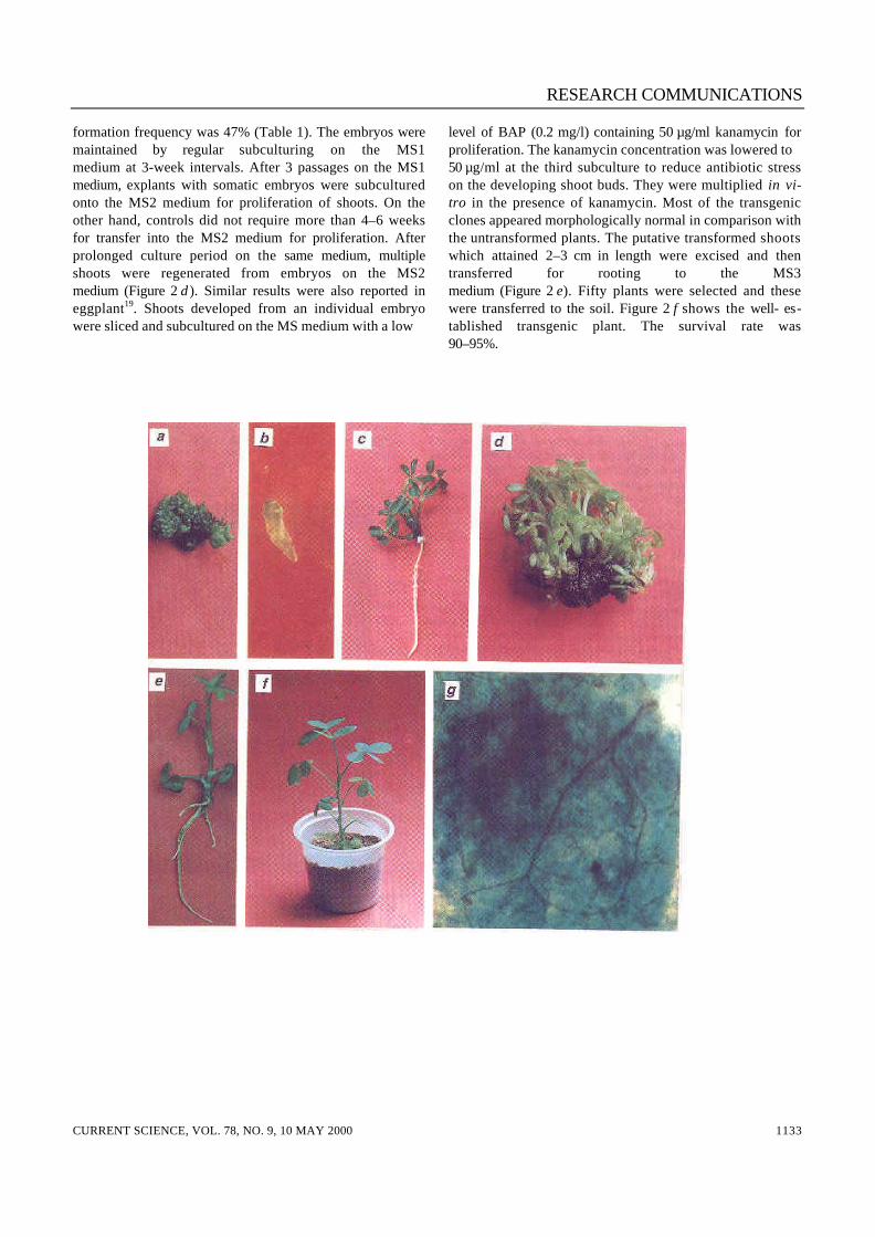
viously by Eapen and George2 and Cheng et al.3 in ground-nut. Presence of the uidA and nptII genes in the plantgenome was confirmed by PCR analysis with specific prim-ers using DNA from leaves. PCR analysis revealed that0.53 kb uidA and 0.8 kb nptII DNA fragments amplified fromgenomic DNA of all putative transgenic plants. However, nouidA and nptII PCR products were seen for untransformedcontrol plants. Figure 3 a shows that all the samples oftransgenic plants (lanes 4–9) gave the predicted size ofDNA fragment of 0.53 kb of uidA gene. No band was de-tected in the DNA sample from an untransformed controlplant (lane 3). The 0.53 kb DNA fragment was also amplifiedfrom the pBI121 plasmid as positive control (lane 2). DNAproducts with the expected size of 0.8 kb were amplifiedfrom total genomic DNAs of the putative transgenic plants(Figure 3 b, lanes 5–11). These DNA fragments were not
Table 1. Transformation frequency of putatively transformedembryos from groundnut cotyledon explants on MS medium con-
tainingNAA (0.5 mg/l), BAP (5.0 mg/l), kanamycin (75 µg/ml) and
cefotaxime (300 µg/ml)
Experiment
No. ofexplants
co-cultured
No. ofexplants
respondedFrequency
of embryos
Average no.of embryos/cotyledon
Control* 75 65 86.6±4.3 65.0±3.7Control** 86 0 00.0±0.0 00.0±0.0Expt. 1 45 17 37.7±2.6 52.0±3.4Expt. 2 36 15 41.6±3.8 48.0±3.1Expt. 3 51 24 47.0±4.2 56.0±4.7Expt. 4 39 17 43.5±3.6 32.0±2.8Expt. 5 40 15 37.5±3.3 41.0±3.9
*No co-cultivation, non-selective medium (without kanamycin).**No co-cultivation, selection medium (with 75 µg/ml of kanamy-cin).
Figure 3. PCR analysis of transgenic shoots by amplication of theuidA (GUS) and nptII genes from total plant DNA extracts. a, Detec-tion of the uidA (GUS) gene. Lane 1, DNA size marker; lane 2,Plasmid PBI121 (positive control); lane 3, Untransformed plant(negative control); lanes 4–9, Transformed plants; and b, Detectionof the nptII gene. Lane 1, DNA size marker; lanes 2, 4, Untrans-formed plants (negative control); lane 3, Plasmid pBI121 (positivecontrol); lanes 5–11, Transformed plants.
RESEARCH COMMUNICATIONS
CURRENT SCIENCE, VOL. 78, NO. 9, 10 MAY 2000 1135
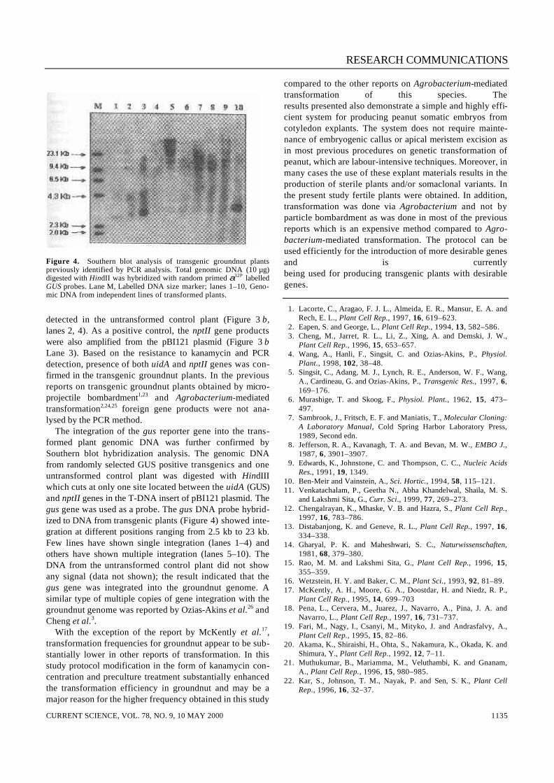
detected in the untransformed control plant (Figure 3 b,lanes 2, 4). As a positive control, the nptII gene productswere also amplified from the pBI121 plasmid (Figure 3 bLane 3). Based on the resistance to kanamycin and PCRdetection, presence of both uidA and nptII genes was con-firmed in the transgenic groundnut plants. In the previousreports on transgenic groundnut plants obtained by micro-projectile bombardment1,23 and Agrobacterium-mediatedtransformation2,24,25 foreign gene products were not ana-lysed by the PCR method. The integration of the gus reporter gene into the trans-formed plant genomic DNA was further confirmed bySouthern blot hybridization analysis. The genomic DNAfrom randomly selected GUS positive transgenics and oneuntransformed control plant was digested with HindIIIwhich cuts at only one site located between the uidA (GUS)and nptII genes in the T-DNA insert of pBI121 plasmid. Thegus gene was used as a probe. The gus DNA probe hybrid-ized to DNA from transgenic plants (Figure 4) showed inte-gration at different positions ranging from 2.5 kb to 23 kb.Few lines have shown single integration (lanes 1–4) andothers have shown multiple integration (lanes 5–10). TheDNA from the untransformed control plant did not showany signal (data not shown); the result indicated that thegus gene was integrated into the groundnut genome. Asimilar type of multiple copies of gene integration with thegroundnut genome was reported by Ozias-Akins et al.26 andCheng et al.3. With the exception of the report by McKently et al.17,transformation frequencies for groundnut appear to be sub-stantially lower in other reports of transformation. In thisstudy protocol modification in the form of kanamycin con-centration and preculture treatment substantially enhancedthe transformation efficiency in groundnut and may be amajor reason for the higher frequency obtained in this study
compared to the other reports on Agrobacterium-mediatedtransformation of this species. Theresults presented also demonstrate a simple and highly effi-cient system for producing peanut somatic embryos fromcotyledon explants. The system does not require mainte-nance of embryogenic callus or apical meristem excision asin most previous procedures on genetic transformation ofpeanut, which are labour-intensive techniques. Moreover, inmany cases the use of these explant materials results in theproduction of sterile plants and/or somaclonal variants. Inthe present study fertile plants were obtained. In addition,transformation was done via Agrobacterium and not byparticle bombardment as was done in most of the previousreports which is an expensive method compared to Agro-bacterium-mediated transformation. The protocol can beused efficiently for the introduction of more desirable genesand is currentlybeing used for producing transgenic plants with desirablegenes.
1. Lacorte, C., Aragao, F. J. L., Almeida, E. R., Mansur, E. A. andRech, E. L., Plant Cell Rep., 1997, 16, 619–623.
2. Eapen, S. and George, L., Plant Cell Rep., 1994, 13, 582–586. 3. Cheng, M., Jarret, R. L., Li, Z., Xing, A. and Demski, J. W.,
Plant Cell Rep., 1996, 15, 653–657. 4. Wang, A., Hanli, F., Singsit, C. and Ozias-Akins, P., Physiol.
Plant., 1998, 102, 38–48. 5. Singsit, C., Adang, M. J., Lynch, R. E., Anderson, W. F., Wang,
A., Cardineau, G. and Ozias-Akins, P., Transgenic Res., 1997, 6,169–176.
6. Murashige, T. and Skoog, F., Physiol. Plant., 1962, 15, 473–497.
7. Sambrook, J., Fritsch, E. F. and Maniatis, T., Molecular Cloning:A Laboratory Manual, Cold Spring Harbor Laboratory Press,1989, Second edn.
8. Jefferson, R. A., Kavanagh, T. A. and Bevan, M. W., EMBO J.,1987, 6, 3901–3907.
9. Edwards, K., Johnstone, C. and Thompson, C. C., Nucleic AcidsRes., 1991, 19, 1349.
10. Ben-Meir and Vainstein, A., Sci. Hortic., 1994, 58, 115–121. 11. Venkatachalam, P., Geetha N., Abha Khandelwal, Shaila, M. S.
and Lakshmi Sita, G., Curr. Sci., 1999, 77, 269–273. 12. Chengalrayan, K., Mhaske, V. B. and Hazra, S., Plant Cell Rep.,
1997, 16, 783–786. 13. Distabanjong, K. and Geneve, R. L., Plant Cell Rep., 1997, 16,
334–338. 14. Gharyal, P. K. and Maheshwari, S. C., Naturwissenschaften,
1981, 68, 379–380. 15. Rao, M. M. and Lakshmi Sita, G., Plant Cell Rep., 1996, 15,
355–359. 16. Wetzstein, H. Y. and Baker, C. M., Plant Sci., 1993, 92, 81–89. 17. McKently, A. H., Moore, G. A., Doostdar, H. and Niedz, R. P.,
Plant Cell Rep., 1995, 14, 699–703 18. Pena, L., Cervera, M., Juarez, J., Navarro, A., Pina, J. A. and
Navarro, L., Plant Cell Rep., 1997, 16, 731–737. 19. Fari, M., Nagy, I., Csanyi, M., Mityko, J. and Andrasfalvy, A.,
Plant Cell Rep., 1995, 15, 82–86. 20. Akama, K., Shiraishi, H., Ohta, S., Nakamura, K., Okada, K. and
Shimura, Y., Plant Cell Rep., 1992, 12, 7–11. 21. Muthukumar, B., Mariamma, M., Veluthambi, K. and Gnanam,
A., Plant Cell Rep., 1996, 15, 980–985. 22. Kar, S., Johnson, T. M., Nayak, P. and Sen, S. K., Plant Cell
Rep., 1996, 16, 32–37.
Figure 4. Southern blot analysis of transgenic groundnut plantspreviously identified by PCR analysis. Total genomic DNA (10 µg)digested with HindII was hybridized with random primed α32P labelledGUS probes. Lane M, Labelled DNA size marker; lanes 1–10, Geno-mic DNA from independent lines of transformed plants.

RESEARCH COMMUNICATIONS
CURRENT SCIENCE, VOL. 78, NO. 9, 10 MAY 20001136
23. Schnall, J. A. and Weissinger, A. K., Plant Cell Rep., 1993, 12,316–319.
24. Mansur, E. A., Lacorte, C., deFreitas, V. G., deOliveria, D. E.,Timmerman, B. and Cordeiro, A. R., Plant Sci., 1993, 89, 93–99.
25. Lacorte, C., Mansur, E. A., Timmerman, B. and Cordeiro, A. R.,Plant Cell Rep., 1991, 10, 354–357.
26. Ozias-Akins, P., Schnall, J. A., Anderson, W. F., Singsit, C.,Clemente, T. E., Adang, M. J. and Weissinger, A. K., Plant Sci.,1993, 93, 185–194.
ACKNOWLEDGEMENT. P. V. is grateful to Department of Bio-technology DBT, Govt. of India, New Delhi for the financial assis-tance in the form of post-doctoral fellowship.
Received 14 October 1999; revised accepted 29 February 2000
Blinding induces copulatory activityamong inactive males of South Indiangerbils
Biju B. Thomas and Mathew M. Oommen*Department of Zoology, University of Kerala, Kariavattom,Thiruvananthapuram 695 581, India
The effect of blinding on reproductive behavioural physiol-ogy of male South Indian gerbils (Tatera indica cuvieri)was assessed. Blinding of reproductively inactive adultmales resulted in 60% of them exhibiting sexual activity.Reproductive behavioural variables studied, such as intro-mission frequency, thrust frequency, ejaculation frequencyand postejaculatory copulation frequency were exhibited tothe level of reproductively active males. Such reproductiveactivation was also reflected in the epididymal sperm count.These studies reveal that blinding (constant darkness) mayhave a stimulatory effect in the reproduction of the tropicalrodent T. indica cuvieri.
AMONG mammals, photoperiodic cues determine the peri-ods of sexual activity and inactivity especially in temperatespecies1,2. Several investigations have been carried out toestablish the influence of photoperiodic changes on repro-duction. In hamsters, exposure to short photoperiod re-sulted in gonadal regression and loss of sexual behaviour3,4.Constant darkness (blinding) also influencesreproduction of male rodents. According to Hard and Lars-son5, though mating was not fully impaired, the intervalsbetween ejaculations were shorter for blinded male labora-tory rats. In laboratory rats whose visual function was de-stroyed neonatally, a deficit in copulatory behaviouralvariables coupled with a decreased androgen level wereobserved6. Lack of responsiveness of the reproductive sys-
tem to photoperiodic changes has been reported for malelaboratory rats7 and prairie voles8. Among tropical rodentsgonadal regression consequent to short photoperiod hasbeen reported for Indian palm squirrel Funambulus pen-nanti9. Another tropical rodent, the cane mouse (Zygodon-tomys brevicauda), however, was not responsive tovariation in photoperiod with regard to the weight of testisand seminal vesicle10. Sinhasane and Joshi11 reported that inIndian desert gerbil, Meriones hurrianae, constant lightresulted in reproductive inhibition whereas constant dark-ness was stimulatory. The South Indian gerbil Tatera indica cuvieri is a burrowdwelling tropical rodent with adult males and females occu-pying separate closed burrows12. They were found to bebreeding throughout the year13. Our field observationsshow that the animal is nocturnal with foraging habits dur-ing the night after the onset of darkness. The malereproductive behaviour pattern of this rodent has been de-scribed14. The mating pattern consists of a courtship fight-ing (upright boxing), several mountings by the male, andintromissions. Ejaculation takes place during one of theintromissions. Typically a male exhibits one to four ejacula-tions during a complete mating period, and following thefinal ejaculation the male continued the copulatory activitywithout any ejaculation. However about 50% of the maleswere found to be reproductively inactive and did not exhibitthese mating sequences15. Though photoperiodic altera-tions do influence the reproductive phenomenon of ro-dents, the response depends on the experimental scheduleand the species employed. Hence further investigations onthe male reproductive mechanism of this rodent is worthundertaking. As a first step we carried out the present studyby assessing the impact of blinding on the reproduction ofadult reproductively inactive male gerbils. Gerbils were collected regularly from the field throughoutthe year, brought and acclimatized to the laboratory condi-tions under light/dark cycle of 12 : 12 h. Adult animals (165–185 g) were selected from among a stock of healthy ones.Only those males (n = 8) which did not copulate in severalmating tests (range 4–6) were employed for blinding experi-ments. These reproductively inactive males did not showany mating behaviour other than random mounting at-tempts. Animals were housed individually in polypropylenecages provided with bedding of wood shavings and paperbits. They were maintained on laboratory rat feed and waterad libitum. Blinding (constant darkness) was effected according tothe method described by Thomas and Oommen16. For this,0.2–0.3 ml of absolute alcohol (Merck, Germany) was ad-ministered into the vitreous chamber of the eye under mildetherization. This volume ensured a rapid anduniform distribution of the alcohol within the vitreouschamber under a pressure, with the excess flowing out.Following this the iris turned white, and an antiseptic eyeointment was applied to prevent any possible infection.
*For correspondence.