Aging Suppresses Skin-Derived Circulating SDF1 to Promote ...
37
JC WS 2019/20 Brunner Tabea 1 Aging Suppresses Skin-Derived Circulating SDF1 to Promote Full- Thickness Tissue Regeneration Mailyn A. Nishiguchi, Casey A. Spencer, Denis H. Leung, Thomas H. Leung
Transcript of Aging Suppresses Skin-Derived Circulating SDF1 to Promote ...

JC WS 2019/20
Brunner Tabea
1
Aging Suppresses Skin-Derived Circulating SDF1 to Promote Full-Thickness Tissue Regeneration Mailyn A. Nishiguchi, Casey A. Spencer,
Denis H. Leung, Thomas H. Leung

Introduction
• Wound repair two biological
processes:
• Scar formation
• Tissue regeneration
• Human skin wounds invariably form scars
• Aging: slows skin re-epithelialization &
rate of wound repair
• Strength of re-epithelized skin remains
the same of any age
2
• Amphibians regenerate lost limbs
• Mammals repair injured skin with scar
formation
• Exception: liver regeneration,
pediatric traumatic digit tip
amputation, fetal skin wounds
• Skin wounds in elderly close with
thinner scar formation
• Incidence of keloid and hypertrophic
scar formation peaks in second
decade of life and decreases with age

Introduction • Surgical wounds in elderly heal with
thinner scars than wound in young
patients
• Exposure of aged mice to blood
from young mice by parabiosis
SDF1 stromal derived factor 1 • CXC motif chemokine 12 (CXCL12)
• Binds chemokine receptor type 4 and 7
• Expressed by several cell types (osteoblasten, fibroblasten, endothelial cells)
• Important in stem cell migration and proliferation
• Higher levels in young mice
• Genetic deletion of SDF enhanced tissue regeneration
3
EZH2 • Histone
methyltransferase
• Histone methylation – suppress activity of certain genes
• Change due to circulating factor in blood?
• Identification of this factor
• SDF1, generated mouse that lacked SDF1 protein in the
skin
• How does getting old shut of SDF1 production?
• EZH2 inhibition?
• Findings also true in human skin?
4 JC WS 2019/20
Brunner Tabea

Methods
5
• Injury models
• Murine excisional back wound model
• Parabiosis
• Histology and Immunohistochemistry
• Real-time RT-PCR
• Chromatin Immunoprecipitation (ChIP)
• ELISA
• Pharmacologic inhibitor experiments

Methods
Animals
• C57BL/6 female mice (18 month) national institute on
Aging
• C57BL/6 male mice (18
month) Jackson Labs
• C57BL/5 female mice (1 month)
• K5-rtTA;tetO-Cre mice
(Sarah Miller)
• Doxycycline food pellets (6gm/kg) for one week or
• Tamoxifen (1mg) daily for 5 days (intraperitoneal injection)
6

Methods
Cell culture and human skin oragnoids
• Primary human keratinocytes of different ages and gender obtained from University of
Pennsylvania
• Cells were grown in
supplemented media (50:50, keratinocytes-SFM und Medium
154)
• Nutrient deprivation: media was
removed and replaced with un-supplemented media, for 24h
• Primary human keratinocyten were seeded onto acellular human dermis, in growth media
• Organoids maintained at 37°C for 4 days prior to being wounded
7

Methods
Injury models
• Standard 2mm mechanical punch
• Create a hole in the center of each outer ear
• Ear hole diameter was measured weekly
• Ears were excluded if there were signs of wound infection, tearing of the ear, or abnormal geometric shape
Murine excisional back wound model • 6mm disposable biopsy
punch • Two circular full thickness
wounds on the dorsal back skin of mice
• Silicone wound splints were sutured with 4-0 Nylon to prevent skin contracture
• Borders were monitored by application of permanent marker
8

Methods Parabiosis
• Mirror-image incisions at the left and right flanks
• Elbow and knee joints were
sutured together
• 1 month after parabiosis
surgery – standard ear punch assay
Histology, ChIP, PCR
• Standard histology and immunostaining protocols were performed
• Chromatin Immunoprecipitation (ChIP) • Ear tissue 4%
formaldehyde, frozen in nitrogen, buffer, antibody (H3K27me3)
• PCR
9
Methods
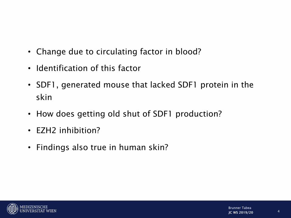
Pharmacologic inhibitor experiments • Mice treated with DZNep
received IP injections – three
times/week
• Pre-treated for 1 week
before ear punch
Knockdown experiments • Obtained Lentiviral
knockdown vectors
• Vectors used to create EZH2-knockdown keratinocytes
10

RESULTS
11

AGING PROMOTES TISSUE REGENERATION AND DECREASES SCAR FORMATION IN MOUSE EARS
12
• Young mice • Horizontally oriented
fibroblasts and glassy tickenend collagend – consistent with tissue fibrosis and scar formation
• Cartilage end plates 2mm apart – absence of cartilage regeneration
• Old mice • Normal tissue
architecture, hair follicels, sebaceous glands, subcutaneous fat
• Opposing cartilage end plates re-anastomosed (black arrow)
• 1 month old wild-type mice
• 2-mm ear holes
• Closed to significantly larger size compared with 18 month old WT mice

AGING PROMOTES TISSUE REGENERATION AND DECREASES SCAR FORMATION IN MOUSE EARS
13
• Significantly more chondrocytes expressed Ki-67

14
AGING PROMOTES TISSUE REGENERATION AND DECREASES SCAR FORMATION IN MOUSE EARS
• Injured aged miced expressed significantly lower levels of alpha smooth muscl actin
• Marker of myofibroblasts involved in scar formation
• AlphaSMA brown cells
• Immunostaining of ears fom young and aged mice

AGING PROMOTES TISSUE REGENERATION AND DECREASES SCAR FORMATION IN MOUSE EARS
15
Dark blue collagen represents normal skin, pale light blue collagen represents sacar

CIRCULATINIG FACTOR PROMOTES SCAR FORMATION
16

17
Lengthened time between Parabiosis procedure and ear injury - hole closure improved significantly Extending the time period between parabiosis procedure and ear injury improves ear hole closure in aged:aged isochronic pairs. After 12-weeks, a second distinct ear hole punch was performed and followed

A circulating factor in young blood promotes scar formation and blocks skin tissue regeneration in aged mice
18

Identification of potential circulating factors
19
Ear hole injury induces SDF1 expression in injured keratinocytes
SDF1 = green
Immunostaining from young and aged mice
Dotted lines = epidermal border
Hole is located to the right of the section

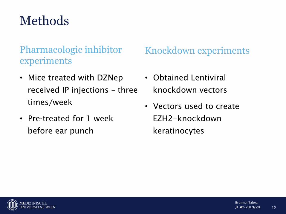
INJURED YOUNG KERATINOCYTES SECRETE SDF1 TO PROMOTE SCAR FORMATION
20
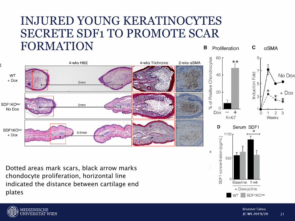
INJURED YOUNG KERATINOCYTES SECRETE SDF1 TO PROMOTE SCAR FORMATION
21
Dotted areas mark scars, black arrow marks chondocyte proliferation, horizontal line indicated the distance between cartilage end plates

22
Young doxy treated SDF1KOker mice
Aged WT mice
• Both parabiont closed ear holes to a significantly smaller size
• Cartilage regeneration and decreased scar formation

23
• Compared with non doxycycline treated SDF1KOker mice, silicone-stented back wound on docycycline treated SDF1KOker mice exhibited diminished scar formation, evidenced by return of hair follicles and reduced levels of alpha SMA
Non-doxycycline treated SDF1Koker mice
Silicone-stented back wounds on doxycycline-treated SDF1Koker mice

Fazit: circulating SDF1 in young blood originates from wounded keratinocytes to drive scar formation
24
25
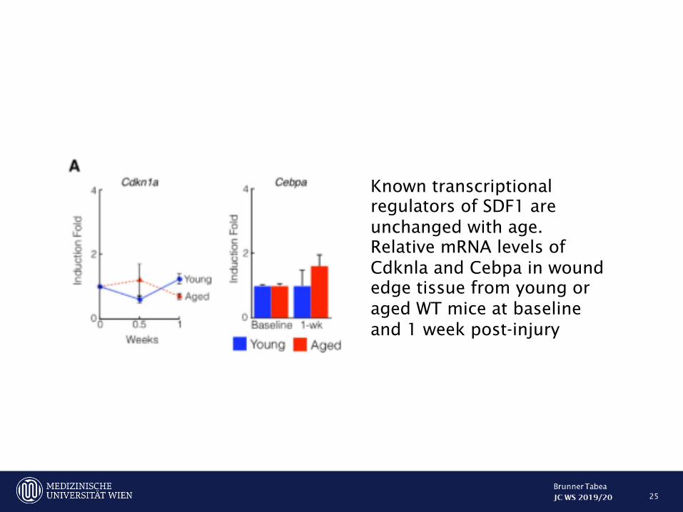
Known transcriptional regulators of SDF1 are unchanged with age. Relative mRNA levels of Cdknla and Cebpa in wound edge tissue from young or aged WT mice at baseline and 1 week post-injury

AGING SUPRESSES SDF1 ACTIVATION VIA INCREASED RECRUITMENT OF EZH2 AND H3K27m3 TO THE SDF1 GENE
26
• Wound edge tissue injured aged mice: • increased enrichment of histone H3 lysine 27 trimethylation (H3K27me)
= epigenetic marker of gene inhibition at the SDF1 promoter
• Decreased histone H3 lysine 4 trimethylation (HeK4me3) enrichment = epigenetic marker of gene activation
• Increased levels of EZH2 transcript and protein and increased EZH2 enrichment at the SDF1 promoter and transcription start
• EZH2 catalyzes the addition of methyl groups to H3K27

27
Inhibition
Activation
Shown are H3K27 me3, H3K4me3 and EZH2 chromatin immunoprecipitation of ear wound edge tissue at baseline and 1 week post-injury at 3 different locations of the SDF1 gene

28
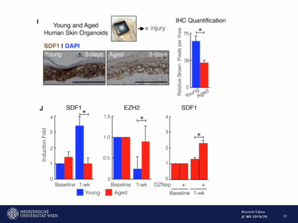
• Aged mice treated with 3-Deazaneplanocin = pharmacologic inhibitor of EZH2à restored SDF1 induction, and ear holes closed with larger sizes
Aging supresses SDF1 induction through increased recruitment of EZH2 to the SDF1 promoter
29

Human skin exhibits age-dependet EZH2 mediated SDF1 induction
30
31
Inhibition of SDF1 or EZH2 may be used to decrease scar formation in humans in potential future clinial trials
32

Discussion • Results counter current dogma that tissue function inevitably worsens with age and uncovers
potential mechanisms to explain the paradoxical effect of againg on skin tissue regeneration • Aging slows the speed of skin re-epithelialization • Young ears repair faster but a scar develops • Aged ears repair slower but to a better resolution • Overexpression of SDF1 speeds up skin re-epithelialization • Alternative interpretation is: regenerative healing is the “default” program and young age
inhibits this process • Scar formation is the dominant form of wound repair in mammals at any age • Ear and back wound models, represent different systems with different cell types involved • Keratinocyte secreted SDF1 regulates the choice between tissue regeneration and scar
formation • Increased SDF1 also drives scar formation in other organs (mouse lung, zebra fish fin) • Future studies needed to elucidate wether the precise cellular and molecular mechanisms are
conserved in other organs • Although skin specific loss of SDF1 significantly improves skin tissue regeneration, knockout
mice do not fully close injured ears holes à suggests that other factors also likely participate in tissue regeneration
33

Highlights
• Full-thickness skin wounds in aged but not young mice fully
regenerate
• Genetic deletion of SDF1 in young skin enhanced tissue
regeneration
• Aging remodels chromatin accessibility at the SDF1 gene to
inhibit SDF1 transcription
• Human skin also exhibits age-dependent SDF1 suppression
34
CXC motif chemokine 12 CXCL 12
JC WS 2019/20
Brunner Tabea

35

36

37