Ageing Research Reviews Anti-aging.pdf · the longevity dividend, i.e. the idea that extending...
27
Ageing Research Reviews 31 (2016) 9–35 Contents lists available at ScienceDirect Ageing Research Reviews jou rn al hom epage: www.elsevier.com/locate/arr Review Anti-aging pharmacology: Promises and pitfalls Alexander M. Vaiserman a,∗ , Oleh V. Lushchak b , Alexander K. Koliada a a D.F. Chebotarev Institute of Gerontology, NAMS, Kiev, Ukraine b Vasyl Stefanyk Precarpathian National University, Ivano-Frankivsk, Ukraine a r t i c l e i n f o Article history: Received 6 March 2016 Received in revised form 6 August 2016 Accepted 9 August 2016 Available online 11 August 2016 Keywords: Anti-aging drug Age-related pathology Lifespan Healthspan Antioxidant Calorie restriction mimetic a b s t r a c t Life expectancy has grown dramatically in modern times. This increase, however, is not accompanied by the same increase in healthspan. Efforts to extend healthspan through pharmacological agents targeting aging-related pathological changes are now in the spotlight of geroscience, the main idea of which is that delaying of aging is far more effective than preventing the particular chronic disorders. Currently, anti- aging pharmacology is a rapidly developing discipline. It is a preventive field of health care, as opposed to conventional medicine which focuses on treating symptoms rather than root causes of illness. A number of pharmacological agents targeting basic aging pathways (i.e., calorie restriction mimetics, autophagy inducers, senolytics etc.) are now under investigation. This review summarizes the literature related to advances, perspectives and challenges in the field of anti-aging pharmacology. © 2016 Elsevier B.V. All rights reserved. Contents 1. Introduction . . . . . . . . . . . . . . . . . . . . . . . . . . . . . . . . . . . . . . . . . . . . . . . . . . . . . . . . . . . . . . . . . . . . . . . . . . . . . . . . . . . . . . . . . . . . . . . . . . . . . . . . . . . . . . . . . . . . . . . . . . . . . . . . . . . . . . . . . . . . . 10 2. The free radical theory of aging: conceptual issues and clinical applicability . . . . . . . . . . . . . . . . . . . . . . . . . . . . . . . . . . . . . . . . . . . . . . . . . . . . . . . . . . . . . . . . . . . . . . . . . 10 2.1. ARE/Nrf2 pathway . . . . . . . . . . . . . . . . . . . . . . . . . . . . . . . . . . . . . . . . . . . . . . . . . . . . . . . . . . . . . . . . . . . . . . . . . . . . . . . . . . . . . . . . . . . . . . . . . . . . . . . . . . . . . . . . . . . . . . . . . . . . . . . 11 2.2. Life extension by antioxidants . . . . . . . . . . . . . . . . . . . . . . . . . . . . . . . . . . . . . . . . . . . . . . . . . . . . . . . . . . . . . . . . . . . . . . . . . . . . . . . . . . . . . . . . . . . . . . . . . . . . . . . . . . . . . . . . . . 11 2.3. Melatonin . . . . . . . . . . . . . . . . . . . . . . . . . . . . . . . . . . . . . . . . . . . . . . . . . . . . . . . . . . . . . . . . . . . . . . . . . . . . . . . . . . . . . . . . . . . . . . . . . . . . . . . . . . . . . . . . . . . . . . . . . . . . . . . . . . . . . . . .13 2.4. Antioxidant intake: contradictions, controversies and paradoxes . . . . . . . . . . . . . . . . . . . . . . . . . . . . . . . . . . . . . . . . . . . . . . . . . . . . . . . . . . . . . . . . . . . . . . . . . . . . . 13 3. Calorie restriction mimetics . . . . . . . . . . . . . . . . . . . . . . . . . . . . . . . . . . . . . . . . . . . . . . . . . . . . . . . . . . . . . . . . . . . . . . . . . . . . . . . . . . . . . . . . . . . . . . . . . . . . . . . . . . . . . . . . . . . . . . . . . . . . 14 3.1. Inhibitors of glycolytic metabolism . . . . . . . . . . . . . . . . . . . . . . . . . . . . . . . . . . . . . . . . . . . . . . . . . . . . . . . . . . . . . . . . . . . . . . . . . . . . . . . . . . . . . . . . . . . . . . . . . . . . . . . . . . . . . 15 3.2. Inhibitors of GH/IGF-1 pathway . . . . . . . . . . . . . . . . . . . . . . . . . . . . . . . . . . . . . . . . . . . . . . . . . . . . . . . . . . . . . . . . . . . . . . . . . . . . . . . . . . . . . . . . . . . . . . . . . . . . . . . . . . . . . . . . .15 3.3. Pharmacological inhibition of mTOR pathway . . . . . . . . . . . . . . . . . . . . . . . . . . . . . . . . . . . . . . . . . . . . . . . . . . . . . . . . . . . . . . . . . . . . . . . . . . . . . . . . . . . . . . . . . . . . . . . . . 16 3.4. Rapamycin . . . . . . . . . . . . . . . . . . . . . . . . . . . . . . . . . . . . . . . . . . . . . . . . . . . . . . . . . . . . . . . . . . . . . . . . . . . . . . . . . . . . . . . . . . . . . . . . . . . . . . . . . . . . . . . . . . . . . . . . . . . . . . . . . . . . . . . 16 3.5. AMPK pathway . . . . . . . . . . . . . . . . . . . . . . . . . . . . . . . . . . . . . . . . . . . . . . . . . . . . . . . . . . . . . . . . . . . . . . . . . . . . . . . . . . . . . . . . . . . . . . . . . . . . . . . . . . . . . . . . . . . . . . . . . . . . . . . . . . 17 3.6. Metformin . . . . . . . . . . . . . . . . . . . . . . . . . . . . . . . . . . . . . . . . . . . . . . . . . . . . . . . . . . . . . . . . . . . . . . . . . . . . . . . . . . . . . . . . . . . . . . . . . . . . . . . . . . . . . . . . . . . . . . . . . . . . . . . . . . . . . . . 18 4. Pharmacological induction of autophagy . . . . . . . . . . . . . . . . . . . . . . . . . . . . . . . . . . . . . . . . . . . . . . . . . . . . . . . . . . . . . . . . . . . . . . . . . . . . . . . . . . . . . . . . . . . . . . . . . . . . . . . . . . . . . . .18 5. Senolytic drugs . . . . . . . . . . . . . . . . . . . . . . . . . . . . . . . . . . . . . . . . . . . . . . . . . . . . . . . . . . . . . . . . . . . . . . . . . . . . . . . . . . . . . . . . . . . . . . . . . . . . . . . . . . . . . . . . . . . . . . . . . . . . . . . . . . . . . . . . . . 20 6. Telomerase activators . . . . . . . . . . . . . . . . . . . . . . . . . . . . . . . . . . . . . . . . . . . . . . . . . . . . . . . . . . . . . . . . . . . . . . . . . . . . . . . . . . . . . . . . . . . . . . . . . . . . . . . . . . . . . . . . . . . . . . . . . . . . . . . . . . .22 7. Epigenetic drugs . . . . . . . . . . . . . . . . . . . . . . . . . . . . . . . . . . . . . . . . . . . . . . . . . . . . . . . . . . . . . . . . . . . . . . . . . . . . . . . . . . . . . . . . . . . . . . . . . . . . . . . . . . . . . . . . . . . . . . . . . . . . . . . . . . . . . . . . 23 7.1. DNMT inhibitors . . . . . . . . . . . . . . . . . . . . . . . . . . . . . . . . . . . . . . . . . . . . . . . . . . . . . . . . . . . . . . . . . . . . . . . . . . . . . . . . . . . . . . . . . . . . . . . . . . . . . . . . . . . . . . . . . . . . . . . . . . . . . . . . .23 7.2. HDAC inhibitors . . . . . . . . . . . . . . . . . . . . . . . . . . . . . . . . . . . . . . . . . . . . . . . . . . . . . . . . . . . . . . . . . . . . . . . . . . . . . . . . . . . . . . . . . . . . . . . . . . . . . . . . . . . . . . . . . . . . . . . . . . . . . . . . . 23 7.3. Sirtuin-activating compounds . . . . . . . . . . . . . . . . . . . . . . . . . . . . . . . . . . . . . . . . . . . . . . . . . . . . . . . . . . . . . . . . . . . . . . . . . . . . . . . . . . . . . . . . . . . . . . . . . . . . . . . . . . . . . . . . . . 25 7.3.1. Resveratrol . . . . . . . . . . . . . . . . . . . . . . . . . . . . . . . . . . . . . . . . . . . . . . . . . . . . . . . . . . . . . . . . . . . . . . . . . . . . . . . . . . . . . . . . . . . . . . . . . . . . . . . . . . . . . . . . . . . . . . . . . . . . .25 7.3.2. Synthetic sirtuin activators . . . . . . . . . . . . . . . . . . . . . . . . . . . . . . . . . . . . . . . . . . . . . . . . . . . . . . . . . . . . . . . . . . . . . . . . . . . . . . . . . . . . . . . . . . . . . . . . . . . . . . . . . . . . 25 7.4. Micro RNA . . . . . . . . . . . . . . . . . . . . . . . . . . . . . . . . . . . . . . . . . . . . . . . . . . . . . . . . . . . . . . . . . . . . . . . . . . . . . . . . . . . . . . . . . . . . . . . . . . . . . . . . . . . . . . . . . . . . . . . . . . . . . . . . . . . . . . . 26 ∗ Corresponding author at: Institute of Gerontology, Vyshgorodskaya st. 67, Kiev 04114, Ukraine. E-mail address: [email protected] (A.M. Vaiserman). http://dx.doi.org/10.1016/j.arr.2016.08.004 1568-1637/© 2016 Elsevier B.V. All rights reserved.
Transcript of Ageing Research Reviews Anti-aging.pdf · the longevity dividend, i.e. the idea that extending...

R
A
Aa
b
a
ARRAA
KAALHAC
C
h1
Ageing Research Reviews 31 (2016) 9–35
Contents lists available at ScienceDirect
Ageing Research Reviews
jou rn al hom epage: www.elsev ier .com/ locate /ar r
eview
nti-aging pharmacology: Promises and pitfalls
lexander M. Vaisermana,∗, Oleh V. Lushchakb, Alexander K. Koliadaa
D.F. Chebotarev Institute of Gerontology, NAMS, Kiev, UkraineVasyl Stefanyk Precarpathian National University, Ivano-Frankivsk, Ukraine
r t i c l e i n f o
rticle history:eceived 6 March 2016eceived in revised form 6 August 2016ccepted 9 August 2016vailable online 11 August 2016
a b s t r a c t
Life expectancy has grown dramatically in modern times. This increase, however, is not accompanied bythe same increase in healthspan. Efforts to extend healthspan through pharmacological agents targetingaging-related pathological changes are now in the spotlight of geroscience, the main idea of which is thatdelaying of aging is far more effective than preventing the particular chronic disorders. Currently, anti-aging pharmacology is a rapidly developing discipline. It is a preventive field of health care, as opposed toconventional medicine which focuses on treating symptoms rather than root causes of illness. A number
eywords:nti-aging drugge-related pathologyifespanealthspanntioxidant
of pharmacological agents targeting basic aging pathways (i.e., calorie restriction mimetics, autophagyinducers, senolytics etc.) are now under investigation. This review summarizes the literature related toadvances, perspectives and challenges in the field of anti-aging pharmacology.
© 2016 Elsevier B.V. All rights reserved.
alorie restriction mimetic
ontents
1. Introduction . . . . . . . . . . . . . . . . . . . . . . . . . . . . . . . . . . . . . . . . . . . . . . . . . . . . . . . . . . . . . . . . . . . . . . . . . . . . . . . . . . . . . . . . . . . . . . . . . . . . . . . . . . . . . . . . . . . . . . . . . . . . . . . . . . . . . . . . . . . . . 102. The free radical theory of aging: conceptual issues and clinical applicability . . . . . . . . . . . . . . . . . . . . . . . . . . . . . . . . . . . . . . . . . . . . . . . . . . . . . . . . . . . . . . . . . . . . . . . . . 10
2.1. ARE/Nrf2 pathway . . . . . . . . . . . . . . . . . . . . . . . . . . . . . . . . . . . . . . . . . . . . . . . . . . . . . . . . . . . . . . . . . . . . . . . . . . . . . . . . . . . . . . . . . . . . . . . . . . . . . . . . . . . . . . . . . . . . . . . . . . . . . . . 112.2. Life extension by antioxidants . . . . . . . . . . . . . . . . . . . . . . . . . . . . . . . . . . . . . . . . . . . . . . . . . . . . . . . . . . . . . . . . . . . . . . . . . . . . . . . . . . . . . . . . . . . . . . . . . . . . . . . . . . . . . . . . . . 112.3. Melatonin . . . . . . . . . . . . . . . . . . . . . . . . . . . . . . . . . . . . . . . . . . . . . . . . . . . . . . . . . . . . . . . . . . . . . . . . . . . . . . . . . . . . . . . . . . . . . . . . . . . . . . . . . . . . . . . . . . . . . . . . . . . . . . . . . . . . . . . .132.4. Antioxidant intake: contradictions, controversies and paradoxes . . . . . . . . . . . . . . . . . . . . . . . . . . . . . . . . . . . . . . . . . . . . . . . . . . . . . . . . . . . . . . . . . . . . . . . . . . . . . 13
3. Calorie restriction mimetics . . . . . . . . . . . . . . . . . . . . . . . . . . . . . . . . . . . . . . . . . . . . . . . . . . . . . . . . . . . . . . . . . . . . . . . . . . . . . . . . . . . . . . . . . . . . . . . . . . . . . . . . . . . . . . . . . . . . . . . . . . . . 143.1. Inhibitors of glycolytic metabolism . . . . . . . . . . . . . . . . . . . . . . . . . . . . . . . . . . . . . . . . . . . . . . . . . . . . . . . . . . . . . . . . . . . . . . . . . . . . . . . . . . . . . . . . . . . . . . . . . . . . . . . . . . . . . 153.2. Inhibitors of GH/IGF-1 pathway. . . . . . . . . . . . . . . . . . . . . . . . . . . . . . . . . . . . . . . . . . . . . . . . . . . . . . . . . . . . . . . . . . . . . . . . . . . . . . . . . . . . . . . . . . . . . . . . . . . . . . . . . . . . . . . . .153.3. Pharmacological inhibition of mTOR pathway . . . . . . . . . . . . . . . . . . . . . . . . . . . . . . . . . . . . . . . . . . . . . . . . . . . . . . . . . . . . . . . . . . . . . . . . . . . . . . . . . . . . . . . . . . . . . . . . . 163.4. Rapamycin . . . . . . . . . . . . . . . . . . . . . . . . . . . . . . . . . . . . . . . . . . . . . . . . . . . . . . . . . . . . . . . . . . . . . . . . . . . . . . . . . . . . . . . . . . . . . . . . . . . . . . . . . . . . . . . . . . . . . . . . . . . . . . . . . . . . . . . 163.5. AMPK pathway . . . . . . . . . . . . . . . . . . . . . . . . . . . . . . . . . . . . . . . . . . . . . . . . . . . . . . . . . . . . . . . . . . . . . . . . . . . . . . . . . . . . . . . . . . . . . . . . . . . . . . . . . . . . . . . . . . . . . . . . . . . . . . . . . . 173.6. Metformin . . . . . . . . . . . . . . . . . . . . . . . . . . . . . . . . . . . . . . . . . . . . . . . . . . . . . . . . . . . . . . . . . . . . . . . . . . . . . . . . . . . . . . . . . . . . . . . . . . . . . . . . . . . . . . . . . . . . . . . . . . . . . . . . . . . . . . . 18
4. Pharmacological induction of autophagy. . . . . . . . . . . . . . . . . . . . . . . . . . . . . . . . . . . . . . . . . . . . . . . . . . . . . . . . . . . . . . . . . . . . . . . . . . . . . . . . . . . . . . . . . . . . . . . . . . . . . . . . . . . . . . .185. Senolytic drugs . . . . . . . . . . . . . . . . . . . . . . . . . . . . . . . . . . . . . . . . . . . . . . . . . . . . . . . . . . . . . . . . . . . . . . . . . . . . . . . . . . . . . . . . . . . . . . . . . . . . . . . . . . . . . . . . . . . . . . . . . . . . . . . . . . . . . . . . . . 206. Telomerase activators . . . . . . . . . . . . . . . . . . . . . . . . . . . . . . . . . . . . . . . . . . . . . . . . . . . . . . . . . . . . . . . . . . . . . . . . . . . . . . . . . . . . . . . . . . . . . . . . . . . . . . . . . . . . . . . . . . . . . . . . . . . . . . . . . . .227. Epigenetic drugs . . . . . . . . . . . . . . . . . . . . . . . . . . . . . . . . . . . . . . . . . . . . . . . . . . . . . . . . . . . . . . . . . . . . . . . . . . . . . . . . . . . . . . . . . . . . . . . . . . . . . . . . . . . . . . . . . . . . . . . . . . . . . . . . . . . . . . . . 23
7.1. DNMT inhibitors . . . . . . . . . . . . . . . . . . . . . . . . . . . . . . . . . . . . . . . . . . . . . . . . . . . . . . . . . . . . . . . . . . . . . . . . . . . . . . . . . . . . . . . . . . . . . . . . . . . . . . . . . . . . . . . . . . . . . . . . . . . . . . . . .237.2. HDAC inhibitors . . . . . . . . . . . . . . . . . . . . . . . . . . . . . . . . . . . . . . . . . . . . . . . . . . . . . . . . . . . . . . . . . . . . . . . . . . . . . . . . . . . . . . . . . . . . . . . . . . . . . . . . . . . . . . . . . . . . . . . . . . . . . . . . . 237.3. Sirtuin-activating compounds . . . . . . . . . . . . . . . . . . . . . . . . . . . . . . . . . . . . . . . . . . . . . . . . . . . . . . . . . . . . . . . . . . . . . . . . . . . . . . . . . . . . . . . . . . . . . . . . . . . . . . . . . . . . . . . . . . 25
7.3.1. Resveratrol . . . . . . . . . . . . . . . . . . . . . . . . . . . . . . . . . . . . . . . . . . . . . . . .
7.3.2. Synthetic sirtuin activators . . . . . . . . . . . . . . . . . . . . . . . . . . . . . . .
7.4. Micro RNA . . . . . . . . . . . . . . . . . . . . . . . . . . . . . . . . . . . . . . . . . . . . . . . . . . . . . . . . . . .
∗ Corresponding author at: Institute of Gerontology, Vyshgorodskaya st. 67, Kiev 04114E-mail address: [email protected] (A.M. Vaiserman).
ttp://dx.doi.org/10.1016/j.arr.2016.08.004568-1637/© 2016 Elsevier B.V. All rights reserved.
. . . . . . . . . . . . . . . . . . . . . . . . . . . . . . . . . . . . . . . . . . . . . . . . . . . . . . . . . . . . . . . . . . . . . . . . . . . . .25
. . . . . . . . . . . . . . . . . . . . . . . . . . . . . . . . . . . . . . . . . . . . . . . . . . . . . . . . . . . . . . . . . . . . . . . . . . . . . 25 . . . . . . . . . . . . . . . . . . . . . . . . . . . . . . . . . . . . . . . . . . . . . . . . . . . . . . . . . . . . . . . . . . . . . . . . . . . . 26
, Ukraine.

10 A.M. Vaiserman et al. / Ageing Research Reviews 31 (2016) 9–35
8. Conclusions . . . . . . . . . . . . . . . . . . . . . . . . . . . . . . . . . . . . . . . . . . . . . . . . . . . . . . . . . . . . . . . . . . . . . . . . . . . . . . . . . . . . . . . . . . . . . . . . . . . . . . . . . . . . . . . . . . . . . . . . . . . . . . . . . . . . . . . . . . . . . 27Conflict of interests . . . . . . . . . . . . . . . . . . . . . . . . . . . . . . . . . . . . . . . . . . . . . . . . . . . . . . . . . . . . . . . . . . . . . . . . . . . . . . . . . . . . . . . . . . . . . . . . . . . . . . . . . . . . . . . . . . . . . . . . . . . . . . . . . . . . . 28Acknowledgment . . . . . . . . . . . . . . . . . . . . . . . . . . . . . . . . . . . . . . . . . . . . . . . . . . . . . . . . . . . . . . . . . . . . . . . . . . . . . . . . . . . . . . . . . . . . . . . . . . . . . . . . . . . . . . . . . . . . . . . . . . . . . . . . . . . . . . . 28
. . . . . .
1
catdot2oaseB
saigRdscsTfcawtsioiBtihss(nnfa
a(telpgSoarc
References . . . . . . . . . . . . . . . . . . . . . . . . . . . . . . . . . . . . . . . . . . . . . . . . . . . . . . . . . . . .
. Introduction
Human longevity had dramatically increased during the pastentury when implementation of vaccination, disinfectants andntibiotics caused significant reduction in the importance of infec-ious diseases as a cause of death worldwide. The continuingecline in the mortality rates among the elderly had most probablyccurred due to the preventative factors, including improved nutri-ion, as well as exercise and reduction of smoking (de Magalhães,014; Vijg and de Grey, 2014). As a consequence, the majorityf modern societies are characterized by rapid population aging,nd the rise of age-related diseases prevalence such as cancer,troke, heart failure, as well as Alzheimer’s and Parkinson’s dis-ases becomes a considerable socio-economic challenge (Beard andloom, 2015; Harper, 2014).
Over the decades, the compression of morbidity was the basictrategy in geriatric research. According to this strategy, it isssumed that morbidity should be restricted to a short period laten life by delaying the onset of age-associated chronic patholo-ies, thereby reducing the disability burden (Seals et al., 2016).ecently, a new field focused on healthspan extension has started toevelop within the aging research. This field is referred to as ‘gero-cience’ (Kennedy et al., 2014). Extending healthspan is a majoromponent of ‘optimal longevity’, defined as living long, but withatisfactory health, well-being and life quality (Seals et al., 2016).he attempts to extend healthspan are now focused on slowing theundamental biological processes underlying aging such as mito-hondrial dysfunction, impaired proteostasis, cellular senescence,ge-associated decline of stress resistance, dysregulation of path-ays involved in growth and cellular energy sensing, deteriorating
he function and/or bioavailability of stem cells, as well as oxidativetress and inflammation (Fontana et al., 2014; Kirkland, 2013a). Thenvestigation dedicated to extending the human lifespan is a partf novel research field, ‘anti-aging medicine’ which is an increas-ngly debated topic throughout the last years (Anton et al., 2005;arazzetti and Reichlin, 2011; de Cabo et al., 2014). Traditionally,he research aimed at human life extension raises the concern thatt can result in the rise of the elderly population and, thereby, in theigh prevalence of aging-related chronic diseases. Experimentaltudies, however, have repeatedly demonstrated that life exten-ion is generally accompanied by delayed and/or reduced morbidityFontana et al., 2010). Consistent with these animal findings, cente-arian studies have demonstrated that the majority of centenariansot only exhibit the extraordinary longevity, but usually remain
ree from chronic disorders and disability until the very advancedge (Willcox et al., 2008).
The development of pharmacological treatments targetingge-related functional decline and pathological manifestationso-called ‘anti-aging drugs’) is currently in the focus of biogeron-ological research (Kennedy and Pennypacker, 2014; Verdaguert al., 2012). A number of genes playing pivotal roles in regu-ating aging and longevity have been identified over this timeeriod, and most of these genes represent promising drug tar-ets (Lindborg et al., 2015; Moskalev et al., 2014; Paul et al., 2010;hadyab and LaCroix, 2015). Another area in anti-aging pharmacol-
gy is evaluation of pharmacological potential of agents which werepproved by the U.S. Food and Drug Administration (FDA) and otheregulatory agencies for the treatment of particular pathologicalonditions related to aging. Beta-blockers, metformin, thiazoli-. . . . . . . . . . . . . . . . . . . . . . . . . . . . . . . . . . . . . . . . . . . . . . . . . . . . . . . . . . . . . . . . . . . . . . . . . . . . 28
dinediones, renin-angiotensin-aldosterone system inhibitors andseveral anti-inflammatory drugs (Seals et al., 2016) are among thesemedications. These preparations are commonly used for the treat-ment of persons with particular chronic illnesses and their safetyand efficiency was confirmed in numerous clinical trials. In severaltrials, they have also demonstrated the improvement of physio-logical functioning, well-being and health status in patients withchronic diseases (Seals et al., 2014). Such medications are not usednow in treatment of age-related decline in physiological and cogni-tive functions in the absence of clinical manifestations of particulardiseases. However, these drugs would likely be redirected to pre-venting or treating specific conditions and/or syndromes that arecommonly associated with aging.
By slowing and/or delaying the process of aging per se, it appar-ently would be possible to prevent the majority of age-relatedpathological changes rather than to overcome them one by one,which is the current approach of a disease-centered paradigmof drug development (Seals and Melov, 2014). Furthermore, pre-venting the progression of a particular age-associated chronicpathology, e.g. cardio-vascular disease, would apparently haveonly a modest impact on the population life expectancy since co-morbidity such as cancer or neurodegenerative disorders wouldto a great extent substitute the reduction of the mortality riskdue to the prevention of targeted pathology. As a consequence,the longevity dividend, i.e. the idea that extending healthspan byslowing aging can prove to be the most effective way to combatthe disabling and fatal disorders that plague us today (Olshansky,2013), may provide a great opportunity to revitalize the pipelinefor drug development.
This review is focused on current advances and perspectives inthe field of anti-aging pharmacology.
2. The free radical theory of aging: conceptual issues andclinical applicability
Historically, the free radical theory of aging has probably beenthe most influential theory of aging. This theory was first postulatedby Denham Harman in the mid of the last century (Harman, 1956).In modern literature, this concept is more commonly termed theoxidative damage theory of aging. It has been one of the earliestconcepts to try to explain the basic cause of age-related functionaldecline and accompanying pathology. This hypothesis supposesthat free radicals and other reactive oxygen species (ROS), formedin the course of mitochondrial metabolism as inevitable side prod-ucts of the oxidative energy metabolism, may result in damagedmolecules such as carbonylated proteins, lipid peroxides, and oxi-dized DNA (Liu et al., 2014a; Payne and Chinnery, 2015; Pinto andMoraes, 2015). The accumulation of this damage is believed to bethe leading cause of cellular senescence, age-dependent telomereattrition and aging-related diseases (Koliada et al., 2015). In addi-tion, it is assumed that longevity is determined by the rate at whichsuch damage occurs.
Physiologically, ROS excess in the body can be eliminated byendogenous antioxidative defense systems, including superox-ide dismutase (SOD), the enzyme detoxifying superoxide radical
(O2•−), glutathione reductase (GR) involved in the regeneration ofglutathione, as well as catalase (CAT) and glutathione peroxidase(GPX) both implicated in the detoxification of peroxides. Togetherthese antioxidant systems allow to keep the balance between

Resea
ooboelgsaBtofmtRf
pamsb(tVaoie(topoiotpscebScitmaS
2
vacrnaK
tKpm
A.M. Vaiserman et al. / Ageing
xidative and anti-oxidative processes. In the conditions of chronicxidative stress, the endogenous antioxidant systems are, however,elieved to be insufficiently effective. According to Harman’s the-ry, it is assumed that in such conditions supplementation withxogenous antioxidants such as vitamins E and C, beta-carotene,ipoic acid, coenzyme Q10, glutathione, polyphenols, phytoestro-ens, as well as several minerals including zinc, manganese andelenium can play a role in maintaining homeostasis (Pandeynd Rizvi, 2010; Bouayed and Bohn, 2010; Sadowska-Bartosz andartosz, 2014). Thereby, the proponents of this theory believe thathere is a demand for exogenous antioxidant sources to preventxidative stress, representing an imbalance in the redox state in theavor of oxidation. However, excessive doses of these substances
ay be toxic due to their pro-oxidative effects at high concentra-ions or their potential to eliminate beneficial concentrations ofOS normally occurring at physiological conditions and required
or the effective cellular functioning (Bouayed and Bohn, 2010).According to the oxidative damage theory of aging, it is sup-
osed that longevity could be extended by activation of endogenousntioxidant defense systems or by exogenous antioxidant supple-entation. On the basis of this assumption, a large number of
tudies have been attempted to delay aging and promote lifespany dietary supplementation with synthetic or natural antioxidantsSadowska-Bartosz and Bartosz, 2014; Si and Liu, 2014). The oneshat have been the most studied in this regard are vitamins C and E.itamin C (ascorbic acid) is the major hydrophilic (water-soluble)ntioxidant. It effectively reduces �-tocopheroxyl radicals and levelf low-density lipoprotein (LDL) in cell membranes, thereby restor-ng �-tocopherol and inhibiting the generation of free radicals (Nikit al., 1995). Vitamin E (�-tocopherol) is the main hydrophobicfat-soluble) antioxidant protecting cell membranes from oxida-ive damage by reaction with lipid radicals produced in the coursef the lipid peroxidation chain reaction. The dietary vitamin E sup-lementation has been shown to be associated with reduced riskf atherosclerosis through reducing oxidative stress and inhibit-ng LDL oxidation (Meydani, 2001). Currently, the developmentf novel means of antioxidants delivery into cells or construc-ion of novel antioxidant compounds is believed to open newrospects for the modulating the aging rate and healthspan exten-ion. Because mitochondria are the main source of ROS in theell, mitochondria-targeted antioxidants are suggested to be moreffective than traditional ones. Such compounds are suggestedy several authors (see, e.g., Oyewole and Birch-Machin, 2015;kulachev, 2013) to have a considerable potential since they canross the mitochondrial phospholipid bilayer and eliminate ROSn the heart of the source. Some preliminary evidence favoringhat conclusion originates from recent in vivo findings of effects of
itochondria-targeted antioxidants including plastoquinone (SkQ)nd mitoquinone mesylate (MitoQ) (Apostolova and Victor, 2015;kulachev, 2013).
.1. ARE/Nrf2 pathway
Generally, cellular protection against oxidative stress is pro-ided by both direct and indirect antioxidant activities. Directntioxidants are compounds that directly neutralize ROS. Theseompounds are themselves redox active and are consumed uponeaction with ROS. Indirect antioxidants activate the body’s ownatural detoxification systems, causing transcriptional activation of
battery of cytoprotective proteins acting catalytically (Dinkova-ostova and Talalay, 2008).
The nuclear factor-erythroid 2-related factor 2 (Nrf2) is a master
ranscriptional regulator of indirect antioxidant activity (Jung andwak, 2010). In response to oxidative stress, it regulates a battery ofrotective genes by binding to regulatory antioxidant response ele-ents (AREs) in the promoter regions of antioxidant genes, as wellrch Reviews 31 (2016) 9–35 11
as genes implicated in other cytoprotective functions (Xue et al.,2015). SOD, catalase and glutathione peroxidase are among theantioxidant genes activated by Nrf2 (Pall and Levine, 2015). TheNrf2-signaling pathway promotes cytoprotection by inducing thetranscription of more than 200 genes involved in the metabolismof toxins and drugs, protection against inflammation and oxida-tive stress, stability of proteins and removal of damaged proteinsvia proteasomal degradation or autophagy (Lewis et al., 2010). Inaddition, Nrf2 interacts with other crucial cell regulators includ-ing nuclear factor-kappa beta (NF-kappaB) and tumor suppressorprotein 53 (p53). Nrf2 is constitutively expressed in all tissues,although key detoxification organs such as liver and kidney exhibitthe highest levels (Lewis et al., 2010).
Studies in transgenic mice showed that elevated level of Nrf2activity can promote healthspan and longevity, whereas loweredlevel is associated with lifespan shortening (Pall and Levine, 2015).This apparently is because of the fact that activation of Nrf2 can pos-itively influence pathways involved in cellular senescence (Kapetaet al., 2010). Because of these properties, it is proposed to be themaster regulator of the aging process, protecting against many age-related diseases including cardiovascular disorders (Silva-Palacioset al., 2015) and neurodegeneration (Johnson and Johnson, 2015).In addition, Nrf2 plays a dual role in carcinogenesis. On theone hand, it is known to be required for systemic protectionagainst redox-mediated injury and tumorigenesis. On the otherhand, however, its constitutive activation is strongly associatedwith cancer progression and aggressiveness. Therefore, a betterunderstanding of Nrf2 regulation will be necessary to maintainthe pro-oxidant/antioxidant balance to inhibit tumor progression(Moon and Giaccia, 2015).
Several factors were identified demonstrating health-promoting properties attributed to the Nrf2 activation. Amongthese factors, there are phenolic antioxidants, carotenoids, �,�-tocopherols and tocotrienols, isothiocyanates from cabbage,broccoli and other cruciferous food, terpenes, fish oil and sulfurcompounds (Kumar et al., 2014; Pall and Levine, 2015). Taking thatinto account, the ARE/Nrf2 pathway seems to be a promising targetfor the pharmacological control of chronic diseases, such as cardio-vascular diseases as well as immunological and neurodegenerativedisorders (Al-Sawaf et al., 2015; Gao et al., 2014).
2.2. Life extension by antioxidants
In experimental studies, supplementation with antioxidant sub-stances caused controversial effects on longevity. Major findings onthe effects of exogenous antioxidants on lifespan in various exper-imental models are summarized in Table 1.
As we can see from this table, antioxidant supplementation ledin most cases to the life extension, and also to reduced levels ofaging-related oxidative stress, enhanced antioxidant enzyme activ-ity, reproductive activity and stress resistance, as well as to changedexpression of aging-associated genes. In some studies, however,the lifespan and fertility were found to be unchanged or evendiminished. In some cases, life-promoting capacity of antioxidantswas observed in superoxide dismutase (SOD)-deficient mutantswith compromised antioxidant defense or in normal animals understressful conditions, but it has not been demonstrated if wild-typeanimals reared in normal conditions have been studied. In addi-tion, antioxidants have been shown to be able to influence the meanlifespan but they have failed to show consistent effects in extendingmaximal lifespan of wild-type animals, assuming that these effectswould be caused by reduction of mortality rate via delaying or pre-
venting aging-related pathologies rather than via interfering withthe aging process per se. Moreover, it is necessary to consider thatlife extension in many animal models can be caused by mechanismsother than those attributable to aging per se. For example, antibi-
12 A.M. Vaiserman et al. / Ageing Research Reviews 31 (2016) 9–35
Table 1Summary of the effects of substances having antioxidant properties on the lifespan and associated functional traits of model organisms.
Model/Agent Longevity effect Associated functional traits ReferenceRotiferVitamin E N.D. extended pre-reproductive stage Sawada and Enesco (1984)Indolepropionamide increased lifespan N.D. Poeggeler et al. (2010)
Caenorhabditis elegansVitamin E; CoQ increased lifespan reduced superoxide anion levels Ishii et al. (2004)Vitamin E increased lifespan delayed reproduction Harrington and Harley (1988)Polydatin, a natural resveratrolglycoside
increase of mean lifespan by 31% and62% under normal and acute stressconditions, respectively
N.D. Wen et al. (2014)
Curcumin increased lifespan N.D. Liao et al. (2011)Quercetin, a major flavonoid in thehuman diet
increased lifespan increased reproductive capacity; enlarged bodysize
Surco-Laos et al. (2011)
Quercetin-3-O-glucoside increase of mean lifespan by 23% bylow concentrations, decrease oflifespan by high concentrations
N.D. Duenas et al. (2013)
Flavonoids myricetin, quercetin,kaempferol, naringenin
increased lifespan in wild-type worms N.D. Grünz et al. (2012)
Caffeic acid; rosmarinic acid increased lifespan reduced body size; altered lipid metabolism;delayed reproduction
Pietsch et al. (2011)
Flavanol catechin increased lifespan enhanced stress resistance; reduced body length Saul et al. (2009)Epigallocatechin gallate increased lifespan enhanced stress resistance Abbas and Wink (2009)Epigallocatechin gallate increased lifespan under stress
conditions; no effect under normalconditions
N.D. Zhang et al. (2009a,b)
Ferulsinaic acid increased lifespan improved resistance for heat and oxidativestresses; attenuated lipid peroxidation
Sayed (2011)
EUK-8 and EUK-134, SOD/CATmimetics
increased mean lifespan of wild-typeworms by 44% and prematurely agingworms by 67%
N.D. Melov et al. (2000)
Tyrosol, an olive oil phenol increased lifespan N.D. Canuelo et al. (2012)KPG-7, herb mixture complex increased lifespan protection against oxidative stress Moriwaki et al. (2013)EUK-8 dose-dependent reduction of lifespan dose-dependent reduction of fertility Keaney and Gems (2003)Platinum nanoparticles (nano-Pt), aSOD/CAT mimetic
increased lifespan reduced accumulation of lipofuscin and ROSinduced by paraquat
Kim et al. (2008)
N-acetyl-l-cysteine increased lifespan increased resistance to oxidative, UV and heatstresses
Oh et al. (2015)
Drosophila melanogasterLipoic acid and resveratrol increased lifespan N.D. Bauer et al. (2004)Melatonin increased lifespan N.D. Izmaylov and Obukhova (1999)EUK-8 and EUK-134;MitoQ
increased lifespan in SOD-deficientflies; no effect in wild type flies
N.D. Magwere et al. (2006)
SkQ1 increased lifespan N.D. Krementsova et al. (2012)Epitalon increase of mean lifespan by 11–16% N.D. Khavinson et al. (2000)Carnosine,S,S-Trolox-carnosine, STC
20% increase of male mean lifespan, noeffect on females; increase of malemean lifespan by 16%, female by 36%
N.D. Stvolinsky et al. (2010)
Curcumin increased lifespan enhanced SOD activity Suckow and Suckow (2006)Curcumin increased lifespan reduced oxidative stress; improved locomotion;
modulated expression of aging-related genesLee et al. (2010)
Curcumin increased lifespan increased SOD activity; reduced accumulation ofmalondialdehyde and lipid peroxidation
Shen et al. (2013)
Sesamin, a major lignan in sesame oil increase of mean lifespan up −regulation of SOD1, SOD2, CAT and Rpn11genes; attenuation of neurodegeneration
Zuo et al. (2013)
Musca domesticaEUK-8 and EUK-134 no effect on lifespan under normoxic
conditions; shortened lifespan underhyperoxic conditions
N.D. Bayne and Sohal (2002)
Mus musculusVitamin E increased median lifespan in C57BL/6
mice by 15%anti-cancer effect via inducing the P21 signalingpathway
Banks et al. (2010)
Vitamin E no effect on lifespan N.D. Morley and Trainor (2001)Vitamin E increase in mean but not maximum
lifespanlower fatal tumor incidence Blackett and Hall (1981)
Tetrahydrocurcumin; polyphenol fromgreen tea
increased survival in male C57BL/6mice
N.D. Kitani et al. (2007)
Commercial nutraceuticalcombinations
no or detrimental effect on lifespan N.D. Spindler et al. (2014)
Carboxyfullerene, SOD mimetic increased lifespan; reduced age-associated oxidative stress; rescuedage-related cognitive impairment
Quick et al. (2008)
Alpha-lipoic acid decreased lifespan in senescent proneSAMP8 mice
reduced oxidative stress and improved memory inold mice
Farr et al. (2012)
SkQ1 increased lifespan prevented age-dependent disappearance of estrouscycles; retardation of age-linked immune decline
Anisimov et al. (2011a)
In this and subsequent tables, N.D. – not determined.

Resea
obpcthwieltr
2
opietiIeopcoaiMaeos
olattate
TS
A.M. Vaiserman et al. / Ageing
tics may promote longevity in C. elegans by killing pathogenicacteria, and agents reducing egg-laying rate may increase lifes-an in Drosophila. Life-extending effects of antioxidants may be alsoaused by non-specific (hormetic) response, a dose-response rela-ionship phenomenon characterized by low-dose stimulation andigh-dose inhibition (Calabrese et al., 2015; Rattan, 2014). Note-orthy, lifespan-extending effects were so far mainly observed
n species that are evolutionarily distant from mammals. All life-xtending strategies used till now, however, did not result in similarifespan extension in mammals, where more complex control sys-ems are apparently working over the aging process, probablyequiring more elaborated interventions.
.3. Melatonin
Among the agents with antioxidant activity, melatonin standsut due to its highly pleiotropic properties. It is a natural hormoneroduced predominantly by pineal gland in the brain and released
nto circulation in a pulsatile fashion with the sharpest peaks in thearly morning hours (Claustrat and Leston, 2015). In addition tohe direct free radical scavenging and indirect antioxidant effects,t demonstrates a variety of metabolic and physiological activities.t was shown to modulate activity of antioxidant and pro-oxidantnzymes thereby reducing the oxidative damage. These actionsf melatonin are believed to be mediated by the Keap1-Nrf2-AREathway. This highly pleiotropic agent is also well known as cir-adian rhythm regulator, as well as a modulator and protectantf mitochondrial electron flux, a coregulator of metabolic sensingnd antagonist of insulin resistance, immunoregulating and anti-nflammatory molecule, and oncostatic agent (Hardeland, 2015;
anchester et al., 2015). In addition, its effects are mediated byutophagy and regulation of some signaling pathways that influ-nce energy metabolism, including insulin/IGF1, mammalian targetf rapamycin (mTOR), and Akt and phosphoinositide 3 kinase (PI3K)ignaling (Jenwitheesuk et al., 2014).
The role of melatonin in aging processes is evident from thebservation that both aging and aging-related pathologies areinked to the loss of melatonin secretion and decline of circadianmplitude of the melatonin rhythm. Recently, it was also assumedhat lifespan-promoting effects of melatonin could be attributedo activation of sirtuin 1 (SIRT1) (Ramis et al., 2015). Its potential
ging-modulating properties were studied in a range of experimen-al models including Drosophila melanogaster and mice (Anisimovt al., 2006; Hardeland, 2013).Findings from these studies are summarized in Table 2.
able 2ummary of the effects of melatonin on the lifespan and associated functional traits of m
Model Longevity effect Associate
Drosophilamelanogaster
increase of lifespan up to 19% in malesand up to 12% in females
N.D.
13.5% increase in median lifespan;33.2% increase in maximum lifespan;
increasedtemperat
27.8% decrease in mean lifespan, 25.4%increase in maximum lifespan
increaseddecreased
Mus musculus 5.4% increase in mean lifespan offemale CBA mice
decreasedtemperatswitchingmalignan
10% increase in maximum lifespan offemale SHR mice
delay of tfunction
17.4% increase in maximum lifespan ofSAMP8 mice
normalizemitochon
rch Reviews 31 (2016) 9–35 13
2.4. Antioxidant intake: contradictions, controversies andparadoxes
Findings from the research of antioxidant effects of dietary sup-plements collectively suggest that these effects could be explainedby mechanisms other than antioxidant activity per se. To maintainthe redox homeostasis, exogenous antioxidants act synergisticallywith endogenous antioxidant systems (Bouayed and Bohn, 2010).In several cases, the effects of antioxidant compounds may belinked not with their own antioxidant activity, but with induc-ing the endogenous antioxidant mechanisms; the intake of excesslevels of exogenous antioxidants may, however, suppress the syn-thesis of endogenous antioxidant enzymes (Sadowska-Bartosz andBartosz, 2014). This is an important point since a fine-tuned equi-librium among the oxidative and antioxidative processes is criticalin maintaining homeostatic stability (Bouayed and Bohn, 2010).The excessive antioxidant supplementation can result in destroyingthe delicate control mechanisms of homeostasis thereby leading todeteriorating health and life shortening (Vaiserman, 2014). More-over, since it is difficult to evaluate the quality and quantity of foodconsumed by experimental organisms, intake of high amounts ofantioxidants may likely modulate their eating behavior and therebyresult in malnutrition or even starvation (Le Bourg, 2001). If so, life-extending effects of exogenous antioxidants could be explained,at least partly, by dietary restriction (DR) known to dramaticallyslow the aging rate in various experimental models and delaythe onset of age-associated diseases, such as neurodegenerativedisease, cancer, cardiovascular disease and type 2 diabetes. Further-more, life-promoting effects of antioxidants could be a by-productof other, rather adverse, effects such as reduced level of metabolicrate, lower weight and decreased rate of development (Le Bourg,2001). This is all the more true that most substances with antiox-idant properties are multi-functional. The mechanisms which aredominant in a certain situation are dependent on the particular bio-logical conditions and those conditions may influence the kineticsand thereby the antioxidant activity. The ascorbic acid, e.g., mayact, depending on conditions, as antioxidant, but also as oxygenscavenger, metal chelating agent or reducing agent (Frankel, 1996).The active substance in turmeric, curcumin, is another example.Although it is known to demonstrate great antioxidant activity,its lifespan-extending properties can also be caused by inductionof exogenous antioxidant enzymes, or by interference with var-ious cellular signaling pathways including apoptosis-associatedand cell-cycle proteins, growth factors and their receptors, inflam-
matory cytokines, adhesion molecules, transcription factors suchas a major transcription factor for inflammatory responses, NF-kappaB, AP-1 and STAT, as well as with several longevity-associatedodel organisms.
d functional traits Reference
Izmaylov and Obukhova (1999)
resistance to paraquat and to heightenedure (36◦ C)
Bonilla et al. (2002)
eclosion rate and the locomotor activity; concentration of malondialdehyde
Terán et al. (2012)
locomotor activity and bodyure; delay of the age-related-off of estrous function; 20% increase int tumor incidence
Anisimov et al. (2001)
he age-related switching-off of estrous Anisimov et al. (2003a)
d redox and bioenergetic status of thedria and increased ATP levels
Rodríguez et al. (2008)

1 Resea
eaFvt(cepdauFc
iccihmmiRlwoitw
iauar2asiia(eiMid(Stb2
erssnEtdapil
4 A.M. Vaiserman et al. / Ageing
nzymes like mTOR, mitogen-activated protein kinase (MAPK)nd protein kinase B (Akt) (Sadowska-Bartosz and Bartosz, 2014).urthermore, antioxidants demonstrate inconsistent behavior inarying environments. Depending on their concentrations and onhe nature of neighboring molecules, they can act as pro-oxidantsCarocho and Ferreira, 2013). In addition, while high ROS con-entrations are hazardous and damage basic cellular components,vidence have been obtained that at proper concentrations theylay a key mediator role in signaling processes such as signal trans-uction from membrane receptors and control of gene expression,s well as in cellular proliferation and differentiation, cell cycle reg-lation and autophagy stimulation (Juránek et al., 2013; Mao andranke, 2013). Therefore, excessive antioxidant supplementationan lead to disruption of the pro/antioxidant balance.
Similarly to all hormesis-inducing agents (hormetins), antiox-dants act in certain concentration range, and their higheroncentrations are commonly toxic to most organisms. On theontrary, longevity hormesis may paradoxically be mediated byncreased ROS formation (Mao and Franke, 2013). According to theypothesis of ‘mitohormesis’, while high ROS level generated in theitochondria can cause cellular damage and promote aging, theiroderate level might induce an adaptive response thus improv-
ng the systemic defense mechanisms. This concept postulates thatOS act as important signaling molecules to promote health and
ifespan, and that consumption of exogenous antioxidants may beorthless or even harmful (Ristow and Schmeisser, 2014). Based
n these facts, Bouayed and Bohn (2010) stated in their review thatntake of antioxidants causes double-edged effects depending onheir concentrations: physiologic doses lead to beneficial effectshile high doses cause harmful effects.
Given these contradictions, it is not surprising that conflict-ng data were obtained on the health consequences of long-termntioxidant intake in human populations. Oxidative macromolec-lar damage is known to be significantly involved in the etiology ofge-related disorders including metabolic, cardiovascular and neu-odegenerative diseases, as well as cancer (Richardson and Schadt,014). Several authors therefore suppose that enhancement of thentioxidant defense systems may be valuable in reducing oxidativetress and risk for aging-associated pathological conditions. Indeed,t has been found in animal studies that intake of exogenous antiox-dants can reduce the progression of aging-related pathologies suchs Parkinson disease, amyotrophic lateral sclerosis and heart failureJung et al., 2001; Kawakami et al., 2009; Makino et al., 2011; Pengt al., 2005; Redout et al., 2010; van Empel et al., 2006). Apply-ng that idea to humans, however, appeared rather problematic.
ost clinical trials failed to show any clinical benefits of antiox-dant therapy for aging-related disorders such as cardiovascularisease (Kris-Etherton et al., 2004; Sahebkar et al., 2015), strokeSchürks et al., 2010), and type 2 diabetes (Sheikh-Ali et al., 2011;uksomboon et al., 2015). This controversy could likely be relatedo the excessive antioxidant intake or to insufficient antioxidantioavailability in relevant organs to reduce oxidative damage (Doss,012).
The excessive levels of exogenous antioxidants can disturbndogenous signaling mechanisms and thereby might be delete-ious (Halliwell, 2011). So, the use of multivitamins more thaneven times per week has been demonstrated to be related to aignificantly enhanced risk for prostate cancer in comparison toever users (Lawson et al., 2007). Dietary intake of vitamins C and
combined precluded the health-promoting effects of exercise inhe Ristow et al. (2009) study. A substantial increase in lung canceriagnosis has been found in those Finnish heavy smokers who were
t high risk for lung cancer and who took the beta-carotene sup-lement relative to those taking a placebo (Albanes et al., 1996). Anncreased incidence of lung cancer and elevated risk of death fromung cancer, cardiovascular disease, and any cause were found in
rch Reviews 31 (2016) 9–35
both heavy smokers and workers exposed to asbestos taking thecombination of beta carotene and vitamin A (Omenn et al., 1996).In meta-analyses of randomized controlled trials and observationalstudies in healthy and well-nourished populations, antioxidantsupplementation was repeatedly shown to be associated with dis-advantageous health consequences (Bjelakovic et al., 2014; Curtiset al., 2014; Dolara et al., 2012). Collectively, these findings demon-strate that supplementation with antioxidants can not always bebeneficial to the human health status (Bast and Haenen 2013; Liu,2014).
The contradiction lies in the fact that ROS are involved in chronichuman disorders, but intake of large doses of dietary antioxidantsleads to little or no preventive effect in most cases. Currently, thiscontradiction is commonly referred to as ‘antioxidant paradox’.Based on that, some authors suggest that manipulation of endoge-nous antioxidants level (e.g., by intake of mild pro-oxidants) islikely a more appropriate approach for prevention and treatmentof ROS-mediated diseases than the consumption of large doses ofdietary antioxidants (Halliwell, 2013). While benefits of dietaryantioxidant intake seem to be clear in case of enhanced oxidativestress and endogenous antioxidant deficiency, further research isrequired to determine the potential risks and benefits linked to thesupplement of antioxidants by healthy persons.
In summary, it should be noted that a body of recent evi-dence raised serious doubts whether the oxidative damage theoryremains still valuable for understanding the processes underlyingaging and longevity (Gladyshev, 2014; Liochev, 2015; Speakmanand Selman, 2011). One example is the research on mice withknockout of antioxidant genes. These mice exhibited expected riseof oxidative damage but any impact on longevity (Zhang et al.,2009a,b). Some authors claim that the oxidative damage theoryof aging is fundamentally wrong, though this has been a topic ofintense debate within the field over the past decade. This theory,however, still has many adherents and needs to be decisively veri-fied in further studies.
3. Calorie restriction mimetics
Current doubts about the efficiency of antioxidant consumptionled to a growing interest in alternative healthspan-promoting inter-ventions. Among them, DR- or, more specifically, calorie restriction(CR)-based strategies are likely the most hopeful now. CR refers toa reduced calorie intake without malnutrition in animals normallyfed ad libitum (Masoro, 2005). Generally, CR is a diet providingall essential nutrients, vitamins and minerals but having 30–70%reduced amount of calories.
Presently, DR is the most well-established experimental pro-cedure for life extension, although some authors suggest that therestriction of specific dietary components such as protein and par-ticular amino acids like methionine can play a crucial role (Fontanaand Partridge, 2015). The ability of CR to promote lifespan and slowaging have attracted significant scientific attention since the pio-neering work conducted by McCay and colleagues eighty years ago(McCay et al., 1935). In this research, it has been shown that ratssubjected to 40% CR had up to 50% longer median and maximumlifespans than rats fed a standard diet. Over the years, a numberof studies have shown that DR can retard aging and extend lifes-pan in various experimental models including yeast, worms, insectsand rodents (Ingram and Roth, 2015; Selman, 2014; Testa et al.,2014). More recently, evidence has been obtained that DR can leadto a delayed onset of age-associated pathologies in non-human
primates such as rhesus monkeys (Colman et al., 2009; Kemnitz,2011; Mattison et al., 2012). Specifically, DR leads to reduced lev-els of body fat, slower rate of aging-related muscle loss, reducedincidence of cardio-vascular disease, type 2 diabetes and cancer,
Resea
aakmetF
aimaswhactgapct
tnCsa2(mnDdktema
esbiaaepde
tps
3
awingsp
A.M. Vaiserman et al. / Ageing
s well as improved glucose tolerance and insulin sensitivity. Inddition, DR resulted in reducing the age-related mortality of mon-eys (Colman et al., 2014). Accumulating data demonstrate thatoderate DR may reduce the risk of cancer, and also cause pow-
rful protective effect against inflammation, hypertension, obesity,ype 2 diabetes and cardio-vascular disease in humans (Cava andontana, 2013; Holloszy and Fontana, 2007).
However, although DR was shown to clearly reduce risk factorsssociated with human aging, it is still a subject of debate whethert may increase the human life expectancy. One challenge of the DR
odel is that the DR-mediated lifespan extension is not universalnd can not even be shared between various strains of the samepecies. In addition, some authors suggest that control animalshich fed ad libitum are overweight and tend to have associatedealth problems, and thus they could not be appropriate controlnimals for longevity studies. Increasing evidence suggest that DRan promote lifespan of genotypes that develop energy imbalancehrough the ad libitum feeding (Sohal and Forster, 2014). In suchroups, DR results in decrease of body temperature, metabolic rate,nd oxidant production and retardation of the aging-associatedro-oxidative shift of the redox state. Nevertheless, despite thesehallenges and limitations, the DR concept continues to be one ofhe leading paradigms in modern gerontology (Ingram et al., 2006).
The beneficial effects of DR were found to be mediated byhe same pathways which are also involved in regulating immu-ity, tissue repair, metabolism, thermoregulation and appetite (Leouteur et al., 2012). These effects of DR likely result not from pas-ive metabolic changes, but rather from highly regulated processesssociated with activation of specific effectors (Mouchiroud et al.,010). Adenosine monophosphate (AMP)-activated protein kinaseAMPK), the insulin/insulin-like growth factor signaling (IIS) and
TOR pathways, as well as sirtuins, especially SIRT1 are the basicutrient-sensing pathways related to the control of the lifespan byR. These pathways are known to be crucial regulators of mitochon-rial function, cell growth, proliferation and autophagy, and are alsonown to be regulated by crosstalk with one another. Importantly,he long-term DR was shown to be necessary to produce beneficialffects on healthspan and lifespan. It is apparently problematic forost modern people. Thus, such interventions may not be readily
pplicable.The doubt on the applicability of DR-based treatments has gen-
rated the growing interest in developing alternative treatmenttrategy which can provide pro-healthspan and pro-longevity DRenefits without the substantial reduction in the long-term food
ntake. Compounds used in this treatment strategy are referred tos CR mimetics. Basic characteristic properties of CR mimetics ares follows: (1) inducing the hormonal, metabolic and physiologicalffects similar to those of CR; (2) activation of the stress responseathways like the CR; (3) lifespan extension and reducing the inci-ence or delaying the onset of age-associated disorders (Ingramt al., 2006).
The main signaling pathways mediating the CR effects andhe most promising pharmacological substances modulating theseathways and thus mimicking the CR effects are reviewed in theubsections below.
.1. Inhibitors of glycolytic metabolism
Hyperinsulinemia and hyperglycemia are widespread in oldge. Age-associated metabolic disorders such as type 2 diabetes,hich are accompanied by disturbance in glucose homeostasis and
nsulin signaling, are known to produce various aging-like phe-
otypes. Untreated diabetes patients commonly have enhancedlucose levels and they are more susceptible to chronic disordersuch as vascular and microvascular damage, heart disease, kidneyroblems, impaired wound healing and cataracts, and neurologicalrch Reviews 31 (2016) 9–35 15
degeneration, which typically occur in non-diabetics much later inlife (Anisimov, 2013; Bonomini et al., 2015).
Oxidative stress induced by hyperglycemia is known to promotethe generation of advanced glycosylation end products (AGEs),thus substantially accelerating the aging rate (Anisimov, 2013). Theaccumulation of AGEs has been found to be enhanced in personswith type 2 diabetes and this parameter was proposed as a reliablebiomarker for both aging rate and ‘metabolic memory’ (the persis-tence of adverse effects of prior hyperglycaemia on the onset andprogression of diabetic vasculopathies even after the normaliza-tion of glucose levels) (Rajaobelina et al., 2015; Sato et al., 2001).Furthermore, hyperinsulinemia, a marker of insulin resistance thatalso shown to increase with age is a substantial causal factor in thecancer etiology (Gristina et al., 2015).
In a number of studies, CR has been demonstrated to reducethe levels of both glucose and insulin, and to improve the insulinsensitivity (Anisimov, 2003). Therefore, the inhibition of enzymesof the glycolytic pathway is believed to be a promising strategy toproduce CR-like effects (Longo et al., 2015). In this pharmacologicalcategory, an analog of glucose 2-deoxy-d-glucose is the most stud-ied candidate agent (Minor et al., 2009). In the course of glycolysis,2-deoxy-d-glucose is converted by hexokinase to 2-deoxyglucose6-phosphate that may in turn inhibit phosphoglucose isomeraseand imped processing of glucose to fructose 6-phosphate. Owingto these properties, the capability of 2-deoxy-d-glucose to affectglycolysis has been investigated for several decades in a variety ofmodel organisms from bacteria to mammals. In these studies, thetherapeutic benefits of this preparation for treating viral infection,epilepsy and cancer have been revealed (Xi et al., 2014). It is con-sidered to be especially promising in anti-cancer therapy. Indeed,tumor cells exhibit elevated glycolysis and use this metabolic path-way for generating ATP as a primary source of the energy supply.The enhanced dependence of malignant cells on glycolytic path-way for the generation of ATP could provide a mechanistic basisfor the development of novel therapeutic strategies to preferen-tially kill cancer cells by the pharmacological inhibition of glycolysis(Ganapathy-Kanniappan and Geschwind, 2013; Ingram and Roth,2010).
2-Deoxy-d-glucose has also been found to reproduce someaspects of the CR phenotype including reduced levels of circulat-ing insulin and body temperature in rodents, whereas its apparenttoxic effect has been demonstrated until now only in one chronicfeeding research (Minor et al., 2009). In this study, CR-like phe-notype has been revealed in rats treated with this medication,such as reduced dietary intake and decreased weight gain. Thesechanges have been accompanied, however, by cardiac vacuoliza-tion and increased mortality in treated rats. The potential toxicityof 2-deoxy-d-glucose leads to serious doubts in the CR-based ther-apeutic application of this preparation.
3.2. Inhibitors of GH/IGF-1 pathway
Among the signaling pathways linked to DR, insulin/insulin-likegrowth factor-1 (IGF-1) pathway is known to play a crucial role inthe lifespan regulation. The bulk of the research findings suggeststhat CR, as well as intermittent fasting and protein or amino acidrestriction, all can extend the mammalian lifespan via modulatingthe IGF-1 signaling (Taormina and Mirisola, 2015). Several poly-morphic variants of the IGF-1 receptor gene have been found to belinked to the human longevity (Pawlikowska et al., 2009), in partic-ular, to the exceptional lifespan of centenarians (Suh et al., 2008).In addition, low circulating IGF-I bioactivity was shown to be asso-
ciated with longevity of the exceptionally long-lived individuals(Milman et al., 2014), and their offspring (Vitale et al., 2012). Thelong-lived growth hormone (GH) receptor knockout Laron dwarfmice (GHR−/−) have been found to be insulin sensitive despite
1 Resea
olIIsAwmh
fG(aGsst
aacGradaaelnppa
ipphisPHn
isoafse
3
na(edatcac
6 A.M. Vaiserman et al. / Ageing
besity, and were also shown to have substantially reduced IGF-1evels and decreased risks of cancer and diabetes (Zhou et al., 1997;keno et al., 2009). The Laron syndrome patients, who have reducedGF1 levels despite normal or elevated levels of GH, have beenimilarly found to be protected from cancer and diabetes (Guevara-guirre et al., 2011; Steuerman et al., 2011). As a consequence, itas suggested that treatments targeted at reducing IGF-1 levelsay be a promising pharmacological strategy for the extension of
ealthspan.Recently, inhibitors of the GH/IGF-1 axis have been proposed
or the treatment of patients with acromegaly, a disease caused byH/IGF-I hypersecretion and associated with a high mortality rate
Giustina et al., 2014). Such medications include, e.g., somatostatinnalogues which may reduce the GH serum levels via suppressingH secretion by pituitary somatotrophs, thus resulting in reducederum IGF-1 levels. Such therapy, however, can potentially causeevere unfavorable effects since such agents may suppress secre-ion of some hormones including insulin.
Indeed, the adverse health consequences such as diarrhoea,norexia and gallstones, were revealed in 20–50% of treatedcromegalic patients. Another medicine from that pharmacologicallass is the GH receptor antagonist pegvisomant known to inhibitH secretion and GH action by binding to and blocking the GH
eceptor (Kopchick et al., 2002; Kopchick, 2016). In patients withcromegaly treated with pegvisomant, a significant (up to 90%)ose-dependent decrease of IGF-1 levels was revealed (van der Lelynd Kopchick, 2006). Pegvisomant is believed to be a promisinggent for healthspan and lifespan extension because it led to ben-ficial effects on the glucose metabolism by lowering serum IGF-1evels and increasing the insulin sensitivity. However, even thougho harmful effects have been reported in the long-term research ofegvisomant (van der Lely et al., 2012), further research is needed torove its safety. Moreover, this medication requires daily injectionsnd is very expensive (Higham et al., 2009).
Modulating the IGF-1 availability could be another way tonhibit IGF-1 action. For that, pharmaceuticals modulating theregnancy-associated plasma protein-A (PAPP-A), a zinc metallo-roteinase known to enhance the local bioavailability of the IGFs,ave been developed. Such medications are believed to be promis-
ng as PAPP-A knockout mice have been previously found to haveubstantially extended healthspan, suggesting an important role ofAPP-A in aging and associated disease risk (Conover, 2010, 2013).ealthspan-promoting effects of these medications, however, haveot been examined yet and are the subject of further research.
Recently, a high life-extending potential of modulating thensulin/IGF-1 pathway was reported by Slack et al. (2015). In thistudy, a role for Ras-Erk-ETS pathway, implicated in the effectsf reduced insulin/IGF-1 signaling, was identified. Specifically, thedult-onset supplementation with an FDA-approved medicationor the treatment of melanoma, trametinib, which is a highlypecific inhibitor of Ras-Erk-ETS signaling, caused significant lifextension in Drosophila.
.3. Pharmacological inhibition of mTOR pathway
The mammalian target of rapamycin (mTOR) pathway is anotherutrient-sensing pathway which can play a role in mediatingdvantageous effects of CR on the aging rate and longevityEhninger et al., 2014). mTOR is a serine/threonine protein kinasevolutionarily conserved from yeast to human in the catalyticomain and known to be implicated in cell growth, metabolismnd regulation of energy homeostasis (Johnson et al., 2015). Within
he cell, it exists as a part of two multiprotein complexes, mTORomplex 1 (mTORC1) and mTOR complex 2 (mTORC2) (Laplantend Sabatini, 2012). The mTORC1 regulates important cellular pro-esses including transcription, translation and autophagy (Inokirch Reviews 31 (2016) 9–35
et al., 2012). The mTORC1 was also found to be the main mTORcomplex involved in lifespan regulation (Kaeberlein, 2014). Inthe presence of sufficient amounts of nutrients, growth factorsand energy availability, mTOR switches off stress resistance andautophagy, and switches on cell growth pathways, including trans-lation and ribosomal biogenesis (Kapahi et al., 2010). Since it hasbeen demonstrated in lower animal models that decreasing proteinsynthesis can increase lifespan (Hipkiss, 2007), inhibition of proteinsynthesis by reducing the activity of mTOR pathway can likely beinvolved in life-extending effects across species. In several studies,it was shown that mTOR can be activated by single amino acids. Itmay provide a causal mechanism for recent findings that decreasein the proportion of protein in the diet, rather than CR per se, cancause lifespan-promoting effects in various experimental models(Lee, 2015; Solon-Biet et al., 2014, 2015). In addition, mTOR sig-naling was shown to play a central role in mediating aging andassociated metabolic disorders, including obesity, type 2 diabetesand cancer (Albert and Hall, 2015). Therefore, mTOR is supposed tobe a leading target for pharmacological intervention to modulatethe nutrient response pathways and to decelerate the aging pro-cess. Inhibition of this pathway has been repeatedly demonstratedto confer protection against aging-associated pathological condi-tions and to extend lifespan in different model organisms (Johnsonet al., 2013). Genetic inhibition of mTOR signaling caused life exten-sion in worms (Jia et al., 2004; Vellai et al., 2003), fruit flies (Kapahiet al., 2004), and mice (Lamming et al., 2012). Recent gene expres-sion analysis showed that mTOR pathway is strongly associatedwith human health and longevity (Passtoors et al., 2013).
3.4. Rapamycin
Rapamycin (also known as sirolimus), a natural compound pro-duced by bacteria Streptomyces hygroscopicus that was first foundin an Easter Island soil sample around 1970, is the most widely usedinhibitor of mTOR. It has been initially developed as an antifungalagent but has subsequently been found to have substantial regula-tory effects on crucial biological processes such as cellular growth,proliferation, and inflammation via its inhibitory action on mTOR(Lamming et al., 2013). Since rapamycin was shown to inhibit theimmune response, it was subsequently used in immunosuppres-sive therapy to prevent graft rejection and to treat autoimmunedisorders (Ingle et al., 2000). Currently, rapamycin and its analogs(rapalogs), e.g., temsirolimus and everolimus, are suggested to beamong the most promising anti-aging drugs (Blagosklonny, 2007).Rapamycin therapy has also been revealed to delay aging-relatedchanges in mice, including tendon stiffening, accumulation of sub-cellular alterations in the myocardium, endometrial hyperplasia,liver degeneration, and decline in physical activity (Wilkinson et al.,2012). Furthermore, it proved to be efficient against aging-linkedpathologies including cognitive decline, retinopathy, neurode-generative disorders, cardiac hypertrophy, and loss of stem cellfunction (Chen et al., 2009; Halloran et al., 2012; Kolosova et al.,2012; Shioi et al., 2003; Spilman et al., 2010).
Life-extending properties of rapamycin have been examined inmany studies from yeast to mammals. A summary of these studiesis presented in Table 3.
The potential mechanisms of rapamycin-induced life extensionare assumed to include antitumor effect (Blagosklonny, 2012; Saranet al., 2015), autophagy induction (Perluigi et al., 2015), stem cellguidance (Maiese, 2015), as well as immune-modulating and anti-inflammatory activity (Araki et al., 2011). In addition, these effectscan be mediated by the reduction of the rate of protein synthesis,
because the decrease of the overall translation rate may preventthe accumulation of damaged or misfolded proteins which mightin turn affect lifespan (Hipkiss, 2007). The mTOR was also shown tobe negatively regulated by other nutrient-sensing pathways such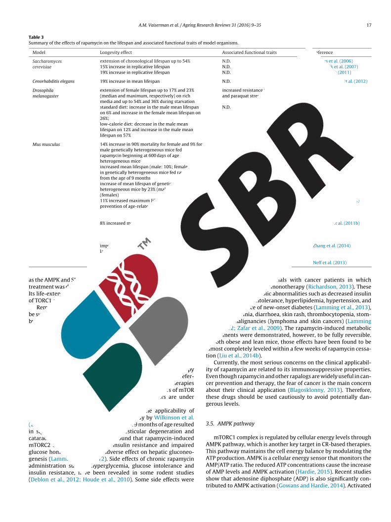
A.M. Vaiserman et al. / Ageing Research Reviews 31 (2016) 9–35 17
Table 3Summary of the effects of rapamycin on the lifespan and associated functional traits of model organisms.
Model Longevity effect Associated functional traits Reference
Saccharomycescerevisiae
extension of chronological lifespan up to 54% N.D. Powers et al. (2006)15% increase in replicative lifespan N.D. Medvedik et al. (2007)19% increase in replicative lifespan N.D. Ha and Huh (2011)
Cenorhabditis elegans 19% increase in mean lifespan N.D. Robida-Stubbs et al. (2012)
Drosophilamelanogaster
extension of female lifespan up to 17% and 23%(median and maximum, respectively) on richmedia and up to 54% and 36% during starvation
increased resistance to starvationand paraquat stress
Bjedov et al. (2010)
standard diet: increase in the male mean lifespanon 6% and increase in the female mean lifespan on26%;low-calorie diet: decrease in the male meanlifespan on 12% and increase in the male meanlifespan on 57%
N.D. Sun et al. (2012)
Mus musculus 14% increase in 90% mortality for female and 9% formale genetically heterogeneous mice fedrapamycin beginning at 600 days of ageheterogeneous mice
N.D. Harrison et al. (2009)
increased mean lifespan (male: 10%; female: 18%)in genetically heterogeneous mice fed rapamycinfrom the age of 9 months
attenuating age-associated declinein spontaneous activity in malesbut not in females
Miller et al. (2011)
increase of mean lifespan of geneticallyheterogeneous mice by 23% (males) to 26%(females)
N.D. Miller et al. (2014)
11% increased maximum lifespan in females,prevention of age-related weight gain
decreased rate of aging andsuppressed carcinogenesis intransgenic HER-2/neucancer-pronemice
Anisimov et al. (2010a)
8% increased maximum lifespan, inhibited age-related weight gain,decreased aging rate, and delayedspontaneous cancer in inbredfemales
Anisimov et al. (2011b)
improved survival of C57BL/6 mice after the reduced total and resting Zhang et al. (2014)
e
atIo
bbrwetdbesatpd
r(icmggai(
late-life rapamycin feeding
increased median lifespan in male C57BL/6J mic
s the AMPK and SIRT1 pathways (Cetrullo et al., 2015). Rapamycinreatment was demonstrated to inhibit both mTORC1 and mTORC2.ts life-extending effects, however, are mediated by the inhibitionf TORC1 activity only.
Remarkably, the rapamycin treatment starting in later life maye sufficient to extend lifespan; similar benefits were also achievedy transient late-life treatments (Kaeberlein, 2014). In mice studies,apamycin supplementation initiated late in life (20 months of age)as nearly as efficient as if beginning it at 9 months of age (Harrison
t al., 2009; Miller et al., 2011). Late-life transient rapamycinreatment was also demonstrated to reverse the age-linked heartysfunction, and also caused the beneficial motor, skeletal andehavioral changes in 24-month-old female C57BL/6J mice (Flynnt al., 2013). The benefits of a mid- or late-life rapamycin therapyeem particularly promising since such interventions are prefer-ble when considering potential translation of anti-aging therapieso humans (Kaeberlein, 2014). Currently, some inhibitors of mTORathway are already clinically approved, and others are underevelopment.
There are some concerns, however, on the applicability ofapamycin in anti-aging therapy. In the study by Wilkinson et al.2012), treating with rapamycin starting at 9 months of age resultedn significantly higher incidence of testicular degeneration andataracts in mice. It has been also found that rapamycin-inducedTORC2 suppression can cause insulin resistance and impaired
lucose homeostasis through adverse effect on hepatic gluconeo-enesis (Lamming et al., 2012). Side effects of chronic rapamycin
dministration such as hyperglycemia, glucose intolerance andnsulin resistance, have been revealed in some rodent studiesDeblon et al., 2012; Houde et al., 2010). Some side effects weremetabolic rate during the light(inactive) phaseN.D. Neff et al. (2013)
also observed in clinical trials with cancer patients in whichrapamycin was used as a monotherapy (Richardson, 2013). Theseeffects included metabolic abnormalities such as decreased insulinsensitivity, glucose intolerance, hyperlipidemia, hypertension, andenhanced incidence of new-onset diabetes (Lamming et al., 2013),as well as anaemia, diarrhoea, skin rash, thrombocytopenia, stom-atitis, and malignancies (lymphoma and skin cancers) (Lamminget al., 2012; Zafar et al., 2009). The rapamycin-induced metabolicimpairments were demonstrated, however, to be fully reversible.In both obese and lean mice, those effects have been found to bealmost completely leveled within a few weeks of rapamycin cessa-tion (Liu et al., 2014b).
Currently, the most serious concerns on the clinical applicabil-ity of rapamycin are related to its immunosuppressive properties.Even though rapamycin and other rapalogs are widely useful in can-cer prevention and therapy, the fear of cancer is the main concernabout their clinical application (Blagosklonny, 2013). Therefore,these drugs should be used cautiously to avoid potentially dan-gerous levels.
3.5. AMPK pathway
mTORC1 complex is regulated by cellular energy levels throughAMPK pathway, which is another key target in CR-based therapies.This pathway maintains the cell energy balance by modulating theATP production. AMPK is a cellular energy sensor that monitors the
AMP/ATP ratio. The reduced ATP concentrations cause the increaseof AMP levels and AMPK activation (Hardie, 2015). Recent studiesshow that adenosine diphosphate (ADP) is also significantly con-tributed to AMPK activation (Gowans and Hardie, 2014). Activated
1 Resea
Aca2gsmei2aalwiiecp(mso
m(hrwetwpSnptpl
3
hFdemdacgMtamctfsh
ittc
8 A.M. Vaiserman et al. / Ageing
MPK increases the cellular ATP production and decreases the ATPonsumption by shutting down energy-consuming processes suchs the mTOR-dependent protein translation (Towler and Hardie,007). Upon activating, AMPK switches on catabolic pathways thatenerate ATP, both in a short time frame by promoting glycoly-is and fatty acid oxidation and in a long time frame by enhancingitochondrial content and using mitochondrial substrates as an
nergy source, and switches off ATP-consuming processes includ-ng cell growth, biosynthesis and proliferation (Cantó and Auwerx,010). All these processes occur to respond to the metabolic stresst the cellular level. Cytokines and hormones such as leptin, insulinnd adiponectin interact with this system, and these processesikely play an important role in maintaining energy balance at the
hole-body level. AMPK responds to energy stress by suppress-ng cellular growth and biosynthetic processes, partially throughnhibiting the mTORC1 pathway (Gwinn et al., 2008; Hindupurt al., 2015). Moreover, AMPK activation results in enhanced glu-ose uptake in skeletal muscles, and in decreased hepatic glucoseroduction and increased fatty acid oxidation in various tissuesRuderman et al., 2013). Consistent data indicate that AMPK inhibits
TOR through the phosphorylation and activation of the tuberousclerosis complex (TSC), and also through direct phosphorylationf the RAPTOR subunit of mTORC1 (Potter et al., 2010).
Recently, the tumor suppressor LKB1 was identified as theajor upstream kinase in the AMPK cascade in mammalian cells
Hardie et al., 2012). So, AMPK is a key player in control of energyomeostasis at both cellular and whole-body levels through theegulation of mitochondrial biogenesis and insulin sensitivity, asell as in the processes of autophagy and stress resistance (Ha
t al., 2015). Therefore, it seems promising in development of novelreatments for obesity, type 2 diabetes and metabolic syndrome. Itas also demonstrated to be the key mediator and integrator ofrocesses linking metabolism to longevity (Burkewitz et al., 2014;alminen and Kaarniranta, 2012). Because changes in AMPK sig-aling result in modulating key anti-aging pathways such as theeroxisome proliferator-activated receptor (PPAR) gamma coac-ivator a (PGC-1a), SIRT1 and TOR (Verdaguer et al., 2012), thisathway is supposed to be a promising drug target for health- and
ifespan extension.
.6. Metformin
Several drugs demonstrating the AMPK-activating propertiesave been recently developed. These substances include someDA-approved drugs including biguanides, resveratrol, thiazoli-inediones, salicylates, glucagon-like peptide-1 receptor agoniststc (Coughlan et al., 2014). Among them, antidiabetic biguanideetformin (1,1-dimethylbiguanide hydrochloride), a medication
erived from French lilac, is currently in the focus of researchctivity. Metformin is known to specifically inhibit the hepatic glu-oneogenesis without enhancing insulin secretion, inducing weightain and posing a risk of hypoglycaemia (Madiraju et al., 2014;oreira, 2014). Therefore, it is considered as one of the most effec-
ive preparation for treating type 2 diabetes. It may also act as CR mimetic since it can decrease hepatic glucose production,ostly through mild inhibition of the mitochondrial respiratory-
hain complex 1 (Viollet et al., 2012). In experimental studies,reatment with metformin and other biguanides, including phen-ormin and buformin, caused life extension in many animal modelsuch as nematode worms, fruit flies and rodents, although findingsave been contradictory (Table 4).
Evidence for beneficial effects of metformin has been provided
n human studies as well. For example, significant improvement inotal cholesterol, body mass index, low-density lipoprotein choles-erol, and non-high density lipoprotein cholesterol was observed inhildren with metabolic syndrome treated with metformin (Luongrch Reviews 31 (2016) 9–35
et al., 2015). In both diabetic and non-diabetic patients, anti-cancereffect of metformin has been also revealed (Coperchini et al., 2015).Metformin is assumed to directly affect tumor cells, primarily influ-encing the mitochondrial respiratory chain and, thereby, activatingAMPK.
It is far from clear, however, to what extent the beneficialeffects of metformin are mediated by activation of AMPK. AMPK-independent pathways can also be involved in both beneficialand unfavorable effects of this drug (Zheng et al., 2015). Thesepathways are believed to include suppression of glucagon signal-ing, inhibition of mitochondrial shuttles, induction of autophagyand mitochondrial stress, attenuation of inflammasome activation,reduction of terminal endoplasmic reticulum stress and changesin intestinal microbiota (Hur and Lee, 2015). Remarkably, microar-ray analysis showed marked similarity between gene expressionpatterns induced by both metformin and CR (Dhahbi et al., 2005).
By arguing that CR and its mimetics may promote their bene-ficial effects through various signaling pathways, Ingram and Roth(2015) have proposed to apply the “cocktails” of CR mimetics tocontemporaneously affect multiple pathways. One example of suchtreatment strategy would be the usage of the metformin to blockthe glucose dysmetabolism caused by rapamycin treatment viainhibiting the gluconeogenesis by downregulation of expression ofglucose-6-phosphatase and phosphoenolpyruvate carboxykinase(Mendelsohn and Larrick, 2012). This combined approach wouldlikely be more secure and provide a more balanced therapeuticstrategy, especially in healthy subjects.
4. Pharmacological induction of autophagy
Autophagy is a process by which defective or damaged cellu-lar components are transferred into the lysosomes for degradationand recycling; then they may be used for protein synthesis andcellular repair (Gelino and Hansen, 2012). Autophagy is com-monly classified into three major categories depending on themode of delivery of cellular material to lysosomes: macroau-tophagy, microautophagy and chaperone-mediated autophagy(Jia and Sowers, 2015). Among them, macroautophagy (usuallyreferred to as ‘autophagy’) is the most studied form of autophagy. Itis a degradative pathway by which aggregated/misfolded proteinsand organelles are engulfed within double-membrane vesicles(autophagosomes) and then delivered to lysosomes for degrada-tion.
For decades, this process has been regarded as a largelyunspecific and destructive, contributing to autophagic cell death.Recent research, however, has revealed important cytoprotec-tive functions of this pathway including cellular differentiation,fuel utilization and protection against cell death (Martinez-Lopezet al., 2015). The cytoprotective effects of autophagy are commonlyattributed to the digestion of potentially harmful intracellularstructures, including aggregates of misfolded dysfunctional pro-teins and damaged organelles (Martins et al., 2011; Morselli et al.,2009). Moreover, cytoprotection by autophagy may be attributedto buffering of cellular stress in conditions of fluctuating nutrientavailability (Madeo et al., 2015).
On the organismal level, these effects can be mediated byimmune regulation, suppression of carcinogenesis and hormeticresponses (Madeo et al., 2015). Energy resources mobilized throughautophagic cytoprotective response allow cell to cope with stresseslike genotoxic stress, amino acid and/or glucose deprivation,hypoxia and viral infection (Filomeni et al., 2015; He and Klionsky,
2009). As a consequence, autophagy enhances cellular survival,thereby improving cellular fitness, while inhibiting this pathwaymay cause bioenergetic failure and cell death (Tasdemir et al.,2008). The induction of autophagy is assumed to be contributed
A.M
. V
aiserman
et al.
/ A
geing R
esearch R
eviews
31 (2016)
9–35
19
Table 4Summary of the effects of biguanides on the lifespan and associated functional traits of model organisms.
Model Biguanide Longevity effect Associated functional traits Reference
Caenorhabditis elegans Metformin extended median lifespan slowed lipofuscinaccumulation, prolongedyouthful locomotory ability
Onken and Driscoll (2010)
MetforminPhenformin increase of mean lifespan by 18%, 36%, and 3% in worms treated withdoses of 25, 50, and 100 mM, respectively;increase of mean lifespan by 5%, 21%, and 26% in worms treated withdoses of 1.5, 3, and 4.5 mM, respectively
altered microbial folate andmethionine metabolism
Cabreiro et al. (2013)
Metformin significant life extension 45% increase in respiration and38% increase in metabolic heatproduction
De Haes et al. (2014)
Drosophila melanogaster Metformin males: no effect on lifespan of flies supplemented with 1–50 mM,decreased lifespan at 100 mM; females: no effect on survival of fliessupplemented with 1–10 mM, dose-dependent decrease in lifespan atdoses above 10 mM
activation of AMPK andreduced lipid store
Slack et al. (2012)
Mus musculus Metformin C57BL/6 strain: 5.83%, increase of mean lifespan in males treated with0.1%; 14.4% decrease of mean lifespan in mice treated with 1%;B6C3F1 strain: 4.15% increase of mean lifespan in males treated with0.1%
N.D. Martin-Montalvo et al. (2013)
Metformin 8% increase of mean lifespan in female transgenic HER-2/neu micesupplemented with 100 mg/kg
decreased food consumption;slowdown of the age-relatedrise in blood glucose andtriglycerides level, as well asthe age-related switch-off ofestrous function
Anisimov et al. (2005)
Metformin Increase of mean lifespan up to 37.8% and maximum lifespan up to10.3% in female outbred SHR mice supplemented with 100 mg/kg;slowdown the age-related switch-off of estrous function
N.D. Anisimov et al. (2008)
Metformin 8% increase of mean lifespan in transgenic HER2/neu micesupplemented with 0.5 mg/ml
increased latency of mammaryadenocarcinoma by 13.2%
Anisimov et al. (2010b)
Metformin 13.4% decreased mean lifespan of inbred 129/Sv male micetreated with 100 mg/kg; no significant effect on female lifespan
3.5 times decreased incidenceof malignant neoplasms infemale mice
Anisimov et al. (2010c)
Metformin 14% increase of mean lifespan in mice treated started at the age of 3months; no effects on longevity in mice treated starting from the agesof 9 or 15 months
decreased body temperatureand postponed age-relatedswitch-off of estrous functionin female outbred SHR mice;delayed detection of the firsttumor by 22% and 25% in micetreated since the ages of 3 and9 months, respectively
Anisimov et al. (2011c)
Phenformin Increase of mean lifespan of female C3H/Sn mice by 21.1% and themaximum lifespan by 26%.
N.D. Anisimov et al. (2003b)
Rattus norvegicus Buformin 9% increase of mean lifespan in LIO rats 1.6-fold reduction incumulative incidence ofspontaneous tumors
Anisimov (2001)
Metformin no effect on lifespan in Fischer-344 rats reduced body weight Smith et al. (2010)
2 Resea
tbetotssgesr2beTepAieropatcasc
ccltuooiRbtetai
ooFpw1tsbfdcectthMf
0 A.M. Vaiserman et al. / Ageing
o the beneficial effects of fasting and hormetic effects inducedy low doses of radiation, toxins and other stressors (Martinst al., 2011; Moore et al., 2015; Szumiel, 2012). It may also coun-eract the age-related accumulation of damaged proteins andrganelles. Since the capacity of the cell to autophagic degrada-ion declines with age, it is not surprising that autophagy has atrong impact on pathological processes common to the elderlyuch as glucose intolerance, excessive lipid accumulation, neurode-eneration, sarcopenia, and cardiac malfunction (Martinez-Lopezt al., 2015). Autophagy-related genes (ATGs) have been demon-trated to be substantially implicated in the life-extending effectseported in fruit flies, nematodes and mice (Yamaguchi and Otsu,012). In experimental models, autophagy has been shown toe triggered by genetic manipulations similar to that required toxtend longevity, such as mutations in signaling pathways likeOR, SIRT1 and insulin/IGF-1 (Gelino and Hansen, 2012; Morsellit al., 2010a). Moreover, life extending effects were often accom-anied by autophagy in experimental studies (Madeo et al., 2015).lthough biological mechanisms underlying the role of autophagy
n health and disease are not clear yet, there are some plausiblexplanations for that. Enhanced autophagy may improve the stressesistance of the cell by elevating its metabolic buffering abilityr, alternatively, it may enhance the organelle turnover, therebyreventing the accumulation of damaged or old organelles (Levinend Kroemer, 2009). Moreover, the reduced autophagy may con-ribute to cancer progression. The increased autophagy levels areharacteristic to cancerous cells where the availability of oxygennd nutrients is restricted, and this represents a mechanism of cellurvival aimed to overcome the cellular stress and drug-inducedytotoxicity (Tucci, 2012).
Pharmacological induction or suppression of autophagy mayause therapeutic effects, depending on the pathophysiologicalontext, through promoting either survival or death of particu-ar cell groups that can be critically important to prevent andreat chronic diseases including cancer (Cheng et al., 2013). Mod-lating the autophagy activity by targeting specific componentsf its molecular machinery may affect the onset and progressionf chronic pathological processes, thereby representing a promis-ng pharmacological target in treatment of age-related disorders.ecent findings indicate that induction of autophagy may mediateoth DR- and drug-induced life extension in various experimen-al models (He et al., 2013). In yeast, rapamycin was not able toxtend the chronological lifespan in mutants lacking the essen-ial autophagic genes, ATG1 and ATG7 (Alvers et al., 2009). Role ofutophagy in life extension by rapamycin was also demonstratedn Drosophila melanogaster (Bjedov et al., 2010).
Autophagy is also believed to mediate health-promoting effectsf resveratrol. It is suggested that this substance increase the levelf SIRT1 that is a key regulator of both autophagy and longevity.or example, resveratrol caused life extension in the autophagy-roficient nematodes in conditions of CR. This effect, however,as abolished by knockdown of the autophagic modulator Beclin-
(Morselli et al., 2010b). Similarly to the resveratrol, which ishe histone deacetylase activator, the histone acetylase inhibitor,permidine, was shown to extend lifespan and induce autophagyy up-regulation of ATG genes in a range of model organismsrom yeast to mice (He et al., 2013; Marino et al., 2011). Spermi-ine is a naturally occurring polyamine interacting with negativelyharged molecules, including DNA, RNA and lipids, thereby influ-ncing basic cellular functions such as DNA stability, as well asell growth, proliferation and death. The intracellular concentra-ions of this substance have been shown to decline throughout
he aging process (Eisenberg et al., 2009). Recently, spermidineas emerged as promising anti-aging agent (Madeo et al., 2010;inois, 2014). Dietary supplementation with this compound wasound to increase the stress resistance and lifespan as well as to
rch Reviews 31 (2016) 9–35
decrease the age-related loss of locomotor activity and the inci-dence of aging-linked impairments in yeast, worms and fruit flies(Minois et al., 2014). Supplementation with spermidine and otherpolyamines, such as spermine and putrescine, also increased bothhealthspan and lifespan in mice (Soda et al., 2009, 2013), althoughrole of autophagy has not been examined in these studies. Sincespermidine may influence acetylation profiles of many proteins(primarily belonging to the autophagy protein network), autophagyis currently postulated to be the main mechanism underlying itsanti-aging effects.
Involvement of autophagy in the life-extending effects of sper-midine is evident from the fact that these effects were abolishedin yeast, worms and fruit flies if autophagy has been blocked byknockout or knockdown of autophagy genes ATG7 or BECN1 (Heet al., 2013). The deletion of SIRT1 did not abolish the life-extendingcapability of spermidine, indicating that it may promote longevityby pathways other than SIRT1. Other candidate pathways, AMPKand mTOR, are also unlikely to play a crucial role because spermi-dine does not alter the phosphorylation of these enzymes and theirsubstrates (Morselli et al., 2011). The signaling pathway mediatingthe effects of spermidine could be the MAPK pathway, known tointeract with polyamines. This pathway is involved in the controlof cellular functions such as inflammation, cell proliferation, pro-grammed cell death, and also in autophagy modulation (Vellai et al.,2009; Yang et al., 2013).
Remarkably, health benefits of spermidine are mediated byinhibition of histone acetylase activity, whereas similar ben-eficial effects of resveratrol are due to the activation of thehistone deacetylase, SIRT1. This can indicate on the role of his-tone acetylation and other epigenetic pathways in both theautophagy and longevity regulation (Eisenberg et al., 2009).Recently, other autophagy-inducing compounds have emergedas exhibiting potent life-extending capacities. For example, �-guanidinopropionic acid (a creatine analog that causes ATPdepletion) was shown to extend lifespan of Drosophila through theAMP-activated protein kinase-dependent increase in autophagy(Yang et al., 2015). Supplementation with ethanolamine (a precur-sor of phosphatidylethanolamine that is an essential componentof biological membranes) was also found to positively regulate theautophagy and longevity in yeast (Rockenfeller et al., 2015).
Experimental findings on the lifespan-modulating effects ofautophagy-inducing agents are summarized in Table 5.
5. Senolytic drugs
Intracellular damage occurring during the aging process, such asgenomic instability, epigenetic alterations, telomere attrition andloss of proteostasis, can lead to loss of functionality of the cells.Cellular senescence, a permanent proliferative cell-cycle arrest,occurs in damaged cells and prevents their propagation in theorganism (Ovadya and Krizhanovsky, 2014). Under normal con-ditions, senescent cells recruit the immune system that providestheir removal from tissues. During aging, however, senescentcells tend to accumulate, and can adversely affect their microen-vironment (Tchkonia et al., 2013). Even though proportion ofsenescent cell constitutes a maximum of 15 percent in aged tis-sues (Herbig et al., 2006), these cells secrete pro-inflammatorychemokines, cytokines, and extracellular matrix proteases, whichcollectively constitute the senescence-associated secretory pheno-type (SASP), likely contributing to systemic dysfunction and chronicdisease (Byun et al., 2015). It is thought that chronic inflamma-
tion triggered by senescent cells is among the main drivers ofaging-associated pathologies. Increasing evidence suggests thatimmune cells recruited through proinflammatory chemotactic fac-tors secreted as a part of the SASP, can effectively remove damaged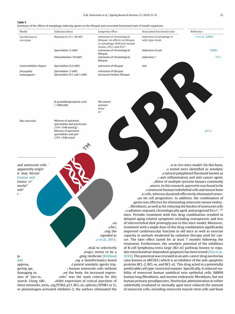
A.M. Vaiserman et al. / Ageing Research Reviews 31 (2016) 9–35 21
Table 5Summary of the effects of autophagy-inducing agents on the lifespan and associated functional traits of model organisms.
Model Substance/dose Longevity effect Associated functional traits Reference
Saccharomycescerevisiae
Rapamycin (0.1–40 nM) extension of chronologicallifespan; no effects on lifespanin autophagy-deficient mutantstrains, ATG1 and ATG7
induction of autophagy inwild-type strain
Alvers et al. (2009)
Spermidine (1 mM) extension of chronologicallifespan
induction of autophagy Eisenberg et al. (2009)
Ethanolamine (10 mM) extension of chronologicallifespan
induction of autophagy Rockenfeller et al. (2015)
Caenorhabditis elegans Spermidine (0.2 mM) extension of lifespan induction of autophagy Eisenberg et al. (2009)
Drosophilamelanogaster
Spermidine (1 mM) extension of lifespan induction of autophagy Eisenberg et al. (2009)Spermidine (0.1 and 1 mM) increased median lifespan enhances starvation resistance,
improved survival on paraquat,reduced female fecundity;abolished life extension bydownregulation of autophagythrough the reduction inexpression of ATG5 gene by RNAinterference
Bjedov et al. (2010)
�-guanidinopropionic acid( > 900 mM)
life extension; abolished lifeextension by downregulationof autophagy through thereduction in expression ofATG5 gene by RNA interference
AMP-activated proteinkinase-dependent increase inautophagy
Yang et al. (2015)
Mus musculus Mixture of spermine,spermidine and putrescine(374–1540 nmol/g)
increased survival lower incidence ofglomerulosclerosis in kidney
Soda et al. (2009)
Mixture of spermine,spermidine and putrescine(374–1540 nmol/g)
reduced mortality in advancedage
decreased age-associatedpathological changes, suppressedage-associated increase inpro-inflammatory status, inhibitedage-associated global alteration in
Soda et al. (2013)
aai(twwcrpratacianaK
ipaagdssto
nd senescent cells. Moreover, although the senescence responsepparently originally appeared as a tumor suppressor mechanism,t may become a cancer promoter as a consequence of agingLoaiza and Demaria, 2016). Indeed, senescent cells, similarly toumor cells, exhibit enhanced expression of “pro-survival net-orks” which help them to resist apoptosis. Thereby, SASP, alongith age-related immune decline and genomic instability, may
ontribute to deterioration of the effectiveness of the anti-canceresponse. In addition, cell-autonomous and paracrine mediatorsroduced by senescent cells can influence the processes of self-enewal and differentiation of stem cells, thus, persistent growthrrest may contribute to the decline of tissue regenerative poten-ial (Childs et al., 2015). For example, in BubR1 progeroid mice, fatnd muscle progenitor cells are shown to be highly susceptible toellular senescence (Baker et al., 2011). Optimal stem cell functions known to strongly depend on their microenvironment (niche),nd, therefore, SASP can impair this function by modulating theiche (Jang et al., 2011). As a result, senescent cells are regarded asttractive targets for therapeutic interventions (Childs et al., 2015;irkland, 2013b).
Identification of small molecules with potential to selectivelynduce death of senescent cells (senolytic drugs) seems to be aromising therapeutic approach in anti-aging medicine (Kirklandnd Tchkonia, 2015). Recently, by using a bioinformatics-basedpproach, Zhu et al. (2015) identified potent senolytic agents trig-ering apoptosis preferentially in human senescent cells withoutamaging other cells throughout the body. An increased expres-ion of “pro-survival networks” was the main criteria for this
earch. Using siRNA to inhibit expression of critical junctions ofhese networks, including PI3Kd, p21, BCL-xL, ephrins (EFNB1 or 3),r plasminogen-activated inhibitor-2, the authors eliminated theDNA methylation status and1,2-dimethylhydrazine-inducedtumorigenesis
senescent but not normal cells in in vivo mice model. On this basis,two compounds of 46 agents tested were identified as senolyticdrugs, namely, quercetin, a natural polyphenol flavonoid known asantihistamine, and also anti-inflammatory and anti-cancer agent,and dasatinib, an inhibitor of multiple tyrosine kinases commonlyused for treating cancers. In this research, quercetin was found to beefficient against senescent human endothelial cells and mouse bonemarrow stem cells, whereas dasatinib effectively eliminated senes-cent human fat cell progenitors. In addition, the combination ofthese agents was effective for eliminating senescent mouse embry-onic fibroblasts, as well as for reducing the burden of senescent cellsin radiation-exposed, chronologically aged, and progeroid Ercc1−/�
mice. Periodic treatment with this drug combination resulted indelayed aging-related symptoms including osteoporosis and lossof intervertebral disk proteoglycans in this mice model. Moreover,treatment with a single dose of this drug combination significantlyimproved cardiovascular function in old mice as well as exercisecapacity in animals weakened by radiation therapy used for can-cer. The later effect lasted for at least 7 months following thetreatment. Furthermore, the senolytic potential of the inhibitorsof B-cell lymphoma-extra large (Bcl-xl) pathway known to regu-late mitochondrial-dependent apoptosis has been tested (Zhu et al.,2016). This potential was revealed in an anti-cancer drug navitoclax(also known as ABT263) which is an inhibitor of the anti-apoptoticproteins BCL-2, BCL-w, and BCL-xL. This drug acted in a potentiallypredictable cell type-restricted manner. Specifically, it reduced via-bility of senescent human umbilical vein epithelial cells, IMR90human lung fibroblasts, and murine embryonic fibroblasts, but not
human primary preadipocytes. Navitoclax administration to eithersublethally irradiated or normally aged mice reduced the amountof senescent cells, including senescent muscle stem cells and bone
2 Resea
mras
metBbtpFmsdadiloeamIwaaoeatmcetA4ttthcp2
coppeepto2ocpdatroMp
2 A.M. Vaiserman et al. / Ageing
arrow hematopoietic stem cells. That, in turn, diminished theadiation-induced premature aging of the hematopoietic systemnd rejuvenated the aged muscle stem cells and hematopoietictem cells in normally aged mice.
Data demonstrating that selective elimination of senescent cellsay prevent or delay age-related functional impairments and
xtend healthspan were obtained in transgenic progeroid mousehat expresses low amounts of the mitotic checkpoint protein,ubR1 (Baker et al., 2011; Naylor et al., 2013). In this research, aiomarker of senescence, p16(Ink4a), was used to design a novelransgene, INK-ATTAC, for inducible elimination of p16(Ink4a)-ositive senescent cells. The authors based their approach on theAT-ATTAC (fat apoptosis through targeted activation of caspase)ouse model, where adipocytes are selectively killed by apopto-
is upon the administration of the synthetic membrane-permeablerug AP20187. In tissues such as skeletal muscle, adipose tissuend eye in which p16(Ink4a) contributes to age-associated disor-ers, long-term removal of p16(Ink4a)-expressing cells resulted
n delayed onset of aging-related phenotypes, such as cataracts,ipodystrophy and lordokyphosis (curvature of the spine). More-ver, later-life clearance retarded the progression of alreadystablished chronic pathologies. In continuing these studies, theuthors conducted experiments in normal (i.e. non-BubR1 mutant)ice. Specifically, they used a previously established transgene,
NK-ATTAC, to induce apoptosis in p16Ink4a-expressing cells ofild-type mice by injections of cell-permeant small molecule lig-
nd, AP20187, since one year of age (Baker et al., 2016). In both malend female mice belonged to two distinct genetic backgrounds (onef mixed genetic origin and one a pure C57BL/6 line), AP20187xtended median lifespan by 17–35% compared with the vehiclelone. Furthermore, the clearance of p16Ink4a-positive cells ledo a delay of age-related decline in kidney function and improve-
ent in some markers of cardiac function. Deleting senescentells also resulted in decreased expression of inflammatory mark-rs and protection from cataract formation, as well as in delayedumor appearance. Remarkably, extension of median lifespan inP20187-treated mice dying without tumors ranged from 24% to2%, indicating that this life extension was not merely owing to theumor-protective effect. Collectively, these findings demonstratehe feasibility of selectively removing of senescent cells and effec-iveness of senolytics for mitigating the symptoms of frailty andealthspan extension. Another related approach is pharmacologi-al SASP suppression, which might in part explain the anti-agingroperties of rapamycin (Serrano, 2015; Tomimatsu and Narita,015).
Recently, a pro- aging role of mitochondria in cellular senes-ence was reported (Correia-Melo and Passos, 2015). Inductionf mitochondria-dependent pro-oxidant and pro-inflammatoryathways is known to occur at both cellular senescence-relatedrocesses such as tumor suppression and aging. To study to whatxtent mitochondria are implicated in senescence, Correia-Melot al. (2016) have completely eliminated the mitochondrial com-artment by inducing mitophagy [a kind of autophagy ensuringhe preservation of healthy mitochondria due to the removalf damaged or excessive mitochondria (Patergnani and Pinton,015)] in mice model. For this, they used the depolarizationf mitochondria by uncouplers, such as carbonyl cyanide m-hlorophenyl hydrazone (CCCP), a chemical inhibitor of oxidativehosphorylation, targeting the ubiquitin ligase Parkin to mitochon-ria and promoting their degradation through proteasome andutophagy pathways. Elimination of mitochondria caused reduc-ion of a range of senescence effectors and phenotypes, including
educed oxidative stress, inflammation markers and expressionf the cyclin-dependent kinase inhibitors p21 and p16 (Correia-elo et al., 2016). These data suggest that mitochondria are aromising candidate target for interventions aimed to reduce the
rch Reviews 31 (2016) 9–35
adverse impacts of senescence in aging tissues. The concern is, how-ever, whether there is a possibility that targeting mitochondriawould adversely affect non-senescent cells too. This issue shouldbe addressed in further studies.
6. Telomerase activators
Pharmacological targeting of telomerase activity is anotherpromising anti-aging approach. Proper maintenance of telomeres(nucleoprotein structures at the end of linear eukaryotic chro-mosomes) is crucial for genome stability (Chiodi and Mondello,2016). Age-related telomere shortening is known to play a majorrole in senescence and aging-associated conditions (Zhang et al.,2016). Telomerase is the specialized reverse transcriptase capa-ble of maintaining telomere length through the template-mediatedaddition of telomeric repeats onto the ends of newly synthesizedchromosomes (Sishc et al., 2015). In multicellular organisms, tel-omerase activity is limited to specific cell types. Activation oftelomerase in somatic cells is a key step toward cell immortaliza-tion and transformation. Targeting telomerase is believed to be apromising antitumor therapeutic strategy, and also may provideclinical benefits in non-cancer pathological conditions, includingaging-related ones (Sprouse et al., 2012). The ectopic expressionof the telomerase catalytic subunit (telomerase reverse transcrip-tase, TERT) in mice epithelial cells was shown to extend lifespanby up to 40% in the cancer-resistant animals (Tomás-Loba et al.,2008); it, however, resulted in increased cancer risk in wild-typemice (Gonzalez-Suarez et al., 2001; Artandi et al., 2002). Reacti-vation of telomerase in the telomerase-negative normal humancells, exhibiting telomere shortening and senescence, by transfec-tion with vectors encoding the human TERT (hTERT), resulted inelongation of telomeres and reduced level of beta-galactosidase (abiomarker for senescence) in these cells (Bodnar et al., 1998). Retro-viral introduction of hTERT into senescent normal human diploidfibroblasts lacking telomerase activity also resulted in reconstitu-tion of telomerase activity, telomere maintenance, and significantcell life extension in vitro (Forsythe et al., 2002; Steinert et al., 2000;Vaziri and Benchimol, 1998). Treatment with an adeno-associatedvirus (AAV) carrying the mouse TERT (mTERT) cDNA resultedin substantial beneficial effects on health and fitness in mice(Bernardes de Jesus et al., 2012). These effects included decreasedosteoporosis risk and enhancement of insulin sensitivity, neuro-muscular coordination and several molecular biomarkers of aging.Importantly, AAV9-mTERT-treated 1- and 2-year old mice did notdevelop more cancers than control animals, and their median lifes-pans were increased by 24 and 13%, respectively. These beneficialeffects were not revealed with a reporter virus or a catalyticallyinactive TERT, indicating the strong dependence of an active telo-merase complex. Interestingly, CR caused significantly decreasedcancer incidence in TERT transgenic (TgTERT) mice and extendedtheir lifespan compared with the wild-type controls, demonstrat-ing a synergy between TgTERT and CR in increasing the longevity(Vera et al., 2013).
A pharmacological telomerase-based approach to enhanceimmune function was first applied by Fauce et al. (2008). In thisstudy, exposure with a small molecule telomerase activator TAT2retarded telomere shortening and enhanced proliferative potential,cytokine/chemokine production and antiviral activity of humanCD8(+) T lymphocytes. A small-molecule telomerase activator,TA-65, extracted from the roots of the plant Astragalus mem-branaceus was shown to be able to activate telomerase in human
fibroblasts, keratinocytes, and immune cells (Harley et al., 2011).In human T cells treated with TA-65, MAPK-specific telomeraseactivation and significant increase in proliferative activity wereobserved (Molgora et al., 2013). Dietary supplementation with TA-
Resea
6ae6oi6flr2e(od2
odtrcabit2oaioAsiIstcTigre
7
gtt(acbEetaaobpf
oD
A.M. Vaiserman et al. / Ageing
5 resulted in telomerase-dependent elongation of short telomeresnd rescue of associated DNA damage in mice (Bernardes de Jesust al., 2011). Moreover, supplementation of female mice with TA-5 led to an improvement of several healthspan indicators such assteoporosis, glucose tolerance and skin fitness, without increasen cancer incidence. More recently, the stimulating effect of TA-5 on the telomerase activity and telomere lengths, as well asight feather renewal capacity and faster feather regeneration wasevealed in captive zebra finches, Taenopygia guttata (Reichert et al.,014). TA-65 supplementation also caused improvement of mark-rs of bone, metabolic, and cardiovascular health in human trialsHarley et al., 2013). Recently, an efficiency of TA-65 in treatmentf early age-related macular degeneration was reported in a ran-omized placebo-controlled interventional study (Dow and Harley,016).
Collectively, these findings indicate that activation of tel-merase can be an effective treatment option, especially forisorders related to impaired telomere maintenance such aselomeropathies. Both direct and indirect strategies of telome-ase activation, however, have raised safety concerns becauseonstitutive reactivation of endogenous telomerase is apparentlyssociated with risk of cancer development. In particular, it haseen reported in several studies that activation of TERT by TA-65
s mediated by inducing mitogenic pathways causing activation ofhe oncogene c-myc (Bernardes de Jesus et al., 2011; Molgora et al.,013), thereby driving tumorigenesis. Therefore, the developmentf safe clinical applications for telomerase activation representsn important goal for further research. AAV-based gene therapys suggested to be of particular value for TERT activation, becausef the non-integrative and replication incompetent properties ofAVs allow for cell division-associated telomere elongation andubsequent loss of TERT expression as cells divide, thereby restrict-ng TERT expression to a few cell divisions (Ramunas et al., 2015).ndeed, the unfavorable side effects of telomerase activation werehown to be avoided via direct delivery of TERT using AAV vec-ors (Bernardes de Jesus et al., 2012). Thus, AAV-based treatmentan apparently provide a transient and relatively safe strategy toERT activation. Another prospective way to extend telomeres andncrease cell proliferative capacity without risk of insertional muta-enesis by transient delivery of modified mRNA encoding TERT wasecently reported in the in vitro human fibroblast study (Ramunast al., 2015).
. Epigenetic drugs
In the last years, pharmacological compounds targeted to epi-enetic regulators of gene expression are actively studied inhe context of geroscience. Epigenetic mechanisms including his-one modifications, DNA methylation, and changes in microRNAmiRNA) expression play central role in regulating gene expressionnd genomic instability throughout the lifespan. Epigenetic modifi-ations are known to be finely balanced in normal tissues. They cane, however, unbalanced in malignant and other transformed cells.pigenetic dysregulation was shown to contribute to the pathogen-sis of age-associated pathologies such as cancer, atherosclerosis,ype 2 diabetes, psychiatric and neurodegenerative diseases, as wells decline in immune response (Brunet and Berger, 2014; Cacabelosnd Torrellas, 2015; Vaiserman, 2015). Therefore, the modulatorsf the activity of enzymes involved in epigenetic regulation mighte clinically applicable. Epigenetic modifications are known to beotentially reversible; this feature makes them attractive targets
or pharmacological intervention (Cacabelos and Torrellas, 2015).Over the past years, a series of medications have been devel-ped targeted to epigenetic regulators, including modulators ofNA methyltransferases (DNMTs), histone deacetylases (HDACs),
rch Reviews 31 (2016) 9–35 23
histone acetyltransferases (HATs) and noncoding miRNAs, withpotential effects against various types of tumors, myelodysplas-tic syndromes, and neurodegenerative disorders (Arguelles et al.,2016; Cacabelos, 2014).
7.1. DNMT inhibitors
Over the past years, DNMT inhibitors have become the promis-ing drugs in the treatment of several chronic diseases, primarilycancers; their preventive role in carcinogenesis, however, remainsunclear. It was shown that inhibition of DNMT1 is able toreverse the malignant phenotype of transformed cells by restoringexpression of aberrantly silenced genes involved in differentia-tion, senescence, and apoptosis (Zhong et al., 2016). Among thedrugs targeted to inhibit DNMTs, FDA-approved drugs such as 5-azacytidine (azacitidine) and 5-aza-2′-deoxycytidine (decitabine)are the most intensively studied. Several clinical trials havedemonstrated that these medications have therapeutic potentialagainst leukemias including myelodysplastic syndrome, chronicmyelogenous leukemia, acute myeloid leukemia and chronicmyelomonocytic leukemia, while their efficacy in solid tumorsseems rather limited (Ghoshal and Bai, 2007). A major problem inthe usage of these substances as anti-cancer agents is their insta-bility in vivo and toxicity, causing cell cycle arrest. DNMT inhibitorswere also demonstrated to be able to modulate vascular biologyand atherosclerosis development (Dunn et al., 2015). It shouldbe noted, however, that DNA-demethylating agents can benefi-cially reactivate tumor suppressor genes, but also activate silencedprometastatic genes thereby inducing metastasis (Cheishvili et al.,2015). Therefore, caution is required in using DNMT inhibitors incancer patients.
7.2. HDAC inhibitors
Histone acetylation is the main histone modification affectinggene transcription. The main enzymes controlling process of his-tone acetylation are HDACs and histone acetyltransferases (HATs).HATs are acetylate lysines of histone proteins, causing relaxationof chromatin structure and promoting gene activation. HDACs,conversely, remove acetyl groups from hyperacetylated histonesand thereby suppress gene transcription. Currently, HDACs aredivided into four classes (Eom and Kook, 2014). Class I HDACsundergo posttranslational modifications including sumoylation,phosphorylation and S-nitrosylation. Class II HDACs work mainlyin association with other transcription factors to target binding ele-ments in a phosphorylation-dependent manner. In addition, HDACsbelonging to class I demonstrate much higher enzymatic activitythan class II HDACs and target many non-histone proteins as wellas the histone-core complex. In recent years, increasing evidencehas been accumulated that HDACs play key role in diverse biolog-ical processes such as cell proliferation, apoptosis, inflammationand carcinogenesis (Chen et al., 2015). Several modulators of HDACactivity (primarily, HDAC inhibitors), among other drugs target-ing epigenetic machinery, have been recently examined in humanclinical trials, and some have been proposed as promising anti-aging drug candidates (Vaiserman and Pasyukova, 2012). HDACinhibitors include four chemical classes: cyclic peptides, hydrox-amic acids, short chain fatty acids and synthetic benzamides, andthey substantially vary in biological activity, structure and speci-ficity (Lakshmaiah et al., 2014). The anti-tumor effects of thesemedications may be attributed to the transcriptional reactivationof silent tumor suppressor genes and the transcriptional repression
of proto-oncogenes (Boumber and Issa, 2011).Among the chemicals affecting HDAC activity, HDAC inhibitorsseem to be the most promising in the field of geroscience. Sincethe level of transcription of many genes, primarily metabolic

2 Resea
aeHdgwT
bHllfticietueiosoriaaptaMn‘tdlnsfisds
siH(vsdlabGaettestomt
4 A.M. Vaiserman et al. / Ageing
nd biosynthetic ones, is known to decrease with aging (Seroudet al., 2002), the restoration of the transcriptional activity viaDAC inhibitors could likely delay the age-related functionalecline. Moreover, HDAC inhibition can cause up-regulation ofenes involved in response to stress and inflammation − path-ays generally involved in the regulation of longevity (Kourtis and
avernarakis, 2011).Several convincing lines of experimental evidence have also
een obtained indicating substantial life-extending potential ofDAC inhibitors. A significant increase in both mean and maximum
ifespans by up to 30–50% without diminution of stress-resistance,ocomotor activity and reproductive ability has been revealed byeeding Drosophila with HDAC inhibitor, 4-phenylbutyrate (PBA),hroughout adulthood (Kang et al., 2002). Flies fed PBA exhib-ted a global increase in histone acetylation and dramaticallyhanged patterns of gene expression of various genes, includ-ng those supposedly implicated in the life extending effects:longation factor 1, superoxide dismutase, cytochrome P450, glu-athione S-transferase, and three chaperones. All these genes werep-regulated by PBA. Life extension accompanied by elevatedxpression of hsp22 gene was observed in flies fed with HDACnhibitor such as trichostatin A (TSA) (Tao et al., 2004). TSA andther HDAC inhibitor, sodium butyrate, have also been demon-trated to significantly extend longevity and promote expressionf hsp22 and hsp70 in fruit flies (Zhao et al., 2005). In a moreecent study, significant increase of the mean lifespan was revealedn both males and females fed sodium butyrate during both pre-dult and adult stages; treatment with sodium butyrate during thedult stage only caused an increase in male (but not female) lifes-an (Vaiserman et al., 2012). The effects of sodium butyrate onhe flies’ longevity were also studied simultaneously in normal-nd long-lived strains of Drosophila melanogaster (Arking, 2015;cDonald et al., 2013). This agent increased longevity in the
ormal-lived Ra strain when administered in the ‘transition’ orsenescent’ spans, but decreased longevity when administered overhe ‘health’ span only or over the entire adult lifespan. Mostlyeleterious effects were revealed when administered to the long-
ived La strain. Late-life extending effects were observed in theormal-lived strain administered with another HDAC inhibitor,uberoylanilide hydroxamic acid (SAHA). On the basis of thesendings, the authors concluded that use of HDAC inhibitors mayignificantly alter the mortality rate of the ‘senescent’ span byecreasing its vulnerability, or short-term risk of death, in a mannerimilar to that of CR (McDonald et al., 2013).
Since the onset and progression of various cancers involve sub-tantial dysregulation of HDAC activity, a wide range of HDACnhibitors are emerging as promising anticancer pharmaceuticals.DAC inhibitors such as belinostat, panobinostat, SAHA and FK228
Ma et al., 2016), as well as trichostatin A (TSA), sodium butyrate,orinostat, valproic acid and romidepsinor (Chun, 2015) showedubstantial activity in both haematological and solid tumors inifferent tissues. Their effects are apparently mediated by the regu-
ation of gene expression and DNA repair, inducing cell cycle arrestnd apoptosis and inhibiting angiogenesis. They can also be causedy the long-term stimulation of immune response (Leggatt andabrielli, 2012). In the last years, HDAC inhibitors have undergone
rapid phase of clinical development in different cancer types,ither as monotherapy or combined with other anticancer modali-ies. To date, three HDAC inhibitors have been approved by FDA forhe treatment of cutaneous/peripheral T-cell lymphoma (Mottamalt al., 2015), and four HDAC inhibitors, namely vorinostat, belino-tat, romidepsin and panobinostat have been approved by FDA for
he treatment of hematologic cancers (Yoon and Eom, 2016). Manyther HDAC inhibitors are at different stages of clinical develop-ent for the treatment of haematological malignancies and solidumors (Mottamal et al., 2015).
rch Reviews 31 (2016) 9–35
The FDA’s approval of HDAC inhibitors as anticancer agentshas provided the motivation to use these medicines as treat-ment options for non-malignant diseases. The beneficial outcomesof HDAC inhibition were obtained in treatment of various typesof inflammatory, neurodegenerative and cardiovascular disorders(Yoon and Eom, 2016). In particular, experimental evidence hasindicated that inhibitors of Class I HDACs can attenuate thedevelopment of cardiac hypertrophy and preserve cardiac func-tion in several small animal models (Abend and Kehat, 2015).In addition, HDAC inhibitors have been found to be beneficial inpreventing myocardial infarction, hypertension, atherosclerosis,vascular calcification, supraventricular arrhythmia, cardiac remod-eling, fibrosis, and neointima formation (Eom and Kook, 2014).The putative mechanisms mediating beneficial effects of HDACinhibitors on the heart function include suppression of oxidativestress and inflammation, enhancement of cardiac protein aggregateclearance and autophagic flux, as well as inhibition of MAP kinasesignaling (Ferguson and McKinsey, 2015). In addition, since HDACinhibitors were reported to promote �-cell proliferation, differenti-ation and function and positively affect late diabetic microvascularcomplications, HDAC inhibition was proposed as a novel treatmentstrategy for type 2 diabetes (Christensen et al., 2011).
HDAC inhibitors show great promise to combat the aging-associated cognitive decline and to ameliorate the symptoms ofposttraumatic stress disorder and depression (Penney and Tsai,2014). Neurodegenerative diseases such as amyotrophic lateralsclerosis, polyglutamine-related diseases, as well as Parkinson’sand Alzheimer’s diseases are known to be accompanied by tran-scriptional dysfunctions, leading to neuronal death (Selvi et al.,2010). Several recent studies have highlighted the importance ofHDACs in neuronal memory, learning, synaptic plasticity and neuralregeneration (Ganai et al., 2016). Accumulating evidence indicatethat histone acetylation plays a crucial role in the etiology of neu-rodegenerative disorders. Some studies demonstrate that increaseof histone acetylation can be involved in Alzheimer’s disease, andthat HDAC inhibitors may be neuroprotective by regulating mem-ory and synaptic dysfunctions in both in vitro and in vivo models ofthis pathology (Lu et al., 2015). HDAC inhibitors were also reportedto cause beneficial effects in both in vitro and in vivo models ofParkinson’s disease. The potential mechanisms underlying theseeffects include maintenance of histone acetylation homeostasisand transcriptional activation of neuronal survival genes (Sharmaand Taliyan, 2015). In the last years, clinical trials have been ini-tiated to examine the effectiveness of HDAC inhibitors in patientswith Parkinson’s disease. The loss of functional activity of HATs(enzymes with activities antagonistic to those of HDACs) is likelya common mechanism related to the impairment of the chro-matin acetylation status throughout the lifetime of neurons. Thetherapeutic potential of HAT activators in the treatment of neurode-generative disorders has been established in preclinical studies.Substantial neuroprotective properties were revealed for one ofthe HATs termed cAMP response element binding protein (CREB)-binding protein (CBP), an also for several other HATs which wereshown to be essential for processes of neuronal plasticity and mem-ory formation (Selvi et al., 2010).
Non-specific HDAC inhibitors also demonstrated anti-inflammatory effects in both in vitro and in vivo models. Recently,evidence was obtained for the role of the NF-�B signal trans-duction pathway in mediating the effects of HDAC inhibitors oninflammatory responses (Dekker et al., 2014). The important pointis that such effects were reported at concentrations which were10–100-fold lower than anti-cancer effects of these compounds.
Clinical application of these substances for treating inflammatorydiseases is, however, hampered due to their low specificity anda wide variety of HDACs that they affect throughout the body(Cantley and Haynes, 2013).
Resea
ssi
7
ttadeTwiiStssg(Sgtoaiddtm
Sicadtip1mtrefets
7
lp(piLtv(
cw
A.M. Vaiserman et al. / Ageing
Overall, the therapeutic strategy based on HDAC inhibitioneems very promising. However, as most HDAC inhibitors lackpecificity (Pirooznia and Elefant, 2013), their wide applicabilitys still questionable.
.3. Sirtuin-activating compounds
The term ‘sirtuins’ is derived from the ‘sir-two-ins’ in referenceo homology to the yeast Sir2 protein (silent information regula-or) (North and Sinclair, 2007). Silent information regulators (SIRTs)re nicotinamide adenine dinucleotide (NAD + )–dependent proteineacetylases playing a crucial role in epigenetic regulation of genexpression by mediating the assembly of the silenced chromatin.he yeast deacetylase Sir2 was initially identified as a moleculeith potent anti-aging properties (Imai et al., 2000). Subsequently,
t has been demonstrated to be implicated in the control of lifespann yeast, nematode, and Drosophila. In mammals, a homologue ofir2, SIRT1, has been found to act as anti-aging molecule in the con-ext of CR (Boily et al., 2008). Apart from gene silencing, SIRT1 washown to regulate other important biological processes includingtress resistance, cell survival, inflammation, mitochondrial bio-enesis, glucose and lipid metabolism, apoptosis and autophagyGiblin et al., 2014; Liu and McCall, 2013). Moreover, the control ofIRT1 activity is closely related to those of the AMPK, that regulateslucose, lipid and cholesterol metabolism in specialized metabolicissues, such as muscle, liver and adipose tissue and which isne of the key regulators of eukaryotic metabolism (Shackelfordnd Shaw, 2009). AMPK is activated in conditions where nutritions limited and promotes oxidative metabolism. It has also beenemonstrated that AMPK is activated by increasing NAD+ pro-uction, and activation of SIRT1 in turn leads to AMPK activationhrough the deacetylation of liver kinase B1 (LKB1), which is the
ajor upstream AMPK kinase (Cantó et al., 2009; Hardie, 2015).The importance of SIRT1 for the CR effects has been shown in
IRT1 transgenic mice that manifest a CR-like phenotype, show-ng lowered levels of adipokines, insulin, fasting glucose bloodholesterol, and greater glucose tolerance compared to the controlnimals (Bordone et al., 2007). In a controlled intervention trial con-ucted in healthy individuals, with measurements before and afterhe CR treatment, it has been shown that 25% reduction in calorientake during six months leads to the overexpression of SIRT1 anderoxisome proliferator-activated receptor (PPAR)-� coactivator-� (PGC-1�) in the skeletal muscles, accompanied by the enhanceditochondrial function and reduced levels of the body tempera-
ure, metabolic rate, oxidative stress, visceral fat mass and insulinesistance in non-obese young adults (Civitarese et al., 2007). How-ver, in several studies in yeast, nematodes and Drosophila, CR wasound to be able to extend lifespan in the absence of Sir2 (Burnettt al., 2011; Kaeberlein et al., 2004; Smith et al., 2007). Therefore,he role of sirtuins, particularly SIRT1, in CR-mediated life extensiontill remains unclear.
.3.1. ResveratrolLarge-scale screening of drugs capable of activating SIRTs
ed to the discovery of 18 molecules demonstrating suchroperties (Kitada and Koya, 2013). Among them, resveratrol3,5,40-trihydroxystilbene), a naturally occurring polyphenolichytoalexin present mostly in red wine and grape skins, but also
n berries and peanuts, has been investigated most thoroughly (deigt et al., 2015). The research of this compound was initially startedhrough the “French paradox” which describes improved cardio-ascular outcomes despite a high-fat diet in the French population
Catalgol et al., 2012).Dietary resveratrol administration in doses of 100–1000 �Maused life extension in short-lived model organisms, such as yeast,orms, honey bee and fruit flies (Table 6).
rch Reviews 31 (2016) 9–35 25
Resveratrol has also been shown to prolong lifespan and retardthe onset of aging-associated markers in short-lived vertebratessuch as fish (Valenzano et al., 2006; Yu and Li, 2012). In micemodel, the beneficial effects of resveratrol on metabolism and lifes-pan were revealed in high-fat diet-induced obese animals (Bauret al., 2006). In mice fed a standard diet, resveratrol administrationdid not prolong life, but resulted in delayed aging-related impair-ments and transcriptional changes similar to those observed in CRanimals (Pearson et al., 2008). Resveratrol has also been shownto be able to protect against a variety of aging-related disordersincluding type 2 diabetes, Alzheimer’s disease and cancer (Bhullarand Hubbard, 2015). In obese male individuals, supplementationwith resveratrol during 30 days prevented the obesity-associatedmetabolic dysregulation by activating the AMPK/SIRT1 pathwayin skeletal muscle (Timmers et al., 2011). Furthermore, resveratrolintake was associated with elevated intramyocellular lipid levels,and decreased intrahepatic lipid content, triglycerides, circulat-ing glucose, alanine-aminotransferase and inflammatory markers.More recently, 30-day supplementation with the same dose ofresveratrol caused a significantly decreased adipocyte size, witha shift toward a decrease in the proportion of large adipocytes andan increase in the proportion of small adipocytes (Konings et al.,2014). A widespread view is that longevity effects of resveratrol aremediated by activation of sirtuins. It, however, had no detectableeffects on Sir2 activity in in vivo yeast studies (Kaeberlein et al.,2005). Thereby, the direct activation of sirtuins by resveratrol isstill under debate (Fernández and Fraga, 2011).
Currently, resveratrol is one of the most popular nutritionalsupplements on the market. In the U.S., two-thirds of personsconsuming multiple dietary supplements, consume this supple-ment (Hubbard and Sinclair, 2014). However, despite the highexpectations placed on resveratrol, some challenges are relatedto its application. Recently, in discussing these issues Vang (2015)stressed that: ‘(1) many different cellular effects are observed forresveratrol, but it is not known whether they arise from one point ofaction (or a few) or whether resveratrol is non-specific in its action;(2) the health-promotion effect of dietary resveratrol is likely acombinatory effect of various bioactive components in the mix-ture (diet); (3) the known cell biological response to resveratrolis presently based on exposure to short-term high levels, and bet-ter in vitro analyses have to be developed; (4) the actual level ofresveratrol and resveratrol metabolites present in vitro and in vivoduring experiments may be over- and underestimated, respec-tively, because resveratrol is not very soluble in water; and (5) onlya few small clinical studies have been published to date, focusingon the therapeutic effects of resveratrol.’
7.3.2. Synthetic sirtuin activatorsSeveral synthetic SIRT1 activators such as SRT1460, SRT1720,
SRT2104 and SRT2183 have been synthesized over the last decade.Among them, SRT1720 is the most studied. This agent has beenshown to cause life extension and to ameliorate the metabolicimpairments in mice fed both a normal diet (Mitchell et al., 2014)and a high-fat diet (Minor et al., 2011). Moreover, it has alsobeen found to be able to reverse adverse aging-associated changesincluding vascular endothelial dysfunction, excessive superoxideproduction and inflammation (Gano et al., 2014). Recently, help-ful effects of another SIRT1 activator, SRT2104, on muscle and bonemass as well as on the survival rate in male mice have been detected(Mercken et al., 2014). Based on this research background, smallSIRT1 activators such as SRT501, SRT2104 and SRT2379 are cur-rently undergoing clinical trials for the treatment of obesity, type 2
diabetes and metabolic syndrome, among other chronic disorders(Camins et al., 2010).As the activation of SIRT1 has been shown in several studiesto be able to delay the progression of aging-associated conditions
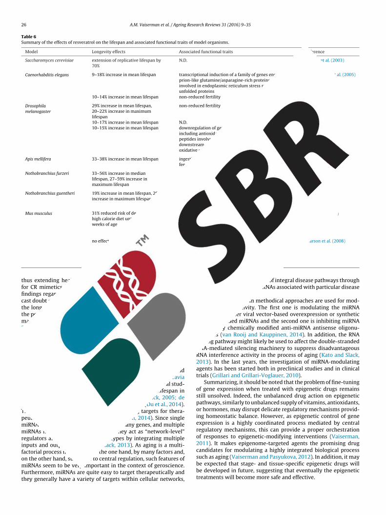
26 A.M. Vaiserman et al. / Ageing Research Reviews 31 (2016) 9–35
Table 6Summary of the effects of resveratrol on the lifespan and associated functional traits of model organisms.
Model Longevity effects Associated functional traits Reference
Saccharomyces cerevisiae extension of replicative lifespan by70%
N.D. Howitz et al. (2003)
Caenorhabditis elegans 9–18% increase in mean lifespan transcriptional induction of a family of genes encodingprion-like glutamine/asparagine-rich proteinsinvolved in endoplasmic reticulum stress response tounfolded proteins
Viswanathan et al. (2005)
10–14% increase in mean lifespan non-reduced fertility Wood et al. (2004)
Drosophilamelanogaster
29% increase in mean lifespan,20–22% increase in maximumlifespan
non-reduced fertility Wood et al. (2004)
10–17% increase in mean lifespan N.D. Bauer et al. (2004)10–15% increase in mean lifespan downregulation of genes in aging-related pathways,
including antioxidant peroxiredoxins, insulin-likepeptides involved in insulin-like signaling and severaldownstream genes in Jun-kinase signaling involved inoxidative stress response
Wang et al. (2013)
Apis mellifera 33–38% increase in mean lifespan ingestion of fewer quantities of food under ad libitumfeeding conditions
Rascón et al. (2012)
Nothobranchius furzeri 33–56% increase in medianlifespan, 27–59% increase inmaximum lifespan
delaying the age-dependent decay of locomotoractivity and cognitive performances and reducing theexpression of neurofibrillary degeneration in the brain
Valenzano et al. (2006)
Nothobranchius guentheri 19% increase in mean lifespan, 28%increase in maximum lifespan
enhanced cognitive ability and locomotor activity;protection from neurodegeneration; reduced rate oflipofuscin formation
Yu and Li (2012)
Mus musculus 31% reduced risk of death from thehigh calorie diet until the 114weeks of age
increased insulin sensitivity, reduced insulin-likegrowth factor-1 levels, increased AMPK andperoxisome proliferator-activated receptor-gammacoactivator 1alpha activity, increased mitochondrialnumber, improved motor function
Baur et al. (2006)
no effect on lifespan marked reduction in signs of aging, including reducedalbuminuria, decreased inflammation, and apoptosis in
vascuater mmatio
Pearson et al. (2008)
tfficttm2
7
ckaaimLTpmmrifomFt
thegrefor
hus extending healthspan, SIRT1 regarded as a potential targetor CR mimetics (Baur et al., 2012). Several recent controversialndings regarding the role of SIRTs in the effects of CR, however,ast doubt on their relevance to aging, particularly as mediators ofhe longevity effects induced by CR (Park et al., 2013). Therefore,he potential of SIRTs as therapeutic targets is being questioned by
any researchers and is a subject of intense debate now (Baur et al.,012; Dang, 2014; Park et al., 2013).
.4. Micro RNA
MicroRNAs (miRNAs) are small non-coding RNAs which play arucial role in epigenetic regulation of gene expression. They arenown to be crucially involved in various biological processes andre dysregulated in a variety of aging-related pathologies (Caraviand López-Otín, 2015; Kato and Slack, 2013). In experimental stud-es, miRNAs have been observed to be able to extend lifespan in
odel organisms such as C. elegans (Boehm and Slack, 2005; deencastre et al., 2010; Wang et al., 2015) and mice (Du et al., 2014).hereby, miRNAs are believed to be promising targets for thera-eutic applications (van Rooij and Kauppinen, 2014). Since singleiRNAs may regulate the expression of many genes, and multipleiRNAs may target the same mRNA, they act as “network-level”
egulators affecting complex phenotypes by integrating multiplenputs and outputs (Inukai and Slack, 2013). As aging is a multi-actorial process influenced, on the one hand, by many factors and,
n the other hand, subjected to central regulation, such features ofiRNAs seem to be very important in the context of geroscience.urthermore, miRNAs are quite easy to target therapeutically andhey generally have a variety of targets within cellular networks,
lar endothelium, increased aortic elasticity,otor coordination, reduced cataract
n, and preserved bone mineral density
thereby allowing modulation of integral disease pathways throughtherapeutic targeting of miRNAs associated with particular diseasestates.
Currently, two main methodical approaches are used for mod-ulating miRNA activity. The first one is modulating the miRNAfunction by either viral vector-based overexpression or syntheticdouble-stranded miRNAs and the second one is inhibiting miRNAfunction by chemically modified anti-miRNA antisense oligonu-cleotides (van Rooij and Kauppinen, 2014). In addition, the RNAediting pathway might likely be used to affect the double-strandedRNA-mediated silencing machinery to suppress disadvantageousRNA interference activity in the process of aging (Kato and Slack,2013). In the last years, the investigation of miRNA-modulatingagents has been started both in preclinical studies and in clinicaltrials (Grillari and Grillari-Voglauer, 2010).
Summarizing, it should be noted that the problem of fine-tuningof gene expression when treated with epigenetic drugs remainsstill unsolved. Indeed, the unbalanced drug action on epigeneticpathways, similarly to unbalanced supply of vitamins, antioxidants,or hormones, may disrupt delicate regulatory mechanisms provid-ing homeostatic balance. However, as epigenetic control of geneexpression is a highly coordinated process mediated by centralregulatory mechanisms, this can provide a proper orchestrationof responses to epigenetic-modifying interventions (Vaiserman,2011). It makes epigenome-targeted agents the promising drugcandidates for modulating a highly integrated biological process
such as aging (Vaiserman and Pasyukova, 2012). In addition, it maybe expected that stage- and tissue-specific epigenetic drugs willbe developed in future, suggesting that eventually the epigenetictreatments will become more safe and effective.
A.M. Vaiserman et al. / Ageing Research Reviews 31 (2016) 9–35 27
Table 7Summary and categorization of the most promising anti-aging agents.
Preparation Effects observed
AO DR AI AC AP NP AA GHS TA LE
Agents with no observed demographic effects until now2-deoxy-d-glucose + + +Pegvisomant + + +TA-65 + +
Agents suggesting to promote longevity mainly by inhibiting particular disease(s)Statins + + + + + +Rapamycin + + + + + + + +Metformin + + + + + + +
Agents with both anti-aging and pro-longevity effectsAspirin + + +Coenzyme Q10 + + + + +Spermidine + + + + +Vitamin E + +Sodium butyrate + + + + + +Suberoylanilide hydroxamic acid + + + + + +Melatonin + + + + + + +Epigallocatechin-3-gallate + + + + + + +Curcumin + + + + + + +Quercetin + + + + + + + +Resveratrol + + + + + + + + + +
A us; APs
8
itsiepec(iovhpttia
apdFoldtodfrf
c
-
O: antioxidant; DR: dietary restriction; AI: anti-inflammatory; AC: anti-cancerouppression; TA: telomerase activation; LE: life extension.
. Conclusions
Few decades ago, it was generally believed that damagenduced by free radicals is a primary cause of aging and, thereby,hat antioxidants may protect against this damage, consequentlylowing the aging process and improving health. Currently, its becoming apparent that a single cause could not properlyxplain the health- and lifespan-promoting effects of differentharmacological classes. It is increasingly clear that pro-longevityffects of many prospective anti-aging agents are complex andould be attributable to numerous interacting signaling pathwaysVaiserman, 2014). Taking into account the exceptional complex-ty of mechanistic pathways underlying aging, the identificationf these pathways and development of relevant anti-aging inter-entions seems to be a challenging task. Considerable progress,owever, has been made in this research field throughout theast years. A large number of pharmacological substances havinghe potential to target molecular pathways underlying aging ando induce protective responses against aging-associated diseasesncluding, e.g., mTOR inhibitors such as rapamycin, senolytics, etc,re presently under investigation.
One important issue in the study of potential geroprotectivegents is that their pro-longevity effects might be mediated by therevention and/or delay of onset of age-associated, life-shorteningiseases, rather than through inhibiting the aging processes itself.or example, Neff et al. (2013) and Ehninger et al. (2014) basedn a large-scale assessment of more than 150 cellular, molecu-ar, histopathological, and functional traits, which typically changeuring the course of aging, in rapamycin-treated mice, concludedhat rapamycin can extend longevity, but it has only limited effectn the aging process per se. Another promising anti-aging drug can-idate, resveratrol, was found to be able to extend life span in miceed with high-fat diet by reversing the initial steps of particular age-elated diseases induced by that diet, but had no effect on animalsed with healthy diet (Baur et al., 2006).
Thus, all known pro-longevity drugs can be divided in three
ategories in accordance with the suggested mode of action:those demonstrating anti-aging effects, but without any evidenceyet of their ability to prolong life;
: autophagy; NP: neuroprotective; AA: anti-atherogenic; GHS: growth hormone
- those that are suggested to extend longevity primarily becausethey can prevent or postpone the progression of particular age-related disease, such as cancer, but which are not proven to delaythe aging process(es) per se;
- those that extend longevity because they are actually suggested toreverse the aging process itself, at least in certain environments.
Based on these considerations, the most promising agents withsuspected anti-aging properties are categorized into three maingroups in Table 7. Certainly, the classification presented in this tableis quite conditional since only relatively few research findings havebeen reported until now in this area of investigation, thus any newdata can move a particular substance from one category to another.
In summarizing it may be suggested that targeting aging perse may be more efficient approach to prevent or postpone age-related diseases than treatments targeted to particular pathologicalconditions. Because of the aging population, this therapeutic strat-egy undoubtedly can be an area of increasing relevance for thepharmaceutical industry and public health organizations. It is cur-rently assumed that great socio-economic benefits can be obtainedfrom the approach based on the longevity dividend paradigm incomparison with modern public health strategy aimed on the pre-vention of particular diseases. Presently, the consensus is amongmedical and health professionals that the optimization of physio-logical and mental functioning through the life course should be amain focus of public health policy addressing the problem of globalaging. A healthy lifestyle including a balanced diet, regular exerciseand smoking cessation is the first-line strategy. Using the pharma-cological substances, both potential and existing, may serve as aprospective additional approach (Seals et al., 2016).
The great expectations related to further development of anti-aging interventions, however, must certainly be critically discussedand explored in the light of their economic, social and ethicalimplications. The implementation of these approaches in clinicalpractice will be possible only after in-depth examination and fur-
ther comprehensive discussion. To meet the needs caused by therapid population aging across the globe, a novel emphasis in physi-ological geroscience is needed which will require the joint efforts ofinterdisciplinary investigators working throughout the continuum
2 Resea
ot
C
A
a
R
A
A
A
A
A
A
A
A
A
A
A
A
A
A
A
A
A
8 A.M. Vaiserman et al. / Ageing
f translational medicine from basic research to clinical applica-ions.
onflict of interests
The authors have no conflict of interests.
cknowledgment
The authors would like to thank Oksana Zabuga for the helpfulssistance in preparing the manuscript.
eferences
bbas, S., Wink, M., 2009. Epigallocatechin gallate from green tea (Camelliasinensis) increases lifespan and stress resistance in Caenorhabditis elegans.Planta Med. 75 (3), 216–221.
bend, A., Kehat, I., 2015. Histone deacetylases as therapeutic targets–from cancerto cardiac disease. Pharmacol. Ther. 147, 55–62.
l-Sawaf, O., Clarner, T., Fragoulis, A., Kan, Y.W., Pufe, T., Streetz, K., Wruck, C.J.,2015. Nrf2 in health and disease: current and future clinical implications. Clin.Sci. (Lond.) 129 (12), 989–999.
lbanes, D., Heinonen, O.P., Taylor, P.R., Virtamo, J., Edwards, B.K., Rautalahti, M.,Hartman, A.M., Palmgren, J., Freedman, L.S., Haapakoski, J., Barrett, M.J.,Pietinen, P., Malila, N., Tala, E., Liippo, K., Salomaa, E.R., Tangrea, J.A., Teppo, L.,Askin, F.B., Taskinen, E., Erozan, Y., Greenwald, P., Huttunen, J.K., 1996.Alpha-tocopherol and beta-carotene supplements and lung cancer incidence inthe alpha-tocopherol, beta-carotene cancer prevention study: effects ofbase-line characteristics and study compliance. J. Natl. Cancer Inst. 88,1560–1570.
lbert, V., Hall, M.N., 2015. mTOR signaling in cellular and organismal energetic.Curr. Opin. Cell Biol. 33, 55–66.
lvers, A.L., Wood, M.S., Hu, D., Kaywell, A.C., Dunn Jr., W.A., Aris, J.P., 2009.Autophagy is required for extension of yeast chronological life span byrapamycin. Autophagy 5 (6), 847–849.
nisimov, V.N., Zavarzina, N.Y., Zabezhinski, M.A., Popovich, I.G., Zimina, O.A.,Shtylick, A.V., Arutjunyan, A.V., Oparina, T.I., Prokopenko, V.M., Mikhalski, A.I.,Yashin, A.I., 2001. Melatonin increases both life span and tumor incidence infemale CBA mice. J. Gerontol. A Biol. Sci. Med. Sci. 56 (7), B311–323.
nisimov, V.N., Alimova, I.N., Baturin, D.A., Popovich, I.G., Zabezhinski, M.A.,Rosenfeld, S.V., Manton, K.G., Semenchenko, A.V., Yashin, A.I., 2003a.Dose-dependent effect of melatonin on life span and spontaneous tumorincidence in female SHR mice. Exp. Gerontol. 38 (4), 449–461.
nisimov, V.N., Semenchenko, A.V., Yashin, A.I., 2003b. Insulin and longevity:antidiabetic biguanides as geroprotectors. Biogerontology 4, 297–307.
nisimov, V.N., Berstein, L.M., Egormin, P.A., Piskunova, T.S., Popovich, I.G.,Zabezhinski, M.A., Kovalenko, I.G., Poroshina, T.E., Semenchenko, A.V.,Provinciali, M., Re, F., Franceschi, C., 2005. Effect of metformin on life span andon the development of spontaneous mammary tumors in HER-2/neutransgenic mice. Exp. Gerontol. 40 (8–9), 685–693.
nisimov, V.N., Popovich, I.G., Zabezhinski, M.A., Anisimov, S.V., Vesnushkin, G.M.,Vinogradova, I.A., 2006. Melatonin as antioxidant, geroprotector andanticarcinogen. Biochim. Biophys. Acta 1757 (5–6), 573–589.
nisimov, V.N., Berstein, L.M., Egormin, P.A., Piskunova, T.S., Popovich, I.G.,Zabezhinski, M.A., Tyndyk, M.L., Yurova, M.V., Kovalenko, I.G., Poroshina, T.E.,Semenchenko, A.V., 2008. Metformin slows down aging and extends life spanof female SHR mice. ABBV Cell Cycle 7 (17), 2769–2773.
nisimov, V.N., Zabezhinski, M.A., Popovich, I.G., Piskunova, T.S., Semenchenko,A.V., Tyndyk, M.L., Yurova, M.N., Antoch, M.P., Blagosklonny, M.V., 2010a.Rapamycin extends maximal lifespan in cancer-prone mice. Am. J. Pathol. 176(5), 2092–2097.
nisimov, V.N., Egormin, P.A., Piskunova, T.S., Popovich, I.G., Tyndyk, M.L., Yurova,M.N., Zabezhinski, M.A., Anikin, I.V., Karkach, A.S., Romanyukha, A.A., 2010b.Metformin extends life span of HER-2/neu transgenic mice and in combinationwith melatonin inhibits growth of transplantable tumors in vivo. ABBV CellCycle 9 (1), 188–197.
nisimov, V.N., Piskunova, T.S., Popovich, I.G., Zabezhinski, M.A., Tyndyk, M.L.,Egormin, P.A., Yurova, M.V., Rosenfeld, S.V., Semenchenko, A.V., Kovalenko, I.G.,Poroshina, T.E., Berstein, L.M., 2010c. Gender differences in metformin effecton aging, life span and spontaneous tumorigenesis in 129/Sv mice. Aging(Albany NY) 2 (12), 945–958.
nisimov, V.N., Egorov, M.V., Krasilshchikova, M.S., Lyamzaev, K.G., Manskikh, V.N.,Moshkin, M.P., Novikov, E.A., Popovich, I.G., Rogovin, K.A., Shabalina, I.G.,Shekarova, O.N., Skulachev, M.V., Titova, T.V., Vygodin, V.A., Vyssokikh, M.Y.,Yurova, M.N., Zabezhinsky, M.A., Skulachev, V.P., 2011a. Effects of themitochondria-targeted antioxidant SkQ1 on lifespan of rodents. Aging (Albany
NY) 3 (11), 1110–1119.nisimov, V.N., Zabezhinski, M.A., Popovich, I.G., Piskunova, T.S., Semenchenko,A.V., Tyndyk, M.L., Yurova, M.N., Rosenfeld, S.V., Blagosklonny, M.V., 2011b.Rapamycin increases lifespan and inhibits spontaneous tumorigenesis ininbred female mice. ABBV Cell Cycle 10 (24), 4230–4236.
rch Reviews 31 (2016) 9–35
Anisimov, V.N., Berstein, L.M., Popovich, I.G., Zabezhinski, M.A., Egormin, P.A.,Piskunova, T.S., Semenchenko, A.V., Tyndyk, M.L., Yurova, M.N., Kovalenko, I.G.,Poroshina, T.E., 2011c. If started early in life, metformin treatment increaseslife span and postpones tumors in female SHR mice. Aging (Albany NY) 3 (2),148–157.
Anisimov, V.N., 2001. Life span extension and cancer risk: myths and reality. Exp.Gerontol. 36 (7), 1101–1136.
Anisimov, V.N., 2003. Insulin/IGF-1 signaling pathway driving aging and cancer asa target for pharmacological intervention. Exp. Gerontol. 38, 1041–1049.
Anisimov, V.N., 2013. Metformin: do we finally have an anti-aging drug? ABBV CellCycle 12 (22), 3483–3489.
Anton, B., Vitetta, L., Cortizo, F., Sali, A., 2005. Can we delay aging? The biology andscience of aging. Ann. N.Y. Acad. Sci. 1057, 525–535.
Apostolova, N., Victor, V.M., 2015. Molecular strategies for targeting antioxidantsto mitochondria: therapeutic implications. Antioxid. Redox. Signal. 22 (8),686–729.
Araki, K., Ellebedy, A.H., Ahmed, R., 2011. TOR in the immune system. Curr. Opin.Cell Biol. 23 (6), 707–715.
Arguelles, A.O., Meruvu, S., Bowman, J.D., Choudhury, M., 2016. Are epigeneticdrugs for diabetes and obesity at our door step? Drug Discov. Today. 21 (3),499–509.
Arking, R., 2015. Independent chemical regulation of health and senescent spans inDrosophila. Invertebr. Reprod. Dev. 59 (Supp 1), 28–32.
Artandi, S.E., Alson, S., Tietze, M.K., Sharpless, N.E., Ye, S., Greenberg, R.A.,Castrillon, D.H., Horner, J.W., Weiler, S.R., Carrasco, R.D., DePinho, R.A., 2002.Constitutive telomerase expression promotes mammary carcinomas in agingmice. Proc. Natl. Acad. Sci. U. S. A. 99, 8191–8196.
Baker, D.J., Wijshake, T., Tchkonia, T., LeBrasseur, N.K., Childs, B.G., van de Sluis, B.,Kirkland, J.L., van Deursen, J.M., 2011. Clearance of p16Ink4a-positive senescentcells delays ageing-associated disorders. Nature 479 (7372), 232–236.
Baker, D.J., Childs, B.G., Durik, M., Wijers, M.E., Sieben, C.J., Zhong, J., Saltness, A.R.,Jeganathan, K.B., Verzosa, G.C., Pezeshki, A., Khazaie, K., Miller, J.D., vanDeursen, J.M., 2016. Naturally occurring p16Ink4a-positive cells shortenhealthy lifespan. Nature 530 (7589), 184–189.
Banks, R., Speakman, J.R., Selman, C., 2010. Vitamin E supplementation andmammalian lifespan. Mol. Nutr. Food Res. 54 (5), 719–725.
Barazzetti, G., Reichlin, M., 2011. Life-extension: a biomedical goal? Scientificprospects, ethical concerns. Swiss. Med. Wkly. 141, w13181.
Bast, A., Haenen, G.R., 2013. Ten misconceptions about antioxidants. TrendsPharmacol. Sci. 34 (8), 430–436.
Bauer, J.H., Goupil, S., Garber, G.B., Helfand, S.L., 2004. An accelerated assay for theidentification of lifespan-extending interventions in Drosophila melanogaster.Proc. Natl. Acad. Sci. U. S. A. 101 (35), 12980–12985.
Baur, J.A., Pearson, K.J., Price, N.L., Jamieson, H., A, Lerin, C., Kalra, A., Prabhu, V.V.,Allard, J.S., Lopez-Lluch, G., Lewis, K., Pistell, P.J., Poosala, S., Becker, K.G., Boss,O., Gwinn, D., Wang, M., Ramaswamy, S., Fishbein, K.W., Spencer, R.G., Lakatta,E.G., Le Couteur, D., Shaw, R.J., Navas, P., Puigserver, P., Ingram, D.K., de Cabo,R., Sinclair, D.A., 2006. Resveratrol improves health and survival of mice on ahigh-calorie diet. Nature 444, 337–342.
Baur, J.A., Ungvari, Z., Minor, R.K., Le Couteur, D.G., de Cabo, R., 2012. Are sirtuinsviable targets for improving healthspan and lifespan? Nat. Rev. Drug Discov.11, 443–461.
Bayne, A.C., Sohal, R.S., 2002. Effects of superoxide dismutase/catalase mimetics onlifespan and oxidative stress resistance in the housefly Musca domestica. FreeRadic. Biol. Med. 32, 1229–1234.
Beard, J.R., Bloom, D.E., 2015. Towards a comprehensive public health response topopulation ageing. Lancet 385 (9968), 658–661.
Bernardes de Jesus, B., Schneeberger, K., Vera, E., Tejera, A., Harley, C.B., Blasco,M.A., 2011. The telomerase activator TA-65 elongates short telomeres andincreases health span of adult/old mice without increasing cancer incidence.Aging Cell. 10 (4), 604–621.
Bernardes de Jesus, B., Vera, E., Schneeberger, K., Tejera, A.M., Ayuso, E., Bosch, F.,Blasco, M.A., 2012. Telomerase gene therapy in adult and old mice delays agingand increases longevity without increasing cancer. EMBO Mol. Med. 4 (8),691–704.
Bhullar, K.S., Hubbard, B.P., 2015. Lifespan and healthspan extension byresveratrol. Biochim. Biophys. Acta 1852 (6), 1209–1218.
Bjedov, I., Toivonen, J.M., Kerr, F., Slack, C., Jacobson, J., Foley, A., Partridge, L., 2010.Mechanisms of life span extension by rapamycin in the fruit fly Drosophilamelanogaster. Cell Metab. 11 (1), 35–46.
Bjelakovic, G., Nikolova, D., Gluud, C., 2014. Antioxidant supplements andmortality. Curr. Opin. Clin. Nutr. Metab. Care 17, 40–44.
Blackett, A.D., Hall, D.A., 1981. Vitamin E – its significance in mouse ageing. AgeAgeing 10 (3), 191–195.
Blagosklonny, M.V., 2007. An anti-aging drug today: from senescence-promotinggenes to anti-aging pill. Drug Discov. Today 12, 218–224.
Blagosklonny, M.V., 2012. Rapalogs in cancer prevention: anti-aging or anticancer?Cancer Biol. Ther. 13 (14), 1349–1354.
Blagosklonny, M.V., 2013. Immunosuppressants in cancer prevention and therapy.Oncoimmunology 2 (12), e26961.
Bodnar, A.G., Ouellette, M., Frolkis, M., Holt, S.E., Chiu, C.P., Morin, G.B., Harley, C.B.,
Shay, J.W., Lichtsteiner, S., Wright, W.E., 1998. Extension of life-span byintroduction of telomerase into normal human cells. Science 279 (5349),349–352.Boehm, M., Slack, F., 2005. A developmental timing microRNA and its targetregulate life span in C elegans. Science 310, 1954–1957.

Resea
B
B
B
B
B
B
B
B
B
B
C
C
C
C
C
C
C
C
C
C
C
C
C
C
C
C
C
C
C
C
C
A.M. Vaiserman et al. / Ageing
oily, G., Seifert, E.L., Bevilacqua, L., He, X.H., Sabourin, G., Estey, C., Moffat, C.,Crawford, S., Saliba, S., Jardine, K., Xuan, J., Evans, M., Harper, M.E., McBurney,M.W., 2008. SirT1 regulates energy metabolism and response to caloricrestriction in mice. PLoS One 3, e1759.
onilla, E., Medina-Leendertz, S., Díaz, S., 2002. Extension of life span and stressresistance of Drosophila melanogaster by long-term supplementation withmelatonin. Exp. Gerontol. 37 (5), 629–638.
onomini, F., Rodella, L.F., Rezzani, R., 2015. Metabolic syndrome, aging andinvolvement of oxidative stress. Aging Dis. 6 (2), 109–120.
ordone, L., Cohen, D., Robinson, A., Motta, M.C., van Veen, E., Czopik, A., Steele,A.D., Crowe, H., Marmor, S., Luo, J., Gu, W., Guarente, L., 2007. SIRT1 transgenicmice show phenotypes resembling calorie restriction. Aging Cell. 6, 759–767.
ouayed, J., Bohn, T., 2010. Exogenous antioxidants – double-edged swords incellular redox state: health beneficial effects at physiologic doses versusdeleterious effects at high doses. Oxid. Med. Cell Longev. 3 (4), 228–237.
oumber, Y., Issa, J.P., 2011. Epigenetics in cancer: what’s the future? Oncology(Williston Park) 25, 220–226 (228).
runet, A., Berger, S.L., 2014. Epigenetics of aging and aging-related disease. J.Gerontol. A Biol. Sci. Med. Sci. 69 (Suppl 1), S17–S20.
urkewitz, K., Zhang, Y., Mair, W.B., 2014. AMPK at the nexus of energetics andaging. Cell Metab. 20 (1), 10–25.
urnett, C., Valentini, S., Cabreiro, F., Goss, M., Somogyvári, M., Piper, M.D.,Hoddinott, M., Sutphin, G.L., Leko, V., McElwee, J.J., Vazquez-Manrique, R.P.,Orfila, A.M., Ackerman, D., Au, C., Vinti, G., Riesen, M., Howard, K., Neri, C.,Bedalov, A., Kaeberlein, M., Soti, C., Partridge, L., Gems, D., 2011. Absence ofeffects of Sir2 overexpression on lifespan in C elegans and Drosophila. Nature477, 482–485.
yun, H.O., Lee, Y.K., Kim, J.M., Yoon, G., 2015. From cell senescence to age-relateddiseases: differential mechanisms of action of senescence-associated secretoryphenotypes. BMB Rep. 48 (10), 549–558.
anuelo, A., Gilbert-López, B., Pacheco-Linán, P., Martínez-Lara, E., Siles, E.,Miranda-Vizuete, A., 2012. Tyrosol, a main phenol present in extra virgin oliveoil, increases lifespan and stress resistance in Caenorhabditis elegans. Mech.Ageing Dev. 133 (8), 563–574.
abreiro, F., Au, C., Leung, K.Y., Vergara-Irigaray, N., Cochemé, H.M., Noori, T.,Weinkove, D., Schuster, E., Greene, N.D., Gems, D., 2013. Metformin retardsaging in C: elegans by altering microbial folate and methionine metabolism.Cell 153 (1), 228–239.
acabelos, R., Torrellas, C., 2015. Epigenetics of aging and Alzheimer’s disease:implications for pharmacogenomics and drug response. Int. J. Mol. Sci. 16 (12),30483–30543.
acabelos, R., 2014. Epigenomic networking in drug development: from pathogenicmechanisms to pharmacogenomics. Drug Dev. Res. 75 (6), 348–365.
alabrese, E.J., Dhawan, G., Kapoor, R., Iavicoli, I., Calabrese, V., 2015. What ishormesis and its relevance to healthy aging and longevity? Biogerontology 16(6), 693–707.
amins, A., Sureda, F.X., Junyent, F., Verdaguer, E., Folch, J., Pelegri, C., Vilaplana, J.,Beas-Zarate, C., Pallàs, M., 2010. Sirtuin activators: designing molecules toextend life span. Biochim. Biophys. Acta 1799 (10–12), 740–749.
antó, C., Auwerx, J., 2010. AMP-activated protein kinase and its downstreamtranscriptional pathways. Cell Mol. Life Sci. 67, 3407–3423.
antó, C., Gerhart-Hines, Z., Feige, J.N., Lagouge, M., Noriega, L., Milne, J.C., Elliott,P.J., Puigserver, P., Auwerx, J., 2009. AMPK regulates energy expenditure bymodulating NAD+ metabolism and SIRT1 activity. Nature 458, 1056–1060.
antley, M.D., Haynes, D.R., 2013. Epigenetic regulation of inflammation:progressing from broad acting histone deacetylase (HDAC) inhibitors totargeting specific HDACs. Inflammopharmacology 21 (4), 301–307.
aravia, X.M., López-Otín, C., 2015. Regulatory roles of miRNAs in aging. Adv. Exp.Med. Biol. 887, 213–230.
arocho, M., Ferreira, I.C., 2013. A review on antioxidants, prooxidants and relatedcontroversy: natural and synthetic compounds: screening and analysismethodologies and future perspectives. Food Chem. Toxicol. 51, 15–25.
atalgol, B., Batirel, S., Taga, Y., Ozer, N.K., 2012. Resveratrol: french paradoxrevisited. Front. Pharmacol. 3, 141.
ava, E., Fontana, L., 2013. Will calorie restriction work in humans? Aging (AlbanyNY) 5 (7), 507–514.
etrullo, S., D’Adamo, S., Tantini, B., Borzi, R.M., Flamigni, F., 2015. mTOR, AMPK,and Sirt1: key players in metabolic stress management. Crit. Rev. Eukaryot.Gene Expr. 25 (1), 59–75.
heishvili, D., Boureau, L., Szyf, M., 2015. DNA demethylation and invasive cancer:implications for therapeutics. Br. J. Pharmacol. 172 (11), 2705–2715.
hen, C., Liu, Y., Zheng, P., 2009. mTOR regulation and therapeutic rejuvenation ofaging hematopoietic stem cells. Sci. Signal. 2 (98), ra75.
hen, H.P., Zhao, Y.T., Zhao, T.C., 2015. Histone deacetylases and mechanisms ofregulation of gene expression. Crit. Rev. Oncog. 20 (1-2), 35–47.
heng, Y., Ren, X., Hait, W.N., Yang, J.M., 2013. Therapeutic targeting of autophagyin disease: biology and pharmacology. Pharmacol. Rev. 65 (4), 1162–1197.
hilds, B.G., Durik, M., Baker, D.J., van Deursen, J.M., 2015. Cellular senescence inaging and age-related disease: from mechanisms to therapy. Nat. Med. 21 (12),1424–1435.
hiodi, I., Mondello, C., 2016. Telomere and telomerase stability in human diseases
and cancer. Front Biosci. (Landmark Ed.) 21, 203–224.hristensen, D.P., Dahllöf, M., Lundh, M., Rasmussen, D.N., Nielsen, M.D., Billestrup,N., Grunnet, L.G., Mandrup-Poulsen, T., 2011. Histone deacetylase (HDAC)inhibition as a novel treatment for diabetes mellitus. Mol. Med. 17 (5–6),378–390.
rch Reviews 31 (2016) 9–35 29
Chun, P., 2015. Histone deacetylase inhibitors in hematological malignancies andsolid tumors. Arch. Pharm. Res. 38 (6), 933–949.
Civitarese, A.E., Carling, S., Heilbronn, L.K., Hulver, M.H., Ukropcova, B., Deutsch,W.A., Smith, S.R., Ravussin, E., 2007. CALERIE Pennington Team, Calorierestriction increases muscle mitochondrial biogenesis in healthy humans. PLoSMed. 4, e76.
Claustrat, B., Leston, J., 2015. Melatonin: physiological effects in humans.Neurochirurgie 61 (2–3), 77–84.
Colman, R.J., Anderson, R.M., Johnson, S.C., Kastman, E.K., Kosmatka, K.J., Beasley,T.M., Allison, D.B., Cruzen, C., Simmons, H.A., Kemnitz, J.W., Weindruch, R.,2009. Caloric restriction delays disease onset and mortality in rhesus monkeys.Science 325 (5937), 201–204.
Colman, R.J., Beasley, T.M., Kemnitz, J.W., Johnson, S.C., Weindruch, R., Anderson,R.M., 2014. Caloric restriction reduces age-related and all-cause mortality inrhesus monkeys. Nat. Commun. 5, 3557.
Conover, C.A., 2010. PAPP-A: a new anti-aging target? Aging Cell. 9 (6), 942–946.Conover, C.A., 2013. Role of PAPP-A in aging and age-related disease. Exp. Gerontol.
48, 612–613.Coperchini, F., Leporati, P., Rotondi, M., Chiovato, L., 2015. Expanding the
therapeutic spectrum of metformin: from diabetes to cancer. J. Endocrinol.Invest. 38 (10), 1047–1055.
Correia-Melo, C., Passos, J.F., 2015. Mitochondria: are they causal players in cellularsenescence? Biochim. Biophys. Acta. 1847 (11), 1373–1379.
Correia-Melo, C., Marques, F.D., Anderson, R., Hewitt, G., Hewitt, R., Cole, J., Carroll,B.M., Miwa, S., Birch, J., Merz, A., Rushton, M.D., Charles, M., Jurk, D., Tait, S.W.,Czapiewski, R., Greaves, L., Nelson, G., Bohlooly-Y, M., Rodriguez-Cuenca, S.,Vidal-Puig, A., Mann, D., Saretzki, G., Quarato, G., Green, D.R., Adams, P.D., vonZglinicki, T., Korolchuk, V.I., Passos, J.F., 2016. Mitochondria are required forpro-ageing features of the senescent phenotype. EMBO J. 35 (7), 724–742.
Coughlan, K.A., Valentine, R.J., Ruderman, N.B., Saha, A.K., 2014. AMPK activation: atherapeutic target for type 2 diabetes? Diabetes Metab. Syndr. Obes. 7,241–253.
Curtis, A.J., Bullen, M., Piccenna, L., McNeil, J.J., 2014. Vitamin E supplementationand mortality in healthy people: a meta-analysis of randomised controlledtrials. Cardiovasc. Drugs Ther. 28 (6), 563–573.
Dang, W., 2014. The controversial world of sirtuins. Drug Discov. Today Technol.12, e9–e17.
De Haes, W., Frooninckx, L., Van Assche, R., Smolders, A., Depuydt, G., Billen, J.,Braeckman, B.P., Schoofs, L., Temmerman, L., 2014. Metformin promoteslifespan through mitohormesis via the peroxiredoxin PRDX-2. Proc. Natl. Acad.Sci. U. S. A. 111 (24), E2501–2509.
Deblon, N., Bourgoin, L., Veyrat-Durebex, C., Peyrou, M., Vinciguerra, M., Caillon, A.,Maeder, C., Fournier, M., Montet, X., Rohner-Jeanrenaud, F., Foti, M., 2012.Chronic mTOR inhibition by rapamycin induces muscle insulin resistancedespite weight loss in rats. Br. J. Pharmacol. 165 (7), 2325–2340.
de Cabo, R., Carmona-Gutierrez, D., Bernier, M., Hall, M.N., Madeo, F., 2014. Thesearch for antiaging interventions: from elixirs to fasting regimens. Cell 157(7), 1515–1526.
Dekker, F.J., van den Bosch, T., Martin, N.I., 2014. Small molecule inhibitors ofhistone acetyltransferases and deacetylases are potential drugs forinflammatory diseases. Drug Discov. Today. 19 (5), 654–660.
de Lencastre, A., Pincus, Z., Zhou, K., Kato, M., Lee, S.S., Slack, F.J., 2010. MicroRNAsboth promote and antagonize longevity in C elegans. Curr. Biol. 20, 2159–2168.
de Ligt, M., Timmers, S., Schrauwen, P., 2015. Resveratrol and obesity: canresveratrol relieve metabolic disturbances? Biochim. Biophys. Acta 1852 (6),1137–1144.
de Magalhães, J.P., 2014. The scientific quest for lasting youth: prospects for curingaging. Rejuvenation Res. 17 (5), 458–467.
Dhahbi, J.M., Mote, P.L., Fahy, G.M., Spindler, S.R., 2005. Identification of potentialcaloric restriction mimetics by microarray profiling. Physiol. Genom. 23,343–350.
Dinkova-Kostova, A.T., Talalay, P., 2008. Direct and indirect antioxidant propertiesof inducers of cytoprotective proteins. Mol. Nutr. Food Res. 52 (Suppl 1),S128–S138.
Dolara, P., Bigagli, E., Collins, A., 2012. Antioxidant vitamins and mineralsupplementation, lifespan expansion and cancer incidence: a criticalcommentary. Eur. J. Nutr. 51, 769–781.
Doss, M., 2012. Shifting the paradigm in radiation safety. Dose Response 10 (4),562–583.
Dow, C.T., Harley, C.B., 2016. Evaluation of an oral telomerase activator for earlyage-related macular degeneration – a pilot study. Clin. Ophthalmol. 10,243–249.
Du, W.W., Yang, W., Fang, L., Xuan, J., Li, H., Khorshidi, A., Gupta, S., Li, X., Yang, B.B.,2014. miR-17 extends mouse lifespan by inhibiting senescence signalingmediated by MKP7. Cell. Death. Dis. 5, e1355.
Duenas, M., Surco-Laos, F., González-Manzano, S., González-Paramás, A.M.,Gómez-Orte, E., Cabello, J., Santos-Buelga, C., 2013. Deglycosylation is a keystep in biotransformation and lifespan effects of quercetin-3-O-glucoside inCaenorhabditis elegans. Pharmacol. Res. 76, 41–48.
Dunn, J., Thabet, S., Jo, H., 2015. Flow-dependent epigenetic DNA methylation inendothelial gene expression and atherosclerosis. Arterioscler. Thromb. Vasc.
Biol. 35 (7), 1562–1569.Ehninger, D., Neff, F., Xie, K., 2014. Longevity, aging and rapamycin. Cell Mol. LifeSci. 71 (22), 4325–4346.
Eisenberg, T., Knauer, H., Schauer, A., Büttner, S., Ruckenstuhl, C.,Carmona-Gutierrez, D., Ring, J., Schroeder, S., Magnes, C., Antonacci, L., Fussi,

3 Resea
E
F
F
F
F
F
F
F
F
F
F
F
G
G
G
G
G
G
G
G
G
G
G
G
G
G
G
G
H
0 A.M. Vaiserman et al. / Ageing
H., Deszcz, L., Hartl, R., Schraml, E., Criollo, A., Megalou, E., Weiskopf, D., Laun,P., Heeren, G., Breitenbach, M., Grubeck-Loebenstein, B., Herker, E., Fahrenkrog,B., Fröhlich, K.U., Sinner, F., Tavernarakis, N., Minois, N., Kroemer, G., Madeo, F.,2009. Induction of autophagy by spermidine promotes longevity. Nat. Cell Biol.11 (11), 1305–1314.
om, G.H., Kook, H., 2014. Posttranslational modifications of histone deacetylases:implications for cardiovascular diseases. Pharmacol. Ther. 143 (2), 168–180.
arr, S.A., Price, T.O., Banks, W.A., Ercal, N., Morley, J.E., 2012. Effect of alpha-lipoicacid on memory, oxidation, and lifespan in SAMP8 mice. J. Alzheimers Dis. 32(2), 447–455.
auce, S.R., Jamieson, B.D., Chin, A.C., Mitsuyasu, R.T., Parish, S.T., Ng, H.L., Kitchen,C.M., Yang, O.O., Harley, C.B., Effros, R.B., 2008. Telomerase-basedpharmacologic enhancement of antiviral function of human CD8+ Tlymphocytes. J. Immunol. 181 (10), 7400–7406.
erguson, B.S., McKinsey, T.A., 2015. Non-sirtuin histone deacetylases in thecontrol of cardiac aging. J. Mol. Cell Cardiol. 83, 14–20.
ernández, A.F., Fraga, M.F., 2011. The effects of the dietary polyphenol resveratrolon human healthy aging and lifespan. Epigenetics 6 (7), 870–874.
ilomeni, G., De Zio, D., Cecconi, F., 2015. Oxidative stress and autophagy: the clashbetween damage and metabolic needs. Cell Death Differ. 22 (3), 377–388.
lynn, J.M., O’Leary, M.N., Zambataro, C.A., Academia, E.C., Presley, M.P., Garrett,B.J., Zykovich, A., Mooney, S.D., Strong, R., Rosen, C.J., Kapahi, P., Nelson, M.D.,Kennedy, B.K., Melov, S., 2013. Late-life rapamycin treatment reversesage-related heart dysfunction. Aging Cell. 12 (5), 851–862.
ontana, L., Partridge, L., 2015. Promoting health and longevity through diet: frommodel organisms to humans. Cell 161 (1), 106–118.
ontana, L., Partridge, L., Longo, V.D., 2010. Extending healthy life span – fromyeast to humans. Science 328, 321–326.
ontana, L., Kennedy, B.K., Longo, V.D., Seals, D., Melov, S., 2014. Medical research:treat ageing. Nature 511, 405–407.
orsythe, H.L., Elmore, L.W., Jensen, K.O., Landon, M.R., Holt, S.E., 2002.Retroviral-mediated expression of telomerase in normal human cells providesa selective growth advantage. Int. J. Oncol. 20 (6), 1137–1143.
rankel, E.N., 1996. Antioxidants in lipid foods and their impact on food quality.Food Chem. 57, 51–55.
anai, S.A., Ramadoss, M., Mahadevan, V., 2016. Histone Deacetylase (HDAC)Inhibitors – emerging roles in neuronal memory, learning, synaptic plasticityand neural regeneration. Curr. Neuropharmacol. 14 (1), 55–71.
anapathy-Kanniappan, S., Geschwind, J.F., 2013. Tumor glycolysis as a target forcancer therapy: progress and prospects. Mol. Cancer. 12, 152.
ano, L.B., Donato, A.J., Pasha, H.M., Hearon Jr., C.M., Sindler, A.L., Seals, D.R., 2014.The SIRT1 activator SRT1720 reverses vascular endothelial dysfunction,excessive superoxide production, and inflammation with aging in mice. Am. J.Physiol. Heart Circ. Physiol. 307 (12), H1754–H1763.
ao, B., Doan, A., Hybertson, B.M., 2014. The clinical potential of Nrf2 signaling indegenerative and immunological disorders. Clin. Pharmacol. 6, 19–34.
elino, S., Hansen, M., 2012. Autophagy – an emerging anti-aging mechanism? J.Clin. Exp. Pathol. 4 (pii:006).
hoshal, K., Bai, S., 2007. DNA methyltransferases as targets for cancer therapy.Drugs Today (Barc.) 43 (6), 395–422.
iblin, W., Skinner, M.E., Lombard, D.B., 2014. Sirtuins: guardians of mammalianhealthspan. Trends Genet. 30 (7), 271–286.
iustina, A., Chanson, P., Kleinberg, D., Bronstein, M.D., Clemmons, D.R., Klibanski,A., van der Lely, A.J., Strasburger, C.J., Lamberts, S.W., Ho, K.K., Casanueva, F.F.,Melmed, S., 2014. Expert consensus document: a consensus on the medicaltreatment of acromegaly. Nat. Rev. Endocrinol. 10, 243–248.
ladyshev, V.N., 2014. The free radical theory of aging is dead: long live thedamage theory! Antioxid. Redox. Signal. 20 (4), 727–731.
onzalez-Suarez, E., Samper, E., Ramırez, A., Flores, J.M., Martın-Caballero, J.,Jorcano, J.L., Blasco, M.A., 2001. Increased epidermal tumors and increased skinwound healing in transgenic mice overexpressing the catalytic subunit oftelomerase, mTERT, in basal keratinocytes. EMBO J. 20, 2619–2630.
owans, G.J., Hardie, D.G., 2014. AMPK: a cellular energy sensor primarilyregulated by AMP. Biochem. Soc. Trans. 42 (1), 71–75.
rünz, G., Haas, K., Soukup, S., Klingenspor, M., Kulling, S.E., Daniel, H., Spanier, B.,2012. Structural features and bioavailability of four flavonoids and theirimplications for lifespan-extending and antioxidant actions in C elegans. Mech.Ageing Dev. 133 (1), 1–10.
rillari, J., Grillari-Voglauer, R., 2010. Novel modulators of senescence, aging, andlongevity: small non-coding RNAs enter the stage. Exp. Gerontol. 45 (4),302–311.
ristina, V., Cupri, M.G., Torchio, M., Mezzogori, C., Cacciabue, L., Danova, M., 2015.Diabetes and cancer: a critical appraisal of the pathogenetic and therapeuticlinks. Biomed. Rep. 3 (2), 131–136.
uevara-Aguirre, J., Balasubramanian, P., Guevara-Aguirre, M., Wei, M., Madia, F.,Cheng, C.W., Hwang, D., Martin-Montalvo, A., Saavedra, J., Ingles, S., de Cabo, R.,Cohen, P., Longo, V.D., 2011. Growth hormone receptor deficiency is associatedwith a major reduction in pro-aging signaling, cancer, and diabetes in humans.Sci. Transl. Med. 3 (70), 70ra13.
winn, D.M., Shackelford, D.B., Egan, D.F., Mihaylova, M.M., Mery, A., Vasquez, D.S.,Turk, B.E., Shaw, R.J., 2008. AMPK phosphorylation of raptor mediates a
metabolic checkpoint. Mol. Cell. 30 (2), 214–226.a, C.W., Huh, W.K., 2011. Rapamycin increases rDNA stability by enhancingassociation of Sir2 with rDNA in Saccharomyces cerevisiae. Nucleic Acids Res.39 (4), 1336–1350.
rch Reviews 31 (2016) 9–35
Ha, J., Guan, K.L., Kim, J., 2015. AMPK and autophagy in glucose/glycogenmetabolism. Mol. Aspects Med. 46, 46–62.
Halliwell, B., 2011. Free radicals and antioxidants – quo vadis? Trends Pharmacol.Sci. 32, 125–130.
Halliwell, B., 2013. The antioxidant paradox: less paradoxical now? Br. J. Clin.Pharmacol. 75 (3), 637–644.
Halloran, J., Hussong, S.A., Burbank, R., Podlutskaya, N., Fischer, K.E., Sloane, L.B.,Austad, S.N., Strong, R., Richardson, A., Hart, M.J., Galvan, V., 2012. Chronicinhibition of mammalian target of rapamycin by rapamycin modulatescognitive and non-cognitive components of behavior throughout lifespan inmice. Neuroscience 223, 102–113.
Hardeland, R., 2013. Melatonin and the theories of aging: a critical appraisal ofmelatonin’s role in antiaging mechanisms. J. Pineal. Res. 55 (4), 325–356.
Hardeland, R., 2015. Melatonin and circadian oscillators in aging −a dynamicapproach to the multiply connected players. Interdiscip. Top. Gerontol. 40,128–140.
Hardie, D.G., Ross, F.A., Hawley, S.A., 2012. AMPK: a nutrient and energy sensorthat maintains energy homeostasis. Nat. Rev. Mol. Cell Biol. 13 (4), 251–262.
Hardie, D.G., 2015. AMPK: positive and negative regulation: and its role inwhole-body energy homeostasis. Curr. Opin. Cell Biol. 33, 1–7.
Harley, C.B., Liu, W., Blasco, M., Vera, E., Andrews, W.H., Briggs, L.A., Raffaele, J.M.,2011. A natural product telomerase activator as part of a health maintenanceprogram. Rejuvenation Res. 14 (1), 45–56.
Harley, C.B., Liu, W., Flom, P.L., Raffaele, J.M., 2013. A natural product telomeraseactivator as part of a health maintenance program: metabolic andcardiovascular response. Rejuvenation Res. 16 (5), 386–395.
Harman, D., 1956. Aging: a theory based on free radical and radiation chemistry. J.Gerontol. 11, 298–300.
Harper, S., 2014. Economic and social implications of aging societies. Science 346,587–591.
Harrington, L.A., Harley, C.B., 1988. Effect of vitamin E on lifespan and reproductionin Caenorhabditis elegans. Mech. Ageing Dev. 43 (1), 71–78.
Harrison, D.E., Strong, R., Sharp, Z.D., Nelson, J.F., Astle, C.M., Flurkey, K., Nadon,N.L., Wilkinson, J.E., Frenkel, K., Carter, C.S., Pahor, M., Javors, M.A., Fernandez,E., Miller, R.A., 2009. Rapamycin fed late in life extends lifespan in geneticallyheterogeneous mice. Nature 460 (7253), 392–395.
He, C., Klionsky, D.J., 2009. Regulation mechanisms and signaling pathways ofautophagy. Annu. Rev. Genet. 43, 67–93.
He, L.Q., Lu, J.H., Yue, Z.Y., 2013. Autophagy in ageing and ageing-associateddiseases. Acta Pharmacol. Sin. 34 (5), 605–611.
Herbig, U., Ferreira, M., Condel, L., Carey, D., Sedivy, J.M., 2006. Cellular senescencein aging primates. Science 311, 1257.
Higham, C.E., Chung, T.T., Lawrance, J., Drake, W.M., Trainer, P.J., 2009. Long-termexperience of pegvisomant therapy as a treatment for acromegaly. Clin.Endocrinol. (Oxf.) 71 (1), 86–91.
Hindupur, S.K., González, A., Hall, M.N., 2015. The opposing actions of target ofrapamycin and AMP-activated protein kinase in cell growth control. ColdSpring Harb. Perspect. Med. 5 (7), a019141.
Hipkiss, A.R., 2007. On why decreasing protein synthesis can increase lifespan.Mech. Ageing Dev. 128 (5-6), 412–414.
Holloszy, J.O., Fontana, L., 2007. Caloric restriction in humans. Exp. Gerontol. 42 (8),709–712.
Houde, V.P., BrÛlé, S., Festuccia, W.T., Blanchard, P.G., Bellmann, K., Deshaies, Y.,Marette, A., 2010. Chronic rapamycin treatment causes glucose intoleranceand hyperlipidemia by upregulating hepatic gluconeogenesis and impairinglipid deposition in adipose tissue. Diabetes 59 (6), 1338–1348.
Howitz, K.T., Bitterman, K.J., Cohen, H.Y., Lamming, D.W., Lavu, S., Wood, J.G.,Zipkin, R.E., Chung, P., Kisielewski, A., Zhang, L.L., Scherer, B., Sinclair, D.A.,2003. Small molecule activators of sirtuins extend Saccharomyces cerevisiaelifespan. Nature 425, 191–196.
Hubbard, B.P., Sinclair, D.A., 2014. Small molecule SIRT1 activators for thetreatment of aging and age-related diseases. Trends Pharmacol. Sci. 35 (3),146–154.
Hur, K.Y., Lee, M-Sh., 2015. New mechanisms of metformin action: focusing onmitochondria and the gut. J. Diab. Invest. 6 (6), 600–609.
Ikeno, Y., Hubbard, G.B., Lee, S., Cortez, L.A., Lew, C.M., Webb, C.R., Berryman, D.E.,List, E.O., Kopchick, J.J., Bartke, A., 2009. Reduced incidence and delayedoccurrence of fatal neoplastic diseases in growth hormone receptor/bindingprotein knockout mice. J. Gerontol. A Biol. Sci. Med. Sci. 64, 522–529.
Imai, S., Armstrong, C.M., Kaeberlein, M., Guarente, L., 2000. Transcriptionalsilencing and longevity protein Sir2 is an NAD-dependent histone deacetylase.Nature 403 (6771), 795–800.
Ingle, G.R., Sievers, T.M., Holt, C.D., 2000. Sirolimus: continuing the evolution oftransplant immunosuppression. Ann. Pharmacother. 34 (9), 1044–1055.
Ingram, D.K., Roth, G.S., 2010. Glycolytic inhibition as a strategy for developingcalorie restriction mimetics. Exp. Gerontol. 46, 148–154.
Ingram, D.K., Roth, G.S., 2015. Calorie restriction mimetics: can you have your cakeand eat it, too? Ageing Res. Rev 20, 46–62.
Ingram, D.K., Zhu, M., Mamczarz, J., Zou, S., Lane, M.A., Roth, G.S., deCabo, R., 2006.Calorie restriction mimetics: an emerging research field. Aging Cell. 5 (2),97–108.
Inoki, K., Kim, J., Guan, K.L., 2012. AMPK and mTOR in cellular energy homeostasisand drug targets. Annu. Rev. Pharmacol. Toxicol. 52, 381–400.
Inukai, S., Slack, F., 2013. MicroRNAs and the genetic network in aging. J. Mol. Biol.425 (19), 3601–3608.

Resea
I
I
J
J
J
J
J
J
J
J
J
J
K
K
K
K
K
K
K
K
K
K
K
K
K
K
K
K
K
K
K
A.M. Vaiserman et al. / Ageing
shii, N., Senoo-Matsuda, N., Miyake, K., Yasuda, K., Ishii, T., Hartman, P.S.,Furukawa, S., 2004. Coenzyme Q10 can prolong C: elegans lifespan by loweringoxidative stress. Mech. Ageing Dev. 125 (1), 41–46.
zmaylov, D.M., Obukhova, L.K., 1999. Geroprotector effectiveness of melatonin:investigation of lifespan of Drosophila melanogaster. Mech. Ageing Dev. 106(3), 233–240.
ang, Y.C., Sinha, M., Cerletti, M., Dall’Osso, C., Wagers, A.J., 2011. Skeletal musclestem cells: effects of aging and metabolism on muscle regenerative function.Cold Spring Harb. Symp. Quant. Biol. 76, 101–111.
enwitheesuk, A., Nopparat, C., Mukda, S., Wongchitrat, P., Govitrapong, P., 2014.Melatonin regulates aging and neurodegeneration through energymetabolism, epigenetics, autophagy and circadian rhythm pathways. Int. J.Mol. Sci. 15 (9), 16848–16884.
ia, G., Sowers, J.R., 2015. Autophagy: a housekeeper in cardiorenal metabolichealth and disease. Biochim. Biophys. Acta 1852 (2), 219–224.
ia, K., Chen, D., Riddle, D.L., 2004. The TOR pathway interacts with the insulinsignaling pathway to regulate C. elegans larval development metabolism andlife span. Development 131, 3897–3906.
ohnson, D.A., Johnson, J.A., 2015. Nrf2–a therapeutic target for the treatment ofneurodegenerative diseases. Free Radic. Biol. Med. 88 (Pt B), 253–267.
ohnson, S.C., Rabinovitch, P.S., Kaeberlein, M., 2013. mTOR is a key modulator ofageing and age-related disease. Nature 493 (7432), 338–345.
ohnson, S.C., Sangesland, M., Kaeberlein, M., Rabinovitch, P.S., 2015. ModulatingmTOR in aging and health. Interdiscip. Top. Gerontol. 40, 107–127.
ung, K.A., Kwak, M.K., 2010. The Nrf2 system as a potential target for thedevelopment of indirect antioxidants. Molecules 15 (10), 7266–7291.
ung, C., Rong, Y., Doctrow, S., Baudry, M., Malfroy, B., Xu, Z., 2001. Syntheticsuperoxide dismutase/catalase mimetics reduce oxidative stress and prolongsurvival in a mouse amyotrophic lateral sclerosis model. Neurosci. Lett. 304,157–160.
uránek, I., Nikitovic, D., Kouretas, D., Hayes, A.W., Tsatsakis, A.M., 2013. Biologicalimportance of reactive oxygen species in relation to difficulties of treatingpathologies involving oxidative stress by exogenous antioxidants. Food Chem.Toxicol. 61, 240–247.
aeberlein, M., Kirkland, K.T., Fields, S., Kennedy, B.K., 2004. Sir2-independent lifespan extension by calorie restriction in yeast. PLoS Biol. 2, E296.
aeberlein, M., McDonagh, T., Heltweg, B., Hixon, J., Westman, E.A., Caldwell, S.D.,Napper, A., Curtis, R., DiStefano, P.S., Fields, S., Bedalov, A., Kennedy, B.K., 2005.Substrate-specific activation of sirtuins by resveratrol. J. Biol. Chem. 280 (17),17038–17045.
aeberlein, M., 2014. Rapamycin and ageing: when, for how long, and how much?J. Genet. Genom. 41 (9), 459–463.
ang, H.-L., Benzer, S., Min, K.-T., 2002. Life extension in Drosophila by feeding adrug. Proc. Natl. Acad. Sci. U. S. A. 99, 838–843.
apahi, P., Zid, B.M., Harper, T., Koslover, D., Sapin, V., Benzer, S., 2004. Regulationof lifespan in Drosophila by modulation of genes in the TOR signaling pathway.Curr. Biol. 14, 885–890.
apahi, P., Chen, D., Rogers, A.N., Katewa, S.D., Li, P.W., Thomas, E.L., Kockel, L.,2010. With TOR, less is more: a key role for the conserved nutrient-sensingTOR pathway in aging. Cell Metab. 11, 453–465.
apeta, S., Chondrogianni, N., Gonos, E.S., 2010. Nuclear erythroid factor2-mediated proteasome activation delays senescence in human fibroblasts. J.Biol. Chem. 285, 8171–8184.
ato, M., Slack, F.J., 2013. Ageing and the small, non-coding RNA world. Ageing Res.Rev. 12 (1), 429–435.
awakami, S., Matsuda, A., Sunagawa, T., Noda, Y., Kaneko, T., Tahara, S., Hiraumi,Y., Adachi, S., Matsui, H., Ando, K., Fujita, T., Maruyama, N., Shirasawa, T.,Shimizu, T., 2009. Antioxidant, EUK-8, prevents murine dilatedcardiomyopathy. Circ. J. 73 (11), 2125–2134.
eaney, M., Gems, D., 2003. No increase in lifespan in Caenorhabditis elegans upontreatment with the superoxide dismutase mimetic EUK-8. Free Radic. Biol.Med. 34 (2), 277–282.
emnitz, J.W., 2011. Calorie restriction and aging in nonhuman primates. ILAR J. 52(1), 66–77.
ennedy, B.K., Pennypacker, J.K., 2014. Drugs that modulate aging: the promisingyet difficult path ahead. Transl. Res. 163 (5), 456–465.
ennedy, B.K., Berger, S.L., Brunet, A., Campisi, J., Cuervo, A.M., Epel, E.S.,Franceschi, C., Lithgow, G.J., Morimoto, R.I., Pessin, J.E., Rando, T.A., Richardson,A., Schadt, E.E., Wyss-Coray, T., Sierra, F., 2014. Geroscience: linking aging tochronic disease. Cell 159, 709–713.
havinson, V.K., Izmaylov, D.M., Obukhova, L.K., Malinin, V.V., 2000. Effect ofepitalon on the lifespan increase in Drosophila melanogaster. Mech. AgeingDev. 120 (1-3), 141–149.
im, J., Takahashi, M., Shimizu, T., Shirasawa, T., Kajita, M., Kanayama, A.,Miyamoto, Y., 2008. Effects of a potent antioxidant, platinum nanoparticle, onthe lifespan of Caenorhabditis elegans. Mech. Ageing Dev. 129 (6), 322–331.
irkland, J.L., Tchkonia, T., 2015. Clinical strategies and animal models fordeveloping senolytic agents. Exp. Gerontol. 68, 19–25.
irkland, J.L., 2013a. Translating advances from the basic biology of aging intoclinical application. Exp. Gerontol. 48, 1–5.
irkland, J.L., 2013b. Inflammation and cellular senescence: potential contribution
to chronic diseases and disabilities with aging. Public Policy Aging Rep. 23,12–15.itada, M., Koya, D., 2013. Renal protective effects of resveratrol. Oxid. Med. CellLongev. 2013, 568093.
rch Reviews 31 (2016) 9–35 31
Kitani, K., Osawa, T., Yokozawa, T., 2007. The effects of tetrahydrocurcumin andgreen tea polyphenol on the survival of male C57BL/6 mice. Biogerontology 8(5), 567–573.
Koliada, A.K., Krasnenkov, D.S., Vaiserman, A.M., 2015. Telomeric aging: mitoticclock or stress indicator? Front. Genet. 6, 82.
Kolosova, N.G., Muraleva, N.A., Zhdankina, A.A., Stefanova, N.A., Fursova, A.Z.,Blagosklonny, M.V., 2012. Prevention of age-related macular degeneration-likeretinopathy by rapamycin in rats. Am. J. Pathol. 181 (2), 472–477.
Konings, E., Timmers, S., Boekschoten, M.V., Goossens, G.H., Jocken, J.W., Afman,L.A., Müller, M., Schrauwen, P., Mariman, E.C., Blaak, E.E., 2014. The effects of30 days resveratrol supplementation on adipose tissue morphology and geneexpression patterns in obese men. Int. J. Obes. (Lond.) 38 (3), 470–473.
Kopchick, J.J., Parkinson, C., Stevens, E.C., Trainer, P.J., 2002. Growth hormonereceptor antagonists: discovery, development, and use in patients withacromegaly. Endocr. Rev. 23, 623–646.
Kopchick, J.J., 2016. Lessons learned from studies with the growth hormonereceptor. Growth Horm. IGF Res. 28, 21–25.
Kourtis, N., Tavernarakis, N., 2011. Cellular stress response pathways and ageing:intricate molecular relationships. EMBO J. 30, 2520–2531.
Krementsova, A.V., Roshina, N.V., Tsybul’ko, E.A., Rybina, O.Y., Symonenko, A.V.,Pasyukova, E.G., 2012. Reproducible effects of the mitochondria-targetedplastoquinone derivative SkQ1 on Drosophila melanogaster lifespan underdifferent experimental scenarios. Biogerontology 13 (6), 595–607.
Kris-Etherton, P.M., Lichtenstein, A.H., Howard, B.V., Steinberg, D., Witztum, J.L.,2004. Nutrition committee of the american heart association council onnutrition, physical activity, and metabolism. antioxidant vitamin supplementsand cardiovascular disease. Circulation 110 (5), 637–641.
Kumar, H., Kim, I.S., More, S.V., Kim, B.W., Choi, D.K., 2014. Natural product-derivedpharmacological modulators of Nrf2/ARE pathway for chronic disease. Nat.Prod. Rep. 31, 109–139.
Lakshmaiah, K.C., Jacob, L.A., Aparna, S., Lokanatha, D., Saldanha, S.C., 2014.Epigenetic therapy of cancer with histone deacetylase inhibitors. J. Cancer Res.Ther. 10 (3), 469–478.
Lamming, D.W., Ye, L., Katajisto, P., Goncalves, M.D., Saitoh, M., Stevens, D.M.,Davis, J.G., Salmon, A.B., Richardson, A., Ahima, R.S., Guertin, D.A., Sabatini,D.M., Baur, J.A., 2012. Rapamycin-induced insulin resistance is mediated bymTORC2 loss and uncoupled from longevity. Science 335 (6076), 1638–1643.
Lamming, D.W., Ye, L., Sabatini, D.M., Baur, J.A., 2013. Rapalogs and mTORinhibitors as anti-aging therapeutics. J. Clin. Invest. 123 (3), 980–989.
Laplante, M., Sabatini, D.M., 2012. mTOR signaling in growth control and disease.Cell 149, 274–293.
Lawson, K.A., Wright, M.E., Subar, A., Mouw, T., Hollenbeck, A., Schatzkin, A.,Leitzmann, M.F., 2007. Multivitamin use and risk of prostate cancer in theNational Institutes of Health-AARP Diet and Health Study. J. Natl. Cancer. Inst.99, 754–764.
Le Bourg, E., 2001. Oxidative stress, aging and longevity in Drosophilamelanogaster. FEBS Lett. 498 (2-3), 183–186.
Le Couteur, D.G., McLachlan, A.J., Quinn, R.J., Simpson, S.J., de Cabo, R., 2012. Agingbiology and novel targets for drug discovery. J. Gerontol. A Biol. Sci. Med. Sci.67 (2), 168–174.
Lee, K.S., Lee, B.S., Semnani, S., Avanesian, A., Um, C.Y., Jeon, H.J., Seong, K.M., Yu, K.,Min, K.J., Jafari, M., 2010. Curcumin extends life span, improves health span,and modulates the expression of age-associated aging genes in Drosophilamelanogaster. Rejuvenation Res. 13 (5), 561–570.
Lee, K.P., 2015. Dietary protein: carbohydrate balance is a critical modulator oflifespan and reproduction in Drosophila melanogaster: a test using achemically defined diet. J. Insect. Physiol. 75, 12–19.
Leggatt, G.R., Gabrielli, B., 2012. Histone deacetylase inhibitors in the generation ofthe anti-tumour immune response. Immunol. Cell Biol. 90, 33–38.
Levine, B., Kroemer, G., 2009. Autophagy in aging, disease and death: the trueidentity of a cell death impostor. Cell Death Differ. 16, 1–2.
Lewis, K.N., Mele, J., Hayes, J.D., Buffenstein, R., 2010. Nrf2, a guardian of healthspanand gatekeeper of species longevity. Integr. Comp. Biol. 50 (5), 829–843.
Liao, V.H., Yu, C.W., Chu, Y.J., Li, W.H., Hsieh, Y.C., Wang, T.T., 2011.Curcumin-mediated lifespan extension in Caenorhabditis elegans. Mech.Ageing Dev. 132 (10), 480–487.
Lindborg, C.M., Property, K.J., Pignolo, R.J., 2015. Conservation of pro-longevitygenes among mammals. Mech. Ageing Dev. 146–148, 23–27.
Liochev, S.I., 2015. Reflections on the theories of aging, of oxidative stress, and ofscience in general: is it time to abandon the free radical (oxidative stress)theory of aging? Antioxid. Redox Signal. 23 (3), 187–207.
Liu, T.F., McCall, C.E., 2013. Deacetylation by SIRT1 reprograms inflammation andcancer. Genes Cancer 4 (3-4), 135–147.
Liu, Y., Long, J., Liu, J., 2014a. Mitochondrial free radical theory of aging: whomoved my premise? Geriatr. Gerontol. Int. 14 (4), 740–749.
Liu, Y., Diaz, V., Fernandez, E., Strong, R., Ye, L., Baur, J.A., Lamming, D.W.,Richardson, A., Salmon, A.B., 2014b. Rapamycin-induced metabolic defects arereversible in both lean and obese mice. Aging (Albany NY) 6 (9), 742–754.
Liu, Z.Q., 2014. Antioxidants may not always be beneficial to health. Nutrition 30(2), 131–133.
Loaiza, N., Demaria, M., 2016. Cellular senescence and tumor promotion: is aging
the key? Biochim. Biophys. Acta 1865 (2), 155–167.Longo, V.D., Antebi, A., Bartke, A., Barzilai, N., Brown-Borg, H.M., Caruso, C., Curiel,T.J., de Cabo, R., Franceschi, C., Gems, D., Ingram, D.K., Johnson, T.E., Kennedy,B.K., Kenyon, C., Klein, S., Kopchick, J.J., Lepperdinger, G., Madeo, F., Mirisola,M.G., Mitchell, J.R., Passarino, G., Rudolph, K.L., Sedivy, J.M., Shadel, G.S.,

3 Resea
L
L
M
M
M
M
M
M
M
M
M
M
M
M
M
M
M
M
M
M
M
M
M
M
M
M
2 A.M. Vaiserman et al. / Ageing
Sinclair, D.A., Spindler, S.R., Suh, Y., Vijg, J., Vinciguerra, M., Fontana, L., 2015.Interventions to slow aging in humans: are we ready? Aging Cell. 14 (4),497–510.
u, X., Wang, L., Yu, C., Yu, D., Yu, G., 2015. Histone acetylation modifiers in thepathogenesis of Alzheimer’s disease. Front. Cell. Neurosci. 9, 226.
uong, D.Q., Oster, R., Ashraf, A.P., 2015. Metformin treatment improves weightand dyslipidemia in children with metabolic syndrome. J. Pediatr. Endocrinol.Metab. 28 (5–6), 649–655.
a, N., Luo, Y., Wang, Y., Liao, C., Ye, W.C., Jiang, S., 2016. Selective histonedeacetylase inhibitors with anticancer activity. Curr. Top. Med. Chem. 16 (4),415–426.
adeo, F., Eisenberg, T., Büttner, S., Ruckenstuhl, C., Kroemer, G., 2010. Spermidine:a novel autophagy inducer and longevity elixir. Autophagy 6 (1), 160–162.
adeo, F., Zimmermann, A., Maiuri, M.C., Kroemer, G., 2015. Essential role forautophagy in life span extension. J. Clin. Invest. 125 (1), 85–93.
adiraju, A.K., Erion, D.M., Rahimi, Y., Zhang, X.M., Braddock, D.T., Albright, R.A.,Prigaro, B.J., Wood, J.L., Bhanot, S., MacDonald, M.J., Jurczak, M.J., Camporez,J.P., Lee, H.Y., Cline, G.W., Samuel, V.T., Kibbey, R.G., Shulman, G.I., 2014.Metformin suppresses gluconeogenesis by inhibiting mitochondrialglycerophosphate dehydrogenase. Nature 510, 542–546.
agwere, T., West, M., Riyahi, K., Murphy, M.P., Smith, R.A., Partridge, L., 2006. Theeffects of exogenous antioxidants on lifespan and oxidative stress resistance inDrosophila melanogaster. Mech. Ageing Dev. 127 (4), 356–370.
aiese, K., 2015. Stem cell guidance through the mechanistic target of rapamycin.World J. Stem Cells. 7 (7), 999–1009.
akino, N., Maeda, T., Oyama, J., Sasaki, M., Higuchi, Y., Mimori, K., Shimizu, T.,2011. Antioxidant therapy attenuates myocardial telomerase activity reductionin superoxide dismutase-deficient mice. J. Mol. Cell Cardiol. 50 (4), 670–677.
anchester, L.C., Coto-Montes, A., Boga, J.A., Andersen, L.P., Zhou, Z., Galano, A.,Vriend, J., Tan, D.X., Reiter, R.J., 2015. Melatonin: an ancient molecule thatmakes oxygen metabolically tolerable. J. Pineal. Res. 59 (4), 403–419.
ao, L., Franke, J., 2013. Hormesis in aging and neurodegeneration −a prodigyawaiting dissection. Int. J. Mol. Sci. 14 (7), 13109–13128.
arino, G., Morselli, E., Bennetzen, M.V., Eisenberg, T., Megalou, E., Schroeder, S.,Cabrera, S., Bénit, P., Rustin, P., Criollo, A., Kepp, O., Galluzzi, L., Shen, S., Malik,S.A., Maiuri, M.C., Horio, Y., López-Otín, C., Andersen, J.S., Tavernarakis, N.,Madeo, F., Kroemer, G., 2011. Longevity-relevant regulation of autophagy atthe level of the acetylproteome. Autophagy 7 (6), 647–649.
artin-Montalvo, A., Mercken, E.M., Mitchell, S.J., Palacios, H.H., Mote, P.L.,Scheibye-Knudsen, M., Gomes, A.P., Ward, T.M., Minor, R.K., Blouin, M.J.,Schwab, M., Pollak, M., Zhang, Y., Yu, Y., Becker, K.G., Bohr, V.A., Ingram, D.K.,Sinclair, D.A., Wolf, N.S., Spindler, S.R., Bernier, M., de Cabo, R., 2013.Metformin improves healthspan and lifespan in mice. Nat. Commun. 4, 2192.
artinez-Lopez, N., Athonvarangkul, D., Singh, R., 2015. Autophagy and aging. Adv.Exp. Med. Biol. 847, 73–87.
artins, I., Galluzzi, L., Kroemer, G., 2011. Hormesis, cell death and aging. Aging(Albany NY) 3 (9), 821–828.
asoro, E.J., 2005. Overview of caloric restriction and ageing. Mech. Ageing Dev.126, 913–922.
attison, J.A., Roth, G.S., Beasley, T.M., Tilmont, E.M., Handy, A.M., Herbert, R.L.,Longo, D.L., Allison, D.B., Young, J.E., Bryant, M., Barnard, D., Ward, W.F., Qi, W.,Ingram, D.K., de Cabo, R., 2012. Impact of caloric restriction on health andsurvival in rhesus monkeys from the NIA study. Nature 489 (7415), 318–321.
cCay, C.M., Crowell, M.F., Maynard, L.A., 1935. The effect of retarded growth uponthe length of life span and upon the ultimate body size. Nutrition 5 (3),155–171.
cDonald, P., Maizi, B.M., Arking, R., 2013. Chemical regulation of mid- andlate-life longevities in Drosophila. Exp. Gerontol. 48 (2), 240–249.
edvedik, O., Lamming, D.W., Kim, K.D., Sinclair, D.A., 2007. MSN2 and MSN4 linkcalorie restriction and TOR to sirtuin-mediated lifespan extension inSaccharomyces cerevisiae. PLoS Biol. 5, e261.
elov, S., Ravenscroft, J., Malik, S., Gill, M.S., Walker, D.W., Clayton, P.E., Wallace,D.C., Malfroy, B., Doctrow, S.R., Lithgow, G.J., 2000. Extension of life-span withsuperoxide dismutase/catalase mimetics. Science 289, 1567–1569.
endelsohn, A.R., Larrick, J.W., 2012. Dissecting mammalian target of rapamycinto promote longevity. Rejuvenation Res. 15 (3), 334–337.
ercken, E.M., Mitchell, S.J., Martin-Montalvo, A., Minor, R.K., Almeida, M., Gomes,A.P., Scheibye-Knudsen, M., Palacios, H.H., Licata, J.J., Zhang, Y., Becker, K.G.,Khraiwesh, H., González-Reyes, J.A., Villalba, J.M., Baur, J.A., Elliott, P.,Westphal, C., Vlasuk, G.P., Ellis, J.L., Sinclair, D.A., Bernier, M., de Cabo, R., 2014.SRT2104 extends survival of male mice on a standard diet and preserves boneand muscle mass. Aging Cell. 13 (5), 787–796.
eydani, M., 2001. Vitamin E and atherosclerosis: beyond prevention of LDLoxidation. J. Nutr. 131 (2), 366S–368S.
iller, R.A., Harrison, D.E., Astle, C.M., Baur, J.A., Boyd, A.R., de Cabo, R., Fernandez,E., Flurkey, K., Javors, M.A., Nelson, J.F., Orihuela, C.J., Pletcher, S., Sharp, Z.D.,Sinclair, D., Starnes, J.W., Wilkinson, J.E., Nadon, N.L., Strong, R., 2011.Rapamycin, but not resveratrol or simvastatin, extends life span of geneticallyheterogeneous mice. J. Gerontol. A Biol. Sci. Med. Sci. 66 (2), 191–201.
iller, R.A., Harrison, D.E., Astle, C.M., Fernandez, E., Flurkey, K., Han, M., Javors,M.A., Li, X., Nadon, N.L., Nelson, J.F., Pletcher, S., Salmon, A.B., Sharp, Z.D., Van
Roekel, S., Winkleman, L., Strong, R., 2014. Rapamycin-mediated lifespanincrease in mice is dose and sex dependent and metabolically distinct fromdietary restriction. Aging Cell. 13 (3), 468–477.rch Reviews 31 (2016) 9–35
Milman, S., Atzmon, G., Huffman, D.M., Wan, J., Crandall, J.P., Cohen, P., Barzilai, N.,2014. Low insulin-like growth factor-1 level predicts survival in humans withexceptional longevity. Aging Cell. 13, 769–771.
Minois, N., Rockenfeller, P., Smith, T.K., Carmona-Gutierrez, D., 2014. Spermidinefeeding decreases age-related locomotor activity loss and induces changes inlipid composition. PLoS One 9 (7), e102435.
Minois, N., 2014. Molecular basis of the ‘anti-aging’ effect of spermidine and othernatural polyamines −a mini-review. Gerontology 60 (4), 319–326.
Minor, R.K., Smith D.L.Jr. Sossong, A.M., Kaushik, S., Poosala, S., Spangler, E.L., Roth,G.S., Lane, M., Allison, D.B., de Cabo, R., Ingram, D.K., Mattison, J.A., 2009.Chronic ingestion of 2-deoxy-D-glucose induces cardiac vacuolization andincreases mortality in rats. Toxicol. Appl. Pharmacol. 243, 332–339.
Minor, R.K., Baur, J.A., Gomes, A.P., Ward, T.M., Csiszar, A., Mercken, E.M.,Abdelmohsen, K., Shin, Y.K., Canto, C., Scheibye-Knudsen, M., Krawczyk, M.,Irusta, P.M., Martín-Montalvo, A., Hubbard, B.P., Zhang, Y., Lehrmann, E., White,A.A., Price, N.L., Swindell, W.R., Pearson, K.J., Becker, K.G., Bohr, V.A., Gorospe,M., Egan, J.M., Talan, M.I., Auwerx, J., Westphal, C.H., Ellis, J.L., Ungvari, Z.,Vlasuk, G.P., Elliott, P.J., Sinclair, D.A., de Cabo, R., 2011. SRT1720 improvessurvival and healthspan of obese mice. Sci. Rep. 1, 70.
Mitchell, S.J., Martin-Montalvo, A., Mercken, E.M., Palacios, H.H., Ward, T.M.,Abulwerdi, G., Minor, R.K., Vlasuk, G.P., Ellis, J.L., Sinclair, D.A., Dawson, J.,Allison, D.B., Zhang, Y., Becker, K.G., Bernier, M., de Cabo, R., 2014. The SIRT1activator SRT1720 extends lifespan and improves health of mice fed a standarddiet. Cell Rep. 6 (5), 836–843.
Molgora, B., Bateman, R., Sweeney, G., Finger, D., Dimler, T., Effros, R.B., Valenzuela,H.F., 2013. Functional assessment of pharmacological telomerase activators inhuman T cells. Cells 2 (1), 57–66.
Moon, E.J., Giaccia, A., 2015. Dual roles of NRF2 in tumor prevention andprogression: possible implications in cancer treatment. Free Radic. Biol. Med.79, 292–299.
Moore, M.N., Shaw, J.P., Ferrar Adams, D.R., Viarengo, A., 2015. Anti-oxidativecellular protection effect of fasting-induced autophagy as a mechanism forhormesis. Mar. Environ. Res. 107, 35–44.
Moreira, P.I., 2014. Metformin in the diabetic brain: friend or foe? Ann. Transl.Med. 2 (6), 54.
Moriwaki, T., Kato, S., Kato, Y., Hosoki, A., Zhang-Akiyama, Q.M., 2013. Extension oflifespan and protection against oxidative stress by an antioxidant herb mixturecomplex (KPG-7) in Caenorhabditis elegans. J. Clin. Biochem. Nutr. 53 (2),81–88.
Morley, A.A., Trainor, K.J., 2001. Lack of an effect of vitamin E on lifespan of mice.Biogerontology 2 (2), 109–112.
Morselli, E., Galluzzi, L., Kepp, O., Criollo, A., Maiuri, M.C., Tavernarakis, N., Madeo,F., Kroemer, G., 2009. Autophagy mediates pharmacological lifespan extensionby spermidine and resveratrol. Aging (Albany NY) 1 (12), 961–970.
Morselli, E., Maiuri, M.C., Markaki, M., Megalou, E., Pasparaki, A., Palikaras, K.,Criollo, A., Galluzzi, L., Malik, S.A., Vitale, I., Michaud, M., Madeo, F.,Tavernarakis, N., Kroemer, G., 2010a. The life span-prolonging effect ofsirtuin-1 is mediated by autophagy. Autophagy 6 (1), 186–188.
Morselli, E., Maiuri, M.C., Markaki, M., Megalou, E., Pasparaki, A., Palikaras, K.,Criollo, A., Galluzzi, L., Malik, S.A., Vitale, I., Michaud, M., Madeo, F.,Tavernarakis, N., Kroemer, G., 2010b. Caloric restriction and resveratrolpromote longevity through the Sirtuin-1-dependent induction of autophagy.Cell. Death. Dis. 1, e10.
Morselli, E., Marino, G., Bennetzen, M.V., Eisenberg, T., Megalou, E., Schroeder, S.,Cabrera, S., Benit, P., Rustin, P., Criollo, A., Kepp, O., Galluzzi, L., Shen, S., Malik,S.A., Maiuri, M.C., Horio, Y., Lopez-Otin, C., Andersen, J.S., Tavernarakis, N.,Madeo, F., Kroemer, G., 2011. Spermidine and resveratrol induce autophagy bydistinct pathways converging on the acetylproteome. J. Cell Biol. 192, 615–629.
Moskalev, A.A., Aliper, A.M., Smit-McBride, Z., Buzdin, A., Zhavoronkov, A., 2014.Genetics and epigenetics of aging and longevity. ABBV Cell Cycle 13 (7),1063–1077.
Mottamal, M., Zheng, S., Huang, T.L., Wang, G., 2015. Histone deacetylase inhibitorsin clinical studies as templates for new anticancer agents. Molecules 20 (3),3898–3941.
Mouchiroud, L., Molin, L., Dallière, N., Solari, F., 2010. Life span extension byresveratrol, rapamycin, and metformin: the promise of dietary restrictionmimetics for an healthy aging. Biofactors 36 (5), 377–382.
Naylor, R.M., Baker, D.J., van Deursen, J.M., 2013. Senescent cells: a noveltherapeutic target for aging and age-related diseases. Clin. Pharmacol. Ther. 93(1), 105–116.
Neff, F., Flores-Dominguez, D., Ryan, D.P., Horsch, M., Schröder, S., Adler, T., Afonso,L.C., Aguilar-Pimentel, J.A., Becker, L., Garrett, L., Hans, W., Hettich, M.M.,Holtmeier, R., Hölter, S.M., Moreth, K., Prehn, C., Puk, O., Rácz, I., Rathkolb, B.,Rozman, J., Naton, B., Ordemann, R., Adamski, J., Beckers, J., Bekeredjian, R.,Busch, D.H., Ehninger, G., Graw, J., Höfler, H., Klingenspor, M., Klopstock, T.,Ollert, M., Stypmann, J., Wolf, E., Wurst, W., Zimmer, A., Fuchs, H.,Gailus-Durner, V., Hrabe de Angelis, M., Ehninger, D., 2013. Rapamycin extendsmurine lifespan but has limited effects on aging. J. Clin. Invest. 123 (8),3272–3291.
Niki, E., Noguchi, N., Tsuchihashi, H., Gotoh, N., 1995. Interaction among vitamin C,vitamin E, and beta-carotene. Am. J. Clin. Nutr. 62 (6), 1322S–1326S.
North, B.J., Sinclair, D.A., 2007. Sirtuins: a conserved key unlocking AceCS activity.Trends Biochem. Sci. 32 (1), 1–4.
Oh, S.I., Park, J.K., Park, S.K., 2015. Lifespan extension and increased resistance toenvironmental stressors by N-Acetyl-L-Cysteine in Caenorhabditis elegans.Clinics (Sao Paulo) 70 (5), 380–386.

Resea
O
O
O
O
O
P
P
P
P
P
P
P
P
P
P
P
P
P
P
P
P
P
P
Q
R
R
R
A.M. Vaiserman et al. / Ageing
lshansky, S.J., 2013. Articulating the case for the longevity dividend. Public PolicyAging Rep. 23 (4), 3–6.
menn, G.S., Goodman, G.E., Thornquist, M.D., Balmes, J., Cullen, M.R., Glass, A.,Keogh, J.P., Meyskens, F.L., Valanis, B., Williams, J.H., Barnhart, S., Hammar, S.,1996. Effects of a combination of beta-carotene and vitamin A on lung cancerand cardiovascular disease. N. Engl. J. Med. 334 (18), 1150–1155.
nken, B., Driscoll, M., 2010. Metformin induces a dietary restriction-like state andthe oxidative stress response to extend C elegans healthspan via AMPK, LKB1,and SKN-1. PLoS One 5, e8758.
vadya, Y., Krizhanovsky, V., 2014. Senescent cells: sASPected drivers ofage-related pathologies. Biogerontology 15 (6), 627–642.
yewole, A.O., Birch-Machin, M.A., 2015. Mitochondria-targeted antioxidants.FASEB J. 29 (12), 4766–4771.
all, M.L., Levine, S., 2015. Nrf2, a master regulator of detoxification and alsoantioxidant, anti-inflammatory and other cytoprotective mechanisms, is raisedby health promoting factors. Sheng Li Xue Bao. 67 (1), 1–18.
andey, K.B., Rizvi, S.I., 2010. Markers of oxidative stress in erythrocytes andplasma during aging in humans. Oxid. Med. Cell Longev. 3 (1), 2–12.
ark, S., Mori, R., Shimokawa, I., 2013. Do sirtuins promote mammalian longevity?A critical review on its relevance to the longevity effect induced by calorierestriction. Mol. Cells. 35 (6), 474–480.
asstoors, W.M., Beekman, M., Deelen, J., van der Breggen, R., Maier, A.B., Guigas,B., Derhovanessian, E., van Heemst, D., de Craen, A.J., Gunn, D.A., Pawelec, G.,Slagboom, P.E., 2013. Gene expression analysis of mTOR pathway: associationwith human longevity. Aging Cell. 12 (1), 24–31.
atergnani, S., Pinton, P., 2015. Mitophagy and mitochondrial balance. MethodsMol. Biol. 1241, 181–194.
aul, S.M., Mytelka, D.S., Dunwiddie, C.T., Persinger, C.C., Munos, B.H., Lindborg,S.R., Schacht, A.L., 2010. How to improve R&D productivity: the pharmaceuticalindustry’s grand challenge. Nat. Rev. Drug Discov. 9, 203–214.
awlikowska, L., Hu, D., Huntsman, S., Sung, A., Chu, C., Chen, J., Joyner, A.H.,Schork, N.J., Hsueh, W.C., Reiner, A.P., Psaty, B.M., Atzmon, G., Barzilai, N.,Cummings, S.R., Browner, W.S., Kwok, P.Y., Ziv, E., 2009. Study of OsteoporoticFractures: association of common genetic variation in the insulin/IGF1signaling pathway with human longevity. Aging Cell. 8 (4), 460–472.
ayne, B.A., Chinnery, P.F., 2015. Mitochondrial dysfunction in aging: muchprogress but many unresolved questions. Biochim. Biophys. Acta 1847 (11),1347–1353.
earson, K.J., Baur, J.A., Lewis, K.N., Peshkin, L., Price, N.L., Labinskyy, N., Swindell,W.R., Kamara, D., Minor, R.K., Perez, E., Jamieson, H.A., Zhang, Y., Dunn, S.R.,Sharma, K., Pleshko, N., Woollett, L.A., Csiszar, A., Ikeno, Y., Le Couteur, D.,Elliott, P.J., Becker, K.G., Navas, P., Ingram, D.K., Wolf, N.S., Ungvari, Z., Sinclair,D.A., de Cabo, R., 2008. Resveratrol delays age-related deterioration andmimics transcriptional aspects of dietary restriction without extending lifespan. Cell Metab. 8 (2), 157–168.
eng, J., Stevenson, F.F., Doctrow, S.R., Andersen, J.K., 2005. Superoxidedismutase/catalase mimetics are neuroprotective against selectiveparaquat-mediated dopaminergic neuron death in the substantial nigra:implications for Parkinson disease. J. Biol. Chem. 280 (32), 29194–29198.
enney, J., Tsai, L.H., 2014. Histone deacetylases in memory and cognition. Sci.Signal. 7 (355), re12.
erluigi, M., Di Domenico, F., Butterfield, D.A., 2015. mTOR signaling in aging andneurodegeneration: at the crossroad between metabolism dysfunction andimpairment of autophagy. Neurobiol. Dis. 84, 39–49.
ietsch, K., Saul, N., Chakrabarti, S., Stürzenbaum, S.R., Menzel, R., Steinberg, C.E.,2011. Hormetins, antioxidants and prooxidants: defining quercetin-, caffeicacid- and rosmarinic acid-mediated life extension in C elegans. Biogerontology12 (4), 329–347.
into, M., Moraes, C.T., 2015. Mechanisms linking mtDNA damage and aging. FreeRadic. Biol. Med. 85, 250–258.
irooznia, S.K., Elefant, F., 2013. Targeting specific HATs for neurodegenerativedisease treatment: translating basic biology to therapeutic possibilities. Front.Cell. Neurosci. 7, 30.
oeggeler, B., Sambamurti, K., Siedlak, S.L., Perry, G., Smith, M.A., Pappolla, M.A.,2010. A novel endogenous indole protects rodent mitochondria and extendsrotifer lifespan. PLoS One 5 (4), e10206.
otter, W.B., O’Riordan, K.J., Barnett, D., Osting, S.M., Wagoner, M., Burger, C.,Roopra, A., 2010. Metabolic regulation of neuronal plasticity by the energysensor AMPK. PLoS One 5 (2), e8996.
owers 3rd, R.W., Kaeberlein, M., Caldwell, S.D., Kennedy, B.K., Fields, S., 2006.Extension of chronological life span in yeast by decreased TOR pathwaysignalling. Genes Dev. 20 (2), 174–184.
uick, K.L., Ali, S.S., Arch, R., Xiong, C., Wozniak, D., Dugan, L.L., 2008. Acarboxyfullerene SOD mimetic improves cognition and extends the lifespan ofmice. Neurobiol. Aging 29 (1), 117–128.
ajaobelina, K., Cougnard-Gregoire, A., Delcourt, C., Gin, H., Barberger-Gateau, P.,Rigalleau, V., 2015. Autofluorescence of skin advanced glycation end products:marker of metabolic memory in elderly population. J. Gerontol. A Biol. Sci.Med. Sci. 70 (7), 841–846.
amis, M.R., Esteban, S., Miralles, A., Tan, D.X., Reiter, R.J., 2015. Caloric restriction,resveratrol and melatonin: role of SIRT1 and implications for aging and
related-diseases. Mech. Ageing Dev. 146–148, 28–41.amunas, J., Yakubov, E., Brady, J.J., et al., 2015. Transient delivery of modifiedmRNA encoding TERT rapidly extends telomeres in human cells. FASEB J. 29(5), 1930–1939.
rch Reviews 31 (2016) 9–35 33
Rascón, B., Hubbard, B.P., Sinclair, D.A., Amdam, G.V., 2012. The lifespan extensioneffects of resveratrol are conserved in the honey bee and may be driven by amechanism related to caloric restriction. Aging (Albany NY) 4 (7), 499–508.
Rattan, S.I., 2014. Molecular gerontology: from homeodynamics to hormesis. Curr.Pharm. Des. 20 (18), 3036–3039.
Redout, E.M., van der Toorn, A., Zuidwijk, M.J., van de Kolk, C.W., van Echteld, C.J.,Musters, R.J., van Hardeveld, C., Paulus, W.J., Simonides, W.S., 2010.Antioxidant treatment attenuates pulmonary arterial hypertension-inducedheart failure. Am. J. Physiol. Heart Circ. Physiol. 298 (3), H1038–1047.
Reichert, S., Bize, P., Arrivé, M., Zahn, S., Massemin, S., Criscuolo, F., 2014.Experimental increase in telomere length leads to faster feather regeneration.Exp. Gerontol. 52, 36–38.
Richardson, A.G., Schadt, E.E., 2014. The role of macromolecular damage in agingand age-related disease. J. Gerontol. A Biol. Sci. Med. Sci. 69, 28–32.
Richardson, A., 2013. Rapamycin, anti-aging, and avoiding the fate of Tithonus. J.Clin. Invest. 123 (8), 3204–3206.
Ristow, M., Schmeisser, K., 2014. Mitohormesis: promoting health and lifespan byincreased levels of reactive oxygen species (ROS). Dose Response 12 (2),288–341.
Ristow, M., Zarse, K., Oberbach, A., Kloting, N., Birringer, M., Kiehntopf, M.,Stumvoll, M., Kahn, C.R., Bluher, M., 2009. Antioxidants preventhealth-promoting effects of physical exercise in humans. Proc. Natl. Acad. Sci.U. S. A. 106, 8665–8670.
Robida-Stubbs, S., Glover-Cutter, K., Lamming, D.W., Mizunuma, M., Narasimhan,S.D., Neumann-Haefelin, E., Sabatini, D.M., Blackwell, T.K., 2012. TOR signalingand rapamycin influence longevity by regulating SKN-1/Nrf and DAF-16/FoxO.Cell Metab. 15 (5), 713–724.
Rockenfeller, P., Koska, M., Pietrocola, F., Minois, N., Knittelfelder, O., Sica, V., Franz,J., Carmona-Gutierrez, D., Kroemer, G., Madeo, F., 2015.Phosphatidylethanolamine positively regulates autophagy and longevity. CellDeath Differ. 22 (3), 499–508.
Rodríguez, M.I., Escames, G., López, L.C., López, A., García, J.A., Ortiz, F., Sánchez, V.,Romeu, M., Acuna-Castroviejo, D., 2008. Improved mitochondrial function andincreased life span after chronic melatonin treatment in senescent prone mice.Exp. Gerontol. 43 (8), 749–756.
Ruderman, N.B., Carling, D., Prentki, M., Cacicedo, J.M., 2013. AMPK, insulinresistance, and the metabolic syndrome. J. Clin. Investig. 123, 2764–2772.
Sadowska-Bartosz, I., Bartosz, G., 2014. Effect of antioxidants supplementation onaging and longevity. Biomed. Res. Int. 2014, 404680.
Sahebkar, A., Serban, C., Ursoniu, S., Wong, N.D., Muntner, P., Graham, I.M.,Mikhailidis, D.P., Rizzo, M., Rysz, J., Sperling, L.S., Lip, G.Y., Banach, M., 2015.Lipid and Blood Pressure Meta-analysis Collaboration Group: lack of efficacy ofresveratrol on C-reactive protein and selected cardiovascular risk factors−Results from a systematic review and meta-analysis of randomizedcontrolled trials. Int. J. Cardiol. 189, 47–55.
Salminen, A., Kaarniranta, K., 2012. AMP-activated protein kinase (AMPK) controlsthe aging process via an integrated signaling network. Ageing Res. Rev. 11 (2),230–241.
Saran, U., Foti, M., Dufour, J.F., 2015. Cellular and molecular effects of the mTORinhibitor everolimus. Clin. Sci. (Lond.) 129 (10), 895–914.
Sato, Y., Kondo, T., Ohshima, T., 2001. Estimation of age of human cadavers byimmunohistochemical assessment of advanced glycation end products in thehippocampus. Histopathology 38 (3), 217–220.
Saul, N., Pietsch, K., Menzel, R., Stürzenbaum, S.R., Steinberg, C.E., 2009. Catechininduced longevity in C: elegans: from key regulator genes to disposable soma.Mech. Ageing Dev. 130 (8), 477–486.
Sawada, M., Enesco, H.E., 1984. Vitamin E extends lifespan in the short-lived rotiferAsplanchna brightwelli. Exp. Gerontol. 19 (3), 179–183.
Sayed, A.A., 2011. Ferulsinaic acid attenuation of advanced glycation end productsextends the lifespan of Caenorhabditis elegans. J. Pharm. Pharmacol. 63 (3),423–428.
Schürks, M., Glynn, R.J., Rist, P.M., Tzourio, C., Kurth, T., 2010. Effects of vitamin Eon stroke subtypes: meta-analysis of randomised controlled trials. BMJ 341,c5702.
Seals, D.R., Melov, S., 2014. Translational Geroscience: emphasizing function toachieve optimal longevity. Aging (Albany) 6, 718–730.
Seals, D.R., Kaplon, R.E., Gioscia-Ryan, R.A., LaRocca, T.J., 2014. You’re only as old asyour arteries: translational strategies for preserving vascular endothelialfunction with aging. Physiology (Bethesda) 29, 250–264.
Seals, D.R., Justice, J.N., LaRocca, T.J., 2016. Physiological geroscience: targetingfunction to increase healthspan and achieve optimal longevity. J. Physiol. 594(8), 2001–2024.
Selman, C., 2014. Dietary restriction and the pursuit of effective mimetics. Proc.Nutr. Soc. 73 (2), 260–270.
Selvi, B.R., Cassel, J.C., Kundu, T.K., Boutillier, A.L., 2010. Tuning acetylation levelswith HAT activators: therapeutic strategy in neurodegenerative diseases.Biochim. Biophys. Acta. 1799 (10–12), 840–853.
Seroude, L., Brummel, T., Kapahi, P., Benzer, S., 2002. Spatio-temporal analysis ofgene expression during aging in Drosophila melanogaster. Aging Cell. 1, 47–56.
Serrano, M., 2015. The InflammTORy powers of senescence. Trends Cell Biol. 25(11), 634–636.
Shackelford, D.B., Shaw, R.J., 2009. The LKB1-AMPK pathway: metabolism andgrowth control in tumour suppression. Nat. Rev. Cancer 9 (8), 563–575.
Shadyab, A.H., LaCroix, A.Z., 2015. Genetic factors associated with longevity: areview of recent findings. Ageing Res. Rev. 19, 1–7.

3 Resea
S
S
S
S
S
S
S
S
S
S
S
S
S
S
S
S
S
S
S
S
S
S
S
S
S
S
S
S
S
4 A.M. Vaiserman et al. / Ageing
harma, S., Taliyan, R., 2015. Targeting histone deacetylases: a novel approach inParkinson’s disease. Parkinsons Dis. 2015, 303294.
heikh-Ali, M., Chehade, J.M., Mooradian, A.D., 2011. The antioxidant paradox indiabetes mellitus. Am. J. Ther. 18 (3), 266–278.
hen, L.R., Xiao, F., Yuan, P., Chen, Y., Gao, Q.K., Parnell, L.D., Meydani, M., Ordovas,J.M., Li, D., Lai, C.Q., 2013. Curcumin-supplemented diets increase superoxidedismutase activity and mean lifespan in Drosophila. Age (Dordr.) 35 (4),1133–1142.
hioi, T., McMullen, J.R., Tarnavski, O., Converso, K., Sherwood, M.C., Manning, W.J.,Izumo, S., 2003. Rapamycin attenuates load-induced cardiac hypertrophy inmice. Circulation 107 (12), 1664–1670.
i, H., Liu, D., 2014. Dietary antiaging phytochemicals and mechanisms associatedwith prolonged survival. J. Nutr. Biochem. 25 (6), 581–591.
ilva-Palacios, A., Königsberg, M., Zazueta, C., 2015. Nrf2 signaling and redoxhomeostasis in the aging heart: a potential target to prevent cardiovasculardiseases? Ageing Res. Rev. 26, 81–95.
ishc, B.J., Nelson, C.B., McKenna, M.J., Battaglia, C.L., Herndon, A., Idate, R., Liber,H.L., Bailey, S.M., 2015. Telomeres and telomerase in the radiation response:implications for instability, reprograming, and carcinogenesis. Front. Oncol. 5,257.
kulachev, V.P., 2013. Cationic antioxidants as a powerful tool againstmitochondrial oxidative stress. Biochem. Biophys. Res. Commun. 441 (2),275–279.
lack, C., Foley, A., Partridge, L., 2012. Activation of AMPK by the putative dietaryrestriction mimetic metformin is insufficient to extend lifespan in Drosophila.PLoS One 7 (10), e47699.
lack, C., Alic, N., Foley, A., Cabecinha, M., Hoddinott, M.P., Partridge, L., 2015. TheRas-Erk-ETS-signaling pathway is a drug target for longevity. Cell 162 (1),72–83.
mith Jr., D.L., McClure, J.M., Matecic, M., Smith, J.S., 2007. Calorie restrictionextends the chronological lifespan of Saccharomyces cerevisiae independentlyof the sirtuins. Aging Cell. 6, 649–662.
mith Jr., D.L., Elam Jr., C.F., Mattison, J.A., Lane, M.A., Roth, G.S., Ingram, D.K.,Allison, D.B., 2010. Metformin supplementation and life span in Fischer-344rats. J. Gerontol. A Biol. Sci. Med. Sci. 65 (5), 468–474.
oda, K., Dobashi, Y., Kano, Y., Tsujinaka, S., Konishi, F., 2009. Polyamine-rich fooddecreases age-associated pathology and mortality in aged mice. Exp. Gerontol.44, 727–732.
oda, K., Kano, Y., Chiba, F., Koizumi, K., Miyaki, Y., 2013. Increased polyamineintake inhibits age-associated alteration in global DNA methylation and1,2-dimethylhydrazine-induced tumorigenesis. PLoS One 8 (5), e64357.
ohal, R.S., Forster, M.J., 2014. Caloric restriction and the aging process: a critique.Free Radic. Biol. Med. 73, 366–382.
olon-Biet, S.M., McMahon, A.C., Ballard, J.W., Ruohonen, K., Wu, L.E., Cogger, V.C.,Warren, A., Huang, X., Pichaud, N., Melvin, R.G., Gokarn, R., Khalil, M., Turner,N., Cooney, G.J., Sinclair, D.A., Raubenheimer, D., Le Couteur, D.G., Simpson, S.J.,2014. The ratio of macronutrients, not caloric intake, dictates cardiometabolichealth, aging, and longevity in ad libitum-fed mice. Cell Metab. 19 (3), 418–430.
olon-Biet, S.M., Mitchell, S.J., de Cabo, R., Raubenheimer, D., Le Couteur, D.G.,Simpson, S.J., 2015. Macronutrients and caloric intake in health and longevity.J. Endocrinol. 226 (1), R17–28.
peakman, J.R., Selman, C., 2011. The free-radical damage theory: accumulatingevidence against a simple link of oxidative stress to ageing and lifespan.Bioessays 33 (4), 255–259.
pilman, P., Podlutskaya, N., Hart, M.J., Debnath, J., Gorostiza, O., Bredesen, D.,Richardson, A., Strong, R., Galvan, V., 2010. Inhibition of mTOR by rapamycinabolishes cognitive deficits and reduces amyloid-beta levels in a mouse modelof Alzheimer’s disease. PLoS One 5 (4), e9979.
pindler, S.R., Mote, P.L., Flegal, J.M., 2014. Lifespan effects of simple and complexnutraceutical combinations fed isocalorically to mice. Age (Dordr.) 36 (2),705–718.
prouse, A.A., Steding, C.E., Herbert, B.S., 2012. Pharmaceutical regulation oftelomerase and its clinical potential. J. Cell Mol. Med. 16 (1), 1–7.
teinert, S., Shay, J.W., Wright, W.E., 2000. Transient expression of humantelomerase extends the life span of normal human fibroblasts. Biochem.Biophys. Res. Commun. 273 (3), 1095–1098.
teuerman, R., Shevah, O., Laron, Z., 2011. Congenital IGF1 deficiency tends toconfer protection against post-natal development of malignancies. Eur. J.Endocrinol. 164, 485–489.
tvolinsky, S., Antipin, M., Meguro, K., Sato, T., Abe, H., Boldyrev, A., 2010. Effect ofcarnosine and its Trolox-modified derivatives on life span of Drosophilamelanogaster. Rejuvenation Res. 13 (4), 453–457.
uckow, B.K., Suckow, M.A., 2006. Lifespan extension by the antioxidant curcuminin Drosophila melanogaste. Int. J. Biomed. Sci. 2 (4), 402–405.
uh, Y., Atzmon, G., Cho, M.O., Hwang, D., Liu, B., Leahy, D.J., Barzilai, N., Cohen, P.,2008. Functionally significant insulin-like growth factor I receptor mutationsin centenarians. Proc. Natl. Acad. Sci. U. S. A. 105, 3438–3442.
uksomboon, N., Poolsup, N., Juanak, N., 2015. Effects of coenzyme Q10supplementation on metabolic profile in diabetes: a systematic review andmeta-analysis. J. Clin. Pharm. Ther. 40 (4), 413–418.
un, X., Komatsu, T., Lim, J., Laslo, M., Yolitz, J., Wang, C., Poirier, L., Alberico, T., Zou,
S., 2012. Nutrient-dependent requirement for SOD1 in lifespan extension byprotein restriction in Drosophila melanogaster. Aging Cell. 11 (5), 783–793.urco-Laos, F., Cabello, J., Gómez-Orte, E., González-Manzano, S.,González-Paramás, A.M., Santos-Buelga, C., Duenas, M., 2011. Effects ofO-methylated metabolites of quercetin on oxidative stress, thermotolerance,
rch Reviews 31 (2016) 9–35
lifespan and bioavailability on Caenorhabditis elegans. Food Funct. 2 (8),445–456.
Szumiel, I., 2012. Radiation hormesis: autophagy and other cellular mechanisms.Int. J. Radiat. Biol. 88 (9), 619–628.
Tao, D., Lu, J., Sun, H., Zhao, Y.M., Yuan, Z.G., Li, X.X., Huang, B.Q., 2004. TrichostatinA extends the lifespan of Drosophila melanogaster by elevating hsp22expression. Acta Biochim. Biophys. Sin. 36, 618–622.
Taormina, G., Mirisola, M.G., 2015. Longevity: epigenetic and biomolecular aspects.Biomol. Concepts 6 (2), 105–117.
Tasdemir, E., Maiuri, M.C., Galluzzi, L., Vitale, I., Djavaheri-Mergny, M., D’Amelio,M., Criollo, A., Morselli, E., Zhu, C., Harper, F., Nannmark, U., Samara, C., Pinton,P., Vicencio, J.M., Carnuccio, R., Moll, U.M., Madeo, F., Paterlini-Brechot, P.,Rizzuto, R., Szabadkai, G., Pierron, G., Blomgren, K., Tavernarakis, N., Codogno,P., Cecconi, F., Kroemer, G., 2008. Regulation of autophagy by cytoplasmic p53.Nat. Cell Biol. 10 (6), 676–687.
Tchkonia, T., Zhu, Y., van Deursen, J., Campisi, J., Kirkland, J.L., 2013. Cellularsenescence and the senescent secretory phenotype: therapeutic opportunities.J. Clin. Invest. 123, 966–972.
Terán, R., Bonilla, E., Medina-Leendertz, S., Mora, M., Villalobos, V., Paz, M., Arcaya,J.L., 2012. The life span of Drosophila melanogaster is affected by melatoninand thioctic acid. Invest. Clin. 53 (3), 250–261.
Testa, G., Biasi, F., Poli, G., Chiarpotto, E., 2014. Calorie restriction and dietaryrestriction mimetics: a strategy for improving healthy aging and longevity.Curr. Pharm. Des. 20 (18), 2950–2977.
Timmers, S., Konings, E., Bilet, L., Houtkooper, R.H., van de Weijer, T., Goossens,G.H., Hoeks, J., van der Krieken, S., Ryu, D., Kersten, S., Moonen-Kornips, E.,Hesselink, M.K., Kunz, I., Schrauwen-Hinderling, V.B., Blaak, E.E., Auwerx, J.,Schrauwen, P., 2011. Calorie restriction-like effects of 30 days of resveratrolsupplementation on energy metabolism and metabolic profile in obesehumans. Cell Metab. 14, 612–622.
Tomás-Loba, A., Flores, I., Fernández-Marcos, P.J., Cayuela, M.L., Maraver, A., Tejera,A., Borrás, C., Matheu, A., Klatt, P., Flores, J.M., Vina, J., Serrano, M., Blasco, M.A.,2008. Telomerase reverse transcriptase delays aging in cancer-resistant mice.Cell 135 (4), 609–622.
Tomimatsu, K., Narita, M., 2015. Translating the effects of mTOR on secretorysenescence. Nat. Cell Biol. 17 (10), 1230–1232.
Towler, M.C., Hardie, D.G., 2007. AMP-activated protein kinase in metabolic controland insulin signalling. Circ. Res. 100 (3), 328–341.
Tucci, P., 2012. Caloric restriction: is mammalian life extension linked to p53?Aging (Albany NY) 4 (8), 525–534.
Vaiserman, A.M., Pasyukova, E.G., 2012. Epigenetic drugs: a novel anti-agingstrategy? Front. Genet. 3, 224.
Vaiserman, A.M., Kolyada, A.K., Koshel, N.M., Simonenko, A.V., Pasyukova, E.G.,2012. Effect of the histone deacetylase inhibitor sodium butyrate on theviability and life span in Drosophila melanogaster. Adv. Gerontol. 25, 126–131(Article in Russian).
Vaiserman, A.M., 2011. Hormesis and epigenetics: is there a link? Ageing Res. Rev10, 413–421.
Vaiserman, A.M., 2014. Aging-modulating treatments: from reductionism to asystem-oriented perspective. Front. Genet. 5, 446.
Vaiserman, A., 2015. Epidemiologic evidence for association between adverseenvironmental exposures in early life and epigenetic variation: a potential linkto disease susceptibility? Clin. Epigenet. 7 (1), 96.
Valenzano, D.R., Terzibasi, E., Genade, T., Cattaneo, A., Domenici, L., Cellerino, A.,2006. Resveratrol prolongs lifespan and retards the onset of age-relatedmarkers in a short-lived vertebrate. Curr. Biol. 16, 296–300.
van der Lely, A.J., Kopchick, J.J., 2006. Growth hormone receptor antagonists.Neuroendocrinology 83 (3–4), 264–268.
van der Lely, A.J., Biller, B.M., Brue, T., Buchfelder, M., Ghigo, E., Gomez, R.,Hey-Hadavi, J., Lundgren, F., Rajicic, N., Strasburger, C.J., Webb, S.M.,Koltowska-Haggstrom, M., 2012. Long-term safety of pegvisomant in patientswith acromegaly: comprehensive review of 1288 subjects in ACROSTUDY. J.Clin. Endocrinol. Metab. 97, 1589–1597.
van Empel, V.P., Bertrand, A.T., van Oort, R.J., van der Nagel, R., Engelen, M., vanRijen, H.V., Doevendans, P.A., Crijns, H.J., Ackerman, S.L., Sluiter, W., De Windt,L.J., 2006. EUK-8, a superoxide dismutase and catalase mimetic, reducescardiac oxidative stress and ameliorates pressure overload-induced heartfailure in the harlequin mouse mutant. J. Am. Coll. Cardiol. 48 (4), 824–832.
Vang, O., 2015. Resveratrol: challenges in analyzing its biological effects. Ann. N. Y.Acad. Sci. 1348 (1), 161–170.
van Rooij, E., Kauppinen, S., 2014. Development of microRNA therapeutics iscoming of age. EMBO Mol. Med. 6 (7), 851–864.
Vaziri, H., Benchimol, S., 1998. Reconstitution of telomerase activity in normalhuman cells leads to elongation of telomeres and extended replicative lifespan. Curr. Biol. 8 (5), 279–282.
Vellai, T., Takacs-Vellai, K., Zhang, Y., Kovacs, A.L., Orosz, L., Müller, F., 2003.Genetics: influence of TOR kinase on lifespan in C. elegans. Nature 426 (6967),620.
Vellai, T., Takacs-Vellai, K., Sass, M., Klionsky, D.J., 2009. The regulation of aging:does autophagy underlie longevity? Trends Cell Biol. 19, 487–494.
Vera, E., Bernardes de Jesus, B., Foronda, M., Flores, J.M., Blasco, M.A., 2013.
Telomerase reverse transcriptase synergizes with calorie restriction toincrease health span and extend mouse longevity. PLoS One 8 (1), e53760.Verdaguer, E., Junyent, F., Folch, J., Beas-Zarate, C., Auladell, C., Pallàs, M., Camins,A., 2012. Aging biology: a new frontier for drug discovery. Expert Opin. DrugDiscov. 7 (3), 217–229.
Resea
V
V
V
V
W
W
W
W
W
W
X
X
Y
Y
Y
Y
A.M. Vaiserman et al. / Ageing
ijg, J., de Grey, A.D., 2014. Innovating aging: promises and pitfalls on the road tolife extension. Gerontology 60 (4), 373–380.
iollet, B., Guigas, B., Sanz Garcia, N., Leclerc, J., Foretz, M., Andreelli, F., 2012.Cellular and molecular mechanisms of metformin: an overview. Clin. Sci.(Lond.) 122 (6), 253–270.
iswanathan, M., Kim, S.K., Berdichevsky, A., Guarente, L., 2005. A role for SIR-2.1regulation of ER stress response genes in determining C. elegans life span. Dev.Cell. 9 (5), 605–615.
itale, G., Brugts, M.P., Ogliari, G., Castaldi, D., Fatti, L.M., Varewijck, A.J., Lamberts,S.W., Monti, D., Bucci, L., Cevenini, E., Cavagnini, F., Franceschi, C., Hofland, L.J.,Mari, D., Janssen, J., 2012. Low circulating IGF-I bioactivity is associated withhuman longevity: findings in centenarians’ offspring. Aging (Albany NY) 4 (9),580–589.
ang, C., Wheeler, C.T., Alberico, T., Sun, X., Seeberger, J., Laslo, M., Spangler, E.,Kern, B., de Cabo, R., Zou, S., 2013. The effect of resveratrol on lifespan dependson both gender and dietary nutrient composition in Drosophila melanogaster.Age (Dordr.) 35 (1), 69–81.
ang, N., Liu, J., Xie, F., Gao, X., Ye, J.H., Sun, L.Y., Wei, R., Ai, J., 2015.miR-124/ATF-6, a novel lifespan extension pathway of Astragaluspolysaccharide in Caenorhabditis elegans. J. Cell Biochem. 116 (2),242–251.
en, H., Gao, X., Qin, J., 2014. Probing the anti-aging role of polydatin inCaenorhabditis elegans on a chip. Integr. Biol. (Camb.) 6 (1), 35–43.
ilkinson, J.E., Burmeister, L., Brooks, S.V., Chan, C.C., Friedline, S., Harrison, D.E.,Hejtmancik, J.F., Nadon, N., Strong, R., Wood, L.K., Woodward, M.A., Miller, R.A.,2012. Rapamycin slows aging in mice. Aging Cell. 11 (4), 675–682.
illcox, B.J., Willcox, D.C., Ferrucci, L., 2008. Secrets of healthy aging and longevityfrom exceptional survivors around the globe: lessons from octogenarians tosupercentenarians. J. Gerontol. A Biol. Sci. Med. Sci. 63, 1181–1185.
ood, J.G., Rogina, B., Lavu, S., Howitz, K., Helfand, S.L., Tatar, M., Sinclair, D., 2004.Sirtuin activators mimic caloric restriction and delay ageing in metazoans.Nature 430, 686–689.
i, H., Kurtoglu, M., Lampidis, T.J., 2014. The wonders of 2-deoxy-D-glucose.IUBMB Life 66.2, 110–121.
ue, M., Momiji, H., Rabbani, N., Barker, G., Bretschneider, T., Shmygol, A., Rand,D.A., Thornalley, P.J., 2015. Frequency modulated translocational oscillations ofNrf2 mediate the antioxidant response element cytoprotective transcriptionalresponse. Antioxid. Redox. Signal. 23 (7), 613–629.
amaguchi, O., Otsu, K., 2012. Role of autophagy in aging. J. Cardiovasc. Pharmacol.60 (3), 242–247.
ang, S.H., Sharrocks, A.D., Whitmarsh, A.J., 2013. MAP kinase signalling cascadesand transcriptional regulation. Gene 513 (1), 1–13.
ang, S., Long, L.H., Li, D., Zhang, J.K., Jin, S., Wang, F., Chen, J.G., 2015.
�-Guanidinopropionic acid extends the lifespan of Drosophila melanogastervia an AMP-activated protein kinase-dependent increase in autophagy. AgingCell. 14 (6), 1024–1033.oon, S., Eom, G.H., 2016. HDAC and HDAC inhibitor: from cancer to cardiovasculardiseases. Chonnam. Med. J. 52 (1), 1–11.
rch Reviews 31 (2016) 9–35 35
Yu, X., Li, G., 2012. Effects of resveratrol on longevity, cognitive ability andaging-related histological markers in the annual fish Nothobranchiusguentheri. Exp. Gerontol. 47 (12), 940–949.
Zafar, I., Belibi, F.A., He, Z., Edelstein, C.L., 2009. Long-term rapamycin therapy inthe Han: SPRD rat model of polycystic kidney disease (PKD). Nephrol. Dial.Transplant. 24 (8), 2349–2353.
Zhang, L., Jie, G., Zhang, J., Zhao, B., 2009a. Significant longevity-extending effectsof EGCG on Caenorhabditis elegans under stress. Free Radic. Biol. Med. 46 (3),414–421.
Zhang, Y., Ikeno, Y., Qi, W., Chaudhuri, A., Li, Y., Bokov, A., Thorpe, S.R., Baynes, J.W.,Epstein, C., Richardson, A., Van Remmen, H., 2009b. Mice deficient in both Mnsuperoxide dismutase and glutathione peroxidase-1 have increased oxidativedamage and a greater incidence of pathology but no reduction in longevity. J.Gerontol. A Biol. Sci. Med. Sci. 64 (12), 1212–1220.
Zhang, Y., Bokov, A., Gelfond, J., Soto, V., Ikeno, Y., Hubbard, G., Diaz, V., Sloane, L.,Maslin, K., Treaster, S., Réndon, S., van Remmen, H., Ward, W., Javors, M.,Richardson, A., Austad, S.N., Fischer, K., 2014. Rapamycin extends life andhealth in C57BL/6 mice. J. Gerontol. A Biol. Sci. Med. Sci. 69 (2), 119–130.
Zhang, J., Rane, G., Dai, X., Shanmugam, M.K., Arfuso, F., Samy, R.P., Lai, M.K.,Kappei, D., Kumar, A.P., Sethi, G., 2016. Ageing and the telomere connection: anintimate relationship with inflammation. Ageing Res. Rev. 25, 55–69.
Zhao, Y., Sun, H., Lu, J., Li, X., Chen, X., Tao, D., et al., 2005. Lifespan extension andelevated hsp gene expression in Drosophila caused by histone deacetylaseinhibitors. J. Exp. Biol. 208, 697–705.
Zheng, J., Woo, S.L., Hu, X., Botchlett, R., Chen, L., Huo, Y., Wu, C., 2015. Metforminand metabolic diseases: a focus on hepatic aspects. Front. Med. 9 (2), 173–186.
Zhong, B., Vatolin, S., Idippily, N.D., Lama, R., Alhadad, L.A., Reu, F.J., Su, B., 2016.Structural optimization of non-nucleoside DNA methyltransferase inhibitor asanti-cancer agent. Bioorg. Med. Chem. Lett. 26 (4), 1272–1275.
Zhou, Y., Xu, B.C., Maheshwari, H.G., He, L., Reed, M., Lozykowski, M., Okada, S.,Cataldo, L., Coschigamo, K., Wagner, T.E., Baumann, G., Kopchick, J.J., 1997. Amammalian model for Laron syndrome produced by targeted disruption of themouse growth hormone receptor/binding protein gene (the Laron mouse).Proc. Natl. Acad. Sci. U. S. A. 94, 13215–13220.
Zhu, Y., Tchkonia, T., Pirtskhalava, T., Gower, A.C., Ding, H., Giorgadze, N., Palmer,A.K., Ikeno, Y., Hubbard, G.B., Lenburg, M., O’Hara, S.P., LaRusso, N.F., Miller, J.D.,Roos, C.M., Verzosa, G.C., LeBrasseur, N.K., Wren, J.D., Farr, J.N., Khosla, S., Stout,M.B., McGowan, S.J., Fuhrmann-Stroissnigg, H., Gurkar, A.U., Zhao, J., Colangelo,D., Dorronsoro, A., Ling, Y.Y., Barghouthy, A.S., Navarro, D.C., Sano, T., Robbins,P.D., Niedernhofer, L.J., Kirkland, J.L., 2015. The Achilles’ heel of senescent cells:from transcriptome to senolytic drugs. Aging Cell. 14 (4), 644–658.
Zhu, Y., Tchkonia, T., Fuhrmann-Stroissnigg, H., Dai, H.M., Ling, Y.Y., Stout, M.B.,Pirtskhalava, T., Giorgadze, N., Johnson, K.O., Giles, C.B., Wren, J.D.,
Niedernhofer, L.J., Robbins, P.D., Kirkland, J.L., 2016. Identification of a novelsenolytic agent, navitoclax, targeting the bcl-2 family of anti-apoptotic factors.Aging Cell. 15 (3), 428–435.Zuo, Y., Peng, C., Liang, Y., Ma, K.Y., Chan, H.Y., Huang, Y., Chen, Z.Y., 2013. Sesaminextends the mean lifespan of fruit flies. Biogerontology 14 (2), 107–119.