Age-dependent alpha-synuclein accumulation and aggregation ...
9
SHORT REPORT Open Access Age-dependent alpha-synuclein accumulation and aggregation in the colon of a transgenic mouse model of Parkinson’ s disease Qian-Qian Chen 1 , Caroline Haikal 2 , Wen Li 2 , Ming-Tao Li 3 , Zhan-You Wang 4 and Jia-Yi Li 1,2* Abstract Background: Parkinson’s disease (PD) is one of the most common neurodegenerative diseases, neuropathologically characterized by misfolded protein aggregation, called Lewy bodies and Lewy neurites. PD is a slow-progressive disease with colonic dysfunction appearing in the prodromal stage and lasting throughout the course of the disease. Methods: In order to study PD pathology in the colon, we examined the age-dependent morphological and pathological changes in the colon of a PD mouse model expressing human wildtype α-synuclein (α-syn) fused with the green fluorescent protein (GFP), under the endogenous mouse α-syn promoter. Results: We observed an age-dependent progressive expression and accumulation of α-syn-GFP in the enteric neurons of Meissner’ s (submucosal) and Auerbach’s (myenteric) plexuses of the colon. Additionally, the phosphorylation of α-syn at serine 129 also increased with age and the aggregation of α-syn-GFP coincided with the appearance of motor deficits at 9 months of age. Furthermore, α-syn (-GFP) distinctly co-localized with different subtypes of neurons, as identified by immunohistochemical labeling of vasoactive intestinal peptide (VIP), neuronal nitric oxide synthase (nNOS), and calretinin. Conclusions: Our results show the development of α-syn pathology in the enteric neurons of the colon in a PD mouse model, which coincide with the appearance of motor deficits. Our mouse model possesses the potential and uniqueness for studying PD gastrointestinal dysfunction. Keywords: Parkinson’s disease, Colon, α-syn, Phosphorylation, VIP, nNOS, Calretinin, Enteric nervous system Background Parkinson’ s disease (PD) is a common neurodegenerative disease of ageing, pathologically characterized by the loss of dopaminergic neurons in the substantia nigra pars compacta [1] and the formation of intracellular inclu- sions termed Lewy bodies (LBs) and Lewy neurites (LNs) [2, 3]. Immunohistochemical analyses have shown that misfolded α-syn is the primary protein component of LBs and LNs [4], which suggests its potential role in the pathogenesis of PD. Typical motor symptoms of PD include tremor, rigidity, bradykinesia, and postural in- stability. Most, if not all, PD patients also suffer from multiple non-motor features such as depression, rapid eye movement sleep behavioral disorder, excessive day- time sleepiness, constipation, etc [5] Recent evidence suggests that PD pathology can spread from the gut to the brain [6]. Braak and colleagues first proposed this hypothesis based on their systemic exami- nations of tissues from the peripheral organs to the brain: Lewy pathology in the intestinal tract and lower brain stem preceded the appearance of pathology in the upper brain regions in postmortem tissues of PD patients [7, 8]. * Correspondence: [email protected]; [email protected] 1 Institute of Neuroscience, College of Life and Health Sciences, Northeastern University, 110819 Shenyang, People’s Republic of China 2 Neural Plasticity and Repair Unit, Wallenberg Neuroscience Center, Department of Experimental Medical Science, Lund University, BMC A10, 22184 Lund, Sweden Full list of author information is available at the end of the article © The Author(s). 2018 Open Access This article is distributed under the terms of the Creative Commons Attribution 4.0 International License (http://creativecommons.org/licenses/by/4.0/), which permits unrestricted use, distribution, and reproduction in any medium, provided you give appropriate credit to the original author(s) and the source, provide a link to the Creative Commons license, and indicate if changes were made. The Creative Commons Public Domain Dedication waiver (http://creativecommons.org/publicdomain/zero/1.0/) applies to the data made available in this article, unless otherwise stated. Chen et al. Translational Neurodegeneration (2018) 7:13 https://doi.org/10.1186/s40035-018-0118-8
Transcript of Age-dependent alpha-synuclein accumulation and aggregation ...

SHORT REPORT Open Access
Age-dependent alpha-synucleinaccumulation and aggregation in the colonof a transgenic mouse model of Parkinson’sdiseaseQian-Qian Chen1, Caroline Haikal2, Wen Li2, Ming-Tao Li3, Zhan-You Wang4 and Jia-Yi Li1,2*
Abstract
Background: Parkinson’s disease (PD) is one of the most common neurodegenerative diseases, neuropathologicallycharacterized by misfolded protein aggregation, called Lewy bodies and Lewy neurites. PD is a slow-progressivedisease with colonic dysfunction appearing in the prodromal stage and lasting throughout the course of thedisease.
Methods: In order to study PD pathology in the colon, we examined the age-dependent morphological andpathological changes in the colon of a PD mouse model expressing human wildtype α-synuclein (α-syn)fused with the green fluorescent protein (GFP), under the endogenous mouse α-syn promoter.
Results: We observed an age-dependent progressive expression and accumulation of α-syn-GFP in the enteric neuronsof Meissner’s (submucosal) and Auerbach’s (myenteric) plexuses of the colon. Additionally, the phosphorylation of α-synat serine 129 also increased with age and the aggregation of α-syn-GFP coincided with the appearance of motor deficitsat 9 months of age. Furthermore, α-syn (-GFP) distinctly co-localized with different subtypes of neurons, as identified byimmunohistochemical labeling of vasoactive intestinal peptide (VIP), neuronal nitric oxide synthase (nNOS), andcalretinin.
Conclusions: Our results show the development of α-syn pathology in the enteric neurons of the colon in a PDmouse model, which coincide with the appearance of motor deficits. Our mouse model possesses the potential anduniqueness for studying PD gastrointestinal dysfunction.
Keywords: Parkinson’s disease, Colon, α-syn, Phosphorylation, VIP, nNOS, Calretinin, Enteric nervous system
BackgroundParkinson’s disease (PD) is a common neurodegenerativedisease of ageing, pathologically characterized by the lossof dopaminergic neurons in the substantia nigra parscompacta [1] and the formation of intracellular inclu-sions termed Lewy bodies (LBs) and Lewy neurites(LNs) [2, 3]. Immunohistochemical analyses have shownthat misfolded α-syn is the primary protein component
of LBs and LNs [4], which suggests its potential role inthe pathogenesis of PD. Typical motor symptoms of PDinclude tremor, rigidity, bradykinesia, and postural in-stability. Most, if not all, PD patients also suffer frommultiple non-motor features such as depression, rapideye movement sleep behavioral disorder, excessive day-time sleepiness, constipation, etc [5]Recent evidence suggests that PD pathology can spread
from the gut to the brain [6]. Braak and colleagues firstproposed this hypothesis based on their systemic exami-nations of tissues from the peripheral organs to the brain:Lewy pathology in the intestinal tract and lower brainstem preceded the appearance of pathology in the upperbrain regions in postmortem tissues of PD patients [7, 8].
* Correspondence: [email protected]; [email protected] of Neuroscience, College of Life and Health Sciences, NortheasternUniversity, 110819 Shenyang, People’s Republic of China2Neural Plasticity and Repair Unit, Wallenberg Neuroscience Center,Department of Experimental Medical Science, Lund University, BMC A10,22184 Lund, SwedenFull list of author information is available at the end of the article
© The Author(s). 2018 Open Access This article is distributed under the terms of the Creative Commons Attribution 4.0International License (http://creativecommons.org/licenses/by/4.0/), which permits unrestricted use, distribution, andreproduction in any medium, provided you give appropriate credit to the original author(s) and the source, provide a link tothe Creative Commons license, and indicate if changes were made. The Creative Commons Public Domain Dedication waiver(http://creativecommons.org/publicdomain/zero/1.0/) applies to the data made available in this article, unless otherwise stated.
Chen et al. Translational Neurodegeneration (2018) 7:13 https://doi.org/10.1186/s40035-018-0118-8

Recently, we showed direct evidence of PD pathologyspread from the gastrointestinal tract to the brain in a ratmodel after exogenous α-syn was injected into the intes-tinal wall [6]. In addition, retrospective statistical analysesshowed that full truncal vagotomy diminished the risk ofsubsequent PD development, suggesting that the vagalnerve, the neuronal connection between the gut and brain,is a route for PD pathology spread [9]. Moreover, our pre-vious study demonstrated that α-syn was more highlyexpressed in the distal segment of the small intestine thanin the proximal segment. It is consistent with the distribu-tion of the microbiota that: the stomach and upper smallintestine are essentially sterile; the bacteria count rises fur-ther along the intestine [10]. Considering that the large in-testine contains the largest bacterial ecosystem in thehuman body [11], it inspired us to further explore whetherpathological alteration of α-syn is present in the entericnervous system of the colon, which plays an essential rolein the development of constipation in early PD.
Materials and MethodsMouse strainThe transgenic mouse model used in this study has beendescribed previously [12, 13]. Briefly, BAC-α-syn-GFP-transgenic mice were generated by pronuclear injectionof BAC DNA into fertilized eggs of C57 black 6 (C57BL/6) mice. The BAC DNA is composed of the humanfull-length wildtype α-syn gene fused with the GFP se-quence. The human α-syn cDNA was inserted into thepAcGFP1-C1 vector at the initiation codon of the mouseα-syn gene.
Tissue preparationBAC mice aged 3 to 24 months (3 m, n = 3; 6 m, n = 3;9 m, n = 3; 18 m, n = 1; 24 m, n = 3) and C57BL/6 wildtypemice (6 m, n = 3; 9 m, n = 3; 18 m, n = 3) were sacrificedby transcardial perfusion with 0.1 M phosphate buffer so-lution (PBS), followed by 4% paraformaldehyde (PFA).The colon was dissected and post-fixed in 4% PFA over-night. Fixed colon was then transferred to 10, 20, 30% su-crose in 0.1 M PBS, consecutively. The colon was then cuttransversely into 10 μm sections using a cryostat (Leica,Germany) and mounted on gelatin-coated glass slides.
Immunohistochemistry and analysesAntigen retrieval of the samples was performed in heatedcitrate buffer (pH 6). Endogenous peroxidase was blockedby treatment with 3% hydrogen peroxide and 10% metha-nol in 0.1 M PBS at room temperature for 15 min. Thesections were then pre-incubated with blocking solution(5% normal goat serum and 0.3% Triton X 100 in 0.1 MPBS) for 1 h at room temperature. Primary antibody(anti-α-syn (phosphoS129), Abcam, ab51253, rabbit, dilu-tion: 1/500), was incubated on samples at 4 °C overnight,
followed by incubation with the secondary antibody (bio-tinylated goat anti-rabbit IgG antibody, Vector Laborator-ies, BA-1000, dilution: 1/500) at room temperature for 1 h,and ABC solution (Vector Bio labs, Cat. # VEPK-6100) atroom temperature for 1 h. The staining was developedwith diaminobenzidine (VMR chemical, Cat. # 0430, 0.03%dissolved in 0.05 M pH 7.5 tris-HCl) for 5 min. The sec-tions were then dehydrated sequentially with 70, 80, 90,95,100% ethanol and dimethyl benzene, then mountedwith neutral balsam for microscopic analyses. In eachround of immunohistochemical staining, we included“negative controls” without incubation of the primaryantibodies (Additional file 1: Figure S1C).For double fluorescent immunostaining, the process of
the antigen retrieval completely quenched the transgeni-cally expressed GFP (Additional file 1: Figure S1A). Afterpre-incubation with blocking solution (5% normal don-key serum and 0.3% Triton X 100 in 0.1 M PBS), thesections were incubated with primary antibodies (i.e.α-syn antibody with the respective specific neuronalmarker antibodies) and their paired double staining anti-bodies (Table 1) at room temperature for 1 h. The sec-tions were mounted with an anti-fading medium forconfocal microscopic analysis (Leica TCS SP8).Quantification of the intensity of α-syn-GFP, calretinin,
VIP and nNOS immunofluorescence was performed bymeasuring the mean fluorescence intensity in the positivearea of the colonic cross-sections. The measurement ofphospho-α-syn intensity was based on the gray value of im-munohistochemistry staining in Auerbach’s plexus. Themeasurements of co-localization were processed using thePearson’s Correction with the Fiji plugin Coloc 2. Two-wayANOVA tests were performed to statistically analyze theimmunofluorescence intensity and co-localization, followedby Bonferroni adjustment post hoc tests with the softwareIBM SPSS Statistics 2.0.
ResultsAge-dependent expression and accumulation of α-syn-GFP in the enteric neurons of the colonWe first analyzed the expression of α-syn-GFP in thecolon in the proximal (close to the cecum) and distal(close to the rectum) regions. α-Syn-GFP was present inthe nerve terminals in the mucosal layer and Auerbach’sand Meissner’s plexuses (Additional file 1: Figure S1B).The expression of α-syn-GFP was age-dependent; thetransgene expression of α-syn-GFP in both the proximaland distal colon increased with age (Fig. 1). At 3 monthsof age, α-syn-GFP was weakly and sparsely present in thenerve fibers of the myenteric and the mucosal layers.From 6 months onwards, α-syn-GFP signals became moreevident in the nerve fibers both in the myenteric and mu-cosal layers, and around the crypts of Lieberkühn (Fig. 1).
Chen et al. Translational Neurodegeneration (2018) 7:13 Page 2 of 9

Age-dependent phosphorylation of α-syn in the entericneurons of the colonAs one of the most common post-translational modifica-tions, phosphorylation of α-syn plays an important role inPD pathogenesis. We examined the status of α-syn phos-phorylation (ser 129) in the enteric neurons of the colon ofBAC-α-syn-GFP mice at different ages. At 6 months of age,
phosphorylated α-syn was rarely detected in the enteric neu-rons in the proximal and distal colons of the mice (Fig. 2a).However, with age, phospho-α-syn became more abundant(Fig. 2d). α-Syn phosphorylation was more robust in thenerve fibers and terminals in Auerbach’s (myenteric) plexus,compared to Meissner’s plexus. Interestingly, phospho-α-syncontaining cell bodies of myenteric neurons were observed
Table 1 Primary and secondary antibodies in double immunostainings
Double Immunostaining 1st antibodies 2nd antibodies
α- syn & PGP .5 Anti-α-syn: Santa Cruz Biotechnology, sc-12,767,mouse, dilution: 1/200
Cy3-donkey anti-mouse:Jackson ImmunoResearch, 715–165-150, dilution:1/200
Anti-PGP 9.5: Abcam, ab72910,Chicken, dilution:1/800
Alexa 488 goat anti-chicken:Invitrogen, A11039, dilution: 1/1000
α- syn & α-syn (phosphoS129) Anti-α-syn: Santa Cruz Biotechnology, sc-12,767,mouse, dilution: 1/200
Alexa 488-donkey anti-mouse:Jackson ImmunoResearch, 715–546-151, dilution:1/200
Anti-α-syn (phosphoS129): Abcam, ab51253,rabbit, dilution:1/200
Cy3-donkey anti-rabbit: Jackson ImmunoResearch,711–165-152, dilution:1/800
α- syn & calretinin Anti-α-syn: Santa Cruz Biotechnology, sc-7011-r,rabbit, dilution: 1/200
Cy3-donkey anti-rabbit:Jackson ImmunoResearch, 711–165-152, dilution:1/800;
Anti-calretinin: Santa Cruz Biotechnology, sc-11,644,goat, dilution:1/400
Alexa 488-donkey anti-goat:Jackson ImmunoResearch, 705–545-147, dilution:1/400
α- syn & nNOS Anti-α-syn: Santa Cruz Biotechnology, sc-12,767,mouse, dilution: 1/200
Alexa 488-donkey anti-mouse:Jackson ImmunoResearch, 715–546-151, dilution:1/200
Anti-neuronal nitric oxide synthase: Abcam, ab76067,rabbit, dilution:1/400
Cy3-donkey anti-rabbit:Jackson ImmunoResearch, 711–165-152, dilution:1/800
α- syn & VIP Anti-α-syn: Santa Cruz Biotechnology, sc-12,767,mouse, dilution: 1/200
Alexa 488-donkey anti-mouse:Jackson ImmunoResearch, 715–546-151, dilution:1/200
Anti-vasoactive intestinal polypeptide: Abcam, ab22736,rabbit, dilution:1/800
Cy3-donkey anti-rabbit:Jackson ImmunoResearch, 711–165-152, dilution:1/800
Fig. 1 The presence of α-syn in enteric neurons of the colon with age. a. Representative images of α-syn-GFP transgene expression in theproximal (Upper panels) and distal (Lower panels) colon at 3, 6, 9 and 24 months of mice. b. The semi-quantitative analyses of α-syn fluorescence(GFP) intensity in the colon of transgenic mice at different ages [two-way ANOVA (F = 9.233, p = 0.001, in ages. F = 0.089, p = 0.98), followed byBonferroni adjustment post hoc tests (***p<0.001, **p<0.01, *p<0.05)]. Error bars in SEM. Scale bar = 250 μm and 100 μm (zoomed up images) in a
Chen et al. Translational Neurodegeneration (2018) 7:13 Page 3 of 9
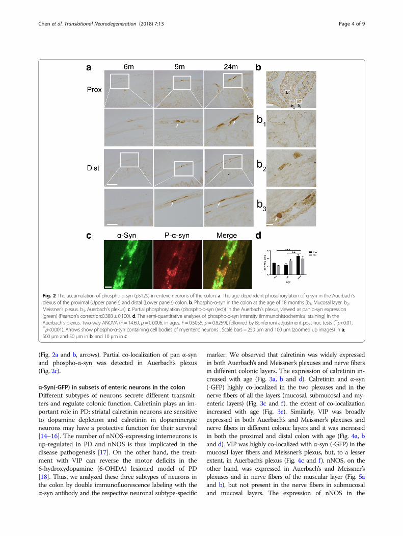
(Fig. 2a and b, arrows). Partial co-localization of pan α-synand phospho-α-syn was detected in Auerbach’s plexus(Fig. 2c).
α-Syn(-GFP) in subsets of enteric neurons in the colonDifferent subtypes of neurons secrete different transmit-ters and regulate colonic function. Calretinin plays an im-portant role in PD: striatal calretinin neurons are sensitiveto dopamine depletion and calretinin in dopaminergicneurons may have a protective function for their survival[14–16]. The number of nNOS-expressing interneurons isup-regulated in PD and nNOS is thus implicated in thedisease pathogenesis [17]. On the other hand, the treat-ment with VIP can reverse the motor deficits in the6-hydroxydopamine (6-OHDA) lesioned model of PD[18]. Thus, we analyzed these three subtypes of neurons inthe colon by double immunofluorescence labeling with theα-syn antibody and the respective neuronal subtype-specific
marker. We observed that calretinin was widely expressedin both Auerbach’s and Meissner’s plexuses and nerve fibersin different colonic layers. The expression of calretinin in-creased with age (Fig. 3a, b and d). Calretinin and α-syn(-GFP) highly co-localized in the two plexuses and in thenerve fibers of all the layers (mucosal, submucosal and my-enteric layers) (Fig. 3c and f). the extent of co-localizationincreased with age (Fig. 3e). Similarly, VIP was broadlyexpressed in both Auerbach’s and Meissner’s plexuses andnerve fibers in different colonic layers and it was increasedin both the proximal and distal colon with age (Fig. 4a, band d). VIP was highly co-localized with α-syn (-GFP) in themucosal layer fibers and Meissner’s plexus, but, to a lesserextent, in Auerbach’s plexus (Fig. 4c and f). nNOS, on theother hand, was expressed in Auerbach’s and Meissner’splexuses and in nerve fibers of the muscular layer (Fig. 5aand b), but not present in the nerve fibers in submucosaland mucosal layers. The expression of nNOS in the
Fig. 2 The accumulation of phospho-α-syn (pS129) in enteric neurons of the colon. a. The age-dependent phosphorylation of α-syn in the Auerbach’splexus of the proximal (Upper panels) and distal (Lower panels) colon. b. Phospho-α-syn in the colon at the age of 18 months (b1, Mucosal layer. b2,Meissner’s plexus. b3, Auerbach’s plexus). c. Partial phosphorylation (phospho-α-syn (red)) in the Auerbach’s plexus, viewed as pan α-syn expression(green) (Pearson’s correction:0.388 ± 0.100). d. The semi-quantitative analyses of phospho-α-syn intensity (immunohistochemical staining) in theAuerbach’s plexus. Two-way ANOVA (F = 14.69, p = 0.0006, in ages. F = 0.5055, p= 0.8259), followed by Bonferroni adjustment post hoc tests (**p<0.01,**p<0.001). Arrows show phospho-α-syn containing cell bodies of myenteric neurons . Scale bars = 250 μm and 100 μm (zoomed up images) in a;500 μm and 50 μm in b; and 10 μm in c
Chen et al. Translational Neurodegeneration (2018) 7:13 Page 4 of 9

proximal and distal colon showed no change with age(Fig. 5a, b and d), which is not consistent with the changesin the small intestine we previously reported [13]. Apartfrom this, nNOS was poorly co-localized with α-syn (-GFP)in both Auerbach’s and Meissner’s plexuses (Fig. 5c and f).
DiscussionA large body of clinical reports have suggested thatgastrointestinal dysfunction, such as constipation, is acommon non-motor symptom in PD, starting from the
prodromal stage and lasting throughout the course ofthe disease [19, 20]. The links between the gut and thebrain are manifold and both direct and indirect in PDdevelopment. Braak and colleagues have suggested thatα-syn pathology could be initiated in the gut and thenpropagate to the brain. In support of this hypothesis, ourlab has previously reported the direct spread of patho-logical α-syn from the small intestine to the lower brainstem via the vagal nerve. Additionally, there have beenstudies suggesting a connection of PD progress and gut
Fig. 3 Co-localization between calretinin and α-syn in enteric neurons in the colon. a α-syn and calretinin expression in the enteric neurons increasedwith age and highly co-localized to each other at 6, 9, and 24 months in the proximal colon. b Expression of calretinin in the distal colon also increasedwith age. c Co-localization between calretinin (CR) and α-syn in the enteric nerve terminals and neuronal cell bodies in the mucosal layers (ML) andAuerbach’s (AP) and Meissner’s plexuses (MP) at 24 months. d Semi-quantitative analyses of calretinin in the colon of BAC α-syn-GFP mice. Two-wayANOVA (F = 6.277, p = 0.014, in ages. F = 0.420, p = 0.529), followed by Bonferroni adjustment post hoc tests (*p<0.05). e Quantitative of co-localizationbetween calretinin and α-syn in the colon in BAC α-syn-GFP mice. Two-way ANOVA (F = 1.442, p= 0.275, in ages. F = 10.840, p= 0.006). f Quantitative ofco-localization between calretinin and α-syn in the mucosal layers (ML) and Auerbach’s (AP) and Meissner’s plexuses (MP) 24 months. Scale bar = 250 μmin a and b; 20 μm in c (MP, ML) and 40 μm in c (AP)
Chen et al. Translational Neurodegeneration (2018) 7:13 Page 5 of 9

inflammation. Bialecka et al. reported that the fre-quency of CARD15/NOD2 gene variants, associatedwith Crohn’s disease, may be a risk factor for spor-adic PD development [19]. In 2013, David et al. alsofound pro-inflammatory cytokines, glial fibrillaryacidic protein and Sox-10 to be elevated in the as-cending colon of PD patients [20]. Other studies haveprovided evidence of intestinal inflammation in PD
patients through fecal examinations [21, 22]. More-over, intestinal microbiota in PD patients were foundto differ from those in healthy controls: a decrease ofanti-inflammatory, butyrate-producing bacteria and anincrease in pro-inflammatory bacteria were observedin PD patient stool samples compared to those ofhealthy controls [20, 21]. In experimental models,microbiota has similarly been shown to regulate
Fig. 4 Co-localization between VIP and α-syn in enteric neurons of the colon. a and b, Age-dependent increase of VIP expression was observedin the enteric neurons of the proximal colon at 6, 9, and 24 months, partial co-localization of VIP and α-syn was seen at 24 months old of age. cCo-localization of VIP and α-syn, and the nucleus (DAPI) at 24 months in mucosal layers (MP) and Auerbach’s (AP) and Meissner’s (MP) plexuses. dSemi-quantitative analyses of VIP in the colon of BAC α-syn-GFP mice. Two-way ANOVA [F = 18.999, p = 0.0002, in ages. F = 19.950, p = 0.0008(***p),followed by Bonferroni adjustment post hoc tests (***p<0.001, **P<0.01). e Quantitative of co-localization between VIP and α-syn in the colon inBAC α-syn-GFP mice. Two-way ANOVA (F = 2.150, p = 0.159, in ages. F = 0.950, p = 0.349)]. f Quantitative of co-localization between VIP and α-synat 24 months in mucosal layers (MP) and Auerbach’s (AP) and Meissner’s (MP) plexuses. Student t test (*p<0.05, **p<0.01). Scale bar = 150 μm in aand b; 50 μm in c (AP, ML) and 15 μm in c (MP)
Chen et al. Translational Neurodegeneration (2018) 7:13 Page 6 of 9

movement deficits providing further support to the linkbetween intestinal inflammation and PD pathology [22].It is of importance to understand the involvement
of the gastrointestinal system in initiation and devel-opment of PD pathology. Our BAC-α-syn-GFP micepossess both the symptomatic and pathological fea-tures of early PD. α-Syn-GFP accumulation and phos-phorylation were found increased with age in thecolon of the transgenic mice, which corresponds toour observations of an increased gut transit time(Additional file 2: Figure S2), implying a sign of
constipation symptoms in mice. Regarding motorsymptoms, in our previous study, the BAC-α-syn-GFPmice displayed progressive behavioral impairment inopen field and rotarod tests at 7 and 12 months of age[12]. Interestingly, in the colon of the mice, phos-phorylated α-syn was detected prior to the onset ofmotor behavioral deficits, indicating that the patho-logical alterations in the gut appear before the motordeficits even occur. The differential co-localizationbetween α-syn (-GFP) with calretinin, VIP and nNOSsuggests that different subtypes of enteric neurons
Fig. 5 Expression of nNOS in enteric neurons of the colon of transgenic mice and co-localization of nNOS and α-syn. a nNOS expression ispredominant in the Auerbach’s plexus without clear age-related changes. b Expression of nNOS in distal colon at 6, 9, and 24 months. c Co-localization of nNOS and α-syn, and the nucleus (DAPI) at 24 months in Auerbach’s (AP) and Meissner’s (MP) plexuses. d Semi-quantitativeanalyses of nNOS in the colon of BAC mice. Two-way ANOVA (F = 2.723, p = 0.106, in ages. F = 0.031, p = 0.862). e Quantitative of co-localizationbetween nNOS and α-syn in the colon in BAC mice. Two-way ANOVA (F = 15.262, p = 0.001, in ages. F = 1.970, p = 0.186), followed by Bonferroniadjustment post hoc tests (*p<0.05,***P<0.001). f Quantitative of co-localization between nNOS and α-syn at 24 months in Auerbach’s (AP) andMeissner’s (MP) plexuses. Scale bar = 150 μm in a and b; and 15 μm in c
Chen et al. Translational Neurodegeneration (2018) 7:13 Page 7 of 9

may possess a distinct role in gastrointestinal dys-function in PD.
ConclusionsUsing our BAC-α-syn-GFP mouse model, we have demon-strated an age-dependent accumulation, phosphorylationand aggregation of α-syn in the enteric neurons of thecolon. Our model presents both the symptomatic andpathological features of early PD, expanding from the gutto the brain. The data indicate that this mouse model pos-sesses the uniqueness and potential for studying PD gastro-intestinal dysfunction.
Additional files
Additional file 1: Figure S1. A. The quenched α-syn-GFP signal, doubleimmunofluorescence images labeled for PGP 9.5 and α-syn, and thephospho-α-syn immunohistochemical staining in the colon. (TIF 63429 kb)
Additional file 2: Figure S2. The gut transit time in the BAC and wildtype male mice with age. (TIF26 kb)
Abbreviations6-OHDA: 6-hydroxydopamine; BAC: Bacterial artificial chromosome; C57BL/6: c57 black 6; GFP: Green fluorescent protein; LB: Lewy bodies; LN: Lewyneurites; nNOS: neuronal nitric oxide synthase; PBS: Phosphate bufferedsaline; PD: Parkinson’s disease; PFA: Paraformaldehyde; VIP: Vasoactiveintestinal peptide; α-syn: α-synuclein
AcknowledgementsWe would like to thank members of the Li lab for their intellectual inputs.We thank Mr. Sheng Yang and Mrs. Alicja Flasch for their technical support.We would also like to thank Dr. Andrew C. McCourt for the language editing.
FundingThis work was supported by the National Natural Science Foundation(81430025) and also to the supports of the Swedish Research Council (K2015-61X-22297-03-4), EU-JPND (aSynProtec) and EU-JPND (REfreAME), EU H2020-MSCA-ITN-2016 (Syndegen), BAGADILICO-Excellence in Parkinson andHuntington Research, the Strong Research Environment MultiPark(Multidisciplinary research on Parkinson’s disease), the Swedish ParkinsonFoundation (Parkinsonfonden), Torsten Söderbergs Foundation, Olle EngkvistByggmästere Foundation. W.L. is supported by a scholarship from the ChinaScholarship Council.
Availability of data and materialsThe materials used and/or analyzed during the current study are availablefrom the corresponding author on reasonable request.
Authors’ contributionsQQC and JYL conceived the study. QQC, CH and WL performed theexperiments and data analyses. MTL, and ZYW provided intellectual inputs.JYL and QQC wrote the manuscript. All authors edited and approved themanuscript.
Ethics approval and consent to participateAll work involving animals was approved by the Ethical Committees for useof laboratory animals at Lund University, Sweden, and at NortheasternUniversity, China.
Competing interestsThe authors declare that they have no competing interests.
Author details1Institute of Neuroscience, College of Life and Health Sciences, NortheasternUniversity, 110819 Shenyang, People’s Republic of China. 2Neural Plasticity
and Repair Unit, Wallenberg Neuroscience Center, Department ofExperimental Medical Science, Lund University, BMC A10, 22184 Lund,Sweden. 3Guangdong Province Key Laboratory of Brain Function andDisease, Zhongshan School of Medicine, Sun Yat-sen University, No. 74Zhongshan Rd.2, Guangzhou 510080, China. 4Institute of Heath Sciences,China Medical University, 110112 Shenyang, People’s Republic of China.
Received: 2 March 2018 Accepted: 6 June 2018
References1. Hassler R. Zur Pathologie der Paralysis agitans und des postenzephalitischen
Parkinsonismus. J Psychol Neurol. 1938;48:387–455.2. Lewy FH. Paralysis agitans: pathologische. In: Handbuch der Neurologie.
Berlin: Julius Springer; 1912.3. Tretiakoff C. Contribution a l’etude de l’anatomie pathologique du locus
niger de Soemmering avec quelques deductions relatives a la pathogeniedes troubles du tonus musculaire et de la maladie de Parkinson 1919.
4. Spillantini MG, Schmidt ML, Lee VM-Y, Trojanowski JQ, Jakes R, Goedert M. α-Synuclein in Lewy bodies. Nature. 1997;388:839–40. https://doi.org/10.1038/42166.
5. Schapira AHV, Tolosa E. Molecular and clinical prodrome of Parkinsondisease: implications for treatment. Nat Rev Neurol. 2010;6:309–17. https://doi.org/10.1038/nrneurol.2010.52 .
6. Holmqvist S, Chutna O, Bousset L, Aldrin-Kirk P, Li W, Björklund T, et al.Direct evidence of Parkinson pathology spread from the gastrointestinaltract to the brain in rats. Acta Neuropathol. 2014;128:805–20. https://doi.org/10.1007/s00401-014-1343-6 .
7. Braak H, de VRAI, Bohl J, Del Tredici K. Gastric alpha-synucleinimmunoreactive inclusions in Meissner’s and Auerbach’s plexuses in casesstaged for Parkinson’s disease-related brain pathology. Neurosci Lett. 2006;396:67–72. https://doi.org/10.1016/j.neulet.2005.11.012 .
8. Braak H, Tredici KD, Rüb U, de VRAI, Jansen Steur ENH, Braak E. Staging ofbrain pathology related to sporadic Parkinson’s disease. Neurobiol Aging.2003;24:197–211. https://doi.org/10.1016/S0197-4580(02)00065-9 .
9. Svensson E, Horváth-Puhó E, Thomsen RW, Djurhuus JC, Pedersen L,Borghammer P, Sørensen HT. Vagotomy and subsequent risk of Parkinson’sdisease. Ann Neurol. 2015;78:522–9. https://doi.org/10.1002/ana.24448 .
10. Schmidt RF, Gerhard T, editors. Human Physiology: Springer Science &Business Media; 2013.
11. Willey JM, Sherwood L, Woolverton CJ. Prescott’s microbiology. New York:McGraw-Hill; 2014.
12. Hansen C, Björklund T, Petit GH, Lundblad M, Murmu RP, Brundin P, Li J-Y. Anovel α-synuclein-GFP mouse model displays progressive motorimpairment, olfactory dysfunction and accumulation of α-synuclein-GFP.Neurobiol Dis. 2013;56:145–55. https://doi.org/10.1016/j.nbd.2013.04.017 .
13. Zhong C-B, Chen Q-Q, Haikal C, Li W, Svanbergsson A, Diepenbroek M, Li J-Y.Age-Dependent Alpha-Synuclein Accumulation and Phosphorylation in theEnteric Nervous System in a Transgenic Mouse Model of Parkinson’s Disease.Neurosci Bull. 2017;33:483–92. https://doi.org/10.1007/s12264-017-0179-1 .
14. Tsuboi K, Kimber TA, Shults CW. Calretinin-containing axons and neuronsare resistant to an intrastriatal 6-hydroxydopamine lesion. Brain Res. 2000;866:55–64. https://doi.org/10.1016/S0006-8993(00)02219-8 .
15. Mura A, Feldon J, Mintz M. The expression of the calcium binding proteincalretinin in the rat striatum: effects of dopamine depletion and L-DOPAtreatment. Exp Neurol. 2000;164:322–32. https://doi.org/10.1006/exnr.2000.7441 .
16. Kim BG, Shin DH, Jeon GS, Seo JH, Kim YW, Jeon BS, Cho SS. Relativesparing of calretinin containing neurons in the substantia nigra of 6-OHDAtreated rat Parkinsonian model. Brain Res. 2000;855:162–5. https://doi.org/10.1016/S0006-8993(99)02374-4 .
17. Padovan-Neto FE, Cavalcanti-Kiwiatkoviski R, Carolino RO, Anselmo-Franci J,Del Bel E. Effects of prolonged neuronal nitric oxide synthase inhibition onthe development and expression of L-DOPA-induced dyskinesia in 6-OHDA-lesioned rats. Neuropharmacology. 2015;89:87–99. https://doi.org/10.1016/j.neuropharm.2014.08.019 .
18. Yelkenli İH, Ulupinar E, Korkmaz OT, Şener E, Kuş G, Filiz Z, Tunçel N.Modulation of Corpus Striatal Neurochemistry by Astrocytes and VasoactiveIntestinal Peptide (VIP) in Parkinsonian Rats. J Mol Neurosci. 2016;59:280–9.https://doi.org/10.1007/s12031-016-0757-0 .
19. Bialecka M, Kurzawski M, Klodowska-Duda G, Opala G, Juzwiak S, KurzawskiG, et al. CARD15 variants in patients with sporadic Parkinson’s disease.Neurosci Res. 2007;57:473–6. https://doi.org/10.1016/j.neures.2006.11.012 .
Chen et al. Translational Neurodegeneration (2018) 7:13 Page 8 of 9

20. Hopfner F, Künstner A, Müller SH, Künzel S, Zeuner KE, Margraf NG, et al.Gut microbiota in Parkinson disease in a northern German cohort. Brain Res.2017;1667:41–5. https://doi.org/10.1016/j.brainres.2017.04.019 .
21. Keshavarzian A, Green SJ, Engen PA, Voigt RM, Naqib A, Forsyth CB, et al.Colonic bacterial composition in Parkinson’s disease. Mov Disord. 2015;30:1351–60. https://doi.org/10.1002/mds.26307 .
22. Sampson TR, Debelius JW, Thron T, Janssen S, Shastri GG, Ilhan ZE, etal. Gut Microbiota Regulate Motor Deficits and Neuroinflammation in aModel of Parkinson’s Disease. Cell. 2016;167:1469–1480.e12. https://doi.org/10.1016/j.cell.2016.11.018 .
Chen et al. Translational Neurodegeneration (2018) 7:13 Page 9 of 9


![Preclinical development of a vaccine against oligomeric alpha-synuclein … · 2017. 11. 15. · gated alpha-synuclein [6–9]. Alpha-synuclein (a-syn) is an abundant protein in the](https://static.fdocuments.us/doc/165x107/5fc07f533588d914ed7a20f9/preclinical-development-of-a-vaccine-against-oligomeric-alpha-synuclein-2017-11.jpg)