AFRL-RH-WP-TR-2011-0010 F-T Jet Fuel Reverse Mutation ...AFRL-RH-WP-TR-2011-0010 . F-T Jet Fuel...
47
AFRL-RH-WP-TR-2011-0010 F-T Jet Fuel Reverse Mutation Assay and Chromosome Aberration Test David R. Mattie Biosciences and Performance Division Applied Biotechnology Branch Teresa R. Sterner Henry M. Jackson Foundation for the Advancement of Military Medicine Wright-Patterson AFB, OH Margit Oppong-Nketiah Thomas Becker Barbara Wallner Wolfram Riedel Angela Lutterbach BSL BIOSERVICE Scientific Laboratories GmbH Behringstrasse 6/8 82152 Planegg Germany Dean J. Wagner Naval Health Research Center Environmental Health Effects Laboratory (NHRC/EHEL) Wright-Patterson AFB, OH November 2010 Interim Report for October 2007 to October 2008 Air Force Research Laboratory 711 th Human Performance Wing Human Effectiveness Directorate Biosciences and Performance Division Applied Biotechnology Branch WPAFB, OH 45433-5707 Distribution A: Approved for public release; distribution unlimited.
Transcript of AFRL-RH-WP-TR-2011-0010 F-T Jet Fuel Reverse Mutation ...AFRL-RH-WP-TR-2011-0010 . F-T Jet Fuel...

AFRL-RH-WP-TR-2011-0010
F-T Jet Fuel Reverse Mutation Assay and Chromosome Aberration
Test
David R. Mattie
Biosciences and Performance Division
Applied Biotechnology Branch
Teresa R. Sterner
Henry M. Jackson Foundation for the Advancement of Military
Medicine
Wright-Patterson AFB, OH
Margit Oppong-Nketiah
Thomas Becker
Barbara Wallner
Wolfram Riedel
Angela Lutterbach
BSL BIOSERVICE
Scientific Laboratories GmbH
Behringstrasse 6/8
82152 Planegg
Germany
Dean J. Wagner
Naval Health Research Center
Environmental Health Effects Laboratory
(NHRC/EHEL)
Wright-Patterson AFB, OH
November 2010
Interim Report for October 2007 to October 2008
Air Force Research Laboratory
711th
Human Performance Wing
Human Effectiveness Directorate
Biosciences and Performance Division
Applied Biotechnology Branch
WPAFB, OH 45433-5707
Distribution A: Approved for
public release; distribution
unlimited.
NOTICE AND SIGNATURE PAGE
Using Government drawings, specifications, or other data included in this document for any purpose other than Government procurement does not in any way obligate the U.S. Government. The fact that the Government formulated or supplied the drawings, specifications, or other data does not license the holder or any other person or corporation; or convey any rights or permission to manufacture, use, or sell any patented invention that may relate to them. This report was cleared for public release by the 88th Air Base Wing Public Affairs Office and is available to the general public, including foreign nationals. Copies may be obtained from the Defense Technical Information Center (DTIC) (http://www.dtic.mil). AFRL-RH-WP-TR-2011-0010 HAS BEEN REVIEWED AND IS APPROVED FOR PUBLICATION IN ACCORDANCE WITH ASSIGNED DISTRIBUTION STATEMENT. ______SIGNED___________________________ ______________________________________ TIMOTHY W. BUCHER, Work Unit Manager WESLEY BAUMGARNDER Applied Biotechnology Branch Biosciences and Performance Division Human Effectiveness Directorate 711th Human Performance Wing Air Force Research Laboratory This report is published in the interest of scientific and technical information exchange, and its publication does not constitute the Government’s approval or disapproval of its ideas or findings.

REPORT DOCUMENTATION PAGE Form Approved
OMB No. 0704-0188 Public reporting burden for this collection of information is estimated to average 1 hour per response, including the time for reviewing instructions, searching data sources, gathering and maintaining the data needed, and completing and reviewing the collection of information. Send comments regarding this burden estimate or any other aspect of this collection of information, including suggestions for reducing this burden to Washington Headquarters Service, Directorate for Information Operations and Reports, 1215 Jefferson Davis Highway, Suite 1204, Arlington, VA 22202-4302, and to the Office of Management and Budget, Paperwork Reduction Project (0704-0188) Washington, DC 20503. PLEASE DO NOT RETURN YOUR FORM TO THE ABOVE ADDRESS. 1. REPORT DATE (DD-MM-YYYY) 24-11-2010
2. REPORT TYPE Interim
3. DATES COVERED (From - To) 1 Oct 2007 – 31 Oct 2008
4. TITLE AND SUBTITLE F-T Jet Fuel Reverse Mutation Assay and Chromosome Aberration Test
5a. CONTRACT NUMBER
5b. GRANT NUMBER NA
5c. PROGRAM ELEMENT NUMBER
62202F 6. AUTHOR(S) Mattie, David R.*, Sterner, Teresa R.**, Oppong-Nketiah, Margit***, Becker, Thomas***, Wallner, Barbara***, Riedel, Wolfram***, Lutterbach, Angela***, Wagner, Dean J.****
5d. PROJECT NUMBER OAFW
5e. TASK NUMBER P0
5f. WORK UNIT NUMBER OAFWP002
7. PERFORMING ORGANIZATION NAME(S) AND ADDRESS(ES) HJF**, 2729 R Street, Bldg 837 BSL BIOSERVICE*** Wright-Patterson AFB, OH 45433-5707 Scientific Laboratories GmbH Behringstrasse 6/8 Naval Health Research Center**** 82152 Planegg, Germany Environmental Effects Laboratory (NHRC/EHEL), Wright-Patterson AFB, OH
8. PERFORMING ORGANIZATION REPORT NUMBER
9. SPONSORING/MONITORING AGENCY NAME(S) AND ADDRESS(ES) Air Force Materiel Command* Air Force Research Laboratory Human Effectiveness Directorate Biosciences and Protection Division Applied Biotechnology Branch Wright Patterson AFB OH 45433-5707
10. SPONSOR/MONITOR'S ACRONYM(S) 711 HPW/RHPB
11. SPONSORING/MONITORING AGENCY REPORT NUMBER AFRL-RH-WP-TR-2011-0010
12. DISTRIBUTION AVAILABILITY STATEMENT Distribution A: Approved for public release; distribution unlimited. 13. SUPPLEMENTARY NOTES 88ABW/PA cleared 3 February 2011 as 88ABW-2011-0314 14. ABSTRACT Two assays were performed in order to investigate the potential of Fischer Tropsch (F-T) jet fuel for its ability to induce genotoxicity. First, to determine its ability to induce gene mutations, the plate incorporation test was performed with the Salmonella typhimurium strains TA 98, TA 100, TA 102, TA 1535 and TA 1537. No toxic effects of the test item were noted in any of the five tester strains used up to the highest dose group evaluated (with and without metabolic activation). F-T jet fuel did not cause gene mutations by base pair changes or frameshifts in the genome of the tester strains used. Therefore, F -T jet fuel is considered to be non-mutagenic in this bacterial reverse mutation assay. Second, to investigate a possible potential of F -T jet fuel for its ability to induce structural chromosome aberrations in human lymphocytes in vitro, a chromosome aberration assay was carried out. During the described in vitro chromosomal aberration test and under the experimental conditions reported, the test item F-T jet fuel did not induce structural chromosomal aberrations in human lymphocyte cells. Therefore, F-T jet fuel is considered to be non-clastogenic in this chromosome aberration test. 15. SUBJECT TERMS jet fuels, mutagenicity, Salmonella microsome plate incorporation assay, Ames test, chromosomal aberration, human lymphocytes, clastogenicity
16. SECURITY CLASSIFICATION OF:
17. LIMITATION OF ABSTRACT SAR
18. NUMBER OF PAGES
47
19a. NAME OF RESPONSIBLE PERSON
Timothy Bucher a. REPORT U
b. ABSTRACT U
c. THIS PAGE U
19b. TELEPONE NUMBER (Include area code) NA
Standard Form 298 (Rev. 8-98) Prescribed by ANSI-Std Z39-18
i

ii
THIS PAGE INTENTIONALLY LEFT BLANK.

iii
TABLE OF CONTENTS
1.0 Executive Summary ..................................................................................................................1 2.0 Introduction ...............................................................................................................................2 3.0 Materials and Methods ..............................................................................................................3
3.1 Characterization of the Test Substance .........................................................................3 3.2 Mammalian Microsomal Fraction S9 Mix ....................................................................3 3.3 Reverse Mutation Assay ...............................................................................................3 3.4 Chromosome Aberration Assay ....................................................................................8
4.0 Results and Discussion ...........................................................................................................12 4.1 Reverse Mutation Assay .............................................................................................12 4.2 Chromosome Aberration Assay ..................................................................................24
5.0 Conclusion ..............................................................................................................................33 6.0 References ...............................................................................................................................33 Appendix A. Historical Laboratory Control Data for Reverse Mutation Assay...........................35 Appendix B. Historical Laboratory Control Data for Chromosomal Aberration Assay ..............37 List of Abbreviations .....................................................................................................................39

iv
LIST OF TABLES
Table 1. Positive controls substances, specific to S. typhimurium strain, with and without
metabolic activation ...................................................................................................................5 Table 2. S. typhimurium strains and characteristics ........................................................................5 Table 3. Control reverse mutation ranges by strain, with and without activation ..........................7 Table 4. Pre-experiment for cytotoxicity ........................................................................................9 Table 5. Results of pre-experiment toxicity testing, with and without activation ........................13 Table 6. Results of Experiment I plate-incorporation test by strain, with and without
activation ..................................................................................................................................14 Table 7. Results of Experiment II plate-incorporation test by strain, with and without
activation ..................................................................................................................................19 Table 8. Experiment III. Structural Chromosomal Aberrations, without metabolic activation:
4 hours treatment, 24 hours fixation period .............................................................................24 Table 9. Experiment III. Structural Chromosomal Aberrations, with metabolic activation: 4
hours treatment, 24 hours fixation period ................................................................................25 Table 10. Experiment IV. Structural Chromosomal Aberrations, without metabolic
activation: 24 hours treatment, 24 hours fixation period .........................................................26 Table 11. Experiment IV. Structural Chromosomal Aberrations, with metabolic activation:
4 hours treatment, 24 hours fixation period .............................................................................27 Table 12. Summary of aberration rates for Experiment III ..........................................................28 Table 13. Summary of aberration rates for Experiment IV ..........................................................29 Table 14. Experiment III. Number of polyploid cells and mitotic index: 4 hours treatment,
24 hours fixation period ...........................................................................................................30 Table 15. Experiment IV. Number of polyploid cells and mitotic index: 4 hours treatment
(with metabolic activation) 24 hours treatment (without metabolic activation), 24 hours fixation period ..........................................................................................................................31
Table 16. Experiment III. Proliferation index determined by BrdU-labeling ...............................32 Table 17. Experiment IV. Proliferation index determined by BrdU-labeling ..............................32

v
PREFACE
Funding for this project was provided through the Air Force Research Laboratory/Propulsion Directorate, Fuels Branch (Dr Tim Edwards, AFRL/RZPF) and the Alternative Fuels Certification Office (AFMC 77 AESW/LF). This research was conducted under contract FA8601-07-P-034. The program manager for the contract was LT Dean Wagner, PhD, USN Naval Health Research Center/Environmental Health Effects Laboratory (NHRC/EHEL). The technical manager for the program under which this project was conducted, Fischer Tropsch (F-T) Jet Fuel Toxicity Assessment, was Dr David Mattie. The authors acknowledge the following individuals who also served on a review panel for this program and this project: John Hinz (USAFSAM/OEHTH, Brooks City Base, TX); Gunda Reddy, PhD (USACHPPM, Aberdeen Proving Ground, MD); David Steup, PhD (Shell Oil Company, Houston, TX & Chairman, API-Toxicology Task Force); and Errol Zeiger, PhD, J.D. (Errol Zeiger Consulting, Chapel Hill, NC). This study was conducted to comply with: Chemikaliengesetz ("Chemicals Act") of the Federal Republic of Germany, Appendix 1 to §19a as amended and promulgated on June 20, 2002 (BGB l. I NrAO S. 2090), revised October 31, 2006 (BGB 1. I Nr. 50 S.2407). The study also complied with the Organisation for Economic Co-operation and Development (OECD) Principles of Good Laboratory Practice (1998). This study was assessed in compliance with the project protocol, the study plan and the Standard Operating Procedures of BSL BIOSERVICE. The study and test facility were periodically inspected by the Quality Assurance. These inspections and audits were carried out by the Quality Assurance Unit, personnel independent of staff involved in the study. The final report of the study was audited. There were no circumstances that may have affected the quality or integrity of the study. All animal procedures used in this study were in strict accordance with the European Community Council Directive of 24 November 1986 (86-609/EEC) (protection of animals used for experimental and other scientific purposes) and Decree of 20 October 1987 (87-848/EEC).

vi
THIS PAGE INTENTIONALLY LEFT BLANK.

1
Distribution A: Approved for public release; distribution unlimited.
1.0 EXECUTIVE SUMMARY
In order to investigate the potential of F-T jet fuel for its ability to induce genotoxicity, two assays were performed. First, to determine its ability to induce gene mutations, the plate incorporation test was performed with the Salmonella typhimurium strains TA 98, TA 100, TA 102, TA 1535 and TA 1537. In two independent experiments, several concentrations of the test item were used. Each assay was conducted with and without metabolic activation. The concentrations, including the controls, were tested in triplicate. The following concentrations of the test item were prepared and used in the experiments: Experiment I: 0.0316, 0.100, 0.316, 1.0, 2.5 and 5.0 µL/plate; and Experiment II: 0.1875, 0.375, 0.75, 1.5, 3.0 and 5.0 µL/plate. No precipitation of the test item on the agar plates was observed in any of the five tester strains used in Experiment I and II (with and without metabolic activation). However, a clouding of the S9 mix and the S9 substitution buffer, respectively, after addition of the test item solution was noted at a concentration of 0.316 µL/plate and higher (with and without metabolic activation) in Experiment I and at a concentration of 0.375 µL/plate and higher (with and without metabolic activation) in Experiment II. No toxic effects of the test item were noted in any of the five tester strains used up to the highest dose group evaluated (with and without metabolic activation) in Experiment I and II. No biologically relevant increases in revertant colony numbers of any of the five tester strains were observed following treatment with F-T jet fuel at any concentration level, neither in the presence nor absence of metabolic activation in Experiment I and II. The reference mutagens induced a distinct increase of revertant colonies indicating the validity of the experiments. In conclusion, it can be stated that during the described mutagenicity test and under the experimental conditions reported, F-T jet fuel did not cause gene mutations by base pair changes or frameshifts in the genome of the tester strains used. Therefore, F -T jet fuel is considered to be non-mutagenic in this bacterial reverse mutation assay. Second, to investigate a possible potential of F -T jet fuel for its ability to induce structural chromosome aberrations in human lymphocytes in vitro a chromosome aberration assay was carried out. The chromosomes were prepared 24 hours after start of treatment with the test item. The treatment interval was 4 hours with and without metabolic activation (Experiment III) and 4 hours with and 24 hours without metabolic activation (Experiment IV). Two parallel cultures were set up. Per culture 100 metaphases were scored for structural chromosomal aberrations. The following concentrations were evaluated. For Experiment III with metabolic activation (4 hours treatment, 24 hours preparation interval), 1.0, 2.5 and 5 µL/mL were used. Without metabolic activation (4 hours treatment, 24 hours preparation interval), 0.16, 0.50, 1.58 and 5 µL/mL were used. For Experiment IV with metabolic activation (4 hours treatment, 24 hours preparation interval), 3, 4 and 5 µL/mL were used. Without metabolic activation (24 hours treatment, 24 hours preparation interval), 0.50, 1.58 and 5 µL/mL were used. A reduction of the mitotic index was observed in all experiments without metabolic activation. In the experiments with metabolic activation, no reduction of the mitotic index was found. In Experiment III and IV no biologically relevant increase of the aberration rates was noted after treatment with the test item with and without metabolic activation. The aberration rates of all dose groups treated with the test item were within the historical control data of the negative control. No substantial increase in the frequencies of polyploid metaphases was found after treatment with the test item compared to the frequencies of the controls. EMS (ethylmethanesulfonate, 400 and 600 µg/mL) and CPA (cyclophosphamide, 7.5 µg/mL) were used as positive controls. They showed a

2
Distribution A: Approved for public release; distribution unlimited.
distinct and biologically relevant increase of cells with structural chromosome aberrations above our historical control level. In conclusion, it can be stated that during the described in vitro chromosomal aberration test and under the experimental conditions reported, the test item F-T jet fuel did not induce structural chromosomal aberrations in human lymphocyte cells. Therefore, F-T jet fuel is considered to be non-clastogenic in this chromosome aberration test. 2.0 INTRODUCTION
The U.S. Air Force is developing alternative fuels with the aim of decreasing dependence on foreign oil. All new fuels are potentially hazardous to Air Force personnel and require evaluation. Fischer Tropsch (F-T) fuel, the first alternative jet fuel to be certified for use in the U.S. Air Force fleet, is undergoing toxicological evaluation by the 711 Human Performance Wing, Human Effectiveness Directorate, Biosciences and Performance Division, Applied Biotechnology Branch (711 HPW/RHPB). These microbial mutagenicity and chromosome aberration assays are part of this evaluation. Microbial mutagenicity assays can rapidly detect mutagenic activity in a wide range of chemical classes. Genotoxic evaluations of chemicals utilizing microbial mutagenicity assays are short term, sensitive, and reliable tests performed in vitro for assessing mutagenic potential (Mortelmans and Zeiger, 2000). Many chemicals that result in mutagenic responses in the Salmonella assay have been found to be potentially mutagenic and carcinogenic to laboratory animals and humans (Zeiger, 1998). Bacterial reverse mutation assays use amino acid requiring strains of Salmonella typhimurium to detect point mutations, which involve substitution, addition or deletion of one or a few DNA base pairs. The principle of these bacterial reversion assays is that they detect mutations which functionally reverse mutations present in the tester strains and restore the capability to synthesize an essential amino acid (Ames et al., 1973; Claxton et al., 1987; Maron and Ames, 1983). The purpose of this study is to establish the potential of the test item to induce gene mutations in bacteria by means of two independent S. typhimurium reverse mutation assays. The S. typhimurium histidine (his) reversion system measures his -·to his + reversions. The S.
typhimurium strains are constructed to differentiate between base pair (TA 100, TA 1535 and TA 102) and frameshift (TA 98 and TA 1537) mutations (Maron and Ames, 1983). These assays directly measure heritable DNA mutations of a type which is associated with adverse effects (McCann et al., 1975; McCann and Ames, 1976; Zeiger et al., 1988; 1992). Point mutations are the cause of many human genetic diseases and there is substantial evidence that somatic cell point mutations in oncogens and tumor suppressor genes are involved in cancer in humans and experimental systems (Ames et al., 1977). The tester strains have several features that make them more sensitive for the detection of mutations. The specificity of the strains can provide useful information on the types of mutations that are induced by mutagenic agents. According to the direct plate incorporation method, the bacteria are exposed to the test item with and without metabolic activation and plated on selective medium. After a suitable period of incubation, revertant colonies are counted (Maron and Ames, 1983). At least five different concentrations of the test item are tested with

3
Distribution A: Approved for public release; distribution unlimited.
approximately half log (i.e., ) intervals between test points for an initial test. More narrow spacing between dose levels may be appropriate when a dose response is investigated. For soluble, non-toxic test compounds, the recommended maximum test concentration is 5 mg/plate or 5 µL/plate. To validate the test, reference mutagens are tested in parallel to the test item (Gatehouse et al., 1994). The purpose of the in vitro chromosome aberration (CA) test is to identify agents that cause structural chromosome aberrations in stimulated cultured human lymphocytes. The chromosomes are prepared 24 hours after start of treatment with the test item. The treatment interval is 4 hours with and without metabolic activation (Experiment III) and 4 hours with and 24 hours without metabolic activation (Experiment IV). Two parallel cultures are set up. Per culture, 100 metaphases are scored for structural chromosomal aberrations. Chromosome aberration assays aim to detect the induction of chromosome breakage (clastogenesis). Although substances produce structural chromosome aberrations by a variety of mechanisms, the endpoint is a discontinuity in the chromosomal DNA which is left unrejoined, or rejoined inaccurately to produce a mutated chromosome. Many of these changes will be lethal to the cell during the first few cell cycles after their induction, but are used as indicators of the presence of non-lethal changes such as reciprocal translocations, inversions and small deletions. These more subtle changes may have important consequences in both germ and somatic cells. Chromosomal mutations and related events are the cause of many human genetic diseases and there is substantial evidence that these changes including oncogens and tumor suppressor genes are involved in cancer in humans and experimental systems. CAs are generally evaluated in first post-treatment mitoses. The majority of chemical mutagens induce aberration of the chromatid type, but chromosome type aberrations also occur. Short-term cultures of peripheral blood lymphocytes are stimulated to divide by the addition of a mitogen (e.g., phytohemagglutinin: PHA) to the culture medium. Mitotic activity begins at about 40 hours after PHA stimulation and reaches a maximum at around 3 days. The chromosome constitution remains diploid during short-term culture. Treatments should commence at around 48 hours after culture initiation, when the cells are actively proliferating and should be sampled first at about 24 hours later (1 - 1.5 fold of the normal cell cycle time), i.e., at 72 hours after culture initiation (the cycle time of lymphocytes, except first cycle averages about 11 - 17 hours). The cell cycle of the actual lymphocyte cultures is monitored using a BrdU (bromodeoxyuridine)-labeling technique. If toxicity occurs or cell cycle delay is indicated, an additional sampling time should be used at about 24 hours after the first fixation (e.g., 48 hours after beginning of treatment or 96 hours after culture initiation). At least three concentrations of the test item should be used at fixation time (24 hours). The highest concentration should be in the toxic range and should show a significant reduction in mitotic index or in degree of cell confluency (50 percent or greater). The lowest dose should be in the range of the negative control. In the additional sampling time (delayed fixation time = 48 hours) during the second experiment, the same dose that induced a suitable degree of mitotic inhibition at the earlier fixation time should be chosen. Though the purpose of the assay is to detect structural chromosome aberrations, it is important to report polyploidy and/or

4
Distribution A: Approved for public release; distribution unlimited.
endoreduplication when this is seen. To validate the test, reference mutagens are tested in parallel to the test item. 3.0 MATERIALS AND METHODS
3.1 Characterization of the Test Substance
F-T jet fuel, Synthetic Jet Fuel (Batch No.: POSF5109), was provided by Air Force Research Laboratory, Propulsion Directorate, Fuels Branch (AFRL/RZPF). The purity of the test substance was 100 percent. Routine hygienic procedures were sufficient to assure personnel health and safety. 3.2 Mammalian Microsomal Fraction S9 Mix An advantage of using in vitro cell cultures is the accurate control of the concentration and exposure time of cells to the test item under study. However, the bacteria most commonly used in these reverse mutation assays do not possess the enzyme system which, in mammals, is known to convert promutagens into active DNA damaging metabolites necessary (Bradley et al., 1981). In order to overcome this major drawback, an exogenous metabolic system was added in the form of mammalian microsome enzyme activation mixture. The S9 liver microsomal fraction was prepared at BSL BIOSERVICE GmbH. Male Wistar rats were induced with Phenobarbital (80 mg/kg bodyweight) and β-naphtoflavone (100 mg/kg bodyweight) for three consecutive days by the oral route. The rats were humanely euthanized and the livers harvested. Livers were homogenized and then centrifuged at 9000 g for 20 minutes. The resulting supernatant containing the microsomes was frozen in ampoules of 2.0 and 4.5 mL and stored at ≤-75°C. The protein concentration in the S9 preparation (Lot: 140607 (Experiments I through IV) and Lot 291107 (Experiments III and IV)) were 34 mg/mL and 35 mg/mL, respectively. The S9 cofactor solution preparation was performed according to Ames et al. (1973). Ice-cold sodium-ortho-phosphate buffer (pH 7.4, 100 mM) was added to sterilized pre-weighed reagents to give final concentrations of 8 mM MgCb, 33 mM KCI, 5 mM Glucose-6-phosphate, and 4 mM NADP in the S9 mix. This solution was mixed with the liver supernatant fluid (9.5 parts and 0.5 parts, respectively). During the experiment, the S9 mix was stored on ice. Quality control determinations were performed to verify the biological activity in the S. typhimurium assay and the sterility of the mix. 3.3 Reverse Mutation Assay
The test item was dissolved in ethanol and diluted prior to treatment. The solvent was compatible with the survival of the bacteria and the S9 activity. Positive and negative controls were included in each experiment. Strain specific positive controls were included in the assay, which demonstrated the effective performance of the test. Negative solvent controls, consisting of solvent or vehicle alone as well as untreated controls were treated in the same way as the treatment groups. Positive controls were tester strain specific (Table 1). The stability of the

5
Distribution A: Approved for public release; distribution unlimited.
positive control substances in solution is unknown but a mutagenic response in the expected range is sufficient evidence of biological stability. Table 1. Positive Controls Substances, Specific to S. typhimurium Strain, with and without
Metabolic Activation
S. typhimurium
Strain
Control Supplier Purity Solvent Concentration
Without metabolic activation
TA 100, TA 1535
Sodium azide, NaN3 Merck ≥99% Aqua dest 10 µg/plate
TA 98, TA 1537
4-nitro-o-phenylene-diamine,4-NOPD
Fluka >97% DMSO 10 µg/plate
TA 102 Methyl methane sulfonate, MMS
Sigma 99.0% Aqua dest 1 µg/plate
With metabolic activation
TA 98, TA 100, TA 1535, TA 1537
2-aminoanthracene, 2-AA
Aldrich 96% DMSO 2.5 µg/plate
TA 102 2-aminoanthracene, 2-AA
Aldrich 96% DMSO 10 µg/plate
Notes: Aqua dest = top quality distilled water; DMSO = dimethylsulfoxide 3.3.1 Bacteria. Five strains of S. typhimurium were used (Table 2). The Salmonella tester strains TA 98, TA 100, TA 102 and TA 1535 were obtained from Xenometrix, San Diego, CA, USA. Tester strain TA 1537 was obtained from MOLTOX, Inc., NC, USA. Bacteria were stored as stock cultures in ampoules with nutrient broth (OXOID, Basingstoke, Hampshire, UK) supplemented with DMSO (dimethyl sulfoxide, approximately 8 percent volume/volume) over liquid nitrogen.
Table 2. S. typhimurium Strains and Characteristics
Strain Histidine Mutation Mutation Type
TA98 his D 3052 R-factor: frame shift mutations TA 100 his G 46 R-factor: base-pair substitutions TA 1535 his G 46 base-pair substitutions TA 1537 his C 3076 frame shift mutations TA 102 his G 428 (PAQ1) R-factor: base-pair substitutions
All S. typhimurium strains contain mutations in the histidine operon, thereby imposing a requirement for histidine in the growth medium. They contain the deep rough (rfa) mutation, which deletes the polysaccharide side chain of the lipopolysaccharides of the bacterial cell surface. This increases cell permeability of larger substances. The other mutation is a deletion of the uvrB gene coding for the DNA excision repair system resulting in an increased sensitivity

6
Distribution A: Approved for public release; distribution unlimited.
in detecting many mutagens. This deletion also includes the nitrate reductase (chI) and biotin (bio) genes (bacteria require biotin for growth). The tester strains TA 98, TA 100 and TA 102 contain the R-factor plasmid, pkM 101. These strains are reverted by a number of mutagens that are detected weakly or not at all with the non R-factor parent strains. pkM 101 increases chemical and spontaneous mutagenesis by enhancing an error-prone DNA repair system which is normally present in these organisms (Maron and Ames, 1983; Mortelmans and Zeiger, 2000). The properties of the S. typhimurium strains with regard to membrane permeability, ampicillin- and tetracycline-resistance as well as normal spontaneous mutation rates are checked regularly as required by Ames et al. (1973). In this way it is ensured that the experimental conditions set up by Ames are fulfilled. 3.3.2 Preparation of Bacteria and Media. Samples of each tester strain were grown by culturing the bacteria for 12 hours at 38.5°C in nutrient broth to the late exponential or early stationary phase of growth (approximately 109 cells/mL). The nutrient medium consists of 8 g Nutrient Broth and 5 g NaCl per liter. A solution of 125 µL ampicillin (10 mg/mL) (TA 98, TA 100, TA 102) was added in order to retain the phenotypic characteristics of the strain. Vogel-Bonner Medium E agar plates with 2 percent glucose used in the Ames test were prepared by BSL BIOSERVICE or provided by an appropriate supplier. Quality controls were performed. Vogel-Bonner Medium E agar plates contained 15 g agar, 20 mL Vogel-Bonner salts, and 50 mL glucose-solvent (40 percent) per liter. Vogel-Bonner-salts consist of 10 g MgS04 X 7 H20; 100 g citric acid; 175 g NaNH4HP04 x 4 H20; and 500 g K2HP04 per liter. The overlay agar contained 7.0 g agar; 6.0 g NaCl; 10.5 mg L-histidine x HCl x H20; and 12.2 mg biotin per liter. Agars were sterilized at 121°C in an autoclave.
3.3.3 Exposure Concentration Determination. The toxicity of the test item was determined with tester strains TA 98 and TA 100 in a pre-experiment. Eight concentrations were tested for toxicity and induction of mutations with three plates each. The experimental conditions in this pre-experiment were the same as described below for the main Experiment I (plate incorporation test). F-T fuel was tested in the pre-experiment at 0.00316, 0.0100, 0.0316, 0.100, 0.316, 1.0, 2.5 and 5.0 µL/plate. Test item concentrations to be applied in the main experiments were chosen according to the results of the pre-experiment, with a maximum concentration of 5.0 µL/plate. The concentration range covered two logarithmic decades. Two independent experiments were performed with the following concentrations. Experiment I tested 0.0316, 0.100, 0.316, 1.0, 2.5 and 5.0 µL/plate. Experiment II used 0.1875, 0.375, 0.75, 1.5, 3.0 and 5.0 µL/plate. As the results of the pre-experiment were in accordance with the criteria described above, these were reported as a part of Experiment I. 3.3.4 Plate Incorporation Method. For the plate incorporation method, 100 µL test solution (each dose level, solvent control, negative control or reference mutagen solution (positive control)); 500 µL S9 mix (for testing with metabolic activation) or S9 mix substitution buffer

7
Distribution A: Approved for public release; distribution unlimited.
(for testing without metabolic activation); 100 µL bacteria suspension preparation preculture of the strain); and 2000 µL overlay agar were mixed in a test tube and poured over the surface of a minimal agar plate. For each strain and dose level, including the controls, three plates were used. After solidification, the plates were inverted and incubated at 37°C for at least 48 hours in the dark. 3.3.5 Cytotoxicity. The colonies were counted using a ProtoCOL counter (Meintrup DWS Laborgerate GmbH, Lähden, Germany). If precipitation of the test item precluded automatic counting, the revertant colonies were counted by hand. In addition, tester strains with a low spontaneous mutation frequency such as TA 1535 and TA 1537 were counted manually. Cytotoxicity was determined either by a clearing or a diminution of the background lawn (indicated as "B" in the result tables) or a reduction in the number of revertants down to a mutation factor of approximately ≤0.5 in relation to the solvent control. A test was considered acceptable, if, for each strain: the bacteria demonstrate their typical responses to ampicillin (TA 98, TA 100, TA 102) the control plates with and without S9 mix are within the ranges shown in Table 3 (mean
values of the spontaneous reversion frequency are within the historical control data range) corresponding background growth on negative control, solvent control and test plates is
observed, or the positive controls show a distinct enhancement of revertant rates over the control plate.
Table 3. Control Reverse Mutation Ranges by Strain, with and without Activation
Strain -S9 +S9
TA 98 18 - 54 16 - 71 TA 100 75 -171 83 – 168 TA 1535 6 - 30 6 – 31 TA 1537 5 - 31 6 – 36 TA 102 166 - 394 153 - 594
3.3.6 Evaluation of Mutagenicity. The Mutation Factor is calculated by dividing the mean value of the revertant counts by the mean values of the solvent control (the exact, not the rounded values, are used for calculation). A test item is considered as mutagenic if a clear and dose-related increase in the number of revertants occurs and/or a biologically relevant positive response for at least one of the dose groups occurs in at least one tester strain with or without metabolic activation. A biologically relevant increase is strain dependent. In tester strains TA 100 and TA 102, the number of reversions must be at least twice as high as the solvent control. In tester strains TA 98, TA 1535 and TA 1537, the number of reversions should be at least three times higher than the reversion rate of the solvent control (Kier et al., 1986). According to Organisation for Economic Co-operation and Development (OECD) guidelines (1997a), the biological relevance of the results is the criterion for the interpretation of results. A

8
Distribution A: Approved for public release; distribution unlimited.
statistical evaluation of the results is not regarded as necessary. A test item producing neither a dose related increase in the number of revertants nor a reproducible biologically relevant positive response at any of the dose groups is considered to be non-mutagenic in this system. 3.4 Chromosome Aberration Assay
The test item F-T jet fuel was dissolved in 500 µL/mL ethanol; no precipitation of the test item was indicated. During this assay, 1 percent of this solution was diluted in cell culture medium (RPMI 1640) prior to treatment. The solvent was compatible with the survival of the cells and the S9 activity. After dilution with cell culture medium, precipitation of the test item appeared in a concentration of 5 µL/mL. Positive and negative controls were included. Negative controls, consisting of vehicle alone and treated in the same way as the treatment groups were included. Concurrent negative and/or solvent controls were performed. The positive control, without metabolic activation, was ethylmethanesulfonate (EMS, Sigma, purity > 98 percent) dissolved in nutrient medium at final concentrations of 400 and 600 µg/mL. These solutions were prepared on the day of the experiment. The positive control with metabolic activation was cyclophosphamide (CPA, Sigma, purity ≥ 98 percent) dissolved in nutrient medium at a final concentration of 7.5 µg/mL. The stability of CPA at room temperature was good. The stability of the positive control substance in solution was proven by the mutagenic response in the expected range. At 25°C only 3.5 percent of its potency was lost after 24 hours (Gallelli, 1967). The solution was stored in aliquots at 15°C. Blood samples were obtained from healthy donors not receiving medication. In each experiment, blood was collected only from a single donor to reduce inter-individual variability. Blood samples were drawn by venous puncture and collected in heparinized tubes. Before use, the blood was stored under sterile conditions at 4°C for a maximum of 4 hours. 3.4.1 Pre-Experiment for Toxicity and Exposure Concentrations. According to the relevant guidelines, the highest recommended dose is 5 mg/mL, 5 µL/mL or 10 mM, whichever is the lowest. The highest dose group evaluated in the pre-experiment was 5 µL/mL. The relative mitotic index was used as a parameter for toxicity. The concentrations evaluated in the main experiment were based on the results obtained in the pre-experiment.

9
Distribution A: Approved for public release; distribution unlimited.
Table 4. Pre-Experiment for Cytotoxicity
Duplicate cultures were treated at each concentration. The selection of the concentrations used in Experiments III and IV were based on data from the pre-experiment. In Experiment III, concentrations used with metabolic activation were 0.125, 0.25, 0.5, 1.0, 2.5 and 5 µL/mL, and without metabolic activation were 0.0016, 0.005, 0.016, 0.05, 0.16, 0.50, 1.58 and 5 µL/mL. In Experiment IV, concentrations used with metabolic activation were 0.5, 1, 2, 3, 4 and 5 µL/mL, and without metabolic activation were 0.00016, 0.0005, 0.0016, 0.005, 0.016, 0.05, 0.16, 0.50, l.58 and 5 µL/mL. The cells were treated in Experiment III (with and without metabolic activation) for 4 hours with the test item. The metaphases were prepared 24 hours after the treatment. In Experiment IV with metabolic activation, the cells were treated for 4 hours and prepared 24 hours after the treatment. In Experiment IV without metabolic activation, the cells were treated for 24 hours

10
Distribution A: Approved for public release; distribution unlimited.
and prepared at the end of the treatment. The dose group selection for microscopic analyses of chromosomal aberrations was based on the mitotic index in accordance with the guidelines. The following concentrations were selected in the main experiments for the microscopic analysis: Experiment III with metabolic activation (4 hours treatment, 24 hours preparation interval, 1.0, 2.5 and 5 µL/mL) and without metabolic activation (4 hours treatment, 24 hours preparation interval, 0.16, 0.50, 1.58 and 5 µL/mL); Experiment IV with metabolic activation (4 hours treatment, 24 hours preparation interval, 3, 4 and 5 µL/mL) and without metabolic activation (24 hours treatment, 24 hours preparation interval, 0.50, 1.58 and 5 µL/mL). At least three analyzable concentrations of the test item were used for the 24 hours preparation. 3.4.2 Culture Initiation, Treatment and Preparation. Blood cell cultures were set up within 4 hours after collection. The following volumes were added to the plastic culture vessels: 8.45 mL culture medium, 0.90 mL whole blood, 0.50 mL PHA, 0.10 mL antibiotic solution, and 0.05 mL heparin. All incubations were done at 37C in humidified atmosphere with 5 percent CO2.
Experiment III. Short time exposure 4 hours (with and without S9 mix): After 48 hours, the culture medium was replaced with serum-free medium containing the test item (without metabolic activation) or serum-free medium containing the test item with 50 µL/mL S9 mix (with metabolic activation). After 4 hours, the cells were spun down by centrifugation at 1000 rotations per minute for 5 minutes. The supernatant with the dissolved test item was discarded and the cells were resuspended in PBS. The washing procedure was repeated once as described. After washing, the cells were resuspended in complete cell culture medium. The cells were prepared 24 hours after the beginning of the treatment.
Experiment IV. Long time exposure 24 hours (without S9 mix): After 48 hours the culture
medium was replaced with complete medium (with 15 percent FCS) containing the test item without S9 mix. This medium was not changed until preparation of the cells 24 hours after the beginning of the treatment.
Experiment IV. Exposure time with metabolic activation: The cells were treated as
described for Experiment III (with metabolic activation). The cells were prepared 24 hours after the beginning of the treatment.
Two to three hours before harvesting, Colcemid was added to the cultures (final concentration 0.2 µg/mL). The cultures were harvested by centrifugation 24 hours after beginning of treatment. The supernatant was discarded and the cells were resuspended in approximately 5 mL hypotonic solution (0.4 percent KCI). The cell suspension was incubated at room temperature for 20 minutes. After removal of the hypotonic solution by centrifugation, the cells were fixed with 3 + 1 methanol + glacial acetic acid. The fixation procedure was repeated twice. Slides were prepared by dropping the cell suspension on to a clean microscopic slide. The cells were then stained with Giemsa or in accordance with the fluorescent plus Giemsa technique. 3.4.3 Proliferation Index. The negative control and the highest dose group evaluated were treated in the presence of BrdU to measure the proliferation index and/or replication time of the

11
Distribution A: Approved for public release; distribution unlimited.
cultured lymphocytes. The proliferation index was determined by scoring the number of first, second and third metaphases in 100 cells per culture. The proliferation index (PI) was calculated as:
(1) Where M1 = first mitosis, M2 = second mitosis, M3 = third mitosis, with respect to the beginning of the exposure.
3.4.4 Analysis of Metaphase Cells. All slides, including those of positive and negative controls, were independently coded before microscopic analysis. Evaluation of the cultures was performed (according to the standard protocol of the "Arbeitsgruppe der Industrie, Cytogenetik" (Engelhardt, 1987)) using OLYMPUS microscopes with 100x oil immersion objectives. Structural chromosomal aberrations including breaks, fragments, deletions, exchanges and chromosomal disintegrations were recorded. Gaps were recorded as well, but not included in the calculation of the aberration rates. The definition of a gap was as follows: an achromatic region (occurring in one or both chromatids) independent of its width. The remaining visible chromosome regions should not be dislocated longitudinally or laterally. At least 200 well spread metaphases per concentration and control were scored for cytogenetic damage. Metaphases with 46±2 centromer regions were included in the analysis (Engelhardt, 1987; Scott et al., 1990). To describe a cytotoxic effect, the mitotic index (percent cells in mitosis) was determined. Additionally the number of polyploidy cells was scored. 3.4.5 Data Recording. The data generated were recorded in the raw data file. The results are presented in tables, including experimental groups with the test item plus negative and positive controls. The experimental unit is the cell and therefore, the percentage of cells with structural aberration was evaluated. Different types of chromosome aberrations are listed with their numbers of frequencies for experimental and control groups. Gaps were recorded separately and reported but generally not included in the aberration frequency. Concurrent measurements of cytotoxicity were also recorded. 3.4.6 Evaluation of Results. The chromosomal aberration assay is considered acceptable if it meets the following criteria. The number of aberrations found in the negative and/or solvent controls must fall within the range of historical laboratory control data: 0.0 - 4.0 percent. Additionally, the positive control substance should produce biologically relevant increases in the number of cells with structural chromosome aberrations. There are several criteria for determining a positive result. A clear and dose-related increase in the number of cells with aberrations must be present. A biologically relevant response is observed for at least one of the dose groups, which is higher than the laboratory negative control range (up to 4.0 percent aberrant cells). According to the OECD guidelines (1997b), the biological relevance of the results will be the criterion for the interpretation of results, a statistical evaluation of the results is not regarded as necessary. However, for the interpretation of the data, both biological and, if evaluated,

12
Distribution A: Approved for public release; distribution unlimited.
statistical significance should be considered together. A test item is considered to be negative if there is no biologically relevant increase in the percentages of aberrant cells above concurrent control levels, at any dose group. 4.0 RESULTS AND DISCUSSION
4.1 Reverse Mutation Assay
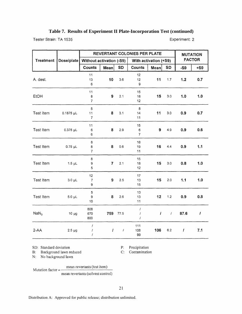
Pre-experimental testing for toxicity was carried out in two strains of S. typhimurium (TA 98 and TA 100), with and without S9 activation. Toxicity may be detected by a clearing or rather diminution of the background lawn or a reduction in the number of revertants down to a mutation factor of approximately ≤0.5 in relation to the solvent control. None of the pre-experiment conditions tested resulted in toxicity, according to this definition (Table 5). The test item F-T jet fuel was investigated for its potential to induce gene mutations according to the plate incorporation test (Experiment I and II, Tables 6 and 7) using S. typhimurium strains TA 98, TA 100, TA 1535, TA 1537 and TA 102. In two independent experiments, several concentrations of the test item were used. Each assay was conducted with and without metabolic activation. The concentrations, including the controls, were tested in triplicate. No precipitation of the test item on the agar plates was observed in any of the five tester strains used in Experiment I and II (with and without metabolic activation). However, a clouding of the S9 mix and the S9 substitution buffer after addition of the test item solution was noted at a concentration of 0.316 µL/plate and higher (with and without metabolic activation) in Experiment I and at a concentration of 0.375 µL/plate and higher (with and without metabolic activation) in Experiment II. No toxic effects of the test item were noted in any of the five tester strains used up to the highest dose group evaluated (with and without metabolic activation) in Experiment I and II. No biologically relevant increases in revertant colony numbers of any of the five tester strains were observed following treatment with F-T jet fuel at any concentration level, neither in the presence nor absence of metabolic activation in Experiment I and II, as compared to solvent controls, positive controls or historical data (Appendix A). The reference mutagens induced a distinct increase of revertant colonies, indicating the validity of the experiments.

13
Distribution A: Approved for public release; distribution unlimited.
Table 5. Results of Pre-Experiment Toxicity Testing, with and without Activation

14
Distribution A: Approved for public release; distribution unlimited.
Table 6. Results of Experiment I Plate-Incorporation Test by Strain, with and without
Activation

15
Distribution A: Approved for public release; distribution unlimited.
Table 6. Results of Experiment I Plate-Incorporation Test (continued)

16
Distribution A: Approved for public release; distribution unlimited.
Table 6. Results of Experiment I Plate-Incorporation Test (continued)

17
Distribution A: Approved for public release; distribution unlimited.
Table 6. Results of Experiment I Plate-Incorporation Test (continued)

18
Distribution A: Approved for public release; distribution unlimited.
Table 6. Results of Experiment I Plate-Incorporation Test (continued)

19
Distribution A: Approved for public release; distribution unlimited.
Table 7. Results of Experiment II Plate-Incorporation Test by Strain, with and without
Activation

20
Distribution A: Approved for public release; distribution unlimited.
Table 7. Results of Experiment II Plate-Incorporation Test (continued)
21
Distribution A: Approved for public release; distribution unlimited.
Table 7. Results of Experiment II Plate-Incorporation Test (continued)

22
Distribution A: Approved for public release; distribution unlimited.
Table 7. Results of Experiment II Plate-Incorporation Test (continued)

23
Distribution A: Approved for public release; distribution unlimited.
Table 7. Results of Experiment II Plate-Incorporation Test (continued)

24
Distribution A: Approved for public release; distribution unlimited.
4.2 Chromosomal Aberration Assay
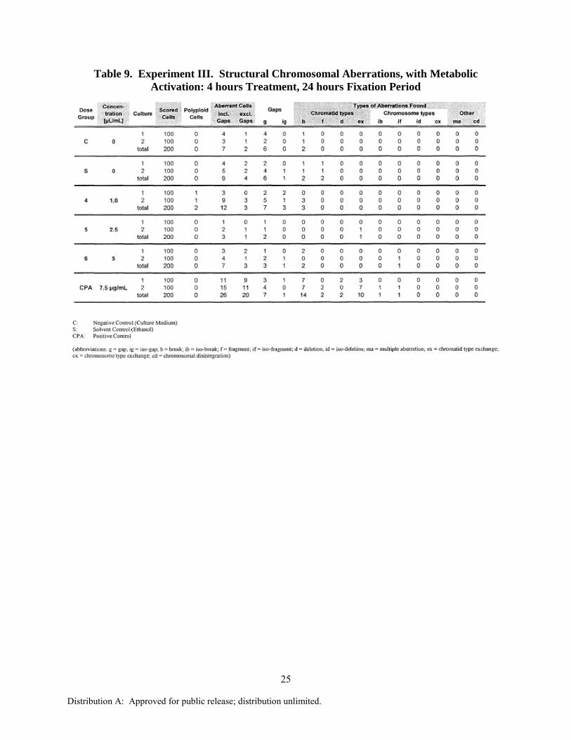
The test item F-T jet fuel was investigated for a possible potential to induce structural chromosomal aberrations in human lymphocytes in vitro in the absence and presence of metabolic activation by S9 homogenate. The chromosomes were prepared 24 hours after start of treatment with the test item. The treatment interval in Experiment III was 4 hours with and without metabolic activation (Tables 8 and 9, respectively). The treatment interval in Experiment IV was 4 hours with metabolic activation and 24 hours without metabolic activation (Tables 10 and 11, respectively). Two parallel cultures were set up per dose group. Per culture, 100 metaphases were scored for structural chromosomal aberrations. A summary of the results is presented in Tables 12 and 13. Precipitation of the test item was noted with and without metabolic activation after the incubation at a concentration of 5 µL/mL.
Table 8. Experiment III. Structural Chromosomal Aberrations, without Metabolic
Activation: 4 hours Treatment, 24 hours Fixation Period
25
Distribution A: Approved for public release; distribution unlimited.
Table 9. Experiment III. Structural Chromosomal Aberrations, with Metabolic
Activation: 4 hours Treatment, 24 hours Fixation Period

26
Distribution A: Approved for public release; distribution unlimited.
Table 10. Experiment IV. Structural Chromosomal Aberrations, without Metabolic
Activation: 24 hours Treatment, 24 hours Fixation Period

27
Distribution A: Approved for public release; distribution unlimited.
Table 11. Experiment IV. Structural Chromosomal Aberrations, with Metabolic
Activation: 4 hours Treatment, 24 hours Fixation Period

28
Distribution A: Approved for public release; distribution unlimited.
Table 12. Summary of Aberration Rates for Experiment III
29
Distribution A: Approved for public release; distribution unlimited.
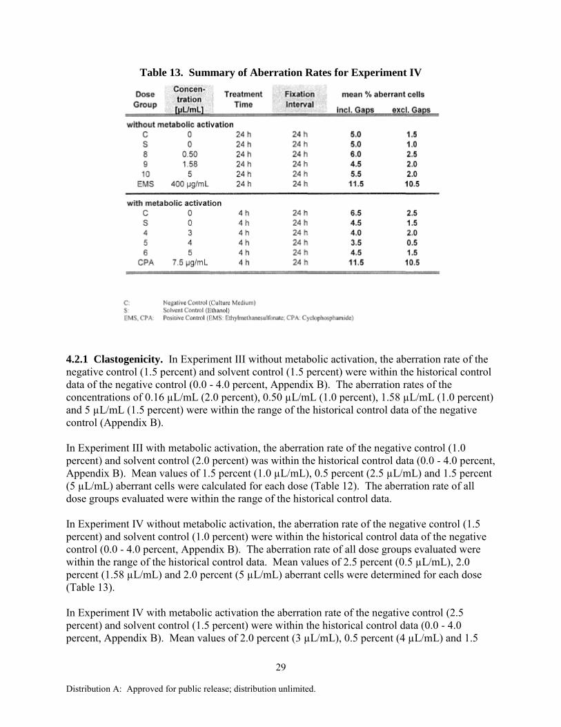
Table 13. Summary of Aberration Rates for Experiment IV
4.2.1 Clastogenicity. In Experiment III without metabolic activation, the aberration rate of the negative control (1.5 percent) and solvent control (1.5 percent) were within the historical control data of the negative control (0.0 - 4.0 percent, Appendix B). The aberration rates of the concentrations of 0.16 µL/mL (2.0 percent), 0.50 µL/mL (1.0 percent), 1.58 µL/mL (1.0 percent) and 5 µL/mL (1.5 percent) were within the range of the historical control data of the negative control (Appendix B). In Experiment III with metabolic activation, the aberration rate of the negative control (1.0 percent) and solvent control (2.0 percent) was within the historical control data (0.0 - 4.0 percent, Appendix B). Mean values of 1.5 percent (1.0 µL/mL), 0.5 percent (2.5 µL/mL) and 1.5 percent (5 µL/mL) aberrant cells were calculated for each dose (Table 12). The aberration rate of all dose groups evaluated were within the range of the historical control data. In Experiment IV without metabolic activation, the aberration rate of the negative control (1.5 percent) and solvent control (1.0 percent) were within the historical control data of the negative control (0.0 - 4.0 percent, Appendix B). The aberration rate of all dose groups evaluated were within the range of the historical control data. Mean values of 2.5 percent (0.5 µL/mL), 2.0 percent (1.58 µL/mL) and 2.0 percent (5 µL/mL) aberrant cells were determined for each dose (Table 13). In Experiment IV with metabolic activation the aberration rate of the negative control (2.5 percent) and solvent control (1.5 percent) were within the historical control data (0.0 - 4.0 percent, Appendix B). Mean values of 2.0 percent (3 µL/mL), 0.5 percent (4 µL/mL) and 1.5

30
Distribution A: Approved for public release; distribution unlimited.
percent (5 µL/mL) aberrant cells were found for each dose (Table 13). The aberration rate of all dose groups evaluated were within the range of the historical control data. 4.2.2 Toxicity. Toxic effects of the test item were observed without metabolic activation. In Experiment III, a biologically relevant decrease of the relative mitotic index (decrease below 70 percent relative mitotic index) was noted at doses of 0.50 µL/mL and higher. The highest dose groups evaluated (0.50, 1.58 and 5 µL/mL) induced a decrease of the relative mitotic index down to 68, 51 and 60 percent, respectively (Table 14). In Experiment IV, a biologically relevant decrease of the relative mitotic index (decrease below 70 percent relative mitotic index) was noted at a concentration of 5 µL/mL; the relative mitotic index was 54 percent (Table 15). In Experiment III and IV with metabolic activation, no biologically relevant decrease of the relative mitotic index was noted at the concentrations evaluated (Tables 8 and 11).
Table 14. Experiment III. Number of Polyploid Cells and Mitotic Index: 4 hours
Treatment, 24 hours Fixation Period

31
Distribution A: Approved for public release; distribution unlimited.
Table 15. Experiment IV. Number of Polyploid Cells and Mitotic Index: 4 hours
Treatment (with metabolic activation) 24 hours Treatment (without metabolic activation),
24 hours Fixation Period
4.2.3 Polyploid Cells. Tables 14 and 15 show the occurrence of polyploid metaphases. No biologically relevant increase in the frequencies of polyploid cells was found after treatment with the test item. EMS (400 and 600 µL/mL) and CPA (7.5 µL/mL) were used as positive controls. They showed a distinct and biologically relevant increase of cells with structural chromosome aberrations above our historical control level.

32
Distribution A: Approved for public release; distribution unlimited.
4.2.4 Proliferation Index. The BrdU-technique was used to detect a possible cell cycle delay after treatment with the test item. In Experiment III, the values of the proliferation index of the negative controls were 1.37 (without metabolic activation) and 1.33 (with metabolic activation) (Table 16). The proliferation index of the highest dose groups evaluated was 1.35 (without metabolic activation) and 1.32 (with metabolic activation) at a concentration of 5 µL/mL. In Experiment IV, the values of the proliferation index of the negative controls were 1.41 (without metabolic activation) and 1.21 (with metabolic activation) (Table 17). The proliferation index of the highest dose groups evaluated without metabolic activation (5 µL/mL) was 1.40. The proliferation index of the highest dose group evaluated with metabolic activation (5 µL/mL) was 1.27. There was no biologically relevant decrease of the proliferation index.
Table 16. Experiment III. Proliferation Index Determined by BrdU-Labeling
Table 17. Experiment IV. Proliferation Index Determined by BrdU-Labeling

33
Distribution A: Approved for public release; distribution unlimited.
5.0 CONCLUSION
In conclusion, it can be stated that during the described mutagenicity test and under the experimental conditions reported, F-T jet fuel did not cause gene mutations by base pair changes or frameshifts in the genome of the tester strains used. Therefore, F-T jet fuel is considered to be non-mutagenic in this bacterial reverse mutation assay. During the described in vitro chromosomal aberration test and under the experimental conditions reported, the test item F-T jet fuel did not induce structural chromosomal aberrations in human lymphocyte cells. Therefore, F-T jet fuel is considered to be non-clastogenic in this chromosome aberration test. 6.0 REFERENCES
Ames, B.N., Durston, W.E., Yamasaki, E. and Lee, F.D. (1973) Carcinogens are mutagens: A
simple test system combining liver homogenates for activation and bacteria for detection. Proc. Natl. Acad. Sci. U.S.A. 70, 2281-2285.
Ames, B.N., McCann, J. and Yamasaki, E. (1977) Methods for detecting carcinogens and mutagens with the Salmonella/mammalian microsome mutagenicity test. In: Handbook of Mutagenicity Test Procedures. Kilbey, B.I., Legator, M., Nichols, W. and Ramel, C. (eds.), Elsevier, Amsterdam, pp. 1-17.
Bradley, M.O., Bhuyan, B., Francis, M.C., Langenbach, R., Peterson, A. and Huberman, E. (1981) Mutagenesis by chemical agents in V79 Chinese hamster cells: A review and analysis of the literature. A report of the Gene-Tox Program. Mutat. Res. 87, 81-142.
Claxton, L.D., Allen, J., Auletta, A., Mortelmans, K., Nestmann, E. and Zeiger, E. (1987). Guide for the Salmonella typhimurium/mammalian microsome tests for bacterial mutagenicity . Mutat. Res. 189, 83-91.
Engelhardt, G. (1987) Cytogenetik Standard-Protokoll zur cytogenetischen Auswertung von Mito- und Meiosechromosomen bei der Routineuntersuchung. Arbeitsgruppe der Industrie.
Gallelli, J.F. (1967) Stability studies of drugs used in intravenous solutions – I. Am. J. Hosp. Pharm. 24, 425-433.
Gatehouse D., Haworth, S., Cebula, T., Gocke, E., Kier, L., Matsushima, T., Melcion, C., Nohmi, T., Ohta, T., Venitt, S., Zeiger, E. (1994) Recommendation for the performance of bacterial mutation assays. Mutat. Res. 312, 217-233.
Kier, L.D., Brusick, D.J., Auletta, A.E., von Halle, E.S., Brown, M.M., Simmon, V.F., Dunkel, V., McCann, J., Mortelmans, K., Prival, M., Rao, T.K. and Ray, V. (1986) The Salmonella
typhimurium/mammalian microsomal assay. A report of the U.S. Environmental Protection Agency Gene-Tox-Program. Mutat. Res. 168, 69-240.
Maron, D.M. and Ames, B.N. (1983) Revised methods for the Salmonella mutagenicity test. Mutat. Res. 113, 173-215.
McCann, J. and Ames, B.N. (1976) Detection of carcinogens as mutagens in the Salmonella/microsome test: Assay of 300 Chemicals: Discussion. Proc. Natl. Acad. Sci. U.S.A. 73, 950-954.
McCann, J., Choi, E., Yamasaki, E. and Ames, B.N. (1975) Detection of carcinogens as mutagens in the Salmonella/microsome test: Assay of 300 Chemicals. Proc. Natl. Acad. Sci.

34
Distribution A: Approved for public release; distribution unlimited.
U.S.A. 72, 5135-5139. Mortelmans, K. and Zeiger, E. (2000) The Ames Salmonella/microsome mutagenicity assay.
Mutat. Res. 455, 29-60. OECD. (1997a) OECD Guideline for Testing of Chemicals: Bacterial Reverse Mutation Assay.
Organisation for Economic Co-operation and Development, Paris. Guideline 471. OECD. (1997b) OECD Guideline for Testing of Chemicals: In Vitro Mammalian Chromosome
Assay. Organisation for Economic Co-operation and Development, Paris. Guideline 473. OECD. (1998) OECD Series on Principles of Good Laboratory Practice and Compliance
Monitoring. Number 1. OECD Principles on Good Laboratory Practice (as revised in 1997). Organisation for Economic Co-operation and Development, Paris. ENV/MC/CHEM(98)17.
Scott, D., Danford N., Dean B. and Kirkland D. (1990) In vitro chromosome aberrations assays. In UKEMS Subcommittee on Guidelines for Mutagenicity Testing. Kirkland D.I. (ed.) Cambridge University Press, Cambridge, New York. Pp. 62-86.
Zeiger, E. (1998) Identification of rodent carcinogens and noncarcinogens using genetic toxicity tests: Premises, promises, and performance. Regul. Toxicol. Pharmacol. 28, 85-95.
Zeiger, E., Anderson, B., Haworth, S., Lawlor, T. and Mortelmans, K. (1992) Salmonella mutagenicity tests: V. Results from the testing of 311 chemicals. Environ. Mol. Mutagen. 19 (Suppl. 21), 2-141.
Zeiger, E., Anderson, B., Haworth, S., Lawlor, T. and Mortelmans, K. (1988) Salmonella mutagenicity tests: IV. Results from the testing of 300 chemicals. Environ. Mol. Mutagen. 11 (Suppl. 12), 1-157.

35
Distribution A: Approved for public release; distribution unlimited.
APPENDIX A. HISTORICAL LABORATORY CONTROL DATA FOR REVERSE
MUTATION ASSAY
Table A-1. Historical laboratory control data of the negative control (in 2004 - 2006)
without S9 (-S9)
Table A-2. Historical laboratory control data of the positive control (in 2004 - 2006)
without S9 (-S9)
36
Distribution A: Approved for public release; distribution unlimited.
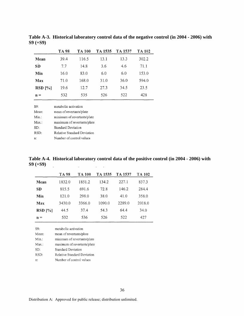
Table A-3. Historical laboratory control data of the negative control (in 2004 - 2006) with
S9 (+S9)
Table A-4. Historical laboratory control data of the positive control (in 2004 - 2006) with
S9 (+S9)

37
Distribution A: Approved for public release; distribution unlimited.
APPENDIX B. HISTORICAL LABORATORY CONTROL DATA FOR
CHROMOSOMAL ABERRATION ASSAY
Table B-1. Historical laboratory control data of the negative control (2000 - 2006)

38
Distribution A: Approved for public release; distribution unlimited.
Table B-2. Historical laboratory control data of the positive control (2000 - 2006)

39
Distribution A: Approved for public release; distribution unlimited.
LIST OF ABBREVIATIONS
bio biotin mutation BrdU bromodeoxyuridine CA chromosome aberration chI nitrate reductase mutation CPA cyclophosphamide DMSO dimethyl sulfoxide EMS ethylmethanesulfonate F-T Fischer Tropsch his histidine OECD Organisation for Economic Co-operation and Development PHA phytohemagglutinin PI proliferation index rfa deep rough mutation