Affinity Chromatography as a Tool for Quantification of Interactions Between Drug Molecules and
32
14 Affinity Chromatography as a Tool for Quantification of Interactions Between Drug Molecules and Their Protein Targets Piotr Draczkowski, Dariusz Matosiuk and Krzysztof Jozwiak Faculty of Pharmacy with Division of Medical Analytics, Medical University of Lublin Lublin Poland 1. Introduction Separations in affinity chromatography are based on specific biological interactions between (bio)molecules and in many aspects resemble processes by which these species interact in a living organism. The technique is widely used in biomedical sciences to separate and detect certain molecules based on their defined specificity to other (bio)molecules immobilized on a stationary phase. Moreover,it can also be used for quantitative determination of affinity interactions and their physiological and pharmacological role in a living system. In both cases the same basic physico-chemical effects (hydrophobic interactions, electrostatic, hydrogen bonds etc.) lead to description of the equilibrium state between unbound, free- floating molecules and those forming a complex with a target. This allows the use of affinity chromatography system as a model for analyzing interactions that normally occur in human body. This approach was suggested by analytical chemists over 50 years ago, e.g. Soczewinski and Bieganowska stated in 1969: "... If the body is regarded as an extremely complex chromatographic system, in which the blood plays the role of the developing solvent, a certain parallelism can be expected between the behaviour of drugs and their chromatographic parameters in common "simple" partition systems." Therefore, affinity chromatography is currently successfully employed in medicinal chemistry projects for detailed characterization of interactions between drug molecules and their protein targets. This type of liquid chromatography is referred to as analytical or quantitative affinity chromatography (QAC). In most applications the assay is performed in HPLC systems using a column with a protein immobilized on the surface of the stationary phase. Relatively simple and rapid procedures (time of a single assay average between 5 to 15 min.), ability to multiple use of the same column without significant loss of properties of immobilized protein (Jozwiak et al., 2004) and the possibility to automate the analysis process, make this technique a promising method for screening and determination of relative affinities of a series of analyzed drug molecules. Loun & Hage (1996) reported that theirs column was stable even to 500-1000 injections of an analyte. Above mentioned advantages of this method additionally increase www.intechopen.com
Transcript of Affinity Chromatography as a Tool for Quantification of Interactions Between Drug Molecules and

14
Affinity Chromatography as a Tool for Quantification of Interactions Between Drug
Molecules and Their Protein Targets
Piotr Draczkowski, Dariusz Matosiuk and Krzysztof Jozwiak Faculty of Pharmacy with Division of Medical Analytics, Medical University of Lublin
Lublin Poland
1. Introduction
Separations in affinity chromatography are based on specific biological interactions between
(bio)molecules and in many aspects resemble processes by which these species interact in a
living organism. The technique is widely used in biomedical sciences to separate and detect
certain molecules based on their defined specificity to other (bio)molecules immobilized on
a stationary phase. Moreover,it can also be used for quantitative determination of affinity
interactions and their physiological and pharmacological role in a living system. In both
cases the same basic physico-chemical effects (hydrophobic interactions, electrostatic,
hydrogen bonds etc.) lead to description of the equilibrium state between unbound, free-
floating molecules and those forming a complex with a target. This allows the use of affinity
chromatography system as a model for analyzing interactions that normally occur in human
body. This approach was suggested by analytical chemists over 50 years ago, e.g.
Soczewinski and Bieganowska stated in 1969:
"... If the body is regarded as an extremely complex chromatographic system, in which the blood plays the role of the developing solvent, a certain parallelism can be expected between the behaviour of drugs and their chromatographic parameters in common "simple" partition systems."
Therefore, affinity chromatography is currently successfully employed in medicinal
chemistry projects for detailed characterization of interactions between drug molecules and
their protein targets. This type of liquid chromatography is referred to as analytical or
quantitative affinity chromatography (QAC).
In most applications the assay is performed in HPLC systems using a column with a protein
immobilized on the surface of the stationary phase. Relatively simple and rapid procedures
(time of a single assay average between 5 to 15 min.), ability to multiple use of the same
column without significant loss of properties of immobilized protein (Jozwiak et al., 2004)
and the possibility to automate the analysis process, make this technique a promising
method for screening and determination of relative affinities of a series of analyzed drug
molecules. Loun & Hage (1996) reported that theirs column was stable even to 500-1000
injections of an analyte. Above mentioned advantages of this method additionally increase
www.intechopen.com

Affinity Chromatography
276
reproducibility of obtained results. Thus, affinity chromatography is considered by many
medicinal chemists as good alternative to tedious in vitro tests using cell cultures (Jozwiak et
al., 2004). It also allows to reduce time needed to perform the assays (Jozwiak et al., 2005).
Special procedures were developed in order to transform retention of a substance or it
elution profiles to characterize the interaction between a drug and the target protein.
Therefore, various protocols can be used to characterize binding equilibrium constants, the
kinetics of drug-protein complex association/dissociation, relative amount of bound drug,
number of binding sites in the system or forces responsible for complex formation.
Simultaneous application of more than one active substance on the column allows defining
interactions between them during binding to immobilized target. It is also possible to
determine the affinity of various isomeric forms of the drug to immobilized protein, which
is extremely important in a pharmaceutical research (Chen & Hage, 2006). Data obtained by
QAC show high correlation with derived from reference methods and can be further used in
studies of quantitative structure - activity relationships (QSAR) (Markuszewski & Kaliszan,
2002; Jozwiak et al., 2004).
This chapter reviews current aspects of application of QAC including basic issues of protein
immobilization on the surface of the stationary phase, advantages and disadvantages of
zonal and frontal elution techniques and vast information which can be provided by
competition displacement in QAC studies. The last subsection provides short review of
application of these techniques in medicinal chemistry investigations.
2. Immobilization of target proteins on the surface of the stationary phase
QAC assay requires a column where one of the partner of the drug-protein complex is
immobilized on the surface of the stationary phase. Both, drug molecules and protein targets
may be immobilized on the chromatographic bead particles. The latter option seems to be
more versatile as it allows to investigate different types of active substances without having
to change the column bead. It directly permits to compare properties of a series of
substances on the basis of obtained chromatograms. The way in which the ligand is attached
to its support is a key factor in any type of affinity chromatography. Immobilization
methods for soluble cytosolic proteins are well established (Taylor, 1991; Turková, 1999;
Kim & Hage, 2006; Scheil et al., 2006) and they are based mainly on chemical or physical
mechanisms. Physical methods include protein adsorption (physical or ionic adsorption) or
protein entrapment within insoluble gel matrix through which only small drug molecules
can diffuse. The advantage of these methods is relatively small perturbation of the protein
native structure, on the other hand immobilisation forces are weak and promotes the leak of
adsorbed protein from the support during use, especially while temperature, pH or ionic
strength is changed. Chemical immobilization methods mainly include protein attachment
to the stationary phase by covalent bonds or cross-linking reactions. Covalent linkage may
alter the native tertiary structure of the protein and cause a change in drug binding
properties, but the target associates strongly with the support preventing the desorption
phenomenon and increasing a column lifetime. The functional groups that usually take part
in this binding are amino, epoxy, carboxyl, sulfhydryl, hydroxyl, diol and phenolic groups
which according to the mode of linkage lead to a wide variety of binding reactions such as
diazotization, amide bond formation, arylation, Schiff’s base formation and amination
www.intechopen.com

Affinity Chromatography as a Tool for Quantification of Interactions Between Drug Molecules and Their Protein Targets
277
(Girelli & Mattei, 2005). Another type of immobilization method is biospecific adsorption. It
uses the binding between the ligand of interest and a secondary ligand attached to the
support. Although a variety of secondary ligands can be used for this purpose, two of the
most common are avidin and streptavidin for the adsorption of biotin-containing
compounds, and protein A or protein G for the adsorption of antibodies (Akerstrom &
Bjorck, 1986; Wilchek & Bayeras, 1990; Bayer & Wilchek, 1996; Wilchek & Bayeras, 1998;
Page & Thorpe, 2002, as cited in: Kim & Hage, 2006).
Various supports are commercially available or have been specifically developed for the immobilization processes, including silica based derivatized matrices (Narayanan et al., 1990; Ruhn et al., 1994; Mateo et al., 2000) and monoliths (Josic & Buchacher, 2001; Lebert, 2008). An important factor is a structure of the support used in preparation of affinity column, since it determines accessibility of protein active sites to substrates. The ideal support must be inert, stable and resistant to mechanical strength, so it can retain its tertiary structure, and this ensures the substrate accessibility to interact with the active sites. Other physical properties, such as porosity, pore size distribution and charge are also important, because they influence the kinetic process (Girelli & Mattei, 2005). Variety of available methods of protein connection with chromatographic beads, gives the opportunity to select and optimize right immobilization method for binding of our protein of interest. It should be noted that the immobilized ligand must as close as possible imitate the behaviour which it exhibits in natural conditions. The method chosen for protein binding should not disrupt the structure crucial for drug binding and provide proper orientation to eliminate any negative steric interactions. Proper immobilization allows retaining activity of the protein on the column, and even its conformational mobility (Beigi et al., 2004), which allows to study allosteric interactions (Chen et al., 2004).
In last years new techniques were developed to immobilize membrane proteins on the surface
of chromatographic bead particles to describe the nature of interactions between drug
molecules and target receptors. This is very valuable from pharmacological point of view
considering that the membrane and transmembrane receptor proteins are targets for almost
75% of current pharmaceuticals (Landry & Gies, 2008). The first membrane protein that was
analysed using QAC was glucose transporter present on the surface of red blood cells
(GLUT1) (Yang and Lundahl, 1995). Two immobilization techniques were used in this system:
in first proteoliposomes or cytoskeleton depleted membrane vesicles containing GLUT1 were
immobilized in the pores of size exclusion chromatography beads (Superdex) (Gottschalk et
al., 2000) by technique of repeated freezing - thawing (Lundqvist et al., 1998). In the second
approach whole cells were immobilized on the surface of beads with positively charged
groups (Zeng et al., 1997) or columns with wheat germ lectin agarose gel beads (Gottschalk &
Lundahl, 2000). Currently, there are many reports in literature describing the columns with
different immobilized receptors and membrane transporters. Most of them use silicon particles
with immobilized phospholipids (IAM) (Pidgeon & Venkataram, 1989) which connect parts of
the cell membranes of tissues showing high expression of the receptor or cultured cells
transformed with receptor gene. It is known as cellular membrane affinity chromatography:
CMAC, or CMC. Using this method columns with immobilized nicotinic receptor (nAChR)
subtypes ┙3┚4, ┙4┚2, ┙3┚2, ┙4┚4, ┙7 and subunits ┚4 and ┙3 (Zhang et al., 1998; Wainer et al.,
1999; Moaddel et al., 2005; Moaddel et al., 2008) purinergic receptors, both acting as ion
channels (P2X family) (Trujillo et al., 2007) as well as G-protein coupled receptors (P2Y family)
www.intechopen.com

Affinity Chromatography
278
(Moaddel et al., 2007), the P2Y-like receptor GRP17 (Temporini et al., 2009), ┚2 adrenergic
receptor (Beigi et al., 2004) or the μ and κ opioid receptors (Beigi & Wainer, 2003) were
constructed. Multi-receptor columns containing nAChRs, ┛-amino-butyric acid receptors
(GABA) and N-methyl D-aspartate receptors (NMDA) (Moaddel et al., 2002) were also
developed. Kitabatake and co-workers (2008) constructed multi-receptor column and
confirmed presence of two types of nAChRs: ┙7 nAChR and heteromeric nAChRs but also
GABA, and NMDA receptors on the surface of 1321N1 and A172 astrocytoma cell lines. The
results indicate that the columns can be used to characterize binding affinities of small
molecules to each of the receptors, and that this approach can be used to probe the expression
of endogenous membrane receptors. With similar immobilization technique beads with
transmembrane transport proteins such as P-glycoprotein (Zhang, 2000), human organic anion
transporter protein (hOAT1 and hOAT2) (Kimura et al., 2007) and human organic cationic
transporter protein (hOCT1) were prepared (Moaddel et al., 2005).
Alternative method was used to immobilize ┙1 adrenergic receptor (Yu et al., 2005) or
muscarinic receptor (Yuan et al., 2005). In this case, the membrane fragments with an
interesting protein were subjected to adsorption on the surface of silica particles under
vacuum and with use of ultrasounds. Since the phospholipid bilayer fragments show a
spontaneous ability to connect, they enfold bead particles forming compact, durable coating
of cell membrane. Same approach was used to construct multi-receptor column for the
simultaneous determination of drug interactions with the purinergic, P2Y1 and histamine 1
receptors (Moaddel et al., 2010) and multi-receptor column prepared using the glioma cell
membranes in order to identify the types of receptors on the surface of these cells by their
specific ligands (Kitabatake et al., 2008).
Immobilization of membrane proteins on the inner surface of silica capillaries (open tubular
column) is also applied. For this purpose, fragments of cell membrane containing membrane
protein bind non-covalently with a capillary using the avidin-biotin pair. Capillaries with
immobilized P-glycoprotein (Moaddel et al., 2004), and recently with immobilized
cannabinoid receptors (CB1/CB2) (Moaddel et al., 2011) were developed using this approach.
When a native structure of a target protein cannot be obtained during immobilization a drug
can be used as an immobilized ligand. However, in this situation any steric interactions
should be eliminated by connecting drug molecule to the bead using a spacer with adequate
length and hydrophobicity, in order to secure free access to the binding site on the protein
molecule. It is important not to connect the drug using functional groups that participate in
binding to the target protein. Application of QAC with immobilized drug molecules is
particularly useful for identifying biomolecules targets for substances with known
therapeutic effect. It is also used as a pre-clinical method of detection of undesired
interactions with other system biomolecules (Guiffant et al., 2007).
3. Elution techniques
3.1 Zonal chromatographic studies of drug-protein binding
One method of QAC is a technique known as the zonal elution technique. It was first used
by Dunn and Chaiken (1974) as modified low-pressure liquid chromatography method used
to investigate the retention of Staphylococcus nuclease on the column with immobilized
www.intechopen.com

Affinity Chromatography as a Tool for Quantification of Interactions Between Drug Molecules and Their Protein Targets
279
thymidine-5'-phosphate-3'-aminophenylphosphate. Zonal elution method can also be
applied using a standard HPLC apparatus equipped with temperature control unit. This
method involves injection on the column small volume of analyte solution and then isocratic
elution with mobile phase, which usually has a composition and pH reflecting the
physiological conditions. Compared to frontal elution (described in subsection 3.2) small
amount of analyte is needed to perform the assay in zonal format. Detection is carried out
on-line, however there are applications with off-line detection when chromatographic
systems has low efficiency (Dunn & Chaiken, 1974).
In result of the analysis the retention factor also called capacity factor is determined. It is
expressed by formula k = (tr/tm) - 1, where tr is the retention time of the test substance, and tm
is the column dead time. Comparing the value of k for different substances, we can
determine their relative affinity for the immobilized protein. Typical chromatogram
obtained from zonal chromatography studies is shown in Figure 1. Studies on the affinity of
benzodiazepines and related coumarins done by Noctor and colleagues using immobilized
human serum albumin (HSA) (Noctor et al., 1993, as cited in Bertucci et al., 2003) have
shown a strong correlation (r = 0.999) between the percentage of bounded drug measured
by the standard method of ultrafiltration, and the data obtained from chromatographic
studies expressed as k/(k + 1).
Fig. 1. Typical chromatogram obtained from zonal AC. Comparison of elution peak profiles of ketamine (solid) and negative control phenylbutazon (dashed line) on the column with immobilized nicotinic receptor (nAChR), subtype ┙3┚4. Adapted with permission from Jozwiak, K., Ravichandran, S., Collins, J. R., & Wainer, I. W. (2004). Interaction of noncompetitive inhibitors with an immobilized ┙3┚4 nicotinic acetylcholine receptor investigated by affinity chromatography, quantitative-structure activity relationship analysis, and molecular docking. J Med Chem, Vol. 47, No. 16, pp. 4008-4021. Copyright 2011 American Chemical Society.
Similar results were reported by Cheng et al. (2004). In this case, the working curve between
literature values of % drug bounded to HSA (by either ultrafiltration or dialysis method)
and k/(k + 1) factor determined by chromatography method showed good linearity with the
www.intechopen.com

Affinity Chromatography
280
coefficient of 0.96, which is acceptable considering diversity of drugs tested and the variety
of %binding data resources used.
According to the Equation 1 (Chaiken, 1987, as cited in Cheng et al., 2004):
a L
M
K mk =
V (1)
compound retention factor is closely related with equilibrium constant of binding reaction to immobilized protein target. This allows to define an order relative binding strength of different compounds by comparing their retention factors (Xuan & Hage, 2005) In Equation 1, VM is the void volume of the column (i.e., the elution volume of a non-retained solute), mL is the moles of active binding site and Ka is association equilibrium constant for the injected solute at this site .
Zonal elution technique is also used to determine the forces that play a fundamental role in the formation of drug-protein complex. Changing assay conditions (e.g., pH, ionic strength, content of organic modifier) allows to determine which factors affect the most binding reaction. For example, retention dependence on mobile phase pH indicates a considerable contribution of Coulomb interactions in the binding of a drug. In turn, addition of organic modifier can accelerate the elution of analyte by disturbing the hydrophobic interactions (Hage & Chen, 2006). It also allows to examine the change in binding of drugs when standard physiological system conditions will be changed as a result of pathological lesions (Basiaga & Hage, 2010).
Temperature studies allow to define changes in enthalpy and entropy of interactions between the drug and immobilized protein. It follows from the equilibrium constant depending on temperature, which can be described by the Equation 2:
ln a
ΔH ΔSK = +
RT R
(2)
where ΔH express a change of enthalpy and ΔS entropy change in a place of interaction, R is the gas constant and T is absolute temperature. So if the system meets the assumption that the number of binding sites (mL) does not change with temperature, and this is a single-site binding then lnKa plotted against 1/T should be linear with a slope equal to -ΔH/R, and intercept to ΔS. The total energy change can be calculated using Equation 3 (Kirkwood & Oppenheim, 1961, as cited in Yang & Hage, 1993):
ln aΔG = RT K (3)
The method, however, requires earlier determination of Ka at given temperature, for example, by conducting self-competition studies with test compound at different temperatures. It is also possible to designate the enthalpy and entropy of binding directly from the value of retention factor. In this case, if a binding has a single-site character, the plot of lnk against 1/T is linear with a slope equal to -ΔH/R and the intercept equal to [ΔS/R + ln(mL/VM)]. However, the calculation of the value of ΔH and ΔS requires the prior determination of concentration of binding sites for the analyte (mL/VM) (Yanda & Hage, 1993).
www.intechopen.com

Affinity Chromatography as a Tool for Quantification of Interactions Between Drug Molecules and Their Protein Targets
281
3.2 Frontal affinity chromatography (FAC) in drug-protein interaction investigations
Another commonly used method of determining the drug-target protein affinity is the frontal technique. Although this elution format is practically no longer used in analytical applications of chromatography, it is still successfully applied in QAC assays, and has some advantages over zonal elution methods. For the first time it was used in 1975 by Kasai and Ishii. In contrast to zonal elution, a test substance is applied continuously on the column as an addition to the mobile phase in specified concentration. The result is essentially a titration of active sites within the column. As the mobile phase flows through the column, the analyte saturates binding sites on the immobilized protein and we can observe a gradual increase of the amount of unbound analyte leaving the column. This produces a vertical rise in the chromatographic trace, called breakthrough curve which ends or plateaus when the immobilized target is fully saturated. Initial, relatively flat portion of the chromatographic traces represents the non-specific and specific binding of the tested compound to the cellular membranes and the target. Inflection points of breakthrough curves shift to shorter breakthrough times (volumes) as the ligand concentration increases (see Figure 2).
Fig. 2. Typical breakthrough curves of two analyte concentration [A]1 (higher) and [A]2 (lower). Vo represents breakthrough volume of the ligand in the absence of the binding event.
Extent and asymmetry of obtained chromatographic profile are related to the analyte-target
protein binding kinetics. Measuring the breakthrough times for several concentrations and
fitting the results to equations based on various reaction models allow to characterize a
nature of binding affinity and the amount of immobilized target on a column. This is
accomplished by plotting number of apparent moles of analyte required to reach the mean
point of the breakthrough curve (1/mLapp) versus 1/[A] (where [A] is applied analyte
concentration). According to Equation 4 (Loun & Hage, 1992), in case of single site reaction,
plot (1/mLapp) versus 1/[A] should give a linear response (see Figure 3) with a slope equal to
1/(KAmL) and an intercept of 1/mL. Dividing the intercept by the slope allows to obtain
information about equilibrium binding constant of analysed interaction.
www.intechopen.com

Affinity Chromatography
282
1
L ALapp
A
m K Am =
+ K A or 1 1 1
Lapp A L L
= +m K m A m
(4)
A similar approach, but using a combination of both nonlinear and linear fits, can be used
for more complex systems that involve multisite interactions (Jacobson et al., 1993; Tweed et
al., 1997, as cited in Hage & Chen, 2006).
Fig. 3. Examples of double-reciprocal frontal analysis plots for systems with (a) single-site binding and (b) multisite binding. Reprinted with permission from: Loun, B., & Hage, D. S. (1994). Chiral separation mechanisms in protein-based HPLC columns. 1. Thermodynamic studies of (R)- and (S)-warfarin binding to immobilized human serum albumin. Anal Chem, Vol. 66, No. 21, pp. 3814-3822; Tweed, S. A., Loun, B., & Hage, D. S. (1997). Effects of ligand heterogeneity in the characterization of affinity columns by frontal analysis. Anal Chem, Vol. 69, No. 23, pp. 4790-4798. Copyright 2011 American Chemical Society.
The simplest binding event, involving the interaction of a ligand with a single type of
binding site can also by described by Equation 5. There may be multiple, equivalent sites in
a given target molecule but the model assumes their independence.
www.intechopen.com

Affinity Chromatography as a Tool for Quantification of Interactions Between Drug Molecules and Their Protein Targets
283
0
0
t
d
BV V =
A + K (5)
In this basic FAC equation (5), two variables are present: [A]0 (ligand infusion concentration) and V-V0 (breakthrough volume V for the ligand corrected by the breakthrough volume of the ligand in the absence of the binding event V0). This simple equation indicates that once Bt (corresponding to the dynamic capacity of the affinity column for the ligand) and the concentration of the analyte are known, the dissociation constant can be determined from a single measurement of its V-V0. In order to determine Bt, various concentrations of the compound are pumped through the column and the corresponding V-V0 values are measured. The analysis of changes in V-V0 versus [A]0 by means of Lineweaver–Burk type double reciprocal plot or standard nonlinear regression analysis, Bt (equal to the reciprocal of the y intercept) and dissociation constant Kd (in M, expressed as negative reciprocal of the x intercept) are obtained (Calleri et al., 2009). Reliable measurement of V0 requires a suppression of specific binding or application of a saturating ligand concentration. In case of membrane proteins it can be done by measuring retention of marker using a column constructed with membranes from cells that do not express the target protein (Moaddel et al., 2005). Other approach that can be employed to differentiate between specific and non-specific interactions is parallel chromatography system in which analysed compounds are simultaneously applied on a column with immobilized membranes containing target protein (experimental) and on a column with immobilized membranes from the same cell line that does not express the target protein (control). The assumption is that all of the non-target interactions between a test compound or protein and the cellular membranes will be the same for the control and experimental cell lines and will be reflected in the chromatographic retention on the control column. Then the difference in compound retention between control and experimental column will reflect only the specific binding. This system may be applied in both, zonal and frontal chromatographic studies (Baynham et al., 2002, as cited in Moaddel & Wainer, 2006).
In frontal QAC connected with mass spectrometry (FAC-MS), ligand is sequentially infused at increasing concentrations, but with no washing steps between infusions. This allows to determine binding parameters in a single experiment. This is accomplished by infusing a FAC–MS column sequentially starting with the lowest of a series of concentrations of analyte. For this approach, referred to as a modified staircase, the summed concentrations ([A]0+y) refer to initial concentration of the ligand for the first step of the staircase but for the second step of the staircase, it will be the sum of the initial concentration plus the concentration of the second step. Similarly, the concentration of the ligand for the third step of the staircase will be the sum of the initial, second and third steps, and so on for the remaining concentrations. A plot of [A]0+y versus reciprocal breakthrough volume supports the determination of Bt and Kd by linear regression analysis. This type of assays referred to as direct measurements might not always be advantageous, especially if each tested ligand required unique mass spectrometric conditions. (Chan et al., 2003; Slon-Usakiewicz et al., 2005). Indirect methods will be described further (see subsection 3.3) since they require usage of known competitive marker ligand. Figure 4 illustrates an ideal chromatogram that would arise from the application of this procedure. Note that successively higher void marker (a compound that has no affinity for the immobilized protein target and gives the same elution front whether the target protein is present in the column or not) concentrations are applied as well, to ensure accurate measurement of V0.
www.intechopen.com

Affinity Chromatography
284
Fig. 4. Depiction of a typical ‘modified staircase’ experiment to evaluate Kd for a protein target (immobilized in the column) and a small molecule ligand. (a) The ligand (blue) is infused at increasing concentrations starting from an initial (i) to a final (j) concentration along with a void marker (red). (b) The reciprocal of the breakthrough volumes, 1/(Vj-V0), are plotted against the summed ligand concentrations, [A]0+yj, to produce a linear correlation from which the Kd value can be determined from the y intercept. The total amount of immobilized protein (Bt) in the column is then obtained from the slope . Reprinted from Drug Discovery Today, Vol. 10, No. 6, Slon-Usakiewicz, J. J., Ng, W., Dai, J. R., Pasternak, A., & Redden, P. R. (2005). Frontal affinity chromatography with MS detection (FAC-MS) in drug discovery, pp. 409-416, Copyright 2011, with permission from Elsevier.
The association constant measured from frontal chromatography can be directly related to the
retention factor obtained from zonal elution chromatography using the same column. Kim &
Wainer (2008) reported a linear relationship (r2 = 0.9993, n = 7) between the standard
association constants from frontal analysis and retention factors from zonal elution using
reference drugs analysed on a column with immobilized HSA. This standard plot was later
used for rapid determination of association constants of various drugs which show low to
www.intechopen.com

Affinity Chromatography as a Tool for Quantification of Interactions Between Drug Molecules and Their Protein Targets
285
medium binding affinity to HSA. Determination of association constants was as fast as 1.5 min
and showed a high correlation to equilibrium dialysis or ultrafiltration. The combination of
frontal and zonal chromatography for determining association constants showed several
advantages, one being rapid determination of association constant of drug to HSA.
Other notable advantages include an ease of automation and simultaneous ability to
distinguish association constants of chiral compounds at the same time. Authors suggested
that the same approach could be used for studying interaction of other drugs and proteins
and should further improve overall drug screening process.
There are several advantages of frontal over zonal chromatographic method. In the FAC
technique, the dynamic capacity of the affinity column for the ligand and the dissociation
constant for the interaction can be obtained from a single set of experiments. In case of
zonal elution technique to determine number of binding sites a separate assay or self-
competition studies are required This makes this approach valuable in characterizing the
properties of a column and in obtaining accurate measurements of binding affinity and
activity.
The direct methods described above are applicable for a broad Kd range from
submicromolar to low millimolar. The lower limit to Kd determination is dictated by the
detectability of a given ligand, and while this is both compound- and detector-dependent.
Zonal chromatographic approach requires a protein stationary phase concentration much
greater than the applied ligand concentration to ensure the experiment is conducted in the
linear region of the binding isotherm. Thus consumption of target protein is increased and
miniaturization capabilities are lower. Smaller column leads to lower amount of
immobilized protein but also require lower ligand concentrations and this challenges the
detectability of the ligand. From the same reasons application of zonal chromatography is
limited to low and mid-micromolar Kd range because either strong (and thus difficult to
remove from a column) or weak (and thus not well retained) interactions can lead to low
concentrations of ligand at the detector (Chan et al., 2003 ).
Frontal elution technique is particularly useful in studies on solvent and temperature
influence on drug interaction with target molecule. As it was mentioned earlier, analyte
retention shifts may be due to alterations in either the affinity or number of binding sites. In
frontal chromatography this is not a problem, since data on both affinity and activity are
provided in the same experiment. However, frontal techniques need to use much larger
amount of analyte (Hage & Chen 2006).
For more theoretical information and practical considerations about frontal and zonal
elution techniques see comprehensive chapters about QAC in book “Handbook of Affinity
Chromatography” (2006) edited by prof. David S. Hage.
3.3 Competition displacement studies
QAC allows to study relationships between different drugs interacting with the same target
protein by observing the effect of the addition of one compound on the retention of the
second. In zonal technique, the retention of a drug is measured in specially prepared mobile
phase with addition of constant, known concentration of competitive agent. Consecutive
www.intechopen.com

Affinity Chromatography
286
injections of an analyte can be applied to a series of mobile phases with increasing
concentration of competitive agent and changes in the retention as a function of competitor
concentration allow describing its interaction with test analyte. In case of direct single site
competitive interactions we should see direct, linear dependence of reciprocal of analyte
retention factor (1/k) on concentration of competitor added to the mobile phase [I].
Increasing value of 1/k (drop in the retention) with an increase of additive concentration
suggest positive competition of both molecules on the binding site. If this relationship is
nonlinear, and the increase of competitive reagent in the mobile phase decreases the
retention of the analyte (increasing 1/k), this suggests that there is negative allosteric
interaction between them or multisite competitive interaction. The nonlinear nature of the
dependence 1/k on [I] characterized by an increase in analyte retention as higher
concentrations of compound are added to the mobile phase, indicates a positive allosteric
interaction (Hage & Chen, 2006). Examples of such plots representing interactions above
mentioned are shown in Figure 5.
Fig. 5. Reciprocal plots prepared for analyte and competing agents with various types of competition on immobilized HSA columns . Reprinted from Journal of Chromatography B, Vol. 768, No. 1, Hage, D. S. (2002). High-performance affinity chromatography: a powerful tool for studying serum protein binding, pp. 3-30, Copyright 2011, with permission from Elsevier.
When the interaction is limited to a single binding site on the protein molecule, and
analysed substances show no other interaction with the stationary phase, the Equation 6
describes the observed retention:
www.intechopen.com

Affinity Chromatography as a Tool for Quantification of Interactions Between Drug Molecules and Their Protein Targets
287
1 I M M
a L a L
K V I V= +
k K m K m (6)
In this equation, the VM determines the volume of eluted substances when they are not interacting with the ligand (the column dead volume) mL describes the number of moles of binding sites for the analyte (A) and competitive agent (I), [I] the concentration of added competitive agent, and Ka and KI binding equilibrium constants, respectively (Hage et al., 2009). In this case, the ratio of the slope of a graph of 1/k dependence on [I] to its intercept will determine the KI. Determination of Ka requires separate measurement of the concentration of binding sites in the column (mL/VM). Similar approach can be used to define binding affinity of multisite and allosteric interaction by choosing the equation best describing the shape of the dependence 1/k on [I] (Hage & Chen, 2006; Joseph & David 2010). In case of simple allosteric interactions this Equation 7 will have the form (Chen & Hage, 2004):
0
0
1 11
1I A IL
k= +
k k β K I (7)
In this equation, k0 defines the retention factor of an analyte without addition of compound I in the mobile phase, and the KIL equilibrium binding constant of I with the immobilized protein. In the case of allosteric interactions between compound A and I, the presence of compound I leads to increase or decrease in the binding of substance A, which in turn changes the equilibrium binding constant (KAL to K'AL). This change is represented in above equation as βA → I, which is equivalent to the ratio of K'AL/KAL. If the analysed interaction fulfill the assumption described by above equation (7), the plot of k0/(k0-k) against 1 / [I] should be linear. Intercept in this case is equivalent to 1/(βA → I - 1), and the slope is 1/[(βA → I - 1)KIL]. Based on these values it is possible to calculate the βA → I and KIL. The value of βA → I > 1 indicates a positive allosteric effect of compound I on binding of the analyte A, while βA → I < 1 indicates a negative allosteric interaction between these two substances. The value of βA → I equal to zero suggests competitive interaction between I and A on the immobilized ligand, while the value of βA → I equal to unity indicates the absence of any effect of compound I on the binding of the substance A (Chen & Hage, 2004). Therefore, it is important that the retention of injected analyte resulted solely from its interaction with the immobilized ligand, and not from the column overload. In order to confirm this, the retention time should remain unchanged at different initial concentration of injected sample. If not, the amount should be decreased until the retention time remains constant and/or increase the volume of the column. Also number of injections at different flow rates should be done to confirm that the processes of association/dissociation are fast enough (compared to how much time analyte spends in the column) to create inside the column local state of a binding equilibrium (Loun & Hage, 1995). Considerations on the factors that need to be pointed out using the zonal QAC are described in publications by prof. D. S. Hage (2002).
Competition and displacement technique allows to study drug-protein interactions
occurring on a single binding site, which interacts with injected compound even though the
drug is bound to several different sites on the protein. In this case, the analysed compound
(drug) should be used as a competitive addition to the mobile phase, and as injected
compound analyte with known, specific binding site on the protein. In addition, this
approach allows to directly calculate the binding equilibrium constant of analysed drug
www.intechopen.com

Affinity Chromatography
288
from the ratio of slope to intercept in plot 1/k against [I] (Loun & Hage, 1995; Chen et. al.,
2004; Yoo et al., 2009, Mallik et al., 2008).
Knowing the location of specific binding site for a substance on the target protein and
applying an analyte on the column in presence of it, becomes possible to define binding site
of analysed drug (Yoo et al., 2009; Mallik et al., 2008). Carrying out series of this type of
experiments we can draw a map of allosteric protein binding sites (Chen et al., 2004).
It is also possible to use the same compound as an injected analyte and a competitive agent
(self-competition studies). In this case the Equation 8 describing single site competitive
interaction will take the form (Hage, 2002):
1 M m
L a L
V A V= +
k m K m (8)
Plotting the dependence of 1/k on [A] we can obtain information about the number of binding sites for the analyte. In case of a single site interactions it should be a linear relationship with a slope equal to mL/VM, and the ratio of the slope to intercept will determine Ka (Xuan & Hage, 2005).
As the zonal elution method, frontal chromatography can also be used in combination with
competition-displacement technique. Increasing concentrations of the competitive ligand in
the presence of constant concentration of a marker are added to the mobile phase and the
effect on the breakthrough volumes of marker is measured. Decreasing breakthrough time
of marker with increasing concentration of competing agent suggests direct competition
between them. If positive or negative allosteric effect of displacement agent on binding
reaction is occurring, shift to lower or higher marker breakthrough times should be
observed. Using Equation 9 the relationship between displacer concentration [D] and
marker retention volume can be used to determine the Kd value of the displacer as well as
the number of active binding sites (Bt).
0t
d
B DD V V =
K + D (9)
In above equation V is the retention volume of marker and V0 is the retention volume of
marker when the specific interaction is completely suppressed. From the plot of [D](V-V0)
versus [D], dissociation constant values for displacer ligand can be obtained (Moaddel &
Wainer, 2006).
Chan and co-workers (2003) reported an alternative ‘‘competitive’’ assay format for Kd
determination of tested ligands. FAC column is equilibrated with increasing concentrations of test ligand, with no washing between infusions. Each equilibration is bracketed by an infusion of the indicator (marker ligand detectable by MS) and void marker, and the adjusted breakthrough volume (V-V0) of the indicator is monitored by mass spectrometry. At high indicator dilution relative to Kd of the particular interaction ([A]0 is negligible compared with Kd ), the breakthrough value is insensitive to slight changes in its concentration and achieves its maximum value (Vlimit). In this mode the indicator is not competing with test ligand, but merely quantitating uncomplexed immobilized protein
www.intechopen.com
Affinity Chromatography as a Tool for Quantification of Interactions Between Drug Molecules and Their Protein Targets
289
(column capacity). This operational mode is very practical because a weak ligand (i.e., with high Kd) can be applied at modest concentrations and still function in the linear region of its binding isotherm. Weak ligands break through the column quickly and are easily washed out, thus providing a rapid probe of column capacity. This method is referred to as the indirect staircase approach for Kd determination (Chan te al., 2003).
The competition displacement methods are insensitive to non-specific binding since they are measuring a retention changes only due to allosteric or competitive interactions between drugs on a specific binding surface of target protein.
Aside from the utility of the FAC technique to provide accurate dissociation constants measurements for individual ligands, combination of frontal QAC with mass spectrometry allows rapid screening of mixtures of substances for their pharmacological activity, and the results show a high correlation with those obtained by traditional methods. FAC-MS screenings can be done with and without an indicator. In the second case detection is performed at selected m/z values to detect individual ligands and void marker. This allows to evaluate set of ligands in a single experiment (Ng et al., 2007).
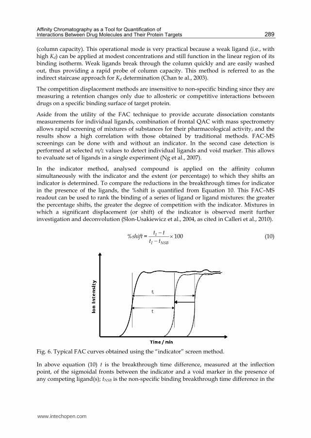
In the indicator method, analysed compound is applied on the affinity column simultaneously with the indicator and the extent (or percentage) to which they shifts an indicator is determined. To compare the reductions in the breakthrough times for indicator in the presence of the ligands, the %shift is quantified from Equation 10. This FAC–MS readout can be used to rank the binding of a series of ligand or ligand mixtures: the greater the percentage shifts, the greater the degree of competition with the indicator. Mixtures in which a significant displacement (or shift) of the indicator is observed merit further investigation and deconvolution (Slon-Usakiewicz et al., 2004, as cited in Calleri et al., 2010).
100I
I NSB
t t%shift =
t t
(10)
Fig. 6. Typical FAC curves obtained using the “indicator” screen method.
In above equation (10) t is the breakthrough time difference, measured at the inflection point, of the sigmoidal fronts between the indicator and a void marker in the presence of any competing ligand(s); tNSB is the non-specific binding breakthrough time difference in the
www.intechopen.com

Affinity Chromatography
290
absence of immobilized protein (and is a constant for the indicator used); and tI is the breakthrough time difference in the absence of any competing ligands. Typical FAC curves obtained using the indicator screen method is shown in Figure 6.
For more detailed information about FAC-MS technique the readers are referenced to
reviews (Chan et al., 2003; Slon-Usakiewicz et al., 2005; Calleri et al., 2009).
3.4 Nonlinear chromatography for determination of kinetics parameters
Peaks obtained in zonal AC differ from Gaussian shape observed frequently in classical
chromatographic analyses. Because we have to deal with column overload and slow kinetics
of adsorption/desorption during assays, peaks exhibit a strong tailing, which increases with
increased concentration of an analyte. Observed asymmetry can arise from a variety of other
factors-including extra column effects, heterogeneity of the stationary phase, heterogeneous
mass transfer or a non-linear isotherm (Wade et al., 1987, as cited in Moaddel & Wainer,
2006). The degree of deviation from a Gaussian distribution is a function of applied ligand
concentration and the kinetics of ligand–receptor interactions occurring during the
chromatographic process. An example of the effect of solute concentration on peak shape is
presented in Figure 7, which shows the example of usage of nonlinear chromatography
(NLC) for determination of kinetic parameters for the ┙3┚4 nAChR allosteric inhibitors
(Jozwiak et al., 2002).
Fig. 7. The effect of increasing concentraions of mecamylamine, from 1 to 1,000 mM, on the
chromatographic profiles of mecamylamine. For experimental details, see reference (Jozwiak
et al., 2002).
While peak tailing (or fronting) is a problem in analytical separations, concentration-
dependent asymmetry can be used with NLC techniques to characterize the separation
processes occurring on the column. When the chromatographic process includes binding
interactions between a ligand and an immobilized membrane bound receptor, the NLC
www.intechopen.com

Affinity Chromatography as a Tool for Quantification of Interactions Between Drug Molecules and Their Protein Targets
291
approach can be used to calculate the association rate constant (kon), dissociation rate
constant (koff) for the ligand–receptor complex and the equilibrium constant for complex
formation (K). One approach to the analysis of NLC data is the Impulse Input Solution for
the mass balance equation developed in 1987 by Wade and collaborators (Wade et al., 1987,
as cited in Moaddel & Wainer, 2006). This approach is based upon the observation that
when adsorption/desorption rates are slow, band broadening is insensitive to a moderate
degree of column overload. In contrast to numerical integration methods this approach uses
the analytical solution, which can be applied directly to fit experimental peak profiles. The
Impulse Input Equation has been included in commercially available deconvolution
software, and can be easily applied to NLC studies. The peak area parameter (a0), peak
center parameter (a1), its width (a2) and distortion (a3) are the parameters used to describe
the chromatographic traces. Thermodynamic and kinetics parameters of a drug-receptor
complex formation are then calculated on the basis of the relationship:
k' = a1 the real thermodynamic capacity factor
kd = 1/a2t0 desorption reaction rate constant
Ka = a3/C0 adsorption equilibrium constants, where C0 is the concentration of solute injected multiplied by the width of the injection pulse (as a fraction of column dead volume)
ka = kd Ka reaction rate constant of adsorption
4. Examples of application
QAC was successfully used to describe drug interactions with multiple different system
proteins. In case of soluble proteins this approach was applied to determine binding of
different active substances (drugs, hormones) with e.g. serum albumins (HSA), ┙1-acid
glycoprotein (AGP) or nucleic estrogen receptors (hER).
Xuan & Hage (2005) conducted research on immobilized ┙1-acid glycoprotein (AGP) demonstrating good correlation (0.954) of observed retention factors of several compounds with their equilibrium binding constants to AGP designated by other methods. In the same work authors, using the self-competition technique, confirmed the literature data on existence of one binding site for propranolol enantiomers on this protein. Determined values of Ka were also included in the range of values known from other experiments. Performing the analysis at different temperatures, changes in enthalpy and entropy of propranolol enantiomers binding to ┙1-acid glycoprotein, and their contribution in the total energy change in the process of binding were determined. It was found, that this reaction depends mainly on the enthalpy, but entropy change also significantly affects the binding of propranolol. Linearity of the plot confirmed the single-side character of binding of this compound by the AGP.
Combination of zonal QAC with competition-displacement studies confirmed the negative allosteric nature of the interaction between verapamil and tamoxifen. It was determined that verapamil causes 41% decrease in binding constant of tamoxifen by immobilized HSA (Malik et al., 2008). As mentioned earlier, using compound of interest as a competitive addition to the mobile phase it is possible to define its interactions with target protein on a single binding site even though the drug is bound to several different sites on the protein.
www.intechopen.com

Affinity Chromatography
292
Such solution has been used in the study of binding of both; hormones (Loun & Hage, 1995) and drugs (Chen et. al., 2004; Yoo et al., 2009; Mallik et al., 2008) to different places in the HSA molecule. Simultaneous injection of imipramine with L-tryptophan on the column with immobilized HSA, showed a competitive interaction between those two drugs, confirming that imipramine specifically connects to the indole-benzodiazepine binding site on the molecule of albumin (Sudlow site II). This analysis also allowed to determine the equilibrium constant of this interaction (Yoo et al., 2009). Analogously was defined binding site for the verapamil on this protein, demonstrating in this case, competitive interaction with warfarin, specifically connecting to Sudlow site I. This was confirmed by elution in the presence of tamoxifen. Nonlinear dependence of retention of this compound on verapamil concentration showed a high correlation to the equation describing the allosteric interaction, characteristic for interaction that is observed between the tamoxifen binding site and the warfarin binding site (Sudlow I) (Mallik et al., 2008). Team of Chen and colleagues (2004) determined the nature of the interaction of phenytoin by drawing a map of interactions of the drug with the major binding sites on the surface of HSA on the basis of the relationship between 1/k of the compounds with confirmed specific binding site on albumin molecule, and various concentrations of phenytoin as a mobile phase additive during elution. Thus suggested the potential impact of other concomitant medications on the efficacy of therapy with phenytoin.
The effect of different assay conditions on drug-protein interaction was used, for example to define the influence of long-chain fatty acids concentration in the plasma and glycation of plasma transport proteins on the binding of sulfonylurea drugs in diabetes. Experiments were carried out using columns with immobilized HSA (Basiaga & Hage, 2010).
Recently Sanghvi and co-workers (2010) successfully immobilized the ligand binding domains of the estrogen related receptors ERR┙ and ERR┛ onto the aminopropyl silica liquid chromatography stationary phase, as well as the surface of the open tubular capillaries, creating the ERR-silica and ERR-OT columns. Both types were characterized using frontal chromatographic techniques with diethylstibesterol and the binding affinities, expressed as Kd values, to the immobilized receptors were consistent with the literature data. Biochanin A, the ERR┙ agonist, was also used to further characterization of properties of the ERR┙-silica column, and obtained Kd value was consistent with the previously reported data. The ERR┛-silica column was characterized using nonlinear chromatographic techniques using a series of tamoxifen derivatives (tamoxifen, 4-hydroxy tamoxifen and N-desmethyl 4-hydroxytamoxifen, Endoxifen). The relative Kd values obtained for the derivatives were consistent with relative ability of the compounds to inhibit the cellular proliferation of the human-derived T98G glioma cell line, expressed as IC50 values. The results indicate that the relative retention of compounds on these columns reflects the magnitude of their inhibitory activity. Therefore columns containing immobilized ERRs can be used for a preliminary screen for anti-glioma agents, such as tamoxifen, which work as selective estrogen receptor modulator and that this method may replace current laborious and time consuming cellular uptake studies.
QAC has been applied to study drug-receptor interactions in the ┙1 adrenergic receptor
system. The results showed a positive correlation of chromatographic data (k') with
literature data defining the affinity of ligands on the basis of radio ligand studies with cell
membrane homogenates containing ┙1 adrenergic receptor (Yu et al., 2005).
www.intechopen.com

Affinity Chromatography as a Tool for Quantification of Interactions Between Drug Molecules and Their Protein Targets
293
QAC was also used in determination of binding site structure of the ┙3┚4 nAChR.
Comparing the retention time of tritium labelled epibatidine ([3H] EB) on four different
columns: with immobilized either receptor subunit ┙3 or ┚4 subunit, both subunits
immobilized on the same column and complete pentameric ┙3┚4 receptor, it was found that
the binding site for this compound is formed by several subunits of the complete receptor,
and not on the individual subunits. This was confirmed by assays with nicotine as a
competitive additive to the mobile phase. No impact of nicotine on the retention of [3H] EB
on columns with immobilized subunits was observed, while the epibatidine retention on the
column with complete receptor decreased from k = 8.4 to k = 1.5, which indicates the
interaction of nicotine on [3H] EB binding site (Wainer et al., 1999). In the same work the
effect of pH and ionic strength of mobile phase on the retention of [3H] epibatidine in the
┙3┚4 nAChR column was also examined using zonal elution technique. Analyses showed an
increase in epibatidine retention with increasing mobile phase pH from pH 4 to pH 7 and a
significant decrease in the retention of this compound with increasing ionic strength of
mobile phase (5-200 mM ammonium acetate concentration). These results showed that the
binding of competitive agonists is mainly electrostatic interaction between drug molecule
and the binding site on the nAChR.
QAC in combination with displacement studies were used in case of ┙3┚4 nAChR to confirm the location of binding sites of allosteric inhibitors of this receptor. Elution of bupropion, ketamine and dextromethorphan was conducted in the presence of mecamylamine (nAChR blocker) as an addition to the mobile phase. The linear dependence of eluted compounds 1/k' on the displacer concentration indicates that all tested inhibitors compete for the same binding site on the receptor molecule. Additional studies also showed no effect of mecamylamine in the mobile phase on epibatidine retention and no effect of nicotine in the mobile phase on the retention of ketamine and bupropion. It indicates that ketamine, bupropion, mecamylamine bind to other sites on the receptor surface than nicotine and epibatidine and do not compete for the binding site (Jozwiak et al., 2002). In the same work nonlinear chromatography (NLC) model was used to analyse interactions between the immobilized ┙3┚4 nAChR, and its allosteric inhibitors. Peak profiles from zonal elution of allosteric inhibitors (ketamine, bupropion, mecamylamine, and dextrometorfan), recorded by the mass spectrometry were numerically fitted to Impulse Input Solution model using PeakFit v4.11 software (SPSS Inc., Chicago, IL). The results (kon, koff, K) were consistent with available literature data, thus confirming the effectiveness of the NLC in receptor drug binding kinetics study, in this case allosteric nAChR inhibitors. In further work enantioselectivity of the interaction of dextromethorphan (DM) and levomethorphan (LM) with an immobilized ┙3┚4 nAChR subtype liquid chromatographic stationary phase has been compared to DM- and LM-induced non-competitive blockade of nicotine-stimulated 86Rb+ efflux from cells expressing the ┙3┚4 nAChR. Since DM and LM are enantiomers and have the same physicochemical properties, any chromatographic and pharmacological differences must be due to specific interactions with nAChR. Asymmetrical peaks were observed for both compounds and DM had significantly longer retention time than LM (Figure 8). Determined by NLC kon values of both compounds did not significantly differ while koff of DM was significantly lower than koff value of LM. That was in good agreement with results of the functional inhibition studies which showed that DM and LM had equivalent potencies, i.e. the same IC50 values, but that DM inhibition lasted longer than the effect produced by LM. The effect of temperature on the chromatographic retention of DM
www.intechopen.com

Affinity Chromatography
294
and LM on the nAChR column was also determined using a sequence of temperature experiments ranging from 5 to 30°C. Respective k’s values of independently injected compound were determined and van’t Hoff plots were constructed by plotting ln k versus 1/T. Results showed that the binding reaction is enthalpy driven. In addition, for DM and LM there was no significant difference in the ΔS° values, while the ΔH° value for the DM-nAChR interaction was significantly lower than respective value for LM-nAChR interaction showing that the enantioselectivity of ┙3┚4 nAChR is mostly enthalpy-based (Jozwiak et al., 2003).The results of those studies demonstrated that non-linear chromatography approach of investigations of immobilized nAChRs can be useful in characterizing ligand–receptor binding interactions and predicting of properties of drugs and drug candidates. Additional NLC studies with this system were used to construct QSAR models of non-competitive inhibitors of the nAChR (Jozwiak et al., 2004), to develop a molecular model of these interactions (Jozwiak et al., 2004) and to predict IC50 values (Jozwiak et al., 2005)
Fig. 8. The comparison of peak profiles of DM and LM obtained in independent experiments of consequent injections. Reprinted from Journal of Chromatography B, Vol. 797, No. 1-2, Jozwiak, K., Hernandez, S. C., Kellar, K. J., & Wainer, I. W. (2003). Enantioselective interactions of dextromethorphan and levomethorphan with the ┙3┚4-nicotinic acetylcholine receptor: comparison of chromatographic and functional data, pp. 373-379, Copyright 2011, with permission from Elsevier
Competition displacement studies with [3H]epibatidine and [125I]┙-bungarotoxin as marker
radioligands specific for ┙3┚4 and ┙7 nAChRs subtypes respectively were applied to
determine Ki values and check subtype selectivity of newly synthesized derivatives of
epiboxidine, synthetic epibatidine-related compounds (Rizzi et al., 2008).
Another interesting application of QAC is use of immobilized-enzyme reactors (IMERs). In
medicinal chemistry research IMERs are applied to drug metabolism studies,
enantioselective analyses and for the identification of substrates and inhibitors as potential
drugs. Interestingly, the investigated enzymatic reaction took place directly on the column.
www.intechopen.com

Affinity Chromatography as a Tool for Quantification of Interactions Between Drug Molecules and Their Protein Targets
295
Attractive features of immobilized enzyme reactors are the increased enzyme stability and
the reusability coupled to accuracy, automation and potential high throughput when they
are inserted in a HPLC system (Bartolini et al., 2005). The approach requires neither highly
purified enzyme nor use of labelled substrates (radio- or color labelled). In immobilized
enzymes, inhibitors efficacies can be measured either as IC50 values or Ki values using
numerical transformation. Enzyme kinetic parameters are determined by successive
injection of a substrate at increasing concentrations and measuring the rate of enzymatic
reaction (V) expressed as peak area of product formed after each injection. Fitting the data to
Lineweaver–Burk double-reciprocal plot of 1/(V) against the substrate concentration [S]
(what is a linear transformation of the Michaelis–Menten plot), Km and Vmax values can be
obtained. The y-intercept of such graph is equivalent to the inverse of 1/Vmax, the x-intercept
of the graph represents -1/Km. In order to obtain correct results the concentration of
substrate should be normalized according to the formula 11.
inj injC VS =
BV
(11)
where Cinj is the injected substrate concentration, Vinj is the injected volume and BV is the
bed volume of the IMER.
To determine the inhibition constant (Ki) for a test drug a set of inhibitor injections in several
different concentrations ([I]) at two or more concentration of a substrate should be
performed. As noted by Dixon in 1953, if 1/V is plotted against inhibitor concentration [I], a
straight line plot is obtained for each substrate concentration [S]. The [I] value of a intersect
of those lines is equal to -Ki. If curves obtained for several different [S] converge in the left
upper quadrant of a chart, the inhibitor is competitive. If curves converge on the [I] axis, the
inhibitor is non-competitive. For not competitive inhibition, the lines are parallel.
Simultaneous injections of both a substrate at a fixed saturating concentration and increasing concentration of an inhibitor, result in increasing reduction of the peak area (Ai) in comparison to area obtained for a substrate alone (A0). The percent inhibition (100 - (Ai/A0 x 100)) is then plotted against the inhibitor concentration to obtain the inhibition curves (Girelli & Mattei, 2005; Nie & Wang, 2009). Recently this technique has been used for the kinetic characterization of inhibitors specific to brain-targeted butyrylcholinesterase (BuChE) (Bartolini et al., 2009), acetylocholinesterase (AchE) (Bartolini et al., 2004; Bartolini et al., 2005; He et al., 2010) and ┚-secretase (human recombinant ┚-amyloid precursor protein cleaving enzyme, hrBACE1)(Mancini & Andrisano, 2010) as potential therapeutics for Alzheimer’s disease. IMER was also used for rapid and cost-effective on-line chromatographic screening of matrix metalloproteinases (MMP-9 (Ma & Chun Yong Chan, 2010) and MMP-8 (Mazzini et al., 2011)) inhibitors that may be useful in cancer therapy and for determining the role of some derived plant products for treating NO-dependent smooth disorders using monolithic micro-IMER with covalently bounded arginase (André et al., 2011).
5. Conclusions
The drive to bring innovative drugs to market faster, without compromising quality and safety, induced need for new experimental techniques and methodologies. It is crucial to
www.intechopen.com

Affinity Chromatography
296
determine biological activity, drug-target interactions and physico-chemical properties of drug candidates as predictors of administration, distribution, metabolism, excretion (ADME) characteristics. In fact, development of rapid methodologies enabling to obtain those information is a key aspects of the drug discovery process. QAC as rapid, relatively simple technique with the possibility of automation proved to be useful alternative to conventional methods in the field of drug discovery and analysis. This method is facilitated by multiple use of the same column with the immobilized target and as a consequence the reproducibility of assays is increased. Thanks to the above features QAC becomes popular method of measuring binding affinity of the drug-protein interactions. Variety of data that we can obtain via this technique allows characterization of binding reactions as well as description of the binding site. The development of techniques of high-yielding synthesis increases demand for technology that would allow pharmaceutical companies for efficient and rapid biological screening of thousands of synthesized compounds (Renaud & Delsuc, 2009). Taking into account that approximately one drug is produced after 8000-10000 compounds were subjected to primary and secondary drug screens, the classical approach, one compound-one assay, becomes unsatisfactory (Caldwell, 2000; as cited in Nie & Wang, 2009) . Studies conducted by Ng and colleagues have shown that use of an automated high performance chromatographic system consisting of two affinity columns and a mass spectrometer as the detector, allow to analyse and rank activity of 10 000 compounds in just 24 hours (Ng et al., 2007). Thus QAC techniques seem to be an promising method of preliminary verification of drug candidates, which is an alternative to expensive and tedious in vitro assays.
6. References
Andre, C., Herlem, G., Gharbi, T., & Guillaume, Y. C. (2011). A new arginase enzymatic
reactor: development and application for the research of plant-derived inhibitors. J
Pharm Biomed Anal, Vol. 55, No. 1, pp. 48-53, ISSN 1873-264X (Electronic)0731-7085
(Linking)
Bartolini, M., Cavrini, V., & Andrisano, V. (2004). Monolithic micro-immobilized-enzyme
reactor with human recombinant acetylcholinesterase for on-line inhibition studies. J
Chromatogr A, Vol. 1031, No. 1-2, pp. 27-34, ISSN 0021-9673 (Print)0021-9673 (Linking)
Bartolini, M., Cavrini, V., & Andrisano, V. (2005). Choosing the right chromatographic
support in making a new acetylcholinesterase-micro-immobilised enzyme reactor
for drug discovery. J Chromatogr A, Vol. 1065, No. 1, pp. 135-144, ISSN 0021-9673
(Print)0021-9673 (Linking)
Bartolini, M., Greig, N. H., Yu, Q. S., & Andrisano, V. (2009). Immobilized
butyrylcholinesterase in the characterization of new inhibitors that could ease
Alzheimer's disease. J Chromatogr A, Vol. 1216, No. 13, pp. 2730-2738, ISSN 1873-
3778 (Electronic)0021-9673 (Linking)
Basiaga, S. B., & Hage, D. S. (2010). Chromatographic studies of changes in binding of
sulfonylurea drugs to human serum albumin due to glycation and fatty acids. J
Chromatogr B Analyt Technol Biomed Life Sci, Vol. 878, No. 30, pp. 3193-3197, ISSN
1873-376X (Electronic)1570-0232 (Linking)
www.intechopen.com

Affinity Chromatography as a Tool for Quantification of Interactions Between Drug Molecules and Their Protein Targets
297
Beigi, F., & Wainer, I. W. (2003). Syntheses of immobilized G protein-coupled receptor
chromatographic stationary phases: characterization of immobilized mu and kappa
opioid receptors. Anal Chem, Vol. 75, No. 17, pp. 4480-4485, ISSN 0003-2700
(Print)0003-2700 (Linking)
Beigi, F., Chakir, K., Xiao, R. P., & Wainer, I. W. (2004). G-protein-coupled receptor
chromatographic stationary phases. 2. Ligand-induced conformational mobility in
an immobilized beta2-adrenergic receptor. Anal Chem, Vol. 76, No. 24, pp. 7187-
7193, ISSN 0003-2700 (Print)0003-2700 (Linking)
Calleri, E., Temporini, C., & Massolini, G. (2011). Frontal affinity chromatography in
characterizing immobilized receptors. J Pharm Biomed Anal, Vol. 54, No. 5, pp. 911-
925, ISSN 1873-264X (Electronic)0731-7085 (Linking)
Calleri, E., Temporini, C., Caccialanza, G., & Massolini, G. (2009). Target-based drug
discovery: the emerging success of frontal affinity chromatography coupled to
mass spectrometry. ChemMedChem, Vol. 4, No. 6, pp. 905-916, ISSN 1860-7187
(Electronic)1860-7179 (Linking)
Chan, N. W., Lewis, D. F., Rosner, P. J., Kelly, M. A., & Schriemer, D. C. (2003). Frontal
affinity chromatography-mass spectrometry assay technology for multiple stages of
drug discovery: applications of a chromatographic biosensor. Anal Biochem, Vol.
319, No. 1, pp. 1-12, ISSN 0003-2697 (Print)0003-2697 (Linking)
Chen, J., & Hage, D. S. (2004). Quantitative analysis of allosteric drug-protein binding by
biointeraction chromatography. Nat Biotechnol, Vol. 22, No. 11, pp. 1445-1448, ISSN
1087-0156 (Print)1087-0156 (Linking)
Chen, J., Ohnmacht, C., & Hage, D. S. (2004). Studies of phenytoin binding to human serum
albumin by high-performance affinity chromatography. J Chromatogr B Analyt Technol
Biomed Life Sci, Vol. 809, No. 1, pp. 137-145, ISSN 1570-0232 (Print)1570-0232 (Linking)
Cheng, Y., Ho, E., Subramanyam, B., & Tseng, J. L. (2004). Measurements of drug-protein
binding by using immobilized human serum albumin liquid chromatography-mass
spectrometry. J Chromatogr B Analyt Technol Biomed Life Sci, Vol. 809, No. 1, pp. 67-
73, ISSN 1570-0232 (Print)1570-0232 (Linking)
Dixon, M. (1953). The determination of enzyme inhibitor constants. Biochem J, Vol. 55, No. 1,
pp. 170-171, ISSN 0264-6021 (Print)0264-6021 (Linking)
Girelli, A. M., & Mattei, E. (2005). Application of immobilized enzyme reactor in on-line
high performance liquid chromatography: a review. J Chromatogr B Analyt Technol
Biomed Life Sci, Vol. 819, No. 1, pp. 3-16, ISSN 1570-0232 (Print)1570-0232 (Linking)
Gottschalk, I., Li, Y. M., & Lundahl, P. (2000). Chromatography on cells: analyses of solute
interactions with the glucose transporter Glut1 in human red cells adsorbed on
lectin-gel beads. J Chromatogr B Biomed Sci Appl, Vol. 739, No. 1, pp. 55-62, ISSN
1387-2273 (Print)1387-2273 (Linking)
Gottschalk, I., Lundqvist, A., Zeng, C. M., Hagglund, C. L., Zuo, S. S., Brekkan, E., Eaker, D.,
& Lundahl, P. (2000). Conversion between two cytochalasin B-binding states of the
human GLUT1 glucose transporter. Eur J Biochem, Vol. 267, No. 23, pp. 6875-6882,
ISSN 0014-2956 (Print)0014-2956 (Linking)
www.intechopen.com

Affinity Chromatography
298
Gritti, F., & Guiochon, G. (2005). Critical contribution of nonlinear chromatography to the
understanding of retention mechanism in reversed-phase liquid chromatography. J
Chromatogr A, Vol. 1099, No. 1-2, pp. 1-42, ISSN 0021-9673 (Print)0021-9673 (Linking)
Guiffant, D., Tribouillard, D., Gug, F., Galons, H., Meijer, L., Blondel, M., & Bach, S. (2007).
Identification of intracellular targets of small molecular weight chemical
compounds using affinity chromatography. Biotechnol J, Vol. 2, No. 1, pp. 68-75,
ISSN 1860-7314 (Electronic)1860-6768 (Linking)
Hage, D. S. (2002). High-performance affinity chromatography: a powerful tool for studying
serum protein binding. J Chromatogr B Analyt Technol Biomed Life Sci, Vol. 768, No. 1,
pp. 3-30, ISSN 1570-0232 (Print)1570-0232 (Linking)
Hage, D. S., Jackson, A., Sobansky, M. R., Schiel, J. E., Yoo, M. J., & Joseph, K. S. (2009).
Characterization of drug-protein interactions in blood using high-performance
affinity chromatography. J Sep Sci, Vol. 32, No. 5-6, pp. 835-853, ISSN 1615-9314
(Electronic)1615-9306 (Linking)
Hage, D. S., Ruhn, P. F. (2006). An Introduction to Affinity Chromatography, In: Handbook of
Affinity Chromatography, Hage, D.S., pp. 3-15, CRC Press/Taylor&Francis, ISBN
978-0-8247-4057-3, Boca Raton
Hage, D.S. (Ed.). (2006). Handbook of Affinity Chromatography, CRC Press/Taylor&Francis,
ISBN 978-0-8247-4057-3, Boca Raton, FL
Hage, D.S., Chen, J. (2006). Quantitative Affinity Chromatography: Practical Aspect, In:
Handbook of Affinity Chromatography, Hage, D.S., pp. 595-628, CRC
Press/Taylor&Francis, ISBN 978-0-8247-4057-3, Boca Raton
He, P., Davies, J., Greenway, G., & Haswell, S. J. (2010). Measurement of acetylcholinesterase
inhibition using bienzymes immobilized monolith micro-reactor with integrated
electrochemical detection. Anal Chim Acta, Vol. 659, No. 1-2, pp. 9-14, ISSN 1873-
4324 (Electronic)0003-2670 (Linking)
Joseph, K. S., & Hage, D. S. (2010). Characterization of the binding of sulfonylurea drugs to
HSA by high-performance affinity chromatography. J Chromatogr B Analyt Technol
Biomed Life Sci, Vol. 878, No. 19, pp. 1590-1598, ISSN 1873-376X (Electronic)1570-
0232 (Linking)
Josic, D., & Buchacher, A. (2001). Application of monoliths as supports for affinity
chromatography and fast enzymatic conversion. J Biochem Biophys Methods, Vol. 49,
No. 1-3, pp. 153-174, ISSN 0165-022X (Print)0165-022X (Linking)
Jozwiak, K., Haginaka, J., Moaddel, R., & Wainer, I. W. (2002). Displacement and nonlinear
chromatographic techniques in the investigation of interaction of noncompetitive
inhibitors with an immobilized alpha3beta4 nicotinic acetylcholine receptor liquid
chromatographic stationary phase. Anal Chem, Vol. 74, No. 18, pp. 4618-4624, ISSN
0003-2700 (Print)0003-2700 (Linking)
Jozwiak, K., Hernandez, S. C., Kellar, K. J., & Wainer, I. W. (2003). Enantioselective
interactions of dextromethorphan and levomethorphan with the alpha 3 beta 4-
nicotinic acetylcholine receptor: comparison of chromatographic and functional
data. J Chromatogr B Analyt Technol Biomed Life Sci, Vol. 797, No. 1-2, pp. 373-379,
ISSN 1570-0232 (Print)1570-0232 (Linking)
www.intechopen.com

Affinity Chromatography as a Tool for Quantification of Interactions Between Drug Molecules and Their Protein Targets
299
Jozwiak, K., Moaddel, R., Yamaguchi, R., Ravichandran, S., Collins, J. R., & Wainer, I. W.
(2005). Qualitative assessment of IC50 values of inhibitors of the neuronal nicotinic
acetylcholine receptor using a single chromatographic experiment and multivariate
cluster analysis. J Chromatogr B Analyt Technol Biomed Life Sci, Vol. 819, No. 1, pp.
169-174, ISSN 1570-0232 (Print)1570-0232 (Linking)
Jozwiak, K., Ravichandran, S., Collins, J. R., & Wainer, I. W. (2004). Interaction of
noncompetitive inhibitors with an immobilized alpha3beta4 nicotinic acetylcholine
receptor investigated by affinity chromatography, quantitative-structure activity
relationship analysis, and molecular docking. J Med Chem, Vol. 47, No. 16, pp. 4008-
4021, ISSN 0022-2623 (Print)0022-2623 (Linking)
Kasai, K., & Ishii, S. (1975). Quantitative analysis of affinity chromatography of trypsin. A
new technique for investigation of protein-ligand interaction. J Biochem, Vol. 77, No.
1?, pp. 261-264, ISSN 0021-924X (Print)0021-924X (Linking)
Kim, H. S., & Wainer, I. W. (2008). Rapid analysis of the interactions between drugs and
human serum albumin (HSA) using high-performance affinity chromatography
(HPAC). J Chromatogr B Analyt Technol Biomed Life Sci, Vol. 870, No. 1, pp. 22-26,
ISSN 1570-0232 (Print)1570-0232 (Linking)
Kim, H.S., Hage, D.S., Immobilization methods for affinity chromatography, In: Handbook of
Affinity Chromatography, Hage, D.S., pp. 35-79, CRC Press/Taylor&Francis, ISBN
978-0-8247-4057-3, Boca Raton
Kimura, T., Perry, J., Anzai, N., Pritchard, J. B., & Moaddel, R. (2007). Development and
characterization of immobilized human organic anion transporter-based liquid
chromatographic stationary phase: hOAT1 and hOAT2. J Chromatogr B Analyt Technol
Biomed Life Sci, Vol. 859, No. 2, pp. 267-271, ISSN 1570-0232 (Print)1570-0232 (Linking)
Kitabatake, T., Moaddel, R., Cole, R., Gandhari, M., Frazier, C., Hartenstein, J., Rosenberg,
A., Bernier, M., & Wainer, I. W. (2008). Characterization of a multiple ligand-gated
ion channel cellular membrane affinity chromatography column and identification
of endogenously expressed receptors in astrocytoma cell lines. Anal Chem, Vol. 80,
No. 22, pp. 8673-8680, ISSN 1520-6882 (Electronic)0003-2700 (Linking)
Landry, Y., & Gies, J. P. (2008). Drugs and their molecular targets: an updated overview.
Fundam Clin Pharmacol, Vol. 22, No. 1, pp. 1-18, ISSN 1472-8206 (Electronic)0767-
3981 (Linking)
Lebert, J. M., Forsberg, E. M., & Brennan, J. D. (2008). Solid-phase assays for small molecule
screening using sol-gel entrapped proteins. Biochem Cell Biol, Vol. 86, No. 2, pp. 100-
110, ISSN 0829-8211 (Print)0829-8211 (Linking)
Loun, B., & Hage, D. S. (1992). Characterization of thyroxine-albumin binding using high-
performance affinity chromatography. I. Interactions at the warfarin and indole
sites of albumin. J Chromatogr, Vol. 579, No. 2, pp. 225-235, ISSN 0021-9673
(Print)0021-9673 (Linking)
Loun, B., & Hage, D. S. (1994). Chiral separation mechanisms in protein-based HPLC
columns. 1. Thermodynamic studies of (R)- and (S)-warfarin binding to
immobilized human serum albumin. Anal Chem, Vol. 66, No. 21, pp. 3814-3822,
ISSN 0003-2700 (Print)0003-2700 (Linking)
www.intechopen.com

Affinity Chromatography
300
Loun, B., & Hage, D. S. (1995). Characterization of thyroxine-albumin binding using high-
performance affinity chromatography. II. Comparison of the binding of thyroxine,
triiodothyronines and related compounds at the warfarin and indole sites of
human serum albumin. J Chromatogr B Biomed Appl, Vol. 665, No. 2, pp. 303-314,
ISSN 1572-6495 (Print)1572-6495 (Linking)
Lundqvist, A., Ocklind, G., Haneskog, L., & Lundahl, P. (1998). Freeze-thaw immobilization
of liposomes in chromatographic gel beads: evaluation by confocal microscopy and
effects of freezing rate. J Mol Recognit, Vol. 11, No. 1-6, pp. 52-57, ISSN 0952-3499
(Print)0952-3499 (Linking)
Ma, X., & Chan, E. C. (2010). On-line chromatographic screening of matrix metalloproteinase
inhibitors using immobilized MMP-9 enzyme reactor. J Chromatogr B Analyt Technol
Biomed Life Sci, Vol. 878, No. 21, pp. 1777-1783, ISSN 1873-376X (Electronic)1570-
0232 (Linking)
Mallik, R., Yoo, M. J., Chen, S., & Hage, D. S. (2008). Studies of verapamil binding to human
serum albumin by high-performance affinity chromatography. J Chromatogr B
Analyt Technol Biomed Life Sci, Vol. 876, No. 1, pp. 69-75, ISSN 1570-0232 (Print)1570-
0232 (Linking)
Mancini, F., & Andrisano, V. (2010). Development of a liquid chromatographic system with
fluorescent detection for beta-secretase immobilized enzyme reactor on-line
enzymatic studies. J Pharm Biomed Anal, Vol. 52, No. 3, pp. 355-361, ISSN 1873-264X
(Electronic)0731-7085 (Linking)
Markuszewski, M., & Kaliszan, R. (2002). Quantitative structure-retention relationships in
affinity high-performance liquid chromatography. J Chromatogr B Analyt Technol
Biomed Life Sci, Vol. 768, No. 1, pp. 55-66, ISSN 1570-0232 (Print)1570-0232 (Linking)
Mateo, C., Fernandez-Lorente, G., Abian, O., Fernandez-Lafuente, R., & Guisan, J. M. (2000).
Multifunctional epoxy supports: a new tool to improve the covalent immobilization
of proteins. The promotion of physical adsorptions of proteins on the supports
before their covalent linkage. Biomacromolecules, Vol. 1, No. 4, pp. 739-745, ISSN
1525-7797 (Print)1525-7797 (Linking)
Mazzini, F., Nuti, E., Petri, A., & Rossello, A. (2011). Immobilization of matrix
metalloproteinase 8 (MMP-8) for online drug screening. J Chromatogr B Analyt
Technol Biomed Life Sci, Vol. 879, No. 11-12, pp. 756-762, ISSN 1873-376X
(Electronic)1570-0232 (Linking)
Moaddel, R., & Wainer, I. W. (2006). Development of immobilized membrane-based affinity
columns for use in the online characterization of membrane bound proteins and for
targeted affinity isolations. Anal Chim Acta, Vol. 564, No. 1, pp. 97-105, ISSN 1873-
4324 (Electronic)0003-2670 (Linking)
Moaddel, R., Bullock, P. L., & Wainer, I. W. (2004). Development and characterization of an
open tubular column containing immobilized P-glycoprotein for rapid on-line
screening for P-glycoprotein substrates. J Chromatogr B Analyt Technol Biomed Life
Sci, Vol. 799, No. 2, pp. 255-263, ISSN 1570-0232 (Print)1570-0232 (Linking)
Moaddel, R., Calleri, E., Massolini, G., Frazier, C. R., & Wainer, I. W. (2007). The synthesis
and initial characterization of an immobilized purinergic receptor (P2Y1) liquid
www.intechopen.com

Affinity Chromatography as a Tool for Quantification of Interactions Between Drug Molecules and Their Protein Targets
301
chromatography stationary phase for online screening. Anal Biochem, Vol. 364, No.
2, pp. 216-218, ISSN 0003-2697 (Print)0003-2697 (Linking)
Moaddel, R., Cloix, J. F., Ertem, G., & Wainer, I. W. (2002). Multiple receptor liquid
chromatographic stationary phases: the co-immobilization of nicotinic receptors,
gamma-amino-butyric acid receptors, and N-methyl D-aspartate receptors. Pharm
Res, Vol. 19, No. 1, pp. 104-107, ISSN 0724-8741 (Print)0724-8741 (Linking)
Moaddel, R., Jozwiak, K., Whittington, K., & Wainer, I. W. (2005). Conformational mobility
of immobilized alpha3beta2, alpha3beta4, alpha4beta2, and alpha4beta4 nicotinic
acetylcholine receptors. Anal Chem, Vol. 77, No. 3, pp. 895-901, ISSN 0003-2700
(Print)0003-2700 (Linking)
Moaddel, R., Lu, L., Baynham, M., & Wainer, I. W. (2002). Immobilized receptor- and
transporter-based liquid chromatographic phases for on-line pharmacological and
biochemical studies: a mini-review. J Chromatogr B Analyt Technol Biomed Life Sci,
Vol. 768, No. 1, pp. 41-53, ISSN 1570-0232 (Print)1570-0232 (Linking)
Moaddel, R., Musyimi, H. K., Sanghvi, M., Bashore, C., Frazier, C. R., Khadeer, M., Bhatia,
P., & Wainer, I. W. (2010). Synthesis and characterization of a cellular membrane
affinity chromatography column containing histamine 1 and P2Y(1) receptors: a
multiple G-protein coupled receptor column. J Pharm Biomed Anal, Vol. 52, No. 3,
pp. 416-419, ISSN 1873-264X (Electronic)0731-7085 (Linking)
Moaddel, R., Oliveira, R. V., Kimura, T., Hyppolite, P., Juhaszova, M., Xiao, Y., Kellar, K. J.,
Bernier, M., & Wainer, I. W. (2008). Initial synthesis and characterization of an
alpha7 nicotinic receptor cellular membrane affinity chromatography column:
effect of receptor subtype and cell type. Anal Chem, Vol. 80, No. 1, pp. 48-54, ISSN
0003-2700 (Print)0003-2700 (Linking)
Moaddel, R., Rosenberg, A., Spelman, K., Frazier, J., Frazier, C., Nocerino, S., Brizzi, A.,
Mugnaini, C., & Wainer, I. W. (2011). Development and characterization of
immobilized cannabinoid receptor (CB1/CB2) open tubular column for on-line
screening. Anal Biochem, Vol. 412, No. 1, pp. 85-91, ISSN 1096-0309 (Electronic)0003-
2697 (Linking)
Moaddel, R., Yamaguchi, R., Ho, P. C., Patel, S., Hsu, C. P., Subrahmanyam, V., & Wainer, I.
W. (2005). Development and characterization of an immobilized human organic
cation transporter based liquid chromatographic stationary phase. J Chromatogr B
Analyt Technol Biomed Life Sci, Vol. 818, No. 2, pp. 263-268, ISSN 1570-0232
(Print)1570-0232 (Linking)
Narayanan, S. R., Kakodkar, S. V., & Crane, L. J. (1990). "Glutaraldehyde-P", a stable,
reactive aldehyde matrix for affinity chromatography. Anal Biochem, Vol. 188, No. 2,
pp. 278-284, ISSN 0003-2697 (Print)0003-2697 (Linking)
Ng, W., Dai, J. R., Slon-Usakiewicz, J. J., Redden, P. R., Pasternak, A., & Reid, N. (2007).
Automated multiple ligand screening by frontal affinity chromatography-mass
spectrometry (FAC-MS). J Biomol Screen, Vol. 12, No. 2, pp. 167-174, ISSN 1087-0571
(Print)1087-0571 (Linking)
Nie, Y.-l., Wang, W.-h. (2009) .Immobilized Enzyme Reactor in On-line LC and Its
Application in Drug Screening. Chromatographia, Vol. 69, pp. S5–S12
www.intechopen.com

Affinity Chromatography
302
Pidgeon, C., & Venkataram, U. V. (1989). Immobilized artificial membrane chromatography:
supports composed of membrane lipids. Anal Biochem, Vol. 176, No. 1, pp. 36-47,
ISSN 0003-2697 (Print)0003-2697 (Linking)
Renaud, J. P., & Delsuc, M. A. (2009). Biophysical techniques for ligand screening and drug
design. Curr Opin Pharmacol, Vol. 9, No. 5, pp. 622-628, ISSN 1471-4973
(Electronic)1471-4892 (Linking)
Rizzi, L., Dallanoce, C., Matera, C., Magrone, P., Pucci, L., Gotti, C., Clementi, F., & De
Amici, M. (2008). Epiboxidine and novel-related analogues: a convenient synthetic
approach and estimation of their affinity at neuronal nicotinic acetylcholine
receptor subtypes. Bioorg Med Chem Lett, Vol. 18, No. 16, pp. 4651-4654, ISSN 1464-
3405 (Electronic)0960-894X (Linking)
Ruhn, P. F., Garver, S., & Hage, D. S. (1994). Development of dihydrazide-activated silica
supports for high-performance affinity chromatography. J Chromatogr A, Vol. 669,
No. 1-2, pp. 9-19, ISSN 0021-9673 (Print)0021-9673 (Linking)
Sanghvi, M., Moaddel, R., Frazier, C., & Wainer, I. W. (2010). Synthesis and characterization
of liquid chromatographic columns containing the immobilized ligand binding
domain of the estrogen related receptor alpha and estrogen related receptor
gamma. J Pharm Biomed Anal, Vol. 53, No. 3, pp. 777-780, ISSN 1873-264X
(Electronic)0731-7085 (Linking)
Slon-Usakiewicz, J. J., Ng, W., Dai, J. R., Pasternak, A., & Redden, P. R. (2005). Frontal
affinity chromatography with MS detection (FAC-MS) in drug discovery. Drug
Discov Today, Vol. 10, No. 6, pp. 409-416, ISSN 1359-6446 (Print)1359-6446 (Linking)
Soczewinski, E., Bieganowska, M. (1969). Investigations on the relationship between
molecular structure and chromatogenic parameters: I. Two homologous series of
pyridine derivatives suitable as reference compounds in liquid-liquid partition
chromatography. J Chromatogr A, Vol. 40, pp. 431-439
Taylor, R.F. (Ed.). (1991). Protein Immobilization: Fundamentals and Applications, Marcel
Dekker, ISBN10 0824782712, New York
Temporini, C., Ceruti, S., Calleri, E., Ferrario, S., Moaddel, R., Abbracchio, M. P., &
Massolini, G. (2009). Development of an immobilized GPR17 receptor stationary
phase for binding determination using frontal affinity chromatography coupled to
mass spectrometry. Anal Biochem, Vol. 384, No. 1, pp. 123-129, ISSN 1096-0309
(Electronic)0003-2697 (Linking)
Trujillo, C. A., Majumder, P., Gonzalez, F. A., Moaddel, R., & Ulrich, H. (2007). Immobilized
P2X2 purinergic receptor stationary phase for chromatographic determination of
pharmacological properties and drug screening. J Pharm Biomed Anal, Vol. 44, No. 3,
pp. 701-710, ISSN 0731-7085 (Print)0731-7085 (Linking)
Turkova, J. (1999). Oriented immobilization of biologically active proteins as a tool for
revealing protein interactions and function. J Chromatogr B Biomed Sci Appl, Vol.
722, No. 1-2, pp. 11-31, ISSN 1387-2273 (Print)1387-2273 (Linking)
Tweed, S. A., Loun, B., & Hage, D. S. (1997). Effects of ligand heterogeneity in the
characterization of affinity columns by frontal analysis. Anal Chem, Vol. 69, No. 23,
pp. 4790-4798, ISSN 0003-2700 (Print)0003-2700 (Linking)
www.intechopen.com

Affinity Chromatography as a Tool for Quantification of Interactions Between Drug Molecules and Their Protein Targets
303
Wade, J.L., Bergold, A.F., Carr, P.W. (1987). Theoretical description of nonlinear
chromatography, with applications to physicochemical measurements in affinity
chromatography and implications for preparative-scale separations. Anal. Chem.,
Vol. 59, No. 9, pp. 1286–1295
Wainer, I. W., Zhang, Y., Xiao, Y., & Kellar, K. J. (1999). Liquid chromatographic studies
with immobilized neuronal nicotinic acetylcholine receptor stationary phases:
effects of receptor subtypes, pH and ionic strength on drug-receptor interactions. J
Chromatogr B Biomed Sci Appl, Vol. 724, No. 1, pp. 65-72, ISSN 1387-2273 (Print)1387-
2273 (Linking)
Winzor, D. J. (2004). Determination of binding constants by affinity chromatography. J
Chromatogr A, Vol. 1037, No. 1-2, pp. 351-367, ISSN 0021-9673 (Print)0021-9673
(Linking)
Xuan, H., & Hage, D. S. (2005). Immobilization of alpha(1)-acid glycoprotein for
chromatographic studies of drug-protein binding. Anal Biochem, Vol. 346, No. 2, pp.
300-310, ISSN 0003-2697 (Print)0003-2697 (Linking)
Yang, J., & Hage, D. S. (1993). Characterization of the binding and chiral separation of D- and
L-tryptophan on a high-performance immobilized human serum albumin column. J
Chromatogr, Vol. 645, No. 2, pp. 241-250, ISSN 0021-9673 (Print)0021-9673 (Linking)
Yang, Q., & Lundahl, P. (1995). Immobilized proteoliposome affinity chromatography for
quantitative analysis of specific interactions between solutes and membrane
proteins. Interaction of cytochalasin B and D-glucose with the glucose transporter
Glut1. Biochemistry, Vol. 34, No. 22, pp. 7289-7294, ISSN 0006-2960 (Print)0006-2960
(Linking)
Yoo, M. J., Smith, Q. R., & Hage, D. S. (2009). Studies of imipramine binding to human
serum albumin by high-performance affinity chromatography. J Chromatogr B
Analyt Technol Biomed Life Sci, Vol. 877, No. 11-12, pp. 1149-1154, ISSN 1873-376X
(Electronic)1570-0232 (Linking)
Yu, W., Yuan, B., Deng, X., He, L., Youyi, Z., & Qide, H. (2005). The preparation of HEK293
alpha1A or HEK293 alpha1B cell membrane stationary phase and the
chromatographic affinity study of ligands of alpha1 adrenoceptor. Anal Biochem,
Vol. 339, No. 2, pp. 198-205, ISSN 0003-2697 (Print)0003-2697 (Linking)
Yuan, B. X., Hou, J., He, L. C., & Yang, G. D. (2005). Evaluation of drug-muscarinic receptor
affinities using cell membrane chromatography and radioligand binding assay in
guinea pig jejunum membrane. Acta Pharmacol Sin, Vol. 26, No. 1, pp. 113-116, ISSN
1671-4083 (Print)1671-4083 (Linking)
Zeng, C. M., Zhang, Y., Lu, L., Brekkan, E., Lundqvist, A., & Lundahl, P. (1997).
Immobilization of human red cells in gel particles for chromatographic activity
studies of the glucose transporter Glut1. Biochim Biophys Acta, Vol. 1325, No. 1, pp.
91-98, ISSN 0006-3002 (Print)0006-3002 (Linking)
Zhang, Y., Leonessa, F., Clarke, R., & Wainer, I. W. (2000). Development of an immobilized
P-glycoprotein stationary phase for on-line liquid chromatographic determination
of drug-binding affinities. J Chromatogr B Biomed Sci Appl, Vol. 739, No. 1, pp. 33-37,
ISSN 1387-2273 (Print)1387-2273 (Linking)
www.intechopen.com

Affinity Chromatography
304
Zhang, Y., Xiao, Y., Kellar, K. J., & Wainer, I. W. (1998). Immobilized nicotinic receptor
stationary phase for on-line liquid chromatographic determination of drug-receptor
affinities. Anal Biochem, Vol. 264, No. 1, pp. 22-25, ISSN 0003-2697 (Print)0003-2697
(Linking)
Zhu, L., Chen, L., Luo, H., & Xu, X. (2003). Frontal affinity chromatography combined on-
line with mass spectrometry: a tool for the binding study of different epidermal
growth factor receptor inhibitors. Anal Chem, Vol. 75, No. 23, pp. 6388-6393, ISSN
0003-2700 (Print)0003-2700 (Linking)
www.intechopen.com

Affinity ChromatographyEdited by Dr. Sameh Magdeldin
ISBN 978-953-51-0325-7Hard cover, 368 pagesPublisher InTechPublished online 21, March, 2012Published in print edition March, 2012
InTech EuropeUniversity Campus STeP Ri Slavka Krautzeka 83/A 51000 Rijeka, Croatia Phone: +385 (51) 770 447 Fax: +385 (51) 686 166www.intechopen.com
InTech ChinaUnit 405, Office Block, Hotel Equatorial Shanghai No.65, Yan An Road (West), Shanghai, 200040, China
Phone: +86-21-62489820 Fax: +86-21-62489821
Most will agree that one major achievement in the bio-separation techniques is affinity chromatography. Thiscoined terminology covers a myriad of separation approaches that relies mainly on reversible adsorption ofbiomolecules through biospecific interactions on the ligand. Within this book, the authors tried to deliver for yousimplified fundamentals of affinity chromatography together with exemplarily applications of this versatiletechnique. We have always been endeavor to keep the contents of the book crisp and easily comprehensive,hoping that this book will receive an overwhelming interest, deliver benefits and valuable information to thereaders.
How to referenceIn order to correctly reference this scholarly work, feel free to copy and paste the following:
Piotr Draczkowski, Dariusz Matosiuk and Krzysztof Jozwiak (2012). Affinity Chromatography as a Tool forQuantification of Interactions Between Drug Molecules and Their Protein Targets, Affinity Chromatography, Dr.Sameh Magdeldin (Ed.), ISBN: 978-953-51-0325-7, InTech, Available from:http://www.intechopen.com/books/affinity-chromatography/affinity-chromatography-as-a-tool-for-quantification-of-interactions-between-drug-molecules-and-thei

© 2012 The Author(s). Licensee IntechOpen. This is an open access articledistributed under the terms of the Creative Commons Attribution 3.0License, which permits unrestricted use, distribution, and reproduction inany medium, provided the original work is properly cited.


![Recent developments in protein ligand affinity mass spectrometry · frontal affinity chromatography (FAC) [1], size-exclusion chromatography (SEC) [2], (pulsed) ultrafiltration [3],](https://static.fdocuments.us/doc/165x107/604c1f4e3a10f26659366e36/recent-developments-in-protein-ligand-affinity-mass-spectrometry-frontal-affinity.jpg)