AFatalCaseofPrimaryAmoebicMeningoencephalitis(PAM ...
3
Case Report A Fatal Case of Primary Amoebic Meningoencephalitis (PAM) Complicated with Diabetes Insipidus (DI): A Case Report and Review of the Literature Muhammad Zain Mushtaq , Saad Bin Zafar Mahmood, and Adil Aziz Department of Medicine, Aga Khan University Hospital, Stadium Road, Karachi, Pakistan Correspondence should be addressed to Muhammad Zain Mushtaq; [email protected] Received 14 February 2020; Accepted 10 July 2020; Published 24 July 2020 Academic Editor: Larry M. Bush Copyright © 2020 Muhammad Zain Mushtaq et al. is is an open access article distributed under the Creative Commons Attribution License, which permits unrestricted use, distribution, and reproduction in any medium, provided the original work is properly cited. Naegleriafowleri is a highly infective free-living amoeba usually isolated from soil and fresh water and is primarily found to infect the central nervous system (CNS) resulting in primary amoebic meningoencephalitis (PAM). PAM as a cause of meningitis is often overlooked for other, more common causes of meningitis. Despite all the advances in antimicrobial therapy and supportive care systems, the mortality rate of this rare infection remains above 95% with the bulk of the cases being found in developed countries. We are presenting a case of a 44-year-old male with fever, worsening headache, and generalized weakness. Lumbar puncture showed a raised leucocyte count of 1100/µL with predominant polymorphonuclear cells, and wet mount prep for Naegleria fowleri was positive further confirmed with PCR. e patient was started Intravenous (IV) and intrathecal ampho- tericin-B, Per Oral (PO) miltefosine, IV rifampin, IV fluconazole, and IV dexamethasone. However, the patient started producing urine at 300–500 ml/hour. e patient’s sodium levels increased from 144 to 175 mmol/L in 12 hours with raised serum osmolality and decreased urine osmolality and urine sodium. e patient was started on PO desmopressin of 0.2 micrograms twice daily after which his urine output dropped to 60–80 ml/hour and sodium decreased from 175 to 162 and, later 155 mmol/L; however, the patient expired. PAM is a rare and extremely fatal illness, but with increasing incidence now being reported in developing countries as a result of better diagnostics. DI is a very rare complication reported in these patients leading to poor outcome. e complication of diabetes insipidus (DI) has not been extensively studied in patients having PAM. Only three cases have been reported with this complication. No mechanism has been mentioned in the literature behind the development of DI in these patients, and no study has mentioned laboratory details of DI as mentioned in this report. 1. Introduction Naegleria fowleri is a highly infective free-living amoeba usually isolated from soil and fresh water mostly during summer times when higher temperatures provide a con- ductive environment for its growth and proliferation [1, 2]. Naegleria fowleri was first found in 1899 and is primarily found to infect the central nervous system (CNS) resulting in primary amoebic meningoencephalitis (PAM) [3]. Infec- tions with Naegleria fowleri are primarily associated with exposure to fresh water during recreational and religious (ablution) activities; however, inadequately chlorinated water supply at homes and in swimming pools are now fast becoming possible causes for infections with the organism [1, 3]. e organism enters the body via the nasal cavity when contaminated water is deeply inhaled. It follows the olfactory nerve, moving along the cribriform plate and invades the CNS via the olfactory neuroepithelium and cribriform plate, causing an infection clinically resembling acute bacterial meningitis (ABM) [1, 2]. PAM, as a cause of meningitis, is often overlooked for other, more common causes of men- ingitis [4]. Despite all the advances in antimicrobial therapy and supportive care systems, the mortality rate of this rare infection remains above 95% with the bulk of the cases being found in developed countries as infections in developing countries remain mostly undiagnosed [3]. We describe here Hindawi Case Reports in Infectious Diseases Volume 2020, Article ID 4925819, 3 pages https://doi.org/10.1155/2020/4925819
Transcript of AFatalCaseofPrimaryAmoebicMeningoencephalitis(PAM ...
Case ReportA Fatal Case of Primary Amoebic Meningoencephalitis (PAM)Complicated with Diabetes Insipidus (DI): A Case Report andReview of the Literature
Muhammad Zain Mushtaq , Saad Bin Zafar Mahmood, and Adil Aziz
Department of Medicine, Aga Khan University Hospital, Stadium Road, Karachi, Pakistan
Correspondence should be addressed to Muhammad Zain Mushtaq; [email protected]
Received 14 February 2020; Accepted 10 July 2020; Published 24 July 2020
Academic Editor: Larry M. Bush
Copyright © 2020 Muhammad Zain Mushtaq et al. *is is an open access article distributed under the Creative CommonsAttribution License, which permits unrestricted use, distribution, and reproduction in anymedium, provided the original work isproperly cited.
Naegleria fowleri is a highly infective free-living amoeba usually isolated from soil and fresh water and is primarily found to infectthe central nervous system (CNS) resulting in primary amoebic meningoencephalitis (PAM). PAM as a cause of meningitis isoften overlooked for other, more common causes of meningitis. Despite all the advances in antimicrobial therapy and supportivecare systems, the mortality rate of this rare infection remains above 95% with the bulk of the cases being found in developedcountries. We are presenting a case of a 44-year-old male with fever, worsening headache, and generalized weakness. Lumbarpuncture showed a raised leucocyte count of 1100/µL with predominant polymorphonuclear cells, and wet mount prep forNaegleria fowleri was positive further confirmed with PCR. *e patient was started Intravenous (IV) and intrathecal ampho-tericin-B, Per Oral (PO) miltefosine, IV rifampin, IV fluconazole, and IV dexamethasone. However, the patient started producingurine at 300–500ml/hour.*e patient’s sodium levels increased from 144 to 175mmol/L in 12 hours with raised serum osmolalityand decreased urine osmolality and urine sodium.*e patient was started on PO desmopressin of 0.2micrograms twice daily afterwhich his urine output dropped to 60–80ml/hour and sodium decreased from 175 to 162 and, later 155mmol/L; however, thepatient expired. PAM is a rare and extremely fatal illness, but with increasing incidence now being reported in developingcountries as a result of better diagnostics. DI is a very rare complication reported in these patients leading to poor outcome. *ecomplication of diabetes insipidus (DI) has not been extensively studied in patients having PAM. Only three cases have beenreported with this complication. No mechanism has been mentioned in the literature behind the development of DI in thesepatients, and no study has mentioned laboratory details of DI as mentioned in this report.
1. Introduction
Naegleria fowleri is a highly infective free-living amoebausually isolated from soil and fresh water mostly duringsummer times when higher temperatures provide a con-ductive environment for its growth and proliferation [1, 2].Naegleria fowleri was first found in 1899 and is primarilyfound to infect the central nervous system (CNS) resulting inprimary amoebic meningoencephalitis (PAM) [3]. Infec-tions with Naegleria fowleri are primarily associated withexposure to fresh water during recreational and religious(ablution) activities; however, inadequately chlorinatedwater supply at homes and in swimming pools are now fast
becoming possible causes for infections with the organism[1, 3].*e organism enters the body via the nasal cavity whencontaminated water is deeply inhaled. It follows the olfactorynerve, moving along the cribriform plate and invades theCNS via the olfactory neuroepithelium and cribriform plate,causing an infection clinically resembling acute bacterialmeningitis (ABM) [1, 2]. PAM, as a cause of meningitis, isoften overlooked for other, more common causes of men-ingitis [4]. Despite all the advances in antimicrobial therapyand supportive care systems, the mortality rate of this rareinfection remains above 95% with the bulk of the cases beingfound in developed countries as infections in developingcountries remain mostly undiagnosed [3]. We describe here
HindawiCase Reports in Infectious DiseasesVolume 2020, Article ID 4925819, 3 pageshttps://doi.org/10.1155/2020/4925819
a case of Naegleria fowleri complicated with diabetesinsipidus (DI). To the best of our knowledge, only three suchcases have been reported in the literature worldwide but allof them being either children or adolescent [5–7].
2. Case Presentation
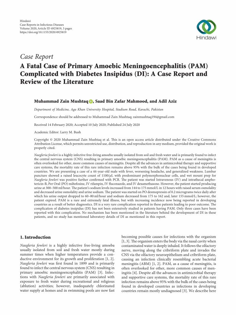
A previously healthy 44-year-old male with no priorcomorbidities presented to emergency with a 2-day historyof fever, worsening headache, and generalized weakness. Hehad no history of exposure to pool water. Examinationrevealed a Glasgow coma scale (GCS) of 14/15 with no focaldeficit or neck stiffness. Laboratory workup was sent whichshowed leukocytosis 13.3×10⁹/L. Rest of the investigationsare summarized in Table 1. Computed tomography (CT)scan brain was performed which was reported normal.Lumbar puncture was performed which showed a raisedopening pressure of 20 cm of water and had a raised leu-cocyte count of 1100/µL with predominant polymorpho-nuclear cells of 60% with a very high protein of 241mg/dland low glucose, i.e., 30mg/dl (Table 2). *e BioFire fil-marray Polymerase chain reaction (PCR) was negative forbacteria and viruses, but wet mount prep for Naegleriafowleri was positive which was further confirmed with PCR.
*e patient was started on Primary amebic meningo-encephalitis (PAM) protocol which included Intravenous(IV) and intrathecal amphotericin-B, Per Oral (PO) milte-fosine, IV rifampin, IV fluconazole, and IV dexamethasone.He was electively intubated and shifted to the intensive careunit (ICU). A repeat CT scan after 24 hours of hospital staywas performed which did not show any infarct or bleed butdid show marked cerebral edema. He was started onmannitol 30 grams every 8 hours, which was stopped after 48hours.
During the ICU stay, the patient remained on me-chanical ventilation and was deeply sedated with agents, IVpropofol and midazolam. Antiepileptic drug IV levetir-acetam was added empirically, and an Electroencephalo-gram (EEG) was obtained which showed theta and deltaslowing downwith no epileptiform activity. He was kept wellhydrated during the ICU stay, and continuous feeding wascarried out through a nasogastric tube.
On the 3rd hospital day, the patient started producingurine at 300–500ml/hour (he made a urine output of morethan 7 liters in 24 hours). His sodium levels increased from144mmol/L to 175mmol/L in 12 hours. His urea was 18mg/dl, ionized calcium was 4.88mg/dl, and blood sugar levelswere 171mg/dl. He was well hydrated with IV ringer lactate.Serum and urinary osmolality were performed which were332mosm/kg and 204mosm/kg, respectively. Urine sodiumwas less than 10mEq/L. He was started on PO desmopressinof 0.2 micrograms twice daily after which his urine outputdropped to 60–80ml/hour and sodium decreased from 175to 162 and, later, 155mmol/L. On the 4th hospital day, hewent in asystole and died. His family had decided for Do NotResuscitate (DNR) during the ICU admission keeping inview of the poor outcome of primary amoebicmeningoencephalitis.
3. Discussion
*e first ever case of primary amoebic meningoencephalitis(PAM) to be reported in the literature was back in 1965 byFowler and Carter in Australia [8]. Since then, around 200total cases have been reported worldwide with the majorityof the cases being reported to have come from the UnitedStates of America (USA) approximating around 138 back in2015 [9, 10]. *e first reported case in Pakistan was in 2008[11]. Previous studies have demonstrated that PAM has apartiality to the male gender; however, no scientific expla-nation is there to support it [12].*e primetime for infectionwith the amoeba is during summer when temperatures arehigh and water recreational activities are up to the maximum[1, 2]. However, as per the literature, the majority of the casesfrom Pakistan have exposure to Naegleria fowleri via tapwater and not through pool water [2]. In our case report, thepatient presented in April 2019; however, he had no previoushistory of exposure to pool water.
*e presenting clinical symptoms of PAM are quitesimilar to those of acute bacterial meningitis such asheadache, fever, nausea, and vomiting, followed by alteredconsciousness and seizures [1, 9]. *is is also one of thereasons that majority of the cases remain undiagnosedresulting in such a low reported incidence of the disease. Ourpatient also presented with fever and headache; however, hedid not have any neurological focal deficits. *e diagnosis ofNaegleria fowleri is predominantly based on the presence oftrophozoites in the cerebrospinal fluid (CSF) detected via thetrichome or giemsa stain. *e other findings of CSF analysisincluding raised opening pressure are not very specific toPAM [9].
*e complication of diabetes insipidus (DI) has not beenextensively studied in patients having PAM. Only three caseshave been reported with this complication [5–7]. *e firstreported instance was from 1969 of a 24-year-old boy whohad a swimming pool exposure 6 days prior to developingsymptoms and expired after 5 days of hospital stay devel-oping DI as a complication during hospital stay. He also had
Table 1: Investigation details on day 1.Total leucocyte count 13.6×10⁹ (L)Neutrophils 92.3%Lymphocytes 4.3%Monocytes 3.3%Eosinophils 0%Random blood sugar 147mg/dLMalaria parasite (ICT) NegativeBlood culture Negative
Table 2: Cerebrospinal fluid detailed report on day 1.White blood cell count 1166 (µL)Red blood cell count 0 (µL)Gram stain NegativeIndia ink NegativeCrypotococcal antigen NegativeWet prep for Naegleria PositiveNaegleria fowleri by PCR Positive
2 Case Reports in Infectious Diseases

a history of head trauma from an auto accident as a child butwas otherwise in relatively good health [5]. *e second casewas from 1995 of an 11-year-old boy who presented withsymptoms of fever, abdominal pain, headache, nausea, andvomiting found to have a CSF picture consistent with that ofABM and was kept on ceftriaxone [7]. However, his conditionquickly deteriorated, and he also developed DI during hisshort of stay of 34 hours in the hospital. Later on, his kidneysand liver were donated, and no complications were noted.*elatest case to observe DI in a patient suffering from PAM wasfrom Madagascar in 2005, where a 7-year-old boy was foundto develop symptoms 10–12 days after exposure and died aweek later having developed myocarditis and DI as a com-plication during the hospital stay [6]. Nomechanism has beenmentioned in the literature behind the development of DI inthese patients, and no study has mentioned laboratory detailsof DI as mentioned in this report.
*e mortality of PAM is exceptionally high with only 7survivors being reported in a study from 2015 [13]. Reportsfrom Pakistan have also reported such high mortality ratewith one study reporting two survivors and the other onlyreporting one [1, 2]. *e treatment protocol designed byCenters for Disease Control and Prevention (CDC) includeshigh doses of intravenous and intrathecal amphotericin-Balong with other therapies that can be useful such as ri-fampin, azithromycin, miltefosine, and miconazole [9, 13].Similarly, the treatment initiated in our patient was in ac-cordance with the guidelines from CDC.
In conclusion, PAM is a rare and extremely fatal illness,but with increasing incidence now being reported in de-veloping countries as a result of better diagnostics. DI is avery rare complication reported in these patients leading topoor outcome. More research is needed with regards to themechanism behind DI and to ascertain the best treatmentapproach in these patients.
Data Availability
Data sharing is not applicable to this study as no datasetswere generated or analyzed during the current study.
Ethical Approval
*is study was reviewed and approved as an exemption bythe Ethical Review Committee of Aga Khan UniversityHospital (Reference # 2019-1982-5060).
Consent
Informed Consent was obtained from the next of kin prior towriting the case report.
Conflicts of Interest
*e authors declare that they have no conflicts of interest.
Acknowledgments
*e authors wish to thank the neurology and infectiousdiseases team at Aga Khan University Hospital for the
management of the patient and the microbiology team forisolating Naegleria fowleri.
References
[1] N. K. Ghanchi, Z. Ansar, B. Jamil et al., “Case series ofNaegleria fowleri primary ameobic meningoencephalitis fromkarachi, Pakistan,”#e American Journal of Tropical Medicineand Hygiene, vol. 97, no. 5, pp. 1600–1602, 2017.
[2] S. Shakoor, M. A. Beg, S. F. Mahmood et al., “Primary amebicmeningoencephalitis caused by Naegleria fowleri, Karachi,Pakistan,” Emerging Infectious Diseases, vol. 17, no. 2,pp. 258–261, 2011.
[3] R. Siddiqui and N. A. Khan, “Primary amoebic meningoen-cephalitis caused by Naegleria fowleri: an old enemy pre-senting new challenges,” PLoS Neglected Tropical Diseases,vol. 8, no. 8, Article ID e3017, 2014.
[4] J. R. Cope, J. Murphy, A. Kahler et al., “Primary amebicmeningoencephalitis associated with rafting on an artificialwhitewater river: case report and environmental investiga-tion,” Clinical Infectious Diseases, vol. 66, no. 4, pp. 548–553,2018.
[5] R. J. Duma, W. I. Rosenblum, R. F. McGehee, M. M. Jones,and E. C. Nelson, “Primary amoebic meningoencephalitiscaused by Naegleria,” Annals of Internal Medicine, vol. 74,no. 6, pp. 923–931, 1971.
[6] M. C. Jaffar-Bandjee, J. L. Alessandri, B. Molet et al., “Primaryamebic meningoencephalitis: 1st case observed in Mada-gascar,” Bulletin de la Societe de Pathologie Exotique, vol. 98,no. 1, pp. 11–13, 2005.
[7] M. H. Kramer, C. J. Lerner, and G. S. Visvesvara, “Kidney andliver transplants from a donor infected with Naegleria fowl-eri,” Journal of Clinical Microbiology, vol. 35, no. 4,pp. 1032-1033, 1997.
[8] M. Fowler and R. F. Carter, “Acute pyogenic meningitisprobably due to Acanthamoeba sp.: a preliminary report,”British Medical Journal, vol. 2, no. 5464, pp. 740–742, 1965.
[9] M. Chen, W. Ruan, L. Zhang, B. Hu, and X. Yang, “Primaryamebic meningoencephalitis: a case report,” #e KoreanJournal of Parasitology, vol. 57, no. 3, pp. 291–294, 2019.
[10] J. R. Cope and I. K. Ali, “Primary amebic meningoenceph-alitis: what have we learned in the last 5 years?” CurrentInfectious Disease Reports, vol. 18, no. 10, p. 31, 2016.
[11] B. I. A. Jamil and V. Zaman, “Primary amoebic meningo-encephalitis,” Journal of Infectious Diseases Pakistan, vol. 17,pp. 66–68, 2008.
[12] J. S. Yoder, B. A. Eddy, G. S. Visvesvara, L. Capewell, andM. J. Beach, “*e epidemiology of primary amoebic me-ningoencephalitis in the USA, 1962-2008,” Epidemiology andInfection, vol. 138, no. 7, pp. 968–975, 2010.
[13] E. Grace, S. Asbill, and K. Virga, “Naegleria fowleri: patho-genesis, diagnosis, and treatment options,” AntimicrobialAgents and Chemotherapy, vol. 59, no. 11, pp. 6677–6681,2015.
Case Reports in Infectious Diseases 3
![Clinical noninvasive imaging and spectroscopic tools for … · rized into two operation modes, that is, acoustic resolu-tion-PAM (AR-PAM) and optical resolution-PAM (OR-PAM) [13].](https://static.fdocuments.us/doc/165x107/6134f35ddfd10f4dd73c0f10/clinical-noninvasive-imaging-and-spectroscopic-tools-for-rized-into-two-operation.jpg)