Advantages of respiratory monitoring during video-EEG evaluation to differentiate epileptic seizures...
3
Advantages of respiratory monitoring during video-EEG evaluation to differentiate epileptic seizures from other events Milena Pavlova a, ⁎ ,1 , Myriam Abdennadher a , Kanwaljit Singh b,c , Eliot Katz d , Nichelle Llewellyn a , Marcin Zarowsky b , David P. White e , Barbara A. Dworetzky a,1 , Sanjeev V. Kothare b,1 a Division of Epilepsy, Neurophysiology, and Sleep, Department of Neurology, Brigham and Women's Hospital, Harvard Medical School, Boston, MA, USA b Division of Clinical Neurophysiology, Department of Neurology, Boston Children's Hospital, Harvard Medical School, Boston, MA, USA c Lurie Center, Massachusetts General Hospital for Children, Harvard Medical School, Boston, MA, USA d Division of Respiratory Disease, Department of Medicine, Boston Children's Hospital, Harvard Medical School, Boston, MA, USA e Division of Sleep Medicine, Department of Medicine, Brigham and Women's Hospital, Harvard Medical School, Boston, MA, USA abstract article info Article history: Received 16 July 2013 Revised 22 December 2013 Accepted 23 December 2013 Available online 19 February 2014 Keywords: Epilepsy Nonepileptic seizures Breathing Cardiac disturbances Seizures Differentiating between epileptic seizures (ES) and seizure-like nonepileptic events (SLNE) is often difficult using descriptions of seizure semiology. Cardiopulmonary dysfunction is frequent in ES but has not been objectively ex- amined in relation to SLNE. Our purpose was to compare cardiopulmonary dysfunction between ES and SLNE. We prospectively recorded cardiopulmonary function using pulse oximetry, EKG, and respiratory inductance plethysmography (RIP) in 52 ES and 22 SLNE. Comparison of cardiopulmonary complications between ES and SLNE was done using two-sample T-tests and logistic regression. Ictal bradypnea and preictal bradycardia were more frequent in ES than SLNE (p b 0.05). Desaturation was found in 57% of ES and in 0% of SLNE (p b 0.0001). Oxygen saturation nadir was significantly lower in ES vs. SLNE (p b 0.0001). Ictal apnea was present in 31% ES and 9% SLNE (p = 0.06). Preictal, ictal, and postictal tachycardia did not significantly differ between ES and SLNE (p N 1.0). Cardiorespiratory dysfunction, specifically bradypnea, apnea, preictal bradycardia, and oxygen desaturation, is more frequently seen in ES than in SLNE. Tachycardia was not discriminant between ES and SLNE. © 2014 Elsevier Inc. All rights reserved. 1. Introduction The differentiating between epileptic seizures (ES) and seizure-like nonepileptic events (SLNE) is often difficult using descriptions of sei- zure semiology alone [1,2]. Cardiorespiratory dysfunction is frequently seen in association with ES [3,4] and may be a contributing factor to the higher risk of unexpected death among patients with epilepsy [5] but has not been examined in relation to SLNE [1,2]. The current report compares respiratory and cardiac changes in ES versus SLNE. 2. Methods We prospectively enrolled patients admitted to the epilepsy moni- toring unit (EMU) for evaluation of seizures. Typically, patients were ad- mitted to the EMU for evaluation of events that were not responsive to appropriate treatment with AEDs. The indications for admission thus mainly fell into two general categories: 1) spell characterization: to deter- mine whether the events in question were truly epileptic in nature; or 2) for presurgical evaluation, mainly for patients with pharmacoresistant epilepsy. Admission duration lasted from 4 to 10 days. Input from a psy- chiatrist, neuropsychological testing, and a social service consultation were provided for appropriate patients. Additionally, we recorded cardiac function using EKG and respiratory function using respiratory inductance plethysmography [6] with thoracic and abdominal belts. Breathing pat- terns, including central versus obstructive events (apneas or hypopneas), and tachypnea versus bradypnea were identified. Oxygenation and heart rate and their relationship to seizure activity were also assessed. Finger pulse oximetry was used to determine oxygen saturation. Type of seizures (ES vs. all SLNE) was determined by trained epileptologists through a review of the video-EEG data. Criteria for diagnosing a nonepileptic event included the absence of typical EEG abnormalities (rhythmic ictal discharges) during the typical events captured during the admission and corroborative evidence from the psychiatrist and social services. Central apnea was defined as ≥2 missed effortless breaths and tachypnea/bradypnea as up to 10% change in respiratory rate from baseline for ≥2 breaths. Tachycardia was defined as heart rate N 100 beats/min and bradycardia as b 60 beats/min. Desaturation was defined as ≥3% decrease from baseline SaO 2 levels or SaO 2 value b 92%. Epilepsy & Behavior 32 (2014) 142–144 ⁎ Corresponding author at: Brigham and Women's Faulkner Hospital, Department of Neurology, Ste 4970, 1153 Centre St., Boston, MA 02130, USA. E-mail addresses: [email protected] (M. Pavlova), [email protected] (M. Abdennadher), [email protected] (K. Singh), [email protected] (E. Katz), [email protected] (N. Llewellyn), [email protected] (M. Zarowsky), [email protected] (D.P. White), [email protected] (B.A. Dworetzky), [email protected] (S.V. Kothare). 1 Drs. Dworetzky and Kothare are joint last authors of the manuscript. 1525-5050/$ – see front matter © 2014 Elsevier Inc. All rights reserved. http://dx.doi.org/10.1016/j.yebeh.2013.12.031 Contents lists available at ScienceDirect Epilepsy & Behavior journal homepage: www.elsevier.com/locate/yebeh
Transcript of Advantages of respiratory monitoring during video-EEG evaluation to differentiate epileptic seizures...
Epilepsy & Behavior 32 (2014) 142–144
Contents lists available at ScienceDirect
Epilepsy & Behavior
j ourna l homepage: www.e lsev ie r .com/ locate /yebeh
Advantages of respiratory monitoring during video-EEG evaluation todifferentiate epileptic seizures from other events
Milena Pavlova a,⁎,1, Myriam Abdennadher a, Kanwaljit Singh b,c, Eliot Katz d, Nichelle Llewellyn a,Marcin Zarowsky b, David P. White e, Barbara A. Dworetzky a,1, Sanjeev V. Kothare b,1
a Division of Epilepsy, Neurophysiology, and Sleep, Department of Neurology, Brigham and Women's Hospital, Harvard Medical School, Boston, MA, USAb Division of Clinical Neurophysiology, Department of Neurology, Boston Children's Hospital, Harvard Medical School, Boston, MA, USAc Lurie Center, Massachusetts General Hospital for Children, Harvard Medical School, Boston, MA, USAd Division of Respiratory Disease, Department of Medicine, Boston Children's Hospital, Harvard Medical School, Boston, MA, USAe Division of Sleep Medicine, Department of Medicine, Brigham and Women's Hospital, Harvard Medical School, Boston, MA, USA
⁎ Corresponding author at: Brigham and Women's FaNeurology, Ste 4970, 1153 Centre St., Boston, MA 02130,
E-mail addresses: [email protected] (M. [email protected] (M. Abdennadher), [email protected] (E. Katz), [email protected]@gmail.com (M. Zarowsky), dpwhite@[email protected] (B.A. Dworetzky), sanjeevkotha
1 Drs. Dworetzky and Kothare are joint last authors of t
1525-5050/$ – see front matter © 2014 Elsevier Inc. All rihttp://dx.doi.org/10.1016/j.yebeh.2013.12.031
a b s t r a c t
a r t i c l e i n f oArticle history:Received 16 July 2013Revised 22 December 2013Accepted 23 December 2013Available online 19 February 2014
Keywords:EpilepsyNonepileptic seizuresBreathingCardiac disturbancesSeizures
Differentiating between epileptic seizures (ES) and seizure-like nonepileptic events (SLNE) is often difficult usingdescriptions of seizure semiology. Cardiopulmonary dysfunction is frequent in ES but has not beenobjectively ex-amined in relation to SLNE. Our purpose was to compare cardiopulmonary dysfunction between ES and SLNE.We prospectively recorded cardiopulmonary function using pulse oximetry, EKG, and respiratory inductanceplethysmography (RIP) in 52 ES and 22 SLNE. Comparison of cardiopulmonary complications between ES andSLNE was done using two-sample T-tests and logistic regression.Ictal bradypnea and preictal bradycardiaweremore frequent in ES than SLNE (p b 0.05). Desaturationwas foundin 57% of ES and in 0% of SLNE (p b 0.0001). Oxygen saturation nadir was significantly lower in ES vs. SLNE(p b 0.0001). Ictal apnea was present in 31% ES and 9% SLNE (p = 0.06). Preictal, ictal, and postictal tachycardiadid not significantly differ between ES and SLNE (p N 1.0).Cardiorespiratory dysfunction, specifically bradypnea, apnea, preictal bradycardia, and oxygen desaturation, ismore frequently seen in ES than in SLNE. Tachycardia was not discriminant between ES and SLNE.
© 2014 Elsevier Inc. All rights reserved.
1. Introduction
The differentiating between epileptic seizures (ES) and seizure-likenonepileptic events (SLNE) is often difficult using descriptions of sei-zure semiology alone [1,2]. Cardiorespiratory dysfunction is frequentlyseen in association with ES [3,4] and may be a contributing factor tothe higher risk of unexpected death among patients with epilepsy [5]but has not been examined in relation to SLNE [1,2]. The current reportcompares respiratory and cardiac changes in ES versus SLNE.
2. Methods
We prospectively enrolled patients admitted to the epilepsy moni-toringunit (EMU) for evaluation of seizures. Typically, patientswere ad-mitted to the EMU for evaluation of events that were not responsive to
ulkner Hospital, Department ofUSA.a),@partners.org (K. Singh),rtners.org (N. Llewellyn),rtners.org (D.P. White),[email protected] (S.V. Kothare).he manuscript.
ghts reserved.
appropriate treatment with AEDs. The indications for admission thusmainly fell into twogeneral categories: 1) spell characterization: to deter-mine whether the events in question were truly epileptic in nature; or2) for presurgical evaluation, mainly for patients with pharmacoresistantepilepsy. Admission duration lasted from 4 to 10 days. Input from a psy-chiatrist, neuropsychological testing, and a social service consultationwere provided for appropriate patients. Additionally,we recorded cardiacfunction using EKG and respiratory function using respiratory inductanceplethysmography [6] with thoracic and abdominal belts. Breathing pat-terns, including central versus obstructive events (apneas or hypopneas),and tachypnea versus bradypnea were identified. Oxygenation and heartrate and their relationship to seizure activity were also assessed. Fingerpulse oximetry was used to determine oxygen saturation. Type ofseizures (ES vs. all SLNE) was determined by trained epileptologiststhrough a review of the video-EEG data. Criteria for diagnosing anonepileptic event included the absence of typical EEG abnormalities(rhythmic ictal discharges) during the typical events captured duringthe admission and corroborative evidence from the psychiatrist andsocial services. Central apneawas defined as≥2missed effortless breathsand tachypnea/bradypnea as up to 10% change in respiratory ratefrom baseline for ≥2 breaths. Tachycardia was defined as heart rateN100 beats/min and bradycardia as b60 beats/min. Desaturation wasdefined as ≥3% decrease from baseline SaO2 levels or SaO2 value b92%.
143M. Pavlova et al. / Epilepsy & Behavior 32 (2014) 142–144
Two-group T-test was performed to compare the average and O2 satura-tion nadir in ES versus SLNE. Odds ratio for occurrence of cardiorespirato-ry events in ES versus SLNE was also calculated. We used SAS v9.3 (SASInc., USA) for statistical analyses. The study was approved by thehuman research committee at Harvard Medical School.
3. Results
Forty-three adult patients were prospectively enrolled fromOctober2010 to August 2011. Subjects ranged in age from 22 to 62 years with amedian age of 32.5 years. We recorded 55 definite ES and a total of 25events were recorded from the 22 patients with SNLE (ten were likelypsychogenic nonepileptic spells (PNES), three were myoclonic move-ments, three consisted of abnormal sensation, and ninewere associatedwith dizziness or other similar symptoms). Seizures per patient rangedfrom 1 to 10, with an average of 3. None of the patients included in thenonepileptic seizure group had epileptic seizures or vice versa.
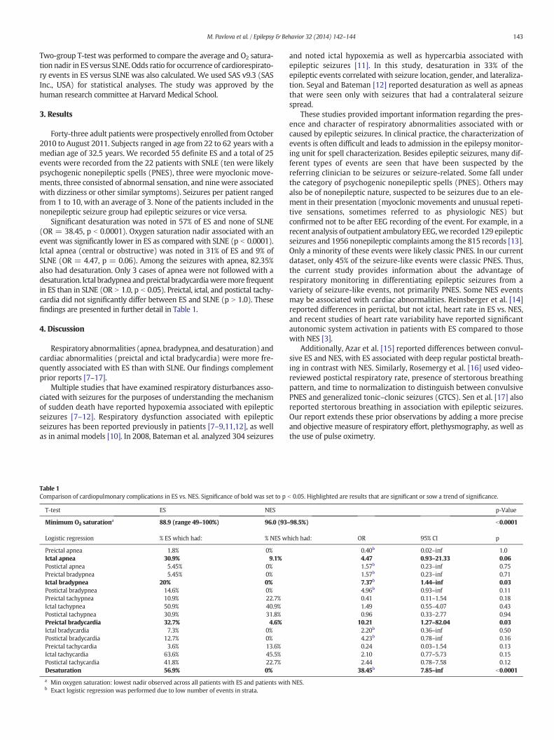
Significant desaturation was noted in 57% of ES and none of SLNE(OR = 38.45, p b 0.0001). Oxygen saturation nadir associated with anevent was significantly lower in ES as compared with SLNE (p b 0.0001).Ictal apnea (central or obstructive) was noted in 31% of ES and 9% ofSLNE (OR = 4.47, p = 0.06). Among the seizures with apnea, 82.35%also had desaturation. Only 3 cases of apnea were not followed with adesaturation. Ictal bradypnea andpreictal bradycardiaweremore frequentin ES than in SLNE (OR N 1.0, p b 0.05). Preictal, ictal, and postictal tachy-cardia did not significantly differ between ES and SLNE (p N 1.0). Thesefindings are presented in further detail in Table 1.
4. Discussion
Respiratory abnormalities (apnea, bradypnea, and desaturation) andcardiac abnormalities (preictal and ictal bradycardia) were more fre-quently associated with ES than with SLNE. Our findings complementprior reports [7–17].
Multiple studies that have examined respiratory disturbances asso-ciated with seizures for the purposes of understanding the mechanismof sudden death have reported hypoxemia associated with epilepticseizures [7–12]. Respiratory dysfunction associated with epilepticseizures has been reported previously in patients [7–9,11,12], as wellas in animal models [10]. In 2008, Bateman et al. analyzed 304 seizures
Table 1Comparison of cardiopulmonary complications in ES vs. NES. Significance of bold was set to p
T-test ES NES
Minimum O2 saturationa 88.9 (range 49–100%) 96.0 (93–
Logistic regression % ES which had: % NES w
Preictal apnea 1.8% 0%Ictal apnea 30.9% 9.1%Postictal apnea 5.45% 0%Preictal bradypnea 5.45% 0%Ictal bradypnea 20% 0%Postictal bradypnea 14.6% 0%Preictal tachypnea 10.9% 22.7%Ictal tachypnea 50.9% 40.9%Postictal tachypnea 30.9% 31.8%Preictal bradycardia 32.7% 4.6%Ictal bradycardia 7.3% 0%Postictal bradycardia 12.7% 0%Preictal tachycardia 3.6% 13.6%Ictal tachycardia 63.6% 45.5%Postictal tachycardia 41.8% 22.7%Desaturation 56.9% 0%
a Min oxygen saturation: lowest nadir observed across all patients with ES and patients withb Exact logistic regression was performed due to low number of events in strata.
and noted ictal hypoxemia as well as hypercarbia associated withepileptic seizures [11]. In this study, desaturation in 33% of theepileptic events correlated with seizure location, gender, and lateraliza-tion. Seyal and Bateman [12] reported desaturation as well as apneasthat were seen only with seizures that had a contralateral seizurespread.
These studies provided important information regarding the pres-ence and character of respiratory abnormalities associated with orcaused by epileptic seizures. In clinical practice, the characterization ofevents is often difficult and leads to admission in the epilepsy monitor-ing unit for spell characterization. Besides epileptic seizures, many dif-ferent types of events are seen that have been suspected by thereferring clinician to be seizures or seizure-related. Some fall underthe category of psychogenic nonepileptic spells (PNES). Others mayalso be of nonepileptic nature, suspected to be seizures due to an ele-ment in their presentation (myoclonic movements and unusual repeti-tive sensations, sometimes referred to as physiologic NES) butconfirmed not to be after EEG recording of the event. For example, in arecent analysis of outpatient ambulatory EEG,we recorded 129 epilepticseizures and 1956 nonepileptic complaints among the 815 records [13].Only a minority of these events were likely classic PNES. In our currentdataset, only 45% of the seizure-like events were classic PNES. Thus,the current study provides information about the advantage ofrespiratory monitoring in differentiating epileptic seizures from avariety of seizure-like events, not primarily PNES. Some NES eventsmay be associated with cardiac abnormalities. Reinsberger et al. [14]reported differences in periictal, but not ictal, heart rate in ES vs. NES,and recent studies of heart rate variability have reported significantautonomic system activation in patients with ES compared to thosewith NES [3].
Additionally, Azar et al. [15] reported differences between convul-sive ES and NES, with ES associated with deep regular postictal breath-ing in contrast with NES. Similarly, Rosemergy et al. [16] used video-reviewed postictal respiratory rate, presence of stertorous breathingpattern, and time to normalization to distinguish between convulsivePNES and generalized tonic–clonic seizures (GTCS). Sen et al. [17] alsoreported stertorous breathing in association with epileptic seizures.Our report extends these prior observations by adding a more preciseand objective measure of respiratory effort, plethysmography, as well asthe use of pulse oximetry.
b 0.05. Highlighted are results that are significant or sow a trend of significance.
p-Value
98.5%) b0.0001
hich had: OR 95% CI p
0.40b 0.02–inf 1.04.47 0.93–21.33 0.061.57b 0.23–inf 0.751.57b 0.23–inf 0.717.37b 1.44–inf 0.034.96b 0.93–inf 0.110.41 0.11–1.54 0.181.49 0.55–4.07 0.430.96 0.33–2.77 0.94
10.21 1.27–82.04 0.032.20b 0.36–inf 0.504.23b 0.78–inf 0.160.24 0.03–1.54 0.132.10 0.77–5.73 0.152.44 0.78–7.58 0.12
38.45b 7.85–inf b0.0001
NES.

144 M. Pavlova et al. / Epilepsy & Behavior 32 (2014) 142–144
While we found a higher rate of preictal and ictal bradycardia withES, we did not find the previously reported [14] difference in the rateof tachycardia pre- or postictally, possibly due to smaller numbersand the inclusion of events that were not psychogenic NES. We also ob-served in our series that tachycardia was nondiscriminant between ESand SLNE.
The advantages of our study include the importance and the physio-logical significance of the hypotheses tested, the prospective experi-mental design, and the robust findings supported by objectivecardiorespiratory abnormalities. Our findings suggest that use of pleth-ysmography and oximetry can help distinguish seizures from otherevents. A significant limitation may be related to the small numberof subjects with SLNE and the potentially biased sample of SLNE,since subjects were selected initially on the basis of the likelihood ofhaving ES. Thus, we could not distinguish psychogenic NES from othernonepileptic symptoms (such as abnormal movements). A largerstudy that allows adequate stratification and comparisons of PNES toall other seizure-like events may further improve our understandingof these abnormalities. Nevertheless, even in a small clinical sample,we describe a dramatic objective difference in cardiorespiratory abnor-malities associated with ES and not with SLNE.
5. Conclusion
Patients with ES had cardiopulmonary abnormalities, specificallybradypnea, apnea, preictal bradycardia, and oxygen desaturation, thatwere not seen with events that are not ES. Therefore in appropriate sit-uations, differentiation of ES from other events may be improved by theaddition of pulse oximetry, respiratory effort plethysmography belts,and EKG.
Author contributions
Drs. Pavlova and Kothare designed and conceptualized the study.Drs. Abdennadher and Zarowsky and Ms. Llewellyn assisted in datacollection. Drs. Pavlova, Kothare, and Dworetzky assisted in the analysisand interpretation of the EEG and clinical data. Drs. Katz and Whiteassisted in the analysis and interpretation of the respiratory data.Dr. Singh performed statistical analysis on the data. Drs. Singh,Abdennadher, Pavlova, and Kothare drafted the manuscript. Revisingand editing the manuscript were primarily performed by Drs. Pavlova,Kothare, and Dworetzky.
Author disclosures
This study was funded with the support of a grant from the HarvardCatalyst (Grant # UL1 RR 025758) in 2010, awarded to Drs. Kothare andPavlova. Dr. Kothare interprets video-EEGs and routine EEG in theDivision of Clinical Neurophysiology at Children's Hospital, Boston andhas received research support from Eisai Inc. and the NIH. None of theother authors have any conflict of interest to report.
References
[1] Syed TU, LaFrance Jr WC, Kahriman ES, Hasan SN, Rajasekaran V, Gulati D, et al. Cansemiology predict psychogenic nonepileptic seizures? A prospective study. AnnNeurol 2011;69:997–1004.
[2] Boon PA, Williamson PD. The diagnosis of pseudoseizures. Clin Neurol Neurosurg1993;95:1–8.
[3] Freeman R, Schachter SC. Autonomic epilepsy. Semin Neurol 1995;15:158–66.[4] Baumgartner C, Lurger S, Leutmezer F. Autonomic symptoms during epileptic sei-
zures. Epileptic Disord 2001;3:103–16.[5] Tomson T,Walczak T, SillanpaaM, Sander JW. Sudden unexpected death in epilepsy:
a review of incidence and risk factors. Epilepsia 2005;46(Suppl. 11):54–61.[6] Iber C, Ancoli-Israel S, Chesson A, Quan SF, for the American Academy of Sleep
Medicine. The AASM manual for the scoring of sleep and associated events: rules,terminology and technical specifications. 1st ed. Westchester: American Academyof Sleep Medicine; 2007.
[7] Seyal M, Bateman LM, Albertson TE, Lin TC, Li CS. Respiratory changes with seizuresin localization-related epilepsy: analysis of periictal hypercapnia and airflow pat-terns. Epilepsia 2010;51:1359–64.
[8] Walker F, FishDR. Recording respiratoryparameters in patientswith epilepsy. Epilepsia1997;38(11 Suppl.):S41–2. http://dx.doi.org/10.1111/j.1528-1157.1997.tb06126.x.
[9] Schomer DL. Oxygen desaturations triggered by partial seizures: implications forcardiopulmonary instability in epilepsy. Epilepsia 2000;41(5):536–41.
[10] St-John WM, Rudkin AH, Homes GL, Leiter JC. Changes in respiratory-modulatedneural activities, consistent with obstructive and central apnea, during fictiveseizures in an in situ anaesthetized rat preparation. Epilepsy Res 2006;70:218–28.
[11] Bateman LM, Li CS, Seyal M. Ictal hypoxemia in localization-related epilepsy: analy-sis of incidence, severity and risk factors. Brain 2008;131(Pt 12):3239–45.
[12] Seyal M, Bateman LM. Ictal apnea linked to contralateral spread of temporal lobe sei-zures: intracranial EEG recordings in refractory temporal lobe epilepsy. Epilepsia2009;50(12):2557–62.
[13] Pavlova MK, Woo Lee J, Yilmaz F, Dworetzky BA. Diurnal pattern of seizures outsidethe hospital: is there a time of circadian vulnerability? Neurology 2012;78(19):1488–92.
[14] Reinsberger C, Perez DL, Murphy MM, Dworetzky BA. Pre- and postictal, not ictal,heart rate distinguishes complex partial and psychogenic nonepileptic seizures.Epilepsy Behav 2012;23:68–70.
[15] Azar NJ, Tayah TF, Wang L, Song Y, Abou-Khalil BW. Postictal breathing pattern dis-tinguishes epileptic from nonepileptic convulsive seizures. Epilepsia 2008;49:132–7.
[16] Rosemergy I, Frith R, Herath S, Walker E. Use of postictal respiratory pattern to dis-criminate between convulsive psychogenic nonepileptic seizures and generalizedtonic–clonic seizures. Epilepsy Behav 2013;27(1):81–4.
[17] Sen A, Scott C, Sisodiya SM. Stertorous breathing is a reliably identified sign thathelps in the differentiation of epileptic from psychogenic non-epileptic convulsions:an audit. Epilepsy Res 2007;77(1):62–4.