Advances in Tumefactive CNS Demyelinating Disease and …€¦ · 29/06/2015 1 Advances in...
27
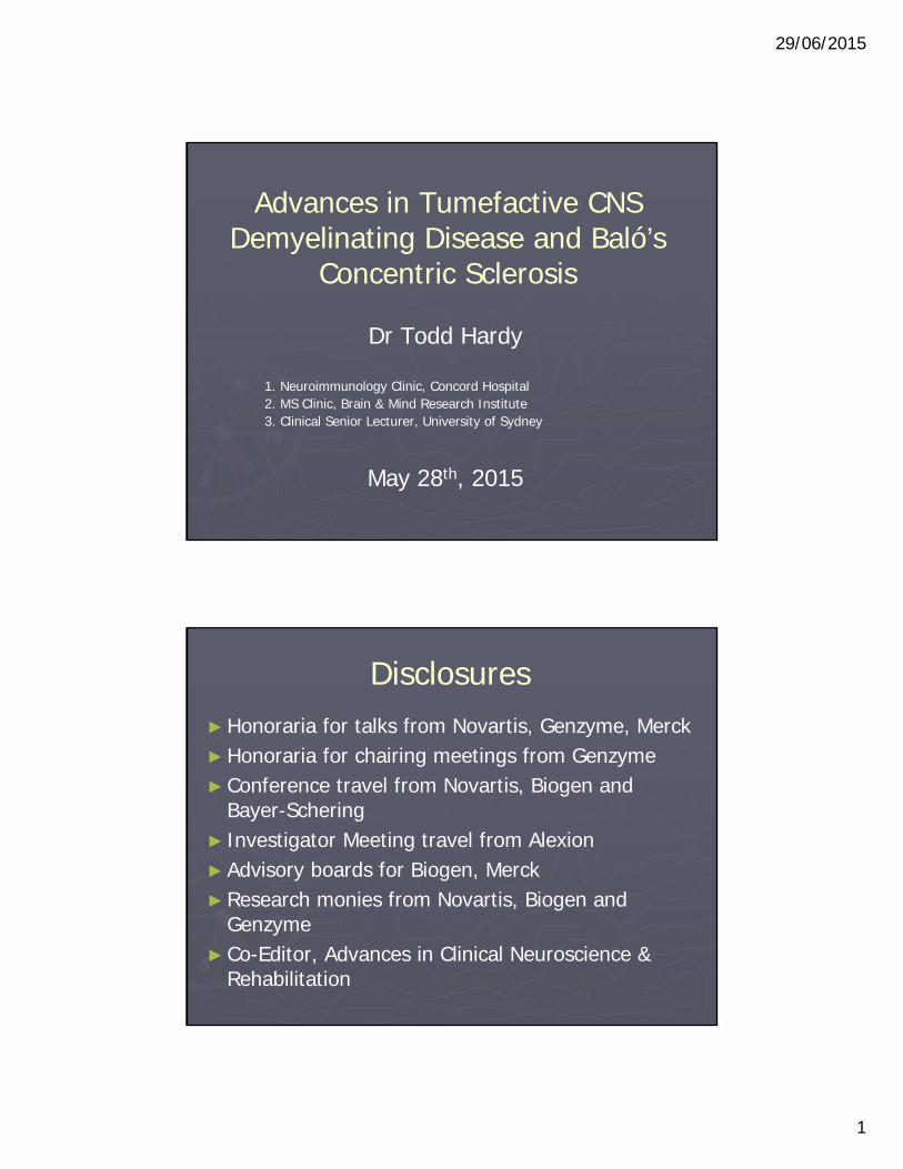
29/06/2015 1 Advances in Tumefactive CNS Demyelinating Disease and Baló’s Concentric Sclerosis Dr Todd Hardy 1. Neuroimmunology Clinic, Concord Hospital 2. MS Clinic, Brain & Mind Research Institute 3. Clinical Senior Lecturer, University of Sydney May 28 th , 2015 Disclosures ► Honoraria for talks from Novartis, Genzyme, Merck ► Honoraria for chairing meetings from Genzyme ► Conference travel from Novartis, Biogen and Bayer-Schering ► Investigator Meeting travel from Alexion ► Advisory boards for Biogen, Merck ► Research monies from Novartis, Biogen and Genzyme ► Co-Editor, Advances in Clinical Neuroscience & Rehabilitation
Transcript of Advances in Tumefactive CNS Demyelinating Disease and …€¦ · 29/06/2015 1 Advances in...
29/06/2015
1
Advances in Tumefactive CNS Demyelinating Disease and Baló’s
Concentric Sclerosis
Dr Todd Hardy
1. Neuroimmunology Clinic, Concord Hospital2. MS Clinic, Brain & Mind Research Institute 3. Clinical Senior Lecturer, University of Sydney
May 28th, 2015
Disclosures►Honoraria for talks from Novartis, Genzyme, Merck►Honoraria for chairing meetings from Genzyme►Conference travel from Novartis, Biogen and
Bayer-Schering► Investigator Meeting travel from Alexion►Advisory boards for Biogen, Merck►Research monies from Novartis, Biogen and
Genzyme►Co-Editor, Advances in Clinical Neuroscience &
Rehabilitation
29/06/2015
2
Atypical Demyelination
►Schilder’s disease►Haemorrhagic leukoencephalitis►Solitary sclerosis►Marburg’s disease
►Tumefactive demyelination►Baló’s concentric sclerosis
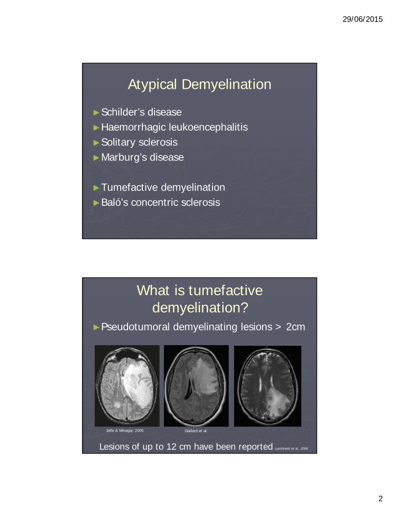
What is tumefactive demyelination?
►Pseudotumoral demyelinating lesions > 2cm
Lesions of up to 12 cm have been reported Lucchinetti et al., 2008
Jaffe & Minagar, 2005 Gaillard et al.
29/06/2015
3
Differential Diagnoses
►Primary neoplasm of the brain e.g. GBM►CNS lymphoma►Abscess►PML►Ischaemic stroke
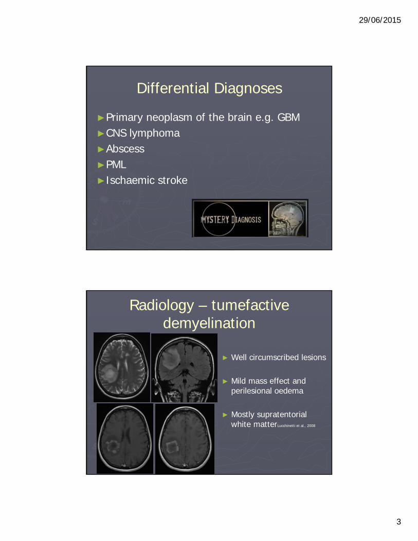
Radiology – tumefactive demyelination
► Well circumscribed lesions
► Mild mass effect and perilesional oedema
► Mostly supratentorialwhite matterLucchinetti et al., 2008
29/06/2015
4
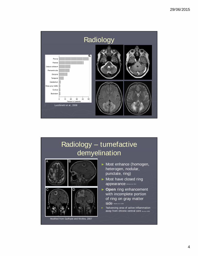
Radiology
Lucchinetti et al., 2008
Radiology – tumefactive demyelination
► Most enhance (homogen, heterogen, nodular, punctate, ring)
► Most have closed ring appearance Altintas et al. 2012
► Open ring enhancement with incomplete portion of ring on gray matter side Masdeu et al. 2000
► ?advancing area of active inflammation away from chronic central core He et al. 2001
Modified from Guilfoyle and Kirollos, 2007

29/06/2015
5
Radiology►Other typical features include: ► a T2 hypointense rim► peripheral restriction on DWI► venular enhancement
Saini et al. 2011Zhang & Metz, 2010
Guilfoyle and Kirollos, 2007
Multifocal and relapsing forms have been described
Among 54 patients with TD, 16.7% developed further TD lesions over 38 months Altintas et al. 2012
29/06/2015
6
Other conditions in which TD lesions described
►Viral infections including HIV►Other autoimmune diseases such as SLE,
Sjogren’s, Behcet’s disease ►Neuromyelitis optica (NMO)►Drugs e.g. tacrolimus►Malignancy; particularly renal cell carcinoma
Terminology
►Tumefactive demyelination NOT tumefactive MS

29/06/2015
7
What is Baló’s concentric sclerosis (BCS)?
►Named after Joseph Baló in 1928
► “Encephalitis periaxalisconcentrica“
Radiology of BCS
► Discrete, concentrically layered demyelinating lesion; “onion-ring” or whorled appearance
► Alternating isointense and hyperintense concentric rings on T2
► Lesion oedema is minimal
Trofimova & Malakhova, 2010
29/06/2015
8
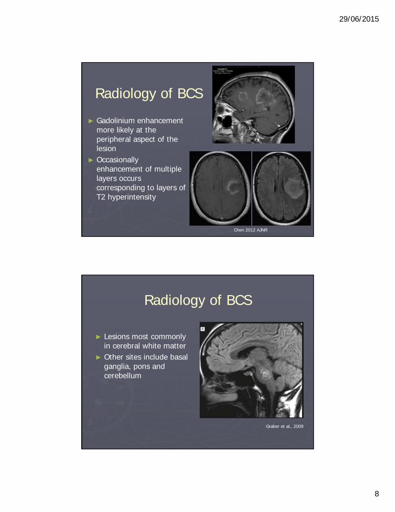
Radiology of BCS
► Gadolinium enhancement more likely at the peripheral aspect of the lesion
► Occasionally enhancement of multiple layers occurs corresponding to layers of T2 hyperintensity
Chen 2012 AJNR
Radiology of BCS
► Lesions most commonly in cerebral white matter
► Other sites include basal ganglia, pons and cerebellum
Graber et al., 2009
29/06/2015
9
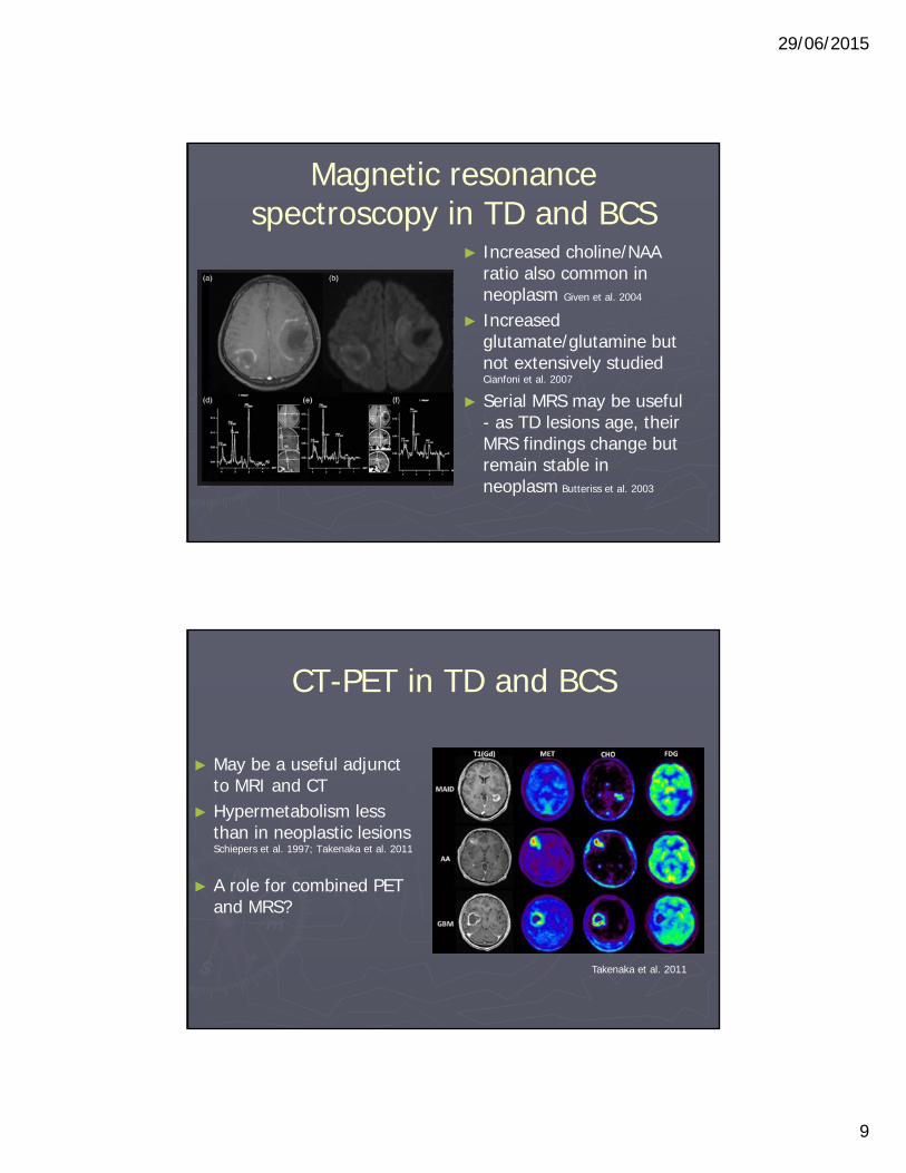
Magnetic resonance spectroscopy in TD and BCS
► Increased choline/NAA ratio also common in neoplasm Given et al. 2004
► Increased glutamate/glutamine but not extensively studied Cianfoni et al. 2007
► Serial MRS may be useful - as TD lesions age, their MRS findings change but remain stable in neoplasm Butteriss et al. 2003
Takenaka et al. 2011
CT-PET in TD and BCS
► May be a useful adjunct to MRI and CT
► Hypermetabolism less than in neoplastic lesions Schiepers et al. 1997; Takenaka et al. 2011
► A role for combined PET and MRS?

29/06/2015
10
Epidemiology – TD and BCS►Both TD and BCS are rare►BCS more common in patients of East Asian
origin (e.g. southern Han Chinese, Taiwanese and Filipinos)
►Female to male ratio of 1-2:1 ►Peak incidence in 20s and 30s►Paediatric and older patients have been
described
Presentation TD and BCS
►Among 17 cases BCS from the Philippines, 50% had prodromal symptoms of mild fever, general malaise and headache (Tabira, 2009)
Lucchinetti et al., 2008
29/06/2015
11
When to biopsy?►Without pre-existing diagnosis of MS►Inconclusive/suspicious imaging ►Older or very young patients►?Negative OCBs
►If typical TD or BCS, treat as presumed demyelination and monitor clinically and radiologically
Hardy & Chataway, 2013
Case 1 - 2011
►58 year old lady►RRMS diagnosed in 1985►Stable course with last relapse 1988►Never required disease modifying therapy►EDSS 1

29/06/2015
12
Case 1
►P/w subacute onset of left hemiparesis►No imaging performed in ED►Given 3 days IVMP for presumed MS relapse►Marked improvement
►2 weeks later left hemiparesis worsens…
MRI brain
29/06/2015
13
Case 1 – Brain biopsy
►Glioblastoma multiforme
Case 1 – Brain biopsy
►Glioblastoma multiforme
►Don’t assume all tumefactive lesions in patients with MS are due to demyelination

29/06/2015
14
Pathology of TD►Not greatly different from typical MS lesions
- Pattern III Lucchinetti et al., 2008
►Demyelination with hypercellularity, reactive astrocytes ± multiple nuclei (Creutzfeldcells), foamy macrophages
►Relative axonal sparing with perivascular and parenchymal lymphocytic infiltrates
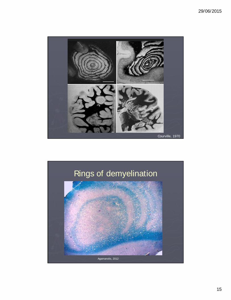
Pathology of BCS►Alternating rings reflects areas of relative
myelin preservation and loss with relative axonal sparing
►Some evidence for a mild astrocytopathy (Kira, 2011)
29/06/2015
15
Courville, 1970
Rings of demyelination
Agamanolis, 2012
29/06/2015
16
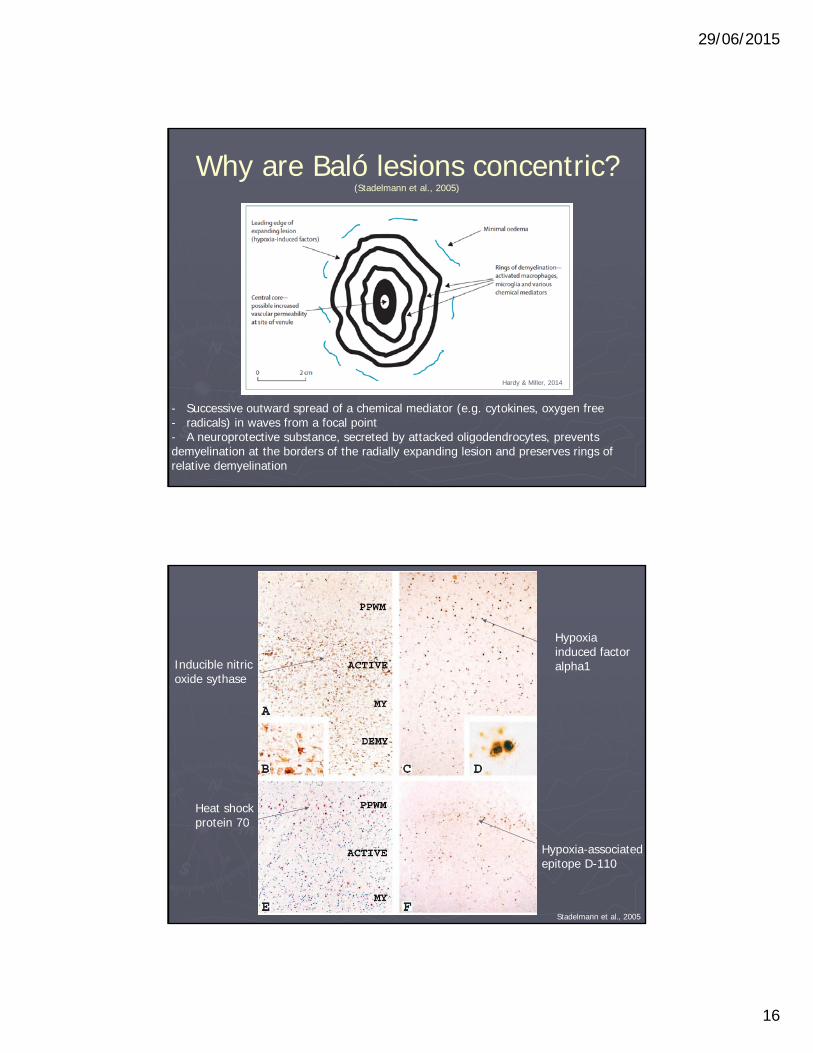
Why are Baló lesions concentric? (Stadelmann et al., 2005)
- Successive outward spread of a chemical mediator (e.g. cytokines, oxygen free - radicals) in waves from a focal point- A neuroprotective substance, secreted by attacked oligodendrocytes, prevents demyelination at the borders of the radially expanding lesion and preserves rings of relative demyelination
Hardy & Miller, 2014
Inducible nitric oxide sythase
Hypoxia induced factor alpha1
Heat shock protein 70
Hypoxia-associated epitope D-110
Stadelmann et al., 2005
29/06/2015
17
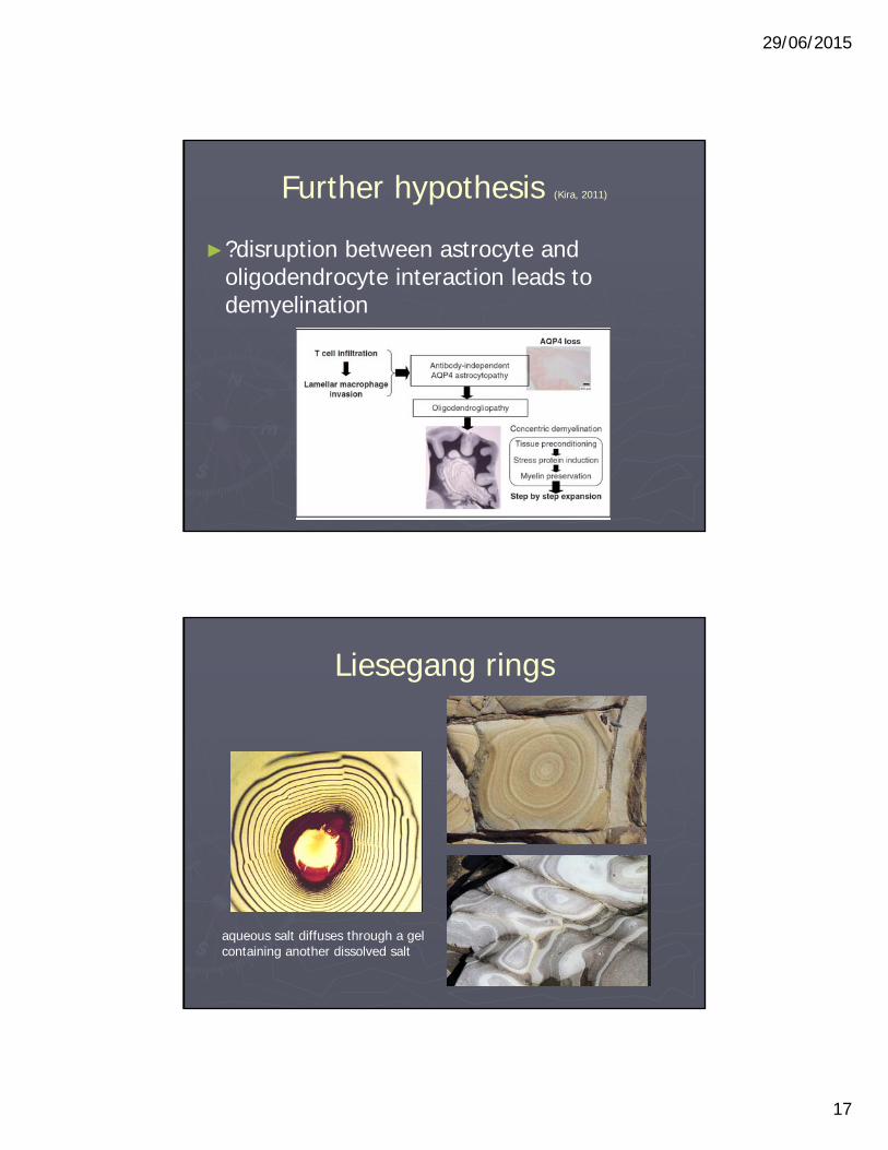
Further hypothesis (Kira, 2011)
►?disruption between astrocyte and oligodendrocyte interaction leads to demyelination
Liesegang rings
aqueous salt diffuses through a gel containing another dissolved salt
29/06/2015
18
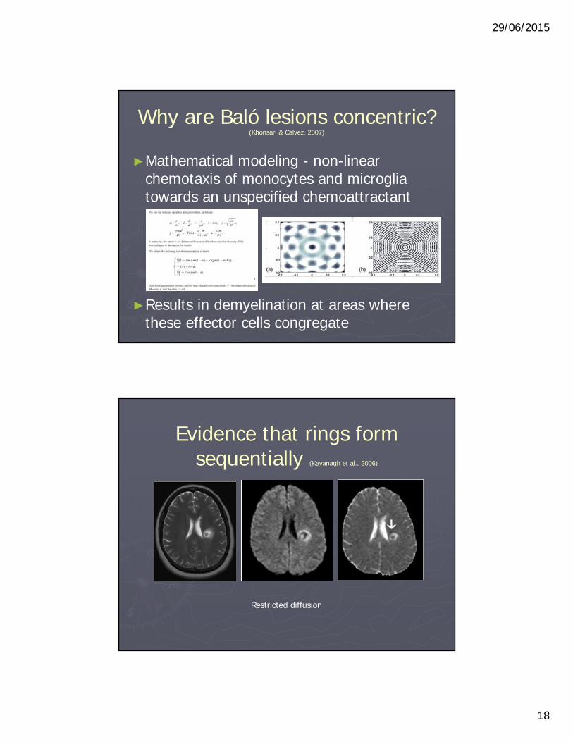
Why are Baló lesions concentric? (Khonsari & Calvez, 2007)
►Mathematical modeling - non-linear chemotaxis of monocytes and microglia towards an unspecified chemoattractant
►Results in demyelination at areas where these effector cells congregate
Evidence that rings form sequentially (Kavanagh et al., 2006)
Restricted diffusion
29/06/2015
19
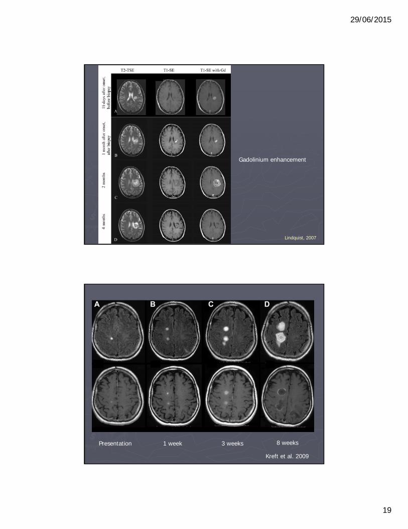
Lindquist, 2007
Gadolinium enhancement
Kreft et al. 2009
1 weekPresentation 3 weeks 8 weeks

29/06/2015
20
What is the clinical relationship of BCS and TD to MS?
► Overlap more than just pathological
► Baló and TD lesions can occur during conventional RRMS
► ~55% who present with a Baló or Baló-like lesion will have typical MS lesions elsewhere on their MRI scan (Chaodong et al., 2008)
What is the clinical relationship of BCS to MS?
►2 of 5 patients with initial Baló lesions relapsed with more typical MS lesions (Chaodong et al., 2008)
►Patients with Baló lesions and positive CSF OCBs can go on to develop MS (Kira, 2011)
►Among 11 patients with Baló-like lesions, CSF OCBs present in only one (Seewan et al., 2008)

29/06/2015
21
BCS and TD in the same patient
Hardy et al., 2015
Acute treatment of TD and BCS►No RCTs to guide►First line - corticosteroids ►Largest case series >80% of TD patients
respond to treatment with corticosteroid Altintaset al. 2012
►Second line - plasma exchange (PEX) ►Randomized trial - PEX beneficial in patients
with mixed CNS inflammatory demyelinating disease who have failed to respond to corticosteroids Weinshenker et al. 1999
29/06/2015
22
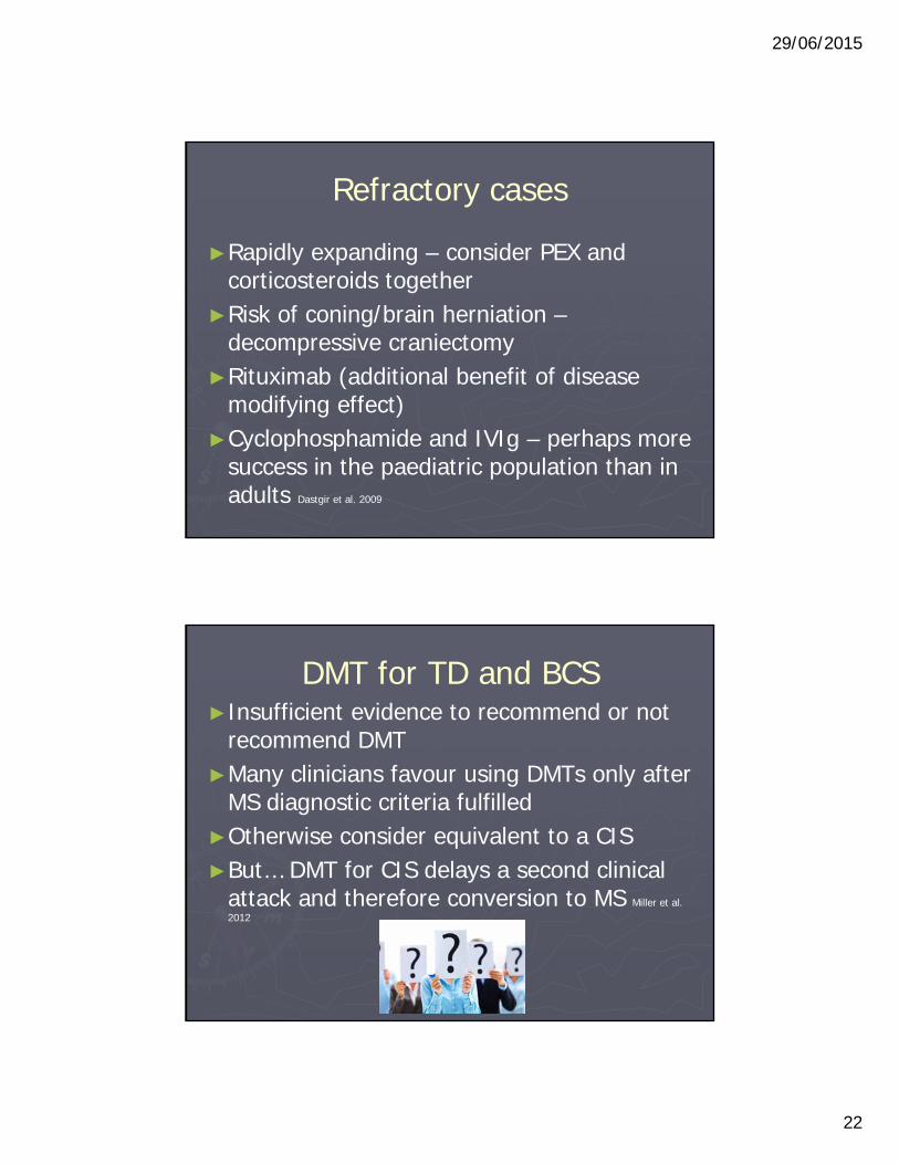
Refractory cases
►Rapidly expanding – consider PEX and corticosteroids together
►Risk of coning/brain herniation –decompressive craniectomy
►Rituximab (additional benefit of disease modifying effect)
►Cyclophosphamide and IVIg – perhaps more success in the paediatric population than in adults Dastgir et al. 2009
DMT for TD and BCS►Insufficient evidence to recommend or not
recommend DMT ►Many clinicians favour using DMTs only after
MS diagnostic criteria fulfilled►Otherwise consider equivalent to a CIS►But… DMT for CIS delays a second clinical
attack and therefore conversion to MS Miller et al. 2012

29/06/2015
23
DMT in TD lesions
►Also, may be improvement in long term disability with early treatment of CIS
►Consider if high risk of conversion to MS e.g. OCBs +ve or multiple typical demyelinating lesions
►Shared decision making between an individual patient and their treating clinician Hardy & Chataway, 2013
DMT in TD lesions
►Start with injectable (interferon beta or glatiramer acetate)
►TD lesions have been reported in patients with MS and NMO undergoing treatment with Natalizumab Berger, 2008; Twyman et al. 2010; Barnett et al. 2012
►One report of Natalizumab beneficial in a patient with rapidly evolving relapsing TD who - relapse-free at 12 months Seifert et al. 2012
29/06/2015
24
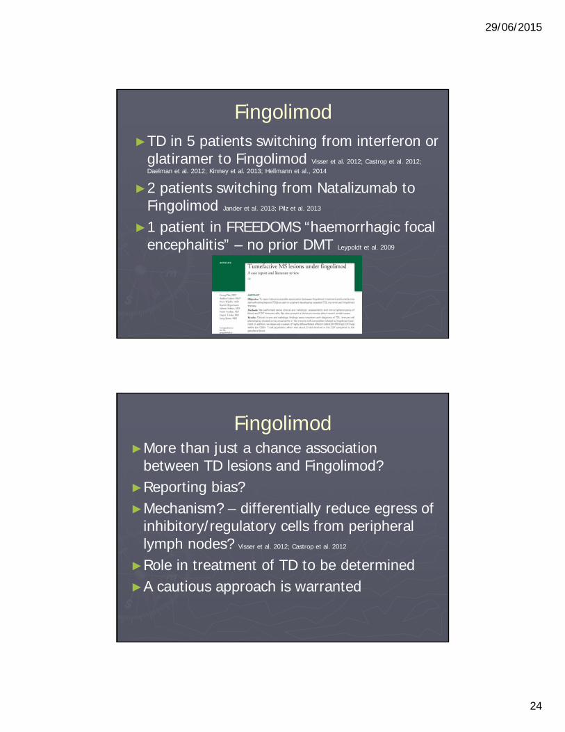
Fingolimod►TD in 5 patients switching from interferon or
glatiramer to Fingolimod Visser et al. 2012; Castrop et al. 2012; Daelman et al. 2012; Kinney et al. 2013; Hellmann et al., 2014
►2 patients switching from Natalizumab to Fingolimod Jander et al. 2013; Pilz et al. 2013
►1 patient in FREEDOMS “haemorrhagic focal encephalitis” – no prior DMT Leypoldt et al. 2009
Fingolimod►More than just a chance association
between TD lesions and Fingolimod?►Reporting bias?►Mechanism? – differentially reduce egress of
inhibitory/regulatory cells from peripheral lymph nodes? Visser et al. 2012; Castrop et al. 2012
►Role in treatment of TD to be determined►A cautious approach is warranted

29/06/2015
25
Prognosis of TD
► 39 patients – 54% complete recovery Nagappa et al., 2013
► 85 patients with biopsy confirmed TD Lucchinetti et al., 2008
► Time to second event in patients presenting with a TD lesion = 4.8 years
► 1.9 to 3 years for a typical demyelinating event Kepes, 1993; Confraveux et al. 2012
Prognosis of TD►Slight trend for better prognosis when TD
lesions accompanied by typical MS lesions on MRI
Lucchinetti et al., 2008
29/06/2015
26
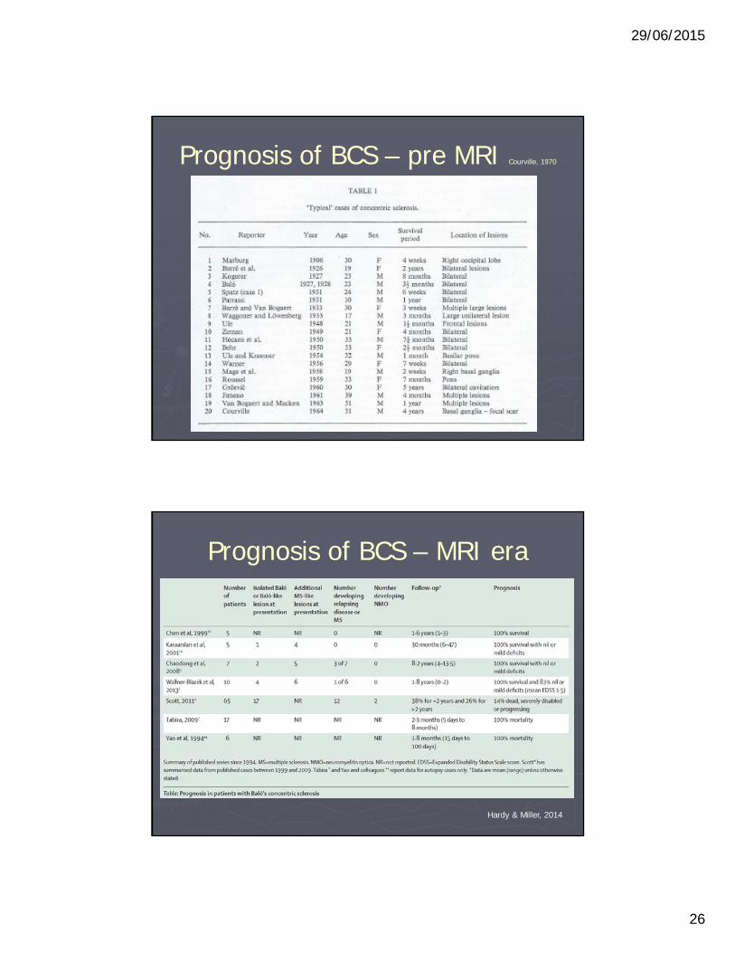
Prognosis of BCS – pre MRI Courville, 1970
Prognosis of BCS – MRI era
Hardy & Miller, 2014
29/06/2015
27
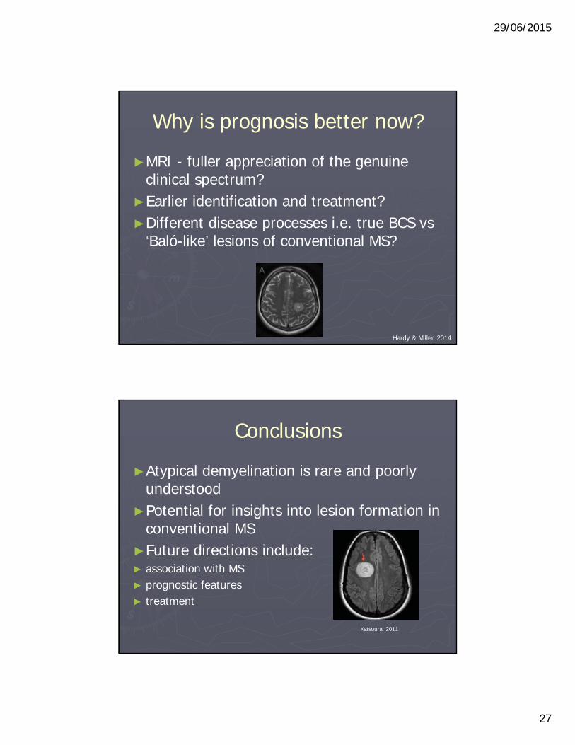
Why is prognosis better now?
►MRI - fuller appreciation of the genuine clinical spectrum?
►Earlier identification and treatment?►Different disease processes i.e. true BCS vs
‘Baló-like’ lesions of conventional MS?
A
Hardy & Miller, 2014
Conclusions
►Atypical demyelination is rare and poorly understood
►Potential for insights into lesion formation in conventional MS
►Future directions include:► association with MS ► prognostic features► treatment
Katsuura, 2011